Secreted Proteins from the Helminth Fasciola hepatica Inhibit the ...
11
Secreted Proteins from the Helminth Fasciola hepatica Inhibit the Initiation of Autoreactive T Cell Responses and Prevent Diabetes in the NOD Mouse Lund, M. E., O'Brien, B. A., Hutchinson, A. T., Robinson, M. W., Simpson, A. M., Dalton, J. P., & Donnelly, S. (2014). Secreted Proteins from the Helminth Fasciola hepatica Inhibit the Initiation of Autoreactive T Cell Responses and Prevent Diabetes in the NOD Mouse. PloS one, 9(1), e86289. [e86289]. https://doi.org/10.1371/journal.pone.0086289 Published in: PloS one Document Version: Publisher's PDF, also known as Version of record Queen's University Belfast - Research Portal: Link to publication record in Queen's University Belfast Research Portal Publisher rights © 2014 The Authors This is an open access article distributed under the terms of the Creative Commons Attribution License (https://creativecommons.org/licenses/by/4.0/), which permits unrestricted use, distribution, and reproduction in any medium, provided the original author and source are credited General rights Copyright for the publications made accessible via the Queen's University Belfast Research Portal is retained by the author(s) and / or other copyright owners and it is a condition of accessing these publications that users recognise and abide by the legal requirements associated with these rights. Take down policy The Research Portal is Queen's institutional repository that provides access to Queen's research output. Every effort has been made to ensure that content in the Research Portal does not infringe any person's rights, or applicable UK laws. If you discover content in the Research Portal that you believe breaches copyright or violates any law, please contact [email protected]. Download date:30. Jan. 2022
-
Upload
khangminh22 -
Category
Documents
-
view
0 -
download
0
Transcript of Secreted Proteins from the Helminth Fasciola hepatica Inhibit the ...
Secreted Proteins from the Helminth Fasciola hepatica Inhibit theInitiation of Autoreactive T Cell Responses and Prevent Diabetes inthe NOD MouseLund, M. E., O'Brien, B. A., Hutchinson, A. T., Robinson, M. W., Simpson, A. M., Dalton, J. P., & Donnelly, S.(2014). Secreted Proteins from the Helminth Fasciola hepatica Inhibit the Initiation of Autoreactive T CellResponses and Prevent Diabetes in the NOD Mouse. PloS one, 9(1), e86289. [e86289].https://doi.org/10.1371/journal.pone.0086289
Published in:PloS one
Document Version:Publisher's PDF, also known as Version of record
Queen's University Belfast - Research Portal:Link to publication record in Queen's University Belfast Research Portal
Publisher rights© 2014 The AuthorsThis is an open access article distributed under the terms of the Creative Commons Attribution License(https://creativecommons.org/licenses/by/4.0/), which permits unrestricted use, distribution, and reproduction in any medium, provided theoriginal author and source are credited
General rightsCopyright for the publications made accessible via the Queen's University Belfast Research Portal is retained by the author(s) and / or othercopyright owners and it is a condition of accessing these publications that users recognise and abide by the legal requirements associatedwith these rights.
Take down policyThe Research Portal is Queen's institutional repository that provides access to Queen's research output. Every effort has been made toensure that content in the Research Portal does not infringe any person's rights, or applicable UK laws. If you discover content in theResearch Portal that you believe breaches copyright or violates any law, please contact [email protected].
Download date:30. Jan. 2022

Secreted Proteins from the Helminth Fasciola hepaticaInhibit the Initiation of Autoreactive T Cell Responsesand Prevent Diabetes in the NOD MouseMaria E. Lund1., Bronwyn A. O’Brien1., Andrew T. Hutchinson1, Mark W. Robinson2, Ann M. Simpson1,
John P. Dalton3, Sheila Donnelly4*
1 School of Medical and Molecular Biosciences, University of Technology Sydney, New South Wales, Australia, 2 School of Biological Sciences, Queen’s University Belfast,
Belfast, Northern Ireland, 3 Institute of Parasitology, McDonald Campus, McGill University, St. Anne de Bellevue, Quebec, Canada, 4 The i3 Institute, University of
Technology Sydney, New South Wales, Australia
Abstract
Infections with helminth parasites prevent/attenuate auto-inflammatory disease. Here we show that molecules secreted bya helminth parasite could prevent Type 1 Diabetes (T1D) in nonobese diabetic (NOD) mice. When delivered at 4 weeks ofage (coincident with the initiation of autoimmunity), the excretory/secretory products of Fasciola hepatica (FhES) preventedthe onset of T1D, with 84% of mice remaining normoglycaemic and insulitis-free at 30 weeks of age. Disease protection wasassociated with suppression of IFN-c secretion from autoreactive T cells and a switch to the production of a regulatoryisotype (from IgG2a to IgG1) of autoantibody. Following FhES injection, peritoneal macrophages converted to a regulatoryM2 phenotype, characterised by increased expression levels of Ym1, Arg-1, TGFb and PD-L1. Expression of these M2 geneticmarkers increased in the pancreatic lymph nodes and the pancreas of FhES-treated mice. In vitro, FhES-stimulated M2macrophages induced the differentiation of Tregs from splenocytes isolated from naıve NOD mice. Collectively, our datashows that FhES contains immune-modulatory molecules that mediate protection from autoimmune diabetes via theinduction and maintenance of a regulatory immune environment.
Citation: Lund ME, O’Brien BA, Hutchinson AT, Robinson MW, Simpson AM, et al. (2014) Secreted Proteins from the Helminth Fasciola hepatica Inhibit theInitiation of Autoreactive T Cell Responses and Prevent Diabetes in the NOD Mouse. PLoS ONE 9(1): e86289. doi:10.1371/journal.pone.0086289
Editor: Lucienne Chatenoud, Universite Paris Descartes, France
Received July 30, 2013; Accepted December 13, 2013; Published January 21, 2014
Copyright: � 2014 Lund et al. This is an open-access article distributed under the terms of the Creative Commons Attribution License, which permitsunrestricted use, distribution, and reproduction in any medium, provided the original author and source are credited.
Funding: This work was supported by a National Health and Medical Research Council (NHMRC) project grant (APP1010197) and the Juvenile Diabetes ResearchFoundation (JDRF) research grant (4-2006-1025). J.P.D. is a Canada Research Chair in Infectious diseases and is funded by the Canadian Institute of HealthResearch (CIHR) and National Science and Engineering Research Council (NSERC). The funders had no role in study design, data collection and analysis, decision topublish, or preparation of the manuscript.
Competing Interests: The authors have declared that no competing interests exist.
* E-mail: [email protected]
. These authors contributed equally to this work.
Introduction
Type 1 Diabetes (T1D) is a multifactorial autoimmune disease
in which the insulin-secreting beta (b) cells within the pancreatic
islets are destroyed. While disease susceptibility is determined by
genetic, immunological and environmental factors, the observed
rising incidence of T1D in recent decades suggests a significant
etiological role for environmental influences, either the removal of
a protective factor(s) or introduction of a susceptibility factor(s) [1].
Post-industrial improvements in sanitation and living conditions
have led to a dramatic decline in exposure to pathogens, notably
parasitic worms (helminths), among Western populations [2–4].
Epidemiological studies have shown that the absence of endemic
helminth infection is inversely correlated with the incidence of
T1D [5,6]. This suggests that exposure to helminths represents a
predominant protective environmental factor against the develop-
ment of T1D, and auto-inflammatory diseases in general. It has
been proposed that the controlled reintroduction of helminth
infection into Western populations could represent an effective
therapy for auto-inflammatory diseases [10,11]. Support for the
therapeutic potential of helminth infection in the prevention of
autoimmune diabetes has come from experimental studies
showing that infection of mice with helminth parasites prevents
the development of T1D [12–17].
Mammals infected with a helminth parasite exhibit a potent and
biased Th2-driven immune response during the acute phase,
which counter-regulates Th1-driven autoimmune pathologies
[12,13,15,18]. During the chronic phase of infection, however,
immune-regulatory networks emerge. These are driven primarily
by regulatory T cells (Tregs) producing IL-10 and TGFb [18],
which has the bystander effect of protecting against Th1-
associated autoimmune diseases, such as T1D [16]. It is likely
that the ability of helminth parasites to modulate host immune
responses towards an anti-inflammatory/regulatory phenotype is
attributable to the molecules that the parasites secrete and/or
excrete which interact with immune effector cells to modulate
their function [19,20].
We have previously reported that the excretory/secretory
products of the helminth parasite Fasciola hepatica, termed FhES,
collected after culturing parasites in vitro, exerts a potent immune-
modulatory effect in the immunocompetent host (Balb/c and
C57BL6 mice). This is achieved by the activation of regulatory M2
macrophages [21,22], suppression of dendritic cell (DC) matura-
PLOS ONE | www.plosone.org 1 January 2014 | Volume 9 | Issue 1 | e86289

tion [23], and inhibition of antigen-specific Th1 and Th17 cell
differentiation [23,24]. Given these profound immune-modulatory
properties, in this study we examined the potential of FhES to
prevent the initiation and perpetuation of the autoreactive
immune responses that underpin T1D development.
Short-term intra-peritoneal administration of FhES to female
non-obese diabetic (NOD) mice resulted in permanent protection
against immune-mediated b-cell destruction. Disease prevention
was associated with the induction of a regulatory immune
environment composed of regulatory B cells (Bregs), and M2
macrophages that induced the differentiation of Tregs. These data
support the proposition that helminth-derived molecules may
represent a more desirable therapeutic alternative to the use of live
parasitic infection as a treatment for T1D, and other auto-
inflammatory diseases.
Materials and Methods
Ethics statementFour week old female NOD/Lt mice were purchased from the
ARC (Perth, Australia) and maintained under an experimental
protocol approved by the University of Technology Sydney (UTS)
Animal Care and Ethics Committee (Approval Number: 2010-
432A). F. hepatica infections in sheep were performed at the
approval of the Animal Ethic Committee (AEC) located at the
Elizabeth Macarthur Agricultural Institute (EMAI, Menangle,
New South Wales (NSW), Australia) and under the guidelines
established by the Animal Research Review Panel (ARRP) of the
NSW Department of Primary Industries (DPI) (www.animalethics.
org.au).
Preparation of FhESMature Fasciola hepatica were recovered from the bile ducts of
Merino sheep 16 weeks after an experimental infection and FhES
was prepared by maintaining the parasites in culture for 8 h as
previously described [21]. The culture medium was concentrated
to 1 mg/ml, using a 3000 Da cut-off centricon, filter-sterilised and
stored at 280uC until required.
Treatment of NOD mice with FhESFhES (10 mg in 100 ml sterile PBS) was delivered to mice
intraperitoneally on alternate days for a total of 6 injections.
Control mice received 100 ml of sterile PBS. Glucose levels were
measured from tail vein blood weekly, from 13 weeks of age, using
Accu-check Advantage blood glucose strips (Roche, Australia).
Animals were sacrificed at diabetes onset; defined by two
consecutive blood glucose concentrations above 14 mmol/L. All
efforts were made to minimize suffering.
Scoring of InsulitisFormalin-fixed paraffin-embedded pancreata were sectioned
(4 mm) at three non-overlapping levels, such that each section was
separated from the preceding one by at least 20 mm. Sections were
stained by hematoxylin and eosin (H&E), studied for their
histological characteristics, and graded for insulitis on a scale of
0–4; whereby 0 = healthy islet or mild peri-insular mononuclear
cell infiltration, 1 = infiltration up to 25% of islet mass, 2 = infil-
tration up to 50% of islet mass, 3 = infiltration from 50% up to
75% of islet mass, and 4 = less than 25% of islet mass present.
Slides were assessed in a blinded fashion and all islets in 10 slides
from each pancreas were scored.
Characterisation of autoantigen-specific immuneresponses
The levels of anti-insulin and anti-glutamic acid decarboxylase
(GAD) immunoglobulin in sera were determined by ELISA, as
previously described [12]. Briefly, plates were coated with bovine
insulin (10 mg/ml; Sigma, Australia) and bound antibodies in sera
detected by the addition of either goat anti-mouse IgG1, IgG2a
(BD Pharmingen, Australia) or IgM (Sigma, Australia) conjugated
to alkaline phosphatase. The development of colour after addition
of p-nitrophenylphosphate (Sigma, Australia) was recorded by
spectrophotometry at 405 nm.
For analysis of T cell responses, single cell suspensions were
prepared from the spleens of treated mice and cultured (16106
cells/ml) in the presence of bovine insulin (10 mg/ml; Sigma,
NSW, Australia) or anti-CD3 (10 mg/ml; BD Pharmingen,
Australia). After 72 h incubation at 37uC, supernatants were
collected and analysed for the presence of IL-4 and IFN-c by
ELISA (BD Pharmingen, Australia)
Characterisation of immune cell populations by flowcytometry
Cells were collected from the peritoneal cavity of treated mice
by lavage with 5 ml sterile PBS/BSA1%/heat inactivated
FCS2%/0.05% sodium azide. Pancreatic lymph nodes (PLNs)
from treated mice were harvested into RPMI (Life Technologies,
Australia). Single cell suspensions from both were blocked with
anti-CD16/32 mouse Fc Block (BD Pharmingen, Australia) and
analysed for the expression of cell surface markers using
combinations of the following antibodies: CD3 (SK7), CD4
(L3T4), CD8a (53-6.7), B220 (RA3-6B2), F4/80 (BM8), CD25
(7D4), PD-L1 (M1H5) or CD19 (1D3) (BD Pharmingen or Life
Technologies, Australia). For the identification of regulatory T
cells, expression of the intracellular marker, Foxp3, was quantified
using a mouse Foxp3 intracellular staining kit (BD Pharmingen,
Australia). Appropriate isotype control antibodies were used.
Labelled cells were analysed using the BD LSRII flow cytometer
(BD Biosciences). Data were analysed using FCS Express 4
Cytometry software (De Novo Software). Gating strategies are
shown in Fig S1.
Characterisation of IL-10 secreting cellsA Mouse IL-10 Secretion Assay (Miltenyi Biotec, Australia) was
used to identify and quantify the IL-10 secreting cells within the
peritoneal cavity and the pancreatic lymph nodes (PLNs) of mice
treated with FhES or PBS. In preparation for the assay, single cell
suspensions of PLNs harvested from treated mice were cultured
overnight in RPMI with 10% v/v heat inactivated FCS (Life
Technologies, Australia). Peritoneal cells were harvested by lavage
and analysed immediately using the IL-10 secretion assay. Initially,
cells were labelled with a capture antibody specific for mouse IL-
10, then returned to culture for 45 min at 37uC in RPMI with
10% v/v heat inactivated FCS. Cells were then stained with an IL-
10 detection antibody or isotype control antibody, before being
counterstained for cell surface markers CD19 or F4/80 to identify
B cells and macrophages, respectively. Dead cells were excluded
using Dapi staining (Life Technologies, Australia). Labelled cells
were analysed using the BD LSRII flow cytometer (BD
Biosciences). Data were analysed using FCS Express 4 Cytometry
software (De Novo Software).
Quantification of macrophage-secreted cytokinesPeritoneal macrophages harvested from PBS or FhES-treated
mice were cultured overnight in RPMI without any further
Helminth Secreted Proteins Prevent Type 1 Diabetes
PLOS ONE | www.plosone.org 2 January 2014 | Volume 9 | Issue 1 | e86289

stimulation. The concentration of IL-10, IL-12 and TGFbsecreted into culture supernatants was measured by ELISA (BD
Biosciences and R&D systems)
Gene expression analysisTotal RNA was extracted from PLNs, peritoneal macrophages
or frozen pancreatic tissue using an RNeasy plus mini kit (Qiagen).
Gene expression levels of Ym1, Retnla, TGFb, Arg-1, Foxp3, and
b-actin were quantified in real time using Taqman gene expression
assays (Applied Biosystems, Australia) and RT-PCR, as previously
described [21]. Gene expression was quantified (in triplicate) using
the change in cycle threshold method (CtGene2Ct
House Keeping)
and normalised to expression of the house-keeping gene,
glyceraldehyde-3-phosphate dehydrogenase. Expression levels of
genes in treatment samples were determined by comparison to the
average DCt of the untreated control cohort.
In vitro macrophage and splenocyte co-culturesMacrophages were harvested from the peritoneal cavity by
lavage and isolated to .94% purity by adherence to plastic for 1 h
at 37uC. Splenocytes were cultured (in 96 well flat bottomed
plates) with FhES (20 mg/ml), soluble egg antigens (SEA; 50 mg/
ml) of the parasitic helminth Schistosoma mansoni (Theodor Bilharz
Research Institute, Cairo, Egypt.), or autologous peritoneal
macrophages (at a ratio of 1:5), and stimulated with anti-CD3
(2 mg/ml; 17A2; BD Pharmingen, Australia) for 72 h at 37uC in
RPMI, supplemented with 10% v/v heat inactivated FCS (Life
Technologies, Australia).
Statistical analysisBlood glucose data was assessed using survival analysis, and
Kaplan-Meier estimates of the survivor functions were compared
using a Tyrone-Ware nonparametric test. For insulitis scores, the
distributions of scores across mice for each group were determined
using a maximum likelihood 82 contingency table test. To
compare the overall distributions of scores for the groups a log-
linear model was used. Statistical analyses of data for cytokine
secretion and immunophenotyping by flow cytometry were
performed using the GraphPad Prism 5 for Windows (GraphPad
Software Inc.). For comparison of two variables the unpaired
Student’s t-test with Welch’s correction for unequal variances, or
the Mann-Whitney two-tailed t test, were used. Error bars
represent 6 standard error of the mean.
Results
Short term peritoneal administration of FhES preventsthe onset of T1D
Female NOD mice were injected intraperitoneally with FhES
beginning at 4 weeks of age (co-incident with the priming of
autoreactive T cell populations and initiation of insulitis) and
continuing on alternate days for a total of 6 treatments (10 mg/
injection). This treatment regime was chosen to replicate the
quantities of FhES that would be secreted during a low-dose
infection by F. hepatica parasites as they migrate through the
peritoneal cavity towards the liver. In addition, we have previously
reported that this treatment regime is sufficient to inhibit the
development of antigen-specific Th1 immune responses in
immune-competent nondiabetes-prone mice [21,22].
As expected with our colony of NOD mice, the cumulative
incidence of diabetes (as determined by two separate blood glucose
concentrations $14 mmol/L) in the PBS-treated NOD mice
reached a maximum at 30 weeks of age, with approximately 80%
of animals developing diabetes and only 19% of animals
remaining normoglycaemic (n = 11; Fig. 1A). In contrast, 84% of
FhES-treated mice remained disease free at 30 weeks of age
(experimental endpoint; n = 12; Fig. 1A). This data is representa-
tive of three independent studies in which there were no significant
differences in survival rates between trials. In the other two trials,
88% (n = 16) and 86% (n = 7) of FhES-treated mice remained
disease-free at 23 and 25 weeks, respectively (experimental
endpoints), as compared to 13% (n = 16) and 17% (n = 12) of
PBS-treated mice, respectively. Consistent with the ability of FhES
to prevent autoimmune diabetes, examination of H&E-stained
sections of pancreas isolated from FhES-treated mice at various
time-points revealed a consistent and significant (p#0.001)
reduction in islet inflammation, as compared to PBS-treated mice
(Fig. 1B–E). Collectively, these data demonstrate that FhES
induced a robust and reproducible disease protection against
autoimmune diabetes development.
Diabetes prevention is associated with the suppressionof autoreactive immune responses
To examine if the disease protection in NOD mice afforded by
FhES treatment was associated with modulation of autoantigen
specific responses, we measured the amount of IFN-c and IL-4
secreted from splenocytes ex vivo in response to stimulation with
insulin (auto-antigen). The quantity of IFN-c released from
splenocytes, isolated from mice immediately after the final
administration of FhES (6 weeks of age; Fig. 2A), were significantly
(p = 0.0164) reduced, as compared to levels secreted by splenocytes
from control (PBS treatment) mice. Even seven weeks after the
final FhES treatment (at 13 weeks of age) when expansion of
diabetogenic T cell clones would have normally already occurred,
insulin-specific IFN-c levels remained lower in the FhES-treated
mice as compared to PBS-treated NOD mice (Fig. 2B). This
suppression was specific to the development of antigen-specific
responses as splenocytes isolated from FhES-treated mice secreted
similar quantities of IFN-c in response to stimulation with aCD3
(667618 pg/ml), as compared to cells isolated from PBS-treated
mice (64667 pg/ml). In addition, the FhES mediated decrease in
IFN-c production was not due to a switch towards an antigen-
specific Th2 response. At both 6 and 13 weeks of age, the levels of
IL-4 secreted by splenocytes in response to insulin were below the
level of detection (3 pg/ml) for both PBS- and FhES-treated mice.
Cells isolated from both FhES- and PBS-treated mice secreted
similar levels of IL-4 in response to stimulation with aCD3 (data
not shown).
Since IFN-c secretion by T cells stimulates the production of
IgG2a [25], we isotyped autoreactive immunoglobulins in the sera
of FhES- and PBS-treated mice. Specifically, we measured the
titres of autoantibodies directed against the dominant auto-
antigens, insulin and GAD. The levels of total IgG and IgM in
sera from FhES-treated and PBS-treated mice did not differ (data
not shown). However, treatment with FhES caused a switch
towards the production of auto-antigen specific IgG1 (Fig. 2C),
which resulted in a significant increase in the ratio of IgG1:IgG2a
autoantibodies against both insulin (Fig. 2D) and GAD (Fig. 2E) in
mice treated with FhES, as compared to PBS-treatment. This data
is consistent with the premise that diminished IFN-c secretion,
induced by FhES treatment, reduces the generation of auto-
antigen-specific IgG2a responses from B cells.
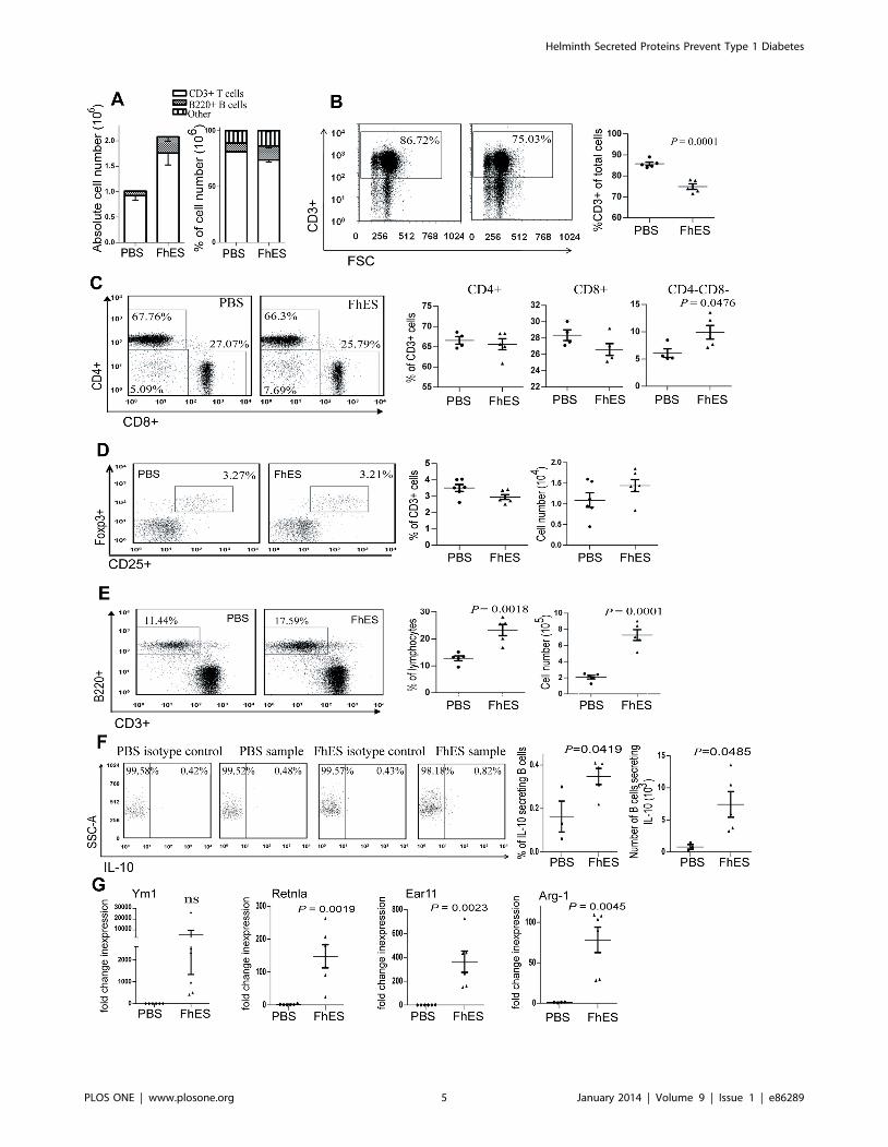
FhES treatment modulates immune cell populations inthe pancreatic lymph nodes
In NOD mice, diabetogenic CD4+ T cells undergo priming and
clonal expansion in the PLNs after presentation of islet autoan-
Helminth Secreted Proteins Prevent Type 1 Diabetes
PLOS ONE | www.plosone.org 3 January 2014 | Volume 9 | Issue 1 | e86289

tigens sequestered in the pancreas by antigen presenting cells
(APCs) [26]. The immunological environment within the PLNs
during the initiation phase of autoimmunity (co-incident with the
delivery of FhES) is therefore a critical determinant for the
expansion of autoreactive T cell clones. Accordingly, 24 h after
the final treatment of FhES, we examined whether the treatment
regime altered immune cell populations within the PLNs.
While FhES treatment induced an increase in total numbers of
immune cells within the PLNs (Fig. 3A), the proportion of CD3+
lymphocytes was significantly decreased (p = 0.0001; Fig. 3A&B).
Within this T cell population, the proportion of CD8+ T cells and
CD4+ T cells among FhES-treated and control mice remained
similar, however the percentage of double negative (CD42CD82)
T cells was elevated (p = 0.0476; Fig. 3C). Neither the proportion
nor number of CD4+CD25+Foxp3+ Tregs differed significantly
between FhES- and PBS- treatment groups (Fig. 3D).
Figure 1. Treatment of NOD mice co-incident with the initiationof autoimmunity prevents T1D. Four-week old female NOD micewere injected intraperitoneally with FhES (10 mg in 100 ml sterile PBS) orvehicle (PBS), on alternate days, for a total of six injections. (A) Bloodglucose levels were monitored and animals were sacrificed when theybecame diabetic (as defined by two consecutive blood glucoseconcentrations $14 mmol/L). The graphs represent an analysis of theage at which each animal was sacrificed and was performed usingsurvival analysis. Data shown is from one of three independentexperiments, all of which produced the same outcome. (B–E) Pancreasisolated from mice at 10 (n = 13), 15 (n = 15), 23 (n = 16) and 30 (n = 16)weeks of age were graded for insulitis on a scale of 0–4; whereby0 = healthy islet or mild peri-insular mononuclear cell infiltration,1 = infiltration up to 25% of islet mass, 2 = infiltration up to 50% ofislet mass, 3 = infiltration from 50% up to 75% of islet mass, and 4 = lessthan 25% of islet mass present. The proportion of islets with each gradeof insulitis is shown.doi:10.1371/journal.pone.0086289.g001
Figure 2. Treatment of NOD mice with FhES prevents thedevelopment of autoreactive immune responses. Female NODmice (at four weeks of age; n = 5) were injected intraperitoneally with10 mg of FhES (or PBS; vehicle control) on alternate days for a total of sixinjections. At (A) 24 h and (B) 7 weeks after the final injection spleencells were isolated, stimulated with auto-antigen (insulin, 10 mg/ml) for72 h and cell supernatants were assayed for IFN-c. Data shown isrepresentative of 3 independent experiments (C) Titres of insulin-specific IgG1 and IgG2a were measured in the sera of PBS- and FhES-treated mice (n = 15) at 15 weeks of age by ELISA. The data shown isrepresentative of 3 independent experiments and displays the inverseof the end point serum dilution. (D) Ratios of IgG1 and IgG2aautoantibodies specific for insulin and (E) GAD in the sera of PBS-and FhES-treated mice (n = 15).doi:10.1371/journal.pone.0086289.g002
Helminth Secreted Proteins Prevent Type 1 Diabetes
PLOS ONE | www.plosone.org 4 January 2014 | Volume 9 | Issue 1 | e86289
Helminth Secreted Proteins Prevent Type 1 Diabetes
PLOS ONE | www.plosone.org 5 January 2014 | Volume 9 | Issue 1 | e86289

A significant increase in both the proportion and absolute
numbers of B220+ B cells in the PLNs of FhES-treated mice, as
compared to PBS-treated mice, was observed (Fig. 3E). Given that
infection with helminth parasites is associated with the generation
of regulatory IL-10 secreting Bregs [27], we further characterised
this expanded B cell population. The proportion of B cells within
the PLNs that secreted IL-10 doubled following FhES treatment,
which represented an overall 9.2-fold increase in the actual
number of IL-10 secreting B cells in the PLNs compared to PBS-
treated mice (Fig. 3F).
We were unable to detect a putative population of macrophages
within the PLNs by flow cytometry. However, microarray analysis
of PLNs showed that the highest increases in gene expression,
following FhES-treatment, were for the characteristic markers of
M2 macrophages (Ym1, Ear 11, Retnla and Arg1; data not
shown). This data was validated by real-time quantitative RT-
PCR, which confirmed a significant increase in the expression of
these genes in the PLNs of FhES-treated mice compared to PBS-
treated mice (Fig. 3G). The expression of these M2 markers was
not detected in B or T cell populations that were purified by FACS
(data not shown), suggesting the likely presence of a population of
regulatory M2 macrophages within the PLNs of FhES-treated
mice.
FhES treatment activates M2 macrophages in theperitoneum
Considering that cells and antigens preferentially home to the
PLNs and pancreas after intraperitoneal injection [28,29], we
postulated that the parasite-induced alterations to PLN immune
cell populations were most likely initiated at the site of injection.
Analyses of cellular populations following the final injection of
FhES (at 6 weeks of age) showed that the total number of cells
within the peritoneal lavage increased 6 fold compared to PBS
treatment (data not shown). Similar to the PLNs, this rise in cell
number was mainly attributable to an increase in the absolute
numbers of macrophages and B cells (Figure 4A).
While there was no increase in the proportion of peritoneal B
cells secreting IL-10 (data not shown), a significantly higher
proportion of peritoneal macrophages from FhES-treated mice
secreted IL-10 when compared to peritoneal macrophages isolated
from PBS-treated mice (23.01% versus 10.68%, respectively;
Fig. 4B). This macrophage population in FhES-treated mice
represented 13.53% of the total numbers of cells and an 11.1-fold
increase in the number of IL-10 secreting macrophages (Fig. 4B).
Consistent with this data, an overall increase in the levels of IL-10
secreted ex vivo by peritoneal macrophages isolated from FhES-
treated mice was observed compared to those isolated from PBS-
treated animals (Fig. 4C). By contrast, no differences in the levels
of IL-12 secreted from peritoneal macrophages isolated from
FhES-treated and control mice were found (Fig. 4C).
Since secretion of IL-10 from macrophages is indicative of the
activation of a regulatory M2 macrophage phenotype, we
examined if FhES treatment also enhanced the expression levels
of the other characteristic M2 markers, namely Ym1, Arg-1 and
TGFb. All of these M2 markers were expressed by macrophages
isolated from FhES-treated mice, while negligible expression levels
were detected in macrophages isolated from PBS-treated mice
(Fig. 4D). In addition, peritoneal macrophages isolated from
FhES-treated mice secreted significantly higher levels of bioactive
TGFb ex vivo compared to macrophages from PBS-treated animals
(Fig. 4E). Furthermore, the surface expression levels of PD-L1 was
increased on macrophages isolated from FhES-treated mice, as
compared to PBS-treatment (Fig. 4F)
FhES-activated M2 macrophages induce the expansionand/or survival of Foxp3+ regulatory T cells
Given that the increase of M2 macrophages at the site of FhES
injection and within the PLNs was the most pronounced effect of
FhES-treatment, we next considered their potential function in
mediating the prevention of T1D in NOD mice. The ability of M2
macrophages to influence the development of adaptive immune
responses occurs via several mechanisms: (i) prevention of T cell
proliferation [30], (ii) stimulation of the differentiation of antigen-
specific Th2 cells [22,31] or (iii) induction of the development of
Tregs [32]. Our data indicated that neither T cell proliferation was
affected by FhES-treatment nor were autoantigen-specific re-
sponses polarised towards a Th2 phenotype. Therefore, we
investigated whether FhES-induced M2 macrophage populations
modulated the development of Tregs. Thus, female NOD mice (at
4 weeks of age) were injected intraperitoneally with FhES (10 mg)
on alternate days for a total of 6 injections and 24 hours after the
final injection peritoneal macrophages were isolated and co-
cultured with naive splenocytes in vitro. Co-incubation of FhES-
derived macrophages led to a significant increase in the percentage
of Foxp3+ Tregs that expanded in culture (Fig. 5A).
Zaccone et al. [33,34] previously showed that, like FhES,
peritoneal administration of SEA of the related helminth parasite
S. mansoni protected NOD mice from T1D. Disease protection was
associated with increased numbers of CD25+Foxp3+ Tregs in the
pancreas of SEA-treated mice [34]. We, and others, have
previously shown that, like FhES, peritoneal injection of SEA,
induced the conversion of peritoneal macrophages to an M2
phenotype [22,35]. Based on our observations of FhES modula-
tion of macrophage activity we investigated whether macrophages
isolated from SEA-treated NOD mice would behave analogously
to those from FhES-treated mice.
Using the same treatment regime as described for FhES, female
NOD mice (at 4 weeks of age) were injected intraperitoneally with
SEA. Co-culture of peritoneal macrophages from these mice with
naive splenocytes in vitro induced the same level of expansion of
Foxp3+ Tregs to that observed after co-incubation of FhES-
derived macrophages with splenocytes (Fig. 5A). The addition of
FhES directly to naive splenocytes did not induce the expression of
Foxp3 (Fig. 5B), further attributing this outcome to the presence of
FhES-elicited M2 macrophages. By contrast, co-incubation of
SEA with splenocytes in vitro led to a significant increase in the
percentage of Tregs (Fig. 5B). This is consistent with previous
Figure 3. FhES treatment modulates the phenotype of immune cells in the pancreatic draining lymph nodes of NOD mice. Four-weekold female NOD mice were treated with 10 mg of FhES or PBS intraperitoneally on alternate days for a total of six injections. The cellular compositionwithin the PLNs was examined by flow cytometry 24 h after the final injection (n = 6; data representative of 5 independent experiments). (A) Absolutecell numbers and percentages of B220+ B cells and CD3+ T cells in the PLNs; (B) representative plots of the proportions of CD3+ T cells; (C) subsets ofCD3+ T cells; (D) proportion and absolute numbers of CD4+CD25+Foxp3+ CD3+ T cells; (E) B220+ B cells; (F) representative dot plots of proportions (leftpanel), and absolute numbers (right panel) of IL-10 secreting CD19+ B cells within the CD19+ gate; (G) expression of Ym1, Retlna, Ear 11 and Arg-1 byquantitative realtime RT-PCR presented as fold change in expression, calculated compared to the average expression of the PBS cohort (each datapoint represents a single mouse; n = 6; data representative of at least 2 independent experiments).doi:10.1371/journal.pone.0086289.g003
Helminth Secreted Proteins Prevent Type 1 Diabetes
PLOS ONE | www.plosone.org 6 January 2014 | Volume 9 | Issue 1 | e86289

reports suggesting that SEA expanded and/or induced the survival
of Tregs via the induction of TGFb secretion from DCs (34).
Expression levels of the M2 markers, Arg1, Ym1, Retlna, and
Ear 11, were significantly increased in the pancreas of FhES-
treated mice compared to controls (Fig. 5C respectively). This
would suggest that FhES mediates protection in the NOD mouse
via the induction of M2 macrophages, which in turn expand
populations of Tregs and/or promote their survival.
Discussion
Helminth parasites exert immune-modulatory effects in their
hosts that prevent and/or attenuate auto-inflammatory diseases,
such as multiple sclerosis, Crohn’s disease and T1D [7–9]. The
induction of Tregs and the associated secretion of IL-10 and
TGFb are events central to the immune responses induced during
helminth infection and are also believed to be the principal
mechanisms by which helminth parasites modulate autoimmune
responses [11,18,34,36]. In the present study, we show that the
administration of molecules excreted/secreted by the helminth
parasite F. hepatica (FhES) to NOD mice, at a time co-incident with
T cell priming events, inhibits the initiation and perpetuation of
autoimmune sequalae to prevent T1D. This finding is in
agreement with previous reports showing that administration of
the soluble homogenate of egg antigens from S. mansoni to NOD
mice prevented the development of T1D [33,34].
In NOD mice, diabetogenic CD4+ T cells undergo priming and
clonal expansion in the PLNs after presentation of islet autoan-
tigens sequestered in the pancreas by APCs [26]. Antigen
availability and the cytokine environment within the PLNs during
Figure 4. FhES treatment of NOD mice alters the populations of immune cells in the peritoneal cavity. Four-week old female NOD mice(n = 6) were treated with 10 mg of FhES or PBS intraperitoneally on alternate days for a total of six injections. The cellular composition and phenotypeof cells within the peritoneal lavage fluid was examined 24 h after the final injection. (A) Absolute numbers and proportions of CD3+ T cells, CD19+ Bcells and F4/80+ macrophages were quantified by flow cytometry; (B) representative dot plots of proportions (left panels) and absolute numbers(right panels) of IL-10 secreting F4/80+ macrophages within the F4/80+ gate; (C) spontaneous secretion of IL-10 and IL-12 by macrophages ex vivo; (D)expression of Ym1, Arg-1, and TGFb in macrophages as determined by RT-PCR; (E) spontaneous secretion of TGFb by macrophages ex vivo; and (F)Expression of PD-L1 on purified CD11b+ macrophages as analysed by flow cytometry. These data are representative of at least three independentexperiments.doi:10.1371/journal.pone.0086289.g004
Helminth Secreted Proteins Prevent Type 1 Diabetes
PLOS ONE | www.plosone.org 7 January 2014 | Volume 9 | Issue 1 | e86289

the initiation phase of autoimmunity (approximately 4 weeks of
age) are critical determinants for the expansion of autoreactive T
cell clones. When NOD mice were treated with FhES we observed
an expansion of B cell populations within the PLNs, among which
a subpopulation of putatively disease protective CD19+ IL-10
secreting B cells was increased. These IL-10-secreting B cells are
characteristic of functional Bregs, which are activated during
parasite infection [27,37] and are thought to play an important
role in controlling inflammation and pathology associated with S.
mansoni infection. Although the specific function of this Breg
population is yet to be elucidated, they can suppress antigen
presentation [38] and promote anti-inflammatory Th2 immune
responses [37]. Activated B cells also prevent autoimmune
diabetes development in NOD mice, via an IL-10 dependent
mechanism [39].
In the initiation stage of pathogenesis in NOD mice, classically-
activated M1 macrophages secrete IL-12 to enhance the priming
of diabetogenic cytotoxic T cells by DCs and B cells [40,41]. In
contrast, we found that M2 macrophages were expanded within
the PLNs of mice treated with FhES. The secretion of IL-10 by
Figure 5. Peritoneal FhES-induced regulatory M2 macrophages expand Foxp3+ regulatory T cells ex vivo. (A) Four-week old femaleNOD mice were treated with 10 mg of FhES, SEA or PBS intraperitoneally on alternate days for a total of six injections. Peritoneal macrophages wereharvested 24 h after the final injection and co-cultured with splenocytes isolated from age matched naive NOD mice in the presence of anti-CD3(2 mg/ml) for 72 h. (B) Splenocytes were co-incubated with FhES (20 mg/ml), SEA (50 mg/ml), or PBS in the presence of anti-CD3 antibody (2 mg/ml) for72 h. Representative flow cytometry dot plots are shown with the numbers representing the percentage of cells expressing both CD25+ and Foxp3.Histograms show the means of triplicate samples 6 SEM, and are representative of two independent experiments. (C) Pancreata were isolated fromfemale NOD mice (n = 3) 24 h after the final (sixth) FhES or PBS treatment and the expression levels of Arg1, Ym1, Retlna and Ear 11 were determinedby quantitative realtime RT-PCR. All fold changes in expression levels were calculated compared to the average expression levels of the PBS cohort.Data shown is representative of at least two repeat experiments.doi:10.1371/journal.pone.0086289.g005
Helminth Secreted Proteins Prevent Type 1 Diabetes
PLOS ONE | www.plosone.org 8 January 2014 | Volume 9 | Issue 1 | e86289

these cells antagonises the activity of IL-12 secreting M1
macrophages, and, together with Breg populations in the PLNs,
likely prevents the development of autoreactive Th1 immune
responses. FhES-treated NOD mice also exhibited significantly
increased numbers of regulatory M2 macrophages in the
peritoneum and pancreas compared to PBS-treated mice and
these populations were still evident at 15 weeks of age (9 weeks
after FhES treatment; data not shown). This finding shows that a
short-term treatment regime with FhES is sufficient to induce
sustained immune-modulatory effects on the priming/expansion
of diabetogenic T cell populations. Consequently, the majority of
FhES-treated NOD mice remained normoglycaemic and had
significantly reduced insulitic lesions, even at 30 weeks of age
(experimental endpoint).
Previous studies have demonstrated that pancreatic M2
macrophages can prevent the development of T1D. For example,
approximately 20% of female NOD mice never develop T1D and
these animals exhibit increased numbers of regulatory M2
macrophages [42]. Calderon et al. [43] showed that transgenic
NOD mice, which do not spontaneously develop T1D, express
M2-associated genes within pancreatic islets. Recently, Parsa et al.
[44] showed that adoptive transfer of M2 macrophages to 16
week-old pre-diabetic NOD mice protected 83% from TID for up
to 3 months after treatment, at which time the mice were aged
approximately 28 weeks. These results are comparable to the
current study in which FhES prevented diabetes development in
NOD mice for up to 30 weeks of age via a mechanism likely
involving the induction of M2 macrophages.
It is plausible that FhES-stimulated M2 macrophages generated
at the site of injection migrate to the pancreas. Indeed, pancreas
isolated from NOD mice that had received an intraperitoneal
injection of M2 macrophages derived from FhES-treated mice
showed increased expression levels of Ym1 compared to mice that
received macrophages isolated from PBS-treated mice (data not
shown). A similar scenario has been reported for M2 macrophages
adoptively transferred to NOD mice [44] and for M2 macro-
phages activated in vivo by the intraperitoneal injection of zymosan
[45]. M2 macrophages have an enhanced phagocytic ability,
which in the pancreas could accelerate clearance of apoptotic beta
cells and thus prevent the initiation of beta cell-specific T cell
responses [44]. However, M2 macrophages are not only directly
protective against b-cell destruction but also indirectly through the
expansion of CD4+CD25+Foxp3+ Tregs induced by TGFbsecretion. Here and elsewhere [21,22], it was shown that
macrophages stimulated with FhES in vivo and in vitro secrete
TGFb. Inhibition of autoantigen-specific Th1 and Th17 responses
in a murine model of experimental allergic encephalomyelitis by
infection with F. hepatica was also mediated by TGFb [46]. In this
case, a high proportion of both macrophages and Tregs produced
TGFb. However, although the authors proposed that Tregs
mediated the protective effect, the role of TGFb-secreting
macrophages was not explored. Therefore, notwithstanding the
fact that Tregs can afford protection from autoimmune disease, as
has been demonstrated in a multitude of adoptive transfer studies,
we suggest that during F. hepatica infection the modulation of
macrophage function into a phenotype that acts to regulate
immune responses (the expansion of Tregs being one such
regulatory mechanism), may be the critical first step in the
prevention of autoreactive immune responses.
Because an increased proportion of Tregs was observed in the
pancreas of SEA-treated NOD mice, Zaccone et al. [34] suggested
that CD25+CD4+Foxp3+ Tregs mediated protection from T1D.
This hypothesis was supported by experiments showing that
disease could be transferred to immunodeficient NOD.scid
recipients using splenocytes isolated from SEA-treated NOD
mice, which were depleted of CD25+CD4+Foxp3+ T cell
populations, but not by non-depleted splenocytes. Despite the
significant increase in M2 macrophage numbers in SEA-treated
mice, a role for these cells in disease protection was not inferred
[34,35]. Here, we have shown that, similar to FhES, intraperito-
neal administration of SEA to NOD mice induced M2 macro-
phages that are capable of expanding Foxp3+ Treg populations.
Therefore, we suggest that these cells may also be pivotal in the
schistosome-mediated protection against T1D in NOD mice.
It is now our interest to identify the individual molecules within
FhES that induce the expansion of CD4+CD25+Foxp3 Tregs via
the activation of M2-TGFb secreting macrophages. Proteomics
analysis revealed that FhES is a much less complex mix than SEA
[47,48]. Interestingly, the two major immune-modulatory mole-
cules found in SEA, IPSE/a-1 [49] and glycoprotein v-1 [50], are
not found in FhES (although the carbohydrate moieties in FhES
have yet to be characterised). Glycoprotein v-1 induced Foxp3+ T
cells from naive NOD CD4+ T cells in vitro via a mechanism
dependent upon the secretion of TGFb from DCs [51], although
the therapeutic potential of this glycoprotein in NOD mice is yet to
be established. By contrast, IPSE/a-1 did not induce the
expansion of Foxp3+ Tregs. While FhES cannot directly induce
Foxp3 expression in naive T cells, we have previously shown that
both FhES and SEA contain a secreted molecule, peroxiredoxin
(Prx), capable of activating macrophages to switch to an M2
phenotype, both in vivo and in vitro [21,22].
We are currently investigating whether the induction of M2-
TGFb secreting macrophages by FhPrx and SmPrx in NOD mice
is sufficient to mimic the protection afforded by FhES and SEA,
respectively. Additionally, we are also investigating other mole-
cules in FhES that influence macrophage function, such as
cathepsin L protease [52] and cathelicidin-like helminth defense
molecule (FhHDM) [53], which may work individually or
synergistically. Most significantly, our data adds credence to the
current thinking of exploiting isolated parasite molecules in the
therapeutic treatment of autoimmune diseases in humans,
including T1D.
Supporting Information
Figure S1 Facs Gating Strategy. (A) Representative forward
and side scatter gating strategy for the identification of
lymphocytes within a single cell suspension of pancreatic lymph
nodes; (B) representative forward and side scatter gating strategy
for the identification of lymphocytes (G2) and monocytes (G3)
within the total PEC; (C) representative gating strategy for single
cells; and (D) representative gating strategy for CD19+ B cells,
CD3+ T cells and F4/80+ macrophages within the PEC.
(TIF)
Acknowledgments
We thank Dr L. Sedger for providing technical training and reagents for
flow cytometry.
Author Contributions
Conceived and designed the experiments: MEL BAO JPD SD. Performed
the experiments: MEL BAO ATH SD. Analyzed the data: MEL BAO
ATH MWR AS JPD SD. Contributed reagents/materials/analysis tools:
MWR JPD SD. Wrote the paper: MEL BAO ATH MWR AS JPD SD.
Helminth Secreted Proteins Prevent Type 1 Diabetes
PLOS ONE | www.plosone.org 9 January 2014 | Volume 9 | Issue 1 | e86289

References
1. McKenna M. (2012) Diabetes Mystery: Why Are Type 1 Cases Surging? SciAmerican January
2. Dunne DW, Cooke A. (2005) A worm’s eye view of the immune system:consequences for evolution of human autoimmune disease. Nat Rev Immunol
5:420–426
3. Gaisford W, Cooke A. (2009) Can infections protect against autoimmunity? CurrOpin Rheumatol 21:391–396.
4. Okada H, Kuhn C, Feillet H, Bach JF. (2010) The ‘hygiene hypothesis’ for
autoimmune and allergic diseases: An update. Clin Expl Immunol 160:1–9.
5. Zaccone P, Fehervari Z, Phillips JM, Dunne DW, Cooke A. (2006) Parasitic
worms and inflammatory diseases. Parasite Immunol 28:515–513.
6. Aravindhan V, Mohan V, Surendar J, Rao MM, Ranjani H, et al. (2010)Decreased prevalence of lymphatic filariasis among subjects with type-1 diabetes.
Am J Trop Med Hyg 83:1336–1339.
7. Correale J, Farez M. (2007) Association between parasite infection and immune
responses in multiple sclerosis. Ann Neurol 61:97–108.
8. Summers RW, Elliott DE, Urban JF Jr, Thompson RA, Weinstock JV. (2005)Trichuris suis therapy for active ulcerative colitis: a randomized controlled trial.
Gastroenterol 128:825–832.
9. Summers RW, Elliott DE, Urban JF Jr, Thompson RA, Weinstock JV. (2005)Trichuris suis therapy in Crohn’s disease. Gut 54:87–90.
10. Bilbo SD, Wray GA, Perkins SE, Parker W. (2011) Reconstitution of the humanbiome as the most reasonable solution for epidemics of allergic and autoimmune
diseases. Med Hypotheses 77:494–504.
11. Osada Y, Kanazawa T. (2010) Parasitic helminths: new weapons againstimmunological disorders. J Biomed Biotechnol 2010:743758
12. Cooke A, Tonks P, Jones FM, O’Shea H, Hutchings P, et al. (1999) Infection
with Schistosoma mansoni prevents insulin dependent diabetes mellitus in non-obese diabetic mice. Parasite Immunol 21:169–176.
13. Saunders KA, Raine T, Cooke A, Lawrence CE. (2007) Inhibition of
autoimmune type 1 diabetes by gastrointestinal helminth infection. InfectImmun 75:397–407.
14. Liu Q, Sundar K, Mishra PK, Mousavi G, Liu Z, et al. (2009) Helminthinfection can reduce insulitis and type 1 diabetes through CD25- and IL-10-
independent mechanisms. Infect Immun 77:5347–5358.
15. Espinoza-Jimenez A, Rivera-Montoya I, Cardenas-Arreola R, Moran L,Terrazas LI. (2010) Taenia crassiceps infection attenuates multiple low-dose
streptozotocin-induced diabetes. J Biomed Biotechnol 2010:850541
16. Hubner MP, Stocker JT, Mitre E. (2009) Inhibition of type 1 diabetes in filaria-infected non-obese diabetic mice is associated with a T helper type 2 shift and
induction of FoxP3+ regulatory T cells. Immunol 127:512–522.
17. Mishra PK, Patel N, Wu W, Bleich D, Gause WC. (2013) Prevention of type 1
diabetes through infection with an intestinal nematode parasite requires IL-10 in
the absence of a Th2-type response. Mucosal Immunol 6:297–308
18. Anthony RM, Rutitzky LI, Urban JF, Stadecker MJ, Gause WC. (2007)
Protective immune mechanisms in helminth infection. Nat Rev Immunol 7:975–987.
19. Harnett W, Harnett M. (2010) Helminth-derived immunomodulators: can
understanding the worm produce the pill? Nat Rev Immunol 10:278–284.
20. Hewitson JP, Grainger JR, Maizels RM. 2009. Helminth immunoregulation: the
role of parasite secreted proteins in modulating host immunity. Mol Biochem
Parasitol 167:1–11.
21. Donnelly S, O’Neill SM, Sekiya M, Mulcahy G, Dalton JP. (2005) Thioredoxin
peroxidase secreted by Fasciola hepatica induces the alternative activation ofmacrophages. Infect Immun 73:166–173.
22. Donnelly S, Stack CM, O’Neill SM, Sayed AA, Williams DL, et al. (2008).
Helminth 2-Cys peroxiredoxin drives Th2 responses through a mechanisminvolving alternatively activated macrophages. FASEB J 22:4022–4032
23. Dowling DJ, Hamilton CM, Donnelly S, La Course J, Brophy PM, et al. (2010)
Major secretory antigens of the helminth Fasciola hepatica activate a suppressivedendritic cell phenotype that attenuates Th17 cells but fails to activate Th2
immune responses. Infect Immun 78:793–801.
24. O’Neill SM, Mills KH, Dalton JP. (2001) Fasciola hepatica cathepsin L cysteine
proteinase suppresses Bordetella pertussis-specific interferon-c production in vivo.
Parasite Immunol 23:541–547.
25. Finkelman FD, Katona IM, Mosmann TR, Coffman RL. (1988) IFN-gamma
regulates the isotypes of Ig secreted during in vivo humoral immune responses.J Immunol 140:1022–1027.
26. Jaakkola I, Jalkanen S, Hanninen A. (2003) Diabetogenic T cells are primed
both in pancreatic and gut-associated lymph nodes in NOD mice. Eur J Immunol33:3255–3264.
27. Wilson MS, Taylor MD, O’Gorman MT, Balic A, Barr TA, et al. (2010)
Helminth-induced CD19+CD23hi B cells modulate experimental allergic andautoimmune inflammation. Eur J Immunol 40:1682–1696.
28. Alam C, Valkonen S, Ohls S, Tornqvist K, Hanninen A. (2010) Enhancedtrafficking to the pancreatic lymph nodes and auto-antigen presentation capacity
distinguishes peritoneal B lymphocytes in non-obese diabetic mice. Diabetologia
53:346–355.
29. Turley SJ, Lee JW, Dutton-Swain N, Mathis D, Benoist C. (2005) Endocrine self
and gut non-self intersect in the pancreatic lymph nodes. Proc Natl AcadSci U S A 102:17729–17733.
30. Schebesch C, Kodelja V, Muller C, Hakij N, Bisson S, et al. (1997). Alternativelyactivated macrophages actively inhibit proliferation of peripheral blood
lymphocytes and CD4+ T cells in vitro. Immunol 92:478–486.
31. Loke P, MacDonald AS, Allen JE. (2000). Antigen-presenting cells recruited byBrugia malayi induce Th2 differentiation of naıve CD4(+) T cells. Eur J Immunol
30:1127–113532. Broadhurst MJ, Leung JM, Lim KC, Girgis NM, Gundra UM, et al. (2012).
Upregulation of retinal dehydrogenase 2 in alternatively activated macrophages
during retinoid-dependent type-2 immunity to helminth infection in mice. PLoSPathog 8:e1002883
33. Zaccone P, Fehervari Z, Jones FM, Sidobre S, Kronenberg M, et al. (2003)Schistosoma mansoni antigens modulate the activity of the innate immune response
and prevent onset of type 1 diabetes. Eur J Immunol 33:1439–1449.34. Zaccone P, Burton O, Miller N, Jones FM. Dunne DW, et al. (2009). Schistosoma
mansoni egg antigens induce Treg that participate in diabetes prevention in NOD
mice. Eur J Immunol 39:1098–1107.35. Zaccone P, Burton OT, Gibbs S, Miller N, Jones FM, et al. (2010) Immune
modulation by Schistosoma mansoni antigens in NOD mice: effects on both innateand adaptive immune systems. J Biomed Biotechnol 2010:795210.
36. Maizels RM, Yazdanbakhsh M. (2008) T-cell regulation in helminth parasite
infections: implications for inflammatory diseases. Chem Immunol Allergy94:112–123.
37. Hernandez HJ, Wang Y, Stadecker MJ. (1997). In infection with Schistosoma
mansoni, B cells are required for T helper type 2 cell responses but not for
granuloma formation. J Immunol 158:4832–4837.38. Gillan V, Lawrence RA, Devaney E. (2005). B cells play a regulatory role in
mice infected with the l3 of Brugia pahangi. Int Immunol 17:373–382.
39. Hussain S, Delovitch TL. (2007). Intravenous transfusion of BCR-activated Bcells protects NOD mice from type 1 diabetes in an IL-10-dependent manner.
J Immunol 179:7225–7232.40. Alleva DG, Pavlovich RP, Grant C, Kaser SB, Beller DI. (2000) Aberrant
macrophage cytokine production is a conserved feature among autoimmune-
prone mouse strains: elevated interleukin (IL)-12 and an imbalance in tumornecrosis factor-alpha and IL-10 define a unique cytokine profile in macrophages
from young nonobese diabetic mice. Diabetes 49:1106–1115.41. Jun HS, Yoon CS, Zbytnuik L, van Rooijen N, Yoon JW. (1999) The role of
macrophages in T cell-mediated autoimmune diabetes in nonobese diabeticmice. J Exp Med 189:347–358
42. Kodama K, Butte AJ, Creusot RJ, Su L, Sheng D, et al. (2008) Tissue- and age-
specific changes in gene expression during disease induction and progression inNOD mice. Clin Immunol 129:195–201.
43. Calderon B, Suri A, Pan XO, Mills JC, Unanue ER. (2008) IFN-gamma-dependent regulatory circuits in immune inflammation highlighted in diabetes.
J Immunol 181:6964–6974.
44. Parsa R, Andresen P, Gillett A, Mia S, Zhang XM, et al. (2012) AdoptiveTransfer of Immunomodulatory M2 Macrophages Prevents Type 1 Diabetes in
NOD Mice. Diabetes 61:2881–2892.45. Burton OT, Zaccone P, Phillips JM, De La Pena H, Fehervari Z, et al. (2010)
Roles for TGF-beta and programmed cell death 1 ligand 1 in regulatory T cellexpansion and diabetes suppression by zymosan in nonobese diabetic mice.
J Immunol 185:2754–2762.
46. Walsh KP, Brady MT, Finlay CM, Boon L, Mills KH. (2009) Infection with ahelminth parasite attenuates autoimmunity through TGF-beta-mediated
suppression of Th17 and Th1 responses. J Immunol 183:1577–1586.47. Boukli NM, Delgado B, Ricaurte M, Espino AM. (2011) Fasciola hepatica and
Schistosoma mansoni: identification of common proteins by comparative proteomic
analysis. J Parasitol 97:852–861.48. Robinson MW, Menon R, Donnelly S, Dalton JP, Ranganathan S. (2009) An
integrated transcriptomic and proteomic analysis of the secretome of thehelminth pathogen, Fasciola hepatica: proteins associated with invasion and
infection of the mammalian host. Mol Cell Proteomics 8:1891–1907
49. Abdulla MH, Lim KC, McKerrow JH, Caffrey CR. (2011) Proteomicidentification of IPSE/alpha-1 as a major hepatotoxin secreted by Schistosoma
mansoni eggs. PLoS Negl Trop Dis 5:e1368.50. Steinfelder S, Andersen JF, Cannons JL, Feng CG, Joshi M, et al. (2009) The
major component in schistosome eggs responsible for conditioning dendritic cellsfor Th2 polarization is a T2 ribonuclease (omega-1). J Exp Med 206:1681–1690.
51. Zaccone P, Burton OT, Gibbs SE, Miller N, Jones FM, et al. (2011) The S.
mansoni glycoprotein v-1 induces Foxp3 expression in NOD mouse CD4+ Tcells. Eur J Immunol 41:2709–2718.
52. Donnelly S, O’Neill SM, Stack CM, Robinson MW, Turnbull L, et al. (2010)Helminth cysteine proteases inhibit TRIF-dependent activation of macrophages
via degradation of TLR3. J Biol Chem 285:3383–3392.
53. Robinson MW, Alvarado R, To J, Hutchinson AT, Dowdell SN, et al. (2012). Ahelminth cathelicidin-like protein suppresses antigen processing and presentation
in macrophages via inhibition of lysosomal vATPase. FASEB J 26: 4614–4627.
Helminth Secreted Proteins Prevent Type 1 Diabetes
PLOS ONE | www.plosone.org 10 January 2014 | Volume 9 | Issue 1 | e86289