A carboxylesterase with broad substrate specificity causes organophosphorus, carbamate

B
A
cttsa©
K
1
rfdtroatBcstmattbi
0d
ARTICLE IN PRESS+ModelIOS-2471; No. of Pages 7
Biosensors and Bioelectronics xxx (2007) xxx–xxx
Highly sensitive detection of organophosphorus insecticides using magneticmicrobeads and genetically engineered acetylcholinesterase
Georges Istamboulie a, Silvana Andreescu b, Jean-Louis Marty a, Thierry Noguer a,∗a Universite de Perpignan Via Domitia, Centre de Phytopharmacie, BIOMEM, 52 Av. Paul Alduy, 66860 Perpignan Cedex, France
b Department of Chemistry and Biomolecular Science Clarkson University, Potsdam, NY 13699-5810, USA
Received 2 March 2007; received in revised form 31 May 2007; accepted 8 June 2007
bstract
This work presents a biosensor for organophosphorus pesticides based on immobilisation of a highly sensitive genetically engineered acetyl-holinesterase (B394) by affinity interactions on metal chelate-functionalised magnetic microbeads. The developed sensor has been compared withhose based on the widely used Electric eel cholinesterase and a classical entrapment procedure in a polyvinylalcohol-based matrix. The use of
he B394 enzyme allowed lowering both IC50 and LOD by a factor of 100 when compared with Electric eel enzyme sensor. The oriented andite-specific immobilisation combined with the high specificity of the B349 mutant allows a more sensitive detection of insecticides, concentrationss low as 1.3 × 10⁻¹¹ M (IC10) being detected for both pesticides chlorpyriphos-oxon and chlorfenvinphos.2007 Elsevier B.V. All rights reserved.
ectici
ml(elAdWortr
tcdr
eywords: Genetically engineered acetylcholinesterase; Organophosphorus ins
. Introduction
The extensive use of insecticides in modern agriculture hasaised serious public concern regarding the environment andood safety, and considerable efforts have been devoted to theevelopment of highly sensitive detection methods. Amonghese methods, chromatographic techniques as gas chromatog-aphy or HPLC have been shown to be suitable for the detectionf pesticides and their metabolites, but they suffer from longnalysis times, required separation/preconcentration steps andhe impossibility to be used in situ (Nogueira et al., 2003).iosensors have been described for many years as being goodandidates as substitutive or complementary tools to theseophisticated methods, as they can provide real-time quali-ative information about the composition of a sample with
inimum preparation. Biosensors based on the inhibition ofcetylcholinesterases (AChE) have been intensively studied inhe aim of detecting carbamate and organophosphorus insec-
Please cite this article in press as: Istamboulie, G., et al., Biosens. Bioelec
icides. In the last years, the sensitivity of these devices haseen considerably improved by using highly sensitive genet-cally modified enzymes as well as original immobilisation
∗ Corresponding author.E-mail address: [email protected] (T. Noguer).
tmltacp
956-5663/$ – see front matter © 2007 Elsevier B.V. All rights reserved.oi:10.1016/j.bios.2007.06.022
des; Magnetic microbeads; Ni–His affinity
ethods such as simple adsorption (Bonnet et al., 2003), cross-inking (Nunes et al., 2004), entrapment in photopolymersJeanty et al., 2001) or xerogels (Dondoi et al., 2006), and ori-nted immobilisation like Ni–His (Andreescu et al., 2002) orectin–glycoprotein affinity methods (Bucur et al., 2004, 2005).
problem related to AChE-based biosensors is associated to theifficulty in performing multiple assays using the same sensor.hile inhibition by carbamates has been shown to be reversible,
rganophosphorus compounds are able to irreversibly phospho-ylate AChE active site. This characteristic hampers seriouslyhe development of biosensors as the inhibited device cannot beeactivated after analysis.
Magnetic particles have recently gained a great attention dueo their potential for providing control of electrochemical pro-esses (Wang et al., 2005) and creating magneto-switchableevices (Katz and Willner, 2002; Katz et al., 2005). Biologicalecognition elements have been attached to magnetic particleso develop various bio and immunosensor devices. The methods
ost commonly used for this purpose are the conventional cova-ent immobilisation procedures. Although very efficient, these
tron. (2007), doi:10.1016/j.bios.2007.06.022
echniques present several limitations such as uncontrollablettachment, high amount of biocomponent, and toxicity of theross-linking agents (e.g. glutaraldehyde), which can induce aartial denaturation of the biomolecule.

IN+ModelB
2 and B
booarbeflcpp
2
2
cmpBt(aAs
(edE(
pa“vucS
4USrpyF0
2
aA
p(
moctts4ib
2
brua
2
c
A
TK1c
E
wg
ouo
v
TseStusc
ARTICLEIOS-2471; No. of Pages 7
G. Istamboulie et al. / Biosensors
In this work, we describe an original immobilisation methodased on Ni–His affinity binding of genetically modified enzymen activated magnetic beads. This represents the first examplef magnetic-controlled affinity immobilisation where magneticnd Ni–His affinity properties are combined in the same mate-ial that can be attached onto the surface of a working electrodey application of a magnetic field. Such an approach has beenxploited for development of an AChE-based inhibition sensoror the detection of two insecticides considered as priority pol-utants by the European Commission: chlorpyriphos-oxon andhlorfenvinphos. The performances of the sensors were com-ared with a classical sensor based on entrapment of AChE in aolyvinyl(alcohol) matrix.
. Materials and methods
.1. Chemicals and stock solutions
Acetylcholinesterase (AChE) from Electric eel was pur-hased form Sigma (St. Quentin-Fallavier, France), geneticallyodified AChE from Drosophila melanogaster type B394 was
roduced by Protein Bio Sensor (PBS, Toulouse, France).efore immobilisation, enzymatic activities were spectropho-
ometrically measured using 5,5′-dithiobis(2-nitrobenzoic acid)DTNB-Ellman’s reagent) provided by Sigma. The substratecetylthiocholine chloride (ATCh) was purchased from Sigma.
0.1 M ATCh stock solution was daily prepared in water andtored at 4 ◦C.
The organophosphorus insecticides chlorpyriphos-oxonphosphoric acid, O,O-diethyl O-(3,5,6-trichloro-2-pyridinyl)ster) and chlorfenvinphos (phosphoric acid, 2-chloro-1-(2,4-ichlorophenyl)ethenyl diethyl ester) were purchased from Dr.hrenstorfer (Augsburg, Germany). Stock solution of pesticides
10−3 M) were prepared in acetonitrile and stored at 4 ◦C.Azide-unit Pendant Water-soluble Photopolymer (AWP) was
rovided by Toyo Gosei Kogyo Co (Chiba, Japan). Iminodiaceticcid-preactivated magnetic beads (diameter 300 nm) were fromHistidine Adem-kit for His-Tagged protein purification” pro-ided by Ademtech SA (Pessac, France). The binding buffersed in the affinity immobilisation was 20 mM Tris, pH 7.5,ontaining 500 mM NaCl and 0.09% sodium azide (AdemtechA, Pessac, France).
The pastes used for screen-printing, i.e. Electrodag PE-410,23SS and 6037SS were obtained from Acheson (Plymouth,K), Timrex T15 graphite was supplied by Timcal (Lonza,witzerland). TCNQ-modified carbon-paste was prepared aseported previously (Andreescu et al., 2002). A glycerophthalicaint (Astral, France) was used as insulating layer. Hydrox-ethyl cellulose (HEC) medium viscosity was purchased fromluka, France). Transparent PVC sheets (200 mm × 100 mm ×.5 mm) were used as printing supports.
.2. Apparatus
Please cite this article in press as: Istamboulie, G., et al., Biosens. Bioelec
Spectrophotometric measurements were performed usingHewlett Packard diode array 8451A spectrophotometer.
mperometric measurements were carried out with a 641VA
abfit
PRESSioelectronics xxx (2007) xxx–xxx
otentiostat (Metrohm, Switzerland), connected to a BD40Kipp & Zonen, The Netherlands) flatbed recorder.
Screen-printed electrodes were produced using a semi-auto-atic DEK 248 printing machine according to a procedure previ-
usly described (Andreescu et al., 2002), but in a three-electrodeonfiguration. The working electrode was a 4 mm-diameter disk,he auxiliary electrode was a 16 mm × 1.5 mm curved line andhe Ag/AgCl pseudo-reference electrode was a 5 mm × 1.5 mmtraight line. For experiments with magnetic beads, a smallmm-diameter magnet was placed on the back side of the work-
ng electrode to magnetically attach the enzyme-functionalisedeads to the electrode surface.
.3. Determination of acetylcholinesterase activity
The activity of AChE was measured spectrophotometricallyy monitoring at 412 nm the appearance of thionitrobenzoateesulting from the reaction of DTNB with thiocholine, the prod-ct of the enzymatic hydrolysis of acetylthiocholine substrate,ccording to the procedure described by Ellman et al. (1961).
.4. Determination of Inhibition constants (ki)
The mechanism of AChE inhibition by organophosphorusompounds can be described by the following reactions:
ChE–OH + PO(OR)3k−1�k+1
AChE–O· · ·PO(OR)3k+2→AChE–OPO(OR)2 + ROH
he reaction is characterised by the dissociation constantd = k−1/k+1 and by the inactivation constant k+2 (Aldridge,950). This scheme can be simplified using the bimolecularonstant ki = k+2/Kd:
+ OPX → EP + X
here E = AChE, OPX = organophosphorus and X = leavingroup.
Inhibition can be followed by varying the incubation timef enzyme and inhibitor, and by measuring the enzyme resid-al activity, the inactivation of enzyme following a pseudo-firstrder kinetic (Worek et al., 2004):
= −d[E]
dt= −d[OPX]
dt= ki [E] [OPX]
he procedure used for the determination of inhibition con-tants (ki) was already described (Villatte et al., 1998; Nunest al., 2001). It was adapted from the methodology described byegel (1975). Basically, the enzymes were incubated for various
imes with different pesticide concentrations and then the resid-al enzyme activity was determined according to the Ellmanpectrophotometric method. Assays to determine the inhibitiononstants were performed according to the following procedure:
tron. (2007), doi:10.1016/j.bios.2007.06.022
liquots of 100 �L from an enzyme–pesticide solution, incu-ated at 30 ◦C in a temperature-controlled bath, were taken atxed time interval and added to a spectrophotometric cell con-
aining 300 �L of 2.5 × 10⁻³ M DTNB solution and 600 �L of

IN PRESS+ModelB
and Bioelectronics xxx (2007) xxx–xxx 3
0sqtwawrbt
2
2
otimiwEwAufgws
2N
C
Fs
Fig. 2. Schematic representation of enzyme immobilisation on magneticmm
ARTICLEIOS-2471; No. of Pages 7
G. Istamboulie et al. / Biosensors
.1 M phosphate buffer at pH 7. Then, 100 �L of ATChCl sub-trate (10−2 M) were added and the residual enzyme activity wasuantified by recording the slope of the plot absorbance versusime. The kinetic spectrometric measurements were performedithin 1 min. The graphs obtained by plotting log of residual
ctivity versus incubation time for each inhibitor concentrationere linear and the slope corresponds to the apparent reaction
ate, kobs, calculated for each concentration of inhibitor. Theimolecular reaction constant ki was calculated as the inverse ofhe slope of 1/kobs versus 1/I plots.
.5. Immobilisation protocol
.5.1. Entrapment in AWPAWP is a water-soluble photopolymer as the base material
f polyvinylalcohol (AWP) which is pendanted with photosensi-ive azide-unit. Enzyme immobilisation in AWP matrix does notnvolve any formation of covalent bonds with the protein, thus
inimising protein denaturation. The immobilisation principles schematically presented in Fig. 1. AWP polymer was mixedith enzymatic solution in a ratio 50:50% (v/v) and 70:30% forlectric eel AChE and B394 AChE, respectively. The mixtureas vortex-mixed and briefly centrifuged to eliminate the foam.volume of 3 �L was then spread onto the working electrode
sing a micropipette. The electrodes were exposed to neon lightor 3 h at 4 ◦C to allow the photo-polymerisation between azideroups (Fig. 1). After drying for 48 h at 4 ◦C, the electrodesere ready to use. The amount of AChE finally entrapped in the
ensing layer was calculated to be 1 mU/electrode.
Please cite this article in press as: Istamboulie, G., et al., Biosens. Bioelec
.5.2. Immobilisation on magnetic nanoparticles viai–His affinity
This method is based on the ability of some metal ion (Ni2+,u2+, Zn2+) to bind strongly but reversibly to peptides and pro-
ig. 1. Schematic representation of the entrapment of enzymes in AWP photo-ensitive polymer.
ta(becahp
so3Mtsii1upetrshe
icrobeads via Ni–His affinity. Black arrows show histidine-exchangeable waterolecules.
eins containing histidine (His) or cysteine (Cys) residues. Inffinity chromatography, chelating agents nitrilotriacetic acidNTA) and iminodiacetic acid (IDA) are generally used toind these metal ions on solid supports to perform His-tagged-nzyme purification. In this work, pre-activated magnetic beadsarrying Ni–IDA complexes have been used to immobilise
genetically modified AChE (B394) having engineered aexa(histidine) tail. The general principle of this procedure isresented in Fig. 2.
The immobilisation protocol and biosensor fabrication con-isted of the following steps: (1) 30 �L of colloidal suspensionf Ni–IDA beads were placed in a 1.5 mL microtube containing00 �L binding buffer. The tube was placed on an Adem-ag SV magnetic support (Ademtech, France) to facilitate
he washing/activation steps and the removal of the washingolutions. (2) The beads were washed twice using 300 �L bind-ng buffer to avoid the non-specific interactions. (3) Enzymemmobilisation was carried out by mixing the beads withmL enzymatic solution, followed by 15 min stirring by man-al inversion of the tube. The solution was then removed bylacing the tube in the magnetic support. (4) To eliminatexcess on unbound enzyme, the activated beads were washedwice with 200 �L binding buffer. (5) The biosensor was fab-icated by placing 1 �L of the enzyme functionalised beads
tron. (2007), doi:10.1016/j.bios.2007.06.022
uspension on the surface of the working electrode, before-and fitted with a 4 mm-diameter magnet on the back of thelectrode.

IN+ModelB
4 and B
2
tpAt((cvrctttm
fowirw
3
3e
ettcato2ttub
3i
t
TIe
E
DDE
tAswawEmabe
emoeodmaabtpiir
moeei
ntatbeTbtt(
ARTICLEIOS-2471; No. of Pages 7
G. Istamboulie et al. / Biosensors
.6. Amperometric measurement principle
Amperometric measurements were performed in stirred solu-ions using a 5 mL cell. The electrodes were tested in 5 mLH 7 PBS solution at a working potential of +100 mV versusg/AgCl, which corresponds to the oxidation of thiocholine,
he product of the enzymatic hydrolysis of the acetylthiocholineATCh) substrate in the presence of tetracyanoquinodimethaneTCNQ) as electron mediator. The TCNQ was included in thearbon paste of the working electrode surface as described pre-iously (Andreescu et al., 2002). The current intensity wasecorded and, after current stabilisation, 1 mM ATCh (final con-entration in the cell) was injected. The time necessary to reachhe plateau was 2–3 min. The measured signal correspondedo the difference of current intensity between the baseline andhe plateau. The cell was washed with distilled water between
easurements.The pesticide detection was made in a three step procedure as
ollows: first, the initial response of the electrode at the injectionf the ATCh (1 mM) was recorded three times, then the electrodeas incubated in a solution containing a known concentration of
nsecticide, and finally the residual response of the electrode wasecorded again. The percentage of the inhibition was correlatedith the insecticide concentration.
. Results and discussion
.1. Determination of the inhibition constants (ki) with freenzyme
The inhibition constants ki were calculated by performingnzyme kinetic measurements using different pesticide concen-rations and by varying the incubation time of the enzyme withhe pesticide. Table 1 shows a comparison between the inhibitiononstants of B394 recombinant AChE, wild-type Drosophila,nd the commercial Electric eel AChE. The results clearly showhat chlorfenvinphos is a weaker inhibitor than chlorpyriphos-xon, the ki of enzymes relatively to this pesticide being 10- to0-fold lower. The ki of the recombinant enzyme was 2.5 and 40imes higher than that of the wild-type enzyme. The ultrasensi-ivity of B394 was obvious when comparing it with the widelysed Electric eel enzyme, the inhibition constant of the recom-inant enzyme being 1000-fold higher for both pesticides tested.
.2. Biosensor characterisation and kinetic study of the
Please cite this article in press as: Istamboulie, G., et al., Biosens. Bioelec
mmobilised enzyme
The AChE biosensors prepared by the two methods were ini-ially characterised with respect to ATCh substrate. We studied
able 1nhibition constants ki (�mol−1 min−1) obtained for the Drosophila and Electricel enzymes using chlorpyriphos-oxon or chlorfenvinphos as inhibitors
nzyme Chlorpyriphos-oxon Chlorfenvinphos
rosophila (B394) 2500 208.3rosophila (wild-type) 980 4.8lectric eel (wild-type) 2.05 0.25
tstre
abttic
PRESSioelectronics xxx (2007) xxx–xxx
he analytical characteristics of the sensors for the detection ofTCh by optimising AWP/enzyme ratio and volume of colloidaluspension per electrode. Electrodes containing 1 mU enzymeere tested for the operational stability and their response to the
ddition of the substrate. The results revealed that stable sensorere obtained with a ratio of 50:50% (v/v) and 70:30% for thelectric eel and the B394 AChE, respectively. For the affinityethod, a volume of 1 �L activated beads suspension ensurescomplete coverage and a good distribution of the magnetic
eads onto the surface of the electrodes, as observed by scanninglectronic microscopy.
The stability of the sensors needs to be evaluated in order tonsure that the decrease in the signal during inhibition measure-ents is due to enzyme inactivation and not enzyme leaking. The
perational stability was estimated by using the same enzymelectrode repetitively and measuring the response to injectionf 1 mM ATCh. The sensors were washed between tests withistilled water. Both types of sensors were stable for at least 10easurements. The biosensors produced with AWP presentedgood reproducibility, with a maximum of 5% variation of the
nalytical signal. The biosensors produced with the magneticeads presented a relatively higher variation. This is mainly dueo the manual deposition of the magnetic beads in colloidal sus-ension. The average of the amperometric signal after substratenjection (1 mM) for n = 5 electrodes prepared in the same exper-mental conditions was 895 ± 56 nA. These reproducibility dataefers to the mean value of ten assays for each electrode.
Using the optimised sensor, sensitivities of 1100 �A/M (foragnetic beads immobilisation) and 205 �A/M (for AWP) were
btained for ATCh at 100 mV versus Ag/AgCl pseudo-referencelectrode. Detection limits of 0.033 mM ATCh (S/N = 3) for thenzyme immobilised by affinity and of 0.1 mM for the enzymemmobilised in the AWP matrix were achieved.
Another parameter that needs to be tested is the presence ofon-specific interactions. Generally, proteins have the tendencyo adsorb easily via electrostatic and/or hydrophobic bonds. Thedsorbed enzyme has the tendency to leach easily, hence leadingo a poor stability of the biosensor. When the enzyme is attachedy specific affinity interactions, simple adsorption needs to beliminated as it might compete with the desired link formations.o prevent the adsorption process we have washed the magneticeads with a binding buffer containing a high salt concentra-ion (500 mM NaCl). To evaluate the non-specific interactions,he response of an electrode containing wild Drosophila AChEcorresponding exclusively to adsorption) was compared withhe response of an electrode with the His-tagged enzyme (corre-ponding to both affinity and adsorption). The results revealedhat electrodes fabricated with the wild-type AChE lost veryapidly their activity, hence showing a fast desorption of thenzyme from the electrode surface.
To further evaluate the effect of the immobilisation on thectivity of the enzyme, the kinetic parameters of the immo-ilised enzyme were measured and compared with those of
tron. (2007), doi:10.1016/j.bios.2007.06.022
he enzyme in solution. These parameters are routinely usedo evaluate enzyme–substrate kinetics and the biological activ-ty of immobilised enzymes. For the free enzyme, the Michaelisonstant was estimated by measuring the initial reaction rate,

IN PRESS+ModelB
and Bioelectronics xxx (2007) xxx–xxx 5
v
mftwatstrATepsriT(
3
3
iwoitfor
3c
b((
FpI(
Table 2The IC50 and IC10 values (M) obtained with the two immobilisation methodsand the two enzymes tested
B 394 Electric eel
CPO CFV CPO CFV
AWPIC50 4.3 × 10⁻¹⁰ 2 × 10⁻⁹ 5.1 × 10⁻⁸ 4.5 × 10⁻⁷
IC10 8.7 × 10⁻¹¹ 1.4 × 10⁻¹⁰ 6 × 10⁻⁹ 1.3 × 10⁻⁷
Magnetic beads
otrCrwtBo
3b
DNeemab
i
ARTICLEIOS-2471; No. of Pages 7
G. Istamboulie et al. / Biosensors
0 as a function of substrate concentration, [S]. v0 was deter-ined by finding the slope during the initial reaction stage (60 s)
or the change in absorbance versus time at each concentra-ion tested. For the immobilised enzyme, the Michaelis constantas estimated by measuring the electrode response versus time
s a function of substrate concentration, [S]. These data werehen plotted using the Lineweaver–Burk equation. A series ofubstrate concentrations ranging from 0.1 mM to 5 mM wereested. A significant decrease was observed for the apparenteaction rate I
appm (260 nA) of the AChE immobilised in the
WP as compared to the magnetic beads method (1520 nA).he difference could be attributed to the confinement of thenzyme molecules within the polymeric matrix and thus theresence of diffusional constraint of the substrate to the activeite of the enzyme. The apparent Michaelis constants K
appm were
espectively 1.04 mM and 2 mM using magnetic beads and AWPmmobilisation, suggesting similar affinity for the substrate.hese values are however higher than that of the soluble enzyme
Km = 0.38 mM).
.3. Detection of insecticides using the AWP-based sensor
.3.1. Optimisation of the incubation timeChlorpyriphos-oxon (CPO) and chlorfenvinphos (CFV)
nsecticides were first detected using the biosensors constructedith 1 mU enzyme immobilised in the AWP photopolymer. Toptimise the incubation time, Electric eel AChE and the genet-cally modified AChE from Drosophila (B394) were used andhe time that give the lowest detection limit was selected foruture inhibition tests. It was observed that an incubation timef 10–15 min was sufficient to attain the maximum inhibitionate whatever the pesticide concentration used.
.3.2. Detection of chlorpyriphos-oxon andhlorfenvinphos
Please cite this article in press as: Istamboulie, G., et al., Biosens. Bioelec
The inhibition effect of CPO and CFV on Electric eel AChE-ased sensor was studied using an incubation time of 10 minFig. 3). As expected from the values of inhibition constantsTable 1), the inhibitory effect of CFV was weaker than the
ig. 3. Inhibition effect of chlorpyriphos-oxon (CPO) (×) and chlorfenvin-hos (CFV) (�) on the sensor based on PVA-immobilised Electric eel AChE.ncubation time = 10 min. Regression equations: y = 42.51 log(x) + 360.17 (×)r2 = 0.997); 73.11 log(x) + 513.95 (�) (r2 = 0.994).
(iblai
3
b2iwInceactn
IC50 1.9 × 10⁻¹⁰ 5.5 × 10⁻¹⁰ – –IC10 1.3 × 10⁻¹¹ 1.3 × 10⁻¹¹ – –
ne of CPO. A total inhibition was obtained using concentra-ions of 7.6 × 10⁻⁷ M CPO and 2.2 × 10⁻⁶ M CFV. Using theecombinant enzyme B394, a total inhibition was observed usingPO and CFV concentrations of 3 × 10⁻⁹ M and 3.4 × 10⁻⁸ M,
espectively. The limit of detection (LOD, IC10) and the IC50ere calculated as the pesticide concentration inducing respec-
ively 10% and 50% inhibition. As shown in Table 2, the use of394 enzyme allowed lowering both IC50 and LOD by a factorf 100.
.4. Detection of insecticides using the magneticeads-based sensor
Of the three AChEs tested, only the B394 recombinant fromrosophila can be immobilised onto the magnetic particles viai–His interactions. His-tag is absent in commercial Electric
el and wild-type Drosophila AChE and consequently, thesenzymes cannot be fixed by this method. In fact, this is theajor limitation of the method, which requires the presence ofHis-tag in the enzyme molecule. However, the His tail coulde incorporated by genetic engineering.
As opposed to entrapment in the AWP matrix, enzymesmmobilised by affinity interactions via an external His-tagattached at a position that is far from the active site) are theoret-cally free on the surface of the electrode. There are no diffusionarriers or chemical bond formation that could affect the bio-ogical activity of the enzyme. Thus, a lower detection limit and
fast response time are expected for biosensors with enzymemmobilised by this method.
.4.1. Effect of the incubation timeFor comparison purposes, we tested the effect of the incu-
ation time of three concentrations of CPO (7.5 × 10⁻¹⁰,× 10−10 and 2 × 10⁻⁹ M) on the inhibition of the B349 AChE
mmobilised on magnetic beads. Biosensors were constructedith an amount of enzyme estimated to 1 mU per electrode.
t was observed that using the enzymes immobilised on mag-etic beads, a shorter contact time with the insecticide providedomparable or even higher inhibition rates than using the AWPntrapment method. For example, after 2 min incubation with
tron. (2007), doi:10.1016/j.bios.2007.06.022
concentration of 2 × 10⁻¹⁰ M CPO, the enzyme B394 physi-ally entrapped in the polymeric matrix was not inhibited whilehe activity of the same amount of enzyme immobilised on mag-etic beads decreased of 13%. This behaviour is explained by
ARTICLE IN+ModelBIOS-2471; No. of Pages 7
6 G. Istamboulie et al. / Biosensors and B
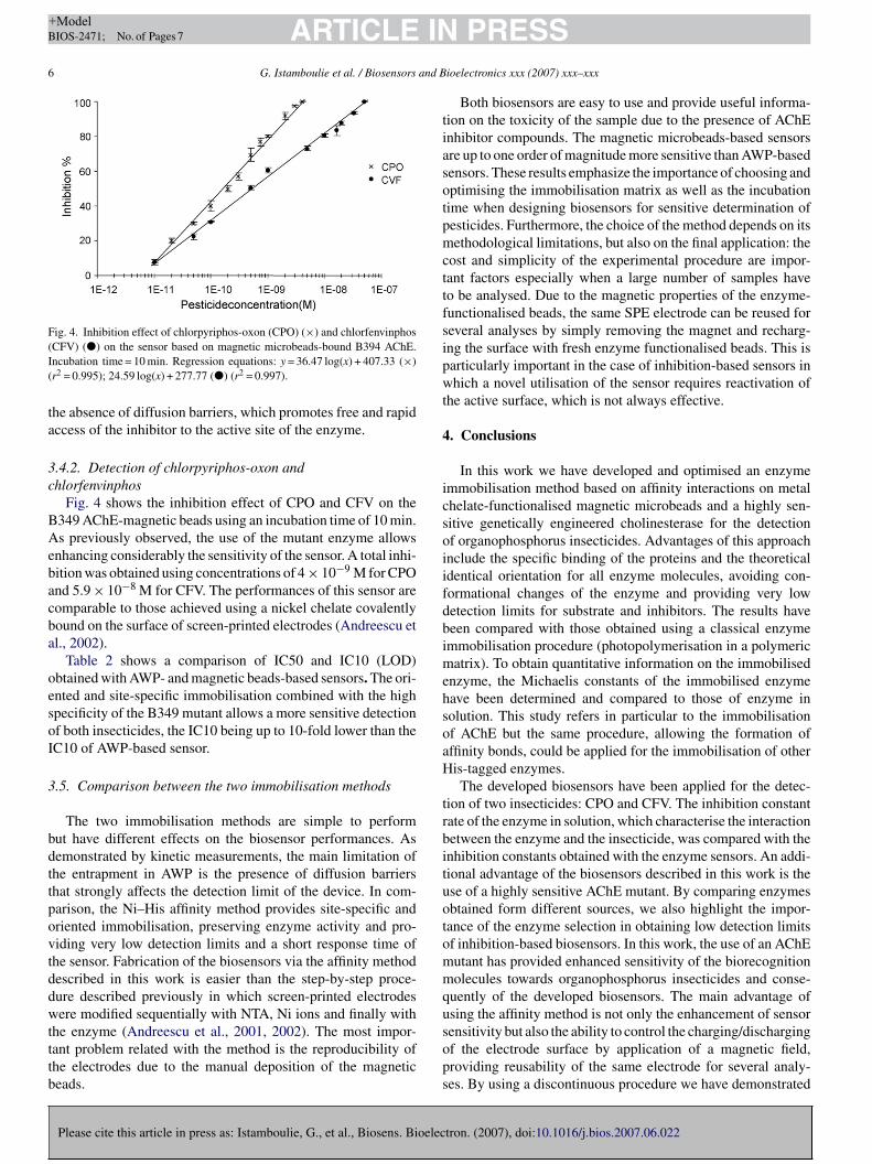
Fig. 4. Inhibition effect of chlorpyriphos-oxon (CPO) (×) and chlorfenvinphos(I(
ta
3c
BAebacba
oesoI
3
bdttpovtddwtttb
tiasotpmcttfsipwt
4
icsoiifdbimehsoaH
trbituotommqu
CFV) (�) on the sensor based on magnetic microbeads-bound B394 AChE.ncubation time = 10 min. Regression equations: y = 36.47 log(x) + 407.33 (×)r2 = 0.995); 24.59 log(x) + 277.77 (�) (r2 = 0.997).
he absence of diffusion barriers, which promotes free and rapidccess of the inhibitor to the active site of the enzyme.
.4.2. Detection of chlorpyriphos-oxon andhlorfenvinphos
Fig. 4 shows the inhibition effect of CPO and CFV on the349 AChE-magnetic beads using an incubation time of 10 min.s previously observed, the use of the mutant enzyme allows
nhancing considerably the sensitivity of the sensor. A total inhi-ition was obtained using concentrations of 4 × 10⁻⁹ M for CPOnd 5.9 × 10⁻⁸ M for CFV. The performances of this sensor areomparable to those achieved using a nickel chelate covalentlyound on the surface of screen-printed electrodes (Andreescu etl., 2002).
Table 2 shows a comparison of IC50 and IC10 (LOD)btained with AWP- and magnetic beads-based sensors. The ori-nted and site-specific immobilisation combined with the highpecificity of the B349 mutant allows a more sensitive detectionf both insecticides, the IC10 being up to 10-fold lower than theC10 of AWP-based sensor.
.5. Comparison between the two immobilisation methods
The two immobilisation methods are simple to performut have different effects on the biosensor performances. Asemonstrated by kinetic measurements, the main limitation ofhe entrapment in AWP is the presence of diffusion barriershat strongly affects the detection limit of the device. In com-arison, the Ni–His affinity method provides site-specific andriented immobilisation, preserving enzyme activity and pro-iding very low detection limits and a short response time ofhe sensor. Fabrication of the biosensors via the affinity methodescribed in this work is easier than the step-by-step proce-ure described previously in which screen-printed electrodesere modified sequentially with NTA, Ni ions and finally with
Please cite this article in press as: Istamboulie, G., et al., Biosens. Bioelec
he enzyme (Andreescu et al., 2001, 2002). The most impor-ant problem related with the method is the reproducibility ofhe electrodes due to the manual deposition of the magneticeads.
sops
PRESSioelectronics xxx (2007) xxx–xxx
Both biosensors are easy to use and provide useful informa-ion on the toxicity of the sample due to the presence of AChEnhibitor compounds. The magnetic microbeads-based sensorsre up to one order of magnitude more sensitive than AWP-basedensors. These results emphasize the importance of choosing andptimising the immobilisation matrix as well as the incubationime when designing biosensors for sensitive determination ofesticides. Furthermore, the choice of the method depends on itsethodological limitations, but also on the final application: the
ost and simplicity of the experimental procedure are impor-ant factors especially when a large number of samples haveo be analysed. Due to the magnetic properties of the enzyme-unctionalised beads, the same SPE electrode can be reused foreveral analyses by simply removing the magnet and recharg-ng the surface with fresh enzyme functionalised beads. This isarticularly important in the case of inhibition-based sensors inhich a novel utilisation of the sensor requires reactivation of
he active surface, which is not always effective.
. Conclusions
In this work we have developed and optimised an enzymemmobilisation method based on affinity interactions on metalhelate-functionalised magnetic microbeads and a highly sen-itive genetically engineered cholinesterase for the detectionf organophosphorus insecticides. Advantages of this approachnclude the specific binding of the proteins and the theoreticaldentical orientation for all enzyme molecules, avoiding con-ormational changes of the enzyme and providing very lowetection limits for substrate and inhibitors. The results haveeen compared with those obtained using a classical enzymemmobilisation procedure (photopolymerisation in a polymeric
atrix). To obtain quantitative information on the immobilisednzyme, the Michaelis constants of the immobilised enzymeave been determined and compared to those of enzyme inolution. This study refers in particular to the immobilisationf AChE but the same procedure, allowing the formation offfinity bonds, could be applied for the immobilisation of otheris-tagged enzymes.The developed biosensors have been applied for the detec-
ion of two insecticides: CPO and CFV. The inhibition constantate of the enzyme in solution, which characterise the interactionetween the enzyme and the insecticide, was compared with thenhibition constants obtained with the enzyme sensors. An addi-ional advantage of the biosensors described in this work is these of a highly sensitive AChE mutant. By comparing enzymesbtained form different sources, we also highlight the impor-ance of the enzyme selection in obtaining low detection limitsf inhibition-based biosensors. In this work, the use of an AChEutant has provided enhanced sensitivity of the biorecognitionolecules towards organophosphorus insecticides and conse-
uently of the developed biosensors. The main advantage ofsing the affinity method is not only the enhancement of sensor
tron. (2007), doi:10.1016/j.bios.2007.06.022
ensitivity but also the ability to control the charging/dischargingf the electrode surface by application of a magnetic field,roviding reusability of the same electrode for several analy-es. By using a discontinuous procedure we have demonstrated

IN+ModelB
and B
tras
R
AA
A
BBBD
E
J
KKN
NN
SV
ARTICLEIOS-2471; No. of Pages 7
G. Istamboulie et al. / Biosensors
he applicability of this approach to measure organophospho-us insecticide induced toxicity. The system could be easilydapted for integration into an autonomously operated magneto-witchable device for monitoring AChE inhibiting activities.
eferences
ldridge, W.N., 1950. Biochem. J. 4, 451–460.ndreescu, S., Magearu, V., Lougarre, A., Fournier, D., Marty, J.-L., 2001. Anal.
Lett. 34, 529–540.ndreescu, S., Barthelmebs, L., Marty, J.-L., 2002. Anal. Chim. Acta 464,
171–180.
Please cite this article in press as: Istamboulie, G., et al., Biosens. Bioelec
onnet, C., Andreescu, S., Marty, J.L., 2003. Anal. Chim. Acta 481, 209–211.ucur, B., Danet, A.F., Marty, J.L., 2004. Biosens. Bioelectron. 20, 217–225.ucur, B., Danet, A.F., Marty, J.L., 2005. Anal. Chim. Acta 530, 1–6.ondoi, M.P., Bucur, B., Danet, A.F., Toader, C.N., Barthelmebs, L., Marty,
J.L., 2006. Anal. Chim. Acta 578, 162–169.
W
W
PRESSioelectronics xxx (2007) xxx–xxx 7
llman, G.L., Coutney, S.M., Andres, V., Featherstne, R.M., 1961. Biochem.Pharm. 7, 88–92.
eanty, G., Ghommidh, Gh., Marty, J.L., 2001. Anal. Chim. Acta 436, 119–128.
atz, E., Baron, R., Willner, I., 2005. J. Am. Chem. Soc. 127, 4060–4070.atz, E., Willner, I., 2002. J. Am. Chem. Soc. 124, 10290–10291.ogueira, J.M.F., Sandra, T., Sandra, P., 2003. J. Chromatogr. A 996, 133–
140.unes, G.S., Jeanty, G., Marty, J.-L., 2004. Anal. Chim. Acta 523, 107–115.unes, G.S., Montesinos, T., Marques, P.B.O., Fournier, D., Marty, J.L., 2001.
Anal. Chim. Acta 434, 1–8.egel, I.H., 1975. Enzyme Kinetics. Wiley/Interscience, New York.illatte, F., Marcel, V., Estrada-Mondaca, S., Fournier, D., 1998. Biosens. Bio-
tron. (2007), doi:10.1016/j.bios.2007.06.022
electron. 13, 157–164.ang, J., Musameh, M., Laocharoensuk, R., 2005. Electrochem. Commun. 7,
652–656.orek, F., Thiermann, H., Szinicz, L., Eyer, P., 2004. Biochem. Pharm. 68,
2237–2248.
Copyright © 2022 FDOKUMEN