In vitro acetylcholinesterase inhibitory activity and the antioxidant properties of Aegle marmelos...
10
ORIGINAL ARTICLE In vitro acetylcholinesterase inhibitory activity and the antioxidant properties of Aegle marmelos leaf extract: implications for the treatment of Alzheimer’s disease Md. ASADUZZAMAN, 1 Md. Josim UDDIN, 1 M.A. KADER, 1 A.H.M.K. ALAM, 1 Aziz Abdur RAHMAN, 1 Mamunur RASHID, 1 Kiyoko KATO, 2 Toshihisa TANAKA, 2 Masatoshi TAKEDA 2 and Golam SADIK 1 1 Department of Pharmacy, University of Rajshahi, Rajshahi, Bangladesh; and 2 Department of Psychia- try, Osaka University Graduate School of Medicine, Osaka, Japan Correspondence: Dr Golam Sadik PhD, Department of Pharmacy, Rajshahi University, Rajshahi 6250, Ban- gladesh. Email: [email protected] Received 1 April 2013; revision received 18 August 2013; accepted 27 August 2013. Abstract Background: Alzheimer’s disease (AD) is a progressive neurodegenerative disorder clinically characterized by loss of memory and cognition. The effec- tive therapeutic options for AD are limited and thus there is a demand for new drugs. Aegle marmelos (Linn.) (A. marmelos) leaves have been used in traditional medicine to promote intellect and enhance memory. In this study, we evaluated A. marmelos for its acetylcholinesterase (AChE) inhibitory activity and antioxidant property in vitro in the treatment of AD. Methods: A crude methanol extract and four fractions (petroleum ether, chloroform, ethyl acetate and aqueous) were prepared from the leaves of A. marmelos. The preparations were assessed for AChE inhibitory activity by the Ellman method, and their antioxidant properties were assessed by several assays: reducing power, scavenging of 1,1-diphenyl-2-picrylhydrazyl free radical and hydroxyl radical, and inhibition of lipid peroxidation. Quali- tative and quantitative analyses of endogenous substances in A. marmelos were performed by the standard phytochemical methods. Results: Among the different extracts tested, the ethyl acetate fraction exhibited the highest inhibition of AChE activity. In the same way, ethyl acetate fraction showed the highest reducing activity and radical scavenging ability towards the 1,1-diphenyl-2-picrylhydrazyl (half maximal inhibitory concentration = 3.84 μg/mL) and hydroxyl free radicals (half maximal inhibi- tory concentration = 5.68 μg/mL). The antiradical activity of the ethyl acetate fraction appeared to be similar to that of the reference standard butylated hydroxytoluene and catechin used in this study. In addition, the ethyl acetate fraction displayed higher inhibition of brain lipid peroxidation. Phytochemical screening of different extractives of A. marmelos showed the presence of phenols and flavonoids, alkaloid, saponin, glycoside, tannin and steroids. Quantitative analysis revealed higher contents of phenolics (58.79-mg gallic acid equivalent/g dried extract) and flavonoids (375.73-mg gallic acid equivalent/g dried extract) in the ethyl acetate fraction. Conclusion: The results suggest that the ethyl acetate fraction of A. marmelos is a significant source of polyphenolic compounds with poten- tial AChE inhibitory property and antioxidant activity and, thus, may be useful in the treatment of AD. Key words: acetylcholinesterase inhibition, Aegle marmelos, Alzheimer’s disease, antioxidant activity, phytochemical screening. doi:10.1111/psyg.12031 PSYCHOGERIATRICS 2014; 14: 1–10 1 © 2014 The Authors Psychogeriatrics © 2014 Japanese Psychogeriatric Society
Transcript of In vitro acetylcholinesterase inhibitory activity and the antioxidant properties of Aegle marmelos...
ORIGINAL ARTICLE
In vitro acetylcholinesterase inhibitory activity and the antioxidantproperties of Aegle marmelos leaf extract: implications for thetreatment of Alzheimer’s disease
Md. ASADUZZAMAN,1 Md. Josim UDDIN,1 M.A. KADER,1 A.H.M.K. ALAM,1 Aziz Abdur RAHMAN,1
Mamunur RASHID,1 Kiyoko KATO,2 Toshihisa TANAKA,2 Masatoshi TAKEDA2 and Golam SADIK1
1Department of Pharmacy, University of Rajshahi,Rajshahi, Bangladesh; and 2Department of Psychia-try, Osaka University Graduate School of Medicine,Osaka, Japan
Correspondence: Dr Golam Sadik PhD, Department ofPharmacy, Rajshahi University, Rajshahi 6250, Ban-gladesh. Email: [email protected]
Received 1 April 2013; revision received 18 August 2013;accepted 27 August 2013.
Abstract
Background: Alzheimer’s disease (AD) is a progressive neurodegenerativedisorder clinically characterized by loss of memory and cognition. The effec-tive therapeutic options for AD are limited and thus there is a demand fornew drugs. Aegle marmelos (Linn.) (A. marmelos) leaves have been used intraditional medicine to promote intellect and enhance memory. In this study,we evaluated A. marmelos for its acetylcholinesterase (AChE) inhibitoryactivity and antioxidant property in vitro in the treatment of AD.Methods: A crude methanol extract and four fractions (petroleum ether,chloroform, ethyl acetate and aqueous) were prepared from the leaves ofA. marmelos. The preparations were assessed for AChE inhibitory activityby the Ellman method, and their antioxidant properties were assessed byseveral assays: reducing power, scavenging of 1,1-diphenyl-2-picrylhydrazylfree radical and hydroxyl radical, and inhibition of lipid peroxidation. Quali-tative and quantitative analyses of endogenous substances in A. marmeloswere performed by the standard phytochemical methods.Results: Among the different extracts tested, the ethyl acetate fractionexhibited the highest inhibition of AChE activity. In the same way, ethylacetate fraction showed the highest reducing activity and radical scavengingability towards the 1,1-diphenyl-2-picrylhydrazyl (half maximal inhibitoryconcentration = 3.84 μg/mL) and hydroxyl free radicals (half maximal inhibi-tory concentration = 5.68 μg/mL). The antiradical activity of the ethyl acetatefraction appeared to be similar to that of the reference standard butylatedhydroxytoluene and catechin used in this study. In addition, the ethyl acetatefraction displayed higher inhibition of brain lipid peroxidation. Phytochemicalscreening of different extractives of A. marmelos showed the presence ofphenols and flavonoids, alkaloid, saponin, glycoside, tannin and steroids.Quantitative analysis revealed higher contents of phenolics (58.79-mggallic acid equivalent/g dried extract) and flavonoids (375.73-mg gallic acidequivalent/g dried extract) in the ethyl acetate fraction.Conclusion: The results suggest that the ethyl acetate fraction ofA. marmelos is a significant source of polyphenolic compounds with poten-tial AChE inhibitory property and antioxidant activity and, thus, may be usefulin the treatment of AD.
Key words: acetylcholinesterase inhibition, Aeglemarmelos, Alzheimer’s disease, antioxidantactivity, phytochemical screening.
bs_bs_banner
doi:10.1111/psyg.12031 PSYCHOGERIATRICS 2014; 14: 1–10
1© 2014 The AuthorsPsychogeriatrics © 2014 Japanese Psychogeriatric Society
INTRODUCTIONAlzheimer’s disease (AD), a major cause of mortality inelderly people, is a progressive neurodegenerative dis-order characterized by a gradual loss of memory, cog-nition and behavioural abnormalities. The pathologicalhallmarks of AD are profound loss of cholinergicneurons, senile plaque consisting of Abeta protein,and neurofibrillary tangles of microtubule-associatedprotein tau.1–5 Although there has been tremendousprogress in understanding the aetiology and patho-genesis of AD, effective drugs remain limited.
Extensive loss of cholinergic neurons, particularly inthe basal forebrain is a prominent feature observed inAD patients that is accompanied by deficiency of ace-tylcholine (ACh), a neurotransmitter found in the syn-apses of the cerebral cortex.6–8 The loss of cholinergicneurons and the associated decrease in levels of AChhas been found to correlate well with the cognitiveimpairment seen in AD patients.9 Therefore, elevatingACh by inhibiting acetylcholinesterase (AChE), which isinvolved in the breakdown of ACh, appears to improvethe symptoms of cognitive deficit in AD and serves asan important strategy in the development of drugs. Todate, only three cholinesterase inhibitors, donepezil,galantamine and rivastigmine, have been approvedby the US Food and Drug Administration to treatAD.10,11 These drugs have ameliorated symptoms andimproved the functioning of patients with AD, but nonecan completely restrict or reverse the progressionof AD.12
A substantial body of evidence implicates oxidativestress in the aetiology and pathogenesis of AD. It hasbeen shown that the reactive oxygen species (ROS)and other free radicals, which are formed and accu-mulated during oxidative stress as a result of an imbal-ance between their production and removal by theantioxidant system, induce cellular and molecularabnormalities in sporadic AD.13–16 Although the exactmechanisms underlying these deleterious effectsremain unclear, it is known that oxidative stress occursbefore the formation of neurofibrillary tangles andsenile plaques, both of which are hallmarks of AD.17,18 Ithas been further demonstrated that Abeta protein, themajor component of senile plaque in the brain of ADpatients, causes an increase in free radical productionin neuronal cells, leading to oxidative stress and celldeath.19,20 Therefore, antioxidants have been sug-gested as therapies to prevent, delay or ameliorate thepathological changes underlying the progression of
AD.21,22 Recently, there has been increasing interest inthe natural antioxidants contained in the medicinalplants, which are candidates to prevent oxidativedamage.23,24
Aegle marmelos (A. marmelos), commonly knownas bael and belonging to the family Rutaceae, is atree widely distributed throughout Bangladesh. Themedicinal properties of A. marmelos are well describedin Ayurveda, traditional Indian medicine.25 The leaf ofthis plant has a folkloric reputation for promoting intel-lect and enhancing memory.26 Traditionally, the planthas been used to treat fever, diabetes, diarrhoea,abscesses and snake bites. Phytochemical investiga-tions of A. marmelos demonstrated several active ele-ments including marmelosin, marmelide, luvangetin,auraptene, psoralen and tannin.27,28 The extract ofthe plant has been reported to possess importantpharmacological effects including anti-diabetic, anti-hyperlipidaemic, contraceptive, antidiarrhoeal, anal-gesic, antipyretic and anti-inflammatory, antimicrobialand anti-proliferative effects.28–34 A preliminary studyhas shown that A. marmelos has 1,1-diphenyl-2-picrylhydrazyl (DPPH) radical scavenging activity.29
Although A. marmelos has important medicinalvalues for the treatment of AD, no studies have yetexamined its anti-AD capabilities. Therefore, the objec-tive of this study was to evaluate the inhibition of AChEactivity and antioxidant properties of A. marmelosleaves in order to treat AD.
MATERIALS AND METHODSChemicalsAluminium chloride, ammonium molybdate, ascorbicacid (AA), bicinchoninic acid, DPPH, Folin–Ciocalteureagent, Tris-HCl and Triton X-100 were obtained fromSigma-Aldrich (Bangalore, India). Gallic acid wasobtained from Wako Pure Chemical Company Ltd(Osaka, Japan). 2-deoxy-D-ribose, thiobarbituric acid,(+)-catechin, 5,5′-dithio-bis-(2-nitro) benzoic acid,acetylthiocholine iodide and donepezil were obtainedfrom Sigma-Aldrich (Tokyo, Japan). Unless otherwisespecified, all other chemicals were of analytical grade.
Plant materialsThe leaves of A. Marmelos were collected from the cityof Rajshahi, Bangladesh, and identified by an experttaxonomist. A voucher specimen was submitted to the
M. Asaduzzaman et al.
2 © 2014 The AuthorsPsychogeriatrics © 2014 Japanese Psychogeriatric Society
herbarium of the Department of Botany, RajshahiUniversity.
ExtractionThe leaves of A. marmelos were dried at room tem-perature for 9 days, finely powdered and used forextraction. Powdered leaves (500 g) were placed in anamber-coloured reagent bottle and soaked in 1.5-Lmethanol. The contents were sealed in the bottle for 7days and occasionally shaken and stirred. The wholemixture was filtered through cotton and then throughWhatman No. 1 filters paper, and the filtrate wasconcentrated with a rotary evaporator under reducedpressure at 50°C to obtain the crude methanol extract(CME) (13.485 g). An aliquot (10 g) of the concentratedmethanol extract was fractionated as described pre-viously,35 and the resultant soluble fractions of petro-leum ether (PEF, 2.693 g), chloroform (CLF, 1.834 g),ethyl acetate (EAF, 1.580 g) and aqueous (AQF,6.195 g) were obtained for the experiment.
Phytochemical screening of the plant extractThe extracts were tested to determine the presence ofvarious phytochemicals, including tannins, flavonoids,alkaloids, saponins, glycosides and steroids in accor-dance with the methods described.35
Determination of total phenolic contentTotal phenolic content of the different extracts fromA. marmelos was determined with the Folin–Ciocalteureagent.36 The reaction mixture contained 0.5-mLplant extract or standard solution at different concen-trations, 2.5-mL Folin–Ciocalteu reagent (diluted 10times with water) and 2.5-m sodium carbonate solu-tion (7.5%). The test tube was incubated for 20 min at25°C to complete the reaction, and the absorbance ofthe reaction mixture was measured at 760 nm. Gallicacid was used as the standard and the results wereexpressed as milligrams of gallic acid equivalent(GAE)/g of dried extractives.
Determination of total flavonoid contentTotal flavonoid content of the different A. marmelosextracts was determined by the aluminium chloridecolorimetric method.37 The plant extract (1.0 mL) wasadded to 3.0-mL methanol, 0.2-mL 10% AlCl3, 0.2-mL1 M potassium acetate and 5.6-mL distilled water. Thetest tube was then incubated at room temperature for30 min to complete the reaction. The absorbance of
the solution was measured at 420 nm. Gallic acid wasused as standard and the results were expressedas mg of gallic acid equivalent (GAE)/g of driedextractives.
Determination of reducing powerThe reducing power of different A. marmelos extractswas evaluated using the method employed byOyaizu.38 Various concentrations of plant extract orstandard solutions (1 mL) were mixed with 2.5-mLpotassium buffer (0.2 M) and 2.5-mL potassiumferricyanide. After heating for 20 min at 50°C, 2.5-mLtrichloroacetic acid (10%) solution was added tothe test tube. The total mixture was centrifuged at3300 g for 10 min. Next, 2.5-mL supernatant solutionwas withdrawn from the mixture and mixedwith 2.5-mL distilled water and 0.5-mL ferric chloride(0.1%) solution. The absorbance was measured at700 nm. AA was used for comparison.
Determination of DPPH radicalscavenging activityDPPH radical scavenging activity of the differentA. marmelos extracts was determined according tothe method reported by Choi et al. with slight modifi-cations: 2-mL methanol solution of plant extract orreference standard butylated hydroxytoluene at differ-ent concentration was mixed with 3-mL methanolsolution of DPPH in the test tube.39 The test tube wasincubated at room temperature for 30 min in a darkplace to complete the reaction. The absorbance ofthe solution was measured at 517 nm. DPPH freeradical scavenging ability (%) was calculated with thefollowing formula:
(( )A AAabsorbance of control absorbance of sample
absorbance
− of control ) × 100
Determination of hydroxyl radicalscavenging activityHydroxyl radical scavenging activity of different A.marmelos extracts was determined by the methoddescribed by Elizabeth et al. but with a slight modifi-cation.40 The assay is based on the quantification of thedegradation product of 2-deoxyribose by condensa-tion with thiobarbituric acid. Hydroxyl radical was gen-erated by the Fe3+-ascorbate-EDTA-H2O2 system (theFenton reaction). In a final volume of 1 mL, the reac-tion mixture contained 2-deoxy-2-ribose (2.8 mM);
Neuroprotective activity of A. marmelos
3© 2014 The AuthorsPsychogeriatrics © 2014 Japanese Psychogeriatric Society
KH2PO4-KOH buffer (20 mM, pH 7.4); FeCl3 (100 μM);EDTA (100 μM); H2O2 (1.0 mM); ascorbic acid(100 μM); and various concentrations of the testsample or reference compound catechin (CA). Afterincubation for 1 h at 37°C, 0.5-mL reaction mixture wasadded to 1-mL 2.8% trichloroacetic acid, then 1-mL1% aqueous thiobarbituric acid was added, and themixture was incubated at 90°C for 15 min to developthe colour. After it cooled, the mixture’s absorbancewas measured at 532 nm against an appropriate blanksolution. Hydroxyl radical scavenging ability (%) wascalculated by using the formula:
(( )A AAabsorbance of control absorbance of sample
absorbance
− of control ) × 100
Determination of lipid peroxidationinhibition activityThe inhibition of lipid peroxidation activity was deter-mined according to the method described by Liu et al.with a slight modification.41 The 150-g adult long Evanrats were anaesthetized with sodium phenobarbitone.The rats’ brain were dissected and homogenizedwith a homogenizer in ice-cold phosphate buffer(50 mM, pH 7.4) to produce a 1/10 homogenate. Thehomogenate was centrifuged at 10 000 g for 15 min at4°C. The supernatant was used as liposome for anin vitro lipid peroxidation assay. The ability of plantextract to inhibit lipid peroxidation was studied byincubating rat brain homogenates treated with hydro-gen peroxide (10 μM) and different concentrationsof plant extract. Hydrogen peroxide induced lipidperoxidation in the rat brain homogenates. Lipid per-oxides reacted with thiobarbituric acid to form a pinkproduct, thiobarbituric acid reacting substances,measurable colorimetrically at 532 nm. The differencebetween the control and plant extract treated samplewas the measured decrease in thiobarbituric acidreacting substances formation, reflecting reducedhydroxyl radical-induced lipid peroxidation. CA wasused as the reference standard for comparison.
Determination of AChE inhibitory activityThe AChE inhibitory assay was performed accordingto the colorimetric method by Ellman et al.,42 withacetylthiocholine iodide as a substrate. For the enzymesource, the rat brains were homogenized in a homog-enizer with five volumes of a homogenation buffer(10 mM Tris-HCl (pH 7.2), which contained 1 M NaCl,
50 mM MgCl2 and 1% Triton X-100), and centrifuged at10 000 g for 30 min. The resulting supernatant wasused as an enzyme source. All of the extraction stepswere carried out at 4°C. Protein concentration wasdetermined by using a bicinchoninic acid kit (SigmaCo., St. Louis, MO, USA) with bovine serum albumin asa protein standard. The rates of hydrolysis by AChEwere monitored spectrophotometrically. Each extractor standard (500 μL) was mixed with an enzyme solu-tion (500 μL) and incubated at 37°C for 15 min. Absor-bance at 405 nm was read immediately after addingEllman’s reaction mixture (3.5-mL 0.5 mM acetyl-thiocholine, 1 mM 5, 5′-dithio-bis (2-nitro benzoicacid)) in a 50 mM sodium phosphate buffer (pH 8.0) tothe above reaction mixture. Reading was repeated for10 min at 2 min intervals to verify that the reactionoccurred linearly. The blank reaction was measured bysubstituting saline for the enzyme. Donepezil was usedas a positive control. The percentage inhibition ofAChE activity was calculated using the followingformula:
(( )A AAabsorbance of control absorbance of sample
absorbance
− of control ) × 100
Statistical analysisAll analyses were carried out in triplicate. Data werepresented as mean 1 SD. Free R-software version2.15.1 (http://www.r-project.org/) and Microsoft Excel2007 (Roselle, IL, USA) were used for the statisticaland graphical evaluations. Significant differences(P-value <0.05) between the means were determinedusing the t-test.
RESULTSAChE inhibitory activityReduction of ACh in the hippocampus and cortex ofthe brain is one of the most important remarkablechanges observed in AD.5–8 Therefore, elevation ofACh level in the synaptic cleft by inhibition of AChE,which is involved in the breakdown of ACh, is anaccepted therapeutic strategy for AD. The inhibitoryactivity of CME derived from A. marmelos against ratbrain AChE was determined by Ellman’s method.41
This method estimates AChE using acetylthiocholineiodide (substrate) and dithiobis nitro benzoic acid. Theenzymatic activity was measured by the yellow colourcompound produced by thiocholine when it reactswith dithiobis nitrobenzoate ion. The result of the
M. Asaduzzaman et al.
4 © 2014 The AuthorsPsychogeriatrics © 2014 Japanese Psychogeriatric Society
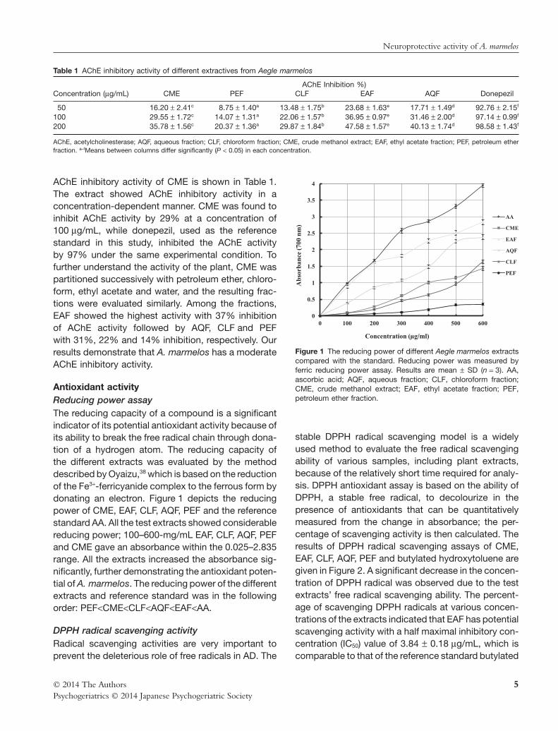
AChE inhibitory activity of CME is shown in Table 1.The extract showed AChE inhibitory activity in aconcentration-dependent manner. CME was found toinhibit AChE activity by 29% at a concentration of100 μg/mL, while donepezil, used as the referencestandard in this study, inhibited the AChE activityby 97% under the same experimental condition. Tofurther understand the activity of the plant, CME waspartitioned successively with petroleum ether, chloro-form, ethyl acetate and water, and the resulting frac-tions were evaluated similarly. Among the fractions,EAF showed the highest activity with 37% inhibitionof AChE activity followed by AQF, CLF and PEFwith 31%, 22% and 14% inhibition, respectively. Ourresults demonstrate that A. marmelos has a moderateAChE inhibitory activity.
Antioxidant activityReducing power assayThe reducing capacity of a compound is a significantindicator of its potential antioxidant activity because ofits ability to break the free radical chain through dona-tion of a hydrogen atom. The reducing capacity ofthe different extracts was evaluated by the methoddescribed by Oyaizu,38 which is based on the reductionof the Fe3+-ferricyanide complex to the ferrous form bydonating an electron. Figure 1 depicts the reducingpower of CME, EAF, CLF, AQF, PEF and the referencestandard AA. All the test extracts showed considerablereducing power; 100–600-mg/mL EAF, CLF, AQF, PEFand CME gave an absorbance within the 0.025–2.835range. All the extracts increased the absorbance sig-nificantly, further demonstrating the antioxidant poten-tial of A. marmelos. The reducing power of the differentextracts and reference standard was in the followingorder: PEF<CME<CLF<AQF<EAF<AA.
DPPH radical scavenging activityRadical scavenging activities are very important toprevent the deleterious role of free radicals in AD. The
stable DPPH radical scavenging model is a widelyused method to evaluate the free radical scavengingability of various samples, including plant extracts,because of the relatively short time required for analy-sis. DPPH antioxidant assay is based on the ability ofDPPH, a stable free radical, to decolourize in thepresence of antioxidants that can be quantitativelymeasured from the change in absorbance; the per-centage of scavenging activity is then calculated. Theresults of DPPH radical scavenging assays of CME,EAF, CLF, AQF, PEF and butylated hydroxytoluene aregiven in Figure 2. A significant decrease in the concen-tration of DPPH radical was observed due to the testextracts’ free radical scavenging ability. The percent-age of scavenging DPPH radicals at various concen-trations of the extracts indicated that EAF has potentialscavenging activity with a half maximal inhibitory con-centration (IC50) value of 3.84 1 0.18 μg/mL, which iscomparable to that of the reference standard butylated
Table 1 AChE inhibitory activity of different extractives from Aegle marmelos
Concentration (μg/mL)AChE Inhibition %)
CME PEF CLF EAF AQF Donepezil
50 16.20 1 2.41c 8.75 1 1.40a 13.48 1 1.75b 23.68 1 1.63e 17.71 1 1.49d 92.76 1 2.15f
100 29.55 1 1.72c 14.07 1 1.31a 22.06 1 1.57b 36.95 1 0.97e 31.46 1 2.00d 97.14 1 0.99f
200 35.78 1 1.56c 20.37 1 1.36a 29.87 1 1.84b 47.58 1 1.57e 40.13 1 1.74d 98.58 1 1.43f
AChE, acetylcholinesterase; AQF, aqueous fraction; CLF, chloroform fraction; CME, crude methanol extract; EAF, ethyl acetate fraction; PEF, petroleum etherfraction. a–fMeans between columns differ significantly (P < 0.05) in each concentration.
0
0.5
1
1.5
2
2.5
3
3.5
4
0 100 200 300 400 500 600
Abs
orba
nce
(700
nm
)
Concentration (µg/ml)
AA
CME
EAF
AQF
CLF
PEF
Figure 1 The reducing power of different Aegle marmelos extractscompared with the standard. Reducing power was measured byferric reducing power assay. Results are mean 1 SD (n = 3). AA,ascorbic acid; AQF, aqueous fraction; CLF, chloroform fraction;CME, crude methanol extract; EAF, ethyl acetate fraction; PEF,petroleum ether fraction.
Neuroprotective activity of A. marmelos
5© 2014 The AuthorsPsychogeriatrics © 2014 Japanese Psychogeriatric Society
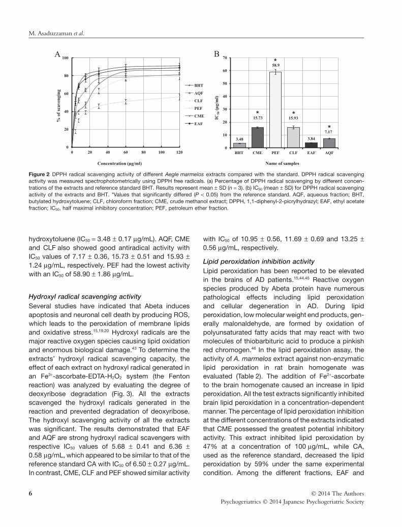
hydroxytoluene (IC50 = 3.48 1 0.17 μg/mL). AQF, CMEand CLF also showed good antiradical activity withIC50 values of 7.17 1 0.36, 15.73 1 0.51 and 15.93 11.24 μg/mL, respectively. PEF had the lowest activitywith an IC50 of 58.90 1 1.86 μg/mL.
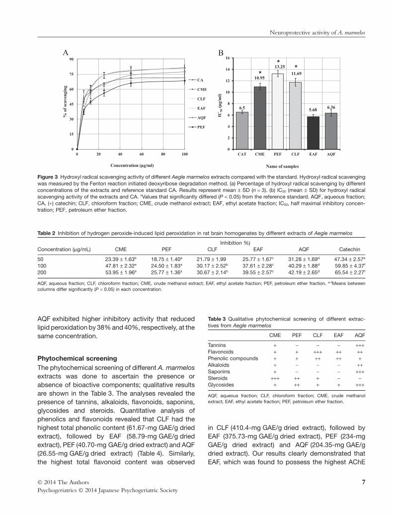
Hydroxyl radical scavenging activitySeveral studies have indicated that Abeta inducesapoptosis and neuronal cell death by producing ROS,which leads to the peroxidation of membrane lipidsand oxidative stress.15,19,20 Hydroxyl radicals are themajor reactive oxygen species causing lipid oxidationand enormous biological damage.43 To determine theextracts’ hydroxyl radical scavenging capacity, theeffect of each extract on hydroxyl radical generated inan Fe3+-ascorbate-EDTA-H2O2 system (the Fentonreaction) was analyzed by evaluating the degree ofdeoxyribose degradation (Fig. 3). All the extractsscavenged the hydroxyl radicals generated in thereaction and prevented degradation of deoxyribose.The hydroxyl scavenging activity of all the extractswas significant. The results demonstrated that EAFand AQF are strong hydroxyl radical scavengers withrespective IC50 values of 5.68 1 0.41 and 6.36 10.58 μg/mL, which appeared to be similar to that of thereference standard CA with IC50 of 6.50 1 0.27 μg/mL.In contrast, CME, CLF and PEF showed similar activity
with IC50 of 10.95 1 0.56, 11.69 1 0.69 and 13.25 10.56 μg/mL, respectively.
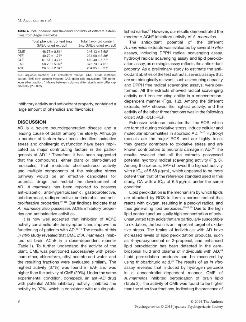
Lipid peroxidation inhibition activityLipid peroxidation has been reported to be elevatedin the brains of AD patients.15,44,45 Reactive oxygenspecies produced by Abeta protein have numerouspathological effects including lipid peroxidationand cellular degeneration in AD. During lipidperoxidation, low molecular weight end products, gen-erally malonaldehyde, are formed by oxidation ofpolyunsaturated fatty acids that may react with twomolecules of thiobarbituric acid to produce a pinkishred chromogen.46 In the lipid peroxidation assay, theactivity of A. marmelos extract against non-enzymaticlipid peroxidation in rat brain homogenate wasevaluated (Table 2). The addition of Fe2+-ascorbateto the brain homogenate caused an increase in lipidperoxidation. All the test extracts significantly inhibitedbrain lipid peroxidation in a concentration-dependentmanner. The percentage of lipid peroxidation inhibitionat the different concentrations of the extracts indicatedthat CME possessed the greatest potential inhibitoryactivity. This extract inhibited lipid peroxidation by47% at a concentration of 100 μg/mL, while CA,used as the reference standard, decreased the lipidperoxidation by 59% under the same experimentalcondition. Among the different fractions, EAF and
0
20
40
60
80
100
0 20 40 60 80 100 120
% o
f sc
aven
ging
Concentration (µg/ml)
BHT
AQF
CLF
PEF
CME
EAF
A
3.48
15.73
58.9
15.93
3.84
7.17
0
10
20
30
40
50
60
70
BHT CME PEF CLF EAF AQF
IC50
(µg/
ml)
Name of samples
B*
* *
*
Figure 2 DPPH radical scavenging activity of different Aegle marmelos extracts compared with the standard. DPPH radical scavengingactivity was measured spectrophotometrically using DPPH free radicals. (a) Percentage of DPPH radical scavenging by different concen-trations of the extracts and reference standard BHT. Results represent mean 1 SD (n = 3). (b) IC50 (mean 1 SD) for DPPH radical scavengingactivity of the extracts and BHT. *Values that significantly differed (P < 0.05) from the reference standard. AQF, aqueous fraction; BHT,butylated hydroxytoluene; CLF, chloroform fraction; CME, crude methanol extract; DPPH, 1,1-diphenyl-2-picrylhydrazyl; EAF, ethyl acetatefraction; IC50, half maximal inhibitory concentration; PEF, petroleum ether fraction.
M. Asaduzzaman et al.
6 © 2014 The AuthorsPsychogeriatrics © 2014 Japanese Psychogeriatric Society
AQF exhibited higher inhibitory activity that reducedlipid peroxidation by 38% and 40%, respectively, at thesame concentration.
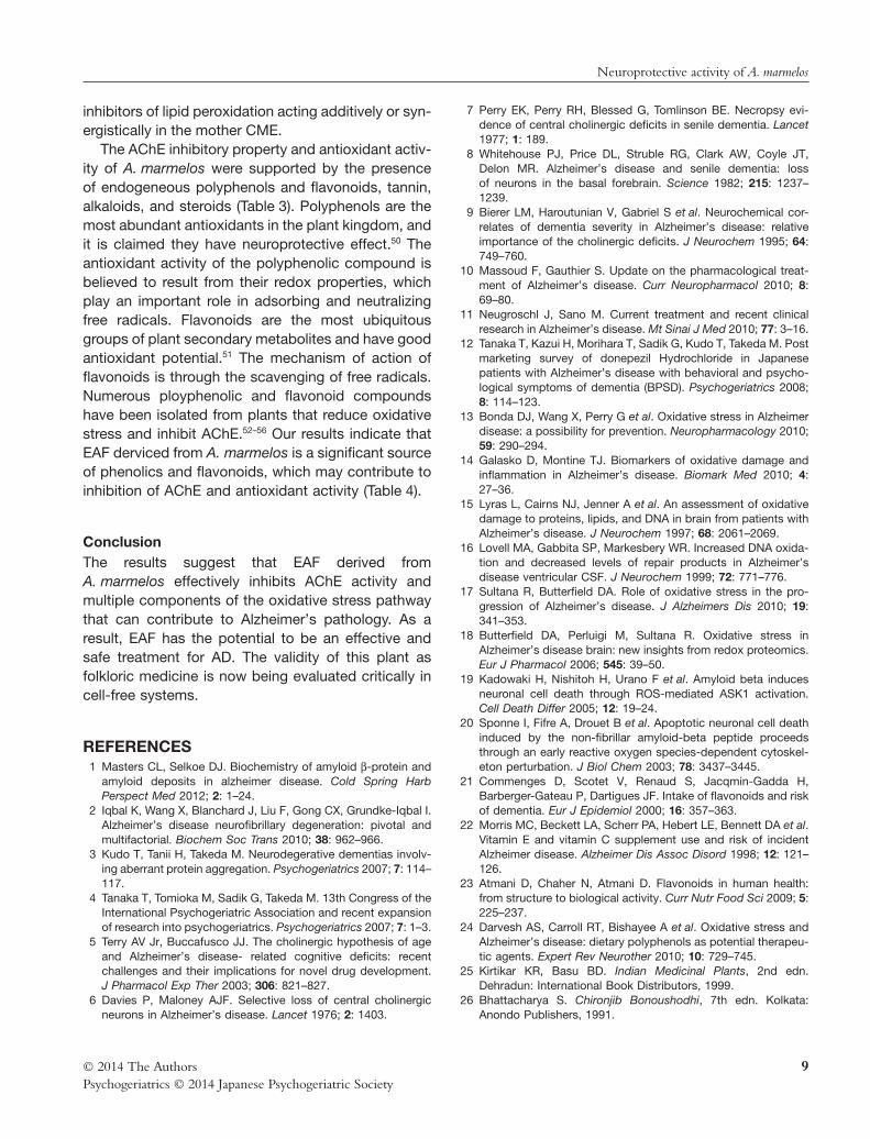
Phytochemical screeningThe phytochemical screening of different A. marmelosextracts was done to ascertain the presence orabsence of bioactive components; qualitative resultsare shown in the Table 3. The analyses revealed thepresence of tannins, alkaloids, flavonoids, saponins,glycosides and steroids. Quantitative analysis ofphenolics and flavonoids revealed that CLF had thehighest total phenolic content (61.67-mg GAE/g driedextract), followed by EAF (58.79-mg GAE/g driedextract), PEF (40.70-mg GAE/g dried extract) and AQF(26.55-mg GAE/g dried extract) (Table 4). Similarly,the highest total flavonoid content was observed
in CLF (410.4-mg GAE/g dried extract), followed byEAF (375.73-mg GAE/g dried extract), PEF (234-mgGAE/g dried extract) and AQF (204.35-mg GAE/gdried extract). Our results clearly demonstrated thatEAF, which was found to possess the highest AChE
Table 3 Qualitative phytochemical screening of different extrac-tives from Aegle marmelos
CME PEF CLF EAF AQF
Tannins + − − − +++Flavonoids + + +++ ++ ++Phenolic compounds + + ++ ++ +Alkaloids + − − − ++Saponins + − − − +++Steroids +++ ++ + − −Glycosides + ++ + + +++
AQF, aqueous fraction; CLF, chloroform fraction; CME, crude methanolextract; EAF, ethyl acetate fraction; PEF, petroleum ether fraction.
0
15
30
45
60
75
90
0 20 40 60 80 100
% o
f sc
aven
ging
Concentration (µg/ml)
CA
CME
CLF
EAF
AQF
PEF
A
6.5
10.95
13.25
11.69
5.68 6.36
0
2
4
6
8
10
12
14
16
CAT CME PEF CLF EAF AQF
IC50
(µg/
ml)
Name of samples
B
**
*
Figure 3 Hydroxyl radical scavenging activity of different Aegle marmelos extracts compared with the standard. Hydroxyl radical scavengingwas measured by the Fenton reaction initiated deoxyribose degradation method. (a) Percentage of hydroxyl radical scavenging by differentconcentrations of the extracts and reference standard CA. Results represent mean 1 SD (n = 3). (b) IC50 (mean 1 SD) for hydroxyl radicalscavenging activity of the extracts and CA. *Values that significantly differed (P < 0.05) from the reference standard. AQF, aqueous fraction;CA, (+) catechin; CLF, chloroform fraction; CME, crude methanol extract; EAF, ethyl acetate fraction; IC50, half maximal inhibitory concen-tration; PEF, petroleum ether fraction.
Table 2 Inhibition of hydrogen peroxide-induced lipid peroxidation in rat brain homogenates by different extracts of Aegle marmelos
Concentration (μg/mL)Inhibition %)
CME PEF CLF EAF AQF Catechin
50 23.39 1 1.63b 18.75 1 1.40a 21.79 1 1.99 25.77 1 1.67c 31.28 1 1.69d 47.34 1 2.57e
100 47.81 1 2.32e 24.50 1 1.83a 30.17 1 2.52b 37.61 1 2.28c 40.29 1 1.88d 59.85 1 4.37f
200 53.95 1 1.96e 25.77 1 1.36a 30.67 1 2.14b 39.55 1 2.57c 42.19 1 2.65d 65.54 1 2.27f
AQF, aqueous fraction; CLF, chloroform fraction; CME, crude methanol extract; EAF, ethyl acetate fraction; PEF, petroleum ether fraction. a–fMeans betweencolumns differ significantly (P < 0.05) in each concentration.
Neuroprotective activity of A. marmelos
7© 2014 The AuthorsPsychogeriatrics © 2014 Japanese Psychogeriatric Society
inhibitory activity and antioxidant property, contained alarge amount of phenolics and flavonoids.
DISCUSSIONAD is a severe neurodegenerative disease and aleading cause of death among the elderly. Althougha number of factors have been identified, oxidativestress and cholinergic dysfunction have been impli-cated as major contributing factors in the patho-genesis of AD.1–5 Therefore, it has been suggestedthat the compounds, either plant or plant-derivedmolecules, that modulate cholinesterase activityand multiple components of the oxidative stresspathway would be an effective candidates forpotential drugs that restrict the development ofAD. A. marmelos has been reported to possessanti-diabetic, anti-hyperlipidaemic, gastroprotective,antidiarrhoeal, radioprotective, antimicrobial and anti-proliferative properties.28–34 Our findings indicate thatA. marmelos also possesses AChE inhibitory proper-ties and antioxidative activities.
It is now well accepted that inhibition of AChEactivity can ameliorate the symptoms and improve thefunctioning of patients with AD.10,11 The results of thisin vitro study revealed that CME of A. marmelos inhib-ited rat brain AChE in a dose-dependent manner(Table 1). To further understand the activity of theplant, CME was partitioned successively with petro-leum ether, chloroform, ethyl acetate and water, andthe resulting fractions were evaluated similarly. Thehighest activity (37%) was found in EAF and washigher than the activity of CME (29%). Under the sameexperimental condition, donepezil, an anti-AD drugwith potential AChE inhibitory activity, inhibited theactivity by 97%, which is consistent with results pub-
lished earlier.47 However, our results demonstrated themoderate AChE inhibitory activity of A. marmelos.
The antioxidant potential of the differentA. marmelos extracts was evaluated by several in vitroassays, including DPPH radical scavenging assay,hydroxyl radical scavenging assay and lipid peroxid-ation assay, as no single assay reflects the antioxidantproperty. As a preliminary study to estimate the anti-oxidant abilities of the test extracts, several assays thatare not biologically relevant, such as reducing capacityand DPPH free radical scavenging assays, were per-formed. All the extracts showed radical scavengingactivity and iron reducing ability in a concentration-dependent manner (Figs. 1,2). Among the differentextracts, EAF showed the highest activity, and theactivity of the other three fractions was in the followingorder: AQF>CLF>PEF.
Extensive evidence indicates that the ROS, whichare formed during oxidative stress, induce cellular andmolecular abnormalities in sporadic AD.13–16 Hydroxylradicals are the major ROS and are highly toxic;they greatly contribute to oxidative stress and areknown contributors to neuronal damage in AD.48 Theresults revealed that all the extracts possessedpotential hydroxyl radical scavenging activity (Fig. 3).Among the extracts, EAF showed the highest activitywith a IC50 of 5.68 μg/mL, which appeared to be morepotent than that of the reference standard used in thisstudy, CA with a IC50 of 6.5 μg/mL under the samecondition.
Lipid peroxidation is the mechanism by which lipidsare attacked by ROS to form a carbon radical thatreacts with oxygen, resulting in a peroxyl radical andthus generating lipid peroxides.15,44,45 Due to the highlipid content and unusually high concentration of poly-unsaturated fatty acids that are particularly susceptibleto oxidation, the brain is an important target of oxida-tive stress. The brains of individuals with AD haveincreased levels of lipid peroxidation products, suchas 4-hydroxynonenal or 2-propenal, and enhancedlipid peroxidation has been detected in the cere-brospinal fluid and plasma of individuals with AD.49
Lipid peroxidation products can be measured byusing thiobarbituric acid.46 The results of an in vitroassay revealed that, induced by hydrogen peroxidein a concentration-dependent manner, CME ofA. marmelos inhibited peroxidation of brain lipid(Table 2). The activity of CME was found to be higherthan the other four fractions, indicating the presence of
Table 4 Total phenolic and flavonoid contents of different extrac-tives from Aegle marmelos
Total phenolic content (mgGAE/g dried extract)
Total flavonoid content(mg GAE/g dried extract)
CME 46.73 1 0.51c 246.13 1 3.60c
PEF 40.70 1 1.77b 234.00 1 3.38b
CLF 61.67 1 3.74e 410.40 1 5.77e
EAF 58.79 1 5.07d 375.73 1 4.01d
AQF 26.55 1 2.04a 204.35 1 6.27a
AQF, aqueous fraction; CLF, chloroform fraction; CME, crude methanolextract; EAF, ethyl acetate fraction; GAE, gallic acid equivalent; PEF, petro-leum ether fraction. a–fMeans between columns differ significantly differ sig-nificantly (P < 0.05).
M. Asaduzzaman et al.
8 © 2014 The AuthorsPsychogeriatrics © 2014 Japanese Psychogeriatric Society
inhibitors of lipid peroxidation acting additively or syn-ergistically in the mother CME.
The AChE inhibitory property and antioxidant activ-ity of A. marmelos were supported by the presenceof endogeneous polyphenols and flavonoids, tannin,alkaloids, and steroids (Table 3). Polyphenols are themost abundant antioxidants in the plant kingdom, andit is claimed they have neuroprotective effect.50 Theantioxidant activity of the polyphenolic compound isbelieved to result from their redox properties, whichplay an important role in adsorbing and neutralizingfree radicals. Flavonoids are the most ubiquitousgroups of plant secondary metabolites and have goodantioxidant potential.51 The mechanism of action offlavonoids is through the scavenging of free radicals.Numerous ployphenolic and flavonoid compoundshave been isolated from plants that reduce oxidativestress and inhibit AChE.52–56 Our results indicate thatEAF derviced from A. marmelos is a significant sourceof phenolics and flavonoids, which may contribute toinhibition of AChE and antioxidant activity (Table 4).
ConclusionThe results suggest that EAF derived fromA. marmelos effectively inhibits AChE activity andmultiple components of the oxidative stress pathwaythat can contribute to Alzheimer’s pathology. As aresult, EAF has the potential to be an effective andsafe treatment for AD. The validity of this plant asfolkloric medicine is now being evaluated critically incell-free systems.
REFERENCES1 Masters CL, Selkoe DJ. Biochemistry of amyloid β-protein and
amyloid deposits in alzheimer disease. Cold Spring HarbPerspect Med 2012; 2: 1–24.
2 Iqbal K, Wang X, Blanchard J, Liu F, Gong CX, Grundke-Iqbal I.Alzheimer’s disease neurofibrillary degeneration: pivotal andmultifactorial. Biochem Soc Trans 2010; 38: 962–966.
3 Kudo T, Tanii H, Takeda M. Neurodegerative dementias involv-ing aberrant protein aggregation. Psychogeriatrics 2007; 7: 114–117.
4 Tanaka T, Tomioka M, Sadik G, Takeda M. 13th Congress of theInternational Psychogeriatric Association and recent expansionof research into psychogeriatrics. Psychogeriatrics 2007; 7: 1–3.
5 Terry AV Jr, Buccafusco JJ. The cholinergic hypothesis of ageand Alzheimer’s disease- related cognitive deficits: recentchallenges and their implications for novel drug development.J Pharmacol Exp Ther 2003; 306: 821–827.
6 Davies P, Maloney AJF. Selective loss of central cholinergicneurons in Alzheimer’s disease. Lancet 1976; 2: 1403.
7 Perry EK, Perry RH, Blessed G, Tomlinson BE. Necropsy evi-dence of central cholinergic deficits in senile dementia. Lancet1977; 1: 189.
8 Whitehouse PJ, Price DL, Struble RG, Clark AW, Coyle JT,Delon MR. Alzheimer’s disease and senile dementia: lossof neurons in the basal forebrain. Science 1982; 215: 1237–1239.
9 Bierer LM, Haroutunian V, Gabriel S et al. Neurochemical cor-relates of dementia severity in Alzheimer’s disease: relativeimportance of the cholinergic deficits. J Neurochem 1995; 64:749–760.
10 Massoud F, Gauthier S. Update on the pharmacological treat-ment of Alzheimer’s disease. Curr Neuropharmacol 2010; 8:69–80.
11 Neugroschl J, Sano M. Current treatment and recent clinicalresearch in Alzheimer’s disease. Mt Sinai J Med 2010; 77: 3–16.
12 Tanaka T, Kazui H, Morihara T, Sadik G, Kudo T, Takeda M. Postmarketing survey of donepezil Hydrochloride in Japanesepatients with Alzheimer’s disease with behavioral and psycho-logical symptoms of dementia (BPSD). Psychogeriatrics 2008;8: 114–123.
13 Bonda DJ, Wang X, Perry G et al. Oxidative stress in Alzheimerdisease: a possibility for prevention. Neuropharmacology 2010;59: 290–294.
14 Galasko D, Montine TJ. Biomarkers of oxidative damage andinflammation in Alzheimer’s disease. Biomark Med 2010; 4:27–36.
15 Lyras L, Cairns NJ, Jenner A et al. An assessment of oxidativedamage to proteins, lipids, and DNA in brain from patients withAlzheimer’s disease. J Neurochem 1997; 68: 2061–2069.
16 Lovell MA, Gabbita SP, Markesbery WR. Increased DNA oxida-tion and decreased levels of repair products in Alzheimer’sdisease ventricular CSF. J Neurochem 1999; 72: 771–776.
17 Sultana R, Butterfield DA. Role of oxidative stress in the pro-gression of Alzheimer’s disease. J Alzheimers Dis 2010; 19:341–353.
18 Butterfield DA, Perluigi M, Sultana R. Oxidative stress inAlzheimer’s disease brain: new insights from redox proteomics.Eur J Pharmacol 2006; 545: 39–50.
19 Kadowaki H, Nishitoh H, Urano F et al. Amyloid beta inducesneuronal cell death through ROS-mediated ASK1 activation.Cell Death Differ 2005; 12: 19–24.
20 Sponne I, Fifre A, Drouet B et al. Apoptotic neuronal cell deathinduced by the non-fibrillar amyloid-beta peptide proceedsthrough an early reactive oxygen species-dependent cytoskel-eton perturbation. J Biol Chem 2003; 78: 3437–3445.
21 Commenges D, Scotet V, Renaud S, Jacqmin-Gadda H,Barberger-Gateau P, Dartigues JF. Intake of flavonoids and riskof dementia. Eur J Epidemiol 2000; 16: 357–363.
22 Morris MC, Beckett LA, Scherr PA, Hebert LE, Bennett DA et al.Vitamin E and vitamin C supplement use and risk of incidentAlzheimer disease. Alzheimer Dis Assoc Disord 1998; 12: 121–126.
23 Atmani D, Chaher N, Atmani D. Flavonoids in human health:from structure to biological activity. Curr Nutr Food Sci 2009; 5:225–237.
24 Darvesh AS, Carroll RT, Bishayee A et al. Oxidative stress andAlzheimer’s disease: dietary polyphenols as potential therapeu-tic agents. Expert Rev Neurother 2010; 10: 729–745.
25 Kirtikar KR, Basu BD. Indian Medicinal Plants, 2nd edn.Dehradun: International Book Distributors, 1999.
26 Bhattacharya S. Chironjib Bonoushodhi, 7th edn. Kolkata:Anondo Publishers, 1991.
Neuroprotective activity of A. marmelos
9© 2014 The AuthorsPsychogeriatrics © 2014 Japanese Psychogeriatric Society
27 Maity P, Hansda D, Bandyopadhyay U, Mishra DK. Biologicalactivities of crude extracts and chemical constituents of Bael.Indian J Exp Biol 2009; 47: 849–861.
28 Narendra T, Sweta S, Tiwari P et al. Antihyperglycemic andantidyslipidemic agent from Aegle marmelos. Bioorg Med Chem2007; 17: 1808–1811.
29 Sabu MC, Ramadasan K. Antidiabetic activity of Aeglemarmelos and its relationship with its antioxidant properties.Indian J Physiol Pharmacol 2004; 48: 81–88.
30 Brijesh S, Daswani P, Tetali P, Antia N, Birdi T. Studies onthe antidiarrhoeal activity of Aegle marmelos unripe fruit: vali-dating its traditional usage. BMC Complement Altern Med 2009;9: 47.
31 Mazumder R, Bhattacharya S, Majumder A, Pattnaik AK,Tiwari PM, Chaudhary S. Antibacterial evaluation of Aeglemarmelos (Correa) Linn. root extract. Phytother Res 2006; 20:82–84.
32 Premchanien M, Kosam N, Luanratana O, JongsomboonkusosS, Ponipon N. Antiproliferative activity of Thai medicinal plantextracts on human breast adenocarcinoma cell line. Fitoterapia2004; 5: 375–377.
33 Chauhan A, Agarwal S, Kwhwaha S, Mutresa A. Suppression offertility in male albino rats following the administration of 50%ethanolic extract of Aegle marmelos. Contraception 2007; 76:474–481.
34 Arul V, Miyazaki S, Dhanjayan R. Studies on antiinflammatory,antipyretic and analgesic properties of leaves of Aeglemarmelos. J Ethnopharmacol 2005; 96: 159–163.
35 Sadik G, Isalm R, Rahman MM, Khondkar P, Rashid MA, SarkerSD. Antimicrobial and cytotoxic constituents of Loranthusglobosus. Fitoterapia 2003; 74: 308–311.
36 Singleton VL, Orthofer R, Lamuela-Raventos RM. Analysis oftotal phenols and other oxidation substrates and antioxidantsby means of Folin–Ciocalteu reagent. Methods Enzymol 1999;299: 152–178.
37 Zhishen J, Mengcheng T, Jianming W. The determinationof flavonoid contents in mulberry and their scavengingeffects on superoxide radicals. Food Chem 1999; 64: 555–559.
38 Oyaizu M. Studies on products of browning reactions:antioxidant activities of products of browning reactionprepared from glucose amine. Jpn J Nutr 1986; 44: 307–315.
39 Choi HY, Jhun EJ, Lim BO. Application of flow injection-chemiluminescence to the study of radical scavenging activityin plants. Phytother Res 2000; 14: 250–253.
40 Elizabeth K, Rao MNA. Oxygen radical scavenging activity ofcurcumin. Int J Pharm 1990; 58: 237–240.
41 Liu F. Antioxidative and free radical scavenging activities ofselected medicinal herbs. Life Sci 2000; 66: 725–735.
42 Ellman GL, Courtney KD, Andres V, Feather-stone RM. A newand rapid colorimetric determination of acetylcholinesteraseactivity. Biochem Pharmacol 1961; 7: 88–95.
43 Gordon MH. The mechanism of antioxidant action in vitro. In:Hudson BJF, ed. Food Antioxidants Elsevier Applied Science.London: Elsevier Science Publishers Ltd, 1990; 1–18.
44 Montine TJ, Neely MD, Quinn JF et al. Lipid peroxidation inaging brain and Alzheimer’s disease. Free Radic Biol Med 2002;33: 620–626.
45 Arlt S, Beisiegel U, Kontush A. Lipid peroxidation inneurodegeneration: new insights into Alzheimer’s disease. CurrOpin Lipidol 2002; 13: 289–294.
46 Okhawa H, Ohishi N, Yagi K. Assay of lipid peroxides in animalstissue by thiobarbituric acid reaction. Anal Biochem 1979; 95:351–358.
47 Choi BW, Ryu G, Park SH et al. Anticholinesterase activityof plastoquinones from Sargassum sagamianum: lead com-pounds for Alzheimer’s disease therapy. Phytother Res 2007;21: 423–426.
48 Tabner BJ, Turnbull S, El-Agnaf OMA, Allsop D. Formation ofhydrogen peroxide and hydroxyl radicals from Aβ andα-synuclein as a possible mechanism of cell death in Alzheim-er’s disease and Parkinson’s disease. Free Radic Biol Med2002; 32: 1076–1083.
49 Mark RJ, Lovell MA, Markesbery WR, Uchida K, Mattson MP.A role for 4-hydroxynonenal, an aldehydic product of lipidperoxidation, in disruption of ion homeostasis and neuronaldeath induced by amyloid β-peptide. J Neurochem 1997; 68:255–264.
50 Rice-Evans CA, Miller NJ, Bolwell PG et al. The relative antioxi-dant activities of plant-derived polyphenolic flavonoids. FreeRadic Res 1995; 22: 375–383.
51 Pietta PG. Flavonoids as antioxidants. J Nat Prod 2000; 63:1035–1042.
52 Gutierrez-Merino C, Lopez-Sanchez C, Lagoa R et al.Neuroprotective actions of flavonoids. Curr Med Chem 2011;18: 1195–1212.
53 Jung HA, Min B-S, Yokozawa T, Lee J-H, Kim YS, Choi JS.Anti-Alzheimer and antioxidant activities of Coptidis Rhizomaalkaloids. Biol Pharm Bull 2009; 32: 1433–1438.
54 Fang Z, Jeong SY, Jung HA, Choi JS, Min BS, Hee M. Anti-cholinesterase and Antioxidant Constituents from Gloiopeltisfurcata. Chem Pharm Bull (Tokyo) 2010; 58: 1236–1239.
55 Cho SO, Ban JY, Kim JY et al. Aralia cordata protects againstamyloid β protein 25–35)–induced neurotoxicity in culturedneurons and has antidementia activities in mice. J PharmacolSci 2009; 111: 22–32.
56 Mishra S, Palanivelu K. The effect of curucumin (turmeric) onAlzheimer’s disease: an overview. Ann Indian Acad Neurol 2008;11: 13–19.
M. Asaduzzaman et al.
10 © 2014 The AuthorsPsychogeriatrics © 2014 Japanese Psychogeriatric Society