A polar tensor calculation of the infrared absorption intensities of formyl fluoride
Journal of Photochemistry and Photobiology B: Biology 130 (2014) 179–187
Contents lists available at ScienceDirect
Journal of Photochemistry and Photobiology B: Biology
journal homepage: www.elsevier .com/locate / jphotobiol
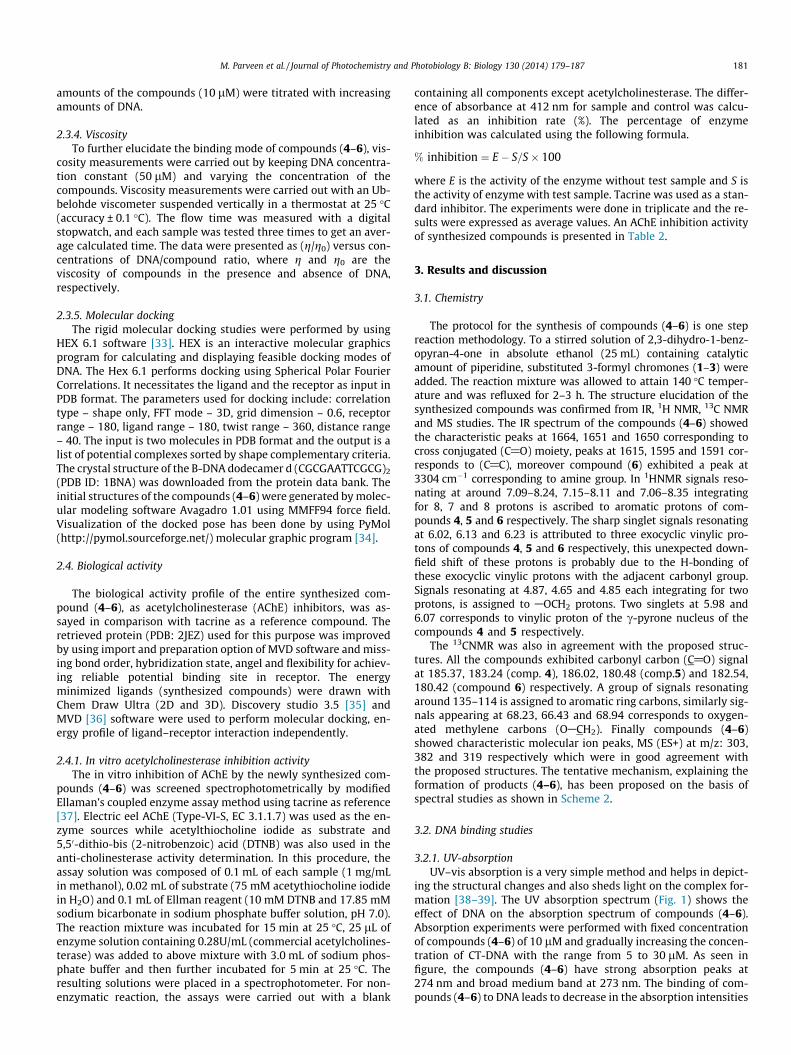
Synthesis, characterization, DNA-binding studies andacetylcholinesterase inhibition activity of new 3-formyl chromonederivatives
1011-1344/$ - see front matter � 2013 Elsevier B.V. All rights reserved.http://dx.doi.org/10.1016/j.jphotobiol.2013.11.019
⇑ Corresponding author. Tel.: +91 9897179498.E-mail address: [email protected] (M. Parveen).
Mehtab Parveen a,⇑, Ali Mohammed Malla a, Zahid Yaseen b, Akhtar Ali a, Mahboob Alam a
a Department of Chemistry, Aligarh Muslim University, Aligarh 202002, Indiab Department of Civil Engineering, Islamic University of Science and Technology, Kashmir 192122, India
a r t i c l e i n f o a b s t r a c t
Article history:Received 25 July 2013Received in revised form 29 October 2013Accepted 18 November 2013Available online 26 November 2013
Keywords:SynthesisChromoneFluorescenceDNA bindingAcetylcholinesterase inhibition
A series of new substituted 3-formyl chromone derivatives (4–6) were synthesized by one step reactionmethodology by knoevenagel condensation, structurally similar to known bisintercalators. The new com-pounds were characterized by IR, 1H NMR, 13C NMR, MS and analytical data. The in vitro DNA bindingprofile of compounds (4–6) was carried out by absorption, fluorescence and viscosity measurements. Itwas found that synthesized compounds, especially compound 6 (evident from binding constant value)bind strongly with calf thymus DNA, presumably via an intercalation mode. Additionally, molecular dock-ing studies of compounds (4–6) were carried out with B–DNA (PDBID: 1BNA) which revealed that partialintercalative mode of mechanism is operational in synthesized compounds (4–6) with CT-DNA. The bind-ing constants evaluated from fluorescence spectroscopy of compounds with CT-DNA follows the ordercompound 6 > compound 5 > compound 4. All the compounds (4–6) were screened for acetylcholinester-ase inhibition assay. It can be inferred from data, that compound (6) showed potent AChE inhibition hav-ing IC50 = 0.27 lM, almost in vicinity to reference drug Tacrine (IC50 = 0.19 lM).
� 2013 Elsevier B.V. All rights reserved.
1. Introduction
Chromones are a group of naturally occurring compounds thatis ubiquitous in nature especially in plants [1]. They areoxygen-containing heterocyclic compounds with a benzo-annelated c-pyrone ring, with the parent compound being chro-mone (4H-chromen-4-one, 4H-1-benzopyran-4-one) [2]. Besidesforming the basic nucleus of an entire class of natural products(i.e., flavones) [3,4], the chromone scaffold forms the key compo-nent of a large number of bioactive molecules of either natural orsynthetic origin, possessing anti-inflammatory, antitumoral, andantimicrobial activities [5]. The benzo-c-pyrone nucleus of chro-mone moiety has been reported to exhibit enzymatic inhibitionproperties toward different systems such as oxidoreductases,kinases, tyrosinases, and cyclooxygenases [6]. The relevance ofthese types of heterocyclic compound as monoamine oxidaseinhibitors (MAOIs) has been recently reported in literature [7].
In recent years, 3-formyl-chromones have attracted consider-able attention as highly reactive compound, which can serve asthe valuable moiety for incorporation in heterocycles with usefulproperties due to the presence of a keto function at C-4, a conju-gated second carbonyl group at C-3, and above all, possess a highly
polarized C2AC3 p-bond [8]. 3-Heteryl-substituted chromones areknown to exhibit wide range of biological activities. The substitu-tion pattern of the chromone scaffolds determines their differentbiological effects. Known effects of these types of compounds areantioxidant [9], antiviral [10], antibacterial activities [11] or kinaseinhibition [12]. Hence, chromones can be considered as privilegedstructures, defined as ‘‘a single molecular framework able to provideligands for diverse receptors’’ [13]. Derivatives of 3-formyl chro-mone are useful synthetic building blocks in both organic andmedicinal chemistry [14]. Introduction of an electron-withdrawinggroup at position 3 of chromones radically changes the reactivity ofthe pyrone ring toward nucleophilic reagents and opens up a broadsynthetic scope of this important oxygen-containing heterocyclicsystem. Being essentially gem-activated alkenes, 3-substitutedchromones exhibit a variety of properties and can undergo addi-tional transformations through opening of the c-pyrone ring[15,16]. The photophysical behavior of 3-hydroxychromones hasbeen carefully studied and efforts have been made to developderivatives with improved and exceptional fluorescent properties.Due to their characteristic fluorescence behavior, 3-hydroxychro-mone derivatives have been used as biosensors, hydrogen bondingsensors and as fluorescent probes for dipotential (WD) measure-ments in lipid bilayers [17–20].They have also been used for stud-ies of DNA interactions and as photochemical dyes for proteinlabeling and apoptosis [21–23].
180 M. Parveen et al. / Journal of Photochemistry and Photobiology B: Biology 130 (2014) 179–187
Metal complexes have been widely investigated as cleavingagents of nucleic acids and are found to be reasonably efficient[24,25], but their use in pharmacy is restricted because of seriousissues over their stability and toxicity, that limits the practicalusage of these compounds [26,27]. To overcome these limitations,Gobel and co-workers [28,29] put forward the concept of‘metal-free cleaving agents’ which are being applied to active phos-phodiesters like ‘nucleic acid mimic’ and RNA. The structure of thechromone ring meets the requirements for the terminal moiety ofbisintercalators. The association of such two almost planar unsatu-rated systems with the unsaturated linker may guide to the forma-tion of molecules which exhibit specific biological properties, e.g.,antitumor activity and are capable of binding to DNA via a bisinter-calative binding mode (simultaneous insertion of two planar sys-tems between the adjacent base-pairs according to a neighborexclusion principle) [30]. Bisintercalators constitute a versatileand very promising group of compounds extensively studied bymany research groups worldwide. Lots of them turned out to bepotent anticancer drugs e.g., elinafide, bisnaphthalimide whichprogressed to clinical trials against solid tumors [31].
Encouraged by the above results, herein, we report the synthe-sis of substituted 3-formyl chromone derivatives by KnoevenagelCondensation, containing chroman–chromen rings tethered atthe 3, 3
0position by ylidenemethyl moiety and behave as metal
free DNA binding agents. The presence of NH2 and CO groups inthe molecules can cooperatively participate in the interaction withDNA via hydrogen bonding. A computer aided molecular dockingstudy was carried out to validate the specific binding mode ofthe synthesized compounds.
2. Experimental
2.1. Material and methods
Melting points were determined on a Kofler apparatus and areuncorrected. Elemental analysis (C, H, N) were conducted usingCarlo Erba analyzer model 1108. The IR spectra were recorded withShimadzu IR-408 Perkin-Elmer 1800 (FTIR) and its values are givenin cm�1. 1H NMR and 13C NMR spectra were run in DMSO-d6 on aBruker Advance-II 400 MHz instrument with TMS as internal stan-dard. Chemical shifts are reported in ppm (d) relative to the TMS.Mass spectra were recorded on a JEOL D-300 mass spectrometer.Thin layer chromatography (TLC) plates were coated with silicagel G and exposed to iodine vapors to check the homogeneity aswell as the progress of reaction. Sodium sulfate (anhydrous) wasused as a drying agent. The calf Thymus DNA was purchased fromBangalore Genei (India). All the reagents were purchased from Sig-ma-Aldrich Chemicals Pvt. Ltd. which were of analytical grade andused without further purification.
2.2. General procedure for the synthesis of compounds (4–6)
The appropriate substituted 3-formylchromones (1–3) and4-chromanone (2 mmol) each, were dissolved in 25 mL of absoluteethanol. To this solution catalytic amount of piperidine was addedand the reaction mixture was stirred at 140 �C for 2–3 h. The com-pletion of the reaction was monitored by TLC. After completion ofthe reaction as evident from TLC, the precipitate formed was fil-tered, thoroughly washed with 5% HCL solution and then with(2 � 100 mL) water, dried and crystallized from CHCl3–MeOH toafford pure products (4–6) (Scheme 1).
2.2.1. 3-(40-Oxo-chroman-3
0-ylidenemethyl)-chromen-4-one (4)
It was crystallized from CHCl3–MeOH as brick red color solid;Yield: 84%; m.p 145–47 �C. Anal. Calc. for C19H12O4: C, 74.99; H,
3.97; found: C, 75.0; H, 3.96; IR (KBr) cm�1: 1650, 1591, 1496,1416, 1373, 1250, 1154. 1H NMR (400 MHz, DMSO-d6, d, ppm):7.09–8.24 (m, 8H, ArAH), 6.02 (s, 1H, Exocyclic vinylic-H), 5.98(s, 1H, vinylicc-pyrone), 4.87 (s, 2H, OACH2). 13C NMR (400 MHz,DMSO-d6, d, ppm): 185.37 (C@O), 183.24 (C@O), 158.21, 154.47,153.55, 135.72, 131.85, 129.59, 128.94, 126.26, 125.82, 124.45,123.2, 122.12, 115.16, 114.68 (Chroman/chromen ring), 68.23(OACH2). MS (ES+) m/z: 303.
2.2.2. 6-Bromo-3-(40-oxo-chroman-3
0-ylidenemethyl)-chromen-4-one (5)
It was crystallized from CHCl3–MeOH as brown color solid;Yield: 73%; m.p 188–90 �C. Anal. Calc. for C19H11BrO4: C, 59.55;H, 2.89; Br, 20.85; found: C, 59.54; H, 2.88; Br, 20.85; IR (KBr)cm�1: 1651, 1595, 1491, 1370, 1298, 1246, 1174. 1H NMR(400 MHz, DMSO-d6, d, ppm): 7.15–8.11 (m, 7H, ArAH), 6.13 (s,1H, exocyclic vinylic-H), 6.07 (s, 1H, vinylicc-pyrone), 4.65 (s, 2H,OACH2)). 13C NMR (400 MHz, DMSO-d6, d, ppm): 186.02 (C@O),180.48 (C@O), 162.21, 156.35, 149.35, 144.75, 141.42, 134.54,131.2, 129.43, 127.21, 126.26, 125.75, 123.85, 121.54, 116.23,114.62 (chroman/chromen ring), 66.43 (OACH2). MS (ES+) m/z:382.
2.2.3. 2-Amino-3-(40-oxo-chroman-3
0-ylidenemethyl)-chromen-4-one (6)
It was crystallized from CHCl3–MeOH as cream color solid;Yield: 78%; m.p 203–05 �C. Anal. Calc. for C19H13NO4: C, 71.47; H,4.10; N, 4.39; found: C, 71.47; H, 4.11; N, 4.39; IR (KBr) cm�1:3304, 1664, 1615, 1410, 1314, 1214, 1154, 874. 1H NMR(400 MHz, DMSO-d6, d, ppm): 9.75 (s, 2H, NH2, D2O exchangeable)7.06–8.35 (m, 8H, ArAH), 6.23 (s, 1H, exocyclic vinylic-H), 4.85 (s,2H, OACH2). 13C NMR (400 MHz, DMSO-d6, d, ppm):182.54 (C@O),180.42 (C@O), 165.34, 158.23, 154.64, 135.86, 134.62, 131.43,130.23, 129.54, 125.97, 123.32, 121.42, 120.73, 119.61, 115.29,114.61, 102.43 (chroman/chromen ring), 68.94 (OACH2). MS(ES+) m/z: 319.
2.3. DNA binding experiments
2.3.1. Preparation of stock solutionThe stock solution of DNA was prepared by dissolving DNA in
Tris–HCl buffer (10 mM, pH 7.5) and compounds in a mixed sol-vent of 1% methanol and 99% Tris–HCL buffer. The purity of DNAwas verified by monitoring the ratio of absorbance at 260 nm tothat at 280 nm, which was in the range 1.8–1.9. The concentrationof the DNA was determined spectrophotometrically usinge260nm = 6600 M�1 cm�1 [32].
2.3.2. Absorbance spectroscopyThe UV–vis absorption of Calf thymus DNA was recorded on a
177 Beckman DU 40 Spectrophotometer (USA) by using a cuvetteof 1 cm path length. The absorbance values of compounds (4–6)in the absence and presence of DNA were recorded in the rangeof 260–380 nm. The reference solution was the correspondingTris–HCl buffer solution. While measuring the absorption spectra,equal amount of DNA was added to both the compounds solutionand the reference solution to eliminate the absorbance of DNAitself.
2.3.3. Emission spectroscopyTo compare quantitatively the affinity of the compounds (4–6)
with DNA, the binding constants K of the compounds binding toDNA were obtained by the fluorescence titration method. Fluores-cence measurements were recorded on a Shimadzu 184 Spectroflu-orimeter-5000 (Japan). The fluorescence quenching withincreasing concentration of DNA was recorded after exciting thecompounds (4–6) at 273 nm, using 10/10 nm as slit widths. Fixed
M. Parveen et al. / Journal of Photochemistry and Photobiology B: Biology 130 (2014) 179–187 181
amounts of the compounds (10 lM) were titrated with increasingamounts of DNA.
2.3.4. ViscosityTo further elucidate the binding mode of compounds (4–6), vis-
cosity measurements were carried out by keeping DNA concentra-tion constant (50 lM) and varying the concentration of thecompounds. Viscosity measurements were carried out with an Ub-belohde viscometer suspended vertically in a thermostat at 25 �C(accuracy ± 0.1 �C). The flow time was measured with a digitalstopwatch, and each sample was tested three times to get an aver-age calculated time. The data were presented as (g/g0) versus con-centrations of DNA/compound ratio, where g and g0 are theviscosity of compounds in the presence and absence of DNA,respectively.
2.3.5. Molecular dockingThe rigid molecular docking studies were performed by using
HEX 6.1 software [33]. HEX is an interactive molecular graphicsprogram for calculating and displaying feasible docking modes ofDNA. The Hex 6.1 performs docking using Spherical Polar FourierCorrelations. It necessitates the ligand and the receptor as input inPDB format. The parameters used for docking include: correlationtype – shape only, FFT mode – 3D, grid dimension – 0.6, receptorrange – 180, ligand range – 180, twist range – 360, distance range– 40. The input is two molecules in PDB format and the output is alist of potential complexes sorted by shape complementary criteria.The crystal structure of the B-DNA dodecamer d (CGCGAATTCGCG)2
(PDB ID: 1BNA) was downloaded from the protein data bank. Theinitial structures of the compounds (4–6) were generated by molec-ular modeling software Avagadro 1.01 using MMFF94 force field.Visualization of the docked pose has been done by using PyMol(http://pymol.sourceforge.net/) molecular graphic program [34].
2.4. Biological activity
The biological activity profile of the entire synthesized com-pound (4–6), as acetylcholinesterase (AChE) inhibitors, was as-sayed in comparison with tacrine as a reference compound. Theretrieved protein (PDB: 2JEZ) used for this purpose was improvedby using import and preparation option of MVD software and miss-ing bond order, hybridization state, angel and flexibility for achiev-ing reliable potential binding site in receptor. The energyminimized ligands (synthesized compounds) were drawn withChem Draw Ultra (2D and 3D). Discovery studio 3.5 [35] andMVD [36] software were used to perform molecular docking, en-ergy profile of ligand–receptor interaction independently.
2.4.1. In vitro acetylcholinesterase inhibition activityThe in vitro inhibition of AChE by the newly synthesized com-
pounds (4–6) was screened spectrophotometrically by modifiedEllaman’s coupled enzyme assay method using tacrine as reference[37]. Electric eel AChE (Type-VI-S, EC 3.1.1.7) was used as the en-zyme sources while acetylthiocholine iodide as substrate and5,50-dithio-bis (2-nitrobenzoic) acid (DTNB) was also used in theanti-cholinesterase activity determination. In this procedure, theassay solution was composed of 0.1 mL of each sample (1 mg/mLin methanol), 0.02 mL of substrate (75 mM acetythiocholine iodidein H2O) and 0.1 mL of Ellman reagent (10 mM DTNB and 17.85 mMsodium bicarbonate in sodium phosphate buffer solution, pH 7.0).The reaction mixture was incubated for 15 min at 25 �C, 25 lL ofenzyme solution containing 0.28U/mL (commercial acetylcholines-terase) was added to above mixture with 3.0 mL of sodium phos-phate buffer and then further incubated for 5 min at 25 �C. Theresulting solutions were placed in a spectrophotometer. For non-enzymatic reaction, the assays were carried out with a blank
containing all components except acetylcholinesterase. The differ-ence of absorbance at 412 nm for sample and control was calcu-lated as an inhibition rate (%). The percentage of enzymeinhibition was calculated using the following formula.
% inhibition ¼ E� S=S� 100
where E is the activity of the enzyme without test sample and S isthe activity of enzyme with test sample. Tacrine was used as a stan-dard inhibitor. The experiments were done in triplicate and the re-sults were expressed as average values. An AChE inhibition activityof synthesized compounds is presented in Table 2.
3. Results and discussion
3.1. Chemistry
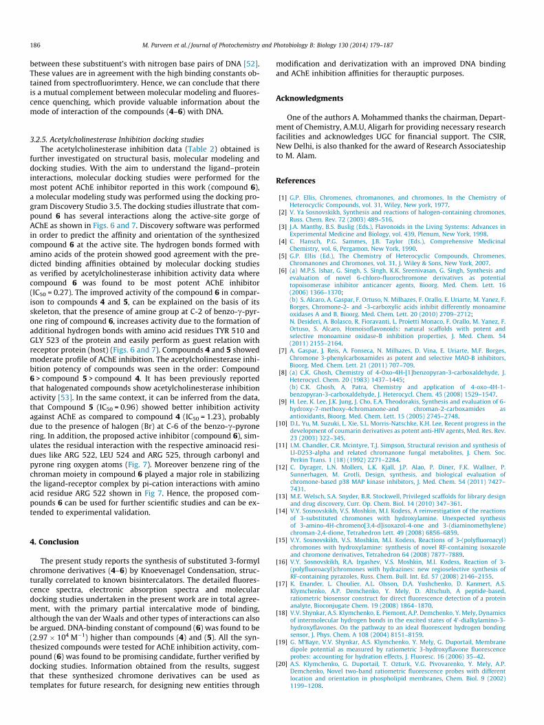
The protocol for the synthesis of compounds (4–6) is one stepreaction methodology. To a stirred solution of 2,3-dihydro-1-benz-opyran-4-one in absolute ethanol (25 mL) containing catalyticamount of piperidine, substituted 3-formyl chromones (1–3) wereadded. The reaction mixture was allowed to attain 140 �C temper-ature and was refluxed for 2–3 h. The structure elucidation of thesynthesized compounds was confirmed from IR, 1H NMR, 13C NMRand MS studies. The IR spectrum of the compounds (4–6) showedthe characteristic peaks at 1664, 1651 and 1650 corresponding tocross conjugated (C@O) moiety, peaks at 1615, 1595 and 1591 cor-responds to (C@C), moreover compound (6) exhibited a peak at3304 cm�1 corresponding to amine group. In 1HNMR signals reso-nating at around 7.09–8.24, 7.15–8.11 and 7.06–8.35 integratingfor 8, 7 and 8 protons is ascribed to aromatic protons of com-pounds 4, 5 and 6 respectively. The sharp singlet signals resonatingat 6.02, 6.13 and 6.23 is attributed to three exocyclic vinylic pro-tons of compounds 4, 5 and 6 respectively, this unexpected down-field shift of these protons is probably due to the H-bonding ofthese exocyclic vinylic protons with the adjacent carbonyl group.Signals resonating at 4.87, 4.65 and 4.85 each integrating for twoprotons, is assigned to AOCH2 protons. Two singlets at 5.98 and6.07 corresponds to vinylic proton of the c-pyrone nucleus of thecompounds 4 and 5 respectively.
The 13CNMR was also in agreement with the proposed struc-tures. All the compounds exhibited carbonyl carbon (C@O) signalat 185.37, 183.24 (comp. 4), 186.02, 180.48 (comp.5) and 182.54,180.42 (compound 6) respectively. A group of signals resonatingaround 135–114 is assigned to aromatic ring carbons, similarly sig-nals appearing at 68.23, 66.43 and 68.94 corresponds to oxygen-ated methylene carbons (OACH2). Finally compounds (4–6)showed characteristic molecular ion peaks, MS (ES+) at m/z: 303,382 and 319 respectively which were in good agreement withthe proposed structures. The tentative mechanism, explaining theformation of products (4–6), has been proposed on the basis ofspectral studies as shown in Scheme 2.
3.2. DNA binding studies
3.2.1. UV-absorptionUV–vis absorption is a very simple method and helps in depict-
ing the structural changes and also sheds light on the complex for-mation [38–39]. The UV absorption spectrum (Fig. 1) shows theeffect of DNA on the absorption spectrum of compounds (4–6).Absorption experiments were performed with fixed concentrationof compounds (4–6) of 10 lM and gradually increasing the concen-tration of CT-DNA with the range from 5 to 30 lM. As seen infigure, the compounds (4–6) have strong absorption peaks at274 nm and broad medium band at 273 nm. The binding of com-pounds (4–6) to DNA leads to decrease in the absorption intensities
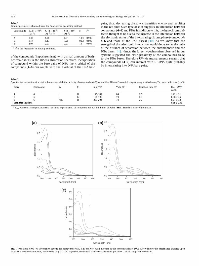
Table 1Binding parameters obtained from the fluorescence quenching method.
Compounds Ksv (1 � 104)(M�1)
Kq (1 � 1012)(M�1 s�1)
K (1 � 104)(M�1)
n r2a
4 1.38 1.38 0.64 1.03 0.9965 1.17 1.17 1.35 0.92 0.9966 2.07 2.07 2.97 1.01 0.994
a r2 is the regression in binding equilibra.
182 M. Parveen et al. / Journal of Photochemistry and Photobiology B: Biology 130 (2014) 179–187
of the compounds (hypochromism), with a small amount of bath-ochrmoic shifts in the UV–vis absorption spectrum. Incorporationof compound within the base pairs of DNA, the p orbital of thecompounds (4–6) can couple with the p orbital of the DNA base
Table 2Quantitative estimation of acetylcholinesterase inhibition activity of compounds (4–6) by
Entry Compound R1 R2 m.p (�C
1 4 H H 145-142 5 H Br 188-193 6 NH2 H 203-20Standard (Tacrine)
a IC50: Concentration (means ± SEM⁄ of three experiments) of compound for 50% inhi
0.0
0.5
1.0
1.5
2.0
2.5
3.0
abso
rban
ce
wavelength (nm)260 280 300 320 340 360 380 400
260 280 3000.0
0.5
1.0
1.5
2.0
2.5
3.0
abso
rban
ce
wavele
(a) (b
(c)
Fig. 1. Variation of UV–vis absorption spectra for compounds 4(a), 5(b) and 6(c) withincreasing DNA concentration, [DNA = 0 to 25 lM]. Data represent mean ± SD of three e
pairs, thus, decreasing the p ? p transition energy and resultingin the red shift. Such type of shift suggests an interaction betweencompounds (4–6) and DNA. In addition to this, the hypochromic ef-fect is thought to be due to the increase in the interaction betweenthe electronic states of the intercalating chromophore (compounds4–6 and those of the DNA bases) [40]. As we know that thestrength of this electronic interaction would decrease as the cubeof the distance of separation between the chromophore and theDNA bases [41]. Hence, the large hypochromism observed in oursystems suggested the close proximity of the compounds (4–6)to the DNA bases. Therefore UV–vis measurements suggest thatthe compounds (4–6) can interact with CT-DNA quite probablyby intercalating into DNA base pairs.
modified Ellaman’s coupled enzyme assay method using Tacrine as reference (n = 3).
) Yield (%) Reaction time (h) IC50 (lM)a
AChE
7 84 2.5 1.23 ± 0.10 73 2.3 0.96 ± 0.54 78 2.7 0.27 ± 0.3
0.19 ± 0.03
bition of AChE, ⁄SEM: Standard error of the mean.
0.0
0.5
1.0
1.5
2.0
2.5
3.0
abso
rban
ce
wavelength (nm)260 280 300 320 340 360 380
320 340 360 380
ngth (nm)
)
increase in the concentration of DNA. Arrow shows the absorbance changes uponxperiments. p value < 0.05 as compared to control.
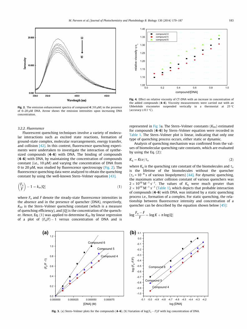
Fig. 2. The emission enhancement spectra of compound 4 (10 lM) in the presenceof 0–20 lM DNA. Arrow shows the emission intensities upon increasing DNAconcentration.
0.0 0.2 0.4 0.6 0.8 1.01.05
1.10
1.15
1.20
1.25
1.30
1.35
1.40
rela
tive
visc
osity
η/η
ο
compound/[DNA]
compound 4 compound 5 compound 6
Fig. 4. Effect on relative viscosity of CT-DNA with an increase in concentration ofthe added compounds (4–6). Viscosity measurements were carried out with anUbbelohde viscometer suspended vertically in a thermostat at 25 �C(accuracy ± 0.1 �C).
M. Parveen et al. / Journal of Photochemistry and Photobiology B: Biology 130 (2014) 179–187 183
3.2.2. FluoresenceFluorescent quenching techniques involve a variety of molecu-
lar interactions such as excited state reactions, formation ofground-state complex, molecular rearrangements, energy transfer,and collision [42]. In this context, fluorescence quenching experi-ments were undertaken to investigate the interaction of synthe-sized compounds (4–6) with DNA. The binding of compounds(4–6) with DNA, by maintaining the concentration of compoundsconstant (i.e., 10 lM) and varying the concentration of DNA from0 to 20 lM, was studied by fluorescence spectroscopy (Fig. 2). Thefluorescence quenching data were analyzed to obtain the quenchingconstant by using the well-known Stern–Volmer equation [43].
Fo
F
� �� 1 ¼ ksv½Q � ð1Þ
where Fo and F denote the steady-state fluorescence intensities inthe absence and in the presence of quencher (DNA), respectively,KSV is the Stern–Volmer quenching constant (which is a measureof quenching efficiency), and [Q] is the concentration of the quench-er. Hence, Eq. (1) was applied to determine KSV by linear regressionof a plot of (Fo/F) � 1 versus concentration of DNA and is
0.0
0.2
0.4
0.6
0.8
1.0
1.2
Compound 4
Compound 5
(F0/F
-1)
[DNA] (M)
Compound 6
0.000000 0.000025 0.000050 0.000075
(a)
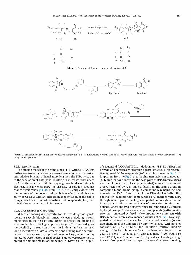
Fig. 3. (a) Stern–Volmer plots for the compounds (4–6). (b) V
represented in Fig 3a. The Stern–Volmer constants (KSV) estimatedfor compounds (4–6) by Stern–Volmer equation were recorded inTable 1. The Stern–Volmer plot is linear, indicating that only onetype of quenching process occurs, either static or dynamic.
Analysis of quenching mechanism was confirmed from the val-ues of biomolecular quenching rate constants, which are evaluatedby using the Eq. (2):
Kq ¼ Ksv=so ð2Þ
where Kq is the quenching rate constant of the biomolecules and so
is the lifetime of the biomolecules without the quencher(so = 10�8 s of various biopolymers) [44]. For dynamic quenching,the maximum scatter collision constant of various quenchers was2 � 1010 M�1 s�1. The values of Kq were much greater than2 � 1010 M�1 s�1 (Table 1), which depicts that probable interactionof compounds (4–6) with DNA, was initiated by a static quenchingprocess i.e., formation of a complex. For static quenching, the rela-tionship between fluorescence intensity and concentration of aquencher can be described by the equation shown below [45]:
logFo � F
F¼ log K þ n log½Q �
-5.1 -5.0 -4.9 -4.8 -4.7 -4.6 -4.5 -4.4 -4.3 -4.2
-1.0
-0.9
-0.8
-0.7
-0.6
-0.5
-0.4
-0.3
-0.2
-0.1
0.0
0.1
Compound 4
Compound 5
Compound 6
log
(F0-F
/F)
log [DNA]
(b)
ariation of log(Fo � F)/F with log concentration of DNA.

Fig. 5. Cartoon representation of DNA with bound compounds. (a)–(c) showsminimum energy poses of DNA–compound 4, DNA–compound 5 and DNA–compound 6 complexes respectively. Each panel contains two figures, which showsolid and stick representation of compounds. In stick model of compound 6(c), thehydrogens attached to carbons are removed, in order to have easy visualization ofamino hydrogen inclined towards A5 of the DNA strand.
Fig. 6. Docking model of compound-enzyme complex. Representation of compound(6) interacting with amino acid residues in the binding site of protein (PDB: 2JEZ).The available binding sites for compound (6) are A:ARG522:N -:DRG1:OAB,A:ARG522:NE:DRG1:OAB, A:GLY523:N -:DRG1:OAC, A:LEU524:N -:DRG1:OAO,A:ARG525:N -:DRG1:OAO, DRG1:HAO - A:ARG522:O, DRG1:HAP -A:TYR510:OH,DRG1:HAP - A:GLY523:O. The compound is shown in green stick model. Picturesare created with Discover Studio 3.5. (For interpretation of the references to color inthis figure legend, the reader is referred to the web version of this article.)
Fig. 7. Ligand (compound 6) displaying various interactions with amino acidresidues at active sites of the protein (PDB: 2JEZ) along with pi-cation interactionwith amino acid residue ARG 522.
184 M. Parveen et al. / Journal of Photochemistry and Photobiology B: Biology 130 (2014) 179–187
where K and n are the binding constant and the number of bindingsites, respectively. From the plot of log(Fo � F)/F vs. log[Q], K and nvalues can be obtained from the intercept and slope (Fig. 3b). The
values of n were approximately equal to 1, which suggests the exis-tence of just one binding site on DNA for compounds (4–6). The va-lue of K is significant, in order to quantify the interaction betweencompounds (4–6) and DNA. The larger values of the K observed inthe present study depicts the stronger interaction between thecompounds (4–6) and DNA. Earlier Wang et al. [46], proposes theintercalative mechanism between the 6-ethoxy chromone-3-carb-aldehyde benzoyl hydrazone and DNA, with a binding constant of8.27 � 105 M�1. Yong. Li et al. [47], reported the binding of 3-carb-aldehyde chromone-(benzoyl) hydrazone with DNA through inter-calative mechanism and the binding constant reported is7.39 � 105 M�1. By comparing these literature values with our data,the binding constant in our system are slightly towards lower side,but these values are too higher than surface binding of compoundswith DNA. Nafisi et al. [48], reported the intercalative and externalbinding mechanism for various fluorescent probes with CT-DNAand their binding constants vary from (6.58–2.13) � 104 M�1.Hence, it may be proposed that compounds (4–6) interact withDNA in an intermediate way between the surface binding at oneend and intercalative at the other (i.e., partially intercalated, dis-cussed in next section). The data shown in (Table 1) also suggeststhat the interaction of the compounds (4–6) with DNA follows theorder: compound 6 > compound 5 > compound 4. This variationlikely reflects the differing ability of these ligands to stack and over-lap well with the base pairs of DNA.
O
O O
H+
O
O
1 - 3 1a
1
R1
H H
2 H Br
3 NH2 H
Ethanol /Piperidine
Reflux, 2-3 hrs, 140 oCR1
R2
O
O
R1
R2
O
O
4- 6
R2R1 R2
4
5
6
H H
H Br
NH2 H
1
2
34
5
6
78 1'
2'
3' 4'
5' 6'7'
8'
Scheme 1. Synthesis of 3-formyl chromone derivatives (4–6).
O
OH
H
O
OH
N+
H
HO
OH-
H
Enol-form keto-form
O
OH-
O
OO
H
R1
R2
O
O-
O
O O
R1
R2
N+
H
H
O
O
O
O O
R1
R2
H
..
NH
..
H
O O
OO
R1
R2
NH
1a
4-6
1-3
Scheme 2. Plausible mechanism for the synthesis of compounds (4–6) via Knoevenagel Condensation of of 4-chromanone (1a) and substituted 3-formyl chromones (1–3)catalyzed by piperidine.
M. Parveen et al. / Journal of Photochemistry and Photobiology B: Biology 130 (2014) 179–187 185
3.2.3. Viscosity resultsThe binding modes of the compounds (4–6) with CT-DNA, was
further confirmed by viscosity measurements. In case of classicalintercalation binding, a ligand must lengthen the DNA helix dueto the separation of base pairs, resulting in increased viscosity ofDNA. On the other hand, if the drug is groove binder or interactselectrostatistically with DNA, the viscosity of solution does notchange significantly [49,50]. From Fig. 4, it is clearly evident thatthe presence of compounds had an obvious effect on relative vis-cosity of CT-DNA with an increase in concentration of the addedcompounds. These results demonstrate that compounds (4–6) bindto DNA through the intercalation mode.
3.2.4. DNA-binding docking studiesMolecular docking is a powerful tool for the design of ligands
toward a specific biopolymer target. Molecular docking is com-monly used in the field of drug design to predict the binding ofsmall molecules to biological protein targets. This method givesthe possibility to study an active site in detail and can be usedfor hit identification, virtual screening and binding mode determi-nation. In our experiment, rigid molecular docking (two interactingmolecules were treated as rigid bodies) studies were performed topredict the binding modes of compounds (4–6) with a DNA duplex
of sequence d (CGCAAATTTCGC)2 dodecamer (PDB ID: 1BNA), andprovide an energetically favorable docked structures (representa-tive figure of DNA–compounds (4–6) complex shown in Fig. 5). Itis apparent from the Fig. 5, that the chromen moiety in compounds(4–6) find its position within the base pairs of DNA (intercalation)and the chroman part of compounds (4–6) remain in the minorgroove region of DNA. In this configuration, the amino group incompound 6 and bromo group in compound 5 remains inclinedtowards the DA5 of strand A of the DNA double helix. Thisobservation suggests that compounds (4–6) interact with DNAthrough minor groove binding and partial intercalation. Partialintercalation is the preferred mode of interaction for the com-pounds, where the two biphenyl rings are connected by unfusedbiphenyl linkage. In the same context, compounds (4–6) containstwo rings connected by fused @CHA linkage, hence interacts withDNA in partial intercalative manner. Amutha et al. [51], have sug-gested partial intercalative mechanism in case of benzidine (wheretwo pheny rings are connected by biphenyl linkage) with bindingconstant of 6.7 � 103 M�1. The resulting relative bindingenergy of docked chromone–DNA complexes was found to be212.10 kJ mole�1 (compound 4), 235.23 kJ mole�1 (compound 5)and 242.11 kJ mole�1 (compound 6). High values of binding energyin case of compound 6 and 5, depicts the role of hydrogen bonding
186 M. Parveen et al. / Journal of Photochemistry and Photobiology B: Biology 130 (2014) 179–187
between these substituent’s with nitrogen base pairs of DNA [52].These values are in agreement with the high binding constants ob-tained from spectrofluorimtery. Hence, we can conclude that thereis a mutual complement between molecular modeling and fluores-cence quenching, which provide valuable information about themode of interaction of the compounds (4–6) with DNA.
3.2.5. Acetylcholinesterase Inhibition docking studiesThe acetylcholinesterase inhibition data (Table 2) obtained is
further investigated on structural basis, molecular modeling anddocking studies. With the aim to understand the ligand–proteininteractions, molecular docking studies were performed for themost potent AChE inhibitor reported in this work (compound 6),a molecular modeling study was performed using the docking pro-gram Discovery Studio 3.5. The docking studies illustrate that com-pound 6 has several interactions along the active-site gorge ofAChE as shown in Figs. 6 and 7. Discovery software was performedin order to predict the affinity and orientation of the synthesizedcompound 6 at the active site. The hydrogen bonds formed withamino acids of the protein showed good agreement with the pre-dicted binding affinities obtained by molecular docking studiesas verified by acetylcholinesterase inhibition activity data wherecompound 6 was found to be most potent AChE inhibitor(IC50 = 0.27). The improved activity of the compound 6 in compar-ison to compounds 4 and 5, can be explained on the basis of itsskeleton, that the presence of amine group at C-2 of benzo-c-pyr-one ring of compound 6, increases activity due to the formation ofadditional hydrogen bonds with amino acid residues TYR 510 andGLY 523 of the protein and easily perform as guest relation withreceptor protein (host) (Figs. 6 and 7). Compounds 4 and 5 showedmoderate profile of AChE inhibition. The acetylcholinesterase inhi-bition potency of compounds was seen in the order: Compound6 > compound 5 > compound 4. It has been previously reportedthat halogenated compounds show acetylcholinesterase inhibitionactivity [53]. In the same context, it can be inferred from the data,that Compound 5 (IC50 = 0.96) showed better inhibition activityagainst AChE as compared to compound 4 (IC50 = 1.23), probablydue to the presence of halogen (Br) at C-6 of the benzo-c-pyronering. In addition, the proposed active inhibitor (compound 6), sim-ulates the residual interaction with the respective aminoacid resi-dues like ARG 522, LEU 524 and ARG 525, through carbonyl andpyrone ring oxygen atoms (Fig. 7). Moreover benzene ring of thechroman moiety in compound 6 played a major role in stabilizingthe ligand-receptor complex by pi-cation interactions with aminoacid residue ARG 522 shown in Fig 7. Hence, the proposed com-pounds 6 can be used for further scientific studies and can be ex-tended to experimental validation.
4. Conclusion
The present study reports the synthesis of substituted 3-formylchromone derivatives (4–6) by Knoevenagel Condensation, struc-turally correlated to known bisintercalators. The detailed fluores-cence spectra, electronic absorption spectra and moleculardocking studies undertaken in the present work are in total agree-ment, with the primary partial intercalative mode of binding,although the van der Waals and other types of interactions can alsobe argued. DNA-binding constant of compound (6) was found to be(2.97 � 104 M�1) higher than compounds (4) and (5). All the syn-thesized compounds were tested for AChE inhibition activity, com-pound (6) was found to be promising candidate, further verified bydocking studies. Information obtained from the results, suggestthat these synthesized chromone derivatives can be used astemplates for future research, for designing new entities through
modification and derivatization with an improved DNA bindingand AChE inhibition affinities for therauptic purposes.
Acknowledgments
One of the authors A. Mohammed thanks the chairman, Depart-ment of Chemistry, A.M.U, Aligarh for providing necessary researchfacilities and acknowledges UGC for financial support. The CSIR,New Delhi, is also thanked for the award of Research Associateshipto M. Alam.
References
[1] G.P. Ellis, Chromenes, chromanones, and chromones, In the Chemistry ofHeterocyclic Compounds, vol. 31, Wiley, New york, 1977.
[2] V. Ya Sosnovskikh, Synthesis and reactions of halogen-containing chromones,Russ. Chem. Rev. 72 (2003) 489–516.
[3] J.A. Manthy, B.S. Buslig (Eds.), Flavonoids in the Living Systems: Advances inExperimental Medicine and Biology, vol. 439, Plenum, New York, 1998.
[4] C. Hansch, P.G. Sammes, J.B. Taylor (Eds.), Comprehensive MedicinalChemistry, vol. 6, Pergamon, New York, 1990.
[5] G.P. Ellis (Ed.), The Chemistry of Heterocyclic Compounds, Chromenes,Chromanones and Chromones, vol. 31, J. Wiley & Sons, New York, 2007.
[6] (a) M.P.S. Ishar, G. Singh, S. Singh, K.K. Sreenivasan, G. Singh, Synthesis andevaluation of novel 6-chloro-fluorochromone derivatives as potentialtopoisomerase inhibitor anticancer agents, Bioorg. Med. Chem. Lett. 16(2006) 1366–1370;(b) S. Alcaro, A. Gaspar, F. Ortuso, N. Milhazes, F. Orallo, E. Uriarte, M. Yanez, F.Borges, Chromone-2- and -3-carboxylic acids inhibit differently monoamineoxidases A and B, Bioorg. Med. Chem. Lett. 20 (2010) 2709–2712;N. Desideri, A. Bolasco, R. Fioravanti, L. Proietti Monaco, F. Orallo, M. Yanez, F.Ortuso, S. Alcaro, Homoisoflavonoids: natural scaffolds with potent andselective monoamine oxidase-B inhibition properties, J. Med. Chem. 54(2011) 2155–2164.
[7] A. Gaspar, J. Reis, A. Fonseca, N. Milhazes, D. Vina, E. Uriarte, M.F. Borges,Chromone 3-phenylcarboxamides as potent and selective MAO-B inhibitors,Bioorg. Med. Chem. Lett. 21 (2011) 707–709.
[8] (a) C.K. Ghosh, Chemistry of 4-Oxo-4H-[1]benzopyran-3-carboxaldehyde, J.Heterocycl. Chem. 20 (1983) 1437–1445;(b) C.K. Ghosh, A. Patra, Chemistry and application of 4-oxo-4H-1-benzopyran-3-carboxaldehyde, J. Heterocycl. Chem. 45 (2008) 1529–1547.
[9] H. Lee, K. Lee, J.K. Jung, J. Cho, E.A. Theodorakis, Synthesis and evaluation of 6-hydroxy-7-methoxy-4chromanone-and chroman-2-carboxamides asantioxidants, Bioorg. Med. Chem. Lett. 15 (2005) 2745–2748.
[10] D.L. Yu, M. Suzuki, L. Xie, S.L. Morris-Natschke, K.H. Lee, Recent progress in thedevelopment of coumarin derivatives as potent anti-HIV agents, Med. Res. Rev.23 (2003) 322–345.
[11] I.M. Chandler, C.R. Mcintyre, T.J. Simpson, Structural revision and synthesis ofLl-D253-alpha and related chromanone fungal metabolites, J. Chem. Soc.Perkin Trans. 1 (18) (1992) 2271–2284.
[12] C. Dyrager, L.N. Mollers, L.K. Kjall, J.P. Alao, P. Diner, F.K. Wallner, P.Sunnerhagen, M. Grotli, Design, synthesis, and biological evaluation ofchromone-based p38 MAP kinase inhibitors, J. Med. Chem. 54 (2011) 7427–7431.
[13] M.E. Welsch, S.A. Snyder, B.R. Stockwell, Privileged scaffolds for library designand drug discovery, Curr. Op. Chem. Biol. 14 (2010) 347–361.
[14] V.Y. Sosnovskikh, V.S. Moshkin, M.I. Kodess, A reinvestigation of the reactionsof 3-substituted chromones with hydroxylamine. Unexpected synthesisof 3-amino-4H-chromeno[3,4-d]isoxazol-4-one and 3-(diaminomethylene)chroman-2,4-dione, Tetrahedron Lett. 49 (2008) 6856–6859.
[15] V.Y. Sosnovskikh, V.S. Moshkin, M.I. Kodess, Reactions of 3-(polyfluoroacyl)chromones with hydroxylamine: synthesis of novel RF-containing isoxazoleand chromone derivatives, Tetrahedron 64 (2008) 7877–7889.
[16] V.Y. Sosnovskikh, R.A. Irgashev, V.S. Moshkin, M.I. Kodess, Reaction of 3-(polyfluoroacyl)chromones with hydrazines: new regioselective synthesis ofRF-containing pyrazoles, Russ. Chem. Bull. Int. Ed. 57 (2008) 2146–2155.
[17] K. Enander, L. Choulier, A.L. Olsson, D.A. Yushchenko, D. Kanmert, A.S.Klymchenko, A.P. Demchenko, Y. Mely, D. Altschuh, A peptide-based,ratiometric biosensor construct for direct fluorescence detection of a proteinanalyte, Bioconjugate Chem. 19 (2008) 1864–1870.
[18] V.V. Shynkar, A.S. Klymchenko, E. Piemont, A.P. Demchenko, Y. Mely, Dynamicsof intermolecular hydrogen bonds in the excited states of 40-dialkylamino-3-hydroxyflavones. On the pathway to an ideal fluorescent hydrogen bondingsensor, J. Phys. Chem. A 108 (2004) 8151–8159.
[19] G. M’Baye, V.V. Shynkar, A.S. Klymchenko, Y. Mely, G. Duportail, Membranedipole potential as measured by ratiometric 3-hydroxyflavone fluorescenceprobes: accounting for hydration effects, J. Fluoresc. 16 (2006) 35–42.
[20] A.S. Klymchenko, G. Duportail, T. Ozturk, V.G. Pivovarenko, Y. Mely, A.P.Demchenko, Novel two-band ratiometric fluorescence probes with differentlocation and orientation in phospholipid membranes, Chem. Biol. 9 (2002)1199–1208.
M. Parveen et al. / Journal of Photochemistry and Photobiology B: Biology 130 (2014) 179–187 187
[21] A.S. Klymchenko, V.V. Shvadchak, D.A. Yushchenko, N. Jain, Y. Mely, Excited-state intramolecular proton transfer distinguishes microenvironments insingle and double-stranded DNA, J. Phys. Chem. B 112 (2008) 12050–12055.
[22] J. Wang, X.Y. Yi, B.D. Wang, Z.Y. Yang, DNA-binding properties studies andspectra of a novel fluorescent Zn(II) complex with a new chromone derivative,J. Photochem. Photobiol., A 201 (2009) 183–190.
[23] R. Torkin, J.F. Lavoie, D.R. Kaplan, H. Yeger, Induction of caspase-dependent,p53-mediated apoptosis by apigenin in human neuroblastoma, Mol. CancerTher. 4 (2005) 1–11.
[24] Y. Jin, J.A. Cowan, DNA cleavage by copper–ATCUN complexes. Factorsinfluencing cleavage mechanism and linearization of dsDNA, J. Am. Chem.Soc. 127 (2005) 8408–8415.
[25] K.E. Erkkila, D.T. Odom, J.K. Barton, Recognition and reaction ofmetallointercalators with DNA, Chem. Rev. 99 (1999) 2777–2796.
[26] J. Smith, K. Ariga, E.V. Anslyn, Enhanced imidazole-catalyzed RNA cleavageinduced by a bis-alkylguanidinium receptor, J. Am. Chem. Soc. 115 (1993)362–364.
[27] J.R. Morrow, L.A. Buttrey, V.M. Shelton, K.A. Berback, Efficient catalyticcleavage of RNA by lanthanide(III) macrocyclic complexes: toward syntheticnucleases for in vivo applications, J. Am. Chem. Soc. 114 (1992) 1903–1905.
[28] M.S. Muche, M.W. Gobel, Bis(guanidinium) alcohols as models ofstaphylococcal nuclease: substrate binding through ion pair complexes andfast phosphoryl transfer reactions, Angew. Chem. Int. Ed. 35 (1996) 2126–2129.
[29] U. Scheffer, A. Strick, V. Ludwig, S. Peter, E. Kalden, M.W. Gobel, Metal-Freecatalysts for the hydrolysis of RNA derived from guanidines, 2-aminopyridines, and 2-aminobenzimidazoles, J. Am. Chem. Soc. 127 (2005)2211–2217.
[30] M.F. Brana, M. Cacho, A. Gradillas, B. de Pascual-Teresa, A. Ramos, Intercalatorsas anticancer drugs, Curr. Pharm. Des. 7 (2001) 1745–1780.
[31] M.F. Brana, M. Cacho, A. Ramos, M.T. Dominguez, J.M. Pozuelo, C. Abradelo,M.F. Rey-Stolle, M. Yuste, C. Carrasco, C. Bailly, Synthesis, biological evaluationand DNA binding properties of novel mono and bisnaphthalimides, Org.Biomol. Chem. 1 (2003) 648–654.
[32] G.S. Son, J-.Ah Yeo, M.-Sun Kim, S.K. Kim, A. Holmen, B. Akerman, B. Norden,Binding mode of norfloxacin to calf thymus DNA, J. Am. Chem. Soc. 120 (1998)6451–6457.
[33] D. Mustard, D.W. Ritchie, Docking essential dynamics eigenstructures, Struct.Funct. Bioinf. 60 (2005) 269–274.
[34] W.L. DeLano, The PyMOL Molecular Graphics System, DeLano Scientific, SanCarlos, CA, USA, 2002.
[35] Accelrys software inc., Discovery Studio Modeling Environment, Release 3.1.,2011.
[36] R. Thomsen, M.H. Christensen, MolDock: a new technique for high-accuracymolecular docking, J. Med. Chem. 11 (2006) 3315–3321.
[37] G.L. Ellman, K.D. Courtney, V.J. Andres, R.M. Feather-Stone, A new and rapidcolorimetric determination of acetylcholinesterase activity, Biochem.Pharmacol. 7 (1961) 88–90.
[38] S.Y. Bi, D.Q. Song, Y. Tian, X. Zhou, Z.Y. Liu, H.Q. Zhang, Molecular spectroscopicstudy on the interaction of tetracyclines with serum albumins, Spectrochim.Acta, Part A 61 (2005) 629–636.
[39] P.B. Kandagal, S. Ashoka, J. Seetharamappa, V. Vanb, S.M.T. Shaikh, Study of theinteraction between doxepin and human serum albumins by spectroscopicmethods, J. Photochem. Photobiol., A 179 (2006) 161–166.
[40] R. Fukuda, S. Takenaka, M. Takagi, Metal ion assisted DNA interaction of crownether-linked acridine derivatives, J. Chem. Soc., Chem. Commun. (1990) 1028–1030.
[41] C. Cantor, P.R. Schimmel, The conformation of biological macromolecules,biophysical chemistry, W.H. Freeman, San Francisco, CA, 1980. p. 287.
[42] M.R. Eftink, Fluorescence Quenching Reaction: Probing Biological MacromolecularStructures, Biophysical and Biochemical Aspects of Fluorescence Spectroscopy,Plenum Press, New York, 1991.
[43] J.R. Lakowicz, Principles of Fluoresence Spectroscopy, Plenum Press, New York,1983.
[44] D.M. Togashi, A.G. Ryder, A fluorescence analysis of ANS bound to bovineserum albumin: binding properties revisited by using energy transfer, J.Floresc. 18 (2008) 519–526.
[45] J. Min, X. Meng-Xia, Z. Dong, L. Yuan, L. Xiao-Yu, C. Xing, Spectroscopic studieson the interaction of cinnamic acid and its hydroxyl derivatives with humanserum albumin, J. Mol. Struct. 692 (2004) 71–80.
[46] J. Wang, Z.-Y. Yang, X.-Y. Yi, B.-D. Wang, DNA-binding properties studies andspectra of a novel fluorescent Zn(II) complex with a new chromone derivative,J. Photochem. Photobiol. A Chem. 201 (2009) 183–190.
[47] Y. Li, Z.-Y. Yang, Rare earth complexes with 3-carbaldehyde chromone-(Benzoyl) hydrazone: synthesis, characterization, DNA binding studies andantioxidant activity, J. Fluoresc. 20 (2010) 329–342.
[48] S. Nafisi, A.A. Saboury, N. Keramat, J.-F. Neault, H.-A. Tajmir-Riahi, Stability andstructural features of DNA intercalation with ethidium bromide, acridineorange and methylene blue, J. Mol. Struct. 827 (2007) 35–43.
[49] I. Haq, P. Lincoln, D. Suh, B. Norden, B.Z. Chowdhry, J.B. Chaires, Interaction ofD- and k- [Ru(phen)2DPPZ]2+ with DNA: a calorimetric and equilibriumbinding study, J. Am. Chem. Soc. 117 (1995) 4788–4796.
[50] S. Satyanarayana, J.C. Dabrowiak, J.B. Chaires, Neither D- nor k-Tris(phenanthroline)ruthenium(II) binds to DNA by classical intercalation,Biochemistry 31 (1992) 9319–9324.
[51] R. Amutha, V. Sbramanian, B.U. Nair, Interaction of benzidine with DNA:experimental and modelling studies, Chem. Phys. Lett. 344 (2001) 40–48.
[52] M.A. Husain, Z. Yaseen, S.U. Rehman, T. Sarwar, M. Tabish, Naproxenintercalates and causes photocleavage of DNA through ROS generation, FEBSJ., DOI:10.1111/febs.12558.
[53] U. Brodbeck, K. Schweikert, R. Gentinetta, M. Rottenberg, Fluorinatedaldehydes and ketones acting as quasi-substrate inhibitors ofacetylcholinesterase, Biochim. et Biophys. Acta (BBA)-Enzymol. 567 (1979)357–369.
Copyright © 2022 FDOKUMEN