N-acetylcysteine prevents memory deficits, the decrease in acetylcholinesterase activity and...
8
Please cite this article in press as: J.F. Gonc ¸ alves, et al., N-acetylcysteine prevents memory deficits, the decrease in acetylcholinesterase activity and oxidative stress in rats exposed to cadmium, Chem. Biol. Interact. (2010), doi:10.1016/j.cbi.2010.04.011 ARTICLE IN PRESS G Model CBI-6259; No. of Pages 8 Chemico-Biological Interactions xxx (2010) xxx–xxx Contents lists available at ScienceDirect Chemico-Biological Interactions journal homepage: www.elsevier.com/locate/chembioint N-acetylcysteine prevents memory deficits, the decrease in acetylcholinesterase activity and oxidative stress in rats exposed to cadmium Jamile F. Gonc ¸ alves a , Amanda M. Fiorenza b , Roselia M. Spanevello b , Cinthia M. Mazzanti c , Guilherme V. Bochi b , Fabiane G. Antes b , Naiara Stefanello b , Maribel A. Rubin b , Valderi L. Dressler b , Vera M. Morsch b , Maria Rosa C. Schetinger a,b,∗ a Departamento de Bioquímica, Instituto de Ciências Básicas da Saúde, Universidade Federal do Rio Grande do Sul, Rua Ramiro Barcellos, 2600-Anexo, 90035-003 Porto Alegre, RS, Brazil b Departamento de Química, Centro de Ciências Naturais e Exatas, Universidade Federal de Santa Maria, Campus Universitário, Camobi, 97105-900 Santa Maria, RS, Brazil c Departamento de Clínica de Pequenos Animais, Setor de Patologia Clínica Veterinária, Universidade Federal de Santa Maria, Campus Universitário, Camobi, 97105-900 Santa Maria, RS, Brazil article info Article history: Received 14 December 2009 Received in revised form 8 April 2010 Accepted 9 April 2010 Available online xxx Keywords: Acetylcholinesterase Cadmium Lipid peroxidation Oxidative stress Memory N-acetylcysteine abstract The present study investigated the effect of the administration of N-acetylcysteine (NAC), on memory, on acetylcholinesterase (AChE) activity and on lipid peroxidation in different brain structures in cadmium (Cd)-exposed rats. The rats received Cd (2 mg/kg) and NAC (150 mg/kg) by gavage every other day for 30 days. The animals were divided into four groups (n = 12–13): control/saline, NAC, Cd, and Cd/NAC. The results showed a decrease in step-down latency in the Cd-group, but NAC reversed the impairment of memory induced by Cd intoxication. Rats exposed to Cd and/or treated with NAC did not demonstrate altered shock sensitivity. Decreased AChE activity was found in hippocampus, cerebellum and hypotha- lamus in the Cd-group but NAC reversed this effect totally or partially while in cortex synaptosomes and striatum there was no alteration in AChE activity. An increase in TBARS levels was found in hippocampus, cerebellum and hypothalamus in the Cd-group and NAC abolished this effect while in striatum there was no alteration in TBARS levels. Urea and creatinine levels were increased in serum of Cd-intoxicated rats, but NAC was able to abolish these undesirable effects. The present findings show that treatment with NAC prevented the Cd-mediated decrease in AChE activity, as well as oxidative stress and consequent memory impairment in Cd-exposed rats, demonstrating that this compound may modulate cholinergic neurotransmission and consequently improve cognition. However, it is necessary to note that the mild renal failure may be a contributor to the behavioral impairment found in this investigation. © 2010 Elsevier Ireland Ltd. All rights reserved. 1. Introduction Cadmium (Cd) is a ubiquitous environmental toxicant, which adversely affects biological systems in various ways. It is present as an industrial pollutant, a food contaminant and as one of the major constituents of cigarette smoke [1]. Accordingly, as the envi- ronment continues to be contaminated with this metal, there is an increasing risk of humans and other mammals being exposed to Cd [2]. As of yet, Cd has not been shown to have any physiological function within the human body [1]. ∗ Corresponding author at: Departamento de Química, Centro de Ciências Naturais e Exatas, Universidade Federal de Santa Maria, Av. Roraima, Campus Universitário, Camobi, 97105-900 Santa Maria, RS, Brazil. Tel.: +55 55 3220 9557; fax: +55 55 3220 9557. E-mail addresses: [email protected], [email protected] (M.R.C. Schetinger). Cd exerts its toxic effects not only on the kidneys, liver and testis but also on the central nervous system (CNS) [3], due to a number of factors, namely, its toxicity at low levels, its long biological half-life (15–30 years in humans) and its low rate of body excretion [2]. The nervous system is protected from many potential toxicants through an anatomically defined barrier, called the blood–brain barrier (BBB) [4]. Cd can penetrate the BBB and accumulate into the brain [4,5]. Acute Cd toxicity led to brain intracellular accu- mulation, cellular dysfunction and lethal cerebral oedema [6,7]. Recent reports have investigated the influence of Cd on synaptic neurotransmission and neurotransmitter and antioxidant levels in animal brain [3,8,9]. Cholinergic neurons and their projections are widely distributed throughout the CNS with an essential role in regulating many vital functions, such as learning, memory, cortical organization of movement and cerebral blood flow control [10]. One of the most important mechanisms responsible for correct cholinergic activ- 0009-2797/$ – see front matter © 2010 Elsevier Ireland Ltd. All rights reserved. doi:10.1016/j.cbi.2010.04.011
Transcript of N-acetylcysteine prevents memory deficits, the decrease in acetylcholinesterase activity and...

G
C
Na
JGVa
2b
Cc
C
a
ARRAA
KACLOMN
1
aamriCf
eCf
(
0d
ARTICLE IN PRESSModel
BI-6259; No. of Pages 8
Chemico-Biological Interactions xxx (2010) xxx–xxx
Contents lists available at ScienceDirect
Chemico-Biological Interactions
journa l homepage: www.e lsev ier .com/ locate /chembio int
-acetylcysteine prevents memory deficits, the decrease in acetylcholinesterasectivity and oxidative stress in rats exposed to cadmium
amile F. Goncalvesa, Amanda M. Fiorenzab, Roselia M. Spanevellob, Cinthia M. Mazzanti c,uilherme V. Bochib, Fabiane G. Antesb, Naiara Stefanellob, Maribel A. Rubinb,alderi L. Dresslerb, Vera M. Morschb, Maria Rosa C. Schetingera,b,∗
Departamento de Bioquímica, Instituto de Ciências Básicas da Saúde, Universidade Federal do Rio Grande do Sul, Rua Ramiro Barcellos,600-Anexo, 90035-003 Porto Alegre, RS, BrazilDepartamento de Química, Centro de Ciências Naturais e Exatas, Universidade Federal de Santa Maria, Campus Universitário,amobi, 97105-900 Santa Maria, RS, BrazilDepartamento de Clínica de Pequenos Animais, Setor de Patologia Clínica Veterinária, Universidade Federal de Santa Maria,ampus Universitário, Camobi, 97105-900 Santa Maria, RS, Brazil
r t i c l e i n f o
rticle history:eceived 14 December 2009eceived in revised form 8 April 2010ccepted 9 April 2010vailable online xxx
eywords:cetylcholinesteraseadmiumipid peroxidation
a b s t r a c t
The present study investigated the effect of the administration of N-acetylcysteine (NAC), on memory, onacetylcholinesterase (AChE) activity and on lipid peroxidation in different brain structures in cadmium(Cd)-exposed rats. The rats received Cd (2 mg/kg) and NAC (150 mg/kg) by gavage every other day for 30days. The animals were divided into four groups (n = 12–13): control/saline, NAC, Cd, and Cd/NAC. Theresults showed a decrease in step-down latency in the Cd-group, but NAC reversed the impairment ofmemory induced by Cd intoxication. Rats exposed to Cd and/or treated with NAC did not demonstratealtered shock sensitivity. Decreased AChE activity was found in hippocampus, cerebellum and hypotha-lamus in the Cd-group but NAC reversed this effect totally or partially while in cortex synaptosomes andstriatum there was no alteration in AChE activity. An increase in TBARS levels was found in hippocampus,
xidative stressemory-acetylcysteine
cerebellum and hypothalamus in the Cd-group and NAC abolished this effect while in striatum there wasno alteration in TBARS levels. Urea and creatinine levels were increased in serum of Cd-intoxicated rats,but NAC was able to abolish these undesirable effects. The present findings show that treatment withNAC prevented the Cd-mediated decrease in AChE activity, as well as oxidative stress and consequentmemory impairment in Cd-exposed rats, demonstrating that this compound may modulate cholinergicneurotransmission and consequently improve cognition. However, it is necessary to note that the mild
tribu
renal failure may be a con. Introduction
Cadmium (Cd) is a ubiquitous environmental toxicant, whichdversely affects biological systems in various ways. It is present
Please cite this article in press as: J.F. Goncalves, et al., N-acetylcysteine prand oxidative stress in rats exposed to cadmium, Chem. Biol. Interact. (201
s an industrial pollutant, a food contaminant and as one of theajor constituents of cigarette smoke [1]. Accordingly, as the envi-
onment continues to be contaminated with this metal, there is anncreasing risk of humans and other mammals being exposed tod [2]. As of yet, Cd has not been shown to have any physiological
unction within the human body [1].
∗ Corresponding author at: Departamento de Química, Centro de Ciências NaturaisExatas, Universidade Federal de Santa Maria, Av. Roraima, Campus Universitário,amobi, 97105-900 Santa Maria, RS, Brazil. Tel.: +55 55 3220 9557;
ax: +55 55 3220 9557.E-mail addresses: [email protected], [email protected]
M.R.C. Schetinger).
009-2797/$ – see front matter © 2010 Elsevier Ireland Ltd. All rights reserved.oi:10.1016/j.cbi.2010.04.011
tor to the behavioral impairment found in this investigation.© 2010 Elsevier Ireland Ltd. All rights reserved.
Cd exerts its toxic effects not only on the kidneys, liver and testisbut also on the central nervous system (CNS) [3], due to a number offactors, namely, its toxicity at low levels, its long biological half-life(15–30 years in humans) and its low rate of body excretion [2].The nervous system is protected from many potential toxicantsthrough an anatomically defined barrier, called the blood–brainbarrier (BBB) [4]. Cd can penetrate the BBB and accumulate intothe brain [4,5]. Acute Cd toxicity led to brain intracellular accu-mulation, cellular dysfunction and lethal cerebral oedema [6,7].Recent reports have investigated the influence of Cd on synapticneurotransmission and neurotransmitter and antioxidant levels inanimal brain [3,8,9].
events memory deficits, the decrease in acetylcholinesterase activity0), doi:10.1016/j.cbi.2010.04.011
Cholinergic neurons and their projections are widely distributedthroughout the CNS with an essential role in regulating manyvital functions, such as learning, memory, cortical organization ofmovement and cerebral blood flow control [10]. One of the mostimportant mechanisms responsible for correct cholinergic activ-

ING
C
2 logica
ihtSinitontad
sTtlacs(a
t[pamp
itoi
2
2
(bMBm
2
AuttA
2
tpsc(Tp
ARTICLEModel
BI-6259; No. of Pages 8
J.F. Goncalves et al. / Chemico-Bio
ty is performed by acetylcholinesterase (AChE) [11]. This enzymeydrolyses the neurotransmitter acetylcholine (ACh) in the synap-ic cleft of cholinergic synapses and neuromuscular junctions [12].ome studies on Cd toxicity have found an increase or a decreasen AChE activity which may indicate alterations in cholinergiceurotransmission and consequently an association to behavioral
mpairments observed in both animal models and humans exposedo Cd [3]. Although there are scarce epidemiological studies in theccupational setting, it is known that Cd exposure can generateeurobehavioral disturbances such as altered attention, psychomo-or, speed, memory and visuomotor functioning in workers [13,14]s well as impaired social memory process and changes in theevelopment of the visual system in rats [15,16].
In addition, AChE responds to various insults including oxidativetress, an important event that has been related to Cd toxicity [4,17].he literature data have demonstrated that the brain is susceptibleo lipid peroxidation (LPO) because of its high rate of oxygen uti-ization, abundant supply of polyunsaturated fatty acids, deficientntioxidant defense and high content of transition metals such asopper and iron in several regions [18]. In fact, increasing evidenceuggests that the excessive production of reactive oxygen speciesROS) in the brain, and the imbalance between oxidative stress andntioxidant defenses is related to Cd exposure [4].
Antioxidant drugs are becoming increasingly popular in oxida-ive stress-related disorders and hold promise as therapeutic agents3]. N-acetylcysteine (NAC) acts as an antioxidant by restoring theool of intracellular reduced glutathione, which is often depleteds a consequence of increased status of oxidative stress and inflam-ation [19]. Furthermore, NAC also has reducing and antioxidant
roperties, acting as a direct scavenger of ROS [20].Therefore, considering that Cd toxicity is associated with behav-
oral impairments and that NAC has important antioxidant actions,he aim of this study was to investigate the effects of this compoundn learning and memory, AChE activity as well as oxidative stressn brain structures of Cd-exposed rats.
. Materials and methods
.1. Chemicals
Acetylthiocholine iodide, 5,5′-dithio-bis-2-nitrobenzoic acidDTNB), tris-(hydroxymethyl)-aminomethane GR and Coomassierilliant blue G were obtained from Sigma Chemical Co. (St. Louis,O, USA). N-acetylcysteine was obtained from Beg (São Paulo, SP,
razil) with 99.1% of purity. All other reagents used in the experi-ents were of analytical grade and of the highest purity.
.2. Animals
Adult male Wistar rats (80 days; 315.7 ± 14 g) from the Centralnimal House of the Federal University of Santa Maria (UFSM) weresed in this experiment. The animals were maintained at a constantemperature (23 ± 1 ◦C) on a 12 h light/dark cycle with free accesso food and water. All animal procedures were approved by thenimal Ethics Committee from UFSM.
.3. Experimental procedure
The body burden of Cd is derived primarily from the inges-ion of food and water contaminated with Cd and CdCl2 is therincipal form of Cd associated with oral exposure, as it is highly
Please cite this article in press as: J.F. Goncalves, et al., N-acetylcysteine prand oxidative stress in rats exposed to cadmium, Chem. Biol. Interact. (201
oluble in water [2]. Thus, in the present study the rats receivedadmium as CdCl2·H2O (Cd; 2 mg/kg) [21,22] and N-acetylcysteineNAC; 150 mg/kg) [20] by gavage every other day for 30 days.he animals were randomly divided into four groups (n = 12er group): control/saline, NAC, Cd and Cd/NAC. The last group
PRESSl Interactions xxx (2010) xxx–xxx
received NAC 30 min after Cd. The solutions were freshly pre-pared in saline and were administered (1 mL/kg) between 9 and11 a.m.
2.4. Behavioral procedure
2.4.1. Inhibitory avoidanceOne day after the end of the treatment, animals were subjected
to training in a step-down inhibitory avoidance apparatus accord-ing to Guerra et al. [23]. Briefly, the rats were subjected to a singletraining session in a step-down inhibitory avoidance apparatus,which consisted of a 25 cm × 25 cm × 35 cm box with a grid floorwhose left portion was covered by a 7 cm × 25 cm platform, 2.5 cmhight. The rat was placed gently on the platform facing the rear leftcorner, and when the rat stepped down with all four paws on thegrid, a 2-s 0.4-mA shock was applied to the grid. The retention testtook place in the same apparatus 24 h later. Test step-down latencywas taken as a measure of retention, and a cut-off time of 600 s wasestablished.
2.4.2. Foot shock sensitivity testReactivity to shock was evaluated in the same apparatus used
for inhibitory avoidance, except that the platform was removed.The modified “up and down” method by Rubin et al. [24] wasused to determine the flinch, jump and vocalization thresholdsin experimentally naïve animals. Animals were placed on the gridand allowed a 3 min habituation period before the start of a seriesof shocks (1 s) delivered at 10 s intervals. Shock intensities rangedfrom 0.1 to 0.6 mA in 0.1 mA increments. The adjustments in shockintensity were made in accordance with each animal’s response.The intensity was raised by one unit when no response occurredand lowered by one unit when there was a response. A flinchresponse was defined as withdrawal of one paw from the gridfloor, and a jump response was defined as withdrawal of three orfour paws. Two measurements of each threshold (flinch, jump andvocalization) were taken and the mean of each score was calculatedfor each animal.
2.5. Brain tissue preparation
After behavioral tests, the animals were anesthetized and sub-mitted to euthanasia. The cranium was opened and the structureswere gently removed and separated into cerebral cortex (CO), stria-tum (ST), hippocampus (HC), cerebellum (CE) and hypothalamus(HT). To verify the cadmium concentration in brain structures, sixor seven animals per group were randomly chosen. For the otheranimals, synaptosomes from the cerebral cortex were obtainedand all the other brain structures were homogenized in a glasspotter in a solution of 10 mM Tris–HCl, pH 7.4, on ice, at aproportion of 1:10 (w/v). The homogenate was centrifuged at1800 rpm for 10 min and the resulting supernatant was stored at−30 ◦C until utilization. Protein was determined previously andadjusted for each structure: CO (0.6–0.8 mg/mL), ST (0.4 mg/mL),HC (0.8 mg/mL), CE (0.5–0.6 mg/mL), and HT (0.6 mg/mL) accord-ing to the Bradford method [25] using bovine serum albumin asstandard solution.
2.6. Cadmium concentration in brain and kidney tissues
Brain structures and kidney were weighted in glass vessels and
events memory deficits, the decrease in acetylcholinesterase activity0), doi:10.1016/j.cbi.2010.04.011
3–8 mL of HNO3 was added for digestion. Digestion was performedusing a block (Velp Scientifica, Milano, Italy) heated at 130 ◦C dur-ing 3 h. After this time, 2 mL of H2O2 was added and the sampleswere heated for 1 h. Digested samples were then transferred topolypropylene flasks for Cd determination. Cd determination was

IN PRESSG
C
logical Interactions xxx (2010) xxx–xxx 3
pMThEa
2
cEhuiade
2
tp(abniTiat
2m
e(ti2Th5t
2
m&9
2
wKstDr
Fig. 1. N-acetylcysteine (NAC) improved memory impairment in the inhibitoryavoidance test in rats exposed to cadmium (Cd). After one treatment-free day ani-
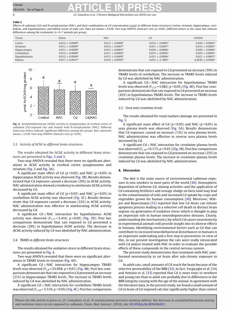
combination with NAC presented an increase of 2800% and 2500%in Cd kidney concentration, respectively. NAC administration wasineffective in restoring Cd levels to normal values in both tissuesevaluated.
Table 1
ARTICLEModel
BI-6259; No. of Pages 8
J.F. Goncalves et al. / Chemico-Bio
erformed by inductively coupled plasma mass spectrometry (ICP-S). An ICP-MS equipment (PerkinElmer Sciex, model ELAN DRC II,
hornhill, Canada), equipped with a concentric nebulizer (Mein-ard Associates, Golden, USA), a cyclonic spray chamber (Glassxpansion, Inc., West Melbourne, Australia) and a quartz torch withn injector tube of 2 mm i.d. was used.
.7. Synaptosome preparation
The cerebral cortex was homogenized in 10 volumes of an ice-old medium (medium I), consisting of 320 mM sucrose, 0.1 mMDTA and 5 mM HEPES, pH 7.5, in a motor driven Teflon-glassomogenizer. The synaptosomes were isolated as described by [26]sing a discontinuous Percoll gradient. The pellet was suspended
n an isoosmotic solution and the final protein concentration wasdjusted to 0.4–0.6 mg/mL. Synaptosomes were prepared freshaily, maintained at 0–4 ◦C throughout the procedure and used fornzymatic assays.
.8. Cerebral AChE enzymatic assay
The AChE enzymatic assay was determined by a modifica-ion of the spectrophotometric method of Ellman et al. [27] asreviously described by Rocha et al. [28]. The reaction mixture2 mL final volume) contained 100 mM K+-phosphate buffer, pH 7.5nd 1 mM 5,5′-dithio-bis-nitrobenzoic acid (DTNB). The method isased on the formation of the yellow anion, 5,5′-dithio-bis-aciditrobenzoic, measured by absorbance at 412 nm during 2-min
ncubation at 25 ◦C. The enzyme was pre-incubated for 2 min.he reaction was initiated by adding 0.8 mM acetylthiocholineodide (AcSCh). All samples were run in duplicate or triplicatend enzyme activity was expressed in �mol AcSCh/h/mg of pro-ein.
.9. Thiobarbituric acid reactive substances (TBARS)easurement
Brain TBARS levels were determined according to Ohkawat al. [29] by measuring the concentration of malondialdehydeMDA) as an end product of lipid peroxidation by reaction withhiobarbituric acid (TBA). Briefly, the reaction mixture, contain-ng 200 �L of brain supernatants or standard (0.03 mM MDA),00 �L of 8.1% sodium dodecyl sulphate (SDS), 500 �L of 0.8%BA and 500 �L of acetic acid solution (2.5 M HCl, pH 3.4), waseated at 95 ◦C for 120 min. The absorbance was measured at32 nm. TBARS tissue levels were expressed as nmol MDA/mg pro-ein.
.10. Urea and creatinine levels
Urea and creatinine levels were measured using standard enzy-atic methods with the use of Ortho-Clinical Diagnostics—JohnsonJohnson reagents, with a fully automated analyzer (Vitros
50®—dry chemistry, Rochester, New York).
.11. Statistical analysis
Please cite this article in press as: J.F. Goncalves, et al., N-acetylcysteine prand oxidative stress in rats exposed to cadmium, Chem. Biol. Interact. (201
Statistical analysis of training and test step-down latenciesas carried out by the Scheirer–Ray–Hare extension of theruskal–Wallis test (nonparametric two-way ANOVA). Foot shockensitivity was analyzed by unpaired t test. All other parame-ers evaluated were analyzed by two-way ANOVA, followed byuncan’s multiple range tests, where p < 0.05 was considered to
epresent a significant difference in all experiments.
mals were tested in a step-down latency test. Data are median ± interquartile rangeof training and test. *p < 0.05 compared with the others groups at testing by thetwo-way ANOVA following by Duncan’s test (n = 6).
3. Results
3.1. Behavioral tests
Fig. 1 shows the effect of NAC per se and in Cd-exposed rats onstep-down latencies. Statistical analysis of testing (nonparamet-ric two-way ANOVA) showed a significant Cd or saline vs NAC orsaline interaction (F1,20 = 4.43; p < 0.05), revealing that treatmentwith NAC reversed the impairment of memory induced by Cd. Sta-tistical analysis of training showed no difference between groups.
Because motivational disparities in the training session mayaccount for differences in inhibitory avoidance at testing, experi-ments were performed to assess whether Cd or NAC affected shockthreshold of the animals. Statistical analysis revealed that nei-ther Cd nor NAC altered foot shock sensitivity, as demonstratedby the similar flinch, jump and vocalization thresholds exhibitedby the animals. These data suggest that neither Cd intoxicationnor treatment with NAC administered before training of inhibitoryavoidance altered foot shock sensitivity (Table 1).
3.2. Cd concentration in brain and kidney tissues
All the brain structures analyzed showed a similar Cd concen-tration within the same group of rats. Control rats (saline) and ratstreated with NAC alone showed Cd concentrations of lower than0.015 �g/g in different brain structures. Rats exposed to Cd alone orCd plus NAC showed Cd concentrations of around 0.020 �g/g in dif-ferent brain structures. A significant main effect of Cd (p < 0.05) andNAC (p < 0.05) in Cd concentrations in different brain structures andin kidney tissue was observed (Table 2). Results demonstrate thatCd exposure alone or in combination with NAC caused an increaseof around 40% in Cd brain concentration. On the other hand, posthoc comparisons demonstrate that rats exposed to Cd alone and in
events memory deficits, the decrease in acetylcholinesterase activity0), doi:10.1016/j.cbi.2010.04.011
Rats exposed to cadmium (Cd) and/or treated with N-acetylcysteine (NAC) did notdemonstrate altered shock sensitivity. Data are means ± S.E.M. of flinch, jump andvocalization thresholds expressed in milliamps (n = 6–7 animals per group).
Saline NAC Cd Cd/NAC
Flinch 0.26 ± 0.033 0.27 ± 0.033 0.27 ± 0.034 0.23 ± 0.019Jump 0.45 ± 0.033 0.43 ± 0.021 0.41 ± 0.040 0.36 ± 0.029Vocalization 0.47 ± 0.048 0.40 ± 0.021 0.43 ± 0.045 0.46 ± 0.040
ARTICLE IN PRESSG Model
CBI-6259; No. of Pages 8
4 J.F. Goncalves et al. / Chemico-Biological Interactions xxx (2010) xxx–xxx
Table 2Effects of cadmium (Cd) and N-acetylcysteine (NAC), and their combination on Cd concentration (�g/g) in different brain structures (cortex, striatum, hippocampus, cere-bellum, and hypothalamus) and kidney tissue of male rats. Data are means ± S.E.M. Two-way ANOVA–Duncan’s test (p < 0.05). Different letters in the same line indicatedifferences among the treatments (n = 6–7 animals per group).
Tissue Saline NAC Cd Cd/NAC
Cortex 0.014 ± 0.0009b 0.013 ± 0.0008b 0.019 ± 0.0005a 0.020 ± 0.0005a
Striatum 0.012 ± 0.0004b 0.014 ± 0.0007b 0.021 ± 0.0007a 0.019 ± 0.0005a
Hippocampus 0.013 ± 0.0009b 0.012 ± 0.00 b a a
Cerebellum 0.014 ± 0.0008b 0.011 ± 0.00Hypothalamus 0.012 ± 0.0004b 0.013 ± 0.00Kidney 0.017 ± 0.0037b 0.019 ± 0.00
Fclm
3
t
as
hsNd
csNd
acdA
3
t
a
lp(i
w
and Antonio et al. [33] reported that Cd is more toxic to newborn
ig. 2. Acetylcholinesterase (AChE) activity in synaptosomes of cerebral cortex ofadmium (Cd)-exposed rats and treated with N-acetylcysteine (NAC). Differentowercase letters indicate significant difference among the groups. Bars represent
eans ± S.E.M. Two-way ANOVA–Duncan’s test (p < 0.05).
.3. Activity of AChE in different brain structures
The results obtained for AChE activity in different brain struc-ures are presented in Figs. 2 and 3.
Two-way ANOVA revealed that there were no significant alter-tions in AChE activity in cerebral cortex synaptosomes andtriatum (Fig. 2 and Fig. 3A).
A significant main effect of Cd (p < 0.05) and NAC (p < 0.05) inippocampus AChE activity was observed (Fig. 3B). Results demon-trated that Cd exposure caused a decrease (29%) in AChE activity.AC administration showed a tendency to ameliorate AChE activityecreased by Cd.
A significant main effect of Cd (p < 0.05) and NAC (p < 0.05) inerebellum AChE activity was observed (Fig. 3C). Results demon-trate that Cd exposure caused a decrease (32%) in AChE activity.AC administration was effective in ameliorating AChE activityecreased by Cd.
A significant Cd × NAC interaction for hypothalamus AChEctivity was observed (F1,17 = 5.416; p < 0.05) (Fig. 3D). Post hocomparisons demonstrate that rats exposed to Cd presented aecrease (29%) in hypothalamus AChE activity. The decrease inChE activity induced by Cd was abolished by NAC administration.
.4. TBARS in different brain structures
The results obtained for oxidative stress in different brain struc-ures are presented in Fig. 4.
Two-way ANOVA revealed that there were no significant alter-tions in TBARS levels in striatum (Fig. 4A).
A significant Cd × NAC interaction for hippocampus TBARSevels was observed (F1,8 = 25.694; p < 0.01) (Fig. 4B). Post hoc com-arisons demonstrate that rats exposed to Cd presented an increase
Please cite this article in press as: J.F. Goncalves, et al., N-acetylcysteine prand oxidative stress in rats exposed to cadmium, Chem. Biol. Interact. (201
21%) in hippocampus TBARS levels. The increase in TBARS levelsnduced by Cd was abolished by NAC administration.
A significant Cd × NAC interaction for cerebellum TBARS levelsas observed (F1,18 = 5.519; p < 0.05) (Fig. 4C). Post hoc comparisons
05 0.020 ± 0.0006 0.020 ± 0.000604b 0.019 ± 0.0005a 0.020 ± 0.0005a
09b 0.021 ± 0.0006a 0.021 ± 0.0007a
56b 4.815 ± 2.1083a 4.2630 ± 0.9966a
demonstrate that rats exposed to Cd presented an increase (59%) inTBARS levels of cerebellum. The increase in TBARS levels inducedby Cd was abolished by NAC administration.
A significant Cd × NAC interaction for hypothalamus TBARSlevels was observed (F1,13 = 5.082; p < 0.05) (Fig. 4D). Post hoc com-parisons demonstrate that rats exposed to Cd presented an increase(42%) in hypothalamus TBARS levels. The increase in TBARS levelsinduced by Cd was abolished by NAC administration.
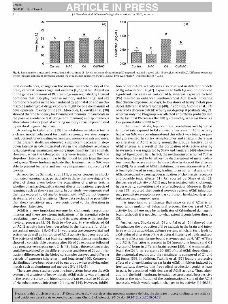
3.5. Urea and creatinine levels
The results obtained for renal markers damage are presented inFig. 5.
A significant main effect of Cd (p < 0.05) and NAC (p < 0.05) inurea plasma levels was observed (Fig. 5A). Results demonstratethat Cd exposure caused an increase (13%) in urea plasma levels.NAC administration was effective in reverse urea plasma levelsincreased by Cd.
A significant Cd × NAC interaction for creatinine plasma levelswas observed (F1,19 = 0.173; p < 0.01) (Fig. 5B). Post hoc comparisonsdemonstrate that rats exposed to Cd presented an increase (25%) increatinine plasma levels. The increase in creatinine plasma levelsinduced by Cd was abolished by NAC administration.
4. Discussion
The diet is the main source of environmental cadmium expo-sure in non-smokers in most parts of the world [30]. Atmosphericdeposition of airborne Cd, mining activities and the application ofCd containing fertilizers and sewage sludge on farm land may leadto the contamination of soils and increased Cd uptake by crops andvegetables grown for human consumption [30]. Moreover, Wät-jen and Beyersmann [31] reported that low Cd doses can initiateapoptosis process leading to a selective cell death in distinct brainregions via generation of oxidative stress which is thought to playan important role in human neurodegenerative diseases. Clearly,understanding the mechanism(s) by which Cd causes neurotoxicityin experimental animals will provide insight into its diverse effectsin humans. Identifying environmental factors such as Cd that cancontribute to increased neurobehavioral disturbances in humans isan important undertaking and a first step to prevention. In view ofthis, in our present investigation the rats were orally intoxicatedwith Cd and/or treated with NAC in order to evaluate the possibleeffects of these compounds in the central nervous system.
The present study demonstrates that treatment with NAC ame-liorated neurotoxicity in rat brain after sub-chronic exposure toCd.
In adult rats, small amounts of Cd reach the brain because of theselective permeability of the BBB [32]. In fact, Yargicoglu et al. [16]
events memory deficits, the decrease in acetylcholinesterase activity0), doi:10.1016/j.cbi.2010.04.011
and young rats than to adult rats probably due to differences in theBBB integrity varying with the age of the animal. In agreement withthe literature data, in the present study, we found a small amount ofCd in brain of Cd-exposed rats but significantly higher than control

ARTICLE IN PRESSG Model
CBI-6259; No. of Pages 8
J.F. Goncalves et al. / Chemico-Biological Interactions xxx (2010) xxx–xxx 5
F pus (w ence a(
vtsdott
F(T
ig. 3. Acetylcholinesterase (AChE) activity in supernatant of striatum (A), hippocamith N-acetylcysteine (NAC). Different lowercase letters indicate significant differ
p < 0.05).
alues and enough to cause brain injury. For all the brain struc-ures analyzed, a similar Cd concentration was found within theame group of rats and co-administration of NAC was ineffective in
Please cite this article in press as: J.F. Goncalves, et al., N-acetylcysteine prand oxidative stress in rats exposed to cadmium, Chem. Biol. Interact. (201
ecreasing this level. These results suggest that the harmful effectsf Cd observed in this investigation were ameliorated mainly byhe antioxidant activity of NAC rather than by Cd removal fromissues. Similar findings were reported by Bludovská et al. [34]
ig. 4. Oxidative stress measured by thiobarbituric acid reactive substances (TBARS) inCd)-exposed rats and treated with N-acetylcysteine (NAC). Different lowercase letterswo-way ANOVA–Duncan’s test (p < 0.05).
B), cerebellum (C) and hypothalamus (D) of cadmium (Cd)-exposed rats and treatedmong the groups. Bars represent means ± S.E.M. Two-way ANOVA–Duncan’s test
for alpha-lipoic acid treatment against Cd toxicity. Nevertheless,it is interesting to point out that although Cd does not accumulatein significant quantities in the adult rat brain following exposure
events memory deficits, the decrease in acetylcholinesterase activity0), doi:10.1016/j.cbi.2010.04.011
[8,35], it severely disturbs the metabolism of trace and essential ele-ments such as copper and zinc [4]. Some studies have demonstratedthat Cd is able to induce neurotoxicity in animals with a widespectrum of clinical entities including neurological and behav-
striatum (A), hippocampus (B), cerebellum (C) and hypothalamus (D) of cadmiumindicate significant difference among the groups. Bars represent means ± S.E.M.
ARTICLE IN PRESSG Model
CBI-6259; No. of Pages 8
6 J.F. Goncalves et al. / Chemico-Biological Interactions xxx (2010) xxx–xxx
F dmiul .M. Tw
ibihhmdstab
anIdtHstat
mewltsts
mrhoeaTsbctpsm
sio
ig. 5. Renal markers measured by urea (A) and creatinine (B) levels in serum of caetters indicate significant difference among the groups. Bars represent means ± S.E
oral disturbances, changes in the normal neurochemistry of therain, cerebral hemorrhage and oedema [6,7,8,14,36]. Alteration
n the gene expressions of RC3 (neurogranin regulated by thyroidormones that may play roles in memory and learning) and sexormone receptors in the brain induced by perinatal Cd and methi-azole (anti-thyroid drug) exposure might be one mechanism of
evelopmental toxicity of Cd [37]. Moreover, Lukawski et al. [38]howed that the tendency for Cd-induced memory impairments inhe passive avoidance task (long-term memory) and spontaneouslternation deficits (spatial working memory) may be potentiatedy cerebral oligemic hypoxia.
According to Cahill et al. [39] the inhibitory avoidance test isclassic model behavioral test, with a strongly aversive compo-
ent, utilized for evaluating learning and memory in rats and mice.n the present study, we observed a significant decrease in step-own latency in Cd-intoxicated rats in the inhibitory avoidanceest, suggesting learning and memory impairment in these animals.owever, when the Cd-exposed rats were treated with NAC the
tep-down latency was similar to that found for rats from the con-rol group. These findings indicate that treatment with NAC wasble to prevent learning and memory impairment induced by Cdoxicity.
As reported by Schmatz et al. [11], a major concern in shock-otivated learning tests, particularly in those that investigate the
ffect of drugs given before the acquisition of a given test, ishether pharmacological treatment affects motivational aspects of
earning, such as shock sensitivity. In our study, we demonstratedhat rats exposed to Cd and/or treated with NAC did not demon-trate altered shock sensitivity. These data exclude the possibilityhat shock sensitivity may have contributed to the alteration intep-down latencies.
AChE is a very important enzyme for cholinergic neurotrans-ission and there are strong indications of its essential role in
egulating many vital functions and its association with neurobe-avioral processes [3,10]. Both in vitro and in vivo effects of Cdn AChE activity have been described in the literature for differ-nt animal models [3,9,40,41,42], yet results are controversial andctivation as well as inhibition of AChE activity has been reported.he activity of AChE in rat brain synaptosomal plasma membraneshowed a considerable decrease after 6 h of Cd exposure, followedy a progressive increase up to 24 h [43]. In fact, these controversiesould be explained by the different routes and doses of Cd adminis-ration, differences in the biological samples assayed and differingeriods of exposure (short-term and long-term) [40]. Controver-ial findings have been observed by our group when studying other
Please cite this article in press as: J.F. Goncalves, et al., N-acetylcysteine prand oxidative stress in rats exposed to cadmium, Chem. Biol. Interact. (201
etals such as aluminum and mercury (Hg) [44,45].There are some studies reporting interactions between the ACh
ystem and a variety of heavy metals. AChE activity was enhancedn the cerebral cortex and hippocampus of rats exposed to 30 dosesf Hg subcutaneous injections (0.1 mg/kg) [44]. However, inhibi-
m (Cd)-exposed rats and treated with N-acetylcysteine (NAC). Different lowercaseo-way ANOVA–Duncan’s test (p < 0.05).
tion of brain AChE activity was also observed in different modelsof Hg intoxication [46,47]. Exposure to both Hg and Cd producedsignificant decreases in cortical ACh, whereas exposure to lead(Pb) resulted in enhanced cerebrocortical ACh levels indicatingthat chronic exposure (45 days) to low doses of heavy metals pro-duces differential ACh response [48]. In addition, Antonio et al. [33]observed a decreased AChE activity in Cd-group at postnatal day 21,whereas only the Pb-group was affected at birthday probably dueto the fact that Pb crosses the BBB quite readily, whereas there is alow permeability of BBB to Cd.
In the present study, hippocampus, cerebellum and hypotha-lamus of rats exposed to Cd showed a decrease in AChE activitybut when NAC was co-administered this effect was totally or par-tially prevented. In cortex synaptosomes and striatum there wasno alteration in AChE activity among the groups. Inactivation ofAChE enzyme as a result of the occupation of its active sites byheavy metals was suggested by Shaw and Panigrahi [49] who inves-tigated Hg-exposed fish. In fact, the mechanism of action of Cd hasbeen hypothesized to be either the displacement of metal cofac-tors from the active site or the direct deactivation of the enzymesite [50]. As a result of AChE inhibition, the neurotransmitter AChis less hydrolysed in synapses, leading to an abnormal amount ofACh, consequently causing overactivation of cholinergic receptorsand possible toxic effects [51]. As reported by Olney et al. [52]the decreased activity of AChE may be associated with cholinergichyperactivity, convulsion and status epilepticus. Moreover, Ecobi-chon [53] reported that central nervous system AChE inhibitionmay precipitate symptoms such as confusion, headache, sleep dis-turbances and memory lapses.
It is important to emphasize that since cerebral AChE is animportant regulator of behavioral process, the decreased AChEactivity found here may be an indicator of Cd-induced damage inbrain, although it is not clear to what extent it contributes directly[3].
Furthermore, Shukla et al. [5] and Pal et al. [54] showed thatCd enhances the production of free radicals in the brain and inter-feres with the antioxidant defense system, which, in turn, leads toa Cd-induced alteration of the structural integrity of lipids and sec-ondarily affects membrane-bound enzymes such as Na+/K+-ATPaseand AChE. The latter is present in G4 (membrane bound) and G1(cytosolic) forms in different brain regions [55]. In the mammalianbrain, the G4 form represents 60–90% of total AChE, depending onthe anatomical region, and the remainder is composed of G1 andG2 forms [56]. In addition, Tsakiris et al. [57] found a protectiveeffect of l-phenylalanine on rat brain AChE inhibition induced by
events memory deficits, the decrease in acetylcholinesterase activity0), doi:10.1016/j.cbi.2010.04.011
free radicals, showing that free radical production could, at leastin part, be associated with decreased AChE activity. Thus, alter-ations in the lipid membrane by oxidative stress could be a decisivefactor in the modification of the conformational state of the AChEmolecule, which would explain changes in its activity [11,49,55].

ING
C
logica
Iaftnap
rNtitadcciocplaascthiescaCNnitaspst
avcaoopadamgas
tporUnt
[
[
ARTICLEModel
BI-6259; No. of Pages 8
J.F. Goncalves et al. / Chemico-Bio
t is well known that peroxidative damage of membrane structurend changes in associated enzymes, receptors, and physiologicalunctions may ultimately result in disturbances in neuronal func-ions [16]. In fact, in the present study, the AChE activity wasegatively correlated with TBARS levels in all cerebral structuresnalyzed (r = 0.79, r = 0.99, r = 0.91 and r = 0.99 to striatum, hip-ocampus, cerebellum and hypothalamus, respectively).
In our study, hippocampus, cerebellum and hypothalamus ofats exposed to Cd presented an increase in TBARS levels but whenAC was co-administered this effect was abolished. In striatum,
here was no alteration in TBARS levels among the groups. Accord-ng to Kumar et al. [58], the brain may be particularly vulnerableo oxidative damage and saturation of the lipid bi-layer of severalreas of the brain as a result of LPO in Cd-exposed animals causedisturbances in membrane fluidity and intracellular calcium con-entrations. Moreover, these authors proposed that Cd exposureaused region-specific biomembrane changes. The ability of Cd tonduce oxidative stress in brain cells is related to the inductionf ROS, LPO and apoptosis, after the interaction of Cd with mito-hondrial sites, leading to the breakdown of the mitochondrialotentials, the consequent reduction of intracellular glutathione
evels (GSH) and changes in catalase and superoxide dismutasectivities [59]. Until rat pups exposed to Cd only gestationallynd lactationally through mothers (20 ppm Cd in drinking water)howed changes in their brain antioxidant defense mechanisms atritical periods of development which may have serious implica-ions in later life [60]. Corroborating with our results, other reportsave shown that Cd induced an elevation of LPO in brain and also
n other tissues [5,16,17,21,39,43,61]. In fact, according to Mancat al. [61] LPO is an early and sensitive consequence of Cd expo-ure. Also similar to our findings, some investigators found that theonsumption of antioxidants such as garlic, vitamin E, carotene,lpha-lipoic acid, diphenyl diselenide, and NAC was able to revertd-induced oxidative stress [3,21,34,62,63,64,65]. As reported byemmiche et al. [65] the mechanism of Cd-induced LPO is stillot fully understood. Available data indicate that the mechanism
s multidirectional and may involve a decrease in the level of glu-athione and the total pool of sulphydryl groups and changes in thectivities of antioxidant enzymes [65]. In fact, some authors havehown a decreased GSH level associated with the increased LPOrocess in rats intoxicated with Cd [34,65]. These factors can con-equently induce a prooxidant state in biological systems and leado peroxidation of polyunsaturated fatty acids.
Another important aspect to be discussed here is that both agingnd age-associated neurodegenerative diseases are associated witharious degrees of behavioral impairments and among the primaryandidates responsible for producing the neuronal changes medi-ting these behavioral deficits appear to be free radicals and thexidative stress they generate [66]. A recent study published byur research group that utilized the same behavioral test as theresent study demonstrated that the antioxidant resveratrol wasble to modulate cerebral AChE activity and to reverse the cognitiveeficits in diabetic rats [11]. Another study showed that vitamin Eppears to have a close interaction with the cholinergic system andemory retention [67]. Taken together, these data allow us to sug-
est that antioxidants such as resveratrol, vitamin E and NAC haven important role in preventing oxidative stress, the cholinergicystem impairment and learning and memory deficits.
In order to explain the possible mechanism by which Cd exert itsoxicity in our study it is important to know whether the exposurerotocol used in this study induced any nephrotoxicity, also in light
Please cite this article in press as: J.F. Goncalves, et al., N-acetylcysteine prand oxidative stress in rats exposed to cadmium, Chem. Biol. Interact. (201
f the fact that the behavioral deficits can be associated with mildenal failure we determine the urea and creatinine levels in serum.rea is the first acute renal marker which increases when the kid-ey suffers any kind of injury otherwise, creatinine is the mostrustable of them [22]. So, in the present study we verified that Cd
[
[
PRESSl Interactions xxx (2010) xxx–xxx 7
induced moderate nephrotoxicity because Cd-exposed rats showedincreased urea and creatinine levels in serum, but no renal histo-logical damage was found (data not shown). The co-administrationof NAC was able to abolish these undesirable effects. These resultsnot decrease the relevance of the present study because we demon-strated the presence of Cd in the brain and its negative effectsin both the cerebral and the kidney tissues. Thus, Cd can induceoxidative stress regulating learning/memory processes through themodulation of brain AChE activity and mild renal injury can con-tribute to these alterations.
In conclusion, our data have shown, for the first time, thatNAC has beneficial actions against Cd-mediated toxic effects inbrain, inhibiting oxidative stress and subsequently restoring cere-bral AChE activity, thus modulating cholinergic neurotransmissionand improving cognition (learning and memory). However, it isnecessary to note that the mild renal failure may be a contribu-tor to the behavioral impairment found in this investigation. It isproposed that NAC acted in this experimental protocol through itsantioxidant properties but not its capacity to form a complex withCd, since NAC did not alter the metal load. Therefore, we can sug-gest that NAC is a promising drug which should be investigated infuture studies in order to improve therapeutic alternatives for braininjury associated with Cd-induced neurotoxicity.
Conflict of interest
There is not conflict of interest.
Acknowledgements
The authors thank the Coordenacão e Aperfeicoamento dePessoal de Nível Superior (CAPES), Conselho Nacional de Desen-volvimento Científico e Tecnológico (CNPq), and Fundacão deAmparo à Pesquisa do Estado do Rio Grande do Sul (FAPERGS) forthe research fellowships.
References
[1] J. Godt, F. Scheidig, C. Grosse-Siestrup, V. Esche, P. Brandenburg, A. Reich, D.A.Groneberg, The toxicity of cadmium and resulting hazards for human health, J.Occup. Environ. Med. 22 (2006) 1–6.
[2] R.K. Zalups, S. Ahmad, Molecular handling of cadmium in transporting epithelia,Toxicol. Appl. Pharmacol. 186 (2003) 163–188.
[3] L. Pari, P. Murugavel, Diallyl tetrasulfide improves cadmium induced alterationsof acetylcholinesterase, ATPases and oxidative stress in brain of rat, Toxicology234 (2007) 44–50.
[4] M. Méndez-Armenta, C. Ríos, Cadmium neurotoxicity, Environ. Toxicol. Appl.Pharmacol. 23 (2007) 350–358.
[5] A. Shukla, G.S. Shukla, R.C. Srimal, Cadmium-induced alterations in blood–brainbarrier permeability and its possible correlation with decreased microvesselantioxidant potential in rat, Hum. Exp. Toxicol. 15 (1996) 400–405.
[6] J.P. Provias, C.A. Ackerley, C. Smith, L.E. Becker, Cadmium encephalopathy: areport with elemental analysis and pathological findings, Acta Neuropathol.Berl. 88 (1994) 583–586.
[7] M. Méndez-Armenta, R. Barroso-Moguel, J. Villeda-Hernández, C. Nava-Ruíz, C.Ríos, Histopathological alterations in brain regions of rats after perinatal com-bined treatment with cadmium and dexamethazone, Toxicology 161 (2001)189–199.
[8] A. Minami, A. Takeda, D. Nishibaba, S. Takefuta, N. Oku, Cadmium toxicity insynaptic neurotransmission in the brain, Brain Res. 894 (2001) 336–339.
[9] C. Luchese, R. Brandão, R. Oliveira, C.W. Nogueira, F.W. Santos, Efficacy ofdiphenyl diselenide against cerebral and pulmonary damage induced by cad-mium in mice, Toxicol. Lett. 173 (2007) 181–190.
10] M.M. Mesulam, A. Guillozet, P. Shaw, A. Levey, Acetylcholinesterase knockoutsestablish central cholinergic pathways and can use butyrylcholinesterase tohydrolyze acetylcholine, Neuroscience 110 (2002) 627–639.
11] R. Schmatz, C.M. Mazzanti, R. Spanevello, N. Stefanello, J. Gutierres, M. Corrêa,M.M. Rosa, M.A. Rubin, M.R.C. Schetinger, V.M. Morsch, Resveratrol pre-
events memory deficits, the decrease in acetylcholinesterase activity0), doi:10.1016/j.cbi.2010.04.011
vents memory deficits and the increase in acetylcholinesterase activity instreptozotocin-induced diabetic rats, Eur. J. Pharmacol. 610 (2009) 42–48.
12] H. Soreq, S. Seidman, Acetylcholinesterase—new roles for an old actor, Nat. Rev.Neurosci. 2 (2001) 294–302.
13] R.P. Hart, C.S. Rose, R.M. Hamer, Neuropsychological effect of occupationalexposure to cadmium, J. Clin. Exp. Neuropsychol. 11 (1989) 933–943.

ING
C
8 logica
[
[
[
[
[
[
[
[
[
[
[
[
[
[
[
[
[
[
[
[
[
[
[
[
[
[
[
[
[
[
[
[
[
[
[
[
[
[
[
[
[[
[
[
[
[
[
[
[
[
[
[
ARTICLEModel
BI-6259; No. of Pages 8
J.F. Goncalves et al. / Chemico-Bio
14] M.K. Viaene, R. Masschelein, J. Leeders, M.De. Groof, L.J.V.C. Swerts, H.A. Roels,Neurobehavioural effects of occupational exposure to cadmium: a cross sec-tional epidemiological study, Occup. Environ. Med. 57 (2000) 19–27.
15] W.R. Holloway, D.H. Thor, Cadmium exposure in infancy: effects on activity andsocial behavior of juvenile rats, Neurotoxicol. Teratol. 10 (1988) 135–142.
16] P. Yargicoglu, A. Agar, Y. Oguz, V.N. Izgut-Uysal, U.K. Senturk, G. Oner, The effectof developmental exposure to cadmium (Cd) on visual evoked potentials (VEPs)and lipid peroxidation, Neurotoxicol. Teratol. 19 (1997) 213–219.
17] M. Méndez-Armenta, J. Villeda-Hernández, R. Barroso-Moguel, C. Nava-Ruíz,M. Jiménez-Capdeville, C. Ríos, Brain regional lipid peroxidation and metal-lothionein levels of developing rats exposed to cadmium and dexamethasone,Toxicol. Lett. 144 (2003) 151–157.
18] V. Calabrese, T.E. Bates, A.M.G. Stella, NO synthase and NO-dependent signalpathways in brain aging and neurodegenerative disorders: the role of oxi-dant/antioxidant balance, Neurochem. Res. 25 (2000) 1315–1341.
19] L. Grinberg, E. Fibach, J. Amer, D. Atlas, N-acetylcysteine amide, a novel cell-permeating thiol, restores cellular glutathione and protects human red bloodcells from oxidative stress, Free Radic. Biol. Med. 38 (2005) 136–145.
20] A.M. Sadowska, B. Manuel-y-Keenoy, W.A. De Backer, Antioxidant and anti-inflammatory efficacy of NAC in the treatment of COPD: discordant in vitroand in vivo dose–effects: a review, Pulm. Pharmacol. Ther. 20 (2007) 9–22.
21] F.M. El-Demerdash, M.I. Yousef, F.S. Kedwany, H.H. Baghdadi, Cadmium-induced changes in lipid peroxidation, blood hematology, biochemicalparameters and semen quality of male rats: protective role of vitamin E andb-carotene, Food Chem. Toxicol. 42 (2004) 1563–1571.
22] L.P. Borges, R. Brandão, B. Godoi, C.W. Nogueira, G. Zeni, Oral administrationof diphenyl diselenide protects against cadmium-induced liver damage in rats,Chem. Biol. Interact. 171 (2008) 15–25.
23] G.P. Guerra, C.F. Mello, P.D. Sauzem, D.B. Berlese, A.F. Furian, Z. Tabarelli, M.A.Rubin, Nitric oxide is involved in the memory facilitation induced by spermi-dine in rats, Psychopharmacology 186 (2006) 150–158.
24] M.A. Rubin, D.B. Berlese, J.A. Stiegemeier, M.A. Volkweis, D.M. Oliveira, T.L. dosSantos, A.C. Fenili, C.F. Mello, Intra-amygdala administration of polyaminesmodulates fear conditioning in rats, J. Neurosci. 24 (2004) 2328–2334.
25] M.M. Bradford, A rapid and sensitive method for quantification of micro-gram quantities of protein utilizing the principle of protein–dye binding, Anal.Biochem. 72 (1976) 248–254.
26] A. Nagy, A.V. Delgado Escueta, Rapid preparation of synaptosomes from 21mammalian brain using non-toxic isosmotic gradient material (Percoll), J. Neu-rochem. 43 (1984) 1114–1123.
27] G.L. Ellman, D.K. Courtney, V. Andres, R.M. Flatherstone, A new and rapid col-orimetric determination of acetylcholinesterase activity, Biochem. Pharmacol.7 (1961) 88–95.
28] J.B.T. Rocha, T. Emanuelli, M.E. Pereira, Effects of early undernutrition on 15kinetic parameters of brain acetylcholinesterase from adult rats, Acta Neuro-biol. Exp. 53 (1993) 431–437.
29] H. Ohkawa, N. Ohishi, K. Yagi, Assay for lipid peroxides in animal tissues bythiobarbituric acid reaction, Anal. Biochem. 95 (1979) 351–358.
30] L. Järup, A. Åkesson, Current status of cadmium as an environmental healthproblem, Toxicol. Appl. Pharmacol. 238 (2009) 201–208.
31] W. Wätjen, D. Beyersmann, Cadmium-induced apoptosis in C6 glioma cells:influence of oxidative stress, Biometals 17 (2004) 65–78.
32] A. Takeda, S. Takefuta, H. Ijiro, S. Okada, N. Oku, 109Cd transport in rat brain,Brain Res. Bull. 49 (1999) 453–459.
33] M.T. Antonio, L. Corredor, M.L. Leret, Study of the activity of several brainenzymes like markers of the neurotoxicity induced by perinatal exposure tolead and/or cadmium, Toxicol. Lett. 143 (2003) 331–340.
34] M. Bludovská, D. Kotyzová, J. Kontensky, V. Eybl, The influence of alpha-lipoicacid on the toxicity of cadmium, Gen. Physiol. Biophys. 18 (1999) 28–32.
35] B. Arvidson, H. Tjälve, Distribution of 109Cd in the nervous system of rats afterintravenous injection, Acta Neuropathol. 69 (1986) 111–116.
36] E.Y. Gutiérrez-Reyes, A. Albores, C. Ríos, Increase of striatal dopamine releaseby cadmium in nursing rats and its prevention by dexamethazone-inducedmetallothionein, Toxicology 131 (1998) 145–154.
37] H. Ishitobi, K. Mori, K. Yoshida, C. Watanabe, Effects of perinatal exposure tolow-dose cadmium on thyroid hormone-related and sex hormone receptorgene expressions in brain of offspring, Neurotoxicology 28 (2007) 790–797.
38] K. Lukawski, B. Nieradko, M. Sieklucka-Dziuba, Effects of cadmium on memoryprocesses in mice exposed to transient cerebral oligemia, Neurotoxicol. Teratol.27 (2005) 575–584.
39] L. Cahill, J. Brioni, I. Izquierdo, Retrograde memory enhancement by diazepam:its relation to anterograde amnesia and some clinical implications, Psychophar-macology 90 (1986) 454–456.
Please cite this article in press as: J.F. Goncalves, et al., N-acetylcysteine prand oxidative stress in rats exposed to cadmium, Chem. Biol. Interact. (201
40] H. Carageorgiou, V. Tzotzes, C. Pantos, C. Mourouzis, A. Zarros, S. Tsakiris, Invivo and in vitro effects of cadmium on adult rat brain total antioxidant status,acetylcholinesterase, (Na+,K+)-ATPase and Mg2+-ATPase activities: protectionby l-cysteine, Basic Clin. Pharmacol. Toxicol. 94 (2004) 112–118.
41] H. Carageorgiou, V. Tzotzes, C. Pantos, C. Mourouzis, A. Zarros, S. Tsakiris, Cad-mium effects on brain acetylcholinesterase activity and antioxidants status of
[
[
PRESSl Interactions xxx (2010) xxx–xxx
adult rats: modulation by zinc, calcium and l-cysteine co-administration, BasicClin. Pharmacol. Toxicol. 97 (2005) 320–324.
42] M.R. Senger, D.B. Rosemberg, E.P. Rico, M.B. Arizi, R.D. Dias, M.R. Bogo, C.D.Bonan, In vitro effect of zinc and cadmium on acetylcholinesterase and ectonu-cleotidase activities in zebrafish (Danio rerio) brain, Toxicol. In Vitro 20 (2006)954–958.
43] C.D. Fasitsas, S.E. Theocharis, D. Zoulas, S. Chrissimou, G. Deliconstantinos,Time-dependent cadmium-neurotoxicity in rat brain synaptosomal plasmamembranes, Comp. Biochem. Physiol. C 100 (1991) 271–275.
44] M.B. Moretto, C.L. Lermen, V.M. Morsch, D. Bohrer, R.P. Ineu, A.C. Silva, D. Balz,M.R.C. Schetinger, Effect of subchronic treatment with mercury chloride onNTPDase, 5′-nucleotidase and acetylcholinesterase from cerebral cortex of rats,J. Trace Elem. Med. Biol. 17 (2004) 255–260.
45] R.R. Kaizer, M.C. Corrêa, R.M. Spanevello, V.M. Morsch, C.M. Mazzanti, J.F.Goncalves, M.R.C. Schetinger, Acetylcholinesterase activation and enhancedlipid peroxidation after long-term exposure to low levels of aluminumon different mouse brain regions, J. Inorg. Biochem. 99 (2005) 1865–1870.
46] M.K. Laksmana, T. Desiraju, T.R. Raju, Mercuric chloride-induced alterations oflevels of noradrenaline, dopa, serotonin and acetylchotine esterase in differ-ent regions of brain during postnatal development, Arch. Toxicol. 67 (1993)422–427.
47] F.M. El-Demerdash, Effects of selenium and mercury on the enzymatic activitiesand lipid peroxidation in brain, liver, and blood of rats, J. Environ. Sci. Health B36 (2001) 489–499.
48] P.D. Hrdina, D.A. Peters, R.L. Singhal, Effects of chronic exposure to cadmium,lead and mercury of brain biogenic amines in the rat, Res. Commun. Chem.Pathol. Pharmacol. 15 (1976) 483–493.
49] B.P. Shaw, A.K. Panigrahi, Brain AChE activity studies in some fish species col-lected from a mercury contaminated estuary, Water Air Soil Pollut. 53 (1990)327–334.
50] E. Casalino, S. Sblano, C. Landriscina, Enzyme activity alteration by cadmiumadministration to rats: the possibility of iron involvement in lipid peroxidation,Arch. Biochem. Biophys. 346 (1997) 171–179.
51] C.H. Walker, Organic Pollutants, in: C.H. Walker (Ed.), An Ecotoxicological Per-spective, Taylor and Francis, New York, USA, 2001.
52] J.W. Olney, R.C. Collins, R.S. Sloviter, Exotoxic mechanism of epileptic braindamage, Adv. Neurol. 44 (1986) 857–877.
53] D.J. Ecobichon, Toxic effects of pesticides, in: C.D. Klaassen (Ed.), Casarett andDoull’s Toxicology: The Basic Science of Poisons, 5th ed., McGraw-Hill, NewYork, 1996, pp. 643–689.
54] R. Pal, R. Nath, D.K. Gill, Neurochem. Int. 23 (1993) 451–458.55] A. Das, M. Dikshit, C. Nath, Profile of acetylcholinesterase in brain areas
of male and female rats of adult and old age, Life Sci. 68 (2001) 1545–1555.
56] L. Descarries, V. Gisiger, M. Steriade, Diffuse transmission by acetylcholine inthe CNS, Prog. Neurobiol. 53 (1997) 603–625.
57] S. Tsakiris, P. Angelogianni, K.H. Schulpis, J.C. Stavridis, Protective effect ofl-phenylalanine on rat brain acetylcholinesterase inhibition induced by freeradicals, Clin. Biochem. 33 (2000) 103–106.
58] R. Kumar, A.K. Agarwal, P.Q. Seth, Oxidative stress-mediated neurotoxicity ofcadmium, Toxicol. Lett. 89 (1996) 65–69.
59] E. López, C. Arce, M.J. Oset-Gasque, S. Canadas, M.P. González, Cadmium inducesreactive oxygen species generation and lipid peroxidation in cortical neuronsin culture, Free Radic. Biol. Med. 40 (2006) 940–951.
60] A. Gupta, A. Gupta, G.S. Shukla, Development of brain free radical scavengingsystem and lipid peroxidation under the influence of gestational and lactationalcadmium exposure, Hum. Exp. Toxicol. 14 (1995) 428–433.
61] D. Manca, A.C. Ricarda, B. Trottiera, G. Chevalier, Studies on lipid peroxidationin rat tissues following administration of low and moderate doses of cadmiumchloride, Toxicology 67 (1991) 303–323.
62] M.A. El-Missiry, F. Shalaby, Role of beta-carotene in ameliorating the cadmim-induced oxidative stress in rat brain and testis, J. Biochem. Mol. Toxicol. 14(2000) 238–243.
63] S.K. Tandon, S. Sing, S. Prasad, K. Khandekar, V.K. Dwivedi, M. Chatter-jee, N. Mathur, Reversal of cadmium induced oxidative stress by chelatingagent, antioxidant or their combination in rat, Toxicol. Lett. 145 (2003) 211–217.
64] F.W. Santos, G. Zeni, J.B.T. Rocha, S.N. Weis, J.M. Fachinetto, A.M. Favero, C.W.Nogueira, Diphenyl diselenide reverses cadmium-induced oxidative damageon mice tissues, Chem. Biol. Interact. 151 (2005) 159–165.
65] S. Nemmiche, D. Chabane-Sari, P. Guiraud, Role of �-tocopherol in cadmium-induced oxidative stress in Wistar rat’s blood, liver and brain, Chem. Biol.
events memory deficits, the decrease in acetylcholinesterase activity0), doi:10.1016/j.cbi.2010.04.011
Interact. 170 (2007) 221–230.66] I. Cantuti-Castelvetri, B. Shukitt-Hale, J.A. Joseph, Neurobehavioral aspects of
antioxidants in aging, Int. J. Dev. Neurosci. 18 (2000) 367–381.67] A. Eidi, M. Eidi, G. Mahmoodi, S. Oryan, Effect of vitamin E on memory retention
in rats: possible involvement of cholinergic system, Eur. Neuropsychopharma-col. 16 (2006) 101–106.