Exercise effects on activities of Na+,K+-ATPase, acetylcholinesterase and adenine nucleotides...
8
Research Report Exercise effects on activities of Na + ,K + -ATPase, acetylcholinesterase and adenine nucleotides hydrolysis in ovariectomized rats Juliana Ben a , Flávia Mahatma Schneider Soares a , Fernanda Cechetti a , Fernanda Cenci Vuaden a , Carla Denise Bonan b , Carlos Alexandre Netto a , Angela Terezinha de Souza Wyse a, ⁎ a Laboratório de Neuroproteção e Doença Metabólica, Departamento de Bioquímica, Instituto de Ciências Básicas da Saúde, Universidade Federal do Rio Grande do Sul, Rua Ramiro Barcelos, 2600-Anexo, CEP 90035-003, Porto Alegre, RS, Brazil b Laboratório de Neuroquímica e Psicofarmacologia, Departamento de Biologia Celular e Molecular, Faculdade de Biociências, Pontifícia Universidade Católica do Rio Grande do Sul, Porto Alegre, RS, Brazil ARTICLE INFO ABSTRACT Article history: Accepted 4 September 2009 Available online 11 September 2009 Hormone deficiency following ovariectomy causes activation of Na + ,K + -ATPase and acetylcholinesterase (AChE) that has been related to cognitive deficits in experimental animals. Considering that physical exercise presents neuroprotector effects, we decide to investigate whether exercise training would affect enzyme activation in hippocampus and cerebral cortex, as well as adenosine nucleotide hydrolysis in synaptosomes from cerebral cortex of ovariectomized rats. Female adult Wistar rats were assigned to one of the following groups: sham (submitted to surgery without removal of the ovaries), exercise, ovariectomized (Ovx) and Ovx plus exercise. Thirty days after surgery, animals were submitted to one month of exercise training, three times per week. After, rats were euthanized, blood serum was collected and hippocampus and cerebral cortex were dissected. Data demonstrated that exercise reversed the activation of Na + ,K + -ATPase and AChE activities both in hippocampus and cerebral cortex of ovariectomized rats. Ovariectomy decreased AMP hydrolysis in cerebral cortex and did not alter adenine nucleotides hydrolysis in blood serum. Exercise per se decreased ADP and AMP hydrolysis in cerebral cortex. On the other hand, AMP hydrolysis in blood serum was increased by exercise in ovariectomized adult rats. Present data support that physical exercise might have beneficial effects and constitute a therapeutic alternative to hormone replacement therapy for estrogen deprivation. © 2009 Elsevier B.V. All rights reserved. Keywords: Ovariectomy Exercise Na + ,K + -ATPase Acetylcholinesterase Adenine nucleotide hydrolysis 1. Introduction Estrogen displays important roles beyond the reproductive system, such as trophic and protective role in the adult brain (Wise et al., 2001b, 2002) and it has been shown that estrogen deprivation is implicated in the pathogenesis of Alzheimer's disease and cerebral ischemia (van Duijn, 1999; Zhang et al., 1998). Studies have suggested that post-menopausal women BRAIN RESEARCH 1302 (2009) 248 – 255 ⁎ Corresponding author. E-mail address: [email protected] (A.T.S. Wyse). 0006-8993/$ – see front matter © 2009 Elsevier B.V. All rights reserved. doi:10.1016/j.brainres.2009.09.013 available at www.sciencedirect.com www.elsevier.com/locate/brainres
Transcript of Exercise effects on activities of Na+,K+-ATPase, acetylcholinesterase and adenine nucleotides...
B R A I N R E S E A R C H 1 3 0 2 ( 2 0 0 9 ) 2 4 8 – 2 5 5
ava i l ab l e a t www.sc i enced i r ec t . com
www.e l sev i e r . com/ loca te /b ra i n res
Research Report
Exercise effects on activities of Na+,K+-ATPase,acetylcholinesterase and adenine nucleotides hydrolysis inovariectomized rats
Juliana Bena, Flávia Mahatma Schneider Soaresa, Fernanda Cechettia,Fernanda Cenci Vuadena, Carla Denise Bonanb,Carlos Alexandre Nettoa, Angela Terezinha de Souza Wysea,⁎aLaboratório de Neuroproteção e Doença Metabólica, Departamento de Bioquímica, Instituto de Ciências Básicas da Saúde,Universidade Federal do Rio Grande do Sul, Rua Ramiro Barcelos, 2600-Anexo, CEP 90035-003, Porto Alegre, RS, BrazilbLaboratório de Neuroquímica e Psicofarmacologia, Departamento de Biologia Celular e Molecular, Faculdade de Biociências,Pontifícia Universidade Católica do Rio Grande do Sul, Porto Alegre, RS, Brazil
A R T I C L E I N F O
⁎ Corresponding author.E-mail address: [email protected] (A.T.S. Wy
0006-8993/$ – see front matter © 2009 Elsevidoi:10.1016/j.brainres.2009.09.013
A B S T R A C T
Article history:Accepted 4 September 2009Available online 11 September 2009
Hormone deficiency following ovariectomy causes activation of Na+,K+-ATPase andacetylcholinesterase (AChE) that has been related to cognitive deficits in experimentalanimals. Considering that physical exercise presents neuroprotector effects, we decide toinvestigate whether exercise training would affect enzyme activation in hippocampus andcerebral cortex, as well as adenosine nucleotide hydrolysis in synaptosomes from cerebralcortex of ovariectomized rats. Female adultWistar rats were assigned to one of the followinggroups: sham (submitted to surgery without removal of the ovaries), exercise,ovariectomized (Ovx) and Ovx plus exercise. Thirty days after surgery, animals weresubmitted to one month of exercise training, three times per week. After, rats wereeuthanized, blood serum was collected and hippocampus and cerebral cortex weredissected. Data demonstrated that exercise reversed the activation of Na+,K+-ATPase andAChE activities both in hippocampus and cerebral cortex of ovariectomized rats.Ovariectomy decreased AMP hydrolysis in cerebral cortex and did not alter adeninenucleotides hydrolysis in blood serum. Exercise per se decreased ADP and AMP hydrolysis incerebral cortex. On the other hand, AMP hydrolysis in blood serum was increased byexercise in ovariectomized adult rats. Present data support that physical exercise mighthave beneficial effects and constitute a therapeutic alternative to hormone replacementtherapy for estrogen deprivation.
© 2009 Elsevier B.V. All rights reserved.
Keywords:OvariectomyExerciseNa+,K+-ATPaseAcetylcholinesteraseAdenine nucleotide hydrolysis
1. Introduction
Estrogen displays important roles beyond the reproductivesystem, such as trophic and protective role in the adult brain
se).
er B.V. All rights reserved
(Wise et al., 2001b, 2002) and it has been shown that estrogendeprivation is implicated in the pathogenesis of Alzheimer'sdisease and cerebral ischemia (van Duijn, 1999; Zhang et al.,1998). Studies have suggested that post-menopausal women
.

249B R A I N R E S E A R C H 1 3 0 2 ( 2 0 0 9 ) 2 4 8 – 2 5 5
are more vulnerable to such diseases and to cognitive deficits(Green and Simpkins, 2000; Wise et al., 2001a,b). Hormonereplacement therapy (HRT), in the form of estrogen andprogesterone or estrogen alone, has been used to treatmenopause symptoms. However, due to the possible sideeffects of HRT, such as breast cancer and increased risk ofthromboembolic accidents, there is a growing demand foralternatives for the treatment of pathological processes andsymptoms associated with menopause (Miquel et al., 2006);physical exercise has been proposed as an alternativetherapeutic tool.
Evidence suggests that exercise may support brain healthand function; consistent to that, there are studies indicatingthat physical activity may reduce age-induced cognitivedecline and it is recommended as a therapeutic strategy toprevent, or recover from, neurodegenerative diseases (Krameret al., 1999; Mattson, 2000). In this context, it has been shownthat exercise increases levels of brain-derived neurotrophicfactor (BDNF) and other growth factors that stimulate neuro-genesis, increases resistance to brain insult and was proposedto improve learning and performance (Cotman and Berchtold,2002; van Praag et al., 2005). Studies suggest that dynamicphysical exercise produces elevated regional cerebral bloodflow (CBF), alterations in endogenous peptides and neuro-transmitters, and increases amino acid transport through theblood brain barrier (Hollmann et al., 1994; Ide et al., 1999).Although the exact molecular mechanisms by which physicalexercise affects brain function are unclear, it has beensuggested that it might activate cellular and molecularpathways that contribute to neuroprotection (Cotman andBerchtold, 2002; van Praag et al., 2005).
Na+,K+-ATPase (E.C 3.6.1.37) is responsible for thegenerationof membrane potential through the active transport of sodiumand potassium ions. This enzyme is necessary to maintain theionic gradient for neuronal excitability, consuming about 40–50% of the ATP generated in brain cells (Erecinska and Silver,1994).Na+,K+-ATPaseactivity is inhibitedby free radicals and itsactivity is reduced incerebral ischemia (Wyseet al., 2000) and inneurodegenerative diseases as Alzheimer's disease (Gómez-Ramos and Morán, 1997; Hattori et al., 1998). In addition, wehave recently demonstrated that ovariectomy increases Na+,K+-ATPase activity in synaptic plasma membranes of rathippocampus (Monteiro et al., 2005b).
ATP and acetylcholine serve as extracellular signalingsubstances in the nervous system and in other tissues. Theycan even be co-storedwithin synaptic vesicles and co-releasedfrom cholinergic nerves. Neither ACh nor ATP can be directlyrecycled. They must first be degraded to either choline oradenosine and those substances are transported back intocells. Acetylcholine is specifically hydrolyzed by acetylcholin-esterase (AChE) (E.C. 3.1.1.7). This enzyme has been implicatedin cholinergic and non-cholinergic actions thatmay play a rolein neurodegenerative diseases (Cummings, 2000); it has beenalso shown that AChE per se activates neuronal cell death(Calder'on et al., 1998). On the other hand, it is known thatestrogen withdrawal and replacement affect the cholinergicsystem in a variety of brain regions (Gibbs and Aggarwal, 1998;Simpkins et al., 1997). In addition, we have shown thatovariectomized female adult rats present an increase ofbrain AChE activity (Monteiro et al., 2005b, 2007).
ATP and the other extracellular nucleoside tri- and diphos-phates can be hydrolyzed by NTPDases (nucleoside triphos-phate diphosphohydrolases), which are enzymes thathydrolyze ATP and ADP, and are present in many tissue,including the vascular system (Ralevic and Burnstock, 2003)and central nervous system (SNC) of several species (Sarkiset al., 1995). The AMP produced is subsequently hydrolyzed toadenosine by an ecto-5′-nucleotidase (CD73, EC 3.1.3.5), whichconstitutes the rate-limiting step in this pathway (Battastiniet al., 1995; Zimmermann, 1992). Extracellular ATP and itsbreakdown products, ADP and adenosine, have pronouncedeffects in a variety of biological processes (Agteresch et al.,1999). It has been suggested that steroid hormone deprivationcan modulate the expression and activity of an ecto-ATPaseof hippocampus and caudate nucleus (Nedeljkovic et al.,2000), and it demonstrated a down regulation in adenosinereceptors in response to ovariectomy using total brain(Rose'Meyer et al., 2003).
In the present study we investigated the influence ofphysical exercise on the effects elicited by ovariectomy onNa+,K+-ATPase, AChE activities and adenine nucleotide hydrolysisin hippocampus and/or cerebral cortex of ovariectomized rats,respectively. The working hypothesis is that exercise wouldreverse the effects of ovariectomy over enzyme activities.
2. Results
The effect of exercise on Na+,K+-ATPase in female adult Wistarrats is shown in Fig. 1. Animals subjected to ovariectomypresented a significant increase (14%) of cerebral cortex Na+,K+-ATPaseactivity (Panel 1A) andexercise reversed the stimulationcaused by ovariectomy [F(3,33)=5.17; p<0.01]. Panel 1B showsthat Na+,K+-ATPase activity was significantly increased (11%) inhippocampus of rats subjected to ovariectomy, which wasreversed by exercise [F(3,30)=3.49; p<0.05]. Exercise per se didnot alterNa+,K+-ATPaseactivity,withexceptionofhippocampalNa+,K+-ATPase, that had a tendency to increase with exercise.
The effect of exercise on acetylcholinesterase in femaleadultWistar rats is showed in Fig. 2. Fig. 2a shows that animalssubjected to ovariectomy presented a significant increase(42%) of cerebral cortex AChE activity and exercise reversedsuch effects [F(3,39)=5.21; p<0.01]. Fig. 2b shows that AChEactivity was significantly increased (29%) in hippocampus ofrats subjected to ovariectomy, and that effect was reversed byexercise [F(3,43)=7.77; p<0.001]. Exercise per se did not alterAChE activity.
The effects of ovariectomy and exercise on hydrolysis ofATP, ADP and AMP in synaptosomes from cerebral cortex andblood serum of female adult Wistar rats are shown in Table 1.When compared to sham group, Ovx group did not show anysignificant difference in ATP hydrolysis and have a tendencyto decrease ADP hydrolysis in cerebral cortex. Animalssubmitted to exercise per se or exercise and Ovx did notshow significant changes in ATP, but decreased significantlyADP hydrolysis in this same cerebral structure [F(3,15)=3.67;p<0.05]. Results demonstrated a decrease in AMP hydrolysisin the cerebral cortex of exercised, ovariectomized, andovariectomized rats submitted to exercise [F(3,14)=3.87;p<0.05] when compared to the sham group (Table 1).
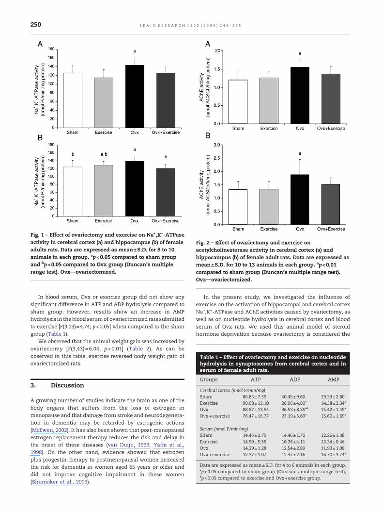
Fig. 2 – Effect of ovariectomy and exercise onacetylcholinesterase activity in cerebral cortex (a) andhippocampus (b) of female adult rats. Data are expressed asmean±S.D. for 10 to 13 animals in each group. ap<0.01compared to sham group (Duncan's multiple range test).Ovx—ovariectomized.
Table 1 – Effect of ovariectomy and exercise on nucleotidehydrolysis in synaptosomes from cerebral cortex and in
Fig. 1 – Effect of ovariectomy and exercise on Na+,K+-ATPaseactivity in cerebral cortex (a) and hippocampus (b) of femaleadults rats. Data are expressed as mean±S.D. for 8 to 10animals in each group. ap<0.05 compared to sham groupand bp<0.05 compared to Ovx group (Duncan's multiplerange test). Ovx—ovariectomized.
250 B R A I N R E S E A R C H 1 3 0 2 ( 2 0 0 9 ) 2 4 8 – 2 5 5
In blood serum, Ovx or exercise group did not show anysignificant difference in ATP and ADP hydrolysis compared tosham group. However, results show an increase in AMPhydrolysis in theblood serumof ovariectomized rats submittedto exercise [F(3,13)=4.74; p<0.05] when compared to the shamgroup (Table 1).
We observed that the animal weight gain was increased byovariectomy [F(3,43)=6.04; p<0.01] (Table 2). As can beobserved in this table, exercise reversed body weight gain ofovariectomized rats.
serum of female adult rats.
Groups ATP ADP AMP
Cerebral cortex (nmol P/min/mg)Sham 86.85±7.55 40.45±9.60 19.39±2.80Exercise 90.68±12.10 26.96±4.80⁎ 14.38±3.34⁎Ovx 88.87±13.54 36.53±8.35⁎# 15.42±1.49⁎Ovx+exercise 76.47±16.77 27.19±5.69⁎ 15.60±1.69⁎
Serum (nmol P/min/mg)Sham 14.45±2.75 14.46±1.70 12.26±1.38Exercise 14.90±5.55 16.36±4.11 13.34±0.46Ovx 14.29±1.28 12.54±2.89 11.93±1.08Ovx+exercise 12.37±1.07 12.47±2.16 16.70±3.74⁎
Data are expressed as mean±S.D. for 4 to 6 animals in each group.⁎p<0.05 compared to sham group (Duncan's multiple range test),#p<0.05 compared to exercise and Ovx+exercise group.
3. Discussion
A growing number of studies indicate the brain as one of thebody organs that suffers from the loss of estrogen inmenopause and that damage from stroke and neurodegenera-tion in dementia may be retarded by estrogenic actions(McEwen, 2002). It has also been shown that post-menopausalestrogen replacement therapy reduces the risk and delay inthe onset of these diseases (van Duijn, 1999; Yaffe et al.,1998). On the other hand, evidence showed that estrogenplus progestin therapy to postmenopausal women increasedthe risk for dementia in women aged 65 years or older anddid not improve cognitive impairment in these women(Shumaker et al., 2003).
In the present study, we investigated the influence ofexercise on the activation of hippocampal and cerebral cortexNa+,K+-ATPase and AChE activities caused by ovariectomy, aswell as on nucleotide hydrolysis in cerebral cortex and bloodserum of Ovx rats. We used this animal model of steroidhormone deprivation because ovariectomy is considered the
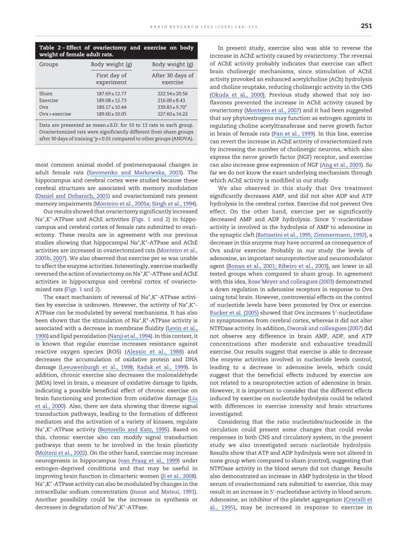
Table 2 – Effect of ovariectomy and exercise on bodyweight of female adult rats.
Groups Body weight (g) Body weight (g)
First day ofexperiment
After 30 days ofexercise
Sham 187.69±12.77 222.54±20.56Exercise 189.08±12.73 216.00±8.43Ovx 189.17±10.44 239.83±9.70⁎Ovx+exercise 189.60±10.05 227.60±14.22
Data are presented as mean±S.D. for 10 to 13 rats in each group.Ovariectomized rats were significantly different from sham groupsafter 30 days of training ⁎p<0.01 compared to other groups (ANOVA).
251B R A I N R E S E A R C H 1 3 0 2 ( 2 0 0 9 ) 2 4 8 – 2 5 5
most common animal model of postmenopausal changes inadult female rats (Savonenko and Markowska, 2003). Thehippocampus and cerebral cortex were studied because thesecerebral structures are associated with memory modulation(Daniel and Dohanich, 2001) and ovariectomized rats presentmemory impairments (Monteiro et al., 2005a; Singh et al., 1994).
Our results showed that ovariectomysignificantly increasedNa+,K+-ATPase and AChE activities (Figs. 1 and 2) in hippo-campus and cerebral cortex of female rats submitted to ovari-ectomy. These results are in agreement with our previousstudies showing that hippocampal Na+,K+-ATPase and AChEactivities are increased in ovariectomized rats (Monteiro et al.,2005b, 2007). We also observed that exercise per se was unableto affect the enzyme activities. Interestingly, exercisemarkedlyreversed theactionof ovariectomyonNa+,K+-ATPase andAChEactivities in hippocampus and cerebral cortex of ovariecto-mized rats (Figs. 1 and 2).
The exact mechanism of reversal of Na+,K+-ATPase activi-ties by exercise is unknown. However, the activity of Na+,K+-ATPase can be modulated by several mechanisms. It has alsobeen shown that the stimulation of Na+,K+-ATPase activity isassociated with a decrease in membrane fluidity (Levin et al.,1990) and lipidperoxidation (Nanji et al., 1994). In this context, itis known that regular exercise increases resistance againstreactive oxygen species (ROS) (Alessio et al., 1988) anddecreases the accumulation of oxidative protein and DNAdamage (Leeuwenburgh et al., 1998; Radak et al., 1999). Inaddition, chronic exercise also decreases the malonaldehyde(MDA) level in brain, a measure of oxidative damage to lipids,indicating a possible beneficial effect of chronic exercise onbrain functioning and protection from oxidative damage (Liuet al., 2000). Also, there are data showing that diverse signaltransduction pathways, leading to the formation of differentmediators and the activation of a variety of kinases, regulateNa+,K+-ATPase activity (Bertorello and Katz, 1995). Based onthis, chronic exercise also can modify signal transductionpathways that seem to be involved in the brain plasticity(Molteni et al., 2002). On the other hand, exercise may increaseneurogenesis in hippocampus (van Praag et al., 1999) underestrogen-deprived conditions and that may be useful inimproving brain function in climacteric women (Ji et al., 2008).Na+,K+-ATPaseactivity canalso bemodulatedby changes in theintracellular sodium concentration (Inoue and Matsui, 1991).Another possibility could be the increase in synthesis ordecreases in degradation of Na+,K+-ATPase.
In present study, exercise also was able to reverse theincrease in AChE activity caused by ovariectomy. The reversalof AChE activity probably indicates that exercise can affectbrain cholinergic mechanisms, since stimulation of AChEactivity provoked an enhanced acetylcholine (ACh) hydrolysisand choline reuptake, reducing cholinergic activity in the CNS(Okuda et al., 2000). Previous study showed that soy iso-flavones prevented the increase in AChE activity caused byovariectomy (Monteiro et al., 2007) and it had been suggestedthat soy phytoestrogens may function as estrogen agonists inregulating choline acetyltransferase and nerve growth factorin brain of female rats (Pan et al., 1999). In this line, exercisecan revert the increase in AChE activity of ovariectomized ratsby increasing the number of cholinergic neurons, which alsoexpress the nerve growth factor (NGF) receptor, and exercisecan also increase gene expression of NGF (Ang et al., 2003). Sofar we do not know the exact underlying mechanism throughwhich AChE activity is modified in our study.
We also observed in this study that Ovx treatmentsignificantly decreases AMP, and did not alter ADP and ATPhydrolysis in the cerebral cortex. Exercise did not prevent Ovxeffect. On the other hand, exercise per se significantlydecreased AMP and ADP hydrolysis. Since 5′-nucleotidaseactivity is involved in the hydrolysis of AMP to adenosine inthe synaptic cleft (Battastini et al., 1995; Zimmermann, 1992), adecrease in this enzymemay have occurred as consequence ofOvx and/or exercise. Probably in our study the levels ofadenosine, an important neuroprotective and neuromodulatoragent (Bonan et al., 2001; Ribeiro et al., 2003), are lower in alltested groups when compared to sham group. In agreementwith this idea, Rose'Meyer and colleagues (2003) demonstrateda down regulation in adenosine receptors in response to Ovxusing total brain. However, controversial effects on the controlof nucleotide levels have been promoted by Ovx or exercise.Rucker et al. (2005) showed that Ovx increases 5′-nucleotidasein synaptosomes from cerebral cortex, whereas it did not alterNTPDase activity. In addition, Dworak and colleagues (2007) didnot observe any difference in brain AMP, ADP, and ATPconcentrations after moderate and exhaustive treadmillexercise. Our results suggest that exercise is able to decreasethe enzyme activities involved in nucleotide levels control,leading to a decrease in adenosine levels, which couldsuggest that the beneficial effects induced by exercise arenot related to a neuroprotective action of adenosine in brain.However, it is important to consider that the different effectsinduced by exercise on nucleotide hydrolysis could be relatedwith differences in exercise intensity and brain structuresinvestigated.
Considering that the ratio nucleotides/nucleoside in thecirculation could present some changes that could evokeresponses in both CNS and circulatory system, in the presentstudy we also investigated serum nucleotide hydrolysis.Results show that ATP and ADP hydrolysis were not altered innone group when compared to sham (control), suggesting thatNTPDase activity in the blood serum did not change. Resultsalso demonstrated an increase in AMP hydrolysis in the bloodserum of ovariectomized rats submitted to exercise, this mayresult in an increase in 5′-nucleotidase activity in blood serum.Adenosine, an inhibitor of the platelet aggregation (Cristalli etal., 1995), may be increased in response to exercise in

252 B R A I N R E S E A R C H 1 3 0 2 ( 2 0 0 9 ) 2 4 8 – 2 5 5
ovariectomized rats, this canbea compensation in situationsofincreased platelet activity, and cardiovascular risk, such aspostmenopausal conditions (Pochmann et al., 2004).
Our previous study showed that ovariectomy significantlydecreases (98%) the estradiol levels in all ovariectomized rats,confirming the efficacy of the surgical procedure of ovari-ectomy (Monteiro et al., 2007). We also observed that theanimalweight gainwas increasedby ovariectomyand exercisereverted weight body gain of ovariectomized rats. Thesefindings are in agreementwith Saengsirisuwan and colleagues(2009), that suggested an increased energy expenditure byexercise training, which could prevent fat accumulation and/or enhanced fat utilization of Ovx rats.
In summary, in the present study we demonstrated thatexercise significantly reverses the action of ovariectomy onNa+,K+-ATPase and AChE activities in hippocampus andcerebral cortex of female adult rats. Ovariectomy decreasedAMP hydrolysis in cerebral cortex and did not alter adeninenucleotidehydrolysis in blood serum. Exercise per sedecreasedthe ADP and AMP hydrolysis and did not alter effects ofovariectomy in cerebral cortex, but increased the AMP hydro-lysis in blood serum of ovariectomized adult rats. Based onthese findings we could suggest that exercise may have aprotective role against the damage brain and increased plateletactivity caused by the loss of estrogen during menopause.Present data support that physical exercise might constitute atherapeutic alternative to hormone replacement therapy forestrogen deprivation.
4. Experimental procedures
4.1. Animals and reagents
Female adult Wistar rats (3 months, 180–210 g BW) wereobtained from the Central Animal House of the Department ofBiochemistry, Instituto de Ciências Básicas da Saúde, Uni-versidade Federal do Rio Grande do Sul, Porto Alegre, RS,Brazil. Animals were maintained on a 12/12 h light/dark cyclein an air-conditioned constant temperature (22±1 °C) colonyroom, with free access to water. Animal care followed theofficial governmental guidelines in compliance with theFederation of Brazilian Societies for Experimental Biologyand was approved by the Ethics Committee of the Federal RioGrande do Sul, Brazil. All chemicals were purchased fromSigma Chemical Co., St. Louis, MO, USA.
Animals were randomly assigned to one of the followinggroups: (n=11-15): sham (only submitted to surgery withoutremoving of ovaries), exercise, ovariectomized (Ovx), andOvx plus exercise. Animals were ovariectomized by thesurgical removal of both ovaries under ketamine anesthesia(90 mg/kg) and xylazine (10 mg/kg) intraperitoneous (i.p.) toeliminate endogenous ovarian steroids (Waynforth andFlecknell, 1992). One month after the surgery, animals weresubmitted to exercise.
4.2. Exercise training
Rats were habituated to the treadmill apparatus to minimizenovelty stressand randomlyassigned todifferentexperimental
groups (n=11-15 in each group): non-exercised (sedentary-control group) andexercisedduring 20min, 3 times aweek. Theexercise training consisted of running sessions on an adaptedmotorized rodent treadmill (INBRAMED TK 01, Porto Alegre,Brazil) at 60% of their maximal oxygen uptake (Brooks andWhite, 1978), a moderate exercise. Measurement of oxygenuptake (VO2) peak was carried out in all animals, indirectlybefore training, considering the exhaustion. Each rat ran on thetreadmill at a low initial speed followedby increases in speedof5m/min every 3minuntil thepoint of exhaustion (i.e., failure ofthe rats to continue running) and the time to fatigue (in min)and workload (in m/min) were taken as indexes of capacity forexercise, that was taken as VO2max. (Arida et al., 1999; Brooksand White, 1978).
Selected animals that initially refused to run were encour-aged by gently tapping their backs. Neither electric shock norphysical prodding was used in this study (Cechetti et al., 2007).The control group was transported to the experimental roomand handled exactly as the experimental animals and weremaintained in the turned off treadmill for 5 min without beingforced to run (Scopel et al., 2006).
The animals were adapted to the treadmill by graduallyincreasing running speed and time, as follows: week 1, at18 m/min for the first 3 min, 24 m/min for the next 3 min,36 m/min for the following 6 min, 24 m/min for the following3min and 18m/min for the last 3 min; week 2, at 18 m/min forthe first 3min, 36m/min for the next 12min, and 18m/min forthe last 3 min; weeks 3 and 4, at 18 m/min for the first 3 min,48 m/min for the next 14 min, and 18 m/min for the last 3 min(Cechetti et al., 2007).
Approximately 12 h after the last exercise session, ratswere euthanized by decapitation without anesthesia and thebrain was immediately isolated, washed with saline solutionand the cerebral cortex and hippocampus were dissected; andthe blood was collected.
4.3. Na+,K+-ATPase activity assay
For determination of Na+,K+-ATPase activity, the hippo-campus and cerebral cortex were homogenized in 10 vol.0.32 mM sucrose solution containing 5.0 mM HEPES and1.0 mM EDTA, pH 7.4.
The reaction mixture for Na+,K+-ATPase activity assaycontained 5.0 mM MgCl2, 80.0 mM NaCl, 20.0 mM KCl and40.0 mM Tris–HCl, pH 7.4, in a final volume of 200 μL. Thereaction was initiated by the addition of ATP. Controls werecarried out under the same conditions with the addition of1.0 mM ouabain. Na+,K+-ATPase activity was calculated by thedifference between the two assays, as previously described(Wyse et al., 2000). Released inorganic phosphate (Pi) was
253B R A I N R E S E A R C H 1 3 0 2 ( 2 0 0 9 ) 2 4 8 – 2 5 5
measured by themethod of (Chan et al., 1986). Specific activityof the enzyme was expressed as nmol Pi released per min permg of protein. All samples were run in duplicate.
4.4. AChE activity assay
For the AChE assay, the hippocampus and cerebral cortexwere homogenized in 10 volumes 0.1 mM potassium phos-phate buffer, pH 7.5, and centrifuged for 10 min at 1000×g. Thesupernatant was used for the enzymatic AChE analyses.
Acetylcholinesterase activity was determined according toEllman et al. (1961), with some modifications (Villescas et al.,1981). Hydrolysis rates were measured at acetylthiocholine (S)concentration of 0.8 mM in 1 mL assay solutions with 30 mMphosphate buffer, pH 7.5, and 1.0 mM 5,5′-dithiobis-(2-nitrobenzoic Acid) (DTNB) at 25 °C. Fifty microliters of rathippocampus and cerebral cortex supernatant was added tothe reaction mixture and preincubated for 3 min. Thehydrolysis was monitored by the formation of the thiolatedianion of DTNB at 412 nm for 2–3 min (intervals of 30 s).Specific enzyme activitywas expressed as μmolASCh per hourper milligram of protein. All samples were run in duplicate.
4.5. Adenine nucleotide hydrolysis
4.5.1. Subcellular fractionationCerebral cortex was removed and placed in ice-cold isolationmedium (320 mM sucrose, 5 mM HEPES, pH 7.5 and 0.1 mMEDTA) and were cut longitudinally. The cerebral cortex wasgently homogenized in 10 volumes, respectively, of ice-coldisolation medium with a motor-driven Teflon-glass homoge-nizer and synaptosomes were isolated as previously described(Nagy and Delgado-Escueta, 1984). Briefly, 0.5 ml of crudemitochondrial fraction wasmixedwith 4.0ml of an 8.5% Percollsolution and layered onto an isoosmotic Percoll/sucrose dis-continuous gradient (10/16%). The synaptosomes that bandedat the 10/16% Percoll interface were collected with wide tipdisposableplastic transfer pipettes. The synaptosomal fractionswerewashed twice at 15,000×g for 20minwith the same ice-coldmedium to remove the contaminating Percoll and the synapto-some pelletwas resuspended to a final protein concentration ofapproximately 0.5mg/ml. Thematerialwasprepared freshdailyand maintained at 0–4 °C throughout preparation.
4.5.2. Isolation of blood serum fractionBlood samples were drawn after decapitation of rats and weresoon centrifuged in plastic tubes at 3000 rpm for 10 min at20 °C. The serumsamples obtainedwere then stored on ice andimmediately used in the experiments (Stefanello et al., 2003).
4.5.3. Measurement of synaptosome ATP, ADP and AMPhydrolysesThe reaction medium used to assay the ATP and ADPhydrolysis was essentially as described previously (Battastiniet al., 1991). The reaction medium contained 5.0 mM KCl,1.5 mM CaCl2, 0.1 mM EDTA, 10 mM glucose, 225 mM sucroseand 45 mM Tris–HCl buffer, pH 8.0, in a final volume of 200 ml.The synaptosome preparation (10–20 μg protein) was added tothe reactionmixture and preincubated for 10min at 37 °C. Thereaction was initiated by the addition of ATP or ADP to a final
concentration of 1.0 mM and the reaction was stopped by theaddition of 200 μl 10% trichloroacetic acid (TCA). The releasedinorganic phosphate (Pi) was measured as previously described(Chan et al., 1986). The reaction medium used to assay the 5′-nucleotidase activity (AMP hydrolysis) contained 10 mM MgCl2,0.1 M Tris–HCl, pH 7.0 and 0.15 M sucrose in a final volume of200 ml (Heymann et al., 1984). The synaptosome preparation(10–20 μg protein) was preincubated for 10 min at 37 °C. Thereaction was initiated by the addition of AMP to a final con-centration of 1.0mMandwas stopped by the addition of 200ml10% TCA; the released inorganic phosphate (Pi) was measuredas previously described (Chan et al., 1986). Controls with theaddition of the enzyme preparation after addition of TCAwereused to correct non-enzymatic hydrolysis of the substrates. Allsamples were run in triplicate. Specific enzyme activity wasexpressed as nmol Pi released per min per mg of protein.
4.5.4. Measurement of blood serum ATP, ADP and AMPhydrolysesATP and ADP hydrolyses were determined using amodificationof the method described by Yegutkin (1997) according toDelwing et al. (2006) The reaction mixture containing 3 mMATP, ADP or AMP as substrate, 112.5 mM Tris–HCl, pH 8.0, wasincubated with approximately 1.0 mg of serum protein at 37 °Cfor 40min in a final volume of 200 μl. The reaction was stoppedby the addition of 200 μl of 10% TCA. The samples were chilledon ice and the amount of inorganic phosphate (Pi) released wasmeasured as described by (Chan et al., 1986). In order to correctnon-enzymatichydrolysis,weperformedcontrolsbyadding theserum after the reaction was stopped with TCA. All sampleswere centrifuged at 5000g for 5 min to eliminate precipitatedproteinandthesupernatantwasused for the colorimetricassay.All samples were assayed in triplicate. Specific enzyme activitywas expressed as nmol Pi released per min per mg of protein.
4.6. Protein determination
Protein was measured by the Comassie Blue method accord-ing to Bradford, using bovine serum albumin as standard(Bradford, 1976).
4.7. Statistical analysis
All assays were performed in duplicate and the mean wasused for statistical analysis. Data were analyzed by one wayANOVA followed by the Duncanmultiple test when F-test wassignificant. All analyses were performed using the StatisticalPackage for the Social Sciences (SPSS) software using a PC-compatible computer. Values of p<0.05 were considered to besignificant.
Acknowledgments
This work was supported in part by grants from ConselhoNacional de Desenvolvimento Científico e Tecnológico (CNPq-Brazil); FINEP Research Grant “Rede Instituto Brasileiro deNeurociência (IBN-Net)-Proc. No 01.06.0842-00”, and “InstitutoNacional de Ciência e Tecnologia (INCT) para Excitotoxicidadee Neuroproteção (INCT/CNPq).”
254 B R A I N R E S E A R C H 1 3 0 2 ( 2 0 0 9 ) 2 4 8 – 2 5 5
R E F E R E N C E S
Agteresch, H.J., Dagnelie, P.C., van den Berg, J.W., Wilson, J.L., 1999.Adenosine triphosphate: established and potential clinicalapplications. Drugs 58, 211–232.
Alessio, H.M., Goldfarb, A.H., Cutler, R.G., 1988. MDA contentincreases in fast and slow-twitch skeletal muscle withintensity of exercise in a rat. Am. J. Physiol. 255, 874–877.
Ang, E.T., Wong, P.T.H., Moochhala, S., Ng, Y.K., 2003.Neuroprotection associated with running: is it a result ofincreased endogenous neurotrophic factors. Neuroscience 118,335–345.
Arida, R.M., Scorza, F.A., Santos, N.F., Peres, C.A., Cavalheiro, E.A.,1999. Effect of physical exercise on seizure occurrence in amodel of temporal lobe epilepsy in rats. Epilepsy Res. 37, 45–52.
Battastini, A.M., da Rocha, J.B., Barcellos, C.K., Dias, R.D., Sarkis, J.J.,1991. Characterization of an ATP diphosphohydrolase(EC 3.6.1.5) in synaptosomes from cerebral cortex of adult rats.Neurochem. Res. 16, 1303–1310.
Battastini, A., Oliveira, E., Moreira, C., Bonan, C., Sarkis, J., Dias, R.,1995. Solubilization and characterization of an ATPdiphosphohydrolase (EC 3.6.1.5.) from rat brain plasmamembranes. Biochem. Mol. Biol. Int. 37, 209–219.
Bertorello, A.M., Katz, A.I., 1995. Regulation of Na+,K+ pumpactivity: pathways between receptors and effectors. NewsPhysiol. Sci. 10, 253–259.
Bonan, C., Schetinger, M., Battastini, A., Sarkis, J., 2001.Ectonucleotidases and synaptic plasticity: implications inphysiological and pathological conditions. Drug Dev. Res. 52,57–65.
Bradford, M.M., 1976. A rapid and sensitive method for thequantification ofmicrograms quantities of protein utilizing theprinciple of protein-die-binding. Anal. Biochem. 72, 248–254.
Brooks, G.A., White, T.P., 1978. Determination of metabolic andheart rate responses of rats to treadmill exercise. J. Appl.Physiol. Res. 45, 1009–1015.
Calder'on, F., Von Bernhardi, R., De Ferrari, G., Luza, S., Aldunate,R., Inestrosa, N., 1998. Toxic effects of acetylcholinesterase onneuronal and glial-like cells in vitro. Mol. Psychiatry 3, 247–255.
Cechetti, F., Rhod, A., Simão, F., Santin, K., Salbego, C., Netto, C.A.,Siqueira, I.R., 2007. Effect of treadmill exercise on cell damagein rat hippocampal slices submitted to oxygen and glucosedeprivation. Brain Res. 1157, 121–125.
Chan, K.M., Delfer, D., Junger, K.D., 1986. A direct colorimetricassay for Ca2+-stimulated ATPase activity. Anal. Biochem. 157,375–380.
Cotman, C.W., Berchtold, N.C., 2002. Exercise: a behavioralintervention to enhance brain health and plasticity. TrendNeurosci. 25, 295–301.
Cristalli, G., Camaioni, E., Vittori, S., Volpini, R., Borea, P.A., Conti,A., Dionisotti, S., Ongini, E., Monopoli, A., 1995. 2-Aralkynyl and2-heteroalkynyl derivatives of adeno-sine-59-N-ethyluronamideas selective A 2A adenosine receptor agonists. J. Med. Chem. 38,1462–1472.
Cummings, J.L., 2000. The role of cholinergic agents in themanagement of behavioral disturbances in Alzheimer'sdisease. Int. J. Neuropsychopharmacol. 3, 21–29.
Daniel, J.M., Dohanich, G.P., 2001. Acetylcholine mediates theestrogen-induced increase in NMDA receptor binding in CA1 ofthe hippocampus and the associated improvement in workingmemory. J. Neurosci. 21, 6949–6956.
Delwing, D., Delwing, D., Sarkis, J.J.F., Wyse, A.T.S., 2006. Prolineinduces alterations in nucleotide hydrolysis in rat blood serum.Mol. Cell. Biochem. 192, 139–144.
Dworak, M., Schierl, T., Bruns, T., Struder, H., 2007. Impact ofsingular excessive computer game and television exposure onsleep patterns and memory performance of school-agedchildren. Pediatrics 120, 978–985.
Ellman, G.L., Courtney, K.D., Andres, V.J., Feather-Stone, R.M.,1961. A new and rapid determination of acetylcholinesteraseactivity. Biochem. Pharmacol. 7, 88–95.
Erecinska, M., Silver, I.A., 1994. Ions and energy in mammalianbrain. Prog. Neurobiol. 43, 37–71.
Gibbs, R.B., Aggarwal, P., 1998. Estrogen and basal forebraincholinergic neurons: implications for brain aging andAlzheimer's disease-related cognitive decline. Horm. Behav.34, 98–111.
Green, P.S., Simpkins, J.W., 2000. Neuroprotective effects ofestrogens: potential mechanisms of action. Int. J. Dev.Neurosci. 18, 347–358.
Gómez-Ramos, P., Morán, M.A., 1997. Ultra structural localizationof butyrylcholinesterase in senile plaques in the brains of agedand Alzheimer's disease patients. Mol. Chem. Neuropathol. 30,161–173.
Hattori, N., Kitagawa, K., Higashida, T., Yagyu, K., Shimohama, S.,Wataya, T., Perry, G., Smith, M.A., Inagaki, C., 1998. Cl−-ATPaseand Na+/K+-ATPase activities in Alzheimer's disease brains.Neurosci. Lett. 254, 141–144.
Heymann, D., Reddington, M., Kreutzberg, G.W., 1984. Subcellularlocalization of 50-nucleotidase in rat brain. J. Neurochem. 43,971–978.
Hollmann, W., Fischer, H., de Meirleir, K., Herzog, H., Herholz, K.,Feinendegen, L.E., 1994. The brain: regional cerebral blood flow,metabolism, and psyche during ergometer exercise. Fitnessand Health: International Proceedings and ConsensusStatement. 490-500.
Ide, K., Horn, A., Secher, N.H., 1999. Cerebral metabolic response tosubmaximal exercise. J. Appl. Physiol. Res. 87, 1604–1608.
Inoue, N., Matsui, H., 1991. Changes in responses of Na pumpisoforms to glutamate excitation of cerebral neurons duringmaturation in culture. Soc. Gen. Physiol. Ser. 46, 597–600.
Ji, J., Jing, H., Choi, G., Oh, M.S., Ryu, J.H., Jeong, J.W., Huh, Y., Park,C., 2008. Voluntary exercise increases the new cell formation inthe hippocampus of ovariectomized mice. Neurosci. Lett. 439,260–263.
Kramer, A.F., Hahn, S., Cohen, N.J., Banich, M.T., McAuley, E.,Harrison, C.R., Chason, J., Vakil, E., Bardell, L., Boileau, R.A.,Colcombe, A., 1999. Ageing, fitness and neurocognitivefunction. Nature 400, 418–419.
Leeuwenburgh, C., Hansen, P., Shaish, A., Holoszy, J.O., Heinrcke,J.W., 1998. Markers of protein oxidation by hydroxyl radical andreactive nitrogen species in tissues of aging rats. Am. J. Physiol.274, 453–461.
Levin, G., Cogan, U., Levy, Y., Mokady, S., 1990. Riboflavindeficiency and the function and fluidity of rat erythrocytemembranes. J. Nutr. 120, 857–861.
Liu, J., Yeo, H.C., Vervik-Douki, E.O., Hagen, T., Doniger, S.J., Chu,D.W., Brooks, G.A., Ames, B.N., 2000. Chronically and acutelyexercised rats: biomarkers of oxidative stress and endogenousantioxidants. J. Appl. Physiol. 89, 21–28.
Mattson, M.P., 2000. Neuroprotective signaling and the agingbrain: take away my food and let me run. Brain Res. 886,47–53.
McEwen, B., 2002. Estrogen actions throughout the brain. RecentProg. Horm. Res. 57, 357–384.
Miquel, J., Ramírez-Boscá, A., Ramírez-Boscá, J.V., Alperi, J.D., 2006.Menopause: a review on the role of oxygen stress and favorableeffects of dietary antioxidants. Arch. Gerontol. Geriatr. 42,289–306.
Molteni, R., Ying, Z., Gómez-Pinilla, F., 2002. Differential effects ofacute and chronic exercise on plasticity-related genes in the rathippocampus revealed by microarray. Eur. J. Neurosci. 16,1107–1116.
Monteiro, S.C., Matté, C., Bavaresco, C.S., Netto, C.A., Wyse, A.T.S.,2005a. Vitamins E and C pretreatment preventsovariectomy-induced memory deficits in water maze.Neurobiol. Learn. Mem. 84, 192–199.
255B R A I N R E S E A R C H 1 3 0 2 ( 2 0 0 9 ) 2 4 8 – 2 5 5
Monteiro, S.C., Matté, C., Delwing, D., Wyse, A.T.S., 2005b.Ovariectomy increases Na+,K+-ATPase, acetylcholinesteraseand catalase in rat hippocampus. Mol. Cell. Endocrinol. 236,9–16.
Monteiro, S.C., Mattos, C.B., Scherer, E.B.S., Wyse, A.T.S., 2007.Supplementation with vitamins E plus C or soy isoflavones inovariectomized rats: effect on the activities of Na+,K+-ATPaseand cholinesterases. Metab. Brain Dis. 22, 156–171.
Nagy, A., Delgado-Escueta, A.V., 1984. Rapid preparation ofsynaptosomes from mammalian brain using nontoxicisoosmotic gradient (Percoll). J. Neurochem. 43, 1114–1123.
Nanji, A.A., Sadrzadeh, S.M., Dannenberg, A.J., 1994.Livermicrosomal fatty acid composition in ethanol-fed rats:effect of different dietary fats and relationship to liver injury.Alcohol., Clin. Exp. Res. 18, 1024–1028.
Nedeljkovic, N., Djordevic, V., Horvat, A., Nikezic, G., Kanazir, D.T.,2000. Effect of steroid hormone deprivation on the expressionof ecto-ATPase in distinct brain regions of female rats. Physiol.Res. 49, 419–426.
Okuda, T., Haga, T., Kanai, Y., Endou, H., Ishihara, T., Katsura, I.,2000. Identification and characterization of the high-affinitycholine transporter. Nat. Neurosci. 3, 120–125.
Pan, Y., Anthony, M., Clarkson, T.B., 1999. Effect of estradioland soy phytoestrogens on choline acetyltransferase andnerve growth factor mRNAs in the frontal cortex andhippocampus of female rats. Proc. Soc. Exp. Biol. Med. 221,118–125.
Pochmann, D., Rücker, B., Battastini, A.M.O., Sarkis, J.J.F., 2004.Ovariectomy and estradiol replacement therapy alters theadenine nucleotide hydrolysis in rat blood serum. Thromb.Res. 114, 275–281.
Radak, Z., Kaneko, T., Tahara, T., Nakamoto, H., Ohno, H., Sasvari,M., Nyakas, C., Goto, S., 1999. The effect of exercise training onoxidative damage of lipids, proteins and DNA in rats skeletalmuscle evidence for beneficial outcomes. Free Radic. Biol. Med.27, 69–74.
Ralevic, V., Burnstock, G., 2003. Involvement of purinergicsignaling in cardiovascular diseases. Drug News Perspect. 16,133–140.
Ribeiro, J.A., Sebastião, A.M., de Mendonça, A., 2003. Adenosinereceptors in the nervous system: pathophysiologicalimplications. Prog. Neurobiol. 68, 377–392.
Rose 'Meyer, R.B., Mellick, A.S., Garnham, B.G., Harrison, G.J.,Massa, H.M., Griffiths, L.R., 2003. The measurement ofadenosine and estrogen receptor expression in rat brainsfollowing ovariectomy using quantitative PCR analysis. BrainRes. Brain Res. Protoc. 11, 9–18.
Rucker, B., Pochmann, D., Furstenau, C., Carneiro-Ramos, M.,Battastini, A., Barreto-Chaves, M., Sarkis, J., 2005. Effects ofsteroid hormones on synaptosomal ectonucleotidase activitiesfrom hippocampus and cortex of adult female rats. Gen. Comp.Endocrinol. 140, 94–100.
Saengsirisuwan, V., Pongseeda, S., Prasannarong, M., Vichaiwong,K., Toskulkao, C., 2009. Modulation of insulin resistance inovariectomized rats by endurance exercise training andestrogen replacement. Metab. Clin. Exp. 58, 38–47.
Sarkis, J.J.F., Battastini, A.M.O., Oliveira, E.M., Frasseto, S.S., Dias,R.D., 1995. ATP diphosphohydrolases: an overview. J. Br. Assoc.Adv. Sci. 47, 131–136.
Savonenko, A.V., Markowska, A.L., 2003. The cognitive effects ofovariectomy and estrogen replacement are modulated byaging. Neurosci. Lett. 119, 821–830.
Scopel, D., Fochesatto, C., Cimarosti, H., Rabbo, M., Belló-Klein, A.,Salbego, C., Netto, C.A., Siqueira, I.R., 2006. Exercise intensityinfluences cell injury in rat hippocampal slices exposed tooxygen and glucose deprivation. Brain Res. Bull. 71, 155–159.
Shumaker, S.A., Legault, C., Raap, S.R., Thal, L., Wallace, R.B.,Ockene, J.K., Hendrix, S.L., Jones, B.N., Assaf, A.R., Jackson, R.D.,Kotchen, J.M., Wassertheil-Smoller, S., Wactawski-Wende, J.,Investigators, W., 2003. Estrogen plus progestin and theincidence of dementia and mild cognitive impairment inpostmenopausal women: the women's health initiativememory study: a randomized controlled trial. JAMA 289,2651–2662.
Simpkins, J.W., Rajakumar, G., Zhang, Y.Q., Simpkins, C.E.,Greenwald, D., Yu, C.J., Bodor, N., Day, A.L., 1997. Estrogensmay reduce mortality and ischemic damage caused by middlecerebral artery occlusion in the female rats. J. Neurosurg. 87,724–730.
Singh, M., Meyer, E.M., Millard, W.J., Simpkins, J.W., 1994. Ovariansteroid deprivation results in a reversible learning impairmentand compromised cholinergic function in femaleSprague-Dawley rats. Brain Res. 644, 305–312.
Stefanello, F.M., Zugno, A.I.,Wannmacher, C.M.,Wajner, M.,Wyse,A.T.S., 2003. Homocysteine inhibits butyrylcholinesteraseactivity in rat serum. Metab. Brain Dis. 18, 187–194.
van Duijn, C.M., 1999. Hormone replacement therapy andAlzheimer's disease. Maturitas 31, 201–205.
van Praag, H., Kempermann, G., Gage, F.H., 1999. Runningincreases cell proliferation and neurogenesis in the adultmouse dentate gyrus. Nat. Neurosci. 2, 266–270.
van Praag, H., Shubert, T., Zhao, C., Gage, F.H., 2005. Exerciseenhances learning and hippocampal neurogenesis in agedmice. J. Neurosci. 25, 8680–8685.
Villescas, R., Ostwald, R., Morimoto, H.D., Bennett, E.L., 1981.Effects of neonatal undernutrition and cold stress on behaviorand biochemical brain parameters in rats. J. Nutr. 111,1103–1110.
Waynforth, H.B., Flecknell, P.A., 1992. Experimental and surgicaltechnique, The rat, 2nd edn. Academic, London, pp. 276–278.
Wise, P.M., 2002. Estrogens and neuroprotection.Trends Endocrinol. Metab. 13, 229–230.
Wise, M.P., Dubal, D.B., Wilson, M.E., Rau, S.W., Böttner, M.,Rosewell, K.L., 2001a. Estradiol is a protective factor in the adultand aging brain: understanding of mechanism derived from invivo and in vitro studies. Brain Res. Brain Res. Rev. 37, 313–319.
Wise, P., Dubal, D., Wilson, M., Rau, S., Liu, Y., 2001b. Estrogens:trophic and protective factors in the adult brain.Front. Neuroendocrinol. 22, 33–66.
Wyse, A.T.S., Streck, E.L., Worm, P., Wajner, A., Ritter, F., Netto,C.A., 2000. Preconditioning prevents the inhibition ofNa+,K+-ATPase activity after brain ischemia. Neurochem. Res.25, 969–973.
Yaffe, K., Sawaya, G., Lieberburg, I., Grady, D., 1998. Estrogentherapy in postmenopausal women: effects on cognitivefunction and dementia. JAMA 279, 688–695.
Yegutkin, G.G., 1997. Kinectic analysis of enzymatic hydrolysis ofATP in human and rat blood serum. Biochemistry (Moscow) 62,724–728.
Zhang, Y.Q., Shi, J., Rajakumar, G., Day, A.L., Simpkins, J.W., 1998.Effects of gender and estradiol treatment on focal brainischemia. Brain Res. 784, 321–324.
Zimmermann, H., 1992. 5′ nucleotidase: molecular structure andfunctional aspects. Biochem. J. 285, 345–365.


![Synthesis, metabolic stability and antiviral evaluation of various alkoxyalkyl esters of cidofovir and 9-( S)-[3-hydroxy-2-(phosphonomethoxy)propyl]adenine](https://static.fdokumen.com/doc/165x107/63122794c3611ef94d0cf37a/synthesis-metabolic-stability-and-antiviral-evaluation-of-various-alkoxyalkyl-esters.jpg)