Applying vibrational spectroscopy to the study of nucleobases – adenine as a case-study
Upload
independentCategory
view
2download
0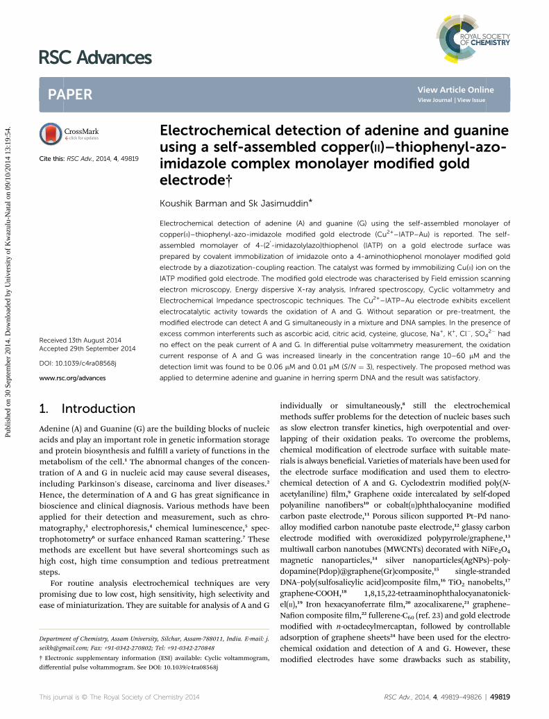
RSC Advances
PAPER
Publ
ishe
d on
30
Sept
embe
r 20
14. D
ownl
oade
d by
Uni
vers
ity o
f K
waz
ulu-
Nat
al o
n 09
/10/
2014
13:
19:5
4.
View Article OnlineView Journal | View Issue
Electrochemical
Department of Chemistry, Assam University
[email protected]; Fax: +91-0342-270802; T
† Electronic supplementary informationdifferential pulse voltammogram. See DO
Cite this: RSC Adv., 2014, 4, 49819
Received 13th August 2014Accepted 29th September 2014
DOI: 10.1039/c4ra08568j
www.rsc.org/advances
This journal is © The Royal Society of C
detection of adenine and guanineusing a self-assembled copper(II)–thiophenyl-azo-imidazole complex monolayer modified goldelectrode†
Koushik Barman and Sk Jasimuddin*
Electrochemical detection of adenine (A) and guanine (G) using the self-assembled monolayer of
copper(II)–thiophenyl-azo-imidazole modified gold electrode (Cu2+–IATP–Au) is reported. The self-
assembled momolayer of 4-(20-imidazolylazo)thiophenol (IATP) on a gold electrode surface was
prepared by covalent immobilization of imidazole onto a 4-aminothiophenol monolayer modified gold
electrode by a diazotization-coupling reaction. The catalyst was formed by immobilizing Cu(II) ion on the
IATP modified gold electrode. The modified gold electrode was characterised by Field emission scanning
electron microscopy, Energy dispersive X-ray analysis, Infrared spectroscopy, Cyclic voltammetry and
Electrochemical Impedance spectroscopic techniques. The Cu2+–IATP–Au electrode exhibits excellent
electrocatalytic activity towards the oxidation of A and G. Without separation or pre-treatment, the
modified electrode can detect A and G simultaneously in a mixture and DNA samples. In the presence of
excess common interferents such as ascorbic acid, citric acid, cysteine, glucose, Na+, K+, Cl�, SO42� had
no effect on the peak current of A and G. In differential pulse voltammetry measurement, the oxidation
current response of A and G was increased linearly in the concentration range 10–60 mM and the
detection limit was found to be 0.06 mM and 0.01 mM (S/N ¼ 3), respectively. The proposed method was
applied to determine adenine and guanine in herring sperm DNA and the result was satisfactory.
1. Introduction
Adenine (A) and Guanine (G) are the building blocks of nucleicacids and play an important role in genetic information storageand protein biosynthesis and fulll a variety of functions in themetabolism of the cell.1 The abnormal changes of the concen-tration of A and G in nucleic acid may cause several diseases,including Parkinson's disease, carcinoma and liver diseases.2
Hence, the determination of A and G has great signicance inbioscience and clinical diagnosis. Various methods have beenapplied for their detection and measurement, such as chro-matography,3 electrophoresis,4 chemical luminescence,5 spec-trophotometry6 or surface enhanced Raman scattering.7 Thesemethods are excellent but have several shortcomings such ashigh cost, high time consumption and tedious pretreatmentsteps.
For routine analysis electrochemical techniques are verypromising due to low cost, high sensitivity, high selectivity andease of miniaturization. They are suitable for analysis of A and G
, Silchar, Assam-788011, India. E-mail: j.
el: +91-0342-270848
(ESI) available: Cyclic voltammogram,I: 10.1039/c4ra08568j
hemistry 2014
individually or simultaneously,8 still the electrochemicalmethods suffer problems for the detection of nucleic bases suchas slow electron transfer kinetics, high overpotential and over-lapping of their oxidation peaks. To overcome the problems,chemical modication of electrode surface with suitable mate-rials is always benecial. Varieties ofmaterials have been used forthe electrode surface modication and used them to electro-chemical detection of A and G. Cyclodextrin modied poly(N-acetylaniline) lm,9 Graphene oxide intercalated by self-dopedpolyaniline nanobers10 or cobalt(II)phthalocyanine modiedcarbon paste electrode,11 Porous silicon supported Pt–Pd nano-alloy modied carbon nanotube paste electrode,12 glassy carbonelectrode modied with overoxidized polypyrrole/graphene,13
multiwall carbon nanotubes (MWCNTs) decorated with NiFe2O4
magnetic nanoparticles,14 silver nanoparticles(AgNPs)–poly-dopamine(Pdop)@graphene(Gr)composite,15 single-strandedDNA–poly(sulfosalicylic acid)composite lm,16 TiO2 nanobelts,17
graphene-COOH,18 1,8,15,22-tetraaminophthalocyanatonick-el(II),19 Iron hexacyanoferrate lm,20 azocalixarene,21 graphene–Naon composite lm,22 fullerene-C60 (ref. 23) and gold electrodemodied with n-octadecylmercaptan, followed by controllableadsorption of graphene sheets24 have been used for the electro-chemical oxidation and detection of A and G. However, thesemodied electrodes have some drawbacks such as stability,
RSC Adv., 2014, 4, 49819–49826 | 49819
RSC Advances Paper
Publ
ishe
d on
30
Sept
embe
r 20
14. D
ownl
oade
d by
Uni
vers
ity o
f K
waz
ulu-
Nat
al o
n 09
/10/
2014
13:
19:5
4.
View Article Online
reproducibility and use of costly chemicals for electrode modi-cation. In this context, the development of novel, chip and simplestrategy for electrode modication is highly desirable. Diazo-nium salt has been extensively used for the electrode surfacemodication.25 Diazonium group functionalized electrodesurface can coupled with phenolic, imidazole or amino groups toform different diazotized compounds26 which are capable offorming metal complexes. There are many reports that metal-azocomplexes can interact with the DNA bases and is important inthe eld of anticancer drug research.27 A and G formed complexeswith metals including copper, has been extensively studied byelectrochemical methods.28 A number of articles have beendevoted to the catalytic and electrocatalytic activity of copper(II)modied electrode for the redox reaction of organic and biolog-ical compounds. For example, electrocatalytic oxidation ofhydroquinone with copper(II)-L-cysteine29 and copper(II)-5-amino-2-mercaptobenzimidazole30 complex monolayer modied goldelectrode, catalytic oxidation of L-cysteine in oxygen-saturatedaqueous solution by copper(II) supported on a polymer,31 deter-mination of cysteine at a glassy carbon electrode modied bycopper(II) ions,32 determination of ascorbic acid using dinuclearcopper salicylaldehyde-glycine schiff base modied GC elec-trode33 and copper(II)–zeolite modied electrode,34 electro-catalytic oxidation of carbohydrates at copper(II) oxide modiedelectrode35 and certain catecholamines such as dopamine,L-dopa, epinephrine and norepinephrine using copper(II)complex and AgNPs modied glassy carbon paste electrode.36
In the present study, we have used a self assembled copper(II)containing azoimidazole complex modied gold electrode forthe detection of purine bases, A and G, individually andsimultaneously in the physiological pH. Electrode modicationprocess and the electrochemical oxidation behaviour of A and Gover themodied electrode were studied in detail. Themodiedelectrode has also been applied for the detection of DNA basesin real sample.
2. Experimental2.1. Chemicals and reagents
4-Aminothiophenol (4-ATP) and NaClO4 were purchased fromSigma-Aldrich (India). Imidazole, CuSO4$5H2O, andK4[Fe(CN)6]$3H2O were purchased fromMerck (India) and wereused as received. Denatured herring sperm DNA, A, G werepurchased from Sigma-Aldrich (India). Phosphate buffer solu-tion was prepared by mixing 0.1 M NaClO4 and 0.01 M H3PO4
and the pH's were adjusted by the addition of 0.11 M NaOHusing Smalley's method.37 Water was puried by double distil-lation with alkaline KMnO4.
Fig. 1 Schematic representation for the fabrication of gold electrodeand the electrochemical oxidation mechanism of adenine andguanine.
2.2. Apparatus and instrumentations
The eld emission scanning electron microscopy (FE SEM)images and Energy dispersive X-ray (EDAX) analysis data wereobtained using FE-SEM, FEI INSPECT F50 operated at anacceleration voltage of 20 kV. FTIR spectra were recorded on aShimadzu 8400S spectrometer. Electrochemical measure-ments were performed on a CHI 660C Electrochemical
49820 | RSC Adv., 2014, 4, 49819–49826
workstation (CH Instrument, USA). A three electrode systemwas employed with gold or modied gold electrode asworking electrode, Pt wire as counter electrode and Ag/AgCl(3 M KCl) as reference electrode. The pH measurements ofsolutions were carried out on a pH meter (Macro Scienticworks (Regd), New Delhi).
2.3. Electrode pre-treatment and immobilization of 4-(20-
imidazolylazo)thiophenol over gold electrode
A gold electrode (2 mm in diameter) was polished with 0.05 mma-alumina on a polishing pad and rinsed extensively withanhydrous ethanol and distilled water. Then the gold electrodewas electrochemically cleaned in 0.5 M H2SO4 until a steadycharacteristic gold oxide cyclic voltammogram was obtained.38
The cleaned gold electrode was immersed into the 4-ATP (1mM)solution for 20 hours. The self-assembled 4-ATP monolayer wasformed over electrode surface via gold–sulfur interaction andthemodied electrode was thoroughly washed with ethanol anddistilled water. Aer that the 4-ATP–Au electrode was dippedinto a 0.1 M HCl solution at 2–4 �C, and 0.1 g NaNO2 solutionwas added slowly. Aer 30 minute incubation, the diazotizedmodied gold electrode (diazo-ATP–Au) was removed andrinsed with ice cold distilled water. For coupling with imidazole,the diazo-ATP–Au electrode was immersed into aqueous 0.025M imidazole solution for 30 minutes at 2–4 �C in stirringcondition. Finally, the 4-(2
0-imidazolylazo)thiophenol modied
gold electrode (IATP–Au) was rinsed with distilled water anddried in air (Fig. 1).
2.4. Copper complexation on IATP–Au electrode
The IATP–Au electrode was immersed into 1 � 10�3 M CuSO4
solution (containing 0.1 M KNO3) at pH 5.6 and stirred for 4hours. Aer complexation, the electrode (Cu2+–IATP–Au) waswashed thoroughly with distilled water and then dried in air forfurther experiment.
This journal is © The Royal Society of Chemistry 2014
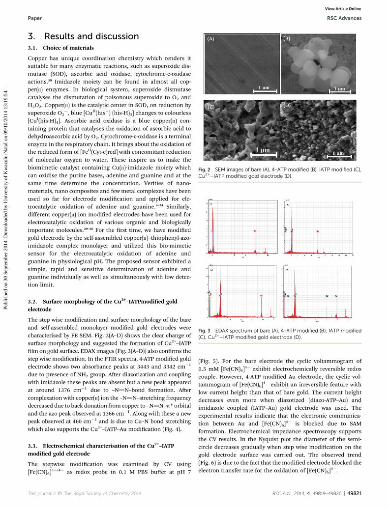
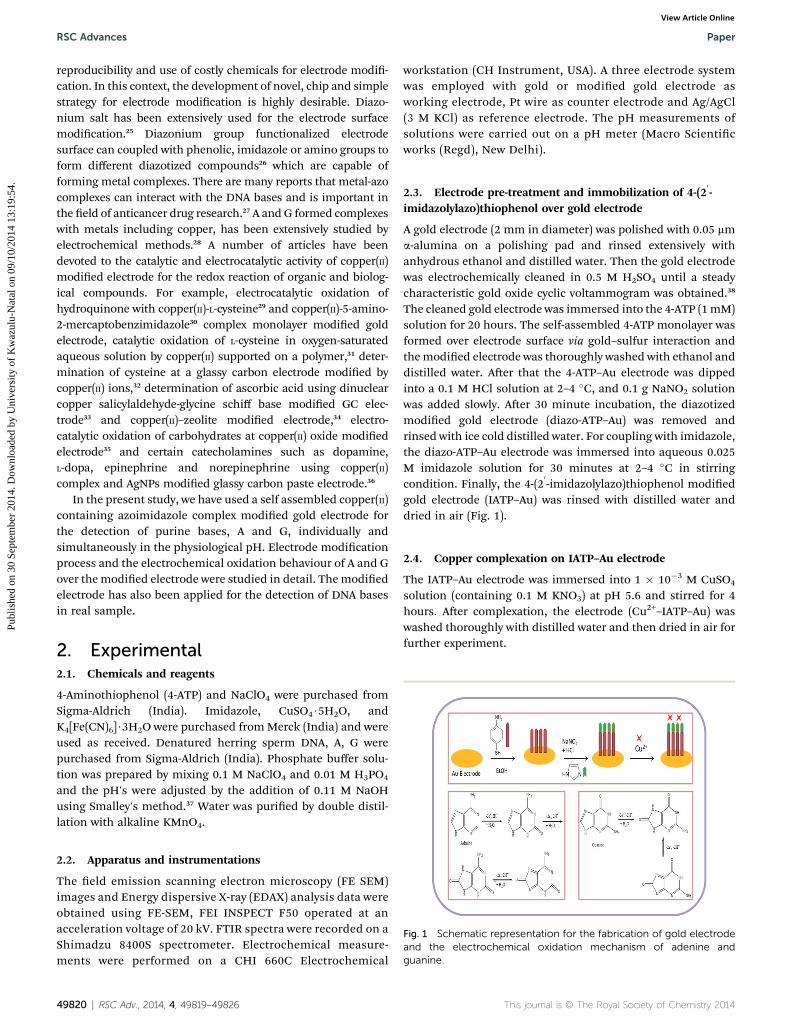
Fig. 2 SEM images of bare (A), 4-ATP modified (B), IATP modified (C),Cu2+–IATP modified gold electrode (D).
Fig. 3 EDAX spectrum of bare (A), 4-ATP modified (B), IATP modified(C), Cu2+–IATP modified gold electrode (D).
Paper RSC Advances
Publ
ishe
d on
30
Sept
embe
r 20
14. D
ownl
oade
d by
Uni
vers
ity o
f K
waz
ulu-
Nat
al o
n 09
/10/
2014
13:
19:5
4.
View Article Online
3. Results and discussion3.1. Choice of materials
Copper has unique coordination chemistry which renders itsuitable for many enzymatic reactions, such as superoxide dis-mutase (SOD), ascorbic acid oxidase, cytochrome-c-oxidaseactions.39 Imidazole moiety can be found in almost all cop-per(II) enzymes. In biological system, superoxide dismutasecatalyses the dismutation of poisonous superoxide to O2 andH2O2. Copper(II) is the catalytic center in SOD, on reduction bysuperoxide O2
�, blue [CuII(his�) (his-H)3] changes to colourless[CuI(his-H)4]. Ascorbic acid oxidase is a blue copper(II) con-taining protein that catalyses the oxidation of ascorbic acid todehydroascorbic acid by O2. Cytochrome-c-oxidase is a terminalenzyme in the respiratory chain. It brings about the oxidation ofthe reduced form of [FeII(Cyt-c)red] with concomitant reductionof molecular oxygen to water. These inspire us to make thebiomimetic catalyst containing Cu(II)-imidazole moiety whichcan oxidise the purine bases, adenine and guanine and at thesame time determine the concentration. Verities of nano-materials, nano composites and fewmetal complexes have beenused so far for electrode modication and applied for elc-trocatalytic oxidation of adenine and guanine.9–24 Similarly,different copper(II) ion modied electrodes have been used forelectrocatalytic oxidation of various organic and biologicallyimportant molecules.29–36 For the rst time, we have modiedgold electrode by the self-assembled copper(II)–thiophenyl-azo-imidazole complex monolayer and utilized this bio-mimeticsensor for the electrocatalytic oxidation of adenine andguanine in physiological pH. The proposed sensor exhibited asimple, rapid and sensitive determination of adenine andguanine individually as well as simultaneously with low detec-tion limit.
3.2. Surface morphology of the Cu2+–IATPmodied goldelectrode
The step wise modication and surface morphology of the bareand self-assembled monolayer modied gold electrodes werecharacterised by FE SEM. Fig. 2(A–D) shows the clear change ofsurface morphology and suggested the formation of Cu2+–IATPlm on gold surface. EDAX images (Fig. 3(A–D)) also conrms thestep wise modication. In the FTIR spectra, 4-ATP modied goldelectrode shows two absorbance peaks at 3443 and 3342 cm�1
due to presence of NH2 group. Aer diazotization and couplingwith imidazole these peaks are absent but a new peak appearedat around 1376 cm�1 due to –N]N–bond formation. Aercomplexation with copper(II) ion the –N]N–stretching frequencydecreased due to back donation from copper to –N]N–p* orbitaland the azo peak observed at 1366 cm�1. Along with these a newpeak observed at 460 cm�1 and is due to Cu–N bond stretchingwhich also supports the Cu2+–IATP–Au modication (Fig. 4).
3.3. Electrochemical characterisation of the Cu2+–IATPmodied gold electrode
The stepwise modication was examined by CV using[Fe(CN)6]
3�/4� as redox probe in 0.1 M PBS buffer at pH 7
This journal is © The Royal Society of Chemistry 2014
(Fig. 5). For the bare electrode the cyclic voltammogram of0.5 mM [Fe(CN)6]
4� exhibit electrochemically reversible redoxcouple. However, 4-ATP modied Au electrode, the cyclic vol-tammogram of [Fe(CN)6]
4� exhibit an irreversible feature withlow current height than that of bare gold. The current heightdecreases even more when diazotized (diazo-ATP–Au) andimidazole coupled (IATP–Au) gold electrode was used. Theexperimental results indicate that the electronic communica-tion between Au and [Fe(CN)6]
4� is blocked due to SAMformation. Electrochemical impedance spectroscopy supportsthe CV results. In the Nyquist plot the diameter of the semi-circle decreases gradually when step wise modication on thegold electrode surface was carried out. The observed trend(Fig. 6) is due to the fact that the modied electrode blocked theelectron transfer rate for the oxidation of [Fe(CN)6]
4�.
RSC Adv., 2014, 4, 49819–49826 | 49821
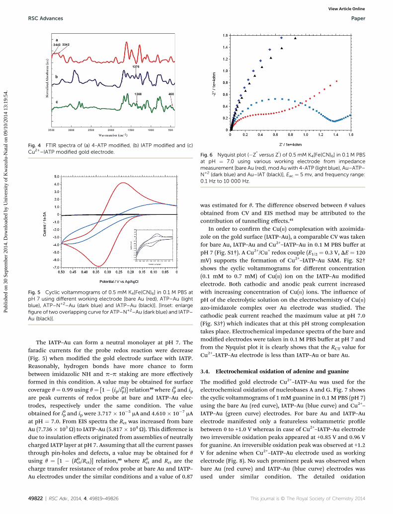
Fig. 4 FTIR spectra of (a) 4-ATP modified, (b) IATP modified and (c)Cu2+–IATP modified gold electrode.
Fig. 5 Cyclic voltammograms of 0.5 mM K4[Fe(CN)6] in 0.1 M PBS atpH 7 using different working electrode [bare Au (red), ATP–Au (lightblue), ATP–N+2–Au (dark blue) and IATP–Au (black)]. [Inset: enlargefigure of two overlapping curve for ATP–N+2–Au (dark blue) and IATP–Au (black)].
Fig. 6 Nyquist plot (�Z00versus Z
0) of 0.5 mM K4[Fe(CN)6] in 0.1 M PBS
at pH ¼ 7.0 using various working electrode from impedancemeasurement [bare Au (red), mod Au with 4-ATP (light blue), Au–ATP–N+2 (dark blue) and Au–IAT (black)], Eac ¼ 5 mv, and frequency range:0.1 Hz to 10 000 Hz.
RSC Advances Paper
Publ
ishe
d on
30
Sept
embe
r 20
14. D
ownl
oade
d by
Uni
vers
ity o
f K
waz
ulu-
Nat
al o
n 09
/10/
2014
13:
19:5
4.
View Article Online
The IATP–Au can form a neutral monolayer at pH 7. Thefaradic currents for the probe redox reaction were decrease(Fig. 5) when modied the gold electrode surface with IATP.Reasonably, hydrogen bonds have more chance to formbetween imidazolic NH and p–p staking are more effectivelyformed in this condition. A value may be obtained for surfacecoverage q¼ 0.99 using q¼ [1� (ip/i
0p)] relation40 where i0p and ip
are peak currents of redox probe at bare and IATP–Au elec-trodes, respectively under the same condition. The valueobtained for i0p and ip were 3.717� 10�5 mA and 4.610 � 10�7 mAat pH ¼ 7.0. From EIS spectra the Rct was increased from bareAu (7.736� 103 U) to IATP–Au (5.817� 104 U). This difference isdue to insulation effects originated from assemblies of neutrallycharged IATP layer at pH 7. Assuming that all the current passesthrough pin-holes and defects, a value may be obtained for q
using q ¼ [1 � (R0ct/Rct)] relation,40 where R0ct and Rct are the
charge transfer resistance of redox probe at bare Au and IATP–Au electrodes under the similar conditions and a value of 0.87
49822 | RSC Adv., 2014, 4, 49819–49826
was estimated for q. The difference observed between q valuesobtained from CV and EIS method may be attributed to thecontribution of tunnelling effects.41
In order to conrm the Cu(II) complexation with azoimida-zole on the gold surface (IATP–Au), a comparable CV was takenfor bare Au, IATP–Au and Cu2+–IATP–Au in 0.1 M PBS buffer atpH 7 (Fig. S1†). A Cu2+/Cu+ redox couple (E1/2 ¼ 0.3 V, DE ¼ 120mV) supports the formation of Cu2+–IATP–Au SAM. Fig. S2†shows the cyclic voltammograms for different concentration(0.1 mM to 0.7 mM) of Cu(II) ion on the IATP–Au modiedelectrode. Both cathodic and anodic peak current increasedwith increasing concentration of Cu(II) ions. The inuence ofpH of the electrolytic solution on the electrochemistry of Cu(II)azo-imidazole complex over Au electrode was studied. Thecathodic peak current reached the maximum value at pH 7.0(Fig. S3†) which indicates that at this pH strong complexationtakes place. Electrochemical impedance spectra of the bare andmodied electrodes were taken in 0.1 M PBS buffer at pH 7 andfrom the Nyquist plot it is clearly shows that the RCT value forCu2+–IATP–Au electrode is less than IATP–Au or bare Au.
3.4. Electrochemical oxidation of adenine and guanine
The modied gold electrode Cu2+–IATP–Au was used for theelectrochemical oxidation of nucleobases A and G. Fig. 7 showsthe cyclic voltammograms of 1 mM guanine in 0.1 M PBS (pH 7)using the bare Au (red curve), IATP–Au (blue curve) and Cu2+–IATP–Au (green curve) electrodes. For bare Au and IATP–Auelectrode manifested only a featureless voltammetric prolebetween 0 to +1.0 V whereas in case of Cu2+–IATP–Au electrodetwo irreversible oxidation peaks appeared at +0.85 V and 0.96 Vfor guanine. An irreversible oxidation peak was observed at +1.2V for adenine when Cu2+–IATP–Au electrode used as workingelectrode (Fig. 8). No such prominent peak was observed whenbare Au (red curve) and IATP–Au (blue curve) electrodes wasused under similar condition. The detailed oxidation
This journal is © The Royal Society of Chemistry 2014
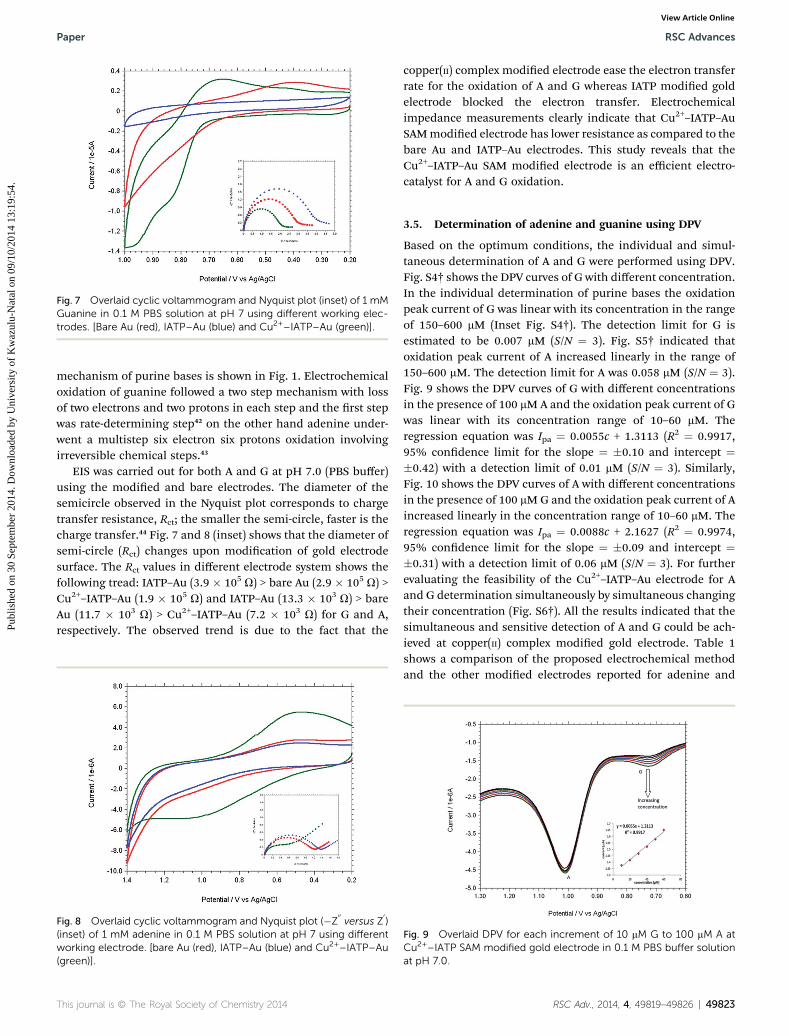
Fig. 7 Overlaid cyclic voltammogram and Nyquist plot (inset) of 1 mMGuanine in 0.1 M PBS solution at pH 7 using different working elec-trodes. [Bare Au (red), IATP–Au (blue) and Cu2+–IATP–Au (green)].
Paper RSC Advances
Publ
ishe
d on
30
Sept
embe
r 20
14. D
ownl
oade
d by
Uni
vers
ity o
f K
waz
ulu-
Nat
al o
n 09
/10/
2014
13:
19:5
4.
View Article Online
mechanism of purine bases is shown in Fig. 1. Electrochemicaloxidation of guanine followed a two step mechanism with lossof two electrons and two protons in each step and the rst stepwas rate-determining step42 on the other hand adenine under-went a multistep six electron six protons oxidation involvingirreversible chemical steps.43
EIS was carried out for both A and G at pH 7.0 (PBS buffer)using the modied and bare electrodes. The diameter of thesemicircle observed in the Nyquist plot corresponds to chargetransfer resistance, Rct; the smaller the semi-circle, faster is thecharge transfer.44 Fig. 7 and 8 (inset) shows that the diameter ofsemi-circle (Rct) changes upon modication of gold electrodesurface. The Rct values in different electrode system shows thefollowing tread: IATP–Au (3.9 � 105 U) > bare Au (2.9 � 105 U) >Cu2+–IATP–Au (1.9 � 105 U) and IATP–Au (13.3 � 103 U) > bareAu (11.7 � 103 U) > Cu2+–IATP–Au (7.2 � 103 U) for G and A,respectively. The observed trend is due to the fact that the
Fig. 8 Overlaid cyclic voltammogram and Nyquist plot (�Z00versus Z
0)
(inset) of 1 mM adenine in 0.1 M PBS solution at pH 7 using differentworking electrode. [bare Au (red), IATP–Au (blue) and Cu2+–IATP–Au(green)].
This journal is © The Royal Society of Chemistry 2014
copper(II) complex modied electrode ease the electron transferrate for the oxidation of A and G whereas IATP modied goldelectrode blocked the electron transfer. Electrochemicalimpedance measurements clearly indicate that Cu2+–IATP–AuSAMmodied electrode has lower resistance as compared to thebare Au and IATP–Au electrodes. This study reveals that theCu2+–IATP–Au SAM modied electrode is an efficient electro-catalyst for A and G oxidation.
3.5. Determination of adenine and guanine using DPV
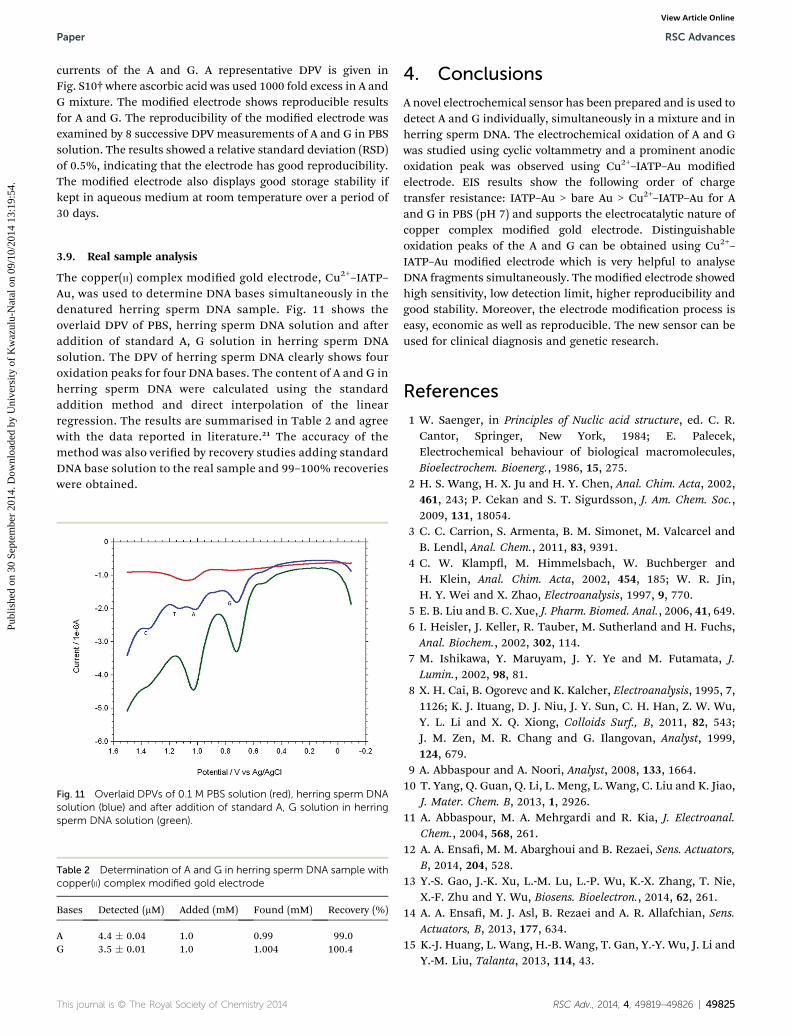
Based on the optimum conditions, the individual and simul-taneous determination of A and G were performed using DPV.Fig. S4† shows the DPV curves of G with different concentration.In the individual determination of purine bases the oxidationpeak current of G was linear with its concentration in the rangeof 150–600 mM (Inset Fig. S4†). The detection limit for G isestimated to be 0.007 mM (S/N ¼ 3). Fig. S5† indicated thatoxidation peak current of A increased linearly in the range of150–600 mM. The detection limit for A was 0.058 mM (S/N ¼ 3).Fig. 9 shows the DPV curves of G with different concentrationsin the presence of 100 mM A and the oxidation peak current of Gwas linear with its concentration range of 10–60 mM. Theregression equation was Ipa ¼ 0.0055c + 1.3113 (R2 ¼ 0.9917,95% condence limit for the slope ¼ �0.10 and intercept ¼�0.42) with a detection limit of 0.01 mM (S/N ¼ 3). Similarly,Fig. 10 shows the DPV curves of A with different concentrationsin the presence of 100 mMG and the oxidation peak current of Aincreased linearly in the concentration range of 10–60 mM. Theregression equation was Ipa ¼ 0.0088c + 2.1627 (R2 ¼ 0.9974,95% condence limit for the slope ¼ �0.09 and intercept ¼�0.31) with a detection limit of 0.06 mM (S/N ¼ 3). For furtherevaluating the feasibility of the Cu2+–IATP–Au electrode for Aand G determination simultaneously by simultaneous changingtheir concentration (Fig. S6†). All the results indicated that thesimultaneous and sensitive detection of A and G could be ach-ieved at copper(II) complex modied gold electrode. Table 1shows a comparison of the proposed electrochemical methodand the other modied electrodes reported for adenine and
Fig. 9 Overlaid DPV for each increment of 10 mM G to 100 mM A atCu2+–IATP SAM modified gold electrode in 0.1 M PBS buffer solutionat pH 7.0.
RSC Adv., 2014, 4, 49819–49826 | 49823
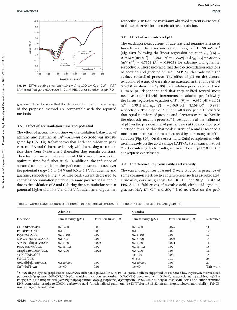
Fig. 10 DPVs obtained for each 10 mM A to 100 mM G at Cu2+–IATPSAM modified gold electrode in 0.1 M PBS buffer solution at pH 7.0.
RSC Advances Paper
Publ
ishe
d on
30
Sept
embe
r 20
14. D
ownl
oade
d by
Uni
vers
ity o
f K
waz
ulu-
Nat
al o
n 09
/10/
2014
13:
19:5
4.
View Article Online
guanine. It can be seen that the detection limit and linear rangeof the proposed method are comparable with the reportedmethods.
3.6. Effect of accumulation time and potential
The effect of accumulation time on the oxidation behaviour ofadenine and guanine at Cu2+–IATP–Au electrode was investi-gated by DPV. Fig. S7(a)† shows that both the oxidation peakcurrent of A and G increased slowly with increasing accumula-tion time from 0–150 s and thereaer they remain constant.Therefore, an accumulation time of 150 s was chosen as theoptimum time for further study. In addition, the inuence ofaccumulation potential on the peak current was examined overthe potential range 0.0 to 0.6 V and 0.0 to 0.5 V for adenine andguanine, respectively Fig. 7(b). The peak current decreased bychanging accumulation potential to more positive value and isdue to the oxidation of A and G during the accumulation step atpotential higher than 0.6 V and 0.5 V for adenine and guanine,
Table 1 Comparative account of different electrochemical sensors for t
Electrode
Adenine
Linear range (mM) Detection limit (mM
GNO–SPAN/CPE 0.5–200 0.05Pt–Pd/PSi/CNPE 0.1–10 0.03PPyox/GR/GCE 0.06–100 0.02MWCNT/NiFe2O4/GCE 0.1–4.0 0.01AgNPs–Pdop@Gr/GCE 0.02–40 0.002PSSA–ssDNA/GCE 0.065–1.1 0.02Graphene-COOH/GCE 0.5–200 0.0254a-NiIITAPc/GCE — —FeHCF/GCE — —Azocalix[4]arene/GCE 0.125–200 0.07Cu2+–IATP–Au 10–60 0.06
a GNO: single-layered graphene oxide, SPANI: sulfonated polyaniline, Pt–Ppolypyrrole/graphene, MWCNT/NiFe2O4: multiwall carbon nanotubes (Pdop@Gr: Ag nanoparticles (AgNPs)–polydopamine(Pdop)@graphene(GrDNA composite, graphene-COOH: carboxylic acid functionalized grapheiron hexacyanoferrate lm.
49824 | RSC Adv., 2014, 4, 49819–49826
respectively. In fact, themaximum observed currents were equalto those observed for open circuit accumulation.
3.7. Effect of scan rate and pH
The oxidation peak current of adenine and guanine increasedlinearly with the scan rate in the range of 10–90 mV s�1
(Fig. S8†) following the linear regression equation Ipa (mA) ¼0.0323 n (mV s�1)� 0.0624 (R2 ¼ 0.9939) and Ipa (mA) ¼ 0.0393 n
(mV s�1) + 4.7321 (R2 ¼ 0.9925) for adenine and guanine,respectively. These indicated that the electrooxidation reactionsof adenine and guanine at Cu2+–IATP–Au electrode were thesurface controlled process. The effect of pH on the electro-oxidation of A and G were also investigated in the range of pH3.0–9.0. As shown in Fig. S9† the oxidation peak potential A andG were pH dependent and that they shied toward morenegative potential with increments in solution pH followingthe linear regression equation of Epa (V) ¼ �0.059 pH + 1.421(R2 ¼ 0.994) and Epa (V) ¼ �0.060 pH + 1.169 (R2 ¼ 0.993),respectively. The slope of 59.0 and 60.0 mV per pH indicatedthat equal numbers of protons and electrons were involved inthe electrode reaction process.45 Investigation of the inuenceof pH on the peak current of purine bases at the modied goldelectrode revealed that that peak current of A and G reached amaximum at pH 7.0 and then decreased by increasing pH of thesolution (Fig. S9†). On the other hand Cu(II) complexation withazoimidazole on the gold surface (IATP–Au) is maximum at pH7.0. Considering both results, we have chosen pH 7.0 for thesubsequent experiments.
3.8. Interference, reproducibility and stability
The current responses of A and G were studied in presence ofsome common electroactive interferences such as ascorbic acid,citric acid, cysteine, glucose, Na+, K+, Cl� and SO4
2� in 0.1 MPBS. A 1000 fold excess of ascorbic acid, citric acid, cysteine,glucose, Na+, K+, Cl� and SO4
2� had no effect on the peak
he determination of adenine and guaninea
Guanine
Reference) Linear range (mM) Detection limit (mM)
0.5–200 0.075 100.1–10 0.02 120.04–100 0.01 130.05–3.0 0.006 140.02–40 0.004 150.065–1.1 0.02 160.5–200 0.05 1810–100 0.03 190–145 0.10 200.125–200 0.05 2110–60 0.01 This work
d/Psi: porous silicon supported Pt–Pd nanoalloy, PPyox/GR: overoxidizedMWCNTs) decorated with NiFe2O4 magnetic nanoparticles, AgNPs–)composite, PSSA–ssDNA: poly(sulfosalicylic acid) and single-strandedne, 4a-NiIITAPc: 1,8,15,22-tetraaminophthalocyanatonickel(II), FeHCF:
This journal is © The Royal Society of Chemistry 2014
Paper RSC Advances
Publ
ishe
d on
30
Sept
embe
r 20
14. D
ownl
oade
d by
Uni
vers
ity o
f K
waz
ulu-
Nat
al o
n 09
/10/
2014
13:
19:5
4.
View Article Online
currents of the A and G. A representative DPV is given inFig. S10†where ascorbic acid was used 1000 fold excess in A andG mixture. The modied electrode shows reproducible resultsfor A and G. The reproducibility of the modied electrode wasexamined by 8 successive DPV measurements of A and G in PBSsolution. The results showed a relative standard deviation (RSD)of 0.5%, indicating that the electrode has good reproducibility.The modied electrode also displays good storage stability ifkept in aqueous medium at room temperature over a period of30 days.
3.9. Real sample analysis
The copper(II) complex modied gold electrode, Cu2+–IATP–Au, was used to determine DNA bases simultaneously in thedenatured herring sperm DNA sample. Fig. 11 shows theoverlaid DPV of PBS, herring sperm DNA solution and aeraddition of standard A, G solution in herring sperm DNAsolution. The DPV of herring sperm DNA clearly shows fouroxidation peaks for four DNA bases. The content of A and G inherring sperm DNA were calculated using the standardaddition method and direct interpolation of the linearregression. The results are summarised in Table 2 and agreewith the data reported in literature.21 The accuracy of themethod was also veried by recovery studies adding standardDNA base solution to the real sample and 99–100% recoverieswere obtained.
Fig. 11 Overlaid DPVs of 0.1 M PBS solution (red), herring sperm DNAsolution (blue) and after addition of standard A, G solution in herringsperm DNA solution (green).
Table 2 Determination of A and G in herring sperm DNA sample withcopper(II) complex modified gold electrode
Bases Detected (mM) Added (mM) Found (mM) Recovery (%)
A 4.4 � 0.04 1.0 0.99 99.0G 3.5 � 0.01 1.0 1.004 100.4
This journal is © The Royal Society of Chemistry 2014
4. Conclusions
A novel electrochemical sensor has been prepared and is used todetect A and G individually, simultaneously in a mixture and inherring sperm DNA. The electrochemical oxidation of A and Gwas studied using cyclic voltammetry and a prominent anodicoxidation peak was observed using Cu2+–IATP–Au modiedelectrode. EIS results show the following order of chargetransfer resistance: IATP–Au > bare Au > Cu2+–IATP–Au for Aand G in PBS (pH 7) and supports the electrocatalytic nature ofcopper complex modied gold electrode. Distinguishableoxidation peaks of the A and G can be obtained using Cu2+–IATP–Au modied electrode which is very helpful to analyseDNA fragments simultaneously. The modied electrode showedhigh sensitivity, low detection limit, higher reproducibility andgood stability. Moreover, the electrode modication process iseasy, economic as well as reproducible. The new sensor can beused for clinical diagnosis and genetic research.
References
1 W. Saenger, in Principles of Nuclic acid structure, ed. C. R.Cantor, Springer, New York, 1984; E. Palecek,Electrochemical behaviour of biological macromolecules,Bioelectrochem. Bioenerg., 1986, 15, 275.
2 H. S. Wang, H. X. Ju and H. Y. Chen, Anal. Chim. Acta, 2002,461, 243; P. Cekan and S. T. Sigurdsson, J. Am. Chem. Soc.,2009, 131, 18054.
3 C. C. Carrion, S. Armenta, B. M. Simonet, M. Valcarcel andB. Lendl, Anal. Chem., 2011, 83, 9391.
4 C. W. Klamp, M. Himmelsbach, W. Buchberger andH. Klein, Anal. Chim. Acta, 2002, 454, 185; W. R. Jin,H. Y. Wei and X. Zhao, Electroanalysis, 1997, 9, 770.
5 E. B. Liu and B. C. Xue, J. Pharm. Biomed. Anal., 2006, 41, 649.6 I. Heisler, J. Keller, R. Tauber, M. Sutherland and H. Fuchs,Anal. Biochem., 2002, 302, 114.
7 M. Ishikawa, Y. Maruyam, J. Y. Ye and M. Futamata, J.Lumin., 2002, 98, 81.
8 X. H. Cai, B. Ogorevc and K. Kalcher, Electroanalysis, 1995, 7,1126; K. J. Ituang, D. J. Niu, J. Y. Sun, C. H. Han, Z. W. Wu,Y. L. Li and X. Q. Xiong, Colloids Surf., B, 2011, 82, 543;J. M. Zen, M. R. Chang and G. Ilangovan, Analyst, 1999,124, 679.
9 A. Abbaspour and A. Noori, Analyst, 2008, 133, 1664.10 T. Yang, Q. Guan, Q. Li, L. Meng, L. Wang, C. Liu and K. Jiao,
J. Mater. Chem. B, 2013, 1, 2926.11 A. Abbaspour, M. A. Mehrgardi and R. Kia, J. Electroanal.
Chem., 2004, 568, 261.12 A. A. Ensa, M. M. Abarghoui and B. Rezaei, Sens. Actuators,
B, 2014, 204, 528.13 Y.-S. Gao, J.-K. Xu, L.-M. Lu, L.-P. Wu, K.-X. Zhang, T. Nie,
X.-F. Zhu and Y. Wu, Biosens. Bioelectron., 2014, 62, 261.14 A. A. Ensa, M. J. Asl, B. Rezaei and A. R. Allafchian, Sens.
Actuators, B, 2013, 177, 634.15 K.-J. Huang, L. Wang, H.-B. Wang, T. Gan, Y.-Y. Wu, J. Li and
Y.-M. Liu, Talanta, 2013, 114, 43.
RSC Adv., 2014, 4, 49819–49826 | 49825
RSC Advances Paper
Publ
ishe
d on
30
Sept
embe
r 20
14. D
ownl
oade
d by
Uni
vers
ity o
f K
waz
ulu-
Nat
al o
n 09
/10/
2014
13:
19:5
4.
View Article Online
16 L. J. Feng, X.-H. Zhang, P. Liu, H.-Y. Xiong and S.-F. Wang,Anal. Biochem., 2011, 419, 71.
17 J. Cui, D. Sun, W. Zhou, H. Liu, P. Hu, N. Ren, H. Qin,Z. Huang, J. Lin and H. Ma, Phys. Chem. Chem. Phys., 2011,13, 9232.
18 K. J. Huang, D. J. Niu, J. Y. Sun, C. H. Han, Z. W. Wu, Y. L. Liand X. Q. Xiong, Colloids Surf., B, 2011, 82, 543.
19 A. J. Jeevagan and S. A. John, Anal. Biochem., 2012, 424, 21.20 S. M. Chen, C. H. Wang and K. C. Lin, Int. J. Electrochem. Sci.,
2012, 7, 405.21 Q. Xu, X. Liu, H. Li, L. Yin and X. Hu, Biosens. Bioelectron.,
2013, 42, 355.22 H. Yin, Y. Zhou, Q. Ma, S. Ai, P. Ju, L. Zhu and L. Lu, Process
Biochem., 2010, 45, 1707.23 R. N. Goyal, V. K. Gupta, M. Oyama and N. Bachheti, Talanta,
2007, 71, 1110.24 X. Xie, K. Zhao, X. Xu, W. Zhao, S. Liu, Z. Zhu, M. Li, Z. Shi
and Y. Shao, J. Phys. Chem. C, 2010, 114, 14243.25 E. Radi, X. M. Berbel, M. C. Puig and J. L. Martyc,
Electroanalysis, 2009, 21, 696; L. S. Jiao, L. Niu, J. Shen,T. Y. You, S. J. Dong and A. Ivaska, Electrochem. Commun.,2005, 7, 219.
26 G. T. Hermenson, Bioconjugate Techniques, Academic Press,San Diego, CA, 2nd edn, 2008; F. Li, Y. Feng, P. Dong andB. Tang, Biosensors Bioelectronics, 2010, 25, 2084.
27 Sk. Jasimuddin, Transition Met. Chem., 2006, 31, 724;Sk. Jasimuddin and C. Sinha, Transition Met. Chem., 2004,29, 566.
28 L. Trnkova, L. Zerzankova, F. Dycka, R. Mikelova andF. Jelen, Sensors, 2008, 8, 429.
29 K. Barman and Sk. Jasimuddin, Indian J. Chem., Sect. A:Inorg., Bio-inorg., Phys., Theor. Anal. Chem., 2013, 52, 217.
30 R. K. Shervedani, F. Yaghoobi, A. H. Mehrjardi andS. M. S. Barzoki, Electrochim. Acta, 2008, 53, 4185.
49826 | RSC Adv., 2014, 4, 49819–49826
31 S. Y. M. Shikov, A. V. Vurasko, L. S. Molechmikov,E. G. Kovalyova and A. A. Ffendieu, J. Mol. Catal. A: Chem.,2000, 158, 447.
32 Z. Dursun, I. Sahbaz, F. N. Ertas and G. Nisli, Turk. J. Chem.,2003, 27, 513.
33 Z. Zhang, X. Li, C. Wang, C. Zhang, P. Liu, T. Fang, Y. Xiongand W. Xu, Dalton Trans., 2012, 41, 1252.
34 T. Rohani and M. A. Taher, Talanta, 2009, 78, 743.35 K. Kano, M. Torimura, Y. Esaka and M. Goto, J. Electroanal.
Chem., 1994, 372, 137.36 B. J. Sanghavi, S. M. Mobin, P. Mathur, G. K. Lahiri and
A. K. Srivastava, Biosens. Bioelectron., 2013, 39, 124.37 J. F. Smalley, K. Chalfant, S. W. Feldberg, T. M. Nahir and
E. F. Bowden, J Phys. Chem. B, 1999, 103, 1676.38 R. K. Shervedani, A. Hate-Mehrjardi and M. Khosravi
Babadi, Electrochim. Acta, 2007, 52, 7051 and referencestherein.
39 G. G. Hammes, Enzyme catalysis and regulation, Academic,New York, 1982; T. Palmer, Understanding Enzymes, Ellis.Horwood, Chicester, 2nd edn, 1985; G. N. Mukherjee andA. Das, Elements of Bioinorganic Chemistry, U. N. Dhur &Sons Pvt. Ltd, Kolkata, 1st edn, 1993.
40 R. K. Shervedani, S. M. S. Barzoki and M. Bagherzadeh,Electroanalysis, 2010, 22, 969.
41 S. Campuzano, M. Pedrero, C. Montemayor, E. Fatas andJ. M. Pingarron, J. Electroanal. Chem., 2006, 586, 112.
42 Q. Li, C. Batchelor-McAuley and R. G. Compton, J. Phys.Chem. B, 2010, 114, 7423; L. M. Goncalves, C. Batchelor-McAuley, A. A. Barros and R. G. Compton, J. Phys. Chem. C,2010, 114, 14213.
43 Y. Wei, Q. A. Huang, M. G. Li, X. J. Huang, B. Fang andL. Wang, Electrochim. Acta, 2011, 56, 8571–8575.
44 P. N. Mashazi, P. Westbroek, K. I. Ozoemena andT. Nyokong, Electrochim. Acta, 2007, 53, 1858.
45 E. Laviron, J. Electroanal. Chem., 1974, 52, 355.
This journal is © The Royal Society of Chemistry 2014
Copyright © 2022 FDOKUMEN