Mechanism and stereochemistry in the anionic polymerization ...
Supported Lipid Monolayer with Improved NanomechanicalStability: Effect of PolymerizationRacha El Zein, Herve Dallaporta, and Anne M. Charrier*
CNRS, UMR7325, 13288, Marseille, France, and Aix-Marseille University, CINaM, 13288, Marseille, France
ABSTRACT: We study the effect of polymerization on the nanomechanical stability ofsupported lipid monolayers consisting of 1,2-di-(10Z,12Z-tricosadiynoyl)-sn-glycero-3-phosphocholine by means of force mapping using an atomic force microscope. For bothnonpolymerized and polymerized lipid monolayers, we investigate the break-through forcesrequired to rupture the monolayers for a whole range of loading velocities. We show thatthe average break-through force exerted by the tip and required to penetrate the monolayerhas a logarithmic dependence on the loading rate. Both Young moduli and intrinsic Gibbsenergies have been determined for the nonpolymerized and polymerized lipid monolayers,and we show a drastic effect of polymerization on the nanomechanical stability of themonolayer with an increase by a factor of ∼100 for the young modulus and ∼3 for theintrinsic Gibbs activation energy.
■ INTRODUCTIONForce measurement using atomic force microscopy (AFM) hasnow become a very common technique used to probe thenanomechanical properties of materials and especially those ofthin films.1−12 Among those, abundant studies realized onsupported lipid bilayers13−16 have shown that the variation ofmechanical stability depends on the ordering of the lipidswithin the bilayer. The solid-like ordered phase with higherdensity of lipids shows higher mechanical stability than lowdensity disordered phase.17,18 That said, one knows that lipidbilayers exhibit gel to fluid phase transition in a range oftemperatures that varies from −70 °C to +80 °C depending ontheir length, headgroup, and number of unsaturated bonds intheir fatty acid chains. In addition, when these bilayers aresupported, their interaction with the substrate may drasticallyincrease their transition temperature.19 It is therefore expectedthat the mechanical properties of lipid layers will stronglydepend on the type of lipid constituting the layer and on thetemperature at which the measurement is realized. Similarly itwas shown that the composition of the lipid bilayer might varyconsiderably its mechanical stability.20 For example, somerecent studies show that increasing the ratio of cholesterol in amixture of dioleoylphosphatidylcholine/sphongomyelin re-leases the stress in the layer and as a result lowers thebreakthrough force required to induce rupture.21 Under-standing the mechanisms that govern the mechanics and elasticproperties of lipid membranes is essential for both thecomprehension of biological processes and the developmentof biotechnologies or bioinspired technologies. For example itwas shown that elastic bilayer forces modulate the free energyand cooperativity of folding of membrane proteins22 andtransmembrane signaling.23 Also, as a biocompatible material,some researchers have been working on the development of
nanoparticles for the targeting and delivery of drugs in whichlipid bilayers are used to encapsulate the drugs. In that field alot of work is dedicated to controlling the stability of thebilayer.24,25 Another example is the stabilization of supportedlipid mono- and bilayers containing acryl and acetylenic speciesin air by polymerization therefore opening new opportunities ofapplications.26 Typically, the electrical properties of poly-merized lipid monolayers of tricosadiynoylphosphatidylcholinewere recently reported27 and suggested that such layers couldbe used as ultrathin dielectrics in electronic devices such as fieldeffect transistors provided these present strong enoughmechanical stability to handle advanced industrial processes.In this work, we investigate the effect of polymerization on
the nanomechanical stability of supported lipid monolayers.Force measurements using AFM were realized before and afterpolymerization on same supported lipid monolayers. In atypical force measurement, the AFM tip, initially far from thesurface approaches the sample and gets into contact with thelipid monolayer. As the tip keeps moving toward the substrate,the cantilever, with a given spring constant, bends over the lipidlayer until the exerted force becomes large enough to rupturethe layer. At this point the tip jumps to the substrate and thejump height of the tip corresponds to the thickness of the layerunder the compression of the tip. Both the break-through forceand the jump height are parameters of the considered lipidlayer that can be utilized to determine properties of the layer. Inaddition, indentation experiments on lipid bilayers haverevealed that the rate of formation of a hole, large enough toinitiate the tip penetration in the bilayer, is governed by an
Received: March 9, 2012Revised: May 31, 2012Published: June 1, 2012
Article
pubs.acs.org/JPCB
© 2012 American Chemical Society 7190 dx.doi.org/10.1021/jp302306r | J. Phys. Chem. B 2012, 116, 7190−7195
activation energy following an Arrhenius law.28−30 It was shownthat this intrinsic Gibbs energy of a bilayer-tip system tospontaneously form a hole in the bilayer can be increased bythe application of a pressure on the bilayer therefore favoring itsrupture.28 The rate at which the pressure increases being tiploading rate dependent, the Gibbs energy not only depends onthe properties of the investigated bilayer but also on the tiploading rate used in the experiment. If this kind of behavior iswell-known for lipid bilayers it was never demonstrated formonolayers. We investigate the effect of the loading rate on themonolayer rupture for both types of monolayers and we willshow how the polymerization impacts the nanomechanicalresistance of the monolayer. From the experimental data wedetermine the Young’s modulus and the intrinsic Gibbs energyof each type of monolayer.
■ MATERIALS AND METHODSMaterials. The lipids, 1,2-di-(10Z,12Z-tricosadiynoyl)-sn-
glycero-3-phosphocholine, DC8,9PC, (Figure 1) were pur-chased from Avanti Polar Lipids (Alabster, AL) and usedwithout further purification. A 1 mg/mL lipid stock solutionwas prepared in chloroform and stored at −20 °C. These lipidswere selected for having two acetylenic groups in each of theiraliphatic chains, therefore allowing for two-dimensionalpolymerization in the plane of the layer. Polymerizationinitiator (2,2-azobis(2-methylpropionamidine)dihydrochloride(AAPH), methanol, chloroform, sulphuric acid, 96%(H2SO4), hydrofluoric acid, 49% (HF) and hydrogen peroxide,and 30% (H2O2) were purchased from Sigma Aldrich.Deionized water (DI, 18 MΩ·cm) was used in all experiments.Silicon Surface Preparation. Silicon wafers ((100), ρ∼5
Ω.cm, Boron doped) were cleaned by soaking in 2:1 H2SO4/H2O2 (piranha solution) at 130 °C for 30 min, followed bythorough rinsing with DI water. Just prior to forming the lipidmonolayer, H-terminated silicon surface is obtained by etchingoff the native silicon oxide from the silicon substrate with HF(2 min in 2% ethanol). The hydrogen passivation results fromefficient removal of fluorine-bonded surface silicon as SiF4leaving behind hydrogen.31
Vesicles Preparations. A total of 50 μL of the 0.1% lipidstock solution were heated at 40 °C to evaporate chloroformand then diluted in 200 μL of DI water. At this stagemultilamellar lipid vesicles are formed in the aqueous solution.
Unilamellar vesicles are then obtained by sonication of the lipidsolution for 15 min. Finally the vesicle radius was reduced byextrusion through 100 nm pores polycarbonate membrane.
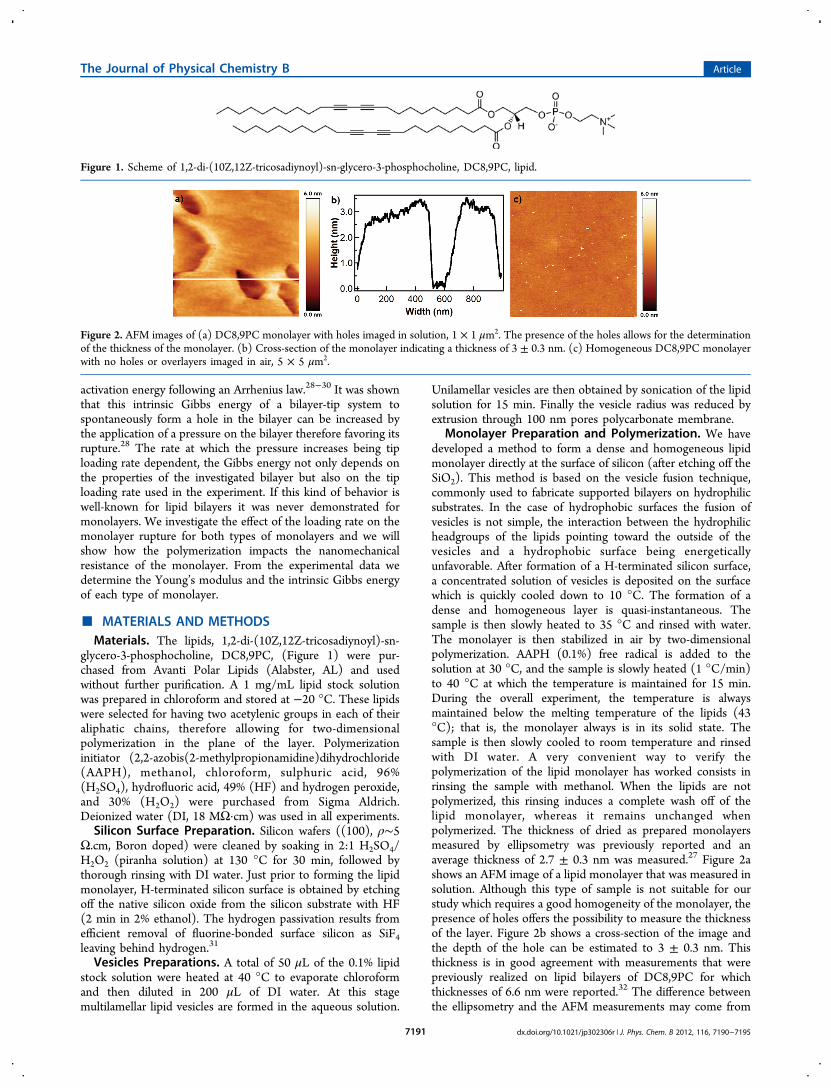
Monolayer Preparation and Polymerization. We havedeveloped a method to form a dense and homogeneous lipidmonolayer directly at the surface of silicon (after etching off theSiO2). This method is based on the vesicle fusion technique,commonly used to fabricate supported bilayers on hydrophilicsubstrates. In the case of hydrophobic surfaces the fusion ofvesicles is not simple, the interaction between the hydrophilicheadgroups of the lipids pointing toward the outside of thevesicles and a hydrophobic surface being energeticallyunfavorable. After formation of a H-terminated silicon surface,a concentrated solution of vesicles is deposited on the surfacewhich is quickly cooled down to 10 °C. The formation of adense and homogeneous layer is quasi-instantaneous. Thesample is then slowly heated to 35 °C and rinsed with water.The monolayer is then stabilized in air by two-dimensionalpolymerization. AAPH (0.1%) free radical is added to thesolution at 30 °C, and the sample is slowly heated (1 °C/min)to 40 °C at which the temperature is maintained for 15 min.During the overall experiment, the temperature is alwaysmaintained below the melting temperature of the lipids (43°C); that is, the monolayer always is in its solid state. Thesample is then slowly cooled to room temperature and rinsedwith DI water. A very convenient way to verify thepolymerization of the lipid monolayer has worked consists inrinsing the sample with methanol. When the lipids are notpolymerized, this rinsing induces a complete wash off of thelipid monolayer, whereas it remains unchanged whenpolymerized. The thickness of dried as prepared monolayersmeasured by ellipsometry was previously reported and anaverage thickness of 2.7 ± 0.3 nm was measured.27 Figure 2ashows an AFM image of a lipid monolayer that was measured insolution. Although this type of sample is not suitable for ourstudy which requires a good homogeneity of the monolayer, thepresence of holes offers the possibility to measure the thicknessof the layer. Figure 2b shows a cross-section of the image andthe depth of the hole can be estimated to 3 ± 0.3 nm. Thisthickness is in good agreement with measurements that werepreviously realized on lipid bilayers of DC8,9PC for whichthicknesses of 6.6 nm were reported.32 The difference betweenthe ellipsometry and the AFM measurements may come from
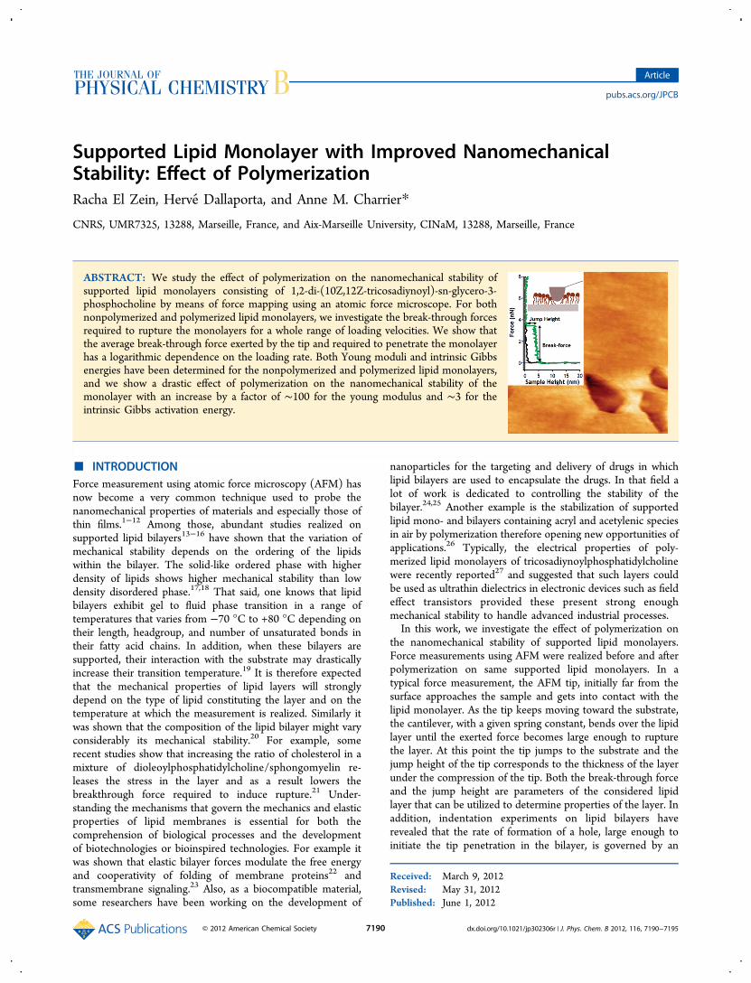
Figure 1. Scheme of 1,2-di-(10Z,12Z-tricosadiynoyl)-sn-glycero-3-phosphocholine, DC8,9PC, lipid.
Figure 2. AFM images of (a) DC8,9PC monolayer with holes imaged in solution, 1 × 1 μm2. The presence of the holes allows for the determinationof the thickness of the monolayer. (b) Cross-section of the monolayer indicating a thickness of 3 ± 0.3 nm. (c) Homogeneous DC8,9PC monolayerwith no holes or overlayers imaged in air, 5 × 5 μm2.
The Journal of Physical Chemistry B Article
dx.doi.org/10.1021/jp302306r | J. Phys. Chem. B 2012, 116, 7190−71957191

the hydration of the layer that may change the moleculesorganization. Figure 2c shows an AFM image of a perfectlyhomogeneous monolayer with no holes or overlayers. Suchmonolayer quality is obtained on a regular base and was verifiedbefore any further measurements.AFM Force Measurements. Force measurements were
carried out in deionized water using AFM (NTEGRA fromNT-MDT). Silicon tips CSC17 from MikroMash with a typicalresonance frequency of 14 kHz in solution and tip radius in therange 8−10 nm were selected for this testing. The AFM tipradii were determined after imaging the tip by scanningelectron microscopy. The spring constants in the range from0.1 to 0.3 N/m were determined using the thermal noisemethod after obtaining the deflection sensitivity of thecantilever by pressing the AFM tip against a hard referencesilicon surface. Cantilever deflection sensitivity measurementswere performed before all sets of measurements. A minimum of200 force measurements per set were realized on a minimum ofthree different areas per sample. To prevent any questioningregarding the reproducibility of the lipid monolayers qualityand a misinterpretation of the data, all of the measurementsreported as before and after polymerization were realized aspairs on the same sample using the same AFM tip.
■ RESULTS AND DISCUSSIONDependence of the Break-through Force on the
Polymerization State. Figure 3a shows two different
representative force curves (loading part only) obtained on ahard silicon surface (black) and a lipid monolayer (green).From this type of measurement, one can determine the forcerequired to rupture the monolayer as well as the height of themonolayer. The later is the sum of the indentation of the tip inthe monolayer before the break-through occurs and the tipjump height at the break-through. Since a higher break-throughforce indicates a more resistant layer, both these parameters canbe used to evaluate the nanomechanical stability of the lipidmonolayers. To evaluate the effect of the polymerization, astatistical analysis of these break-through forces and tip jumpsheights at the lipid monolayers rupture were realized before andafter polymerization. In Figure 3b the break-through forceversus the tip jump height is reported for over 400
measurements acquired before and after polymerization ofthe monolayers on three different samples and for a tip loadingrate of 6 μm/s. The difference between the two types of lipidmonolayers is evidenced in this figure. As an average force of 98± 67 pN is required to rupture the layer before polymerization,a net increase to 1.4 ± 0.72 nN is measured afterpolymerization. Although the large error bars which seem toindicate variability in the quality of the investigated lipidmonolayers, the data clearly reveal an improvement of thenanomechanical resistivity of the monolayers after polymer-ization.
Dependence of the Break-through Force on theLoading Rate. Indentation experiments on lipid bilayershave shown that the average break-through force is logarithmi-cally dependent on the loading rate at which the indentation isperformed, i.e., on the rate at which the tip hits the lipidbilayer.22,28−30 Although this property has now been welldemonstrated for lipid bilayers it was never shown formonolayers. We present a statistical analysis of the force atwhich the break-through occurs for a whole range of loadingrates varying from 30 nm·s−1 to 6 μm·s−1. Because eachmonolayer is not perfectly identical, some differences appearbetween sets of measurements. For clarity, we will show theresults obtained on one representative sample. In the followingthe sets of measurements were performed on the samemonolayer before and after polymerization. These are shownin green square and red circle respectively. For each loadingrate v, the average value F0 is reported in Figure 4, each point
corresponding to a sample of at least 200 force curvemeasurements. The first observation is that for both types ofmonolayer the average break-through force increases linearlywith the logarithm of the loading rate similarly to what wasreported for bilayers. The red and green lines result from fittingequation F0 = a + b log(v) to the experimental data. Thesecond observation is that the dependence of F0 on the loadingvelocity is very different whether the lipid monolayer ispolymerized. Before the polymerization, the break-throughforces are very small with values ranging from 20 to 60 pNwhile still showing some dependence on the loading rate. Toremove the tip curvature effect and compare these results withmeasurements obtained with different tips and samples wenormalized these forces by the tip curvature.16 We obtainnormalized forces ranging from 2 to 7 mN·m−1. Such values of
Figure 3. (a) Representative force measurements loading curvesobtained on a hard silicon surface (black) and a supported lipidmonolayer after polymerization (green). (b) Break-through forcesversus the tip jumpin distance at the lipid monolayer rupture reportedfor over 400 measurements on three different samples before (green)and after (red) polymerization. The tip loading rate was 6 μm/s.
Figure 4. Average break-through forces obtained at loading ratesranging from 30 nm·s−1 to 6 μm·s−1 on both nonpolymerized (greensquares) and polymerized (red circles) lipid monolayers.
The Journal of Physical Chemistry B Article
dx.doi.org/10.1021/jp302306r | J. Phys. Chem. B 2012, 116, 7190−71957192
the break-through forces are relatively small compared to thoseusually reported for bilayers (105 mN·m−1 for DSPE,16 220mN·m−1 for MGDG,16 66−170 mN·m−1 for DOTAP,27 30mN·m−1 for POPE/POPG mixtures,33 900 mN·m−1 forDMPC,34 and 72 mN·m−1 for POPG35) but fall into thesame range of values as previously reported for monolayers ofDPPC36 (DPPC and DC8,9PC have similar melting temper-atures and headgroups. They are therefore expected to behavesimilarly). After polymerization the break-through forces aremuch bigger and vary between 0.2 and 2 nN for theinvestigated range of loading rates, therefore corresponding tonormalized forces varying from 24 to 235 mN·m−1. Thesevalues which compare with those obtained on bilayers evidencethe impact of the polymerization on the nanomechanicalresistance of the monolayer.Determination of the Young’s Modulus: Impact of the
Polymerization. To determine the Young’s modulus of eachtype of monolayer, we describe our experimental data in theframework of the Hertz model of a tip lipid monolayer systemapproximated by a sphere in contact with a flat surface. Usingthe radius of the contact area between the tip and the lipidmonolayer a = (Rh)1/2 and the indentation h = h0 − dj at therupture, one can relate the average break-through force to thetip jump height as
ν=
−−F
ER h d
43 (1 )
( )0 2 0 j3/2
(1)
ν is the Poisson coefficient (we have used ν = 0.3), E is theYoung’s modulus of the monolayer, R is the radius of the tip, h0is the thickness of the monolayer, and dj is the jump height atthe break-through.Like for the break-through force we have determined for
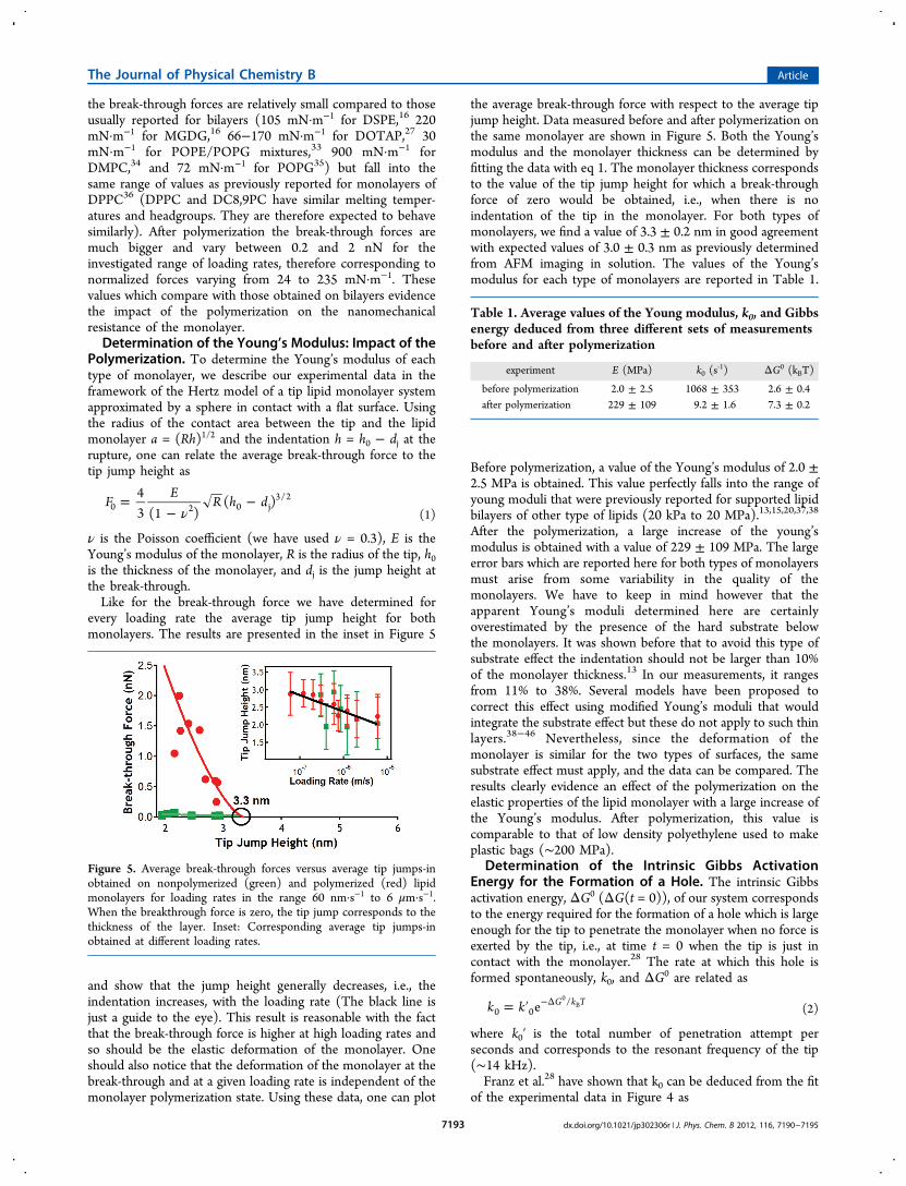
every loading rate the average tip jump height for bothmonolayers. The results are presented in the inset in Figure 5
and show that the jump height generally decreases, i.e., theindentation increases, with the loading rate (The black line isjust a guide to the eye). This result is reasonable with the factthat the break-through force is higher at high loading rates andso should be the elastic deformation of the monolayer. Oneshould also notice that the deformation of the monolayer at thebreak-through and at a given loading rate is independent of themonolayer polymerization state. Using these data, one can plot
the average break-through force with respect to the average tipjump height. Data measured before and after polymerization onthe same monolayer are shown in Figure 5. Both the Young’smodulus and the monolayer thickness can be determined byfitting the data with eq 1. The monolayer thickness correspondsto the value of the tip jump height for which a break-throughforce of zero would be obtained, i.e., when there is noindentation of the tip in the monolayer. For both types ofmonolayers, we find a value of 3.3 ± 0.2 nm in good agreementwith expected values of 3.0 ± 0.3 nm as previously determinedfrom AFM imaging in solution. The values of the Young’smodulus for each type of monolayers are reported in Table 1.
Before polymerization, a value of the Young’s modulus of 2.0 ±2.5 MPa is obtained. This value perfectly falls into the range ofyoung moduli that were previously reported for supported lipidbilayers of other type of lipids (20 kPa to 20 MPa).13,15,20,37,38
After the polymerization, a large increase of the young’smodulus is obtained with a value of 229 ± 109 MPa. The largeerror bars which are reported here for both types of monolayersmust arise from some variability in the quality of themonolayers. We have to keep in mind however that theapparent Young’s moduli determined here are certainlyoverestimated by the presence of the hard substrate belowthe monolayers. It was shown before that to avoid this type ofsubstrate effect the indentation should not be larger than 10%of the monolayer thickness.13 In our measurements, it rangesfrom 11% to 38%. Several models have been proposed tocorrect this effect using modified Young’s moduli that wouldintegrate the substrate effect but these do not apply to such thinlayers.38−46 Nevertheless, since the deformation of themonolayer is similar for the two types of surfaces, the samesubstrate effect must apply, and the data can be compared. Theresults clearly evidence an effect of the polymerization on theelastic properties of the lipid monolayer with a large increase ofthe Young’s modulus. After polymerization, this value iscomparable to that of low density polyethylene used to makeplastic bags (∼200 MPa).
Determination of the Intrinsic Gibbs ActivationEnergy for the Formation of a Hole. The intrinsic Gibbsactivation energy, ΔG0 (ΔG(t = 0)), of our system correspondsto the energy required for the formation of a hole which is largeenough for the tip to penetrate the monolayer when no force isexerted by the tip, i.e., at time t = 0 when the tip is just incontact with the monolayer.28 The rate at which this hole isformed spontaneously, k0, and ΔG0 are related as
= ′ −Δk k e G k T0 0
/0B (2)
where k0′ is the total number of penetration attempt perseconds and corresponds to the resonant frequency of the tip(∼14 kHz).Franz et al.28 have shown that k0 can be deduced from the fit
of the experimental data in Figure 4 as
Figure 5. Average break-through forces versus average tip jumps-inobtained on nonpolymerized (green) and polymerized (red) lipidmonolayers for loading rates in the range 60 nm·s−1 to 6 μm·s−1.When the breakthrough force is zero, the tip jump corresponds to thethickness of the layer. Inset: Corresponding average tip jumps-inobtained at different loading rates.
Table 1. Average values of the Young modulus, k0, and Gibbsenergy deduced from three different sets of measurementsbefore and after polymerization
experiment E (MPa) k0 (s‑1) ΔG0 (kBT)
before polymerization 2.0 ± 2.5 1068 ± 353 2.6 ± 0.4after polymerization 229 ± 109 9.2 ± 1.6 7.3 ± 0.2
The Journal of Physical Chemistry B Article
dx.doi.org/10.1021/jp302306r | J. Phys. Chem. B 2012, 116, 7190−71957193
= −kK
b elog( )10 a b
0/
(3)
with a and b being the two fitting parameters and K being thespring constant of the cantilever. The values of k0 obtained forthe two types of monolayers are reported in Table 1 togetherwith the resulting intrinsic Gibbs activation energiesdetermined using eq 2. Values of k0 of typically 1050 s−1
obtained before the polymerization are reduced by a factor of100 after polymerization therefore indicating a better stabilityof the polymerized lipid monolayer. For both types ofmonolayers, the intrinsic Gibbs activation energies derivedfrom eq 2 are 2.6 ± 0.4 kBT (6.4 ± 0.9 kJ/mol) and 7.3 ± 0.2kBT (18 ± 0.4 kJ/mol) respectively. Considering that theformation of a hole is due to the diffusion of the lipids underthe tip, it is reasonable to compare these Gibbs energies to theenergy of diffusion of lipids in layers. For DPPC bilayers,energies ranging from 20 to 50 kJ/mol have been reported inthe literature using fluorescence technique47,48 and 1H NMR.49
Although a little bit smaller, the values of the Gibbs energiesthat we report here are reasonable considering that ourmeasurements were realized on monolayers instead of bilayers.Indeed, the amount of lipids to displace to allow the tippenetration is twice as high in a bilayer. Similarly, in such forcemeasurement experiments, the size and the shape of the tip willhave an effect on the outcoming values. Nevertheless, the effectof the polymerization on the Gibbs energy of the tip/monolayer system is demonstrated with a variation by a factorof ∼3.
■ CONCLUSIONS
In this work, we have investigated the effect of polymerizationon the nanomechanical properties of supported lipidmonolayers. We have demonstrated that as for bilayers, theforce required to rupture monolayers has a logarithmicdependence on the tip loading rate. We have shown that thisrange of forces varies drastically with the polymerization stateof the monolayer with a large increase by a factor of ∼10 afterpolymerization. Similarly both the Young’s modulus deter-mined for each type of monolayer and the intrinsic Gibbsactivation energies of the tip/monolayer system required toinitiate the rupture of the monolayer evidence an improvementof the nanomechanical stability of the monolayer afterpolymerization. The utilization of lipid monolayers or bilayersin industrial processes has been so far limited by their inherentinstability in air and mechanical fragility. We believe theselayers with improved stability will open a whole range ofpossibilities in applications that require the use of ultrathinlayers. Our interest concerns the development of highsensitivity field effect transistors based biosensors that use alipid monolayer as gate dielectric. More generally these layerscould be used to functionalize flexible materials used inbioelectronics applications for example.
■ AUTHOR INFORMATION
Corresponding Author*E-mail: [email protected]. Phone: +33 (0)6 62 9228 35.
NotesThe authors declare no competing financial interest.
■ ACKNOWLEDGMENTS
We acknowledge the financial support of the PACA regionalcouncil in France (Contract No. DEB 10-1174) as well asPierre-Henri Puech, from the Adhesion and InflammationLaboratory in Marseille, for fruitful scientific discussions.
■ REFERENCES(1) Kovalev, A.; Shulha, H.; Lemieux, M.; Myshkin, N.; Tsukruk, V.V. J. Mater. Res. 2004, 19, 716.(2) VanLandingham, M. R.; Villarrubia, J. S.; Gurthie, W. F.; Meyers,G. F. Macromol. Symp. 2001, 167, 15.(3) Shulha, H.; Kovalev, A.; Myshkin, N.; Tsukruk, V. V. Eur. Polym.J. 2004, 40, 949.(4) Tsukruk, V. V.; Huang, Z.; Chizhik, S. A.; Gorbunov, V. V. J.Mater. Sci. 1998, 33, 4905.(5) Pera, I.; Stark, R.; Kappl, M.; Butt, H. J.; Benfenati, F. Biophys. J.2004, 87, 2446.(6) Kunneke, S.; Kruger, D.; Janshoff, A. Biophys. J. 2004, 86, 1545.(7) Dufrene, Y. F.; Lee, G. U. Biochim. Biophys. Acta 2000, 1509, 14.(8) Park, J.-W. Col. Surf. B 2010, 75, 290.(9) Leonenko, Z.; Finot, E.; Cramb, D. Biochim. Biophys. Acta 2006,1758, 487.(10) Canale, C.; Jacono, M.; Diaspro, A.; Dante, S. Microsci. Res.Technol. 2010, 73, 965.(11) Picas, L.; Scheuring, S. Biophys. J. 2012, 102, L01.(12) Garcia-Manyes, S.; Sanz, F. Biochim. Biophys. Acta 2010, 1798,741.(13) Ngwa, W.; Chen, K.; Sahgal, A.; Stepanov, E. V.; Luo, W. ThinSolid Films 2008, 516, 5039.(14) Dimitriadis, E. K.; Horkay, F.; Maresca, J.; Kachar, B.;Chadwick, R. S. Biophys. J. 2002, 82, 2798.(15) Domke, J.; Radmacher, M. Langmuir 1998, 14, 3320.(16) Schneider, J.; Dufrene, Y. F.; Barger, W. R., Jr.; Lee, G. U.Biophys. J. 2000, 79, 1107.(17) Alessandrini, A.; Seeger, H. M.; Di Cerbo, A.; Caramaschi, T.;Facci, P. Soft Matter 2011, 7, 7054.(18) Goertz, M. P.; Stottrup, B. L.; Houston, J. E.; Zhu, X. Y. J. Phys.Chem. B 2009, 113, 9335.(19) Charrier, A.; Thibaudau, F. Biophys. J. 2005, 89, 1094.(20) Hianik, T.; Haburcak, M.; Lohner, K.; Prenner, E.; Paltauf, F.;Hermetter, A. Colloids Surf. 1998, 139, 189.(21) Sullan, R. M. A.; Li, J. K.; Hao, C.; Walker, G. C.; Zou, S.Biophys. J. 2010, 99, 507.(22) Hong, H.; Tamm, L. K. Proc. Natl. Acad. Sci. 2004, 101, 4065.(23) Anbazhagan, V.; Schneider, D. Biochim. Biophys. Acta 2010,1798, 1899.(24) Uner, M.; Yener, G. Int. J. Nanomed. 2007, 2, 289.(25) Ulrich, A. S. Biosci. Rep. 2002, 22, 129.(26) Charrier, A.; Mischki, T.; Lopinski, G. P. Langmuir 2010, 26,2538.(27) Dumas, C.; El Zein, R.; Dallaporta, H.; Charrier, A. Langmuir2011, 27, 13643.(28) Franz, V.; Loi, S.; Muller, H.; Bamberg, E.; Butt, H. J. ColloidsSurf. 2002, 23, 191.(29) Butt, H. J.; Folker, V. Phys. Rev. E 2002, 66, 031601.(30) Loi, S.; Sun, G.; Franz, V.; Butt, H. J. Phys. Rev. E 2002, 66,031602.(31) Trucks, G. W.; Raghavachari, K.; Higashi, G. S.; Chabal, Y. J.Phys. Rev. Lett. 1990, 65, 504.(32) Zhao, Y.; Mahajan, N.; Fang, J. J. Phys. Chem. B 2006, 110,22060.(33) Picas, L; Montero, M. T.; Morros, A.; Oncins, G.; Hernandez-Borrel, J. J. Phys. Chem. B 2008, 112, 10181.(34) Garcia-Manyes, S.; Redondo-Morata, L.; Oncins, G.; Sanz, F. J.Am. Chem. Soc. 2010, 132, 12874.(35) Picas, L.; Suarez-Germa, C.; Montero, M. T.; Hernandez-Borrell, J. J. Phys. Chem. B 2010, 114, 1543.
The Journal of Physical Chemistry B Article
dx.doi.org/10.1021/jp302306r | J. Phys. Chem. B 2012, 116, 7190−71957194
(36) Oncins, G.; Picas, L.; Hernandez-Borrell, J.; Garcia-Manyes, S.;Sanz, F. Biophys. J. 2007, 938, 2713.(37) Tvarozek, V.; Hianik, T.; Novonty, I.; Rehacek, V.; Ziegler, W.;Ivanic, R.; Andel, M. Vaccum 1998, 50, 251.(38) Hianik, T.; Labajova, A. Bioelectrochem. 2002, 58, 97.(39) Laney, D. E.; Garcia, R. A.; Parsons, S. M.; Hansma, H. G.Biophys. J. 1997, 72, 806.(40) Mencik, J.; Munz, D.; Quandt, E.; Weppelmann, E. R.; Swain,M. V. J. Mater. Res. 1997, 12, 2475.(41) Gao, H.; Chiu, C. H.; Lee, J. Int. J. Sol. Struct. 1992, 29, 2471.(42) King, R. B. Int. J. Sol. Struct. 1987, 23, 1657.(43) Saha, R.; Nix, W. D. Mater. Sci. Eng., A 2001, 319−321, 898.(44) Tricoteaux, A.; Duarte, G.; Chicot, D.; Le Bourhis, E.;Bemporad, E.; Lesage. J. Mech. Mater. 2010, 42, 166.(45) Saha, R.; Nix, W. D. Acta Mater. 2002, 50, 23.(46) Chizhik, S. A.; Gorbunov, V. V.; Luzinov, I.; Fuchigami, N.;Tsukruk, V. V. Macromol. Symp. 2001, 167, 167.(47) Vaz, W.; Clegg, R.; Hallmann, D. Biochemistry 1985, 24, 781.(48) Karakatsanis, P.; Bayerl, T. M. Phys. Rev. E 1996, 54, 1785.(49) Galla, H.-J.; Hartmann, W.; Theilen, U.; Sackmann, E. J. Membr.Biol. 1979, 48, 215.
The Journal of Physical Chemistry B Article
dx.doi.org/10.1021/jp302306r | J. Phys. Chem. B 2012, 116, 7190−71957195
Copyright © 2022 FDOKUMEN