Metformin activates the AMP-activated protein kinase in hepatocytes by changes in adenine...
10
ARTICLE Metformin activates AMP-activated protein kinase in primary human hepatocytes by decreasing cellular energy status X. Stephenne & M. Foretz & N. Taleux & G. C. van der Zon & E. Sokal & L. Hue & B. Viollet & B. Guigas Received: 30 July 2011 /Accepted: 18 August 2011 /Published online: 23 September 2011 Abstract Aim/hypothesis The glucose-lowering drug metformin has been shown to activate hepatic AMP-activated protein kinase (AMPK), a master kinase regulating cellular energy homeostasis. However, the underlying mechanisms remain controversial and have never been investigated in primary human hepatocytes. Methods Hepatocytes isolated from rat, mouse and human livers were treated with various concentrations of metfor- min. Isoform-specific AMPKα abundance and activity, as well as intracellular adenine nucleotide levels and mito- chondrial oxygen consumption rates were determined at different time points. Results Metformin dose- and time-dependently increased AMPK activity in rat and human hepatocytes, an effect associated with a significant rise in cellular AMP:ATP ratio. Surprisingly, we found that AMPKα2 activity was unde- tectable in human compared with rat hepatocytes, while AMPKα1 activities were comparable. Accordingly, metfor- min only increased AMPKα1 activity in human hepato- cytes, although both AMPKα isoforms were activated in rat hepatocytes. Analysis of mRNA expression and protein levels confirmed that only AMPKα1 is present in human hepatocytes; it also showed that the distribution of β and γ regulatory subunits differed between species. Finally, we demonstrated that the increase in AMP:ATP ratio in hepatocytes from liver-specific Ampkα1/2 (also known as Prkaa1/2) knockout mice and humans is due to a similar and specific inhibition of the mitochondrial respiratory- chain complex 1 by metformin. Conclusions/interpretation Activation of hepatic AMPK by metformin results from a decrease in cellular energy status owing to metformin’ s AMPK-independent inhibition of the mitochondrial respiratory-chain complex 1. The unique Electronic supplementary material The online version of this article (doi:10.1007/s00125-011-2311-5) contains peer-reviewed but unedited supplementary material, which is available to authorised users. X. Stephenne : E. Sokal Laboratory of Paediatric Hepatology and Cell Therapy, Université catholique de Louvain and Cliniques St Luc, Brussels, Belgium M. Foretz : B. Viollet Inserm, U1016, Institut Cochin, Paris, France M. Foretz : B. Viollet CNRS, UMR8104, Paris, France M. Foretz : B. Viollet Université Paris Descartes, Paris, France N. Taleux : L. Hue : B. Guigas Hormone and Metabolic Research Unit, Université catholique de Louvain and de Duve Institute, Brussels, Belgium N. Taleux Bioénergétique Fondamentale et Appliquée Inserm-U884, Université J. Fourier, Grenoble, France G. C. van der Zon : B. Guigas (*) Department of Molecular Cell Biology, Leiden University Medical Center, Postzone S1-P, Postbus 9600, 2300 RC Leiden, the Netherlands e-mail: [email protected] Diabetologia (2011) 54:3101–3110 DOI 10.1007/s00125-011-2311-5 # The Author(s) 2011. This article is published with open access at Springerlink.com
-
Upload
independent -
Category
Documents
-
view
0 -
download
0
Transcript of Metformin activates the AMP-activated protein kinase in hepatocytes by changes in adenine...

ARTICLE
Metformin activates AMP-activated protein kinasein primary human hepatocytes by decreasing cellularenergy status
X. Stephenne & M. Foretz & N. Taleux &
G. C. van der Zon & E. Sokal & L. Hue & B. Viollet &B. Guigas
Received: 30 July 2011 /Accepted: 18 August 2011 /Published online: 23 September 2011
AbstractAim/hypothesis The glucose-lowering drug metformin hasbeen shown to activate hepatic AMP-activated proteinkinase (AMPK), a master kinase regulating cellular energyhomeostasis. However, the underlying mechanisms remaincontroversial and have never been investigated in primaryhuman hepatocytes.Methods Hepatocytes isolated from rat, mouse and humanlivers were treated with various concentrations of metfor-min. Isoform-specific AMPKα abundance and activity, aswell as intracellular adenine nucleotide levels and mito-chondrial oxygen consumption rates were determined atdifferent time points.Results Metformin dose- and time-dependently increasedAMPK activity in rat and human hepatocytes, an effectassociated with a significant rise in cellular AMP:ATP ratio.Surprisingly, we found that AMPKα2 activity was unde-
tectable in human compared with rat hepatocytes, whileAMPKα1 activities were comparable. Accordingly, metfor-min only increased AMPKα1 activity in human hepato-cytes, although both AMPKα isoforms were activated in rathepatocytes. Analysis of mRNA expression and proteinlevels confirmed that only AMPKα1 is present in humanhepatocytes; it also showed that the distribution of β and γregulatory subunits differed between species. Finally, wedemonstrated that the increase in AMP:ATP ratio inhepatocytes from liver-specific Ampkα1/2 (also known asPrkaa1/2) knockout mice and humans is due to a similarand specific inhibition of the mitochondrial respiratory-chain complex 1 by metformin.Conclusions/interpretation Activation of hepatic AMPK bymetformin results from a decrease in cellular energy statusowing to metformin’s AMPK-independent inhibition of themitochondrial respiratory-chain complex 1. The unique
Electronic supplementary material The online version of this article(doi:10.1007/s00125-011-2311-5) contains peer-reviewed but uneditedsupplementary material, which is available to authorised users.
X. Stephenne : E. SokalLaboratory of Paediatric Hepatology and Cell Therapy,Université catholique de Louvain and Cliniques St Luc,Brussels, Belgium
M. Foretz :B. ViolletInserm, U1016, Institut Cochin,Paris, France
M. Foretz :B. ViolletCNRS, UMR8104,Paris, France
M. Foretz :B. ViolletUniversité Paris Descartes,Paris, France
N. Taleux : L. Hue :B. GuigasHormone and Metabolic Research Unit,Université catholique de Louvain and de Duve Institute,Brussels, Belgium
N. TaleuxBioénergétique Fondamentale et Appliquée Inserm-U884,Université J. Fourier,Grenoble, France
G. C. van der Zon :B. Guigas (*)Department of Molecular Cell Biology,Leiden University Medical Center,Postzone S1-P, Postbus 9600,2300 RC Leiden, the Netherlandse-mail: [email protected]
Diabetologia (2011) 54:3101–3110DOI 10.1007/s00125-011-2311-5
# The Author(s) 2011. This article is published with open access at Springerlink.com

profile of AMPK subunits found in human hepatocytesshould be considered when developing new pharmacolog-ical agents to target the kinase.
Keywords AMP:ATP ratio . AMPK . Hepatocytes .
Human .Metformin .Mitochondria . Respiratory-chaincomplex 1
AbbreviationsACC Acetyl-CoA carboxylaseAMPK AMP-activated protein kinaseAMPKK AMPK kinaseDNP 2,4-DinitrophenolJO2 Mitochondrial oxygen consumption rateOCT1 Organic cation transporter 1OXPHOS Oxidative phosphorylation
Introduction
Metformin is one of the most widely used glucose-loweringagents to treat type 2 diabetes [1] and is now recommended asthe first-line drug therapy by a recent joint consensusstatement [2]. This unique survivor of the biguanides familyis prescribed for its effective anti-hyperglycaemic action,achieving a potent reduction of hepatic glucose productionthrough inhibition of gluconeogenesis [3]. An increase inperipheral glucose uptake by the drug has also been reported,although the extent and significance of this effect in humansis still being discussed [3]. Metformin also exerts beneficialeffects on circulating lipids and exhibits cardio-protectivefeatures in obese patients treated with the drug comparedwith conventional hypoglycaemic agents [4]. More recently,epidemiological studies have shown a decrease in cancerincidence in metformin-treated patients, suggesting a newpotential application of the drug as an anti-cancer agent [5].However, although prescribed since the end of the 1950s anddespite extensive effort during the last years, the precisemolecular (or biochemical) mechanism or mechanisms ofaction of metformin remain as yet incompletely understood.
At the beginning of the millennium, a remarkable studyby Zhou et al. provided a new mechanistic insight byshowing for the first time that metformin activates theAMP-activated protein kinase (AMPK) in hepatocytes;however, they did not provide an underlying mechanismfor this activation [6]. More recently, Shaw et al. reportedthat activation of the hepatic LKB1/AMPK axis in micecould be involved in the inhibitory effect of metformin ongluconeogenesis, highlighting the putative role of thesekinases in the therapeutic action of the drug [7]. AMPK is awell conserved serine/threonine-protein kinase, which actsas a cellular energy and nutrient sensor, and plays a crucial
role in the regulation of metabolic pathways [8, 9]. AMPKconsists of a heterotrimeric complex containing a catalyticsubunit α and two regulatory β and γ subunits. Eachsubunit has several isoforms (α1, α2, β1, β2, γ1, γ2, γ3),which are encoded by distinct genes, giving multiplecombinations of holoenzyme with different tissue distribu-tion and cellular localisation [8, 9]. The α subunit containsa threonine residue (Thr 172) whose phosphorylation byupstream kinases (AMPK kinase [AMPKK]), such as theserine/threonine-protein kinase LKB1 or calmodulin-dependent protein kinase kinase IIβ (CaMKKIIβ), issufficient and necessary for AMPK activation to occur.The β subunit acts as a scaffold to which the two othersubunits are bound, and also allows AMPK to sense energyreserves in the form of glycogen [8, 9]. Binding of AMP tothe γ subunit activates AMPK via a complex mechanisminvolving direct allosteric activation, phosphorylation onThr172 by AMPKK and inhibition of dephosphorylation ofthis residue by specific protein phosphatases that remain tobe identified [8, 9]. Thus, any increase in the intracellularAMP:ATP ratio activates AMPK, which results, throughphosphorylation of various downstream targets, in concom-itant inhibition of energy-consuming processes and stimu-lation of ATP-generating pathways in order to restoreenergy balance [8, 9]. Finally, it has recently been shownthat ADP, and therefore the ADP:ATP ratio, could also playa regulatory role in AMPK activities by binding to selectiveBateman domains on the γ subunit [10].
While metformin has been previously shown to decreasecellular ATP levels in hepatocytes owing to its specificinhibition of the mitochondrial respiratory-chain complex 1[11–13], some of the early works suggested that the drugactivates AMPK without increasing the AMP:ATP ratio [14,15]. Although these initial conclusions were later refuted bythe authors [16], the absence of clear data linking themitochondrial effect of metformin to activation of hepaticAMPK still nurtures confusion. Thus, an alternative AMP-independent hypothesis involving mitochondrial-derivedperoxynitrite was recently proposed, prolonging the con-troversy [17]. Taken together, a clarification of themechanism by which metformin activates hepatic AMPKis required, especially in humans.
The aim of this study was to test the hypothesis thatactivation of AMPK by metformin in primary hepatocytesis consecutive to change in cellular energy status owing tometformin’s mitochondrial inhibitory effect on therespiratory-chain complex 1.
Methods
Materials All chemicals were purchased from Sigma-Aldrich (St Louis, MO, USA).
3102 Diabetologia (2011) 54:3101–3110

Ethics All experiments performed in animals were done inaccordance with the Institute for Laboratory AnimalResearch Guide for the Care and Use of LaboratoryAnimals. All procedures on human tissues have receivedapproval from the university and hospital Ethical ReviewBoards (St Luc Hospital, Brussels, Belgium).
Generation of liver-specific Ampkα1/2 knockout mice Thegeneration of liver-specific Ampkα1/2 (also known asPrkaa1/2)−/− mice has been described previously [18].
Isolation and primary culture of murine and humanhepatocytes For rodent experiments, liver cells were pre-pared by the collagenase method of Berry and Friend [19],modified by Groen et al. [20], from male Wistar rats (200–300 g) or from male mice (25–30 g) after anaesthesia withsodium pentobarbital (6 mg/100 g body weight) or ketamin/xylazin (8/1 mg/100 g body weight), respectively. Forhuman experiments, hepatocytes were isolated from wholelivers or liver segments not used for transplantation, usingcollagenase P (Roche, Mijdrecht, the Netherlands). Forprimary culture, rat or human hepatocytes were first seededfor 3 to 4 h on type I collagen-coated dishes (2×10⁴ cells/cm2) and then cultured in M199 medium (Invitrogen, Leek,the Netherlands) supplemented with antibiotics in thepresence of the indicated concentrations of metformin.
Western blot analysis Hepatocytes or liver samples werelysed in ice-cold buffer containing: 50 mmol/l HEPES(pH 7.6), 50 mmol/l NaF, 50 mmol/l KCl, 5 mmol/l NaPPi,1 mmol/l EDTA, 1 mmol/l EGTA, 1 mmol/l dithiothreitol,5 mmol/l β-glycerophosphate, 1 mmol/l sodium vanadate,1% NP40 (vol./vol.) and protease inhibitors cocktail(Complete; Roche). Homogenates were centrifuged(16,000g; 15 min, 4°C) and the protein content of thesupernatant fraction was determined using a kit (BCAProtein Assay Kit; Pierce, Rockford, IL, USA). Proteins(10–50 μg) were separated by 7–10% SDS-PAGE, followedby transfer to a polyvinylidene fluoride transfer membrane.Membranes were blocked for 1 h at room temperature inTRIS-buffered saline Tween-20 buffer with 5% non-fat drymilk, followed by overnight incubation with phospho-specific or total antibodies (all of them cross-reacting withhuman, rat and mouse forms, see electronic supplementarymaterial [ESM] Table 1). Blots were then incubated for 1 hwith horseradish peroxidase-conjugated secondary anti-bodies at room temperature. Bands were visualised byenhanced chemiluminescence and quantified using Image J(NIH, Bethesda, MD, USA).
AMPK assays AMPK activity was assayed either afterprecipitation with 10% (wt/vol.) polyethylene glycol 6000or after immunoprecipitation with specific antibodies
directed against α1- or α2-AMPK catalytic subunits(Kinasource, Dundee, UK), as described [18].
Determination of adenine nucleotide concentrations Sam-ples of the cell suspension or cultured hepatocytes werelysed in ice-cold HClO4-EDTA (5% wt/vol., 25 mmol/l)and centrifuged (13000g, 2 min). The supernatant fractionswere immediately neutralised and determination of adeninenucleotides was performed by high-performance liquidchromatography, as previously described [18].
RNA purification and quantitative reverse transcription-coupled real-time PCR RNA was extracted from isolatedhepatocytes using an RNA isolation reagent (Tripure;Roche). Total RNA (2 μg) was reverse-transcribed andquantitative real-time PCR then performed with a kit(SYBR Green Core; Bio-Rad, Veenendaal, the Netherlands)on a thermal cycler (MyIQ; Bio-Rad). mRNA expressionwas normalised to GAPDH mRNA content and expressedas arbitrary units. All the primer sets used were designed tospan an exon (avoiding eventual amplification of gDNA)and have an efficiency of ∼100±5% (ESM Table 2).
Determination of mitochondrial oxygen consumption ratein intact and permeabilised hepatocytes Mouse or humanhepatocytes (7–8 mg dry cells per ml) were incubated in ashaking water bath at 37°C in closed vials containing 2 mlKrebs–Ringer bicarbonate-calcium buffer (120 mmol/l NaCl,4.8 mmol/l KCl, 1.2 mmol/l KH2PO4, 1.2 mmol/l MgSO4,24 mmol/l NaHCO3, 1.3 mmol/l CaCl2, pH 7.4) inequilibrium with a gas phase containing O2/CO2 (19:1) andsupplemented with lactate/pyruvate/octanoate (20/2/4 mmol/l) in the presence or not of 5 mmol/l metformin. After30 min, the cell suspension was saturated again with O2/CO2
for 1 min and immediately transferred into a stirred oxygraphchamber equipped with a Clark oxygen electrode (HEITO,Paris, France). The mitochondrial oxygen consumption rate(JO2) was measured at 37°C before and after successiveaddition of 0.5 μmol/l oligomycin and 150 μmol/l 2,4-dinitrophenol (DNP). To permeabilise hepatocytes, intactcells were first incubated for 30 min as described above, thencollected by centrifugation and resuspended in KCl medium(125 mmol/l KCl, 20 mmol/l TRIS-HCl, 1 mmol/l EGTAand 5 mmol/l Pi-TRIS, pH 7.2) containing 200 μg/mldigitonin. After 3 min at 37°C, the permeabilised hepato-cytes were transferred to the oxygraph. As indicated,5 mmol/l glutamate-TRIS plus 2.5 mmol/l malate-TRIS, or5 mmol/l succinate-TRIS plus 0.5 mmol/l malate-TRIS plus1.25 μmol/l rotenone were added. JO2 was measured beforeand after the successive addition of 1 mmol/l ADP-TRIS,0.5 μg/ml oligomycin and 50 μmol/l DNP. All the results areexpressed in percentage of the maximum activity ofcytochrome oxidase, after addition of 0.15 μg/ml antimycin
Diabetologia (2011) 54:3101–3110 3103
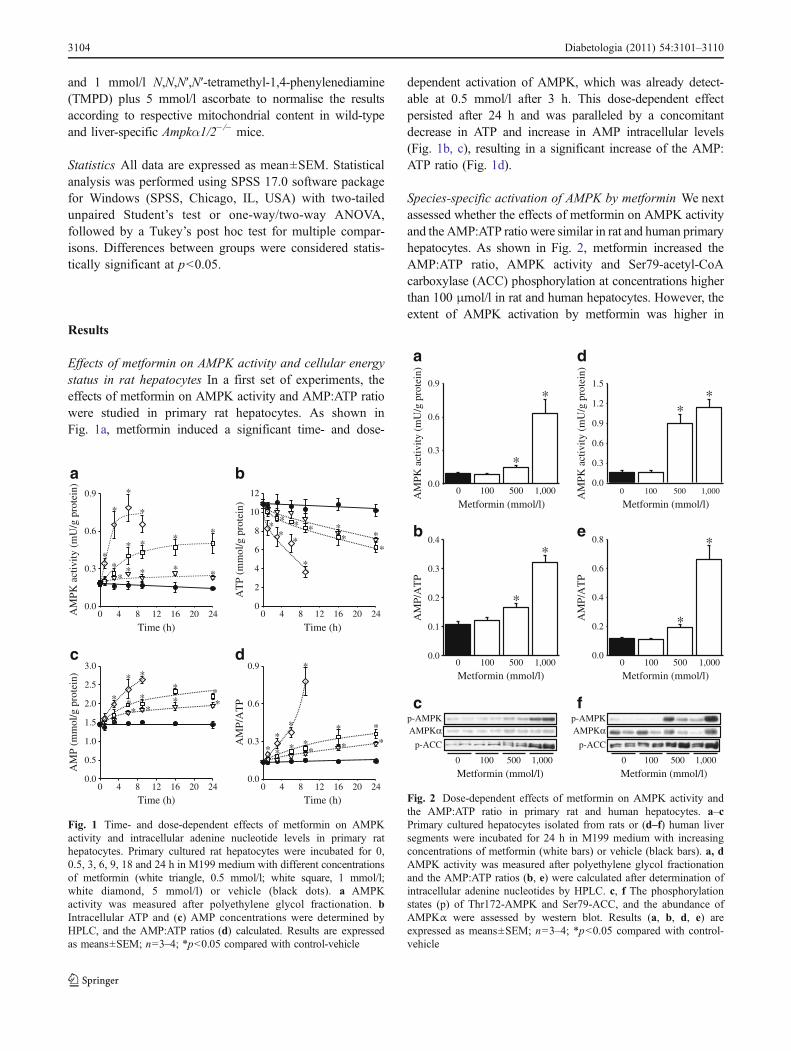
and 1 mmol/l N,N,N′,N′-tetramethyl-1,4-phenylenediamine(TMPD) plus 5 mmol/l ascorbate to normalise the resultsaccording to respective mitochondrial content in wild-typeand liver-specific Ampkα1/2−/− mice.
Statistics All data are expressed as mean±SEM. Statisticalanalysis was performed using SPSS 17.0 software packagefor Windows (SPSS, Chicago, IL, USA) with two-tailedunpaired Student’s test or one-way/two-way ANOVA,followed by a Tukey’s post hoc test for multiple compar-isons. Differences between groups were considered statis-tically significant at p<0.05.
Results
Effects of metformin on AMPK activity and cellular energystatus in rat hepatocytes In a first set of experiments, theeffects of metformin on AMPK activity and AMP:ATP ratiowere studied in primary rat hepatocytes. As shown inFig. 1a, metformin induced a significant time- and dose-
dependent activation of AMPK, which was already detect-able at 0.5 mmol/l after 3 h. This dose-dependent effectpersisted after 24 h and was paralleled by a concomitantdecrease in ATP and increase in AMP intracellular levels(Fig. 1b, c), resulting in a significant increase of the AMP:ATP ratio (Fig. 1d).
Species-specific activation of AMPK by metformin We nextassessed whether the effects of metformin on AMPK activityand the AMP:ATP ratio were similar in rat and human primaryhepatocytes. As shown in Fig. 2, metformin increased theAMP:ATP ratio, AMPK activity and Ser79-acetyl-CoAcarboxylase (ACC) phosphorylation at concentrations higherthan 100 μmol/l in rat and human hepatocytes. However, theextent of AMPK activation by metformin was higher in
Time (h)
0 4 8 12 16 20 24AM
PK a
ctiv
ity (
mU
/g p
rote
in)
0.0
0.3
0.6
0.9
a
c
*
*
*
*
*
* * **
*****
*
AT
P (m
mol
/g p
rote
in)
0
2
4
6
8
10
12
Time (h)
Time (h) Time (h)0 4 8 12 16 20 24
b
0.0
0.5
1.0
1.5
2.0
2.5
3.0
AM
P (m
mol
/g p
rote
in)
0 4 8 12 16 20 24
AM
P/A
TP
0.0
0.3
0.6
0.9
0 4 8 12 16 20 24
d
*
**
**
*
**
***
**
*
*
**
*****
**
** *
***
***
Fig. 1 Time- and dose-dependent effects of metformin on AMPKactivity and intracellular adenine nucleotide levels in primary rathepatocytes. Primary cultured rat hepatocytes were incubated for 0,0.5, 3, 6, 9, 18 and 24 h in M199 medium with different concentrationsof metformin (white triangle, 0.5 mmol/l; white square, 1 mmol/l;white diamond, 5 mmol/l) or vehicle (black dots). a AMPKactivity was measured after polyethylene glycol fractionation. bIntracellular ATP and (c) AMP concentrations were determined byHPLC, and the AMP:ATP ratios (d) calculated. Results are expressedas means±SEM; n=3–4; *p<0.05 compared with control-vehicle
0 100 500 1,000
0 100 500 1,000
Metformin (mmol/l) Metformin (mmol/l)0 100 500 1,000
Metformin (mmol/l) Metformin (mmol/l)0 100 500 1,000
*
*
*
**
*
*
d
e
*A
MPK
act
ivity
(m
U/g
pro
tein
)
0.0
0.3
0.6
0.9
0.0
0.3
0.6
0.9
1.2
1.5
AM
PK a
ctiv
ity (
mU
/g p
rote
in)
AM
P/A
TP
0.0
0.1
0.2
0.3
0.4
0.0
0.2
0.4
0.6
0.8
AM
P/A
TP
0 100 500 1,000
p-ACC
Metformin (mmol/l) Metformin (mmol/l)0 100 500 1,000
p-AMPKAMPKα
p-ACC
p-AMPKAMPKα
a
b
c f
Fig. 2 Dose-dependent effects of metformin on AMPK activity andthe AMP:ATP ratio in primary rat and human hepatocytes. a–cPrimary cultured hepatocytes isolated from rats or (d–f) human liversegments were incubated for 24 h in M199 medium with increasingconcentrations of metformin (white bars) or vehicle (black bars). a, dAMPK activity was measured after polyethylene glycol fractionationand the AMP:ATP ratios (b, e) were calculated after determination ofintracellular adenine nucleotides by HPLC. c, f The phosphorylationstates (p) of Thr172-AMPK and Ser79-ACC, and the abundance ofAMPKα were assessed by western blot. Results (a, b, d, e) areexpressed as means±SEM; n=3–4; *p<0.05 compared with control-vehicle
3104 Diabetologia (2011) 54:3101–3110

human than in rat hepatocytes at intermediate concentrationsof the drug (472% versus 66% at 500 μmol/l, respectively)(Fig. 2a, d), although the increase in the AMP:ATP ratio wassimilar (69% versus 54%, respectively) (Fig. 2b, e),suggesting a species-specific difference in AMPK sensitivitytoward AMP, ADP and/or ATP. Strikingly, the determinationof isoform-specific AMPKα activities after immunoprecipi-tation of the α1 or α2 AMPK catalytic subunits revealed thatAMPKα2 activity was undetectable in human comparedwith rat hepatocytes, while AMPKα1 activity was compa-rable in the basal condition (Fig. 3). Accordingly, metforminonly increased AMPKα1 activity in human hepatocytes,although both AMPKα isoforms were activated by the drugin rat hepatocytes. The apparent species-specific difference inAMPKα2 activity was confirmed by the absence of proteinand mRNA expression of this catalytic subunit in humanhepatocytes (Fig. 4). Interestingly, protein levels and mRNAhepatic expression of the β and γ regulatory subunits alsosignificantly differed between species, showing that the β2,γ1 and truncated γ2 forms are expressed in humanhepatocytes, whereas the β1, γ1 and γ2 forms are mostlypresent in rat and mouse hepatocytes (Fig. 4). We confirmedthat these species-specific differences in AMPK isoformlevels is also found in whole liver (ESM Fig. 1), excludingexperimental bias induced by hepatocytes isolation.
Taken together, the fact that AMPK activation bymetformin is associated with an elevated AMP:ATP ratioin hepatocytes strongly suggests that inhibition of themitochondrial machinery by the drug might be the mainunderlying mechanism for hepatic activation of the kinase.
Mechanism(s) of AMPK activation by metformin To inves-tigate the mechanisms by which metformin affects thecellular energy state, together with the putative involvementof AMPK in this process, we used hepatocytes from wild-
type and liver-specific Ampkα1/2−/− mice. Freshly isolatedhepatocytes were incubated with metformin, and AMPKactivity and expression, as well as the AMP:ATP ratio andJO2 were measured. As expected, metformin increased theAMP:ATP ratio, AMPK activity and Ser79-ACC phosphor-ylation in hepatocytes from wild-type mice (Fig. 5a–c). Inhepatocytes from liver-specific Ampkα1/2−/− mice, AMPKexpression, activity and activation could not be detected(Fig. 5a, b), but the increase in the AMP:ATP ratio inducedby metformin was still present and even significantly higherthan in hepatocytes from wild-type mice (Fig. 5c). Metfor-min induced a similar inhibition of JO2 in wild-type andliver-specific Ampkα1/2−/− mice, an effect that persistedafter addition of the mitochondrial oxidative phosphoryla-tion (OXPHOS) uncoupler DNP (Fig. 5d, e). This clearlyindicates that the inhibitory effect of metformin on JO2 wasexerted on the electron transfer chain rather than ondownstream step(s) linked to ATP synthesis. The effect ofmetformin in intact cells from wild-type and liver-specificAmpkα1/2−/− mice was further investigated after permeabi-lisation of the plasma membrane by digitonin, allowing themitochondrial OXPHOS pathway to be investigated in situ.In the presence of glutamate/malate, a substrate for therespiratory-chain complex 1, a significant decrease inmitochondrial respiratory rates could be detected aftermetformin pre-treatment of cells from wild-type and liver-specific Ampkα1/2−/− mice, occurring regardless of themitochondrial energy state (Fig. 5f, g). By contrast, nodifferences were observed with succinate/malate, a sub-strate for the respiratory-chain complex 2 (Fig. 5h, i).Importantly, a similar specific inhibition of the mitochon-drial respiratory-chain complex 1 by metformin was foundin freshly isolated human hepatocytes incubated in the sameconditions (Fig. 6).
Collectively, these results demonstrate that the inhibitionof JO2 and resultant increase in the AMP:ATP ratio inducedby metformin in hepatocytes is due to a specific andAMPK-independent inhibition of the mitochondrialrespiratory-chain complex 1.
Discussion
Collectively, our results show that activation of AMPK bymetformin in primary hepatocytes from rodents and humansis due to a decrease in cellular energy status resulting frommetformin’s AMPK-independent and specific inhibition ofmitochondrial respiratory-chain complex 1. Strikingly, wereport here for the first time that the distribution of AMPKcatalytic and regulatory subunits in hepatocytes differsbetween rodents and humans. This new important findingsuggests possible consequences for pharmaceutical strate-
a b
α1
*
0
10
20
120
150
*
AM
PK a
ctiv
ity (
mU
/mg
prot
ein)
0
10
20
120
150 *
AM
PK a
ctiv
ity (
mU
/mg
prot
ein)
α2 α1 α2
Fig. 3 Effects of metformin on AMPKα1 and α2 activities. a Primarycultured hepatocytes isolated from rats or (b) human liver segments wereincubated for 24 h in M199 medium in the presence of 500 μmol/lmetformin (white bars) or vehicle (black bars). AMPKα1 and α2activity was measured after immunoprecipitation with isoform-specificantibodies. Results are expressed as means±SEM; n=3–4; *p<0.05compared with control-vehicle
Diabetologia (2011) 54:3101–3110 3105

gies focusing on the development of AMPK activators forthe treatment of metabolic diseases.
The mitochondrial respiratory-chain complex 1 is theprimary target of metformin The inhibition of the mito-chondrial respiratory-chain complex 1 by metformin wasfirst reported in perfused livers and isolated rat hepatocytesby Leverve’s group [12] and later confirmed in variousother cellular models by us [21–25] and others [11, 13, 26,27]. Due to its weaker lipophilic property, metformin, incontrast to the other biguanides, induces only mild andspecific inhibition of the respiratory-chain complex 1 anddoes not affect OXPHOS downstream machinery (Figs 5and 6; B. Guigas, unpublished results). In the presentmanuscript, we show for the first time that this mitochon-drial effect of metformin is also present in humanhepatocytes, confirming that the respiratory-chain complex1 constitutes the primary target of the drug, whatever thespecies.
Although the exact mechanisms by which metformindecreases complex 1 activity remains unknown to date, ithas been shown that this effect requires intact cells [12, 22]and, at least in hepatocytes, is not prevented by inhibitionof nitric oxide synthase or by various reactive oxygenspecies scavengers [12]. On the other hand, the mechanismby which metformin activates AMPK is still beingdiscussed. Despite the indisputable mitochondrial effect of
the drug, Fryer et al. and Hawley et al. initially reported thatmetformin activated AMPK in muscle and rat hepatomacells without affecting the AMP:ATP ratio [14, 15]. In thepresent study, we clearly demonstrate that the activation ofAMPK by metformin is associated with an increased AMP:ATP ratio resulting from inhibition of the respiratory-chaincomplex 1. These discrepancies may be explained bytechnical differences in nucleotide measurement [28] andsubcellular AMP compartmentalisation [29], or by the factthat immortalised cell lines are highly glycolytic andtherefore much less sensitive to impairment of mitochon-drial OXPHOS for ATP supply [30]. Interestingly, metfor-min was shown to increase cytosolic AMP and AMPKactivity in heart [29]. In addition, the involvement ofcomplex 1 in the activation of AMPK by metformin hasalso been elegantly demonstrated in pancreatic MIN6 cells,by showing that methyl succinate, a substrate of complex 2that bypasses the inhibition of complex 1 by the drug,prevented AMPK activation [26]. Finally, two recentpublications also support the finding that metforminactivates AMPK via changes in cellular energy status. ThusHawley et al. showed that the activation of AMPK bymetformin is abolished in a cell line stably expressingAMPK complexes that contain an AMP-insensitive γ2mutant; this indicates that increased cytosolic AMP triggersactivation of the kinase by the drug [28]. On the other hand,Foretz et al. also demonstrated that, contrary to what was
a Human Rat
β1
β2
1
2
3
AMPKα1
β-Actin
AMPKβ2
AMPKβ1+2
AMPKα2
AMPKα
AMPKγ full-length
AMPKγ1
AMPKβ1
AMPKγ
Mouse
0
1
2
3
20
50
mR
NA
exp
ress
ion
(a.u
.)
α1 α2 β1 β2 γ γ all γ
b
AMPKγ truncated321
2
2
3
Fig. 4 a Protein levels of AMPK subunits and (b) the correspondingmRNA gene expression were determined in hepatocytes isolated fromhuman (black bars), rat (hatched bars) or mouse (white bars) livers bywestern blot and RT quantitative PCR, respectively. Western blotanalysis (a) is representative of three separate experiments and showsthat AMPK subunits migrated to their expected molecular mass
(α1, 62 kDa; α2, 62 kDa; β1, 38 kDa; β2, 30 kDa; γ1, 38 kDa;γ2full-length, ∼75 kDa; γ2truncated, ∼55 kDa; γ3, 55 kDa). The westernblots for AMPKα2 were performed using three different primaryantibodies cross-reacting with human, rat and mouse forms (see ESMTable 1). b The results from RT quantitative PCR were corrected forGAPDH and are expressed as means±SEM (n=3). a.u., arbitrary units
3106 Diabetologia (2011) 54:3101–3110

initially suggested by others [7], metformin inhibits hepaticgluconeogenesis by an LKB1- and AMPK-independentmechanism involving an acute decrease in the hepaticenergy state [31].
AMPK is not required for the inhibitory effect of metforminon respiratory-chain complex 1 Inmammalianmitochondria,the respiratory-chain complex 1 is made up of 45 subunits, ofwhich several can be phosphorylated by various Ser/Thrprotein kinases, thus affecting respiratory-chain complex 1
activity [32]. To rule out the possibility that the mitochondrialeffect of metformin is secondary to AMPK activation viasome putative AMP-independent mechanism, we usedAMPK-null hepatocytes isolated from liver-specificAmpkα1/2−/− mice [18, 30]. By showing that metformininhibited the respiratory-chain complex 1 in hepatocytes fromwild-type and liver-specific Ampkα1/2−/− mice, we canclearly rule out the notion that a primary effect of the drugis mediated by AMPK through phosphorylation of somesubunits of the respiratory-chain complex 1 and/or other
b
p-ACC
AMPKα1AMPKα2
Wild-type
β-Actin
a
Control
Wild-type
Metformin Control Metformin
c
*
† †
†*
*
d e
**
**
f g
h i
Basal DNP
State 3 State 4 DNP
*
*
* *
Basal DNP
State 3 State 4 DNP
State 3 State 4 DNP State 3 State 4 DNPWild-type
AM
PK a
ctiv
ity (
U/g
dry
cel
ls)
0
20
40
60
80
100
120
AM
P/A
TP
0.0
0.2
0.4
0.6
0.8
1.0
0
5
10
15
20
25
30
0
10
20
30
40
50
0
10
20
30
40
50
0
5
10
15
20
25
30
0
10
20
30
40
50
0
10
20
30
40
50
JO2
(% c
yt.o
x ac
tivity
) JO
2 (%
cyt
.ox
activ
ity)
JO2
(% c
yt.o
x ac
tivity
)
JO2
(% c
yt.o
x ac
tivity
) JO
2 (%
cyt
.ox
activ
ity)
JO2
(% c
yt.o
x ac
tivity
)
Ampk 1/2−/−α
Ampk 1/2−/−α
Ampk 1/2−/−α
Fig. 5 Effects of metformin on AMPK activity, AMP:ATP ratio, andcellular and mitochondrial JO2 in intact or permeabilised hepatocytesfrom wild-type and liver-specific Ampkα1/2−/− mice. Hepatocytesfrom wild-type or Ampkα1/2−/− mice were incubated at 37°C in aKrebs/bicarbonate medium containing lactate/pyruvate/octanoate (20,2, 4 mmol/l) in the presence of vehicle (black bars) or 5 mmol/lmetformin (white bars). After 30 min incubation, AMPK activity in U/gof dry cells (a), isoform-specific AMPKα abundance and Ser79-ACCphosphorylation (p) state (b), and the AMP:ATP ratio (c) weredetermined. d–i Oligomycin-sensitive JO2 was measured after thesuccessive addition of 6 μg/ml oligomycin (basal) and 100 μmol/l DNPin separate experiments, after hepatocytes from wild-type (d, f, h) and
liver-specific Ampkα1/2−/− (e, g, i) mice had been incubated asdescribed above and permeabilised in a KCl medium containing200 μg/ml digitonine. JO2 was measured in the presence ofglutamate/malate (5 mmol/l; 2.5 mmol/l) (f, g) or succinate/malate/rotenone (5 mmol/l; 0.5 mmol/l; 1.25 μmol/l) (h, i) after the successiveaddition of 1 mmol/l ADP (state 3), 6 μg/ml oligomycin (state 4) and75 μmol/l DNP. All JO2 results are expressed as percentage of themaximum activity of cytochrome oxidase (cyt.ox) determined after thefinal addition of 0.15 μg/ml antimycin and TMPD/ascorbate (1 mmol/l,5 mmol/l). Results are expressed as means±SEM; n=3; *p<0.05compared with control-vehicle; †p<0.05 compared with wild-type mice
Diabetologia (2011) 54:3101–3110 3107

mitochondrial proteins regulating OXPHOS. The more pro-nounced effect of metformin on the AMP:ATP ratio in primaryhepatocytes from liver-specific Ampkα1/2−/− compared withwild-type mice (Fig. 5) could be explained by a decrease inATP synthesis secondary to impaired mitochondrial biogen-esis and/or by a lack of AMPK-mediated inhibition of ATPconsumption, as previously reported [18, 30].
Species-specific differences in hepatic abundance of AMPKsubunits The AMPK protein is a heterotrimer composed of acatalytic α subunit and two regulatory β and γ subunits, in aratio of 1α:1β:1γ, all of which are required for the formation ofa stable and fully functional AMPK complex [8, 9]. Whilesome minor differences could be shown, depending on thefibre types, for the γ regulatory subunit [33], it is wellestablished that the α1 and α2 catalytic subunits are bothexpressed in human and rodent skeletal muscle, and that theα2β2γ1 complex constitutes the majority of AMPK hetero-trimers in this metabolic tissue, showing a high degree ofconsistency between species [33–37]. In the present study, themRNA expression pattern and protein levels of the AMPKsubunits found in rodent livers is in agreement with thosepreviously reported [35, 38], leading to many possiblecombinations of heterotrimers containing either α1 or α2catalytic subunits, and β1, γ1 and γ2 regulatory subunits. Asfar as we know, our finding showing that the abundance ofhepatic AMPK subunits differs in humans compared withrodents is unprecedented. Thus, it might be speculated that theresulting differences in heterotrimeric composition of AMPKcould affect the regulation of kinase activity, notably itssensitivity toward AMP. Indeed, it is striking that theactivation of AMPK by metformin seemed more potent,although the increase of the AMP:ATP ratio in humanscompared with rat primary hepatocytes was similar (Fig. 2),suggesting that the human AMPK α1β2γ1 or α1β2γ2truncated complexes could be more sensitive to subtle
changes in cellular energy status. However, this is incontradiction with previous in vitro results showing that, inrat liver extract, AMPK complexes containing the α2/β2isoforms had a greater dependence on AMP [35, 39].Interestingly, it has been very recently shown that ADP, likeAMP, could also bind to the γ subunit, leading to modulationof the AMP-triggered phosphorylation of AMPK on Thr172[10]. It seems therefore possible that subtle species-specificdifferences in the metformin-induced increase of the ADP:ATP ratio could also be involved. Unfortunately, thedetermination of intracellular ADP levels by HPLC wastechnically not possible in our conditions, so further inves-tigations are required to clarify this point. It is worthmentioning that AMPK has also been reported to have adifferential and tissue-specific localisation pattern in mamma-lian cells, with the AMPKα1 subunit being mainly localisedin the cytosol, and the AMPKα2 and β2 subunits beinglocalised to the nucleus and cytosolic fractions [39–42]. Whilenot as yet clarified, the subcellular localisation of the kinasemight have an important functional role, such as regulation ofgene expression by phosphorylation of nuclear targets ofAMPK. This would therefore suggest that differences in thecomposition of AMPK complexes between rodents andhuman hepatocytes could result in different physiologicaloutcomes, including putative nuclear regulatory functions.
Taken together, the species-specific differences shown inAMPKαβγ complexes in the liver imply that pharmaceu-tical activation of hepatic AMPK could have differenteffects in rodents and humans.
Therapeutic relevance of the metformin concentrationsused in vitro As shown in Fig. 1, the key determinant ofAMPK activation by metformin is a balance between theconcentration and the time of exposure to the drug. It isworth noting that most of the in vitro experiments reportingactivation of AMPK by metformin were generally performedwith drug concentrations far above those found in tissuesfrom rodents after oral administration of metformin [43, 44].In the present study, we show that hepatic AMPK isactivated by metformin at concentrations higher than100 μmol/l, i.e. about five times the highest plasma levelreported in humans after a single oral drug administration[45, 46]. However, the liver is one of the few organs that canaccumulate significant amounts of metformin, with tissueconcentrations that reached hundreds of μmol/l in theperiportal area [43, 44]. In addition, the intracellular transportof metformin is mediated by the organic cation transporter 1(OCT1) [47], the deletion of which in hepatocytes resulted inimpairment of metformin-induced AMPK activation [48].Interestingly, increased OCT1 levels and metformin concen-trations were recently reported in the liver of mice on a high-fat diet [49], suggesting that hepatic accumulation of thedrug could be even higher in diabetic patients.
0
25
50
75
100
State 3 State 4 DNP0
25
50
75
100
State 3 State 4 DNP
JO2
(% c
yt.o
x ac
tivity
)
JO2
(% c
yt.o
x ac
tivity
)
a b
**
Fig. 6 Effects of metformin on JO2 in permeabilised humanhepatocytes. Hepatocytes isolated from human liver segments wereincubated in the presence of vehicle (black bars) or 5 mmol/l metformin(white bars), and the JO2 was measured in the presence of (a)glutamate/malate (5 mmol/l, 2.5 mmol/l) or (b) succinate/malate/rotenone (5 mmol/l; 0.5 mmol/l; 1.25 μmol/l) as described (Fig. 5).The results are expressed as means±SEM; n=3; *p<0.05 comparedwith control-vehicle
3108 Diabetologia (2011) 54:3101–3110

Conclusions
We have demonstrated here that the primary target ofmetformin in rodent and human hepatocytes is themitochondrial machinery. The specific inhibition of therespiratory-chain complex 1 by the drug leads to a dose-and time-dependent decrease in ATP levels, which results ina concomitant increase in cytosolic AMP concentrations(and the AMP:ATP ratio), triggering the activation ofAMPK. One of the important new findings of this studyis the species-specific differences in the profile of AMPKsubunits, suggesting that regulation of the kinase, which iswell characterised in rodents, could differ significantly inhumans. Further investigations are still required, but thispoint should certainly be considered when developing newpharmacological agents that target AMPK.
Acknowledgements The authors are grateful to L. Maisin and M. deCloedt for technical assistance. This work was supported by FNRS,the Inter University Pole of Attraction, the Association pour l’Etudedes Diabètes et des Maladies Métaboliques (ALFEDIAM), AgenceNationale de la Recherche (ANR-10-BLAN-1123-01) and the Euro-pean Union FP6 programme (Exgenesis/LSHM-CT-2004-005272). B.Guigas was recipient of the ICP “Michel de Visscher” Fellowship.
Contribution statement XS, NT and GCvdZ performed experi-ments, analysed data, and critically reviewed the manuscript; MF andBV provided mouse model and reagents, contributed to discussion andcritically reviewed the manuscript; ES and LH contributed todiscussion and critically reviewed the manuscript; BG conceptualisedthe project, performed experiments, analysed data, wrote and editedthe manuscript. All authors approved the final version of themanuscript.
Duality of interest The authors declare that there is no duality ofinterest associated with this manuscript.
Open Access This article is distributed under the terms of the CreativeCommons Attribution Noncommercial License which permits anynoncommercial use, distribution, and reproduction in any medium,provided the original author(s) and source are credited.
References
1. Goodarzi MO, Bryer-Ash M (2005) Metformin revisited: re-evaluation of its properties and role in the pharmacopoeia ofmodern antidiabetic agents. Diabetes Obes Metab 7:654–665
2. Nathan DM, Buse JB, Davidson MB et al (2009) Medicalmanagement of hyperglycaemia in type 2 diabetes mellitus: aconsensus algorithm for the initiation and adjustment of therapy: aconsensus statement from the American Diabetes Association andthe European Association for the Study of Diabetes. Diabetologia52:17–30
3. Natali A, Ferrannini E (2006) Effects of metformin andthiazolidinediones on suppression of hepatic glucose productionand stimulation of glucose uptake in type 2 diabetes: a systematicreview. Diabetologia 49:434–441
4. UKPDS (1998) Effect of intensive blood-glucose control withmetformin on complications in overweight patients with type 2diabetes (UKPDS 34). UK Prospective Diabetes Study (UKPDS)Group. Lancet 352:854–865
5. Ben Sahra I, Le Marchand-Brustel Y, Tanti JF, Bost F (2010)Metformin in cancer therapy: a new perspective for an oldantidiabetic drug? Mol Cancer Ther 9:1092–1099
6. Zhou G, Myers R, Li Y et al (2001) Role of AMP-activatedprotein kinase in mechanism of metformin action. J Clin Invest108:1167–1174
7. Shaw RJ, Lamia KA, Vasquez D et al (2005) The kinase LKB1mediates glucose homeostasis in liver and therapeutic effects ofmetformin. Science 310:1642–1646
8. Steinberg GR, Kemp BE (2009) AMPK in health and disease.Physiol Rev 89:1025–1078
9. Hardie DG (2007) AMP-activated/SNF1 protein kinases: conservedguardians of cellular energy. Nat Rev Mol Cell Biol 8:774–785
10. Oakhill JS, Steel R, Chen ZP et al (2011) AMPK is a direct adenylatecharge-regulated protein kinase. Science 332:1433–1435
11. Brunmair B, Staniek K, Gras F et al (2004) Thiazolidinediones, likemetformin, inhibit respiratory complex I: a common mechanismcontributing to their antidiabetic actions? Diabetes 53:1052–1059
12. El-Mir MY, Nogueira V, Fontaine E, Averet N, Rigoulet M,Leverve X (2000) Dimethylbiguanide inhibits cell respiration viaan indirect effect targeted on the respiratory chain complex I. JBiol Chem 275:223–228
13. Owen MR, Doran E, Halestrap AP (2000) Evidence that metforminexerts its anti-diabetic effects through inhibition of complex 1 of themitochondrial respiratory chain. Biochem J 348:607–614
14. Fryer LG, Parbu-Patel A, Carling D (2002) The anti-diabeticdrugs rosiglitazone and metformin stimulate AMP-activatedprotein kinase through distinct signaling pathways. J Biol Chem277:25226–25232
15. Hawley SA, Gadalla AE, Olsen GS, Hardie DG (2002) Theantidiabetic drug metformin activates the AMP-activated proteinkinase cascade via an adenine nucleotide-independent mechanism.Diabetes 51:2420–2425
16. Hardie DG (2006) Neither LKB1 nor AMPK are the direct targetsof metformin. Gastroenterology 131:973
17. Fujita Y, HosokawaM, Fujimoto S et al (2010) Metformin suppresseshepatic gluconeogenesis and lowers fasting blood glucose levelsthrough reactive nitrogen species inmice. Diabetologia 53:1472–1481
18. Guigas B, Bertrand L, Taleux N et al (2006) 5-Aminoimidazole-4-carboxamide-1-beta-D-ribofuranoside and metformin inhibit hepaticglucose phosphorylation by an AMP-activated protein kinase-independent effect on glucokinase translocation. Diabetes 55:865–874
19. Berry MN, Friend DS (1969) High-yield preparation of isolatedrat liver parenchymal cells: a biochemical and fine structuralstudy. J Cell Biol 43:506–520
20. Groen AK, Sips HJ, Vervoorn RC, Tager JM (1982) Intracellularcompartmentation and control of alanine metabolism in rat liverparenchymal cells. Eur J Biochem 122:87–93
21. Detaille D, Guigas B, Chauvin C et al (2005) Metformin preventshigh-glucose-induced endothelial cell death through a mitochondrialpermeability transition-dependent process. Diabetes 54:2179–2187
22. Detaille D, Guigas B, Leverve X, Wiernsperger N, Devos P(2002) Obligatory role of membrane events in the regulatoryeffect of metformin on the respiratory chain function. BiochemPharmacol 63:1259–1272
23. El-Mir MY, Detaille D, R-Villanueva G et al (2008) Neuro-protective role of antidiabetic drug metformin against apoptoticcell death in primary cortical neurons. J Mol Neurosci 34:77–87
24. Guigas B, Detaille D, Chauvin C et al (2004) Metformin inhibitsmitochondrial permeability transition and cell death: a pharmaco-logical in vitro study. Biochem J 382:877–884
Diabetologia (2011) 54:3101–3110 3109

25. Batandier C, Guigas B, Detaille D et al (2006) The ROS productioninduced by a reverse-electron flux at respiratory-chain complex 1 ishampered by metformin. J Bioenerg Biomembr 38:33–42
26. Hinke SA, Martens GA, Cai Y et al (2007) Methyl succinateantagonises biguanide-induced AMPK-activation and death ofpancreatic beta-cells through restoration of mitochondrial electrontransfer. Br J Pharmacol 150:1031–1043
27. Turner N, Li JY, Gosby A et al (2008) Berberine and its morebiologically available derivative, dihydroberberine, inhibit mito-chondrial respiratory complex I: a mechanism for the action ofberberine to activate AMP-activated protein kinase and improveinsulin action. Diabetes 57:1414–1418
28. Hawley SA, Ross FA, Chevtzoff C et al (2010) Use of cellsexpressing gamma subunit variants to identify diverse mecha-nisms of AMPK activation. Cell Metab 11:554–565
29. Zhang L, He H, Balschi JA (2007) Metformin and phenforminactivate AMP-activated protein kinase in the heart by increasingcytosolic AMP concentration. Am J Physiol Heart Circ Physiol293:H457–H466
30. Guigas B, Taleux N, Foretz M et al (2007) AMP-activated proteinkinase-independent inhibition of hepatic mitochondrial oxidativephosphorylation by AICA riboside. Biochem J 404:499–507
31. Foretz M, Hebrard S, Leclerc J et al (2010) Metformin inhibits hepaticgluconeogenesis in mice independently of the LKB1/AMPK pathwayvia a decrease in hepatic energy state. J Clin Invest 120:2355–2369
32. Papa S, de Rasmo D, Scacco S et al (2008) Mammalian complexI: a regulable and vulnerable pacemaker in mitochondrialrespiratory function. Biochim Biophys Acta 1777:719–728
33. Mahlapuu M, Johansson C, Lindgren K et al (2004) Expressionprofiling of the gamma-subunit isoforms of AMP-activatedprotein kinase suggests a major role for gamma3 in white skeletalmuscle. Am J Physiol Endocrinol Metab 286:E194–E200
34. Wojtaszewski JF, Birk JB, Frosig C, Holten M, Pilegaard H, DelaF (2005) 5'AMP activated protein kinase expression in humanskeletal muscle: effects of strength training and type 2 diabetes. JPhysiol 564:563–573
35. Cheung PC, Salt IP, Davies SP, Hardie DG, Carling D (2000)Characterization of AMP-activated protein kinase gamma-subunitisoforms and their role in AMP binding. Biochem J 346:659–669
36. Birk JB, Wojtaszewski JF (2006) Predominant alpha2/beta2/gamma3 AMPK activation during exercise in human skeletalmuscle. J Physiol 577:1021–1032
37. Treebak JT, Glund S, Deshmukh A et al (2006) AMPK-mediated AS160 phosphorylation in skeletal muscle is depen-
dent on AMPK catalytic and regulatory subunits. Diabetes55:2051–2058
38. Quinn JM, Tam S, Sims NA et al (2010) Germline deletion ofAMP-activated protein kinase beta subunits reduces bone masswithout altering osteoclast differentiation or function. FASEB J24:275–285
39. Salt I, Celler JW, Hawley SA et al (1998) AMP-activated proteinkinase: greater AMP dependence, and preferential nuclearlocalization, of complexes containing the alpha2 isoform. Bio-chem J 334:177–187
40. Kazgan N, Williams T, Forsberg LJ, Brenman JE (2010)Identification of a nuclear export signal in the catalyticsubunit of AMP-activated protein kinase. Mol Biol Cell 21:3433–3442
41. Kodiha M, Rassi JG, Brown CM, Stochaj U (2007) Localizationof AMP kinase is regulated by stress, cell density, and signalingthrough the MEK–>ERK1/2 pathway. Am J Physiol Cell Physiol293:C1427–C1436
42. Suzuki A, Okamoto S, Lee S, Saito K, Shiuchi T, Minokoshi Y(2007) Leptin stimulates fatty acid oxidation and peroxisomeproliferator-activated receptor alpha gene expression in mouseC2C12 myoblasts by changing the subcellular localization of thealpha2 form of AMP-activated protein kinase. Mol Cell Biol27:4317–4327
43. Wilcock C, Bailey CJ (1994) Accumulation of metformin bytissues of the normal and diabetic mouse. Xenobiotica 24:49–57
44. Wilcock C, Wyre ND, Bailey CJ (1991) Subcellular distributionof metformin in rat liver. J Pharm Pharmacol 43:442–444
45. Tucker GT, Casey C, Phillips PJ, Connor H, Ward JD, Woods HF(1981) Metformin kinetics in healthy subjects and in patients withdiabetes mellitus. Br J Clin Pharmacol 12:235–246
46. Graham GG, Punt J, Arora M et al (2011) Clinical pharmacoki-netics of metformin. Clin Pharmacokinet 50:81–98
47. Wang DS, Jonker JW, Kato Y, Kusuhara H, Schinkel AH,Sugiyama Y (2002) Involvement of organic cation transporter 1in hepatic and intestinal distribution of metformin. J PharmacolExp Ther 302:510–515
48. Shu Y, Sheardown SA, Brown C et al (2007) Effect of geneticvariation in the organic cation transporter 1 (OCT1) on metforminaction. J Clin Invest 117:1422–1431
49. Jang EH, Kim HK, Park CS, Kang JH (2010) Increasedexpression of hepatic organic cation transporter 1 and hepaticdistribution of metformin in high-fat diet-induced obese mice.Drug Metab Pharmacokinet 25:392–397
3110 Diabetologia (2011) 54:3101–3110