From Nucleic Acid Therapeutics to COVID‐19 Vaccines - IRIS ...
Upload
khangminh22Category
view
0download
0
In previous chapters, we studiedtwo of the three major kinds ofbiopolymers—polysaccharides and
proteins. Now we will look at thethird—nucleic acids. There are two typesof nucleic acids—deoxyribonucleic acid (DNA)and ribonucleic acid (RNA). DNA encodes an organism’s entire hereditary informa-tion and controls the growth and division of cells. In most organisms, the genetic infor-mation stored in DNA is transcribed into RNA. This information can then be translatedfor the synthesis of all the proteins needed for cellular structure and function.
DNA was first isolated in 1869 from the nuclei of white blood cells. Because thismaterial was found in the nucleus and was acidic, it was called nucleic acid. Eventual-ly, scientists found that the nuclei of all cells contain DNA, but it wasn’t until 1944that they realized that nucleic acids are the carriers of genetic information. In 1953,James Watson and Francis Crick described the three-dimensional structure of DNA—the famed double helix.
27.1 Nucleosides and Nucleotides
Nucleic acids are chains of five-membered-ring sugars linked by phosphate groups(Figure 27.1). The anomeric carbon of each sugar is bonded to a nitrogen of a hetero-cyclic compound in a linkage. (Recall from Section 22.10 that a -linkageis one in which the substituents at C-1 and C-4 are on the same side of the furanose ring.)Because the heterocyclic compounds are amines, they are commonly referred to asbases. In RNA the five-membered-ring sugar is D-ribose. In DNA it is 2-deoxy-D-ribose(D-ribose without an OH group in the 2-position).
Phosphoric acid links the sugars in both RNA and DNA. The acid has threedissociable OH groups with values of 1.9, 6.7, and 12.4. Each of the OH groupscan react with an alcohol to form a phosphomonoester, a phosphodiester, or aphosphotriester. In nucleic acids the phosphate group is a phosphodiester.
pKa
bb-glycosidic
an RNA catalyst
032
27 Nucleosides, Nucleotides,and Nucleic Acids
Studies that determined the structuresof the nucleic acids and paved theway for the discovery of the DNAdouble helix were carried out byPhoebus Levene and elaborated bySir Alexander Todd.
Phoebus Aaron Theodor Levene(1869–1940) was born in Russia.When he immigrated to the UnitedStates with his family in 1891, hisRussian name Fishel was changed toPhoebus. Because his medical schooleducation had been interrupted, hereturned to Russia to complete hisstudies. When he returned to theUnited States, he took chemistrycourses at Columbia University.Deciding to forgo medicine for acareer in chemistry, he went toGermany to study under EmilFischer. He was a professor ofchemistry at the Rockefeller Institute(now Rockefeller University).
BRUI27-32_70r2 26-02-2003 3:51 PM Page 032

Section 27.1 Nucleosides and Nucleotides 033
THE STRUCTURE OF DNA: WATSON, CRICK,FRANKLIN, AND WILKINSJames D. Watson was born in Chicago in 1928. He graduated from
the University of Chicago at the age of 19 and received a Ph.D. three years laterfrom Indiana University. In 1951, as a postdoctoral fellow at Cambridge Universi-ty, Watson worked on determining the three-dimensional structure of DNA.
Francis H. C. Crick was born in Northampton, England, in 1916. Originallytrained as a physicist, Crick was involved in radar research during World War II.After the war, he entered Cambridge University to study for a Ph.D. in chemistry,which he received in 1953. He was a graduate student when he carried out his por-tion of the work that led to the proposal of the double helical structure of DNA.
Rosalind Franklin was born in London in 1920. She graduated from CambridgeUniversity and in 1942 quit her graduate studies to accept a position as a researchofficer in the British Coal Utilisation Research Association. After the war, she stud-ied X-ray diffraction techniques in Paris. In 1951 she returned to England, accept-ing a position to develop an X-ray diffraction unit in the biophysics department atKing’s College. Her X-ray studies showed that DNA was a helix with phosphategroups on the outside of the molecule. Franklin died in 1958 without knowing thesignificance her work had played in determining the structure of DNA.
Watson and Crick shared the 1962 Nobel Prize in medicine or physiology withMaurice Wilkins for determining the double helical structure of DNA. Wilkinscontributed X-ray studies that confirmed the double helical structure. Wilkins wasborn in New Zealand in 1916 and moved to England six years later with his par-ents. During World War II he joined other British scientists who were working withAmerican scientists on the development of the atomic bomb. Rosalind
Franklin
FrancisCrick andJamesWatson
RNA DNA
O base
O
O
O O−P
OH
base
O
O
O O−P
OH
base
O OH
O
O
OO
O
O
O base
O
O
O O−P
base
O
O
O O−P
base
O
a -glycosidiclinkage
2′-OH group
a phosphodiester
a -glycosidiclinkage
anomericcarbon
no 2′-OH group
� Figure 27.1Nucleic acids consist of a chain of five-membered-ring sugars linked by phosphate groups.Each sugar (D-ribose in RNA, -deoxy-D-ribose in DNA) is bonded to a heterocyclic aminein a -glycosidic linkage.b
2¿
Alexander R. Todd (1907–1997)was born in Scotland. He receivedtwo Ph.D. degrees, one from JohannWolfgang Goethe University inFrankfurt (1931) and one fromOxford University (1933). He was aprofessor of chemistry at theUniversity of Edinburgh, at theUniversity of Manchester, and from1944 to 1971 at CambridgeUniversity. He was knighted in 1954and was made a baron in 1962(Baron Todd of Trumpington). For hiswork on nucleotides, he was awardedthe 1957 Nobel Prize in chemistry.
AU: Please identify by
indicating left or right
AU: ok prime here?
BRUI27-32_70r2 26-02-2003 3:51 PM Page 033

The vast differences in heredity among species and among members of the samespecies are determined by the sequence of the bases in DNA. Surprisingly, there areonly four bases in DNA—two are substituted purines (adenine and guanine), and twoare substituted pyrimidines (cytosine and thymine).
RNA also contains only four bases. Three (adenine, guanine, and cytosine) are thesame as those in DNA, but the fourth base in RNA is uracil instead of thymine. Noticethat thymine and uracil differ only by a methyl group—thymine is 5-methyluracil. Thereason DNA contains thymine instead of uracil is explained in Section 27.14.
The purines and pyrimidines are bonded to the anomeric carbon of the furanosering—purines at N-9 and pyrimidines at N-1—in a -glycosidic linkage. A com-pound containing a base bonded to D-ribose or to 2-deoxy-D-ribose is called anucleoside. In a nucleoside the ring positions of the sugar are indicated by primednumbers to distinguish them from the ring positions of the base. This is why thesugar component of DNA is referred to as -deoxy-D-ribose. Notice the differencein the base names and their corresponding nucleoside names in Table 27.1. For ex-ample, adenine is the base, whereas adenosine is the nucleoside. Similarly, cyto-sine is the base, whereas cytidine is the nucleoside, and so forth. Because uracil isfound only in RNA, it is shown attached to D-ribose but not to 2-deoxy-D-ribose;because thymine is found only in DNA, it is shown attached to 2-deoxy-D-ribosebut not to D-ribose.
2¿
b
phosphoric acid
O
OH
POHHO
OHORHO ORRO ORRO
a phosphomonoester
O
P
a phosphodiester
O
OH
P
a phosphotriester
O
OR
P
034 C H A P T E R 2 7 Nucleosides, Nucleotides, and Nucleic Acids
adenosine
nucleosides
N
N N
NH2
O
HO
HO HO
OH
N
guanosine
NN
HN N
O
H2N
cytidine
N
N
NH2
O O
uridine
N
HN
O
O
HO OH
O
HO OH
O
HO OH
HO HO
purine
N
N
N1
2
3
6 7
8
94
5
NH
NH2 NH2CH3
H2N
pyrimidine
N
N3
2
1
4
6
5
NH
N
O
adenine
N
N
N
NH
O
guanine cytosine
O
NH
HN HN
O
uracil
O
NH
O
thymine
N
N
HN
NH
nucleoside = base + sugar
BRUI27-32_70r2 26-02-2003 3:51 PM Page 034

Section 27.1 Nucleosides and Nucleotides 035
CH3
H2N
2′-deoxyadenosine
NN
1′
2′3′4′
5′
N N
NH2
2′-deoxyguanosine
NN
HN N
O
2′-deoxycytidine
N
N
NH2
O O
thymidine
N
HN
O
O
HO
O
HO
O
HO
O
HO
HOHOHOHO
3-D Molecules:Bases; Nucleosides;Nucleotides
Table 27.1 The Names of the Bases, the Nucleosides, and the Nucleotides
Base Nucleoside Ribonucleotide Deoxyribonucleotide
Adenine Adenosine Adenosine
Guanine Guanosine Guanosine
Cytosine Cytidine Cytidine
Thymine — Thymidine — Thymidine
Uracil Uridine — Uridine 5¿-phosphate
5¿-phosphate
5¿-phosphate2¿-Deoxycytidine5¿-phosphate2¿-Deoxycytidine
5¿-phosphate2¿-Deoxyguanosine5¿-phosphate2¿-Deoxyguanosine
5¿-phosphate2¿-Deoxyadenosine5¿-phosphate2¿-Deoxyadenosine
PROBLEM 1
In acidic solutions, nucleosides are hydrolyzed to a sugar and a heterocyclic base. Proposea mechanism for this reaction.
A nucleotide is a nucleoside with either the or the -OH group bonded in anester linkage to phosphoric acid. The nucleotides of RNA—where the sugar isD-ribose—are more precisely called ribonucleotides, whereas the nucleotides ofDNA—where the sugar is 2-deoxy-D-ribose—are called deoxyribonucleotides.
When phosphoric acid is heated with it loses water, forming a phosphoanhy-dride called pyrophosphoric acid. Its name comes from pyr, the Greek word for “fire.”Thus, pyrophosphoric acid is prepared by “fire”—that is, by heating. Triphosphoricacid and higher polyphosphoric acids are also formed.
P2O5
nucleotides
adenosine 5′-monophosphatea ribonucleotide
NH2
N
NN
N
−O
O
O
O
HO OH
O−
P
2′-deoxycytidine 3′-monophosphatea deoxyribonucleotide
NH2
N
N
O
O
O
O−
O
O−
P
HO
5′-position
phosphategroup
3′-position
3¿5¿- nucleotide = base + sugar + phosphate
BRUI27-32_70r2 26-02-2003 3:51 PM Page 035

036 C H A P T E R 2 7 Nucleosides, Nucleotides, and Nucleic Acids
Because phosphoric acid can form an anhydride, nucleotides can exist as monophos-phates, diphosphates, and triphosphates. They are named by adding monophosphate ordiphosphate or triphosphate to the name of the nucleoside.
PROBLEM 2
Draw the structure for each of the following:
a. dCDP c. dUMP e. guanosine b. dTTP d. UDP f. adenosine
27.2 ATP: The Carrier of Chemical Energy
All cells require energy to ensure their survival and reproduction. They get theenergy they need by converting nutrients into a chemically useful form of energy.The most important form of chemical energy is adenosine -triphosphate (ATP).The importance of ATP to biological reactions is shown by its turnover rate in hu-mans—each day, a person uses an amount of ATP equivalent to his or her bodyweight. ATP is known as the universal carrier of chemical energy because, as it iscommonly stated, “the energy of hydrolysis of ATP converts endergonic reactionsinto exergonic reactions.”
In other words, the ability of ATP to enable otherwise unfavorable reactions tooccur is attributed to the large amount of energy released when ATP is hydrolyzed,which can be used to drive an endergonic reaction. For example, the reaction of
5¿
3¿-monophosphate5¿-triphosphate
phosphoric acid pyrophosphoric acid triphosphoric acid
O
OH
P+∆
P2O5OH OHO5
O
OH
POH
O
OH
PHO O
O
OH
PO
O
OH
POH
O
OH
PHO
N
N
N
N
−O O
O
P
O−
NH2
O
HO OHadenosine
5′-monophosphateAMP
N
N
N
N
OO
O
P
O−−O
O
P
O−
NH2
O
HO OHadenosine
5′-diphosphateADP
N
N
N
N
OO
O
P
O−−O
O
P
O−O
O
P
O−
NH2
O
HO OHadenosine
5′-triphosphateATP
N
N
N
N
−O O
O
P
O−
NH2
O
HO2′-deoxyadenosine5′-monophosphate
dAMP
N
N
N
N
OO
O
P
O−−O
O
P
O−
NH2
O
HO2′-deoxyadenosine
5′-diphosphatedADP
N
N
N
N
OO
O
P
O−−O
O
P
O−O
O
P
O−
NH2
O
HO2′-deoxyadenosine
5′-triphosphatedATP
BRUI27-32_70r2 26-02-2003 3:51 PM Page 036

Section 27.2 ATP: The Carrier of Chemical Energy 037
3-D Molecules:Adenosine -triphosphate(ATP)
5¿
1The prime in indicates that two additional parameters have been added to the defined inSection 3.7—the reaction occurs in aqueous solution at and the concentration of water isassumed to be constant.
pH = 7¢G°¢G°¿
D-glucose with hydrogen phosphate to form D-glucose-6-phosphate is endergonicor .1 The hydrolysis of ATP, on the other
hand, is highly exergonic or When the tworeactions are added together (the species occurring on both sides of the reaction arrowcancel), the net reaction is exergonic or Thus, the energy released from the hydrolysis of ATP is more than enough to drive thephosphorylation of D-glucose. Two reactions in which the energy of one is used todrive the other are known as coupled reactions.
This nonmechanistic description of ATP’s power makes ATP look like a magicalsource of energy. Let’s look at the mechanism of the reaction to see what really hap-pens. The reaction is a simple one-step nucleophilic substitution reaction. The 6-OHgroup of glucose attacks the terminal phosphate of ATP, breaking a phosphoanhydridebond without forming an intermediate. Essentially it is an reaction with anadenosine pyrophosphate leaving group.
Now we have a chemical understanding of why the phosphorylation of glucose requiresATP. Without ATP, the 6-OH group of D-glucose would have to displace a very basic
group from hydrogen phosphate. With ATP, the 6-OH group of D-glucose displacesthe weakly basic ADP.
too basic to be displaced
−O OH
O
O−
P+
H
H H
HOHO
HO
H
CH2OH
OH
OH
hydrogenphosphate
-OH
OO
O
P
O−−O
O
P
O−O
O
P
O−
adenosine
ATP
H
OH
O
+
a phosphoanhydride bond
O
O
P
O−−O
−O
O
O
O
P
P
O−
O−
adenosine
ADP
+
D-glucose
D-glucose-6-phosphate
HOOH
CH2OHHO
HO
H
OH
O
HOOH
CH2O
SN2
-16.7 kJ>mol).(¢G°¿ = -4.0 kcal>mol
-30.5 kJ>mol).(¢G°¿ = -7.3 kcal>mol+13.8 kJ>mol)(¢G°¿ = +3.3 kcal>mol
D-glucose hydrogen phosphate+ D-glucose-6-phosphate H2O 3.3 kcal/mol
∆G°′
+ +
D-glucose ATP+ D-glucose-6-phosphate ADP 4.0 kcal/mol+ −
ATP H2O+ ADP hydrogen phosphate 7.3 kcal/mol+ −
13.8 kJ/mol+
16.7 kJ/mol−
30.5 kJ/mol−
or
or
or
BRUI27-32_70r2 26-02-2003 3:51 PM Page 037

038 C H A P T E R 2 7 Nucleosides, Nucleotides, and Nucleic Acids
Although the phosphorylation of glucose is described as being driven by the“hydrolysis” of ATP, you can see from the mechanism that glucose does not reactwith hydrogen phosphate and that ATP is not hydrolyzed because it does notreact with water. In other words, neither of the coupled reactions actually occurs.Instead, the phosphate group of ATP is transferred directly to D-glucose.
The transfer of a phosphate group from ATP to D-glucose is an example of aphosphoryl transfer reaction. There are many phosphoryl transfer reactions inbiological systems. In all of these reactions, the electrophilic phosphate group istransferred to a nucleophile as a result of breaking a phosphoanhydride bond. Thisexample of a phosphoryl transfer reaction demonstrates the actual chemicalfunction of ATP—it provides a reaction pathway involving a good leaving groupfor a reaction that cannot occur (or would occur very slowly) because of a poorleaving group.
PROBLEM 3 SOLVED
The hydrolysis of phosphoenolpyruvate is so highly exergonic orthat it can be used to “drive the formation” of ATP from ADP and hydrogen
phosphate or Propose a mechanism for this reaction.
SOLUTION As we saw in the example with ATP, neither of the coupled reactions actu-ally occurs: phosphoenolpyruvate does not react with water and ADP does not react withhydrogen phosphate. Just as ATP “drives the formation” of glucose-6-phosphate by sup-plying glucose (a nucleophile) with a phosphate that has a good leaving group (ADP),phosphoenolpyruvate “drives the formation” of ATP by supplying ADP (a nucleophile)with a phosphate that has a good leaving group (pyruvate).
PROBLEM 4�
Why is pyruvate a good leaving group?
PROBLEM 5�
Several important biomolecules and the values for their hydrolysis are listed here.Which of them “hydrolyzes” with sufficient energy to “drive the formation” of ATP?
glycerol-1-phosphate: phosphocreatine:
fructose-6-phosphate: glucose-6-phosphate:-3.3 kcal>mol (13.8 kJ>mol).-3.8 kcal>mol3 (-15.9 kJ>mol)
-11.8 kcal>mol (-49.4 kJ>mol)-2.2 kcal>mol (-9.2 kJ>mol)
¢G°¿
+30.5 kJ>mol).(¢G°¿ = +7.3 kcal>mol-61.9 kJ>mol)
(¢G°¿ = -14.8 kcal>mol
OO
O
P
O−
O
P
O−
O
OO
O
P
O−O−
O− adenosineOO
O
P
O−
O
P
O−O
O
P
O−
adenosineCH2 C C
O
O−CH2 C C+ +−
−O
O
OH
O−CH2 C C
−O
H2O
tautomerization
ADP ATP
phosphoenolpyruvate O
O
O−CH3 C C
pyruvate
ATP provides a reaction pathway with agood leaving group for a reaction thatcannot occur because of a poor leavinggroup.
BRUI27-32_70r2 26-02-2003 3:51 PM Page 038

Section 27.3 Three Mechanisms for Phosphoryl Transfer Reactions 039
27.3 Three Mechanisms for PhosphorylTransfer Reactions
There are three possible mechanisms for a phosphoryl transfer reaction. We will illus-trate them using the following nucleophilic acyl substitution reaction.
This reaction does not occur without ATP because the negatively charged carboxylateion resists nucleophilic attack and, if the tetrahedral intermediate could be formed,the incoming nucleophile is a weaker base than the base that would have to be ex-pelled from the tetrahedral intermediate to form the thioester. In other words, thethiol would be expelled from the tetrahedral intermediate, reforming the carboxylateion (Section 17.5).
If ATP is added to the reaction mixture, the reaction occurs. The carboxylate ion at-tacks one of the phosphate groups of ATP, breaking a phosphoanhydride bond. Thisputs a leaving group on the carboxyl group that can be displaced by the thiol. There arethree possible mechanisms for the reaction of a nucleophile with ATP because each ofthe three phosphorus atoms of ATP can undergo nucleophilic attack. Each mechanismputs a different phosphate leaving group on the nucleophile.
If the carboxylate ion attacks the of ATP, an acyl phosphate isformed. The acyl phosphate then reacts with the thiol in a nucleophilic acyl substitu-tion reaction (Section 17.5) to form the thioester.
overall reaction
RSH+ ATP+ ++ ADP −O
O
O−
POHSR
C
O
CH3O−C
O
CH3
g-phosphorus
−O O
O O
O−
P
O−
PO
O
Oadenosine
ATPO−
P
SR
OH
CH3 C C HO+ RSH +
O
SRCH3
C
O
CH3 O−
O− −
Tutorial:Phosphoryl transfer reactions
nucleophilic attack on the -phosphorus
RSH+ +O
O
O−
PO−
C−O
O
O−
POH
O
CH3
OO
O
P
O−
O
P
O−−O O
O
P
O−
adenosine+ +
ATP an acyl phosphate
ADPC
O
CH3 O−
O
O
O−
PCO−
O
CH3
SRC
O
CH3
a phosphoanhydride bond
BRUI27-32_70r2 26-02-2003 3:51 PM Page 039

040 C H A P T E R 2 7 Nucleosides, Nucleotides, and Nucleic Acids
If the carboxylate ion attacks the -phosphorus of ATP, an acyl pyrophosphate isformed. The acyl phosphate then reacts with the thiol in a nucleophilic acyl substitu-tion reaction to form the thioester.
In the third possible mechanism, the carboxylate ion attacks the -phosphorus ofATP, forming an acyl adenylate. The acyl phosphate then reacts with the thiol in a nu-cleophilic acyl substitution reaction to form the thioester.
In Section 17.20 we saw that carboxylic acids in biological systems can be activat-ed by being converted into acyl phosphates, acyl pyrophosphates, and acyl adenylates.Each of these three mechanisms puts a leaving group on the carboxylic acid that caneasily be displaced by a nucleophile. The only difference in the three mechanisms isthe particular phosphate atom that is attacked by the nucleophile and the nature of theintermediate that is formed.
Many different nucleophiles react with ATP in biological systems. Whether nucleo-philic attack occurs on the or -phosphorus in any particular reaction dependsgb-,a-,
a
b
OO
O
P
O−
O
P
O−−O O
O
P
O−
adenosine+ +
ATP an acyl pyrophosphate
AMP
O
P
O−O−O+ RSH + +
O
P
O−−O H+
nucleophilic attack on the -phosphorus
C
O
CH3 O−
O
O
O−
PCO
O
O−
PO−
O
CH3
O
O
O−
PCO
O
O−
PO−
O
CH3
SRC
O
CH3
overall reaction
RSH+ ATP+ + ++ AMP −O
O
O−
PO
O
O−
PO− H+SR
C
O
CH3O−C
O
CH3
ATP an acyl adenylate
nucleophilic attack on the -phosphorus
O
O−
P adenosine+
adenosine+O− −O O
O
O−
PO
O
O−
PO −O
O
O−
PO
O
O−
PO−O
O
O−
PO
adenosineO
O
O−
PO RSH+ + +SR AMP H+
C
O
CH3
C
O
CH3
C
O
CH3
C
O
CH3
overall reaction
RSH+ ATP+ + ++SR AMP −O
O
O−
PO
O
O−
PO− H+O−
C
O
CH3
C
O
CH3
BRUI27-32_70r2 26-02-2003 3:51 PM Page 040

Section 27.4 The “High-Energy” Character of Phosphoanhydride Bonds 041
on the enzyme catalyzing the reaction (Section 27.5). Mechanisms involving nucleo-philic attack on the -phosphorus form ADP and phosphate as side products, whereasmechanisms involving nucleophilic attack on the or -phosphorus form AMP andpyrophosphate as side products.
When pyrophosphate is one of the side products, it is subsequently hydrolyzed totwo equivalents of phosphate. Consequently, in reactions in which pyrophosphate isformed as a product, its subsequent hydrolysis drives the reaction to the right, ensuringits irreversibility.
Therefore, enzyme-catalyzed reactions in which irreversibility is important take placeby one of the mechanisms that form pyrophosphate as a product (attack on the or
-phosphorus of ATP). For example, both the reaction that links nucleotide subunits toform nucleic acids (Section 27.7) and the reaction that binds an amino acid to a tRNA(the first step in translating RNA into a protein; Section 27.12) involve nucleophilicattack on the of ATP.
PROBLEM 6
The of ATP has two phosphoanhydride linkages, but only the one linkingthe -phosphorus to the -phosphorus is broken in phosphoryl transfer reactions. Explainwhy the one linking the -phosphorus to the -phosphorus is never broken.
27.4 The “High-Energy” Characterof Phosphoanhydride Bonds
Because the hydrolysis of a phosphoanhydride bond is a highly exergonic reaction,phosphoanhydride bonds are called “high-energy bonds.” The term “high-energy” inthis context means that a lot of energy is released when a reaction occurs that causesthe bond to break. Do not confuse it with “bond energy,” the term chemists use to de-scribe how difficult it is to break a bond. A bond with a high bond energy is hard tobreak, whereas a high-energy bond breaks readily.
Why is the hydrolysis of a phosphoanhydride bond so exergonic? In other words,why is the value for its hydrolysis large and negative? A large negative means that the products of the reaction are much more stable than the reactants. Let’slook at ATP and its hydrolysis products, phosphate and ADP, to see why this is so.
Three factors contribute to the greater stability of ADP and phosphate compared to ATP:
1. Greater electrostatic repulsion in ATP. At physiological pH ATP has 3.3 negative charges, ADP has 2.8 negative charges, and phosphate has1.1 negative charges (Section 1.20). Because of ATP’s greater negative charge,more electrostatic repulsions are present in ATP than in ADP or phosphate.Electrostatic repulsions destabilize a molecule.
(pH = 7.3),
¢G°¿¢G°¿
gb
ab
b-phosphorus
a-phosphorus
ba-
−O O
O O
O−
P
O−
PO−
pyrophosphate
−O OH
O
O−
P
phosphate
H2O 2+
ba-g
−O O
O O
O−
P
O−
PO
O
OO−
P−O
O
OHO−
P−O
O
OO−
P
O
OO−
Padenosine H2O+ + adenosine H++
ATP phosphate ADP
BRUI27-32_70r2 26-02-2003 3:51 PM Page 041
042 C H A P T E R 2 7 Nucleosides, Nucleotides, and Nucleic Acids
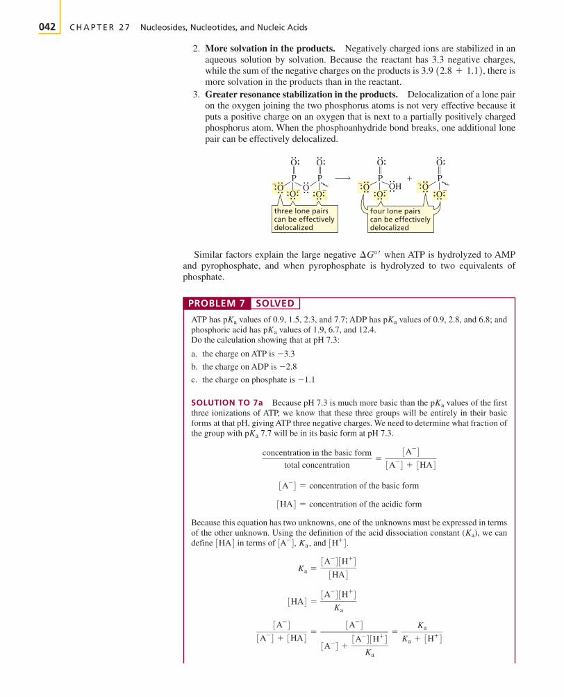
2. More solvation in the products. Negatively charged ions are stabilized in anaqueous solution by solvation. Because the reactant has 3.3 negative charges,while the sum of the negative charges on the products is 3.9 there ismore solvation in the products than in the reactant.
3. Greater resonance stabilization in the products. Delocalization of a lone pairon the oxygen joining the two phosphorus atoms is not very effective because itputs a positive charge on an oxygen that is next to a partially positively chargedphosphorus atom. When the phosphoanhydride bond breaks, one additional lonepair can be effectively delocalized.
Similar factors explain the large negative when ATP is hydrolyzed to AMPand pyrophosphate, and when pyrophosphate is hydrolyzed to two equivalents ofphosphate.
PROBLEM 7 SOLVED
ATP has values of 0.9, 1.5, 2.3, and 7.7; ADP has values of 0.9, 2.8, and 6.8; andphosphoric acid has values of 1.9, 6.7, and 12.4.Do the calculation showing that at pH 7.3:
a. the charge on ATP is
b. the charge on ADP is
c. the charge on phosphate is
SOLUTION TO 7a Because pH 7.3 is much more basic than the values of the firstthree ionizations of ATP, we know that these three groups will be entirely in their basicforms at that pH, giving ATP three negative charges. We need to determine what fraction ofthe group with 7.7 will be in its basic form at pH 7.3.
Because this equation has two unknowns, one of the unknowns must be expressed in termsof the other unknown. Using the definition of the acid dissociation constant we candefine in terms of and
3A-4
3A-4 + 3HA4=
3A-4
3A-4 +3A-43H+4
Ka
=Ka
Ka + 3H+4
3HA4 =3A-43H+4
Ka
Ka =3A-43H+4
3HA4
3H+4.Ka ,3A-4,3HA4(Ka),
3HA4 = concentration of the acidic form
3A-4 = concentration of the basic form
concentration in the basic form
total concentration=
3A-4
3A-4 + 3HA4
pKa
pKa
-1.1
-2.8
-3.3
pKa
pKapKa
¢G°¿
O−OH−− OO
OO
O
P−
− O+
O
P
O
O
P−
O
O
P−
three lone pairscan be effectivelydelocalized
four lone pairscan be effectivelydelocalized
12.8 + 1.12,
BRUI27-32_70r2 26-02-2003 3:51 PM Page 042

Section 27.6 Other Important Nucleotides 043
lysine
arginine
NH2
N N
NN
O
OH
H
HN NC
Mg2+NH2
+
HH
H
N+
O−−O
P
O
OO−
PO
O
O−
P O
OHOH
> Figure 27.2The interactions between ATP,
and arginine and lysineresidues at the active site of anenzyme.
Mg2+,
Now we can calculate the fraction of the group with 7.7 that will be in the basic form.
27.5 Kinetic Stability of ATP in the Cell
Although ATP reacts readily in enzyme-catalyzed reactions, it reacts quite slowly inthe absence of an enzyme. For example, carboxylic acid anhydrides hydrolyze in amatter of minutes, but ATP takes several weeks to hydrolyze. The low rate of ATP hy-drolysis is important because it allows ATP to exist in the cell until it is needed for anenzyme-catalyzed reaction.
The negative charges on ATP are what make it relatively unreactive. These negativecharges repel the approach of nucleophiles. When ATP is bound at an active site of anenzyme, it complexes with magnesium which decreases the overall negativecharge on ATP. (This is why ATP-requiring enzymes also require metal ions;Section 25.5.) The other two negative charges can be stabilized by positively chargedgroups such as arginine or lysine residues at the active site, as shown in Figure 27.2. Inthis form, ATP is readily approached by nucleophiles, so ATP reacts rapidly in anenzyme-catalyzed reaction, but only very slowly in the absence of the enzyme.
(Mg2+),
total negative charge on ATP = 3.0 + 0.3 = 3.3
Ka
Ka + 3H+4=
2.0 * 10-8
2.0 * 10-8 + 5.0 * 10-8 = 0.3
pKa
27.6 Other Important Nucleotides
ATP is not the only biologically important nucleotide. Guanosine -triphosphate(GTP) is used in place of ATP in some phosphoryl transfer reactions. We have alsoseen in Sections 25.2 and 25.3 that dinucleotides are used as oxidizing agents (
FAD, FMN) and reducing agents (NADH, NADPH, ).Another important nucleotide is adenosine -monophosphate, commonly known
as cyclic AMP. Cyclic AMP is called a “second messenger” because it serves as a linkbetween several hormones (the first messengers) and certain enzymes that regulate cel-lular function. Secretion of certain hormones, such as adrenaline, activates adenylatecyclase, the enzyme responsible for the synthesis of cyclic AMP from ATP. CyclicAMP then activates an enzyme, generally by phosphorylating it. Cyclic nucleotides areso important in regulating cellular reactions that an entire scientific journal is devoted tothese processes.
3¿,5¿FMNH2FADH2,NADP+,
NAD+,
5¿
BRUI27-32_70r2 26-02-2003 3:51 PM Page 043

a trinucleotide
OH
A
5′ 5′ 5′
3′ 3′ 3′
C G
PPP
a 5′-triphosphategroup
a 3′-hydroxylgroup
044 C H A P T E R 2 7 Nucleosides, Nucleotides, and Nucleic Acids
3-D Molecule:Cyclic AMP
Erwin Chargaff was born in Austriain 1905 and received a Ph.D. from theUniversity of Vienna. To escape Hitler,he came to the United States in 1935,becoming a professor at ColumbiaUniversity College of Physicians andSurgeons. He modified paper chro-matography, a technique developed toidentify amino acids (Section 23.5), sothat it could be used to quantify thedifferent bases in a sample of DNA.
PROBLEM 8
What products would be obtained from the hydrolysis of cyclic AMP?
27.7 The Nucleic Acids
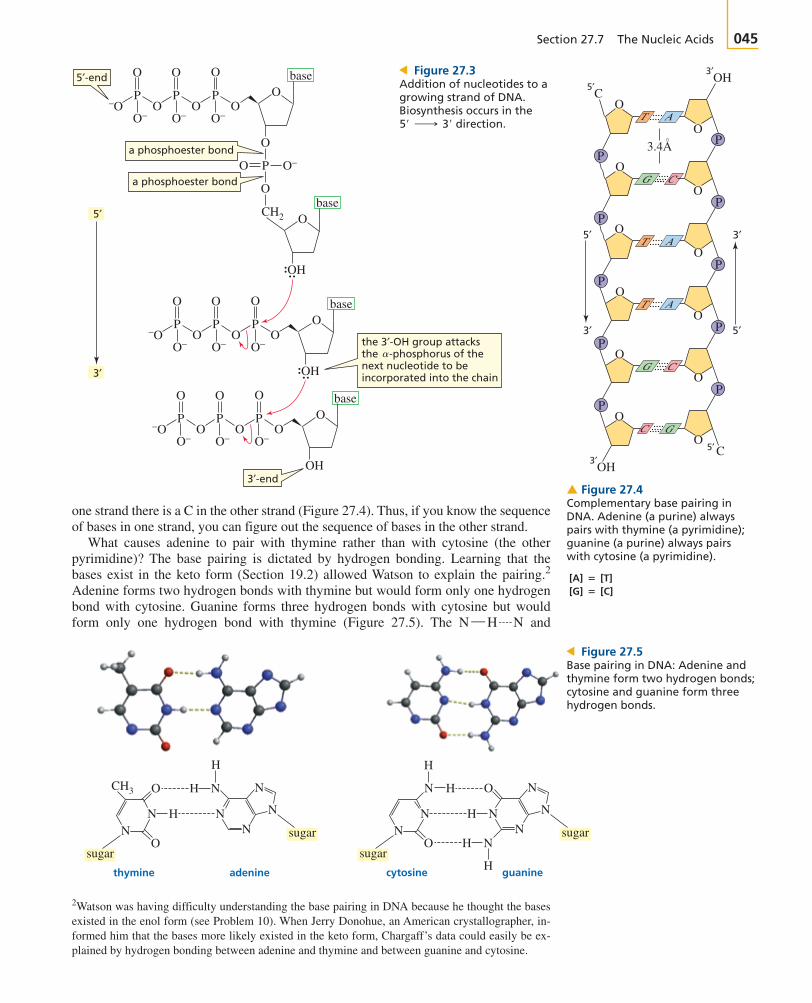
Nucleic acids are composed of long strands of nucleotide subunits linked by phospho-diester bonds. These linkages join the group of one nucleotide to the group of the next nucleotide (Figure 27.1). A dinucleotide contains two nucleotidesubunits, an oligonucleotide contains three to ten subunits, and a polynucleotide con-tains many subunits. DNA and RNA are polynucleotides. Notice that the nucleotide atone end of the strand has an unlinked -triphosphate group, and the nucleotide at theother end of the strand has an unlinked -hydroxyl group.
Nucleotide triphosphates are the starting materials for the biosynthesis of nucleicacids. DNA is synthesized by enzymes called DNA polymerases, and RNA issynthesized by enzymes called RNA polymerases. The nucleotide strand is formed asa result of nucleophilic attack by a group of one nucleotide triphosphate on the
-phosphorus of another nucleotide triphosphate, breaking a phosphoanhydride bondand eliminating pyrophosphate (Figure 27.3). This means that the growing polymer issynthesized in the direction; in other words, new nucleotides are added tothe Pyrophosphate is subsequently hydrolyzed, which makes the reactionirreversible (Section 27.3). RNA strands are biosynthesized in the same way, usingribonucleotides instead of -deoxyribonucleotides. The primary structure of anucleic acid is the sequence of bases in the strand.
Watson and Crick concluded that DNA consists of two strands of nucleic acids withthe sugar–phosphate backbone on the outside and the bases on the inside. The chainsare held together by hydrogen bonds between the bases on one strand and the bases onthe other strand (Figure 27.4). The width of the double-stranded molecule is relativelyconstant, so a purine must pair with a pyrimidine. If the larger purines paired, thestrand would bulge; if the smaller pyrimidines paired, the strands would have to con-tract to bring the two pyrimidines close enough to form hydrogen bonds.
Critical to Watson and Crick’s proposal for the secondary structure of DNA wereexperiments carried out by Erwin Chargaff. These experiments showed that the num-ber of adenines in DNA equals the number of thymines and the number of guaninesequals the number of cytosines. Chargaff also noted that the number of adenines andthymines relative to the number of guanines and cytosines is characteristic of a givenspecies but varies from species to species. In human DNA, for example, 60.4% of thebases are adenines and thymines, whereas 74.2% of them are adenines and thyminesin the DNA of the bacterium Sarcina lutea.
Chargaff’s data showing that and could be explained if adenine (A) always paired with thymine (T) and guanine (G) al-ways paired with cytosine (C). This means the two strands are complementary—wherethere is an A in one strand, there is a T in the opposing strand, and where there is a G in
[guanine] = [cytosine][adenine] = [thymine]
2¿
3¿-end.5¿ ¡ 3¿
a3¿-OH
3¿5¿
5¿-OH3¿-OH
ATPcyclic AMP
adenylate cyclase
NH2
N
NN
N
O
HO OH
P
O
O−O−
P−O
O
O
Mg2+
H+
O−
P
NH2
N
NN
N
OO−
O
P O
O
OH
+ +
O
OO−
P
O
OO−−O
O
O
Mg2+O−
P
BRUI27-32_70r2 26-02-2003 3:51 PM Page 044
Section 27.7 The Nucleic Acids 045
O
O
CH2
O−−O
P
O
OO−
PO
O−
O
P
5′-end
5′
3′
3′-end
a phosphoester bond
a phosphoester bond
the 3′-OH group attacksthe -phosphorus of thenext nucleotide to beincorporated into the chain
base
O
base
OH
O
O O−P
O
OO−
−OP
O
OO−
PO
O−
O
P
base
OH
O
OO−
−OP
O
OO−
PO
O−
O
P
base
OH
O
O
O
O
> Figure 27.3Addition of nucleotides to agrowing strand of DNA.Biosynthesis occurs in the
direction.5¿ ¡ 3¿
O
O
O
O
O
O
O
O
O
O
O
T A
5′
3′
3′
5′
OC
OH5′
C5′
3′
OH3′
PP
T A
PP
T A
PP
CG
PP
CG
PP
C G
3.4A
� Figure 27.4Complementary base pairing inDNA. Adenine (a purine) alwayspairs with thymine (a pyrimidine);guanine (a purine) always pairswith cytosine (a pyrimidine).
2Watson was having difficulty understanding the base pairing in DNA because he thought the basesexisted in the enol form (see Problem 10). When Jerry Donohue, an American crystallographer, in-formed him that the bases more likely existed in the keto form, Chargaff’s data could easily be ex-plained by hydrogen bonding between adenine and thymine and between guanine and cytosine.
one strand there is a C in the other strand (Figure 27.4). Thus, if you know the sequenceof bases in one strand, you can figure out the sequence of bases in the other strand.
What causes adenine to pair with thymine rather than with cytosine (the otherpyrimidine)? The base pairing is dictated by hydrogen bonding. Learning that thebases exist in the keto form (Section 19.2) allowed Watson to explain the pairing.2
Adenine forms two hydrogen bonds with thymine but would form only one hydrogenbond with cytosine. Guanine forms three hydrogen bonds with cytosine but wouldform only one hydrogen bond with thymine (Figure 27.5). The andN ¬ H � N
thymine adenine
sugar
sugar
cytosine guanine
sugar
sugar
O
H
N
H
NHNN
N
NH
H
O
N
NNH
CH3 N
N
H
H
NN
O
O
N
N
> Figure 27.5Base pairing in DNA: Adenine andthymine form two hydrogen bonds;cytosine and guanine form threehydrogen bonds.
[G] � [C] [A] � [T]
BRUI27-32_70r2 26-02-2003 3:51 PM Page 045

046 C H A P T E R 2 7 Nucleosides, Nucleotides, and Nucleic Acids
a.
b.
c.
� Figure 27.7(a) The DNA double helix. (b) View looking down the long axis of the helix. (c) The basesare planar and parallel on the inside of the helix.
C G
O
O
OO
O
O
A T
O
−O OP
O
O O−P
5 ′
3′
3′
5′
O O
OO
� Figure 27.6The sugar–phosphate backbone of DNA is on the outside, and the bases are on the inside,with A’s pairing with T’s and G’s pairing with C’s. The two strands are antiparallel—theyrun in opposite directions.
bonds that hold the bases together are all about the same lengthÅ).
The two DNA strands are antiparallel—they run in opposite directions, with thesugar–phosphate backbone on the outside and the bases on the inside (Figures 27.4and 27.6). By convention, the sequence of bases in a polynucleotide is written in the
direction (the -end is on the left).
The DNA strands are not linear but are twisted into a helix around a common axis(see Figure 27.7a). The base pairs are planar and parallel to each other on the insideof the helix (Figures 27.7b and c). The secondary structure is therefore known as a
5′-end 3′-end
ATGAGCCATGTAGCCTAATCGGC
5¿5¿ ¡ 3¿
(2.9 ; 0.1N ¬ H � O
BRUI27-32_70r2 26-02-2003 3:51 PM Page 046

PROBLEM 9
Indicate whether each functional group of the five heterocyclic bases in nucleic acids canfunction as a hydrogen bond acceptor (A), a hydrogen bond donor (D), or both (D/A).
PROBLEM 10
Using the D, A, and D/A designations in Problem 9, explain how base pairing would beaffected if the bases existed in the enol form.
Section 27.7 The Nucleic Acids 047
OO
O
O
−O OP
base
O
O
base
OH
OH
OO
O O
O
−O −OP
base
O
O
base a 2′′,3′′-cyclic phosphodiester
OH
OO
O O
O−O
P
base
O
O
base
OH
HO
� Figure 27.8Hydrolysis of RNA. The -OH group acts as an intramolecular nucleophilic catalyst. It hasbeen estimated that RNA is hydrolyzed 3 billion times faster than DNA.
2¿
3-D Molecules:Adenine–thymine base pair;Guanine–cytosine base pair
3-D Molecule:DNA double helix
double helix. The double helix resembles a ladder (the base pairs are the rungs)twisted around an axis running down through its rungs (Figures 27.4 and 27.7c). Thesugar–phosphate backbone is wrapped around the bases. The phosphate OH grouphas a of about 2, so it is in its basic form (negatively charged) at physiologicalpH. The negatively charged backbone repels nucleophiles, thereby preventingcleavage of the phosphodiester bonds.
Unlike DNA, RNA is easily cleaved because the group of ribose can act asthe nucleophile that cleaves the strand (Figure 27.8). This explains why the group is absent in DNA. To preserve the genetic information, DNA must remain intactthroughout the life span of a cell. Cleavage of DNA would have disastrous conse-quences for the cell and for life itself. RNA, in contrast, is synthesized as it is neededand is degraded once it has served its purpose.
Hydrogen bonding between base pairs is just one of the forces holding the twostrands of the DNA double helix together. The bases are planar aromatic mole-cules that stack on top of one another. Each pair is slightly rotated with respect tothe next pair, like a partially spread-out hand of cards. There are favorable van derWaals interactions between the mutually induced dipoles of adjacent pairs ofbases. These interactions, known as stacking interactions, are weak attractiveforces, but when added together they contribute significantly to the stability of thedouble helix. Stacking interactions are strongest between two purines and weakestbetween two pyrimidines. Confining the bases to the inside of the helix has anadditional stabilizing effect—it reduces the surface area of the relatively nonpolarresidues exposed to water. This increases the entropy of the surrounding watermolecules (Section 23.14).
2¿-OH2¿-OH
pKa
BRUI27-32_70r2 26-02-2003 3:51 PM Page 047

048 C H A P T E R 2 7 Nucleosides, Nucleotides, and Nucleic Acids
Figure 27.9 NThe three helical forms of DNA.
PROBLEM 11
The phosphodiester, which is formed when RNA is hydrolyzed (Figure 27.8),reacts with water, forming a mixture of nucleotide and -phosphates. Propose a mech-anism for this reaction.
PROBLEM 12�
If one of the strands of DNA has the following sequence of bases running in thedirection,
a. What is the sequence of bases in the complementary strand?b. What base is closest to the in the complementary strand?
27.8 Helical Forms of DNA
Naturally occurring DNA can exist in the three different helical forms shown inFigure 27.9. The B- and A-helices are both right-handed. The B-helix is the predomi-nant form in aqueous solution, while the A-helix is the predominant form in nonpolarsolvents. Nearly all the DNA in living organisms is in a B-helix. The Z-helix is a left-handed helix. It occurs in regions where there is a high content of base pairs.The A-helix is shorter (for a given number of base pairs) and about 3% broader thanthe B-helix, which is shorter and broader than the Z-helix.
Helices are characterized by the number of bases per 360° turn and the distance (therise) between adjacent base pairs. A-DNA has 11 base pairs per turn and a 2.3 Å rise;B-DNA has 10 base pairs per turn and a 3.4 Å rise; and Z-DNA has 12 base pairs perturn and a 3.8 Å rise.
If you examine Figure 27.9, you will see that there are two kinds of alternatinggrooves in DNA. In B-DNA the major groove is wider than the minor groove. Crosssections of the double helix show that one side of each base pair faces into the majorgroove and the other side faces into the minor groove (Figure 27.10).
Proteins and other molecules can bind to the grooves. The hydrogen-bondingproperties of the functional groups facing into each groove determine what kind ofmolecules will bind to the groove. Mitomycin is a naturally occurring compoundthat has been found to have both antibacterial activity and anticancer activity. Itworks by binding to the minor groove of DNA. It binds at regions rich in A’s andT’s (Section 30.10).
G ¬ C
5¿-end
5¿ ¬ G ¬ G ¬ A ¬ C ¬ A ¬ A ¬ T ¬ C ¬ T ¬ G ¬ C ¬ 3¿
5¿ ¡ 3¿
3¿2¿-2¿,3¿-cyclic
AADTYDZ0 missing size
BRUI27-32_70r2 26-02-2003 3:51 PM Page 048

Section 27.9 Biosynthesis of DNA: Replication 049
thymine–adenine cytosine–guanine
sugar
sugar
major groove
minor groove
sugar
sugar
major groove
minor groove
NH
CH3 N
N
H
H
NN
O
OO
N
N
O
H
N
H
NHNN
N
NH
H
N
N
PROBLEM 13�
Calculate the length of a turn in:
a. A-DNA b. B-DNA c. Z-DNA
27.9 Biosynthesis of DNA: Replication
Watson and Crick’s proposal for the structure of DNA was an exciting developmentbecause the structure immediately suggested how DNA is able to pass on genetic in-formation to succeeding generations. Because the two strands are complementary,both carry the same genetic information. Both strands serve as templates for thesynthesis of complementary new strands (Figure 27.11). The new (daughter) DNA
3-D Molecules:B-helix; A-helix; Z-helix
A T
T A T A
T A T A
T A T A
T A
A T
A T A T
A T
A T
A T A T
A T
C G
C G C G
C G
G C
G C
G C
G CG C
G C
G C
G C
G C
C G
parent strands
uncoiling
3′-end
3′-end 5′-end
3′-end3′-end5′-end 5′-end
5′-end
daughterstranddaughter
strand
> Figure 27.11Replication of DNA. The daughterstrand on the left is synthesizedcontinuously in the direction; the daughter strand on the right is synthesizeddiscontinuously in the direction.
5¿ ¡ 3¿
5¿ ¡ 3¿
> Figure 27.10One side of each base pair facesinto the major groove, and theother side faces into the minorgroove.
BRUI27-32_70r2 26-02-2003 3:51 PM Page 049

050 C H A P T E R 2 7 Nucleosides, Nucleotides, and Nucleic Acids
Severo Ochoa was the first toprepare synthetic strands of RNA byincubating nucleotides in thepresence of enzymes that are involvedin the biosynthesis of RNA. ArthurKornberg prepared synthetic strandsof DNA in a similar manner. For thiswork, they shared the 1959 NobelPrize in physiology or medicine.
Severo Ochoa (1905–1993) wasborn in Spain. He graduated from theUniversity of Malaga in 1921 andreceived an M.D. from the Universityof Madrid. He spent the next fouryears studying in Germany andEngland and then joined the facultyat New York University College ofMedicine. He became a U.S. citizenin 1956.
molecules are identical to the original (parent) molecule—they contain all the originalgenetic information. The synthesis of identical copies of DNA is called replication.
All reactions involved in nucleic acid synthesis are catalyzed by enzymes. The syn-thesis of DNA takes place in a region of the molecule where the strands have started toseparate, called a replication fork. Because a nucleic acid can be synthesized only inthe direction, only the daughter strand on the left in Figure 27.11 is syn-thesized continuously in a single piece (because it is synthesized in the di-rection). The other daughter strand needs to grow in the direction, so it issynthesized discontinuously in small pieces. Each piece is synthesized in the
direction and the fragments are joined together by an enzyme called DNAligase. Each of the two resulting daughter molecules of DNA that result contains oneof the original strands (blue strand) plus a newly synthesized strand (green strand).This process is called semiconservative replication.
The genetic information of a human cell is contained in 23 pairs of chromosomes.Each chromosome is composed of several thousand genes (segments of DNA). Thetotal DNA of a human cell—the human genome—contains 3.1 billion base pairs.
PROBLEM 14
Using a dark line for parental DNA and wavy lines for DNA synthesized from parentalDNA, show what the population of DNA molecules would look like in the fourth generation.
PROBLEM 15�
Assuming that the human genome, with its 3.1 billion base pairs, is entirely in a B-helix,how long is the DNA in a human cell?
PROBLEM 16
Why doesn’t DNA unravel completely before replication begins?
27.10 Biosynthesis of RNA: Transcription
The sequence of DNA bases provides the blueprint for the synthesis of RNA. The syn-thesis of RNA from a DNA blueprint, called transcription, takes place in the nucleusof the cell. This initial RNA is the precursor to all RNA: messenger RNA, ribosomalRNA, and transfer RNA. The newly synthesized RNA leaves the nucleus, carrying thegenetic information into the cytoplasm (the cell material outside the nucleus), wheretranslation of this information into proteins takes place (see Figure 27.17).
DNA contains sequences of bases known as promoter sites. The promoter sitesmark the beginning of genes. An enzyme recognizes a promoter site and binds to it,initiating RNA synthesis. The DNA at a promoter site unwinds to give two singlestrands, exposing the bases. One of the strands is called the sense strand orinformational strand. The complementary strand is called the template strand orantisense strand. The template strand is read in the direction, so thatRNA can be synthesized in the direction (Figure 27.12). The bases in thetemplate strand specify the bases that need to be incorporated into RNA, following thesame base pairing found in DNA. For example, each guanine in the template strandspecifies the incorporation of a cytosine into RNA, and each adenine in the templatestrand specifies the incorporation of a uracil into RNA. (Recall that in RNA, uracil isused instead of thymine.). Because both the sense strand and RNA are complementaryto the template strand, the sense strand and RNA have the same base sequence, exceptthat RNA has a uracil wherever the sense strand has a thymine. Just as there are pro-moter sites that signal the places to start RNA synthesis, there are sites in DNA thatsignal that no more bases should be added to the growing strand of RNA, at whichpoint synthesis stops.
5¿ ¡ 3¿3¿ ¡ 5¿
5¿ ¡ 3¿
3¿ ¡ 5¿5¿ ¡ 3¿
5¿ ¡ 3¿
Arthur Kornberg was born in NewYork in 1918. He graduated from theCollege of the City of New York andreceived an M.D. from the Universityof Rochester. He is a member of thefaculty of the biochemistrydepartment at Stanford University.
BRUI27-32_70r2 26-02-2003 3:51 PM Page 050

Section 27.11 Ribosomal RNA 051
5′
5′
3′ 5′
3′
3′
T
A
T
A
T
AG
U C U C G A U A C A C GOH
T
A
C
G
C
G
C
G
T
A
C
G
T
A
C
G
C
G
G
C
G
C
G
C
G
C
GC
G
CT
A G A G C T A T G T G C G T
C
T C T C G A T
direction of transcription
A C A C G C AA
GCCGGAG
RNA
DNA
AUCCpppA
A
T
A
G
C
A
T
A
T
A
T
sense strand
template strand
� Figure 27.12Transcription: using DNA as a blueprint for RNA.
Surprisingly, a gene is not necessarily a continuous sequence of bases. Often thebases of a gene are interrupted by bases that appear to have no informational content.A stretch of bases representing a portion of a gene is called an exon, while a stretch ofbases that contains no genetic information is called an intron. The RNA that is syn-thesized is complementary to the entire sequence of DNA bases—exons and introns.So after the RNA is synthesized, but before it leaves the nucleus, the so-called non-sense bases (encoded by the introns) are cut out and the informational fragments arespliced together, resulting in a much shorter RNA molecule. This RNA processingstep is known as RNA splicing. Scientists have found that only about 2% of DNA con-tains genetic information, while 98% consists of introns.
It has been suggested that the purpose of introns is to make RNA more versatile. Theoriginally synthesized long strand of RNA can be spliced in different ways to create a va-riety of shorter RNAs.
PROBLEM 17
Why do both thymine and uracil specify the incorporation of adenine?
27.11 Ribosomal RNA
RNA is much shorter than DNA and is generally single-stranded. Although DNA mole-cules can have billions of base pairs, RNA molecules rarely have more than 10,000 nu-cleotides. There are three kinds of RNA—messenger RNA (mRNA) whose sequenceof bases determines the sequence of animo acids in a protein, ribosomal RNA (rRNA),a structural component of ribosomes, and transfer RNA (tRNA), the carriers of aminoacids for protein synthesis.
The biosynthesis of proteins takes place on particles known as ribosomes. A ribo-some is composed of about 40% protein and about 60% rRNA. There is increasing evi-dence that protein synthesis is catalyzed by rRNA molecules rather than by enzymes.RNA molecules—found in ribosomes—that act as catalysts are known as ribozymes.The protein molecules in the ribosome enhance the functioning of the rRNA molecules.
Ribosomes are made up of two subunits. The size of the subunits depends on whetherthey are found in prokaryotic organisms or eukaryotic organisms. Prokaryotic organ-isms (pro, Greek for “before”; karyon, Greek for “kernel” or “nut”) are the earliest or-ganisms. They are unicellular and do not have nuclei. A eukaryotic organism (eu,Greek for “well”) is much more complicated. Eukaryotic organisms can be unicellularor multicellular and their cells have nuclei. A prokaryotic ribosome is composed of a50S subunit and a smaller 30S subunit; together they form a 70S ribosome. A eukaryot-ic ribosome has a 60S subunit and a 40S subunit; together they form an 80S ribosome.
Sidney Altman and Thomas R.Cech received the 1989 Nobel Prizein chemistry for their discovery of thecatalytic properties of RNA.
Sidney Altman was born inMontreal in 1939. He received a B.S.from MIT and a Ph.D. from theUniversity of Colorado, Boulder. Hewas a postdoctoral fellow in FrancisCrick’s laboratory at CambridgeUniversity. He is a professor ofbiology at Yale University.
Thomas Cech was born in Chicagoin 1947. He received a B.A. fromGrinnell College and a Ph.D. fromthe University of California,Berkeley. He was a postdoctoralfellow at MIT. He is a professor ofchemistry at the University ofColorado, Boulder.
BRUI27-32_70r2 26-02-2003 3:51 PM Page 051

052 C H A P T E R 2 7 Nucleosides, Nucleotides, and Nucleic Acids
3′
5′a.
OH
anticodon
U
G
C
C
C
G
G
C
UC
A G G C C
C
G
C
A
A
CC
C
AG
C
C
UGC
UG U
GU
G
G
U
G
UA
A
A
U
C
G
C
G
UAG
CC
C
G
G
G
C
G
G
G
GCC
G C
all tRNAs haveCCA at the 3′-end
Figure 27.13 N(a) a transfer RNA thatcarries alanine. Compared withother RNAs, tRNA contains a highpercentage of unusual bases(shown as empty circles). Thesebases result from enzymaticmodification of the four normalbases. (b) The anticodon isshown in red; the serine bindingsite is shown in yellow.
tRNASer:
tRNAAla,
b.
3-D Molecule:tRNA
3Sedimentation constants are not additive, which is why a 50S and a 30S can combine to form a 70S.
The S stands for the sedimentation constant, which designates where a given compo-nent sediments during centrifugation.3
27.12 Transfer RNA
Transfer RNA (tRNA) is much smaller than mRNA or rRNA. It contains only 70 to 90nucleotides. The single strand of tRNA is folded into a characteristic cloverleaf struc-ture strung out with three loops and a little bulge next to the right-hand loop(Figure 27.13a). There are at least four regions with complementary base pairing. AlltRNAs have a CCA sequence at the The three bases at the bottom of the loopdirectly opposite the and are called an anticodon (Figures 27.13a and b).
Each tRNA can carry an amino acid bound as an ester to its terminal group.The amino acid will be inserted into a protein during protein biosynthesis. Each tRNAcan carry only one particular amino acid. A tRNA that carries alanine is designated astRNAAla.
3¿-OH3¿-ends5¿-
3¿-end.
30S
50S
40S
80S70S
eukaryotic ribosomeprokaryotic ribosomeMW 4,200,000MW 2,500,000
60S
BRUI27-32_70r2 26-02-2003 3:51 PM Page 052

Section 27.12 Transfer RNA 053
AMP
ATP
OO
O
P
O−
OO
P
O−O −O O
O
PO−
O
P
O− O−−O
OH
O
P
O−−O
O
P
O−O
O
O
P
O−O
O
O
O
P
O−
adenosine adenosine
adenosine
−
O−
RCHC
+NH3
O
ORCHC
+NH3
O
ORCHC
C
+NH3
RCH
+NH3
+ +
HO ACC
3′-OH group of A
a tetrahedral intermediate
tRNA
H2O
2
ACC
5′ 5′
5′
ACC+
an acyl adenylatean aminoacid
an amino acyl tRNA
pyrophosphate
� Figure 27.14The proposed mechanism foraminoacyl-tRNA synthetase—theenzyme that catalyzes theattachment of an amino acid to a tRNA.
Elizabeth Keller (1918–1997) wasthe first to recognize that tRNA had acloverleaf structure. She received aB.S. from the University of Chicagoin 1940 and a Ph.D. from CornellUniversity Medical College in 1948.She worked at the HuntingtonMemorial Laboratory ofMassachusetts General Hospital and at the United States PublicHealth Service. Later she became a professor at MIT and then at Cornell University.
It is critical that the correct amino acid is attached to the tRNA. Otherwise, thecorrect protein will not be synthesized. Fortunately, the synthetases correct theirown mistakes. For example, valine and threonine are approximately the same size—threonine has an OH group in place of a group of valine. Both amino acids,therefore, can bind at the amino acid binding site of the aminoacyl-tRNA synthetase forvaline, and both can then be activated by reacting with ATP to form an acyl adenylate.The aminoacyl-tRNA synthetase for valine has two adjacent catalytic sites, one for
CH3
aminoacyl-tRNA synthetasespecific for histidine
binding site fortRNAHisbinding site for
histidine
NHN
O−
O
H2N CH C
CH2
OH
ACC
> Figure 27.15An aminoacyl-tRNA synthetase hasa binding site for tRNA and abinding site for the particularamino acid that is to be attached tothat tRNA. Histidine is the aminoacid and is the tRNAmolecule in this example.
tRNAHis
How does an amino acid become attached to a tRNA? Attachment of the amino acidis catalyzed by an enzyme called aminoacyl-tRNA synthetase. In the first step of theenzyme-catalyzed reaction (Figure 27.14), the carboxyl group of the amino acid at-tacks the of ATP, activating the carboxyl group by forming an acyladenylate. The pyrophosphate that is expelled is subsequently hydrolyzed, ensuringthe irreversibility of the phosphoryl transfer reaction (Section 27.3). Then a nucle-ophilic acyl substitution reaction occurs—the group of tRNA attacks the car-bonyl carbon of the acyl adenylate, forming a tetrahedral intermediate. The amino acyltRNA is formed when AMP is expelled from the tetrahedral intermediate. All the stepstake place at the active site of the enzyme. Each amino acid has its own aminoacyl-tRNA synthetase. Each synthetase has two specific binding sites, one for the aminoacid and one for the tRNA that carries that amino acid (Figure 27.15).
3¿-OH
a-phosphorus
BRUI27-32_70r2 26-02-2003 3:51 PM Page 053

054 C H A P T E R 2 7 Nucleosides, Nucleotides, and Nucleic Acids
Table 27.2 The Genetic Code
Middle position
U C A G
U Phe Ser Tyr Cys U
Phe Ser Tyr Cys C
Leu Ser Stop Stop A
Leu Ser Stop Trp G
C Leu Pro His Arg U
Leu Pro His Arg C
Leu Pro Gln Arg A
Leu Pro Gln Arg G
A Ile Thr Asn Ser U
Ile Thr Asn Ser C
Ile Thr Lys Arg A
Met Thr Lys Arg G
G Val Ala Asp Gly U
Val Ala Asp Gly C
Val Ala Glu Gly A
Val Ala Glu Gly G
5¿-Position5¿-Position
The genetic code was worked outindependently by Marshall Nirenbergand Har Gobind Khorana, for whichthey shared the 1968 Nobel Prize inphysiology or medicine. RobertHolley, who worked on the structure of tRNA molecules, also shared thatyear’s prize.
Marshall Nirenberg was born in New York in 1927. He received abachelor’s degree from the Universityof Florida and a Ph.D. from the Uni-versity of Michigan. He is a scientistat the National Institutes of Health.
Har Gobind Khorana was born in India in 1922. He received abachelor’s and a master’s degreefrom Punjab University and a Ph.D.from the University of Liverpool. In 1960 he joined the faculty at theUniversity of Wisconsin and laterbecame a professor at MIT.
attaching the acyl adenylate to tRNA and one for hydrolyzing the acyl adenylate. Theacylation site is hydrophobic, so valine is preferred over threonine for the tRNA acyla-tion reaction. The hydrolytic site is polar, so threonine is preferred over valine for thehydrolysis reaction. Thus, if threonine is activated by the aminoacyl-tRNA synthetasefor valine, it will be hydrolyzed rather than transferred to the tRNA.
27.13 Biosynthesis of Proteins: Translation
A protein is synthesized from its N-terminal end to its C-terminal end by reading thebases along the mRNA strand in the direction. A sequence of three bases,called a codon, specifies a particular amino acid that is to be incorporated into a pro-tein. The bases are read consecutively and are never skipped. A codon is written withthe -nucleotide on the left. Each amino acid is specified by a three-base sequenceknown as the genetic code (Table 27.2). For example, UCA on mRNA codes for theamino acid serine, whereas CAG codes for glutamine.
Because there are four bases and the codons are triplets, different codonsare possible. This is many more than are needed to specify the 20 different aminoacids, so all the amino acids—except methionine and tryptophan—have more than onecodon. It is not surprising, therefore, that methionine and tryptophan are the leastabundant amino acids in proteins. Actually, 61 of the bases specify amino acids, andthree bases are stop codons. Stop codons tell the cell to “stop protein synthesis here.”
Translation is the process by which the genetic message in DNA that has beenpassed to mRNA is decoded and used to build proteins. Each of the approximately100,000 proteins in the human body is synthesized from a different mRNA. Don’t
43 = 64
5¿
5¿ ¡ 3¿
CH3
O
CH3CH
+NH3
CHCO−
valineOH
O
CH3CH
+NH3
CHCO−
threonine
BRUI27-32_70r2 26-02-2003 3:51 PM Page 054

Section 27.13 Biosynthesis of Proteins: Translation 055
confuse transcription and translation—these words are used just as they are used inEnglish. Transcription (DNA to RNA) is copying within the same language ofnucleotides. Translation (RNA to protein) is changing to another language—the lan-guage of amino acids.
How the information in mRNA is translated into a polypeptide is shown inFigure 27.16. In this figure, serine was the last amino acid incorporated into the
codon
anticodon
tRNA
mRNA
growingpolypeptidechain
tRNA thathad carriedserine
the ester and the primary amino groups engage in a nucleophilic acyl substitution reaction
leucine
codes foralanine
serine
AAGAAGCGA UUC CCG5′ 3′
GGC
GCUCGA UUC CCG CUG CCC AAG5′ 3′
R O
NH CH C
R O
O
AC
C
O
NH CH C NH
CH2
OH
CH C
AAG AAG
=
O
AC
C
O
H2N
CH2
CH3
CH3CH
CH C
GCUCGA UUC CCG5′ 3′
R O
NH CH C
R O
O
AC
C
O
NH CH C NH
CH2
OH
CH C
AAG
O
AC
C
O
H2N
CH2
CH3
CH3CH
CH C
R O
NH CH C
R O
O
O
NH CH C
O
CNH
CH2
OH
CH NH
CH2
CH
CH C
O
AC
C
AC
C
OH
AC
C
O
H2N
CH3CH3
CH3 CH C
> Figure 27.16Translation. The sequence of basesin mRNA determines the sequenceof amino acids in a protein.
Transcription: DNA ¡ RNA
Translation: mRNA ¡ protein
Tutorial:Translation
BRUI27-32_70r2 26-02-2003 3:51 PM Page 055

056 C H A P T E R 2 7 Nucleosides, Nucleotides, and Nucleic Acids
amino acid
transcription
modificationof RNA
1
translation5
mRNA
nucleus
cytoplasm
rRNA
mRNA
DNA
tRNA binds anamino acid
initial RNAtranscript
nuclearenvelope
tRNA
2
4
addition ofprotein to RNA
3
ribosome
growing polypeptidechain
growing polypeptide chain. Serine was specified by the AGC codon because theanticodon of the tRNA that carries serine is GCU (Remember that abase sequence is read in the direction, so the sequence of bases in an an-ticodon must be read from right to left.) The next codon is CUU, signaling for atRNA with an anticodon of AAG That particular tRNA carries leucine.The amino group of leucine reacts in an enzyme-catalyzed nucleophilic acyl substi-tution reaction with the ester on the adjacent tRNA, displacing the tRNA. The nextcodon (GCC) brings in a tRNA carrying alanine. The amino group of alanine dis-places the tRNA that brought in leucine. Subsequent amino acids are brought in oneat a time in the same way, with the codon in mRNA specifying the amino acid to beincorporated by complementary base pairing with the anticodon of the tRNA thatcarries that amino acid.
Protein synthesis takes place on the ribosomes (Figure 27.17). The smaller subunitof the ribosome (30S in prokaryotic cells) has three binding sites for RNA molecules.It binds the mRNA whose base sequence is to be read, the tRNA carrying the growingpeptide chain, and the tRNA carrying the next amino acid to be incorporated into theprotein. The larger subunit of the ribosome (50S in prokaryotic cells) catalyzes peptidebond formation.
(3¿-GAA-5¿).
5¿ ¡ 3¿(3¿-UCG-5¿).
Figure 27.17 N1. Transcription of DNA occurs in
the nucleus. The initial RNAtranscript is the precursor of allRNA: tRNA, rRNA, and mRNA.
2. The initially formed RNA oftenmust be chemically modifiedbefore it acquires biologicalactivity. Modification can entailremoving nucleotide segments,adding nucleotides to the - or
-ends, or chemically alteringcertain nucleotides.
3. Proteins are added to rRNA toform ribosomal subunits. tRNA,mRNA, and ribosomal subunitsleave the nucleus.
4. Each tRNA binds the appropriateamino acid.
5. tRNA, mRNA, and a ribosomework together to translate themRNA information into aprotein.
3¿5¿
BRUI27-32_70r2 26-02-2003 3:51 PM Page 056

PROBLEM 18�
If methionine is the first amino acid incorporated into a heptapeptide, what is the sequenceof the amino acids encoded for by the following stretch of mRNA?
PROBLEM 19�
Four C’s occur in a row in the segment of mRNA in Problem 18. What polypeptide wouldbe formed from the mRNA if one of the four C’s were cut out of the strand?
PROBLEM 20
UAA is a stop codon. Why does the UAA sequence in mRNA in Problem 18 not cause pro-tein synthesis to stop?
PROBLEM 21�
Write the sequences of bases in the sense strand of DNA that resulted in the mRNA inProblem 18.
PROBLEM 22
List the possible codons on mRNA that specify each amino acid in Problem 18 and the an-ticodon on the tRNA that carries that amino acid.
U ¬ A ¬ A ¬ A ¬ C ¬ A ¬ C ¬ 3¿5¿ ¬ G ¬ C ¬ A ¬ U ¬ G ¬ G ¬ A ¬ C ¬ C ¬ C ¬ C ¬ G ¬ U ¬ U ¬ A ¬ U ¬
Section 27.13 Biosynthesis of Proteins: Translation 057
3-D Molecules:Chloramphenicol complexedto acetyl-transferase;Tetracycline
A sculpture done by Robert Holley.
Robert W. Holley (1922–1993) wasborn in Illinois and received abachelor’s degree from the Universityof Illinois and a Ph.D. from CornellUniversity. During World War II heworked on the synthesis of penicillinat Cornell Medical School. He was aprofessor at Cornell and later at theUniversity of California, San Diego.He was also a noted sculptor.
SICKLE CELL ANEMIA
Sickle cell anemia is an example of the damage thatcan be caused by a change in a single base of DNA
(Problem 55 in Chapter 23). It is a hereditary disease causedwhen a GAG triplet becomes a GTG triplet in the sense strand ofa section of DNA that codes for the -subunit of hemoglobin. Asa consequence, the mRNA codon becomes GUG—which signals
b
for incorporation of valine—rather than GAG, which wouldhave signaled for incorporation of glutamic acid. The changefrom a polar glutamic acid to a nonpolar valine is sufficient tochange the shape of the deoxyhemoglobin molecule and induceaggregation, causing it to precipitate in red blood cells. Thisstiffens the cells, making it difficult for them to squeezethrough a capillary. Blocked capillaries cause severe pain andcan be fatal.
Normal red blood cells Sickle red blood cells
BRUI27-32_70r2 26-02-2003 4:14 PM Page 057

058 C H A P T E R 2 7 Nucleosides, Nucleotides, and Nucleic Acids
ANTIBIOTICS THAT ACT BYINHIBITING TRANSLATIONPuromycin is a naturally occurring antibiotic. It is
one of several antibiotics that act by inhibiting translation.Puromycin mimics the -CCA-aminoacyl portion of a tRNA.If, during translation, the enzyme is fooled into transferring thegrowing peptide chain to the amino group of puromycin ratherthan to the amino group of the incoming -CCA-aminoacyltRNA, protein synthesis stops. Because puromycin blocks pro-tein synthesis in eukaryotes as well as in prokaryotes, it is poi-sonous to humans and therefore is not a clinically usefulantibiotic. To be clinically useful, an antibiotic must affect pro-tein synthesis only in prokaryotic cells.
3¿
3¿
O
NH
OH
NN
N
H3C CH3
N
N
HO
O
CH2CHC
NH2puromycin
CH3O
Clinically usefulantibiotics Mode of action
Tetracycline Prevents the aminoacyl-tRNA from binding to the ribosomeErythromycin Prevents the incorporation of new amino acids into the proteinStreptomycin Inhibits the initiation of protein synthesisChloramphenicol Prevents the new peptide bond from being formed
27.14 Why DNA Contains Thymine Instead of Uracil
In Section 25.8 we saw that dTMP is formed when dUMP is methylated, with coenzyme-methylenetetrahydrofolate supplying the methyl group. Because the incorpora-
tion of the methyl group into uracil oxidizes tetrahydrofolate to dihydrofolate, dihydrofo-late must be reduced back to tetrahydrofolate to prepare the cofactor for another catalyticcycle. The reducing agent is NADPH. Every NADPH formed in a biological organismcan drive the formation of three ATPs, so using an NADPH to reduce dihydrofolatecomes at the expense of ATP. This means that the synthesis of thymine is energeticallyexpensive, so there must be a good reason for DNA to contain thymine instead of uracil.
The presence of thymine instead of uracil in DNA prevents potentially lethal muta-tions. Cytosine can tautomerize to form an imine, which can be hydrolyzed to uracil(Section 18.6). The overall reaction is called a deamination since it removes an aminogroup.
tautomerization
cytosine uracil
N
O
NH2
NH
NH
NH
HN + NH3H2O
O
NH
HN
O
O
N10N5,
N
R′dUMP
R′ = 2′-deoxyribose-5-P
HN + N5,N10-methylene-THF + dihydrofolate
thymidylatesynthase
O
O
N
R′
HN
O
CH3
O
+tetrahydrofolate NADP++dihydrofolate NADPH + H+dihydrofolate
reductase
dTMP
BRUI27-32_70r2 26-02-2003 3:51 PM Page 058

Section 27.15 Determining the Base Sequence of DNA 059
If a cytosine in DNA is deaminated to a uracil, uracil will specify incorporation ofan adenine into the daughter strand during replication instead of the guanine thatwould have been specified by cytosine. Fortunately, a U in DNA is recognized as a“mistake” by cell enzymes before an incorrect base can be inserted into the daughterstrand. These enzymes cut out the U and replace it with a C. If U’s were normallyfound in DNA, the enzymes could not distinguish between a normal U and a U formedby deamination of cytosine. Having T’s in place of U’s in DNA allows the U’s that arefound in DNA to be recognized as mistakes.
Unlike DNA, which replicates itself, any mistake in RNA does not survive forlong because RNA is constantly being degraded and then resynthesized from theDNA template. Therefore, it is not worth spending the extra energy to incorporateT’s into RNA.
PROBLEM 23�
Adenine can be deaminated to hypoxanthine, and guanine can be deaminated to xanthine.Draw structures for hypoxanthine and xanthine.
PROBLEM 24
Explain why thymine cannot be deaminated.
27.15 Determining the Base Sequence of DNA
In June 2000, two teams of scientists (one from a private biotechnology company andone from the publicly funded Human Genome Project) announced that they had com-pleted the first draft of the sequence of the 3.1 billion base pairs in human DNA. Thisis an enormous accomplishment. For example, if the sequence of 1 million base pairswere determined each day, it would take more than 10 years to complete the sequenceof the human genome.
DNA molecules are too large to sequence as a unit, so DNA is first cleaved at spe-cific base sequences and the resulting DNA fragments are sequenced. The enzymesthat cleave DNA at specific base sequences are called restriction endonucleases,and the DNA fragments that are formed are called restriction fragments. Severalhundred restriction enzymes are now known. A few examples of restriction en-zymes, the base sequence each recognizes, and the point of cleavage in that basesequence are shown here.
The base sequences that most restriction enzymes recognize are palindromes. Apalindrome is a word or a group of words that reads the same forward and backward.“Toot” and “race car” are examples of palindromes.4 A restriction enzyme recognizes
AGCTTCGA
GGCCCCGG
CTGCAGGACGTC
recognition sequencerestriction enzyme
AluI
FnuDI
PstI
4Some other palindromes are “Mom,” “Dad,” “Bob,” “Lil,” “radar,” “noon,” “wow,” “poor Dan in adroop”, “a man, a plan, a canal, Panama,” “Sex at noon taxes,” and “He lived as a devil, eh?”
BRUI27-32_70r2 26-02-2003 3:51 PM Page 059

060 C H A P T E R 2 7 Nucleosides, Nucleotides, and Nucleic Acids
Frederick Sanger (Section 23.12)and Walter Gilbert shared half ofthe 1980 Nobel Prize in chemistry fortheir work on DNA sequencing. Theother half went to Paul Berg, whohad developed a method of cuttingnucleic acids at specific sites andrecombining the fragments in newways, a technique known asrecombinant DNA technology.
Walter Gilbert was born in Bostonin 1932. He received a master’sdegree in physics from Harvard and aPh.D. in mathematics fromCambridge University. In 1958 hejoined the faculty at Harvard, wherehe became interested in molecularbiology.
Paul Berg was born in New York in1926. He received a Ph.D. fromWestern Reserve University (nowCase Western Reserve University).He joined the faculty at WashingtonUniversity in St. Louis in 1955 andbecame a professor of biochemistryat Stanford in 1959.
a piece of DNA in which the template strand is a palindrome of the sense strand. Inother words, the sequence of bases in the template strand (reading from right to left) isidentical to the sequence of bases in the sense strand (reading from left to right).
PROBLEM 25�
Which of the following base sequences would most likely be recognized by a restrictionendonuclease?
a. ACGCGT c. ACGGCA e. ACATCGT
b. ACGGGT d. ACACGT f. CCAACC
The restriction fragments can be sequenced using a chain-terminator procedure de-veloped by Frederick Sanger known as the dideoxy method. This method involvesgenerating fragments whose length depends on the last base added to the fragment.Because of its simplicity, it has superceded alternative methods.
In the dideoxy method a small piece of DNA called a primer, labeled at the with is added to the restriction fragment whose sequence is to be determined.Next, the four -deoxyribonucleoside triphosphates are added as well as DNA poly-merase, the enzyme that adds nucleotides to a strand of DNA. In addition, a smallamount of the -dideoxynucleoside triphosphate of one of the bases is added to thereaction mixture. A -dideoxynucleoside triphosphate has no OH groups at the and -positions.
Nucleotides will be added to the primer by base pairing with the restriction frag-ment. Synthesis will stop if the -dideoxy analog of dATP is added instead ofdATP, because the -dideoxy analog does not have a to which additionalnucleotides can be added. Therefore, three different chain-terminated fragments willbe obtained from the DNA restriction fragment shown here.
The procedure is repeated three more times using a -dideoxy analog of dGTP,then a -dideoxy analog of dCTP, and then a -dideoxy analog of dTTP.
PROBLEM 26�
What labeled fragments would be obtained from the segment of DNA shown above if a-dideoxy analog of dGTP had been added to the reaction mixture instead of a -
dideoxy analog of dATP?2¿,3¿2¿,3¿
2¿,3¿2¿,3¿2¿,3¿
dATP, dGTP,dCTP, dTTP,2′,3′-dATP
DNA polymerase5′3′
32P
AGGCTCCAGTGATCCG
TC
32P TCCGA32P TCCGAGGTCA32P TCCGAGGTCACTA
DNA to be sequenced
primer
3¿-OH2¿,3¿2¿,3¿
a 2′,3′-dideoxynucleoside triphosphate
O
O O
O−
P−O O
O
O−
P
O−
PO
base
O
3¿2¿-2¿,3¿
2¿,3¿
2¿32P,
5¿-end
BRUI27-32_70r2 26-02-2003 3:51 PM Page 060

Section 27.16 Laboratory Synthesis of DNA Strands 061
The chain-terminated fragments obtained from each of the four experiments areloaded onto separate lanes of a buffered polyacrylamide gel—the fragments obtainedfrom using a -dideoxy analog of dATP are loaded onto one lane, the fragmentsobtained from using a -dideoxy analog of dGTP onto another lane, and so on. Anelectric field is applied across the ends of the gel, causing the negatively charged frag-ments to travel toward the positively charged electrode (the anode). The smaller frag-ments fit through the spaces in the gel relatively easily and therefore travel through thegel faster, while the larger fragments pass through the gel more slowly.
After the fragments have been separated, the gel is placed in contact with a photo-graphic plate. Radiation from causes a dark spot to appear on the plate opposite thelocation of each labeled fragment in the gel. This technique is called autoradiography,and the exposed photographic plate is known as an autoradiograph (Figure 27.18).
The sequence of bases in the original restriction fragment can be read directly fromthe autoradiograph. The identity of each base is determined by noting the columnwhere each successive dark spot (larger piece of labeled fragment) appears, starting atthe bottom of the gel. The sequence of the fragment of DNA responsible for the au-toradiograph in Figure 27.18 is shown on the left-hand side of the figure.
Once the sequence of bases in a restriction fragment is determined, the results canbe checked by determining the base sequence of the complementary strand. The basesequence in the original piece of DNA can be determined by repeating the entire pro-cedure with a different restriction endonuclease and noting overlapping fragments.
32P
2¿,3¿2¿,3¿
G
G C TA
TAACGTAATCACAGTCAGCTTACGAC
� Figure 27.18An autoradiograph.
DNA FINGERPRINTINGThe base sequence of the human genome variesfrom individual to individual, generally by a sin-
gle base change every few hundred base pairs. Because some ofthese changes occur in base sequences recognized by restrictionendonucleases, the fragments formed when human DNA reactswith a particular restriction endonuclease vary in size depend-ing on the individual. It is this variation that forms the basis ofDNA fingerprinting (also called DNA profiling or DNA typ-ing). This technique is used by forensic chemists to compareDNA samples collected at the scene of a crime with the DNA ofthe suspected perpetrator. The most powerful technique forDNA identification analyzes restriction fragment length poly-
morphisms (RFLPs) obtained from regions of DNA in whichindividual variations are most common. This technique takesfour to six weeks and requires a blood stain about the size of adime. The chance of identical results from two different per-sons is thought to be one in a million. The second type of DNAprofiling uses a polymerase chain reaction (PCR), which ampli-fies a specific region of DNA and compares differences at thatsite among individuals. This technique can be done in less thana week and requires only 1% of the amount required for RFLP,but does not discriminate as well among individuals. Thechance of identical results from two different people is 1 in 500to 1 in 2000. DNA fingerprinting is also being used to establishpaternity, accounting for about 100,000 DNA profiles a year.
99990000%%%%
27.16 Laboratory Synthesis of DNA Strands
There is a great deal of interest in the synthesis of oligonucleotides with specific basesequences. This would allow scientists to synthesize genes that could be inserted intothe DNA of microorganisms, causing the organisms to synthesize a particular protein.Alternatively, a synthetic gene could be inserted into the DNA of an organism defec-tive in that gene—a process known as gene therapy.
Synthesizing an oligonucleotide with a particular base sequence is an even morechallenging task than synthesizing a polypeptide with a specific amino acid sequencebecause each nucleotide has several groups that must be protected and then deprotect-ed at the proper times. The approach taken was to develop an automated methodsimilar to automated peptide synthesis (Section 23.10). The growing nucleotide is at-tached to a solid support so that it can be purified by flushing the reaction containerwith an appropriate solvent. Therefore, none of the synthesized product will be lostduring purification.
BRUI27-32_70r2 26-02-2003 3:51 PM Page 061
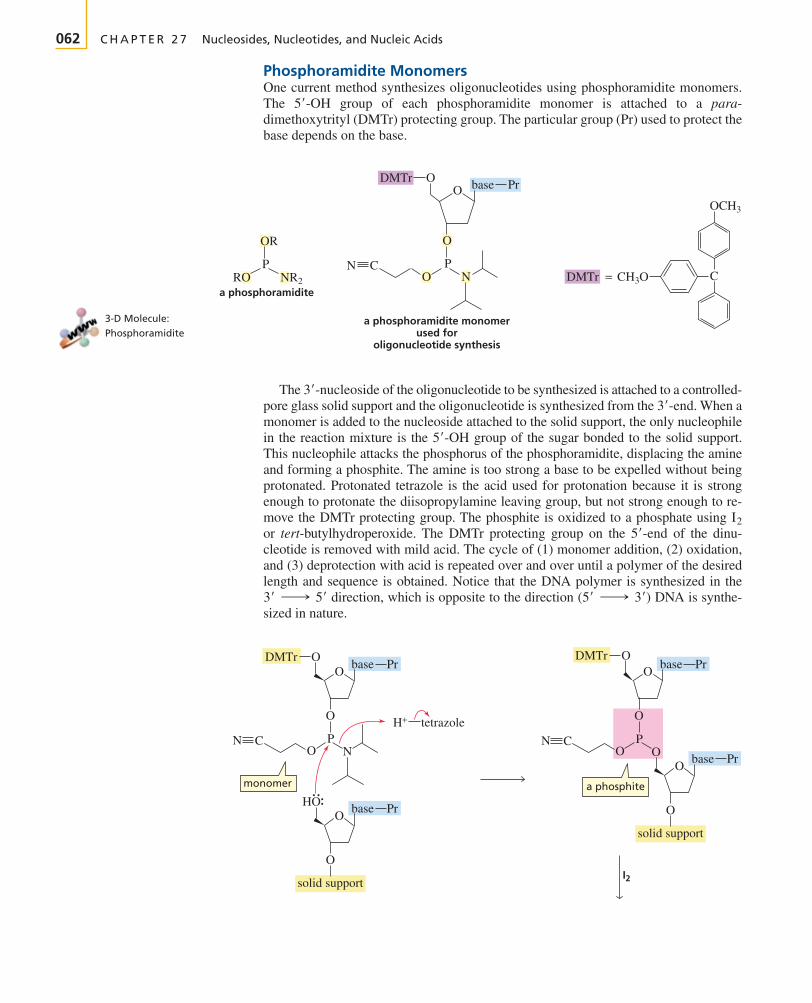
062 C H A P T E R 2 7 Nucleosides, Nucleotides, and Nucleic Acids
Phosphoramidite MonomersOne current method synthesizes oligonucleotides using phosphoramidite monomers.The group of each phosphoramidite monomer is attached to a para-dimethoxytrityl (DMTr) protecting group. The particular group (Pr) used to protect thebase depends on the base.
The of the oligonucleotide to be synthesized is attached to a controlled-pore glass solid support and the oligonucleotide is synthesized from the When amonomer is added to the nucleoside attached to the solid support, the only nucleophilein the reaction mixture is the group of the sugar bonded to the solid support.This nucleophile attacks the phosphorus of the phosphoramidite, displacing the amineand forming a phosphite. The amine is too strong a base to be expelled without beingprotonated. Protonated tetrazole is the acid used for protonation because it is strongenough to protonate the diisopropylamine leaving group, but not strong enough to re-move the DMTr protecting group. The phosphite is oxidized to a phosphate using or tert-butylhydroperoxide. The DMTr protecting group on the of the dinu-cleotide is removed with mild acid. The cycle of (1) monomer addition, (2) oxidation,and (3) deprotection with acid is repeated over and over until a polymer of the desiredlength and sequence is obtained. Notice that the DNA polymer is synthesized in the
direction, which is opposite to the direction DNA is synthe-sized in nature.
(5¿ ¡ 3¿)3¿ ¡ 5¿
5¿-endI2
5¿-OH
3¿-end.3¿-nucleoside
5¿-OH
3-D Molecule:Phosphoramidite
O
OR
CC
NRO NR2
PO N DMTr CH3O
OCH3
=
O
P
PrbaseODMTr
a phosphoramidite
a phosphoramidite monomerused for
oligonucleotide synthesis
O
O
O NN
base
O
O
HO base
Pr
Pr
PC
monomer
solid support
H+ tetrazole
O
O
O
O
N
DMTrODMTrbase
O
O
O base
Pr
Pr
PC
a phosphite
solid support
I2
BRUI27-32_70r2 26-02-2003 3:51 PM Page 062

Section 27.16 Laboratory Synthesis of DNA Strands 063
Tutorial:Oligonucleotide synthesiswith phosphoramidites
O
O
ON
base
O
O
O
O
base
Pr
Pr
PC
solid support
O
O
O
HO
N
base
O
O
O
O
base
Pr
Pr
PC
solid support
O
O
O
HO
N
base
O
O
O
O
base
Pr
Pr
PC
O
ON
OO
O
base Pr
PC
solid support
O
O
O
O
N
base
O
O
O
O
base
Pr
Pr
PC
O
ON
OO
O
base Pr
PC
solid support
H+
1. new monomer H+––tetrazole2. I23. H+
chain extension
O
a phosphate
DMTr
BRUI27-32_70r2 26-02-2003 3:51 PM Page 063

064 C H A P T E R 2 7 Nucleosides, Nucleotides, and Nucleic Acids
The groups of cytosine, adenine, and guanine are nucleophiles and thereforemust be protected to prevent them from reacting with a newly added monomer. The
groups of cytosine and adenine are protected as benzamides and the groupof guanine is protected as a butanamide. Thymine does not contain any nucleophilicgroups, so it does not have to be protected.
After the oligonucleotide is synthesized, the protecting groups on the phosphatesand the protecting groups on the bases must be removed, and the oligonucleotide hasto be detached from the solid support. This can all be done in a single step using aque-ous ammonia. Because a hydrogen bonded to a carbon adjacent to a cyano group isrelatively acidic (Section 19.1), ammonia can be used as the base in the elimination re-action that removes the phosphate protecting group.
Currently, automated DNA synthesizers can synthesize nucleic acids containing asmany as 130 nucleotides in acceptable yields, adding one nucleotide every 2 to 3 min-utes. Longer nucleic acids can be prepared by splicing together two or more individu-ally prepared strands. To ensure a good overall yield of oligonucleotide, the additionof each monomer must occur in high yield This can be accomplished byusing a large excess of monomer. This, however, makes oligonucleotide synthesis veryexpensive because the unreacted monomers are wasted.
H-Phosphonate MonomersA second method for synthesizing oligonucleotides with specific base sequences,using H-phosphonate monomers, has the advantage over the phosphoramiditemethod in that the monomers are easier to handle and phosphate protecting groups
(798%).
ORCN
H H
NH3+NH4
OR
P O +O
OR
OR
P O−OCN
NH2
NH2
N
NN
N
N
N
O
O O
H2N N
NHN
HN
N
O
O
NH N
NHN
N
N
N
N
N
N
unprotected bases protected bases
CH3CH2CH2CCl
O
CCl
O
CCl
O
CH3CH2CH2CHCl+
HCl+
O
N
C
HN
HCl+
O
C
cytosine
adenine
guanine
NH2NH2
NH2
BRUI27-32_70r2 26-02-2003 3:59 PM Page 064

Section 27.16 Laboratory Synthesis of DNA Strands 065
are not needed. However, the yields are not as good. The H-phosphonate monomersare activated by reacting with an acyl chloride, which converts them into phosphoan-hydrides. The group of the nucleoside attached to the solid support reacts withthe phosphoanhydride, forming a dimer. The DMTr protecting group is removedwith acid under mild conditions, and a second activated monomer is added.Monomers are added one at a time in this way until the strand is complete. Oxidationwith (or tert-butyl hydroperoxide) converts the H-phosphonate groups to phos-phate groups. The base-protecting groups are removed by aqueous ammonia as in thephosphoramidite method.
I2
5¿-OH
O
O
O
O−
ODMTrbase Pr
an H-phosphonate
activated monomer
solid support
solid support
PH
O
O
O
O
ODMTrbase Pr
O
O
base Pr
PH
solid support
O
O
O
O
base Pr
O
O
base Pr
PH
O
O
O
O
O
ODMTrbase Pr O
O
base Pr
PH
R C Cl
O
C R
HO
HO
solid support
O
O
O
O
base Pr
O
O
base Pr
PH
O
O
O
O
base Pr
PH
HO
H+
chain extension
1. activated monomer2. H+
5′-OH group
a phosphoanhydride
BRUI27-32_70r2 26-02-2003 3:51 PM Page 065

066 C H A P T E R 2 7 Nucleosides, Nucleotides, and Nucleic Acids
Notice that in the phosphoramidite method an oxidation is done each time amonomer is added, whereas in the H-phosphonate method a single oxidation is carriedout after the entire strand has been synthesized.
PROBLEM 27
Propose a mechanism for removal of the DMTr protecting group by treatment with acid.
27.17 Rational Drug Design
Certain diseases such as acquired immunodeficiency syndrome (AIDS) and herpes arecaused by retroviruses. The genetic information of a retrovirus is contained in itsRNA. The retrovirus uses the sequence of bases in RNA as a template to synthesizeDNA. It is called a retrovirus because its genetic information flows from RNA to DNAinstead of the more typical flow from DNA to RNA.
Drugs that interfere with the synthesis of DNA by retroviruses have been designedand developed. If the retrovirus cannot synthesize DNA, it cannot take over the ge-netic machinery of the cell to produce more retroviral RNA and retroviral proteins.Designing drugs with particular structures to achieve specific purposes is calledrational drug design. AZT is perhaps the best known of the drugs designed to inter-fere with retroviral DNA synthesis. AZT is taken up by the T lymphocytes, cells thatare particularly susceptible to human immunodeficiency virus (HIV), the retrovirusthat causes AIDS. Once in the cell, AZT is converted to AZT-triphosphate. The retro-viral enzyme (reverse transcriptase) that catalyzes DNA formation from RNA bindsAZT-triphosphate more tightly than it binds dTTP. Therefore, AZT rather than T isadded to the growing DNA chain. Because AZT does not have a group, no ad-ditional nucleotides can be added to the chain and DNA synthesis comes to a suddenhalt. Fortunately, the concentration of AZT required to affect reverse transcription istoo low to affect most cellular DNA replication. A newer drug, -dideoxyinosine(ddI), has a similar mechanism of action. Rational drug design is further discussed inChapter 30.
2¿,3¿
3¿-OH
phosphategroup
H-phosphonategroup
solid support
O
O
O
O
base Pr
O
O
base Pr
P
OO
O
O
O
base Pr
PH
H
solid support
O
O
O
O
base Pr
O
O
base Pr
P
OO
O
O
O
base Pr
P−O
−O
I2
BRUI27-32_70r2 26-02-2003 3:51 PM Page 066

� Figure 27.19A triple helix. A synthetic strand ofoligonucleotides (purple atoms) iswrapped around double-strandedDNA.
Section 27.17 Rational Drug Design 067
3-D Molecule:AZT
Because viruses, bacteria, and cancer cells all require DNA to grow and reproduce,chemists are trying to design compounds that will bind to the DNA of invading organ-isms and interfere with their reproduction. Chemists are also attempting to designpolymers that will bind to specific sequences of human DNA. Such compounds coulddisrupt the expression of a gene (interfere with its transcription into RNA). For exam-ple, there is hope that compounds can be designed that will interfere with the expres-sion of genes that contribute to the development of cancer. Polymers that bind to DNAare called antigene agents; those that bind to mRNA are called antisense agents.
For a compound to target a particular gene, the compound must be able to recognizea specific sequence of 15 to 20 bases. A sequence that long might occur only once inthe human genome, so the compound would be specific for a particular site on DNA.In contrast, if the compound recognizes a sequence of only six bases, it could affectthe human genome at more than a million locations because that sequence could occuronce in every 2000 bases. However, since only 10% of the genes are expressed in mostcells, a compound that recognizes a specific sequence of 10 to 12 bases may confer agene-specific effect.
One approach to site-specific recognition uses a synthetic strand of oligonu-cleotides. When a strand of oligonucleotides is added to natural double-stranded DNA,the strand wraps around the DNA, forming a triple helix (Figure 27.19). The hope isthat if the DNA sequence of a particular gene is known, a deoxyribonucleotide can besynthesized that will bind to that gene.
A triple helix is formed through Hoogsteen base pairing between the existing basepairs in DNA and bases in the third synthetic strand. In Hoogsteen base pairing, a Tin the synthetic strand binds to an A of an AT base pair, and a protonated cytosine inthe synthetic strand binds to a G of a GC base pair (Figure 27.20). Thus, oligonu-cleotides can be prepared with sequences that will base-pair to the sense strand of
O O
O O
O
CH3
N
HN
NN− N+
HO
NN
HN N
2′,3′-dideoxyinosineddI3′-azido-2′-deoxythymidine
AZT
HO
3-D Molecule:Triple helix
NH
CH3 N
N
H
H H
NN
O
OO
CH3
N
N
O O
N
N N
O
N
H
H H
NN
N+N O
N
H
N
N
N
NH
H
H
H
thymine–adenine base pair
T in a synthetic strandCH+ in a synthetic strand
cytosine–guanine base pair
sugar
sugarsugar
sugar
> Figure 27.20Hoogsteen base pairing: A T in asynthetic strand of oligonucleotidesbinds to the A of an base pairin double-stranded DNA; a protonated in thesynthetic strand binds to the G of a
base pair in DNA.G ¬ C
C (+CH)
A ¬ T
BRUI27-32_70r2 26-02-2003 3:51 PM Page 067

O
O
P O
base
O
O
P O
baseO
O
O
P O−S
−S
−S
base
O
O
O
� Figure 27.21A synthetic oligonucleotide withnegatively charged sulfurs in placeof negatively charged oxygens.
068 C H A P T E R 2 7 Nucleosides, Nucleotides, and Nucleic Acids
DNA at the desired location. Several methods are being investigated that will cut outthe piece of targeted DNA after the synthetic strand binds to the double helix.
A problem with using synthetic oligonucleotides to target DNA is that the syntheticstrands are susceptible to enzymes, such as restriction endonucleases, that catalyze thehydrolysis of DNA. Consequently, other polymers are being designed that will recognizespecific DNA sequences but will not be enzymatically hydrolyzed. A compound that hasshown some promise is a polymer of phosphorothioates. The polymer differs from DNAin that the negatively charged oxygen bonded to the phosphorus is replaced by anegatively charged sulfur (Figure 27.21). The polymer binds to both DNA and RNAwith complementary base pairing. Oligonucleotide phosphorothioates consisting ofvarious lengths of deoxycytidine residues have recently been found to be effective inprotecting T cells from the HIV virus.
PROBLEM 28
5-Bromouracil, a highly mutagenic compound, is used in cancer chemotherapy. When ad-ministered to a patient, it is converted to the triphosphate and incorporated into DNA inplace of thymine, which it resembles sterically. Why does it cause mutations? (Hint: Thebromo substituent increases the stability of the enol tautomer.)
NH
NH
5-bromouracil
HN
O
BrO
thymine
HN
O
CH3
O
Summary
There are two types of nucleic acids—deoxyribonucleicacid (DNA) and ribonucleic acid (RNA). DNA encodes anorganism’s hereditary information and controls the growthand division of cells. In most organisms the genetic infor-mation stored in DNA is transcribed into RNA. This information can then be translated for the synthesis of allthe proteins needed for cellular structure and function.
ATP is a cell’s most important source of chemical energy;ATP provides a reaction pathway involving a good leavinggroup for a reaction that would not otherwise occur becauseof a poor leaving group. This occurs by way of a phosphoryltransfer reaction in which a phosphate-containing group ofATP is transferred to a nucleophile as a result of breaking aphosphoanhydride bond. The reaction involves one of threeintermediates—an acyl phosphate, an acyl pyrophosphate,or an acyl adenylate. Cleavage of a phosphoanhydride bondis highly exergonic because of electrostatic repulsions, solva-tion, and electron delocalization
A nucleoside contains a base bonded to D-ribose or to 2-deoxy-D-ribose. A nucleotide is a nucleoside with either the
or the group bonded to phosphoric acid by an esterlinkage. Nucleic acids are composed of long strands of nu-cleotide subunits linked by phosphodiester bonds. These link-ages join the group of one nucleotide to the 5¿-OH3¿-OH
3¿-OH5¿-
group of the next nucleotide. A dinucleotide contains twonucleotide subunits, an oligonucleotide contains three to tensubunits, and a polynucleotide contains many subunits. DNAcontains -deoxy-D-ribose while RNA contains D-ribose.The difference in the sugars causes DNA to be stable andRNA to be easily cleaved.
The primary structure of a nucleic acid is the sequenceof bases in its strand. DNA contains A, G, C, and T; RNAcontains A, G, C, and U. RNA has thymine instead of uracilto prevent mutations caused by imine hydrolysis of C to formU. DNA is a double-stranded helix with a major and a minorgroove; the strands run in opposite directions and are twistedinto a helix. The bases are confined to the inside of the helixand the sugar and phosphate groups are on the outside. Thestrands are held together by hydrogen bonds between basesof opposing strands as well as by stacking interactions—van der Waals attractions between adjacent bases on the samestrand. The two strands—one is called a sense strand and theother a template strand—are complementary: A pairs withT, and G pairs with C. DNA is synthesized in the direction by a process called semiconservative replication.
The sequence of bases in DNA provides the blueprintfor the synthesis (transcription) of RNA. RNA is synthe-sized in the direction by transcribing the DNA5¿ ¡ 3¿
5¿ ¡ 3¿
2¿
AU: delete prime here?
BRUI27-32_70r2 26-02-2003 3:51 PM Page 068

Problems 069
template strand in the direction. There are threekinds of RNA: messenger RNA, ribosomal RNA, and trans-fer RNA. Protein synthesis (translation) takes place fromthe N-terminal end to the C-terminal end by reading thebases along the mRNA strand in the direction.Each three-base sequence—a codon—specifies the particu-lar amino to be incorporated into a protein. A tRNA carriesthe amino acid bound as an ester to its -terminal position.The codons and the amino acids they specify are known asthe genetic code.
3¿
5¿ ¡ 3¿
3¿ ¡ 5¿ Restriction endonucleases cleave DNA at specific palin-dromes forming restriction fragments. The dideoxymethod is the preferred method to determine the sequence ofbases in the restriction fragments. Oligonucleotides with spe-cific base sequences can be synthesized using phosphor-amidite monomers or H-phosphonate monomers. Polymersthat bind to DNA are called antigene agents; those that bindto RNA are called antisense agents.
Key Terms
acyl adenylate (p. 040)acyl phosphate (p. 039)acyl pyrophosphate (p. 040)adenosine triphosphate (ATP) (p. 036)anticodon (p. 052)antisense strand (p. 050)autoradiograph (p. 061)base (p. 032)codon (p. 054)deamination (p. 058)deoxyribonucleic acid (DNA) (p. 032)deoxyribonucleotide (p. 035)dideoxy method (p. 060)dinucleotide (p. 044)double helix (p. 047)eukaryotic organism (p. 051)exon (p. 051)gene (p. 050)gene therapy (p. 061)genetic code (p. 054)high-energy bond (p. 041)
Hoogsteen base pairing (p. 067)human genome (p. 050)informational strand (p. 050)intron (p. 051)major groove (p. 048)messenger RNA (mRNA) (p. 051)minor groove (p. 048)nucleic acid (p. 032)nucleoside (p. 034)nucleotide (p. 035)oligonucleotide (p. 044)phosphoanhydride bond (p. 037)phosphodiester (p. 032)phosphoryl transfer reaction (p. 038)polynucleotide (p. 044)primary structure (p. 044)prokaryotic organism (p. 051)promoter site (p. 050)rational drug design (p. 066)replication (p. 050)replication fork (p. 050)
restriction endonuclease (p. 059)restriction fragment (p. 059)retrovirus (p. 066)ribonucleic acid (RNA) (p. 032)ribonucleotide (p. 035)ribosomal RNA (rRNA) (p. 051)ribosome (p. 051)ribozyme (p. 051)RNA splicing (p. 051)sedimentation constant (p. 052)semiconservative replication (p. 050)sense strand (p. 050)site-specific recognition (p. 067)stacking interactions (p. 047)stop codon (p. 054)template strand (p. 050)transcription (p. 050)transfer RNA (tRNA) (p. 051)translation (p. 054)
Problems
29. Name the following compounds:
30. What nonapeptide is coded for by the following piece of mRNA?
31. What would be the base sequence of the segment of DNA that is responsible for the biosynthesis of the following hexapeptide?
Gly-Ser-Arg-Val-His-Glu
5¿ ¬ AAA ¬ GUU ¬ GGC ¬ UAC ¬ CCC ¬ GGA ¬ AUG ¬ GUG ¬ GUC ¬ 3¿
O
d.
O
O
CH3
N
HN
HO
HOO
c.
NN
N N
NH2
HO
−O O
O
O−
PO
b.
NH2
O N
N
HO OH
−O O
O O
O−
P
O−
PO
O
a.
NN
HN
H2N
N
O OH
HO
O
O−
P O−O
AU: Please check Key Term on page 036
BRUI27-32_70r2 26-02-2003 3:51 PM Page 069

34. Using the single-letter abbreviations for the amino acids in Table 23.1, write the sequence of amino acids in a polypeptiderepresented by the first four letters in your first name. Do not use any letter twice. (Because not all letters are assigned to aminoacids, you might have to use one or two letters in your last name.) Write the sequence of bases in mRNA that would result in thesynthesis of that polypeptide. Write the sequence of bases in the sense strand of DNA that would result in formation of theappropriate mRNA.
35. Which of the following pairs of dinucleotides are present in equal amounts in DNA?a. CC and GG c. CA and TG e. GT and CAb. CG and GT d. CG and AT f. TA and AT
36. Why is the codon a triplet rather than a doublet or a quartet?
37. RNAase, the enzyme that catalyzes the hydrolysis of RNA, has two catalytically active histidine residues at its active site. One ofthe histidine residues is catalytically active in its acidic form and the other is catalytically active in its basic form. Propose amechanism for RNAase.
38. The amino acid sequences of peptide fragments obtained from a normal protein and from the same protein synthesized by adefective gene were compared. They were found to differ in only one peptide fragment. The primary sequences of the fragmentsare shown here.
a. What is the defect in DNA?b. It was later determined that the normal peptide fragment is an octapeptide with a C-terminal Val-Leu. What is the C-terminal
amino acid of the mutant peptide?
39. Whether the mechanism requiring activation of a carboxylate ion by ATP involves attack of the carboxylate ion on the -phosphorus or the -phosphorus of ATP cannot be determined from the reaction products because AMP and pyrophosphate are
obtained as products in both mechanisms. The mechanisms, however, can be distinguished by a labeling experiment in which theenzyme, the carboxylate ion, ATP, and radioactively labeled pyrophosphate are incubated, and the ATP is isolated. If the isolatedATP is radioactive, the mechanism involves attack on the -phosphorus. If it is not radioactive, the mechanism involves attack on the -phosphorus. Explain these conclusions.
40. What would be the results of the experiment in Problem 39 if radioactive AMP were added to the incubation mixture instead ofradioactive pyrophosphate?
41. Which cytosine in the following sense strand of DNA could cause the most damage to the organism if it were deaminated?
42. Sodium nitrite, a common food preservative (p. 000), is capable of causing mutations in an acidic environment by convertingcytosines to uracils. Explain how this occurs.
43. The first amino acid incorporated into a polypeptide chain during its biosynthesis in prokaryotes is N-formylmethionine. Explainthe purpose of the formyl group.
5¿ ¬ A ¬ T ¬ G ¬ T ¬ C ¬ G ¬ C ¬ T ¬ A ¬ A ¬ T ¬ C ¬ 3¿
b
a
ba
Mutant: Gln-Ser-Glu-Pro-Gly-ThrNormal: Gln-Tyr-Gly-Thr-Arg-Tyr-Val
070 C H A P T E R 2 7 Nucleosides, Nucleotides, and Nucleic Acids
32. Propose a mechanism for the following reaction:
33. Match the codon with the anticodon:
O O
+−OCCH2CH2CHCO− NH3 + ATP
O O
++ NH3
+ NH3
O
OH
H2NCCH2CH2CHCO− ADP + −O O−P
Codon Anticodon
AAA ACC
GCA CCU
CUU UUU
AGG AGG
CCU UGA
GGU AAG
UCA GUC
GAC UGC
X-ref
BRUI27-32_70r2 26-02-2003 3:51 PM Page 070

071
In previous chapters, the polymers synthe-sized by biological systems—proteins, carbohydrates,and nucleic acids—were discussed.
Chapter 28 discusses polymers synthesized by chemists.These synthetic polymers have physical properties thatmake them useful in everyday life, in applications such asfabrics, bottles, food wrap, automobile parts, and compactdiscs.
Chapter 29 discusses pericyclic reactions—reactions thatoccur as a result of a cyclic reorganization of electrons. In this chapter, you will learn how the conservation oforbital symmetry theory explains the relationships amongreactant, product, and reaction conditions in a pericyclicreaction.
Chapter 30 introduces you to medicinal chemistry. Hereyou will see how many of our commonly used drugs werediscovered, and you will learn about some of the tech-niques used to develop new drugs.
Special Topicsin OrganicChemistry
PA
RT
EI
GH
T
Chapter 28Synthetic Polymers
Chapter 29Pericyclic Reactions
Chapter 30The Organic Chemistry of Drugs
Discovery and Design
BRUI28-71_100r2 27-02-2003 2:47 PM Page 071
Copyright © 2022 FDOKUMEN