From Nucleic Acid Therapeutics to COVID‐19 Vaccines - IRIS ...
49
2100402 (1 of 49) © 2021 Wiley-VCH GmbH www.small-methods.com REVIEW Nanotechnology-Assisted RNA Delivery: From Nucleic Acid Therapeutics to COVID-19 Vaccines Chiara Rinoldi, Seyed Shahrooz Zargarian, Pawel Nakielski, Xiaoran Li, Anna Liguori, Francesca Petronella, Dario Presutti, Qiusheng Wang, Marco Costantini, Luciano De Sio, Chiara Gualandi, Bin Ding, and Filippo Pierini* C. Rinoldi, S. S. Zargarian, P. Nakielski, F. Pierini Department of Biosystems and Soft Matter Institute of Fundamental Technological Research Polish Academy of Sciences ul. Pawińskiego 5B, Warsaw 02-106, Poland E-mail: [email protected] X. Li, Q. Wang, B. Ding Innovation Center for Textile Science and Technology Donghua University West Yan’an Road 1882, Shanghai 200051, China A. Liguori, C. Gualandi Department of Chemistry “Giacomo Ciamician” and INSTM UdR of Bologna University of Bologna Via Selmi 2, Bologna 40126, Italy F. Petronella Institute of Crystallography CNR-IC National Research Council of Italy Via Salaria Km 29.300, Monterotondo – Rome 00015, Italy D. Presutti, M. Costantini Institute of Physical Chemistry Polish Academy of Sciences ul. M. Kasprzaka 44/52, Warsaw 01-224, Poland L. De Sio Department of Medico-Surgical Sciences and Biotechnologies Research Center for Biophotonics Sapienza University of Rome Corso della Repubblica 79, Latina 04100, Italy L. De Sio CNR-Lab. Licryl Institute NANOTEC Arcavacata di Rende 87036, Italy C. Gualandi Interdepartmental Center for Industrial Research on Advanced Applications in Mechanical Engineering and Materials Technology CIRI-MAM University of Bologna Viale Risorgimento 2, Bologna 40136, Italy DOI: 10.1002/smtd.202100402 1. Introduction During the course of human history, the evolution of diseases has brought about a constantly rising demand for novel effective medical treatments. Scientists working in different fields have made a huge effort to pioneer brand-new strate- gies useful for fighting against severe medical conditions and developing new biomedical therapies. Material scientists, supported by biotechnologists and medical doctors, have focused on acquiring a thor- ough understanding of molecular inter- actions in the human body to create new biomedical “weapons”. Their activities have significantly stimulated the design and synthesis of bioactive molecules with innovative functionalities. [1] From a historical perspective, drug research has concentrated on small mole- cules, that is, all the bioactive molecules with a molecular weight lower than 900 In recent years, the main quest of science has been the pioneering of the groundbreaking biomedical strategies needed for achieving a personalized medicine. Ribonucleic acids (RNAs) are outstanding bioactive macromolecules identified as pivotal actors in regulating a wide range of biochemical path- ways. The ability to intimately control the cell fate and tissue activities makes RNA-based drugs the most fascinating family of bioactive agents. However, achieving a widespread application of RNA therapeutics in humans is still a challenging feat, due to both the instability of naked RNA and the presence of biological barriers aimed at hindering the entrance of RNA into cells. Recently, material scientists’ enormous efforts have led to the development of various classes of nanostructured carriers customized to overcome these limitations. This work systematically reviews the current advances in developing the next generation of drugs based on nanotechnology-assisted RNA delivery. The fea- tures of the most used RNA molecules are presented, together with the devel- opment strategies and properties of nanostructured vehicles. Also provided is an in-depth overview of various therapeutic applications of the presented systems, including coronavirus disease vaccines and the newest trends in the field. Lastly, emerging challenges and future perspectives for nanotechnology- mediated RNA therapies are discussed. The ORCID identification number(s) for the author(s) of this article can be found under https://doi.org/10.1002/smtd.202100402. Small Methods 2021, 2100402
-
Upload
khangminh22 -
Category
Documents
-
view
1 -
download
0
Transcript of From Nucleic Acid Therapeutics to COVID‐19 Vaccines - IRIS ...

2100402 (1 of 49) © 2021 Wiley-VCH GmbH
www.small-methods.com
Review
Nanotechnology-Assisted RNA Delivery: From Nucleic Acid Therapeutics to COVID-19 Vaccines
Chiara Rinoldi, Seyed Shahrooz Zargarian, Pawel Nakielski, Xiaoran Li, Anna Liguori, Francesca Petronella, Dario Presutti, Qiusheng Wang, Marco Costantini, Luciano De Sio, Chiara Gualandi, Bin Ding, and Filippo Pierini*
C. Rinoldi, S. S. Zargarian, P. Nakielski, F. PieriniDepartment of Biosystems and Soft MatterInstitute of Fundamental Technological ResearchPolish Academy of Sciencesul. Pawińskiego 5B, Warsaw 02-106, PolandE-mail: [email protected]. Li, Q. Wang, B. DingInnovation Center for Textile Science and TechnologyDonghua UniversityWest Yan’an Road 1882, Shanghai 200051, ChinaA. Liguori, C. GualandiDepartment of Chemistry “Giacomo Ciamician” and INSTM UdR of BolognaUniversity of BolognaVia Selmi 2, Bologna 40126, ItalyF. PetronellaInstitute of Crystallography CNR-ICNational Research Council of ItalyVia Salaria Km 29.300, Monterotondo – Rome 00015, Italy
D. Presutti, M. CostantiniInstitute of Physical ChemistryPolish Academy of Sciencesul. M. Kasprzaka 44/52, Warsaw 01-224, PolandL. De SioDepartment of Medico-Surgical Sciences and BiotechnologiesResearch Center for BiophotonicsSapienza University of RomeCorso della Repubblica 79, Latina 04100, ItalyL. De SioCNR-Lab. LicrylInstitute NANOTECArcavacata di Rende 87036, ItalyC. GualandiInterdepartmental Center for Industrial Research on Advanced Applications in Mechanical Engineering and Materials TechnologyCIRI-MAMUniversity of BolognaViale Risorgimento 2, Bologna 40136, Italy
DOI: 10.1002/smtd.202100402
1. Introduction
During the course of human history, the evolution of diseases has brought about a constantly rising demand for novel effective medical treatments. Scientists working in different fields have made a huge effort to pioneer brand-new strate-gies useful for fighting against severe medical conditions and developing new biomedical therapies. Material scientists, supported by biotechnologists and medical doctors, have focused on acquiring a thor-ough understanding of molecular inter-actions in the human body to create new biomedical “weapons”. Their activities have significantly stimulated the design and synthesis of bioactive molecules with innovative functionalities.[1]
From a historical perspective, drug research has concentrated on small mole-cules, that is, all the bioactive molecules with a molecular weight lower than 900
In recent years, the main quest of science has been the pioneering of the groundbreaking biomedical strategies needed for achieving a personalized medicine. Ribonucleic acids (RNAs) are outstanding bioactive macromolecules identified as pivotal actors in regulating a wide range of biochemical path-ways. The ability to intimately control the cell fate and tissue activities makes RNA-based drugs the most fascinating family of bioactive agents. However, achieving a widespread application of RNA therapeutics in humans is still a challenging feat, due to both the instability of naked RNA and the presence of biological barriers aimed at hindering the entrance of RNA into cells. Recently, material scientists’ enormous efforts have led to the development of various classes of nanostructured carriers customized to overcome these limitations. This work systematically reviews the current advances in developing the next generation of drugs based on nanotechnology-assisted RNA delivery. The fea-tures of the most used RNA molecules are presented, together with the devel-opment strategies and properties of nanostructured vehicles. Also provided is an in-depth overview of various therapeutic applications of the presented systems, including coronavirus disease vaccines and the newest trends in the field. Lastly, emerging challenges and future perspectives for nanotechnology-mediated RNA therapies are discussed.
The ORCID identification number(s) for the author(s) of this article can be found under https://doi.org/10.1002/smtd.202100402.
Small Methods 2021, 2100402

© 2021 Wiley-VCH GmbH2100402 (2 of 49)
www.advancedsciencenews.com www.small-methods.com
Daltons, which can modify biochemical processes and prevent, identify, or cure diseases.[2] Such molecules are easily synthe-sizable with well-optimized chemical processes, scalable from the laboratory to the manufacturing level. However, due to their simple working mechanism and low specificity, they can be less than efficient in reaching the desired effects.[3] In addi-tion, the effectiveness of these bioactive molecules has been greatly hampered by the resistance developed by cancer cells and pathogens.[4]
Over the past decades, a new class of bioactive molecules, called biological drugs, has achieved an increasingly important role in fighting human body diseases.[5] The Food and Drug Administration (FDA) has defined biological drugs as large, complex molecules derived from living cells or biological pro-cesses which are used to diagnose, prevent, treat, and cure a broad spectrum of diseases and medical conditions.[6] Biologics comprise a wide range of substances, including carbohydrates, proteins, nucleic acids, and elaborated composites of these substances.[7] Unlike small molecules, biological drugs have sophisticated structures, from a few hundred to more than one thousand times larger than the classic marketed agents. They boast exceptional therapeutic properties, with an excellent and unique specificity for targeting a precise biological process. Compared with small molecules, bioactive molecules are supe-rior in terms of biomedical efficiency, off-target toxicity, and safety for patients, making them ideal candidates for personal-ized medicine.[8]
Despite all these considerations, small molecules are still leading the drug market due to both the high cost of biologics and the conservative implementation strategy of pharmaceu-tical companies. In any case, the pharmaceutical industry has recognized the possibility of developing new drug-based treatments using biological molecules, employable as vac-cines, gene and cellular therapy drugs, hormones, monoclonal antibodies, and recombinant therapeutic protein allergens, cytokines, growth factors, along with others.[9] In 2016 and 2017, small drugs and biological molecules equally split the top ten positions of the best-selling drug list,[10] while the number of approved biological drugs is progressively rising (Figure 1a). In addition, the advent and application of the fascinating gene-editing tool CRISPR-Cas9 and the new, enormous healthcare needs imposed by the coronavirus disease (COVID-19) out-break are watershed events in the field of drug development, which now lead to a common belief that the next generation of medical treatments will be dominated by biological drugs.
Among biological molecules used as therapeutic agents, ribo-nucleic acids (RNAs) stand out thanks to their unique properties and diverse impacts on the biological processes of the human body.[11] RNA is a family of complex biological molecules made up of linear chains of monomeric nucleotides, playing a fun-damental role in different biochemical cellular mechanisms.[12] RNA was discovered a few decades ago, and in the beginning, it was simply considered as an intermediate product in the genetic information transmission from deoxyribonucleic acid
Figure 1. Impact of nanotechnology-based delivery of biologics and RNAs on drug development and biomedical fields. a) Biological drugs approved by FDA in the last 16 years. Asterisks represent the approved RNA-based pharmaceutical products. b) The number of published articles on RNA delivery during the period 2001–2020, obtained from Web of Science database. c) Unlike small molecules, RNAs cannot overcome the lipid bilayer barrier developed to protect cells from RNA entering. Naked RNAs are too large and too charged to pass through the cell membrane; therefore, they require a delivery agent to invade cells. Reproduced with permission.[16] Copyright 2017, Nature Publishing Group. d) RNA protection from RNase-mediated degradation and capability of RNA internalization into the cell are guaranteed by the use of nanoplatforms as RNA carriers. Fulfilling these objectives allows RNA molecules to exploit their therapeutic effect, directly influencing biochemical cell processes. Reproduced with permission.[20] Copyright 2018, Wiley-VCH.
Small Methods 2021, 2100402

© 2021 Wiley-VCH GmbH2100402 (3 of 49)
www.advancedsciencenews.com www.small-methods.com
(DNA) to ribosomes.[13] Over the past decades, various RNA roles have been discovered, indicating involvement in almost all biochemical paths.[14] These exciting findings have drawn the attention of a large number of scientists toward testing RNA as therapeutic molecules, thus triggering a constant increase in outcomes and scientific discoveries in this field (Figure 1b).
All these efforts have also led to the approval of a few RNA-based drugs in the past 2 years (Figure 1a). Indeed, RNA has gained a central role in pharmacotherapy, achieving a level of complexity and efficiency that would have been unimaginable in the beginning. However, the transition from prospective biomolecules to effective therapeutic agents is ambitious and arduous, since the systemic delivery of naked RNA molecules to a specific target (e.g., cells or tissues) is extremely chal-lenging.[15] Naked RNA are negatively charged large molecules, and cells have a robust defense system to keep exogenous RNAs out of their membranes (Figure 1c).[16] An additional issue is that some naked RNA might trigger an inflammatory response.[17] Lastly, naked RNA is prone to degradation and needs to be protected during the delivery process.[18] For these reasons, there has been a rapid development in groundbreaking nanoplatforms for drug delivery applications during the past 5 years, triggering a remarkable progress in RNA therapy. A vast number of nanostructured vehicles that guarantee the effi-cient delivery of RNA, while protecting their cargo from the threat of the immunological system, have been designed, man-ufactured, and tested.[19] Nanotechnology-based systems make it possible for RNA to overcome human body barriers and exploit their own biochemical functions into the target (Figure 1d).[20] The merging of the striking advances in the nanoscience and bioactive molecule discovery fields opens a new era in drug development, which is bringing about a revolution in the phar-macological treatment of a broad range of diseases.
This paper offers a systematic summarization of the most significant advancements in the field of RNA delivery. First, we briefly introduce RNA-based drugs and their impact on the ongoing revolution in drug development. In the second part, we present the essential features of the key RNA families, focusing on messenger RNA (mRNA), small interfering RNA (siRNA), micro RNA (miRNA), and short hairpin RNA (shRNA). Next, we highlight the recent advances in using nanostructured plat-forms for delivering RNAs to the defined targets, including carbonaceous nanomaterials, inorganic nanoparticles, polymer nanomaterials, virus-like particles, and lipid nanoparticles. In the next section, an in-depth review of the recent progress in applying biomedical treatments based on the delivery of RNA drugs is extensively discussed. The most advanced RNA appli-cation methods, including wound healing, treatments of dif-ferent classes of cancers, various nervous system therapies, and the development of COVID-19 vaccines are reported and enlightened. The newest trends, such as the use of both light-activated nanoplatforms for the on-demand delivery of bioac-tive molecules and RNA for guiding the pluripotent stem cell differentiation and reprogramming, are then illustrated. The review concludes with a valuable discussion of future pros-pects, challenges, and opportunities in designing brand-new healthcare materials and developing innovative therapies based on RNA, while emphasizing the enormous potential impact of artificial intelligence (AI) in this field. This review will provide a
comprehensive panorama of RNA-based therapies; most impor-tantly, it will inspire and offer guidance for developing next-generation biomedical treatments that combine the unique properties of RNA molecules and nanostructured materials.
2. Classification of RNA Therapeutics
Gene therapy using RNAs is a promising treatment because of its therapeutic effect on several types of diseases.[21] It per-mits the delivery of targeted nucleic acid sequences to edit (e.g., downregulate, augment, or correct) specific genetic anomalies or mutations.[22]
Therapies may be broken down into coding and non-coding RNA approaches. Coding RNA therapeutics introduce RNA sequences into the host body to stimulate the synthesis of coded-protein antigens, which resemble antigens of the targeted disease. This stimulates a specific immunological response in terms of antibody and cytotoxic lymphocyte pro-duction.[23] Noncoding RNA approaches aim not to encode a protein, thus silencing one single gene or multiple related genes and inhibiting protein production.
RNA therapies offer enormous advantages, including sim-plicity of sequence design and synthesis, functional versatility, safety and cost, and the possibility of patient-specific treat-ments.[24] Moreover, RNAs can initiate the translation of proteins into the cellular cytoplasm without requiring nuclear entry (as in DNA therapies), and do not integrate with the host genome, thus ensuring the safety of these treatments.[25] In light of this, the development of more accurate and customized therapeutic treatments for various chronic diseases is possible.[26]
In this section, the main RNA therapeutics—including mes-senger RNAs (mRNAs), small interfering RNAs (siRNAs), short hairpin RNAs (shRNAs), and micro RNAs (miRNAs)—are described and discussed.
2.1. mRNAs
mRNAs are single-strand structures made of a sequence of nucleotides (Figure 2a). They participate in the translation of genetic information from genes to proteins.[25] mRNAs are coding RNAs which act as an intermediated agent in the transport of information from the nuclear DNA to the cellular cytoplasm, where the ribosomal translation into functional proteins occurs.[23] More specifically, mRNAs are produced by the gene transcription of complementary DNA, resulting in a transcribed genetic sequence.[24] The dimensions of mRNAs are 300–5000 kDa, while nucleotide sequences are organized into codons formed by three ribonucleotides. Codons encode for a specific structural unit of a protein (i.e., amino acid), resulting in protein synthesis.[27,28] At the end of the process, the mRNA is naturally degraded in the cell body. As mentioned earlier, mRNAs do not have to cross the cellular nuclear barrier to initiate the encoding process, thus offering a high efficacy per dose.[23] Moreover, the mRNA is not inserted into the genome, thus ensuring safety of the treatment. Another advantage of mRNA therapeutic strategies is the fast, cost-effective, and effi-cient development because of its easy in vitro production.[24,29]
Small Methods 2021, 2100402

© 2021 Wiley-VCH GmbH2100402 (4 of 49)
www.advancedsciencenews.com www.small-methods.com
The limitations of mRNA therapies, however, concern mRNA’s large size, stability, biological activity, and immuno-genicity, as well as its translational and delivery efficiency.[24] For these reasons, mRNA modifications have been studied. Within this framework, chemical treatments and the architectonic sta-bilization of mRNAs (e.g., circularization of mRNAs) can sig-nificantly improve the sensitivity to enzymatic degradation and immunity.[29,30] Moreover, the introduction of self-replication
functions may prolong and extend the protein synthesis and ameliorate the mRNA immunogenicity.[23,31]
The therapeutic strategies based on mRNAs have been explored for several applications, such as protein replacement, vaccine design, cancer immunotherapy, genomic editing, genetic engineering, and regenerative medicine.[32,33] More specifically, cancer immunogenic therapies provide the encoding of tumor antigens by mRNAs, in order to promote specific immune
Figure 2. Schematic illustration of mRNA- and siRNA-based therapies. a) mRNA structure and design for engineering vaccines: mRNA single-strands are loaded into nanoparticle carriers and delivered to the host body, encoding virus antigens and inducing the immunological response by producing antigen-specific antibodies. Reproduced with permission.[35] Copyright 2020, Wiley-VCH. b) siRNA structure and design for cancer therapy: siRNA double strands are loaded into nanoparticle carriers and delivered into the host body in order to silence the tumor-associated gene and induce cancer cell apoptosis. Reproduced with permission.[49] Copyright 2018, American Chemical Society.
Small Methods 2021, 2100402

© 2021 Wiley-VCH GmbH2100402 (5 of 49)
www.advancedsciencenews.com www.small-methods.com
responses against tumors.[24,34] In addition, mRNA-based vac-cines have been developed using specific mRNA sequences to induce the production of proteins, which mimic the infectious antigens of the targeted disease, thus leading to the synthesis of antibodies and lymphocytes.[23] mRNA-based vaccines have been developed recently for treating viral diseases (Figure 2a).[35] Some studies have shown that mRNAs can induce the antibody and T cell response to attack and neutralize mutants of virus, thus defending the host body against the viral infection.[36]
Furthermore, mRNA-based therapy has been investigated for liver regeneration purposes. The delivery of mRNAs by injec-tion induced a high hepatocyte proliferation rate, while the liver function was successfully restored and tissue regeneration was accelerated.[37] In the same manner, mRNA therapeutics which encode the vascular endothelial growth factor A (VEGFA) have been researched and developed for the treatment of type 2 dia-betes mellitus. Results show a successful upregulation of VEGFA expression and the subsequently improved skin blood flow, thus evidencing the potential of this therapy for angiogenesis.[38]
2.2. siRNAs
siRNAs are double-stranded RNAs which act during RNA interference (RNAi) pathways in gene silencing mechanisms (Figure 2b).[39] siRNAs have 21–23 nucleotides with 3′ two-nucle-otide overhangs and size of 13–15 kDa.[25] siRNAs are noncoding RNAs which modulate the expression of a specific gene at the post-transcriptional stage by silencing targeted mRNAs.[40,41] More specifically, siRNAs can silence a gene by interaction with a fully complementary mRNA gene sequence, inducing mRNA degradation and translation suppressions.[26] This prevents the encoding of selected genes into proteins and inactivates the gene expression.[40,42] siRNAs can potentially be designed to silence any targeted gene, since they are likely to inhibit the progression of any genetic diseases, attack any viral infections, and prevent cell degeneration processes.[39] However, each siRNA can suc-cessfully target only one specific gene. For this reason, siRNA therapeutic approaches appear particularly suitable for single-gene disorders (e.g., hemophilia and hereditary amyloidosis). On the other hand, siRNA-based strategies are not expected to have a high therapeutic potential for complex multigene-related diseases.[40] The efficiency of siRNA gene silencing is strictly dependent on the target complementary mRNAs that should be cleaved. Within this framework, it has been demonstrated that the length of the double-strand and its thermodynamic sym-metry can positively influence the gene inhibition potency.[40,43]
siRNAs are also widely studied in molecular biology and pharmacology. However, some disadvantages of the applica-tion of siRNA therapeutics include their hydrophobicity and relatively large dimensions, which result in a difficult diffusion of the siRNA through the extracellular membrane and its rapid renal excretion. In addition, the high sensitivity of siRNAs to ribonucleases leads to an ineffective delivery as well as an inefficient targeting of specific cells. To overcome these limita-tions, some chemical modifications of siRNA nucleotides have been introduced, thus improving siRNA potency, stability, and safety.[22,26] More specifically, chemical modification geometries and convenient delivery systems have been proven to enhance
treatment potency, cellular target specificity, and delivery effi-cacy, while reducing siRNA potential immunogenicity and tox-icity.[43–45] siRNA conjugates have also been demonstrated to improve the target delivery efficiency and the higher durability of the silencing treatments.[46] Enhanced long-term activity and organ specificity were found in siRNA conjugated with trivalent N-acetylgalactosamine (GalNAc) therapeutics, allowing a con-tinuous activity of RNAi for months in vitro and in vivo.[47,48]
In light of this, several siRNA-based drugs for gene control during RNAi have been investigated and developed for anti-cancer treatments, gene mutations and disorders, and viral infection therapies.[39] Within this framework, several studies have demonstrated high siRNA silencing efficiency in anti-cancer therapies. Authors have reported a successful siRNA inhibition of targeted gene expression and the induction of cancer cell apoptosis, while also detecting a reduction in tumor size (Figure 2b).[49–53] Furthermore, the effect of siRNAs in neu-rological disorders has been studied to effectively inhibit tar-geted gene expression in the central nervous system by specific gene silencing.[54,55] Additionally, GalNAc-siRNA conjugates are exploited for efficient siRNA delivery to liver hepatocytes, inducing targeted gene silencing while guaranteeing the dura-tion of the effect.[56] Lastly, the suitability of siRNA-therapeutics in cardiovascular diseases has also been proved. The beneficial silencing of a specific gene (i.e., monocyte chemotactic protein 1, Mcp1) led to the specific cell inhibition and decreased leuco-cyte production, thus promoting tissue healing.[57]
2.3. shRNAs
shRNAs are stem-loop RNAs which have a mediating role in RNAi processes (Figure 3a).[21,40] They are noncoding RNAs which perform specific gene silencing functions like those of siRNAs.[58] shRNAs are characterized by a slow degradation and low turnover rate, resulting in a sustained efficacy on cellular functions. The significantly longer effect of shRNAs compared to siRNAs is, most probably, their greatest advantage.[59] How-ever, it should be mentioned that longevity knockdown effect of shRNA appears less significant if compared to GalNac-siRNA conjugates (e.g., inclisiran).[60]
Chemically synthesized shRNAs are considered a promising therapeutic alternative for the treatment of genetic disorders and viral infectious diseases.[61] However, shRNAs’ insuffi-cient silencing potency and off-targets are considered the main limitations of this approach.[62] Another limitation of shRNA therapeutics is the necessity for viral-based delivery systems. Indeed, even though viral vectors are considered more effec-tive with superior delivery and transfection efficiency than non-viral vectors, their safety for medical applications has been strongly argued because of their potential immunogenicity and oncogenicity.[63] Moreover, viral vectors have small insert size and the additional disadvantages of being more costly and less reproducible compared to non-viral vectors.[64]
Today shRNA-based therapy research is mainly focused on cancer treatment, where chemically synthesized shRNAs are introduced to downregulate the expression of specific tumor-related genes. Within the framework of cancer gene therapy, shRNAs are delivered using oncolytic or adeno-viruses
Small Methods 2021, 2100402

© 2021 Wiley-VCH GmbH2100402 (6 of 49)
www.advancedsciencenews.com www.small-methods.com
and act by attacking tumor cancer cells and inhibiting spe-cific gene expressions.[65] Positive results on both the shRNA inhibition of tumor cell proliferation and the high rate of cell apoptosis have been reported.[66]
Besides, it is worth pointing out that the prolonged duration of shRNA activity has not always been considered an advantage. Indeed, since the long-term effects are still unknown, the ben-efits related to long-lasting therapies remain questionable.
2.4. miRNAs
miRNAs are double-stranded stem-loop RNA structures with a length of 21–25 nucleotides and dimensions of 13–15 kDa (Figure 3b).[67] miRNAs are noncoding RNAs which take part in
RNAi mechanisms, playing a crucial role in gene modulation and editing. The miRNA structure is partially complementary to a targeted mRNA sequence and can regulate the gene expres-sion at the post-transcriptional level.[68] miRNAs act selectively by inhibiting the mRNA sequence via mediation of its transla-tional repression, and cleaving or inducing its degradation.[69,70] More specifically, only 2–8 nucleotides of miRNAs can actually interact with the targeted mRNAs via imperfect base-pairing interactions. This short nucleotide sequence of miRNA is called “seed” region and usually pairs with the 3′-untranslated region (UTR) of the target mRNA, inducing post-transcriptional silencing.[71] Less frequently, miRNA can bind to other mRNA sites, such as the 5’-UTR and the coding region.[72]
Because of the short binding region and the non-perfect com-plementarity with mRNA, the specificity of the miRNA action
Figure 3. Schematic illustration of the shRNA- and miRNA-based therapies. a) Schematic representation of shRNA structure (stem-loop), activity, and function. Reproduced with permission.[21] Copyright 2019, Elsevier B.V. b) Schematic representation of miRNA structure (double-stranded stem-loop RNA), functions and activity. Reproduced with permission.[67] Copyright 2018, John Wiley and Sons.
Small Methods 2021, 2100402

© 2021 Wiley-VCH GmbH2100402 (7 of 49)
www.advancedsciencenews.com www.small-methods.com
is lower than that of other RNA therapeutics (e.g., siRNA).[40] Additionally, the low target specificity of miRNAs results in a great ability to target a broader range of mRNAs, by simultane-ously modulating the expression of multiple genes.[41] Moreover, since miRNAs are not required to be perfectly complementary to the mRNA target sequence, the design of miRNAs appears straightforward and much easier to develop than that of siRNAs (which requires the full complementarity of the strands).
The therapeutic approaches of miRNAs may be broken down into two main classes: miRNA inhibition and miRNA replace-ment. The first strategy is based on the silencing of endoge-nous miRNAs using a synthetic miRNA antagonist; the latter provides the introduction of synthetic miRNAs to mimic the activity of endogenous miRNAs.[40]
However, some limitations of miRNA therapies, such as off-target and immunological effects and a less than optimal delivery efficiency, have been pointed out. To overcome these disadvantages, the chemical modification of miRNAs has been studied, leading to improved miRNA properties while pre-serving its silencing activity. Additionally, miRNAs’ hydrophi-licity, high molecular weight, and negative charge result in their poor in vivo stability and consequent difficulties in crossing cel-lular barriers. Thus, in order to guarantee the miRNA clinical application, several delivery systems have been investigated and developed.[40]
Due to their multiple gene targeting ability, miRNAs can modulate one third of mRNAs. Therefore, miRNAs are involved in a large number of biological processes, including cellular activities and functions, as well as degeneration processes and the defense from viral pathogens.[73] Within this framework, miRNA-based therapies can target a wider range of diseases than other RNA therapeutics.[68] More specifically, the miRNA approach can have significant therapeutic effects on complex multigenic diseases (e.g., cancers and neurodegenerative and cardiovascular disorders) which require the control over the expression of entire gene families.[22,67] Thus, the silencing of undesirable and mutated genes as well as the inhibition of path-ological cellular pathways can be targeted by miRNA activity and the recovery of a regular gene expression can be achieved.[42]
In light of all the characteristics and features described, we believe that miRNA therapeutics would be the most promising alternatives for some specific applications, for example, within the wound healing framework. This is because of some signifi-cant miRNA advantages, including its relatively simple synthesis and the possibility to simultaneously regulate the expression of multiple genes to accelerate the cure of wounds. On the other hand, the high efficacy of mRNA-based treatments in terms of encoding processing, as well as their safety and cost-efficiency, make mRNA, in our opinion, the most relevant candidate to produce vaccines for the treatment of viral diseases.
3. Nanocarriers
The use of safe delivery platforms can ensure the efficient delivery of therapeutic RNA and protect it from degradation, and compensate for its inherent hydrophilicity and negatively charged nature when crossing the cellular membrane.[74] RNA carriers should be carefully engineered to adjust to physiological
conditions and overcome the obstacles posed by the human immune system. To this end, a perfect RNA carrier with a high loading capacity should be nontoxic and undetectable by the immune system, retain RNA stability, and protect it from being digested by the nuclease enzymes in living organisms. Lastly, an ideal carrier should be taken up into the desired cell and demonstrate good transfection efficiency in order to ensure a successful endosomal escape of the cargo.[75–77] Recent advances in the field of nanotechnology have shown that nanostructured platforms have remarkable RNA delivery perspectives. These nanocarriers can overcome various biological constraints and spread into the body.[78] In this section, we summarize recent advances in this field and describe the main delivery platforms including carbonaceous nanomaterials, inorganic nanoparti-cles, polymer nanoplatforms, virus-like particles, and lipid nan-oparticles (Figure 4; Table 1).
3.1. Nanocarrier Fate: Biodistribution, Circulation Time, and Cellular Uptake
Ideally, the effectiveness of nanotechnology-assisted strategies is based on the capability to remain in the bloodstream for a con-siderable time and overcome the physical body barriers, repre-sented respectively by i) the first-pass hepatic effect, ii) the spleen sieve activity, and iii) the gap junction between endothelial cells (ECs). Key advantages of nanotechnology-assisted therapies con-sist in increasing the intracellular concentration of drugs and reducing dose-limiting toxicities simultaneously.[79] To tackle these challenges, nanocarriers’ size and surface are the prin-cipal features that should be considered during their design. The size of these vectors, which can be easily adjusted, should be sufficiently small to escape from macrophages present prin-cipally in the reticuloendothelial system of the liver and spleen and, at the same time, should be large enough to prevent their extravasation from the capillaries. A broad literature has identi-fied the best nanocarrier size to be below 100 nm.[80] Addition-ally, the nanocarriers lifespan and destiny in the bloodstream can be increased by modifying nanocarrier surface. Hydrophilic polymers, such as poly(ethylene glycol) (PEG), and repelling plasma proteins—that prevent the opsonization and therefore their phagocytosis—are the principal strategies used to achieve the surface modification.[81] Further, the initial interaction of nanocarriers with the plasma membrane of the target cells can be promoted by introducing positive charges or active targeting ligands on the outer nanocarrier surface, which can interact with the negatively charged cell membrane or specific proteins present on the surface of the target cells.[82]
Nanocarriers should also be designed to prevent non-specific uptake. This feature is particularly important in all the applica-tions—for instance delivery of chemo drugs—where the nano-carrier content must be delivered selectively to certain targeted cells. Passive and active targeting are the two approaches that can be used to tune nanocarrier uptake. The enhanced perme-ability and retention effect (EPR effect), characteristic of the blood vessels committed with the tumor mass, can be used in passive strategies to allow the infiltration of nanocarriers into the cancer tissues. In this case, it has been reported that liposomes up to ≈400 nm are suitable for correct extravasation
Small Methods 2021, 2100402

© 2021 Wiley-VCH GmbH2100402 (8 of 49)
www.advancedsciencenews.com www.small-methods.com
while those having size lower than 200 nm offer higher cell uptake.[83] However, several disadvantages occur with passive targeting approaches. Tumor cells can develop acquired resist-ance to chemotherapies, overexpress the transporter proteins that actively expel drugs from cancer cells, or do not release factors that induce EPR effect, hence limiting the passive strategy. Alternatively, active approaches, in which the nano-carrier surface is modified with specific ligands, can be used to tune the cellular uptake. These ligands can recognize spe-cific receptors present on the surface of the host target cells, thus enhancing their selectivity. Further, to prevent undesired, non-specific uptake, the receptors recognized by the ligands should be overexpressed on target cells. Interestingly, the pro-gress in the nanomaterials field has allowed developing nano-carriers labeled with specific ligands that—after the interaction with the target protein—can be engulfed into the cytoplasm by receptor-mediated internalization machinery. The physical properties of nanocarriers and the internalization machinery of targeted cells govern the uptake of nanoparticles into the cyto-plasm. Remarkably, the same nanocarriers can be internalized by different mechanisms in different cell types, which under-line the relevance to identify the endocytic pathways involved, especially in in vivo models. The direct fusion with the plasma membrane and endocytosis are the two main routes of entry of nanocarriers into the cells. However, the latter is the prin-cipal pathway involved in the internalization process. So far, five major types of endocytosis were investigated: i) clathrin-coated pit-mediated endocytosis (CME; clathrin and dynamin dependent), ii) fast endophilin-mediated endocytosis (FEME, a clathrin-independent but dynamin dependent pathway for
rapid ligand-driven endocytosis of specific membrane proteins), iii) clathrin-independent carrier (CLIC)/glycosylphosphatidylin-ositol-anchored protein-enriched early endocytic compartment (GEEC) endocytosis (clathrin and dynamin independent), iv) macropinocytosis, and v) phagocytosis. A thorough description of all these mechanisms is out of the scope of this review. For more information, the reader can refer to the following refer-ences in which these mechanisms are described in detail.[83,84]
All these mechanisms show early common trafficking. After the interaction with the target cells, the nanocarriers—inde-pendently from the internalization pathways—are conveyed to the early endosomes (EEs) and successively can be recycled back to the plasma membrane or moved forward to the late endosomes (LEs) and consecutively to the lysosomes. However, these pathways can share common components. For example, CLIC/GEEC endocytosis and micropinocytosis boundaries can be unclear because most of the proteins involved are the same.
Internalization of nanocarriers is only the first step for the delivery of therapeutic nanoparticles. Several studies suggest that the delivery of the nanocarriers to the EEs, independently from the internalization pathways, is mediated by the Rab5/EEA1-dependent trafficking pathway.[85] The fate of nanocar-riers and subsequent cellular trafficking can be affected by several factors that relate to the mechanism of internalization. However, so far, it is not clarified if the internalization pathway or the signaling from the receptor drives the nanocarriers traf-ficking. From the EEs, the cargo can be recycled back to the cell surface via the Rab11-positive recycling endosome or can pro-ceed to LEs compartment. It is important to underline that the cargo endocytosed by the same pathway can be delivered into
Figure 4. Schematic representation of nanocarriers for RNA delivery. Fueled by investigation and innovation, nanocarrier functionalities develop pro-gressively as the pursuit continues for achieving higher transfection efficiency balanced with lower immunogenicity.
Small Methods 2021, 2100402

© 2021 Wiley-VCH GmbH2100402 (9 of 49)
www.advancedsciencenews.com www.small-methods.comTa
ble
1. S
elec
ted
nano
carr
ier
plat
form
s fo
r RN
A de
liver
y.
Nan
ocar
rier
plat
form
Nan
ocar
rier t
ype
Shap
eRN
A ty
peCe
ll lin
eAs
say
Cond
ition
Type
of T
hera
pyD
isea
se m
odel
/stu
dyRe
f.
Carb
ona-
ceou
s na
no-
mat
eria
ls
GO
2D, s
heet
sAn
timir-
21 (
cDN
A21)
MD
A-M
B-23
1In
vitr
oG
ene
ther
apy
Canc
erou
s ce
ll[7
8]
GO
2D, s
heet
ssi
RNA
HeL
a an
d H
eLa
puLc
705
cells
, and
U-8
7 M
G-lu
c2
canc
er c
ells
In v
itro
Gen
e th
erap
y[8
2]
GO
2D, s
heet
s, la
tera
l siz
e <1
µm
siRN
AM
ouse
em
bryo
nic
fibro
blas
tsIn
vitr
oG
ene
ther
apy
(Bcl
-2 g
ene
knoc
kdow
n)[7
7]
GO
2D, s
heet
s (1
00 n
m le
ngth
af
ter m
odifi
catio
n)An
ti-VE
GFa
siR
NA
Hep
G2
cells
In v
itro,
in v
ivo
(mic
e)G
ene
ther
apy
(VEG
Fa g
ene
sile
ncin
g)H
epat
ocel
lula
r ca
rcin
oma
[80]
GO
2D, s
heet
s, la
tera
l siz
e 1–
2 µm
Anti-
GFP
siR
NA
Epid
erm
al H
aCaT
cel
lsIn
vitr
oG
ene
ther
apy
(GFP
kno
ckdo
wn)
[79]
GO
2D, s
heet
ssi
RNA
MC
3T3E
1, 7
F2, a
nd 2
E8
oste
obla
st c
ells
, and
MG
-63
and
SaoS
-2 b
one
tum
or
cells
In v
itro,
in v
ivo
(mic
e-as
say
of in
flam
mat
ory
resp
onse
)
Gen
e th
erap
y (B
cl-2
gen
e kn
ockd
own)
Ost
eosa
rcom
a[8
1]
CN
T1D
-cyl
indr
ical
tube
s-si
ngle
wal
l (l
engt
h of
700
nm
bef
ore
and
250
nm a
fter m
odifi
catio
n)
Surv
ivin
siR
NA
A549
tum
or c
ells
In v
itro,
In v
ivo
(mic
e)G
ene
ther
apy
and
chem
othe
rapy
Lung
can
cer
[75]
Inor
gani
c na
nopa
rtic
les
Gol
dSp
heric
al o
r sem
i-sph
eric
al
(1.6
nm
)si
RNA
A549
tum
or c
ells
In v
itro,
In v
ivo
(mic
e)Tu
mor
RT
and
gene
ther
apy
(HIF
-1α
gene
kno
ck d
own)
Lung
can
cer
[98]
Gol
dSp
heric
al (
15 n
m)
siRN
AH
1299
-eG
FP lu
ng e
pith
elia
l ce
llsIn
vitr
oG
ene
ther
apy
(eG
FP k
nock
dow
n)[9
7]
Gol
dSp
heric
al (
20 n
m)
ROR1
siR
NA
MD
A-M
B-23
1In
vitr
oG
ene
ther
apy
(RO
R1 s
uppr
essi
on)
Brea
st c
ance
r[9
3]
Gol
dSp
heric
al (
<50
nm)
siRN
A (s
iPLK
1)M
BA-M
B-23
1 tu
mor
tiss
ueIn
vitr
o, In
viv
o (m
ice)
Gen
e th
erap
y (P
olo-
Like
Kin
ase-
1 re
gula
tion)
Canc
er s
tem
-like
cel
ls,
Brea
st c
ance
r[9
6]
Gol
dN
anor
ods,
hol
low
nan
oshe
lls,
and
hollo
w n
anoc
ages
(4
5−50
nm
)
siRN
AH
eLa-
GFP
cel
lsIn
vitr
oLa
ser c
ance
r the
rapy
, gen
e th
erap
y (d
ownr
egul
atio
n of
GFP
)Ca
ncer
[95]
Sele
nium
Uni
form
sph
eric
al p
artic
les
(80
nm)
MEF
2D-s
iRN
ASK
OV3
cel
lsIn
vitr
o, In
viv
o (m
ice)
Gen
e th
erap
y (M
EF2D
gen
e kn
ock
dow
n)O
varia
n ca
ncer
[103
]
Sele
nium
Uni
form
sph
eric
al p
artic
les
(75
nm)
siRN
A (s
iSox
2)H
epG
2 liv
er c
ance
r cel
lsIn
vitr
o, In
viv
o (m
ice)
Gen
e th
erap
y (S
ox2
gene
kno
ck
dow
n)H
epat
ocel
lula
r ca
rcin
oma
[102
]
Calc
ium
pho
spha
teSp
heric
al (
79 n
m)
miR
NA-
148b
Bone
mar
row
-der
ived
hM
SCs
In v
itro
Gen
etic
man
ipul
atio
n (h
igh
expr
essi
on o
f BM
P-2
and
dow
n re
gula
tion
of N
oggi
n)
Ost
eoge
nic
diffe
rent
iatio
n[1
05]
Iron
oxi
deSp
heric
al (
<100
nm
)si
RNA
(Cy5
-siH
ER2)
HC
C19
54 c
ance
r cel
lsIn
vitr
oG
ene
ther
apy
(HER
2 “g
ene
sile
ncin
g)Br
east
can
cer (
HER
2-po
sitiv
e tu
mor
s)[1
01]
Mes
opor
ous
silic
aSp
heric
al (
Pore
siz
e: 4
nm
, pa
rtic
le s
ize:
100
−200
nm
)si
RNA
(siP
lk1)
and
miR
NA
(miR
-200
c)M
DA-
MB-
231
brea
st tu
mor
sIn
vitr
o, In
viv
o (m
ice)
Canc
er th
erap
yBr
east
can
cer
[109
]
Small Methods 2021, 2100402

© 2021 Wiley-VCH GmbH2100402 (10 of 49)
www.advancedsciencenews.com www.small-methods.com
Nan
ocar
rier
plat
form
Nan
ocar
rier t
ype
Shap
eRN
A ty
peCe
ll lin
eAs
say
Cond
ition
Type
of T
hera
pyD
isea
se m
odel
/stu
dyRe
f.
Mes
opor
ous
silic
aSp
heric
al (
Pore
siz
e: 3
, 5, a
nd
8 nm
, par
ticle
siz
e: 1
00 n
m)
dsRN
A[1
08]
Mes
opor
ous
silic
aSp
heric
al (
Pore
siz
e: 3
nm
, pa
rtic
le s
ize:
100
nm
)M
TH1
siRN
A an
d ph
otos
en-
sitiz
er C
hlor
in e
6U
2OS
and
HC
T116
cel
lsIn
vitr
o, In
viv
o (m
ice)
Phot
odyn
amic
can
cer
ther
apy
and
Gen
e th
erap
y (M
TH1
mRN
A si
lenc
e)
Hum
an c
olon
ca
rcin
oma
[106
]
Poly
mer
na
nopl
at-
form
s
PLG
A-gr
aft-P
EISp
heric
alRN
A-Al
546
MD
A-M
B-23
1 ce
lls, H
EK-
Blue
hTLR
3, a
nd 7
cel
lsIn
vitr
o, In
viv
o (m
ale
CD
-1 m
ice)
Gen
e th
erap
y (R
hoA
gene
kn
ockd
own)
Com
pres
sion
inju
ry—
spin
al c
ord
lesi
on s
ites
[114
]
Phot
oact
ivat
able
Pt(
IV)
prod
rug-
back
bone
pol
ymer
Sphe
rical
(22
0 nm
)si
RNA
(sic
-fos)
and
Pt(
IV)
prod
rug
A278
0 ce
llsIn
vitr
o, In
viv
o (m
ice)
Phot
oact
ivat
ed li
ght c
ontr
olle
d ca
ncer
che
mot
hera
py a
nd g
ene
ther
apy
(c-fo
s si
lenc
ing)
Plat
inum
-res
ista
nt
ovar
ian
canc
er[1
16]
Poly
(β-a
min
o es
ter)
Sphe
rical
(50
–250
nm
)ds
RNA
stru
ctur
al m
imic
: po
lyin
osin
ic:p
olyc
ytid
ylic
ac
id (
Poly
(I:
C))
A549
-dua
l rep
orte
r cel
lsIn
vitr
o, In
viv
o (m
ice)
Gen
e th
erap
yVa
ccin
e st
udy
[118
]
Poly
(β-a
min
o es
ter)
Sphe
rical
(50
–150
nm
)An
ti-m
icro
RNA-
712
ECs
(iM
AEC
s)In
vitr
o, In
viv
o (m
ice)
Gen
e th
erap
y
(red
uctio
n in
miR
-712
exp
ress
ion)
Athe
rosc
lero
sis
[119
]
pDM
AEA
and
pDM
AEM
ASp
heric
al (
120–
200
nm)
saRN
A (e
GFP
, VEE
V, a
nd
fLuc
)H
EK29
3T.1
7 ce
llsEx
viv
o (s
kin
expl
ants
),
in v
ivo
(mur
ine
mod
el)
Gen
e th
erap
yVa
ccin
e st
udy
[120
]
pABO
LSp
heric
al (
120–
400
nm)
Hem
aggl
utin
in
enco
ding
saR
NA
HEK
293T
.17
cell
Ex v
ivo
(ski
n ex
plan
ts),
in
viv
o (m
urin
e sk
in
and
mus
cle)
Gen
e th
erap
yVa
ccin
e st
udy
(infl
uenz
a)[1
22]
Trim
ethy
l chi
tosa
n-cy
stei
neSp
heric
al (
120–
225
nm)
(TN
F)-α
siR
NA
Raw
264
.7 c
ells
In v
itro,
In v
ivo
(mic
e)G
ene
ther
apy
(T
NF-
α kn
ockd
own)
Infla
mm
atio
n th
erap
y-ac
ute
hepa
tic in
jury
[124
]
PLL
and
poly
mer
ic s
iRN
A m
icro
part
icle
sSp
onge
-like
sph
eric
al m
icro
-st
ruct
ures
(18
06 n
m)
siRN
A (s
iPLK
1)SK
OV3
cel
lsIn
vitr
oG
ene
ther
apy
(Pol
o-Li
ke K
inas
e-1
regu
latio
n)O
varia
n ca
ncer
[129
]
(-)-
epi-g
allo
cate
chin
gal
late
(n
atur
al p
olyp
heno
ls)
Sphe
rical
(12
7 nm
)si
RNA
HeL
a, H
eLa-
Luc,
and
A54
9In
vitr
oG
ene
ther
apy
(kno
ck d
own
of
firefl
y lu
cife
rase
gen
e an
d Bc
l-2
dow
nreg
ulat
ion)
[131
]
Amin
e ca
pped
pol
y-(P
1-bi
sacr
ylam
ide-
diam
ine)
Sphe
rical
(10
0–50
0 nm
)si
RNA
and
miR
NA
HeL
a, k
erat
inoc
ytes
, and
EC
sIn
vitr
o, In
viv
o (m
ice)
Gen
e th
erap
y (e
GFP
kno
ckdo
wn)
and
ligh
t-ac
tivat
able
ther
apy
Acut
e sk
in w
ound
s[1
17]
Chi
tosa
nSp
heric
al (
100–
120
nm)
siRN
AT2
4 ce
llIn
vitr
o, In
viv
o (m
ice)
Gen
e th
erap
y (B
cl2
regu
latio
n)Bl
adde
r can
cer
[127
]
Carb
oxym
ethy
l chi
tosa
n an
d (l
abel
ed fl
uore
scei
n is
othi
ocya
nate
) ch
itosa
n hy
droc
hlor
ide
Non
-sph
eric
al (
374
nm)
siRN
AH
T-29
cel
lsIn
vitr
oG
ene
ther
apy
(β-c
aten
in k
nock
-do
wn)
and
ultr
asou
nd-tr
igge
red
ther
apy
Colo
rect
al c
ance
r[1
28]
PEI-g
-PEG
(w
ith a
min
o or
am
ino
acid
term
inal
)Sp
heric
al (
46–8
2 nm
)m
RNA
DC
2.4
mou
se d
endr
itic
cells
, PC
3 hu
man
pro
stat
e ca
ncer
, and
B16
F10
cells
In v
itro,
In v
ivo
(mic
e)Pu
lmon
ary
imm
unom
odul
atio
n an
d ge
ne th
erap
y ((
Gal
8)-G
FP
regu
latio
n)
Lung
dis
ease
[112
]
Tabl
e 1.
Con
tinue
d.
Small Methods 2021, 2100402

© 2021 Wiley-VCH GmbH2100402 (11 of 49)
www.advancedsciencenews.com www.small-methods.com
Nan
ocar
rier
plat
form
Nan
ocar
rier t
ype
Shap
eRN
A ty
peCe
ll lin
eAs
say
Cond
ition
Type
of T
hera
pyD
isea
se m
odel
/stu
dyRe
f.
PEG
-b-p
(DM
AEM
A/D
EAEM
A-co
-AM
A)Sp
heric
al (
64→
500
nm)
mRN
AH
uh7
cells
, Hep
G2,
HeL
a,
and
NC
I-H35
8 ce
llsIn
vitr
oG
ene
ther
apy
(eG
FP re
gula
tion)
Tran
sfec
tion
stud
y on
liv
er, c
ervi
cal,
and
lung
ce
lls
[121
]
Bior
educ
ible
pol
y l-
hist
idin
e,
l-ly
sine
, and
l-tr
ypto
phan
Sphe
rical
(10
0–25
0 nm
)si
RNA,
sgR
NA,
eG
FP
mRN
A, F
luc
mRN
A, C
as9
mRN
A, a
nd R
eplic
on R
NA
HEK
-293
and
NIH
3T3
cel
lsIn
vitr
oG
ene
ther
apy
(luc
ifera
se s
ilenc
ing)
Tran
sfec
tion
stud
y[1
23]
Poly
diac
etyl
enic
Nan
ofibe
r (78
0 nm
in le
ngth
)si
RNA
(siL
im)
786-
O c
ells
In v
itro,
In v
ivo
(mic
e)G
ene
ther
apy
(onc
ogen
e Li
m-1
si
lenc
ing)
Rena
l can
cer
[350
]
PCL
Cylin
dric
al fi
ber (
950
nm)
CRI
SPR/
dCas
9U
2OS
cells
In v
itro
Gen
e th
erap
y (a
ctiv
atio
n of
GD
NF
expr
essi
on)
Neu
ral r
egen
erat
ion
(neu
rite
outg
row
th)
[134
]
Viru
s-lik
e pa
rtic
les
MS2
Icos
ahed
ral s
truc
ture
(27
nm
)M
egaX
DH
10B
E. c
oli e
lec-
troc
ompe
tent
cel
lsIn
vitr
oIn
vest
igat
ion
on m
utab
le lo
op in
the
MS2
coa
t pro
tein
to d
ispl
ay 9
261
non-
nativ
e tr
ipep
tide
inse
rtio
n
Caps
id d
esig
n st
udy
[141
]
Mod
ular
hep
atiti
s B
Icos
ahed
ral s
truc
ture
(3
3.5
nm)
Dim
eric
pro
drug
-act
ivat
ing
enzy
me
Infla
mm
ator
y br
east
can
cer
cells
In v
itro
Suic
ide
enzy
me
deliv
ery
and
cell
killi
ngBr
east
can
cer
[139
]
Bact
erio
phag
e Q
βIc
osah
edra
l str
uctu
re (
36 n
m)
LSP1
, Raf
-1, G
PAD
, and
no
ntar
getin
g si
RNA
moD
Cs,
Raj
i, or
Raj
i-DC
-SI
GN
cel
lsIn
vitr
o, In
viv
o (m
ice)
Gen
e th
erap
y (k
nock
dow
n of
LSP
1 an
d Ra
f-1 in
moD
Cs
and
expr
essi
on
of th
e pr
oinfl
amm
ator
y cy
toki
nes
char
acte
ristic
of a
TH
1 )
Cellu
lar i
mm
unity
[140
]
Bact
erio
phag
e Q
β an
d pl
ant
viru
s co
wpe
a ch
loro
tic m
ottle
vi
rus
Icos
ahed
ral
stru
ctur
e (3
0 nm
)M
imic
SAR
S-Co
V-2
RNA
BL21
(D
E3)
com
pete
nt E
. co
li ce
lls (
for s
ynth
esis
)In
vitr
o, In
viv
o as
sem
bly
Biom
imet
ic S
ARS-
CoV-
2 po
sitiv
e co
ntro
lsSA
RS-C
oV-2
det
ectio
n an
d as
say
deve
lopm
ent
[142
]
Bact
erio
phag
e Q
βIc
osah
edra
l str
uctu
re (
28 n
m)
ID8-
Def
b29/
Vegf
-A a
nd
RAW
264
.7 c
ells
In v
itro,
In v
ivo
(mic
e)Im
mun
ostim
ulat
ion
Ova
rian
canc
er (
perit
o-ne
al o
varia
n tu
mor
s)[1
43]
Cow
pea
chlo
rotic
mot
tle v
irus
Icos
ahed
ral
stru
ctur
e (2
7 nm
)O
DN
1826
and
RN
AM
urin
e co
lon
canc
er C
T26
and
B16F
10 c
ells
In v
itro,
In v
ivo
(mic
e)Im
mun
ostim
ulat
ion
Tum
or-a
ssoc
iate
d m
acro
phag
es[1
45]
Lipi
d na
nopa
rtic
les
DO
TAP
and
hybr
id D
OTA
P/PL
GA
Sphe
rical
(23
0–25
0 nm
)eG
FP-s
iRN
AA5
49-E
GFP
cel
lsIn
vitr
o, In
viv
o (m
ice)
Gen
e th
erap
y (e
GFP
sile
ncin
g)Se
vere
lung
di
sord
ers
[146
]
Serin
e-de
rived
gem
ini s
urfa
c-ta
nts
and
Mon
oole
inSp
heric
al (
100–
250
nm)
eGFP
-siR
NA
HEK
293e
GFP
cel
lsIn
vitr
oG
ene
ther
apy
(eG
FP s
ilenc
ing)
Cyto
toxi
city
ass
ay o
f am
id-a
min
e-es
ter
deriv
ativ
es o
f LN
P
[149
]
pSar
lipi
ds (
pSar
cosi
nyla
ted
lipid
s)Sp
heric
al (
75–1
50 n
m)
Fire
fly lu
cife
rase
-enc
odin
g m
RNA
Hep
G2
cells
In v
itro,
In v
ivo
(mic
e)G
ene
ther
apy
(luc
ifera
se
expr
essi
on)
LNP
form
ulat
ion
stud
y[1
48]
Phyt
oste
rols
(na
tura
lly o
ccur
-rin
g ch
oles
tero
l ana
logs
)Fa
cete
d or
sph
eric
al
(80–
100
nm)
FLuc
mRN
AH
eLa
cells
In v
itro
LNP
form
ulat
ion
stud
y[1
55]
Line
ar-d
endr
itic
PEG
lipi
dsSp
heric
al (
50–1
00 n
m)
siRN
A (s
iLuc
)H
eLa-
Luc
cells
In v
itro,
In
vivo
(m
ice)
Gen
e th
erap
y (l
ucife
rase
ex
pres
sion
)LN
P fo
rmul
atio
n st
udy
[150
]
Bran
ched
-tail
306O
i10
lipid
oid
Sphe
rical
(12
4 nm
)Fi
refly
luci
fera
se, m
Che
rry,
and
eryt
hrop
oiet
in m
RNA
(cod
eliv
ery)
and
Cas
9 m
RNA
and
sgRN
A (s
epar
atel
y)
F4/8
0, C
D31
, and
ASG
R1In
vitr
o, In
v
ivo
(mic
e)G
ene
ther
apy
(luc
ifera
se, E
PO, t
dTo-
mat
o, a
nd m
Che
rry
expr
essi
on)
Live
r dis
ease
[153
]
Tabl
e 1.
Con
tinue
d.
Small Methods 2021, 2100402

© 2021 Wiley-VCH GmbH2100402 (12 of 49)
www.advancedsciencenews.com www.small-methods.com
different EEs and that nanocarriers endocytosed by different signaling can be sorted into the same EEs compartment.[83,86]
So far, the internalization of RNA-loaded lipid-based nano-carriers has not been fully clarified. However, several evidences revealed that the principal uptake mechanism is the clathrin-dependent endocytosis followed by micropinocytosis. Further, it has been determined that only a tiny fraction (1–2%) of lipid nanoparticles can evade the endosomal pathway.[87] The recent advances in our understanding of endocytosis pathways have shown that CME, FEME, and CLIC/GEEC are all involved in the processing of nanocarriers with a diameter smaller than 200 nm, which means it is unlikely that these routes can inter-nalize particles larger than 200 nm.[88]
Importantly, the nanocarriers, after the resuspension in cell medium or injection in vivo, are quickly adsorbed on the surface of serum proteins (for example, vitronectin), forming a protein corona. This interaction, in turn, can influence the binding to specific receptors on the target cells and lead to the selection of the uptake pathway. Unfortunately, the formation of protein corona can favor the aggregation of the nanocarriers and affect the real size of the particles that will be larger than the size meas-ured ex vivo.[83] Therefore, improving our knowledge regarding the fate and the remodeling of nanocarriers in vivo is essential. To face this aspect, the standardization of the synthesis, analysis, and follow-up of in vivo nanocarrier administration represents crucial aspects to be considered. The Minimum Information Reporting in Bio–Nano Experimental Literature (MIRABEL) has been recently proposed to introduce guidelines to improve reproducibility, increase quantitative comparison of bio-nano materials, and facilitate meta-analyses and in silico modeling. To provide data standardization, these guidelines are focalized onto three main categories: i) material characterization, ii) biological characterization, and iii) experimental protocols.[89]
Unfortunately, nowadays, few data are available on the mag-nitude of different endocytic pathways in physiological contexts and on different tissues and how these alternative pathways can impair/improve the delivery of nanocarriers into the cells of interest.
3.2. Carbonaceous Nanomaterials
The unique structural properties of carbonaceous nanomaterials make them promising in many fields, including energy storage and electrochemistry.[90,91] The encouraging potential of these nanomaterials, such as graphene oxide (GO) and carbon nano-tubes (CNTs), has been recently explored in biomedicine and tissue engineering.[92] Therapeutic molecules can bind noncova-lently to GO and CNTs through π−π stacking. CNTs are particu-larly advantageous since they can benefit from the high surface area. In this frame, for the successful integration of CNTs in a biological system, surface functionalization of nanotubes should be practiced to break the relatively high number of nanotube agglomerates in suspension and to improve their low biocom-patibility. The addition of cell-targeting agents to the design of CNT carriers is another requirement of RNA delivery for high cell recognition and efficient internalization. Cao et al. developed functionalized single-walled CNTs for the codelivery of surviving siRNA and 4-substituted-2,5-dimethoxyamphetamines (DOx). N
anoc
arrie
r pl
atfo
rmN
anoc
arrie
r typ
eSh
ape
RNA
type
Cell
line
Assa
y Co
nditi
onTy
pe o
f The
rapy
Dis
ease
mod
el/s
tudy
Ref.
C14
−4Sp
heric
al (
51–9
7 nm
)Lu
cife
rase
mRN
AJu
rkat
cel
ls a
nd N
alm
-6
acut
e ly
mph
obla
stic
leu-
kem
ia c
ell
Ex v
ivo
Gen
e m
anip
ulat
ion
(chi
mer
ic
antig
en re
cept
or e
xpre
ssio
n in
T
cells
)
Acut
e ly
mph
obla
stic
le
ukem
ia a
nd la
rge
B ce
ll ly
mph
oma
[161
]
Conv
entio
nal
Sphe
rical
(<1
00 n
m)
Luci
fera
se m
RNA
and
FVII
I m
RNA
In v
ivo
(mic
e)G
ene
ther
apy
(FVI
II e
xpre
ssio
n)H
emop
hilia
A[1
62]
CL1
5A6,
CL1
5H6,
CL4
H6,
and
YS
K05
(pH
-sen
sitiv
e ca
tioni
c lip
ids)
Sphe
rical
(24
-50
nm)
siRN
A (s
iLuc
)H
eLa
and
HeL
a-dl
uc c
ells
In v
itro
Gen
e th
erap
y (fi
refly
and
reni
lla
luci
fera
se e
xpre
ssio
n)[1
56]
Ioni
zabl
e am
ino
lipid
s (b
ased
on
the
linke
r moi
etie
s su
ch a
s hy
draz
ine,
hyd
roxy
lam
ine,
and
et
hano
lam
ine)
Sphe
rical
(<1
00 n
m)
siRN
A (s
iPLK
and
siL
UC
)U
266
cells
In v
itro,
In v
ivo
(mic
e)G
ene
ther
apy
(sile
ncin
g of
PLK
1)Le
ukoc
ytos
is[1
52]
BAM
EA-O
16B
Sphe
rical
(23
0 nm
)Ca
s9 m
RNA
and
sgRN
AH
EK a
nd H
eLa
cells
In v
itro,
In v
ivo
(mic
e)G
ene
ther
apy
(GFP
and
pro
prot
ein
conv
erta
se s
ubtil
isin
/kex
in ty
pe 9
kn
ock
dow
n)
Hep
atoc
ellu
lar i
njur
y[1
51]
LLN
s (T
T3, D
OPE
, cho
les-
tero
l, D
MG
-PEG
2000
)Sp
heric
al (
150–
190
nm
befo
re a
nd 3
68–5
63 n
m a
fter
mod
ifica
tion)
FLuc
mRN
AH
ep3B
cells
In v
itro,
In v
ivo
(mic
e)G
ene
ther
apy
(luc
ifera
se
expr
essi
on)
Carr
ier s
tora
ge s
tudy
[163
]
Tabl
e 1.
Con
tinue
d.
Small Methods 2021, 2100402

© 2021 Wiley-VCH GmbH2100402 (13 of 49)
www.advancedsciencenews.com www.small-methods.com
A combination of a poly(ethylenimine) (PEI)-betaine conjugate and a targeting peptide was further reacted with oxidized CNTs to grant cell penetration and pH-sensitive endosomal escape characteristics to the carrier. The undesirable size of pristine CNTs was decreased to 250 nm after polymeric modification.[93] A similar approach was used in the study conducted by Edwards et al.[94] In their report, polyamidoamine dendrimer and CNT suspension were placed under sonication to modify the surface of CNTs and improve their functional properties. On the other hand, GO is a sheet of oxidized carbon atoms with a hexagonal conformation resembling a honeycomb, which has a higher spe-cific surface area, suspension stability, and biocompatibility than CNTs.[95] The carrying capacity of bare GO was evaluated via the intracellular delivery of siRNA. The small interfering RNA was complexed at different mass ratios with pristine GO, main-taining an average lateral size of one µm and thickness of two nm. GO accumulated and isolated in large vesicles and the intra-cellular trafficking was hindered, most probably due to the for-mation of GO agglomerates. Furthermore, the charges between cargo and carrier were cancelled out when the complex was introduced into the cell culture medium, which led to low trans-action efficiency.[96] In another study, GO with an average dia-meter of approximately 200 nm was exfoliated under sonication and mixed with a complementary strand of miR-21 and doxo-rubicin hydrochloride as an anticancer drug. Results showed a quick cellular uptake of the carrier and desirable gene silencing
by the cargo in cancer cells.[97] These findings show that although GO benefits from several functional groups, effective delivery of RNA can be further enhanced by polymer and cationic lipid coating of the sheets. These coatings have the capability to extend the presence of nanoparticles in blood circulation, by cir-cumventing immune system recognition, and improving func-tionalization for a targeted delivery of therapeutic agents. Dense polymer brushes were fabricated from fluorescent conjugated polyelectrolyte macro initiators on the surface of GO sheets with a thickness of 1.3 nm, which permitted the cellular tracking of the nanocarrier.[98] Qu et al. constructed a composite of GO and poly(amidoamine) dendrimer incorporated with a PEG-modified glycyrrhetinic acid as targeting ligand, and complexed it with siRNA to demonstrate an active targeting of cancer liver cells. A satisfactory cell uptake of nanocomplex by HepG2 cells and a decreased expression of VEGFA in mRNA and protein levels were observed, and the effective in vitro gene silencing was dem-onstrated.[99] In a recent study, Saravanabhavan et al. introduced a functionalized chitosan GO nanoparticle into traditional pris-tine GO carriers and developed a suitable tumor-targeted mate-rial with good biocompatibility and the potential to regulate the B-cell lymphoma-2 (Bcl-2) expression.[100] In this experiment, chi-tosan was mixed with siRNA prior to GO addition. A composite of loaded chitosan nanoparticles and GO, formed at the weight ratio of 1:1, were complexed with siRNA, and used to reduce the survivability of tumor cells (Figure 5a). It was apparent that
Figure 5. RNA delivery based on carbonaceous, inorganic, and polymer nanocarriers. a) Graphene oxide functionalized with chitosan nanoparticles as a carrier of siRNA for regulating the Bcl-2 expression. Reproduced with permission.[100] Copyright 2019, Elsevier. b) Glucose-linked gold nanoparticles for targeted siRNA delivery to breast cancer stem-like cells. Glucose ligands endow the nanoparticles with target ability toward the breast. Adapted with permission.[115] Copyright 2019, Elsevier. c) Green nanoparticles for siRNA delivery. Natural polyphenol from green tea catechin was complexed with siRNA to form negatively charged nanoparticles, followed by surface coating with PLL. Reproduced under the terms of the Creative Commons Attribution 4.0 International License (http://creativecommons.org/licenses/by/4.0/).[136] Copyright 2020, The Authors. Published by Springer Nature. d) Higher-molecular weight, bioreducible, cationic polymer enhanced saRNA delivery. High-molecular weight pABOLs were achieved by improved aza-Michael addition. Complexation with saRNA happened via titration method and transfection efficacy of the pABOL-100 polyplexes were compared to jetPEI and PEIMAX. Reproduced under the terms of the Creative Commons Attribution 4.0 International License (http://creativecommons.org/licenses/by/4.0/).[133] Copyright 2020, the Authors. Published by American Chemical Society.
Small Methods 2021, 2100402

© 2021 Wiley-VCH GmbH2100402 (14 of 49)
www.advancedsciencenews.com www.small-methods.com
chitosan prevented immunogenicity, while the lateral size of GO reduced the inflammatory response. The Fickian pH-sen-sitive diffusion of siRNA from the carrier complex showed the controlled release required to target tumor cells. In addition, an advanced structure of GO and a porous zeolitic imidazolate framework were designed to enhance RNA-delivery efficiency. The complex of siRNA and positively charged GO-zeolite com-posite improved the cell transfection and demonstrated adequate in-vitro gene knockdown.[101] A prime obstacle in the delivery of nanomaterials to the targeted cells is represented by the liver and spleen sequestration of carriers. Carbonaceous platforms are not exempt from this process.[102] The yet unclear CNT toxicity at the molecular and cellular level makes a future endorsement of this material uncertain and doubtful.[103] Specifically, adopting CNT as a carrier of genetic molecules may carry more risks of malignant transformation, DNA damage, and mutation.[104] On the other hand, the adverse effects of accumulated sequestrated CNTs and GO on zonation, epigenetic changes, and liver func-tion should be studied and addressed.[105,106] Wu et al. set out to find that the controlling mechanisms related to GO liver reten-tion and nano-bio interaction stemmed from the unavoidable liver sequestration of GO nanosheets.[107] GO oxidation level, average lateral size, and the frequency and type of surface func-tional groups dictated the in vivo behavior of GO. The pattern of liver functional zonation appeared to be strikingly disrupted by GO and some notable changes in representative liver gene expression were found. In spite of the minute changes in liver function, the study showed that the transcription and epige-netics of liver cells were largely affected by GO. These pieces of evidence prompted researchers to take cautionary steps toward the consideration of carbonaceous nanomaterials as RNA carriers.[108]
3.3. Inorganic Nanoparticles
Inorganic nanoparticles are another group of materials for RNA delivery which feature several advantages, including their simple synthesis and functionalization, tunable size, distinc-tive optical and electrical properties, as well as good biocompat-ibility and low cytotoxicity.[109]
The advantages of gold nanoparticles are their unique optical properties, ease of functionalization, and tunable size and shape. Gold (Au) nanoparticles can be engineered to protect RNA against degradation through steric hindrance.[110] Further-more, Au nanoparticles benefit from the Compton effect, facili-tating radical production and making them promising vehicles for cancer therapy.[111] The surface plasmon resonance of these nanoparticles, tuned to their size and shape, bestows a property of light-controlled release of the cargo.[112] The quantum-based parameters of the thiolation of a nucleotide and its adsorp-tion on metallic nanoparticles were studied using the density functional theory (DFT).[113] Seed-mediated growth, a simple and low-cost process, was used to synthesize Au nanoparticles from HAuCl4·3H2O and cetyltrimethylammonium bromide (CTAB) solutions. CTAB molecules were later exchanged with a peptide to decorate Au with the regulatory protein HIV-1 TAT and enhance the cellular uptake. The carrier was noncovalently complexed with receptor tyrosine kinase-like orphan receptor 1
(ROR1) siRNA and the apoptosis in MDA-MB-231 breast cancer cells was evaluated.[114]
Thanks to their likeness to carbonaceous nanomaterials and enhanced cargo delivery, the trimming of inorganic nanoparti-cles with polymer brushes or natural substances has been the topic of many studies. Yi et al. introduced glucose-installed PEG-block-poly(l-lysine) (PLL) (PEG-block-PLL) modified with lipoic acid complexed with a single siRNA into a 20 nm gold nanoparticle through sulfur-gold bonding in the presence of sucrose, and a two-step bottom-up self-assembly method.[115] To obtain nanocarriers with a <50 nm size, it was necessary to add sucrose to the buffer during the construction of glucose-modi-fied particles, probably due to the hydrogen bonding hindrance between glucose on the carrier surface. The carrier system prof-ited from stealth, targetability, and uniform size. The unimer polyion complex helped the targeted carrier show a high cel-lular uptake of payloads in a spheroid breast cancer (Figure 5b). An efficient internalization into the glutathione (GSH)-rich environment of the stem-like cancer cells was observed and gene silencing was notably enhanced in a cancer cell orthotopic MDA-MB-231. In vivo investigations showed significant sup-pression of tumor growth. The accumulation of modified nan-oparticles in the brain was reported to be significantly small. The authors attributed this finding to both the low probability of an effective transcytosis of glucose-gold nanoparticles into the brain tissue and the low density of nanoparticles on the luminal plasma membrane of brain capillary endothelial cells (BCECs) in the normal glycemic condition. Cytotoxicity, cel-lular uptake, and downregulation efficiency of chitosan-coated gold nanoparticles were evaluated. The positively charged car-rier was subsequently complexed with siRNA and modified by a final layer of chitosan over the therapeutic molecules.[116] Yang et al. designed an updated nanosystem by decorating amine-ter-minated generation 5 dendrimers with 1,3-propanesultone, and then entrapped it with Au nanoparticles to take advantage of the antifouling and serum-enhanced transfection properties.[111] The carrier was then complexed with hypoxia-inducible factor 1-alpha (HIF1A) siRNAs for a dual sensitization-boosted radio-therapy (RT) of tumors.
Other advantageous inorganic platforms are iron oxide nano-particles which are promising biomaterials for gene delivery and can be bound covalently to nucleic acids.[117] It is worth mentioning that iron oxide particles can be promptly degraded and join the iron stores in the body.[118] To guide nanocarriers toward the desired tissue by an external magnetic field, Cris-tofolini et al. proposed a hybrid of superparamagnetic iron oxide nanoparticles with caffeic acid for siRNA delivery to breast cancer cells.[119] Iron oxide nanoparticles were synthe-sized by a co-precipitation method from degassed solutions of FeCl3·6H2O, FeCl2, and NH4OH, with the subsequent addi-tion of caffeic acid in NaOH. Spherical particles had an average diameter of 14 nm and a hydrodynamic diameter of 93 nm. A hierarchical nanostructure was formed by the agglomeration of 70 magnetic particles when iron oxides were stabilized by layers of calcium phosphate and PEG-polyanion block copolymer. Au and iron oxide nanoparticles are homogeneous nanoscale parti-cles suitable for avoiding biological barricades and for effective RNA delivery. Selenium is also a promising chemotherapeutic gene carrier. The biocompatibility, easy surface modification,
Small Methods 2021, 2100402
© 2021 Wiley-VCH GmbH2100402 (15 of 49)
www.advancedsciencenews.com www.small-methods.com
and low toxicity of these nanoparticles can be compared with that of Au.
In a recent study, a positively charged peptide was installed on the surface of selenium to forge a tumor-targeted siRNA delivery carrier.[120] Selenium was synthesized from a solution of Na2SeO3 and vitamin C. The modified 80 nm selenium nano-particles obtained were able to form a stable suspension, signif-icantly bind siRNA, and protect it from degradation.[121] In cases of bone-related delivery of drugs and genes, calcium phosphate can be considered a good candidate for in vitro and in vivo transfection. The advantages of these nanoparticles are biocom-patibility, low toxicity, remarkable surface-to-volume ratio, sta-bility in the extracellular space, and a strong affinity for binding to nucleic acids.[122] Recently calcium phosphate nanoparticles were stabilized using a conjugate of hyaluronic acid (HA) and 3,4-dihydroxy-l-phenylalanine. The inorganic carrier was syn-thesized from a CaCl2 and Na2HPO4 solution and coated with marine mussel-derived amino acids and anionic non-sulfated glycosaminoglycan in an acidic mixture. The surface-stabilized nanoparticles offered an orderly cellular delivery of microRNA to human mesenchymal stem cells followed by the down-regu-lation of Noggin.[123] Among various types of inorganic carriers, mesoporous silica nanoparticles (MSNPs) have been proven suitable for various uses. Their excellent biocompatibility, large surface area, tunable porosity, and ability to encapsulate and protect nucleic acids turned the focus of many studies toward the development of engineered silica carriers that can ferry different sizes and types of therapeutic molecules. For photo-dynamic therapy, siRNA and photosensitizers were simultane-ously delivered using amine-functionalized mesoporous silica. Nanoparticles were synthesized using a HA-catalyzed sol–gel procedure.[124] Another report showed that siRNA-loaded HA-assembled MSNPs were effective in controlling the drug release and internalization in CAL27 cancer cells. Modified nanoparti-cles of an average size of 184 nm were synthesized from a solu-tion of CTAB, NH4OH, and tetraethyl orthosilicate (TEOS).[125] The design of effective amine-functionalized MSNPs was inves-tigated, and the pore size-dependent thermodynamic driving force of the interactions of double-stranded RNA (dsRNA) with MSNPs depending on dsRNA length was evaluated through isothermal titration calorimetry. During the synthesis of silica nanoparticles with 8 nm pore sizes, triisopropylbenzene (TIPB) was used as a CTAB pore expanding agent. It was shown that the efficient design of dsRNA nanocarriers offers the advan-tage of creating small pores to protect the RNAs from degrada-tion, while a sufficient space is required for dsRNA threading into pores.[126] A layer of lipid conjugated with iRGD peptide was used to stabilize silica nanoparticles by copper-free click-chemistry in a tumor-penetrating siRNA and miRNA codelivery investigation. The carrier efficiency in cytosolic RNA delivery was further enhanced by loading a near-infrared-responsive photosensitizer into nanoparticles for the local generation of reactive oxygen species (ROS). During zeta potential assay, silica particles kept their gross positive charge—an indication of an undersaturated particle surface by the negatively charged molecules leading to high loading capacity. The disruption of the endolysosomal membrane was facilitated by the light-trig-gered photodynamic effect of reactive oxygen production on the surface of silica nanoparticles.[127] The large specific surface
area of inorganic particles ensures the easy adsorption of RNA. As far as loading, complexing efficiency, protection, and release of RNA are concerned, these results suggest the suitability of the design of inorganic particles as nanocarriers.
3.4. Polymer Nanoplatforms
Polymer nanoplatforms are popular materials for RNA delivery due to their great diversity, structural flexibility, and low immu-nogenicity.[128–130] Many studies have adopted new strategies to engineer a new class of synthesized polymers or copolymers with lower toxicity, higher transfection efficiency, and stability. This endeavor involves chemical modification of traditional polymers used to form polyplexes and studying large polymeric libraries through the implementation of high-throughput strat-egies, which ease the assessment of the relationship between structure and activity.[131–133] Moreover, recent investigations on the architectural design of polymeric nanoplatforms can facili-tate tissue integration, promote localized delivery of genetic molecules, and finally affect the development of fibrous cap-sules.[134,135] Finally, and in order to address the unbalanced relationships between cytotoxicity and transfection efficiency of polymeric nanocarriers, the emergence of green nanoparticles will be discussed in this section.[136]
3.4.1. Copolymeric Nanoparticles
Considered one of the most potent carriers, PEI is a cationic polymer currently widely used in clinical trials[137] with dif-ferent formulations. Unfortunately, the high toxicity and low colloidal stability of this cationic polymer cause several limi-tations in physiological conditions. The high positive charge and non-biodegradable nature of PEI can be corrected with PEG grafting. Grafted polymeric nanoparticles offer a low toxicity and high colloidal stability in correlation with PEG chain length and PEG grafting degree. Ke et al. outlined the synthesis of a PEI-g-PEG library for mRNA delivery and exam-ined the effects of complexing volume, efficiency of encapsu-lation, cell penetration, and endosomal escape, with various PEG assemblies and different grafting ratios. An ideal carrier should have a straightforward and consistent manufacturing process. Taking this into account, Ke et al. also assessed a scal-able flash nano complexation production method to manufac-ture nanoparticles in a reproducible manner for future clinical translations.[138] Another scientific work dealt with various PEI nanoparticles such as dexamethasone-conjugated PEI (2 kDa, PEI2k) and deoxycholic acid-conjugated PEI2k for mRNA delivery into the brain.[139] Among the different PEI deriva-tives, branched PEI has the highest transfection efficiency in serum-free conditions owing to both its ability to form stable polyplexes and its buffering capacity. The latter ensures the endosomal escape via the proton sponge effect. The grafting of a branched PEI with poly (lactide-co-glycolide) (PLGA) has been reported to be a viable method for increasing the transfection efficiency of the carrier in the presence of serum, specifically when carried out in vivo. For example, a cationic amphiphilic copolymer of PLGA-graft-PEI nanocarriers of different shapes
Small Methods 2021, 2100402

© 2021 Wiley-VCH GmbH2100402 (16 of 49)
www.advancedsciencenews.com www.small-methods.com
and sizes was used for nucleic acid delivery.[140] To improve the site-specific intracellular delivery of RNA, endolysosomal escape, and temporal delivery of the cargo, a photochemical internalization technology has been devised. This method, however, suffers from both its reduced reactive oxygen genera-tion in the microenvironment of the tumor and the low effi-ciency of RNA and photosensitizer co-loading. To address these issues, Zhang et al. designed a nanoparticle system featuring a photoactivated polyprodrug for the light-controlled codelivery of siRNA for cancer synergistic therapy.[141] The stability and tumor-targeting ability of this carrier were later enhanced by coating the nanoparticles with PEG-grafted HA. The polymeric protective shell and active tumor-targeting ligand protected siRNA from degradation and facilitated the carrier accumu-lation by the tumor.[142] Chitosan is another interesting poly-meric carrier. siRNA-loaded chitosan-cysteine nanoparticles were devised by crosslinking cationic polyplexes with various amounts of anionic cross-linkers: tripolyphosphate, HA, and a copolymer of methacrylic acid-methyl methacrylate.[143,144] To reduce cross-linker cytotoxicity, amine groups of low molecular weight chitosan were covalently reacted with HA dialdehyde, while HA bound to the nanoparticle surface. To enhance the colloidal stability and lengthening the circulation time of the polymeric carrier, the PEGylation of chitosan was also reported.[145] Periodate oxidation in an ethanol-water mixture was used to prepare HA dialdehyde. Monodisperse chitosan polyplexes ranging from 100 to 120 nm, with spherical mor-phology, were achieved. It was demonstrated that the presence of conjugated HA did not affect the particle size. Carrier-cargo complexes accumulated specifically in the tumor site and inhib-ited the targeted oncogene.[146] Since chitosan can only dissolve in acidic solutions, other derivatives of this natural polymer, such as carboxymethyl chitosan and chitosan hydrochloride, have been tested for water-soluble chitosan-based delivery systems. These systems show advantages for the oral route of delivery. An intestinal-targeted siRNA was efficiently delivered by using an external stimulus, while siRNA was released from carboxymethyl chitosan-fluorescein isothiocyanate-chitosan hydrochloride nanoparticles to inhibit the β-catenin protein expression.[147] For a targeted delivery, siRNA microparticles were condensed with cationic PLL and modified using elec-trostatic deposition and click chemistry. Tetrazine-conjugated HA was deposited on condensed nanoparticles via electrostatic interactions, forming a carrier with a single-targeting moiety. Then, trans-cyclooctene-N-hydroxysulfosuccinimide (NHS) ester-modified HER2 antibody was conjugated onto the HA layer of nanoparticles via click chemistry to achieve a dual-tar-geting complexed carrier.[148] Synthesis and the assessment of transfection efficiency, toxicity, and polyplex stability of poly-meric materials have been carried out. In this context, Ulkoski et al. synthesized a library of copolymers with a balance of ion-izable diethyl aminoethyl methacrylate or dimethyl aminoethyl acrylate and hydrophobic alkyl methacrylate monomers to dis-associate and electrostatically interact with genetic molecules under acidic conditions.[132] In this study, pH-responsiveness copolymers self-assembled into micellar nanoparticles and entrapped the cargo once titrated to pH 7.4 to ensure a con-trolled ionizability that promoted membrane destabilization within endosomal compartments.
3.4.2. Electrospun Polymeric Nanofibers
Another strategy for reducing the unwanted carrier uptake and accumulation in tissues is the surface-mediated delivery of genetic molecules via electrospun nanofibers. Therefore, RNA-loaded tissue engineering scaffolds, fabricated by elec-trospinning, have recently been receiving much attention. To minimize systemic side effects and enhance gene trans-fection efficiency, Pinese et al. achieved a sustained delivery of siRNA-MSNP complex, while incorporated into a nanofi-brous scaffold.[149] Poly(caprolactone) (PCL) was chosen as the main material for scaffold fabrication and two methods had been tested for siRNA loading: the first method was the direct fiber surface absorption of the therapeutic molecule using a mussel-inspired bioadhesive, while the second was the direct encapsulation within poly(caprolactone-co-ethyl ethylene phosphate) (PCLEEP) nanofibers. Using a similar method, randomly oriented PCLEEP nanofibers were fabri-cated to encapsulate miRNA and siRNA for modulating mac-rophage phenotypes. The presence of genetic molecules in the electrospinning solution did not affect the average diameter of the fibers and a sustained release was achieved for at least 30 days. It was also demonstrated that the minute changes in average fiber diameter had negligible effects on the variation of miRNA release.[134] Several strategies exist to compensate for the inherent hydrophobicity of PCL fibers. Specifically, and inspired by mussel adhesion chemistry, polydopamine-coated PCL nanofibers showed enhanced cell-substrate interactions and biomolecule immobilizations. Zhang et al. successfully fabricated a layer-by-layer self-assembling peptide coated on PCL nanofibers.[135] The proposed system was able to mediate the localized delivery to promote neural regeneration. In another study, miRNA-gelatin polyplexes were introduced into PCL nanofibers. As a result, the cell viability and osteogenic differentiation were improved.[150]
3.4.3. Advanced Polymeric Nanoparticles
In order to address the shortcomings of common polyca-tionic nanoparticles, such as unfavorable charge density, lack of specific cell targeting, and low RNA uptake, several strate-gies have been devised to alleviate these limitations through functionalizing the constructional polymers of nanoparticles or polymerizing a new advanced category of bioreducible poly-mers. By using a simple high-throughput synthetic scheme, Blersch et al. developed and assessed a nanoparticle library of one hundred sixty polymers for the controlled delivery of RNA therapeutics.[151] Michael-type addition chemistry was selected to synthesize polymers with a photocleavable linker. The set was further introduced to bisacrylamide and monomers of amine. The unreacted acrylamide groups were terminated with an amine to obtain high transfection efficiencies. The advantages of the library were a large variety of disulfide bonds that rendered nanoparticles stable in physiological conditions and promptly degradable in intracellular reductive environments. Moreover, polymeric nanoparticles, with a size ranging from 100 to 500 nm, showed different solubility and hydrophilicity in an aqueous solution. Six polyplexes of this
Small Methods 2021, 2100402

© 2021 Wiley-VCH GmbH2100402 (17 of 49)
www.advancedsciencenews.com www.small-methods.com
library were more efficient than commercial Lipofectamine in knocking down GFP expression. A mixture of di-acrylate and amine monomers was suggested for another library of acrylate-terminated poly(β-amino ester). Polymers were syn-thesized by mixing monomers in a dioxane solution with 1,8-diazobicylcoundecene as a catalyst and then precipitated in diethyl ether. The result was centrifuged, dried in a vacuum, and dissolved in anhydrous tetrahydrofuran to react with a methoxy-PEG amine. From this library, a polyplex formula-tion was detected that increased type I interferon production by 13 times, compared to that of naked dsRNA. Following vac-cination, the mentioned carrier-cargo complex enhanced the magnitude, duration, and affinity maturation of antigen-spe-cific antibodies. Furthermore, the terminal group modification with oligopeptides may suggest a targeted RNA therapy.[152,153] To investigate the hydrolysis rate effect on the pH-independent mechanism, another library of poly(dimethyl aminoethyl) acrylate (pDMAEA) copolymers was synthesized with differ-ences in the co-monomer lipophilicity. Self-immolative charge-altering carriers can be engineered from the polymerization of dimethyl aminoethyl monomers which can effectively con-dense RNA in its positively charged state, followed by self-catalyzed hydrolysis and charge inversion for the repelling and final release of the cargo. While degrading in an aqueous media, the charge of PDMAEA polymeric chains can alter from positive to negative and the polymer releases the non-acrylated monomers. With this knowledge, Gurnani et al. pre-pared a library and analyzed the effects of monomer ratio in the copolymeric library and the alteration of charge on mRNA delivery.[154] Examining the biological properties and struc-ture-activity relationship in large polymeric libraries is time-consuming and costly. Therefore, automated high-throughput screening is beneficial for the production of multivariate delivery carriers. A library of poly(CBA-co-4-amino-1-butanol (ABOL)) (pABOL) polymers was also prepared. Ranging from 5 to 167 kDa, the prepared category contained pABOL with dif-ferent molecular weights, synthesized using an optimized aza-Michael polyaddition protocol, a common method for making poly(amidoamine). To facilitate the reaction rate, triethylamine was used as a Lewis base catalyst. The double-bond conver-sion rate exceeded 99.9% after 4 days of reaction. The cyto-toxicity and in vitro transfection efficiency of the library were characterized and compared to the commercially available PEI (Figure 5c). The molecular weight-dependent pABOLs cytotoxicity was lower than that of PEI. Due to the presence of hierarchical structures and steric hindrances, which cause a reduced accessibility of binding sites on high molecular weight pABOLs to complex self-amplifyng RNA (saRNA), the range of polymer/RNA weight ratios adopted was different from com-monly used values. In spite of the weight ratios, the nano polyplexes formed were in the range of 100 to 400 nm, while pABOL with a molecular weight of more than 5 kDa showed a positive surface charge that was adequate for maintaining a sufficient colloidal stability and good cell permeability. It is easier to obtain a successful endosomal escape when the desired surface charge is reached with a low polymer loading. pABOLs with higher molecular weight make this attainable by means of more effective binding sites per chain, which can then increase the binding constant between polymer chains
and saRNA. As a result, more polymers are incorporated into nanoparticles, while the surface charge increases. Transfec-tion efficiency was also enhanced at high pABOL molecular weights and plateaued for pABOLs with molecular weights between 72 and 167 kDa. Unlike PEI, pABOL is bioreducible and can release saRNA via the intracellular GSH reduction of disulfide bonds on its backbone, where the fast release mecha-nism facilitated a rapid transgene expression. The in vivo pro-tein expression of pABOL polyplexes was 100 times higher than commercially available PEIs, while higher percentages of cells expressing saRNA both ex vivo in human skin explants and in vivo in mice after intramuscular (IM) and intradermal (ID) injections were achieved.[133] Bioreducibility can also be induced by adding a cystine unit into each repeating unit of the polymer backbone. Cystine-containing architectures ensure a low cytotoxicity and facilitate the intracellular disas-sembly of the cargo.[155]
3.4.4. Green Polymeric Nanoparticles
All these strategies, although promising, may not yet be able to counteract either the high cytotoxicity and poor biodistribu-tion of carriers, which may affect RNA therapy, or the adverse effects of polymer accumulation in cells and impaired tissue functions. The undesirable compromise between delivery effi-ciency and cytotoxicity should be addressed.[156] Looking at nature for inspiration, Shen et al. discovered that a catechin derivative from green tea could ease siRNA condensation in carriers made of low molecular-weight polycations with min-imum cytotoxicity.[136] Since these materials are regarded as nontoxic and are produced naturally, “green nanoparticles” was the name chosen for these carriers. In their work, natural poly-phenol (-)-epi-gallocatechin gallate (EGCG) was complexed with siRNA to form negatively charged nanoparticles and a shell of low molecular weight ε-PLL was used as a coating. It was pos-tulated that EGCG could protect siRNA from nuclease degra-dation, while bacterial manufactured PLL ensures an efficient internalization with low cytotoxicity (Figure 5d). Coated green nanoparticles maintained an average hydrodynamic size of 127 nm with a 78% complexing efficiency.
3.5. Virus-Like Particles
Virus-like particles (VLPs) account for another group of mate-rials that are attractive for RNA delivery due to their suitable size, uniform structure, controllable assembly, and easy modifi-cation. This method is based on an artificial pseudovirus pro-gress, using multiprotein structures that resemble viruses in many characteristics but lack any genetic material. By using the natural infectious potential of viruses for gene delivery to living cells, VLPs are excellent vaccine carriers.[157] The remnant of the helical or icosahedral envelope and capsids of the virus in the VLP structure can efficiently encapsidate the genetic molecule within the virions of the host and deliver it to the targeted cells.[158] Viral infection or transfection, recombinant tech-niques, and cell-free systems are the three main categories of viral capsid production. The first category describes a method
Small Methods 2021, 2100402

© 2021 Wiley-VCH GmbH2100402 (18 of 49)
www.advancedsciencenews.com www.small-methods.com
for harvesting the empty capsids being fabricated as by-prod-ucts of the infected cells.[159] One interesting VLP, hepatitis B VLP (HBV VLP), is used for the development of vaccine and drug delivery platforms. The exterior shell of HBV VLP can be easily modified with targeted moieties, such as site-specific pro-tein conjugation with SpyCatcher, while surface spikes are four-helix bundles of the hepatitis dimer. The core component maintains an icosahedral structure with a size of 34 nm and assembles from 240 copies of hepatitis B proteins. Hartzell et al. proposed a modular nanocarrier platform using HBV VLP and inserted SpyCatcher into the c/e1 loop. The high-yield pro-duction of particles took place in Escherichia coli (E. coli) shake flasks. To achieve a modular platform capable of an easy cus-tomization with different targeting and detection components, HBV-SpyCatcher was decorated with 240 functional moieties for different applications.[160] In another study, the conjugated form of truncated hepatitis B core antigen (tHBcAg) VLP with folic acid was used to benefit from attained precise targeting property in comparison with the native form of this viral capsid.[161] The conjugation process was performed by the medi-ation of sulfo-NHS and 1-ethyl-3-(3-dimethylaminopropyl)car-bodiimide (EDC) that activates the carboxylate groups of folic acid and couples the surface primary amine groups of VLP to activated ligands. The urea dissociation and association tech-nique was used prior to folic acid modification in order to load the genetic molecules inside the icosahedral capsid. The internal C-terminal of hepatitis B capsid has an arginine-rich section with positive charge that can complex the negative ther-apeutic molecules. Folate receptors are abundant in cancerous tissues, and nanocarriers modified with folic acid are able to transfect the malignant cancers with high efficiency. Therefore, the targeted delivery of short hairpin RNA, encapsidated in tHBcAg VLP, to HeLa cells was able to downregulate anti-apop-totic Bcl-2 expression and hampered the proliferation of can-cerous cells. To generate type 1 helper T (TH1) immune responses, activate the desired intracellular signaling pathways, and shift dendritic cell (DC) responses to prompt a specific out-come, Alam et al. investigated a DC-targeting strategy using VLPs.[162] Synthetic glycans were linked to a VLP to harness dif-ferent factors using a structurally well-controlled platform. Moreover, to address the selection method of a proper DC tar-geting agent, different criteria—including identity optimiza-tion, spatial distribution, and valency—were taken into account for an efficient antigen uptake and DC activation. Therefore, a highly stable particle, derived from the bacteriophage Qβ, was used and its surface lysine was modified for ligand conjugation. It was demonstrated that the density and the nature of VLP’s binding ligands play a crucial role in signaling, especially in aryl mannoside-induced proinflammatory signalling pathways and cytokine expression, leading to the induction of TH1 cells in vivo. Another well-studied VLP is a derivative of the MS2 bacteriophage (MS2 VLP), which takes advantage of a chemi-cally robust genetic assembly. MS2 VLP comprises 180 copies of identical coat proteins (CP). The coat protein is harvested from bacterial media, such as E. coli. The c-terminus of Q-Beta VLP was engineered to display binding peptides that can evoke an immune response when it resides in its native viral form. Furthermore, to present epitopes on VLPs, a nonfusion tech-nique was investigated to increase the diversity of MS2 VLP
peptide insertion sites. A mutation-prompt region known as the “FG loop” was detected. By inserting all possible three-res-idue peptides into the FG loop of MS2 CP, the “systematic mutagenesis and assembled particle selection” method was adopted to elicit the sequencing of the selected peptide inser-tion library.[163] Positive controls for molecular assays are used in reverse transcription-polymerase chain reactions (RT-PCRs), which serve as a detection tool for COVID-19 infection to vali-date each test with high accuracy. Recently, to overcome the related limitations of positive controls, including cold-chain dis-tribution requirements, a biomimetic virus-like particle was devised from a bacteriophage and a plant virus to encapsidate a SARS-CoV-2 detection module. Two different perpetrations, namely, i) a module capable of target detection during the in vivo coexpression and ii) module assembly, were used to obtain chimeric VLPs. The VLP positive controls produced were able to mimic SARS-CoV-2 packaged RNA and remained noninfec-tious. This important consideration was demonstrated by the deletion of the ribosome binding site and appendage of Qβ hairpin to achieve an efficient in vivo reconstitution. Further-more, the scalability, stability, and the broad use of these posi-tive controls were demonstrated.[164] Qβ VLP was also used to eradicate ovarian cancer. By implementing an Mg-based micro-motor as an active and dynamic Qβ VLP delivery platform in the weak acidic peritoneal cancer ovarian fluid, a motor-based therapy was applied. Interestingly, motor propulsion was engi-neered to be a result of the spontaneous reaction of Mg in acidic environments, which generates hydrogen bubbles. It was postulated that motor propulsion could enable the delivery of the payload in the tumor microenvironment, while enhancing the local distribution and retention time of the carrier (Figure 6a). In this study, Qβ VLP was obtained from E. coli BL21(DE3) cells using a well-documented method and labeled with cyanine3 dye using NHS-activated esters which target sur-face lysine on the viral coat proteins. Magnesium microparti-cles were used to construct micromotors and surface coated with TiO2 and two layers of PLGA for further protection of the cargo via atomic deposition method. Chitosan coating was used as another protective shield to coat the Qβ-loaded Mg-based micromotors as an outermost layer. Using this autonomous propulsion system, the Qβ VLP’s cargo possessed immu-nostimulatory characteristics, and Qβ VLPs distribution and particle-macrophages interactions enhanced.[165] Nanomedical VLP based on plant viruses is another burgeoning area. Their easy and inexpensive production, inability to infect mammals, immunogenic properties, and the ability to induce antitumor responses in the tumor microenvironment are some of the advantages of adopting plant viruses by molecular farming in plants. Plant molecular farming uses transgenic plants to cap-ture naturally occurring empty capsids of plant viruses or parti-cles consisting of the reassembled coat protein to produce plant virus-derived carriers.[166] By using this method and selecting a plant-derived Cowpea chlorotic mottle virus (CCMV), CpG-oli-godeoxynucleotides (ODNs) (CpG-ODNs) were target-delivered to tumor-associated macrophages. It was demonstrated that CCMV is more able to package RNA than ODNs. Biddlecome et al. investigated the encapsidation of a self-amplifying mRNA in CCMV VLPs and its delivery to activate the DCs.[167] The premature DCs antigen-presenting cells of immune system
Small Methods 2021, 2100402

© 2021 Wiley-VCH GmbH2100402 (19 of 49)
www.advancedsciencenews.com www.small-methods.com
activated after being internalized by VLPs and expressed matu-ration markers. Furthermore, and in order to increase the uptake and activation of DCs, CCMV VLPs, carrying non-trans-lated RNA, were first injected into mice and the blood serum of immunized animals was collected prior to exposing the immune cells with mRNA. The results of DCs incubated with anti-VLP suggested improved maturation and increased level of eYFP-Replicon mRNA. Bromoviruses are another plant VLPs that can be used in biomedical applications due to their ease of production and handling. A brome mosaic virus (BMV), which falls in this category, has a simple structure with the size of 28 nm, 180 identical proteins, and an icosahedral shell. BMV has a region at its N-terminal which provides a positive internal surface and through electrostatic interactions is capable of RNA encapsidation inside the VLP. In a study, the potentials of BMV
VLP as a nanocarrier of siRNA were explored, and different aspects of this plant bromoviruses such as biocompatibility, internalization into breast tumor cells and gene silencing effi-ciency were compared against CCMV.[168] It is noteworthy to mention that the immediate resemblance of size, shape, and protein quantity of BMV and CCMV should be considered. The lower immunogenicity observed for BMV was promising, and the in-vivo results indicated a successful inhibition of GFP expression and tumor growth. Moreover, both carriers were internalized by the targeted cells short of the necessity of adding cell targeting ligands to VLPs and despite the low amount of vimentin receptor on the surface of the cancerous cells. These findings hold important prospects for vaccine delivery, when the VLP design is optimized with optimal dynamic immune responses.[169]
Figure 6. RNA delivery based on virus-like and lipid nanocarriers. a) Active delivery of VLPs by Qβ-motors for enhanced therapy of ovarian cancer: fabrication, in vivo administration, and in vivo actuation of Qβ-motors. Reproduced with permission.[165] Copyright 2020, Wiley-VCH. b) Deconvo-luting the structure of lipid nanoparticles for mRNA delivery. Different morphologies of the unilamellar perimeter, “onion” or multilamellar perimeter, bilamellar perimeter, polymorphic or faceted, and polymorphic and multilamellar were achieved. Reproduced with permission.[182] Copyright 2020, American Chemical Society.
Small Methods 2021, 2100402

© 2021 Wiley-VCH GmbH2100402 (20 of 49)
www.advancedsciencenews.com www.small-methods.com
3.6. Lipid Nanoparticles
Lipid nanoparticles (LNPs) are a large, popular class of non-viral carriers for RNA delivery. First introduced in 1989, 1,2-di-O-octadecenyl-3-trimethylammonium-propane (DOTMA) became one of the two most widely used cationic liposomes for therapeutic delivery, the other being 1,2-dioleoyl-3-trimeth-ylammonium-propane (DOTAP). These synthetic lipids differ from their classical structure, which consists of an ester-linked glycerol head and hydrocarbon tail. For instance, DOTMA has two unsaturated aliphatic hydrocarbon chains, ether-linked to a quaternary amine.[170] The new class of synthetic lipids is more efficient in encapsulating genetic molecules when forming spherical-shaped liposome vesicles or lipoplexes. While con-ventional liposomes are composed of only lipid bilayers, the hybrids of lipids and polymers (LPNs) can form more stable nanoparticles with less leakage of the cargo.[171] Benefiting from stealth property, longer in vivo circulation time can also be pro-vided if an outer lipid–PEG layer is incorporated in the design of LPNs. Quite recently, and as an illustration, new lipoplexes made of siRNA and DOTAP have been loaded into PLGA hybrid nanoparticles for the pulmonary delivery of genetic molecules to treat severe lung disorders.[172] Defined by their hydrophobic or amphiphilic nature, LNP formulation is versatile when using different molecular structures of lipids. An ionizable-cationic lipid or lipidoid compound, cholesterol, a phospholipid (helper lipid), and a PEG-conjugated lipid are the four primary com-ponents of LNPs with the role of complexing RNA, enhancing the stability of particles, facilitating endosomal escape, and pre-venting particle aggregation, respectively.[173] PEG-conjugated LNPs benefit from reduced protein opsonization and increased circulation times where the PEG layer provides hydrophilic steric hindrance and acts as a stealth coating. Unfortunately, the issues related to PEG activity and safety are still unclear. Low transfection potency and cellular uptake are two of the draw-backs associated with the presence of PEG in LNP formulation. To deal with this contradiction, a dynamic PEGylation strategy is being used via nanoparticle surface decoration with cleav-able pH-responsive PEG.[174] A notable feature of this strategy is sequential-targeting which involves a two-stage consequent passive and active targeting. The latter occurs when the ligands of a PEG-surface-functionalized nanoparticle are being exposed as a result of pH-triggered de-PEGylation in acidic media. The exposed ligands, at that point, perform an active targeting. Nevertheless, this strategy is well suited for cancer therapies, where the tumor microenvironments are slightly acidic which has prompted many studies considering PEG substitution with amino acid-derived polypeptides. For instance, Nogueira et al. selected polysarcosine (pSar) as a PEG substitute due to its stealth-like characteristic. pSar is made of endogenous amino acid sarcosine (N-methylated glycine) repetitive units and shows no acute immune response and low immunogenicity.[175] Other amino acid-based PEG substitute peptides are gemini surfactants derived from serine. Three variants of this sur-factant, namely (12Ser)2N12 (amine derivative), (12Ser)2COO12 (ester derivative), and (12Ser)2CON12 (amide derivative), were investigated for siRNA delivery in combination with mono-olein, a neutral single-tailed unsaturated lipid. The selection of monoolein was based on its potential to increase the cationic
surfactant transfection efficiency by improving both the sys-tem’s stability in physiological conditions and the endosomal escape ability.[176] Despite all the reported shortcomings of PEG in LNP formulation, research into the chemical structure of this polymer has continued. Linear-dendritic PEG lipids can boost the in vivo delivery of siRNA and increase the presence of LNPs in blood circulation. The unusual topology of PEG lipids warrants diverse chemical and biophysical properties of materials. To perform a systemic investigation into the effects of the hydrophobic domains of PEG lipids on LNP formula-tion, cellular uptake and trafficking, and in vivo RNA delivery, a series of linear-dendritic PEG lipids with different lipid lengths and generation was synthesized. With a siRNA encapsulation efficiency of up to 90% and a size ranging from 50 to 100 nm, the similarities between LNPs and synthesized PEG lipids were established, while only first-generation and second-generation PEG lipids were able to deliver siRNA effectively, in vitro and in vivo.[177]
Bioreducible LNPs were formulated for the systematic simultaneous delivery of CRISPR/Cas9 for an efficient and very rapid genome editing in vitro and in vivo. Disulfide bond-containing hydrophobic tails of these bioreducible LNPs were synthesized by heating amine and acrylates or acrylamides.[178] For the delivery of siRNAs into leukocytes, different linker moieties such as hydrazine, hydroxylamine, and ethanolamine were selected to synthesize novel ionizable amino lipids. The transfection of leukocytes is reported to be a difficult endeavor. Therefore a lipid, beta7 integrin, was formulated into LNPs as a leukocyte-selective targeting agent.[179]
The next-generation branched-tail, ionizable, lipid-like (lipi-doid) material was also used in a recent study. Lipidoids were synthesized from amine 3,3′-diamino-N-methyldipropylamine and reacted with the tail isodecyl acrylate and further combined with dioleoylphosphatidylethanolamine (DOPE), cholesterol, and C14-PEG2000 to form LNPs of a size of 124 nm prior to complexation with mRNA. The loaded carriers were compared with the LNPs organized by ionizable lipid DLin-MC3-DMA, the first FDA-approved lipid in LNP formulation, and achieved a threefold higher total organ expression while their efficacy was sustained with repeated dosing. The key finding was that antibodies were not formed in response to the proposed LNPs, as this issue prevented the repeated dosing of other potent materials.[180] The delivery of a small RNA, in the range of 30 or fewer nucleotides, has been extensively studied by conventional LNPs. Unfortunately, for larger RNA containing more than 100 nucleotides, the LNP formulation should be revised.[181] This can be done through the substitution of cholesterol in LNP with other types of cholesterol derivatives. The deconvolution of the size, shape, and internal structure of LNPs can be achieved by the replacement of phospholipids, PEG-lipids, and ionizable lipids. A major component of LNPs, cholesterol, contributes to nanoparticle morphology and affects gene delivery. Eygeris et al. formulated LNPs with natural phytosterols and observed different degrees of rigidity and crystallinity (Figure 6b). These C24 alkyl derivatives of cholesterol gave rise to a polymorphic shape with various degrees of multi-lamellarity, lipid parti-tioning, thermal response, and more than 90% encapsulation efficiency. Selected phytosterol analogs were fucosterol (Fuco), beta-sitosterol (Sito), campesterol (Camp), and stigmastanol
Small Methods 2021, 2100402

© 2021 Wiley-VCH GmbH2100402 (21 of 49)
www.advancedsciencenews.com www.small-methods.com
(Stig), all of which have at least one additional carbon atom in the C24 aliphatic chain compared to cholesterol. The intriguing multi-lamellar morphology can be tuned by the addition of methyl and ethyl groups to the C24 alkyl tail of the cholesterol backbone where lipid partitioning is induced by the addition of a double bond. This study demonstrated that even minute changes in the chemical structures of cholesterol counterparts could significantly affect the lipid packing capability.[182]
Considering that small-sized LNPs have theoretically a better tissue penetration, microfluidic mixing made possible the generation of small-sized LNPs. The idea is to create the smallest thermodynamically stable aggregates of LNP achiev-able. Unfortunately, these small-sized LNPs are highly sensitive to serum or biological fluids. To overcome the poor stability and weak intracellular trafficking of small-sized LNPs, Sato et al. studied the hydrophobic scaffolds of pH-sensitive cationic lipids with various lengths and shapes. The decreased potency of small-sized LNPs is due to both the diffusion of lipid compo-nents from LNPs and the adsorption of proteins on their sur-face. Interestingly, in this study, the helper lipid was replaced with egg sphingomyelin and formed 22 nm smaller LNPs. A series of examinations concerning the properties of scaffolds with higher molecular weights possessing 18 carbons and more were conducted. It was demonstrated that long-chain, linear scaffolds with hexanol linkers conjugated to fatty acids improved the strength of small-sized LNPs. A greater potency can also be achieved by combining a pH-sensitive cationic lipid and a phosphocholine-containing phospholipid. Moreover, different scaffold lengths can reinforce a weak endosomal escape.[183,184] To further assess the complexing role of amino lipids in LNP formulation for the delivery of siRNA, Ander-luzzi et al. designed four lipid-based nanocarriers including liposomes, solid LNPs, polymeric nanoparticles, and emul-sions.[185] All nanoparticles had either DOTAP or dimethyl dioctadecyl ammonium bromide (DDA) in their formulation, while microfluidic mixers or microfluidization were used to prepare them. These scalable manufacturing methods have the benefit of producing synthetic particles with consistent size and biophysical properties. It is worth mentioning that the in vitro antigen expression result was not in correlation with the in vivo immune response, thus indicating the insufficiency of performing in vitro assays exclusively. This highlighted how reaching the clinic stage requires taking careful and admoni-tory steps. Researchers should acquire a greater understanding of the factors controlling the correlation between in vivo and in vitro assays, because the efficacy of LNP carriers may vary in either condition.[186] As was the case with the recent focus on natural substances in polymeric nanoparticles, LNP formula-tion can also benefit from natural cues, especially for targeted delivery. These natural signals diversify chemical and biological aspects such as membrane fluidity, permeability, and cell sig-nalling. Therefore, mRNA was delivered to the lungs by using natural lipids originating from the cell membrane of plants and microorganisms. A conventional structural lipid, distearoyl-sn-glycero-3-phosphocholine (DSPC), was replaced with gly-colipids, which have sugar moieties in their head groups and originate from the chloroplast of plant cells. In vivo and in vitro LNP transfection results were poorly correlated. It was demon-strated that natural cholesterol analogs enhance the endosomal
escape of LNPs, while the presence of natural structural lipids on nanoparticle surfaces forms a corona when dispersed in bio-logical fluids.[187]
mRNA delivery to T cells was studied by creating a vast library of LNPs. Structural analogs of immune-cell targeted ionizable lipid materials were synthesized via Michael addi-tion chemistry. Alkyl chains terminated with epoxide groups were reacted with polyamine cores of varying lengths. Using microfluidic chips, a combination of 24 ionizable lipids, cho-lesterol, helper lipids, and PEG-lipids were used to encapsulate mRNA and form LNP complexes ranging from 51 to 97 nm. Screening of the process detected improved mRNA delivery for seven LNP formulations, compared to commercially avail-able lipofectamine. No significant correlation was observed among LNP size, concentration of the genetic molecules, or the pKa of ionizable lipid with the enhanced delivery of the cargo. Furthermore, LNPs containing purified saturated ioniz-able lipids improved mRNA delivery over that of crude lipid formulations.[188]
LNPs were also used to deliver mRNA for hemophilia treat-ment. In this investigation, lipid components consisting of ionizable lipid distearoylphosphatidylcholine, cholesterol, and PEG-lipid were mixed with synthesized mRNAs to synthesize mRNA LNPs. LNP complexes of sizes smaller than 100 nm, more than 80% RNA encapsulation efficiency, and less than 10 endotoxin units were obtained. Thanks to a biolumines-cence signal in the liver and a weak or absence of signal in the spleen or other organs, it was suggested that LNPs efficiently delivered mRNA to the liver. Repeated injections of LNPs, how-ever, induced the inhibitor formation of short-lived factors.[189] In the case of long-term storage of LNPs carriers, specifically COVID-19 mRNA vaccines, little is known about the physical stability of lipid nanoparticles. Zhao et al. set out to perform a systematic study of various conditions, such as temperature and physical state (aqueous, freezing, or lyophilized), for the long-term storage of lipidoid nanoparticles complexed with RNA.[190] It was demonstrated that the addition of 5% sucrose or trehalose to LNP formulation could extend mRNA delivery efficiency for at least 3 months in the liquid nitrogen storage condition.
Research on LNP carriers achieved a significant milestone after Patisiran gained FDA approval. A new wave of inspiring studies dedicated to embracing the natural substances in nano-carrier formulation was started, kindling new hope for the future. The lesson learned from COVID-19 vaccine develop-ment evokes the reorganization and improvement of nanocar-riers. The current progress rate of the academic and industrial world in the design of suitable delivery systems ensures the development of an impactful RNA therapy in the near future.
The development of advanced LNPs with state-of-the-art ionizable lipids components helped to break down the trade-off between toxicity and transfection efficacy, further resulting in a series of compatible LNPs with a variety of RNA cargoes. Simultaneously, automated high-throughput screening com-bined with modern synthesis reduced the assessment and evaluation period of LNP libraries and allowed rapid and active responses to the crisis such as the COVID-19 pandemic. In our opinion, all these promising features endorse LNPs as the pri-mary choice for RNA delivery.
Small Methods 2021, 2100402
© 2021 Wiley-VCH GmbH2100402 (22 of 49)
www.advancedsciencenews.com www.small-methods.com
4. RNA Delivery Applications
4.1. Wound Healing
Cutaneous wound healing is of paramount importance in restoring the integrity and proper function of injured skin.[191] Impaired wound healing can be harmful to human health or even life-threatening. It represents a critical global healthcare issue due to an aging population and sharp rise in the inci-dence of diabetes and obesity worldwide.[192]
Wounds heal following a specific, sequenced process: i) the inflammatory phase, which is characterized by hemostasis and inflammatory response; ii) the proliferation phase, including the formation of new blood vessels, granulation tissue, epithe-lialization, and collagen deposition; iii) the remodeling phase, in which the organized wound matrix breakdown and synthesis of the new extracellular matrix take place.[193,194] These stages can overlap over time, while the remodeling phase takes longer, additional months even after the wound has closed. As said, the above-mentioned phases may overlap; however, not all phases will be reached if any of these stages are disrupted.
Despite the development of various wound dressings which, for example, provide moisture balance in the wound and protect it from infection, research in this field is still ongoing and very intense.[195] One of the emerging technologies that may have a significant impact on the healing process is gene therapy.[196] At its origin, gene therapy aimed to modify the human genome to obtain gene improvement. The first clinical trials in the 1990s brought about an eruption of subsequent research, accompa-nied by the discovery of miRNAs in 1993, first identified in the nematode Caenorhabditis elegans.[197,198] These short endogenous non-coding molecules mediate the post-transcriptional regu-lation of gene expression. Unlike growth factors with a short half-life, RNA delivery offers an alternative that promotes cel-lular activity for an extended period.[199] Other genetic therapies explored for wound healing include siRNA and plasmid DNA delivery.[200–202]
4.1.1. Role of RNA in Wound Healing
As many groups showed, the loss of the Dicer enzyme, which plays a vital role in short regulatory RNA biogenesis, leads to delayed wound healing.[203] Moreover, the wound healing pro-cess involves changes in the expression of individual miRNAs in a specific phase of wound healing.[204] The abnormal regula-tion of miRNAs plays a crucial role in transforming a wound into a chronic condition. Despite significant progress in this field, our knowledge of non-coding genes and their effect on wound healing is still limited.
Several miRNAs were identified as playing an important role in each phase of wound healing and scar formation. During the inflammatory response, miR-146a, miR-155, miR-132, miR-21, miR-125b, and miR-223, among others, were identified as playing significant roles.[205,206] For example, miR-21, miR-155, miR-99, and miR-210 expressions in the proliferation phase, and miR-29a, miR-29b, miR-29c, and miR-192/215 expres-sions in remodeling, were found to be upregulated or down-regulated.[207] Since miRNAs were identified as promising
mediators in wound healing, they are attractive candidates for a broad range of innovative applications.[204] The most significant advantage of RNA therapy is that, even though RNA is absent in the plasma, it can exert its functions in cells due to the high biological half-lives.[208]
miR-146a and miR-146b are negative regulators of immune and inflammatory processes in both tissue-resident and special-ized immune cells, through the regulation of Toll-like receptor (TLR) signalling and cytokine responses.[209] One of the strat-egies includes targeting miR-146a downregulation, which pos-sibly contributes to chronic inflammation and, lastly, to delayed healing in diabetic wounds.[210] This miR’s impact is multidirec-tional and includes the stimulation of macrophages to reduce the production of ROS and promote the M2 phenotype.[211]
Other important RNAs impacting the inflammatory phase are miR19 and miR-20, which regulate keratinocyte inflammatory response.[212] These two miRs decreased the TLR3-mediated NF-κB activation by targeting SHCBP1 and SEMA7A, respectively, reducing the production of inflamma-tory chemokines and cytokines by keratinocytes. In vivo mouse models of type 2 diabetes showed significantly accelerated wound closure (Figure 7a) and low detection of both miRs in other organs (Figure 7b). miR19b is also involved in the regula-tion of the Tissue Factor, which is a primary initiator of blood coagulation.[213]
A high level of hypoxia in chronic ischemic wounds induces the expression of miR-210.[214] This was found to inhibit keratinocyte proliferation and impair ischemic wound closure in a murine model by targeting cell-cycle regulatory protein E2F transcription factor 3 (E2F3).[215] In ECs, miR-210 promotes angiogenesis by targeting Ephrin-A3 (EFNA3).[216] After deliv-ering lipid nanoparticles encapsulating the miR-210 inhibitor into murine ischemic skin wounds, the time for wound closure was significantly reduced, thus demonstrating that miR-210 is a promising therapeutic target for improving wound healing.[217]
The microRNA-200b/c-3p expression is abundant in the intact epidermis; in skin wounds; however, it decreases to a considerable extent.[218] As identified in silico and confirmed by luciferase reporter assay, RAC1 is a miR-200b/c-3p target. Forced miR-200b/c-3p expression repressed RAC1 and inhib-ited keratinocyte migration and re-epithelialization in a mouse back skin full-thickness wound healing model.
The few examples described above suggest that microRNAs are fine-tuning regulators that contribute to the highly coor-dinated wound healing process. In most studies devoted to finding the role and mechanism of single miRNA, direct injec-tions of naked miRNA are used in selected wound areas, for example, the wound edge. As was previously described, how-ever, such administration routes entail many limitations, such as low transfection efficacy and degradation. Therefore, most gene therapy systems use vectors to facilitate the access of nucleic acid into target cells.
4.1.2. Gene Delivery Systems for Wound Healing
Two significant challenges in applying RNA are i) the preven-tion of degradation by nucleases, and ii) delivery to specific target sites. Gene delivery systems may be classified as either
Small Methods 2021, 2100402

© 2021 Wiley-VCH GmbH2100402 (23 of 49)
www.advancedsciencenews.com www.small-methods.com
viral or non-viral. Both approaches have been extensively inves-tigated in various models of wound repair.[208] However, non-viral vector delivery systems for wound treatment have many advantages over viral-based systems.[219] These advantages include no inflammation or infection risk, simplicity, and low cost. Although non-viral vectors are considered promising vehi-cles for gene therapy, they are not without faults. For example, the low transfection efficiency is a significant disadvantage for clinical use. This results from the limited ability of nucleic acids to penetrate cell membranes, due to their negative charge and high molecular weight. However, numerous attempts are being made to develop nucleic acid delivery systems that are efficient, safe, and specific for targeting cells to induce the desired long-lasting therapeutic effect.
Chitosan is a natural cationic polysaccharide that is used extensively in the formulation of wound dressings due to its antimicrobial properties.[220] Its functional properties, such as bioactivity, solubility, swelling ratio, and biodegradation, are influenced by the degree of acetylation. In particular, chitosan has been studied as a carrier for gene delivery due to its positive charge that permits easy complexing with negatively charged miRNA or siRNA. Castleberry et al. developed a self-assembled wound dressing made of a nylon bandage with alternating met-alloproteinase-9 siRNA (siMMP-9) matrix and chitosan coat-ings.[221] The sustained delivery of siRNA lasted up to 2 weeks in vitro and in vivo. Released siRNA downregulated MMP-9 levels to 20% and reduced MMP-9 activity by 60% compared to untreated tissue in a diabetic mice model.
Hydrogels have shown great potential in biomedical appli-cations, thanks to their high-water content. They are non-toxic and non-inflammatory; moreover, they are biodegradable, and have viscosity and elasticity properties comparable to those of the surrounding soft tissues. Finally, in the frame of RNA or DNA delivery, they can be injected to provide a local and sus-
tained release.[222] For these reasons, hydrogels have been extensively studied in vitro and in vivo.
Cell-penetrating peptides (CPPs) are a broad group of short peptides that translocate across cell membranes and can make non-covalent complexes with double-stranded nucleic acids driven by ionic interactions. CPPs can be used to deliver poten-tially therapeutic molecules, including DNA,[223] siRNA,[224] and also miRNA.[225]
Yan et al. developed collagen/GAG scaffolds containing MMP-9-targeting siRNA (siMMP-9) to advance diabetic foot ulcer (DFU) healing.[226] A novel cell-penetrating peptide (CPP)—RALA was used to protect the siMMP-9 from degrada-tion and prolong the effects of this therapy by forming RALA-siMMP-9 complexes. The cellular uptake of the cell-penetrating cationic peptide complex with siMMP-9 exceeded 60%. After that, it contributed equally to the reduction in MMP-9 gene expression in a low glucose culture. In an in vitro model of DFU, MMP-9 was downregulated by around 90% (Figure 7c). Taken together, combining the performance of RALA-siMMP-9 complexes with the proven tissue regeneration capacity of col-lagen/GAG scaffolds, the presented scaffold could be a pow-erful candidate for DFU healing.
NickFect (NF) and PepFect (PF) types of cell-penetrating pep-tides were used to deliver miR-146a, which is a negative regulator of inflammatory response in both tissue-resident and specialized immune cells.[227] Both peptides supported the delivery of fluo-rescently labeled miR-146a into keratinocytes (KCs) and dendritic cells (DCs). While in case of KCs they were equally effective, the NFs were more efficient in DCs as assessed by measuring down-regulation of miR-146a-influenced genes. In an in vivo mouse model of irritant contact dermatitis, injected NF71:miR-146a nano complexes confirmed the suppression of inflammatory responses. This was evidenced by the reduced ear swelling and the downregulation of IL-1β, IL-6, IL-33, and TNF-α.
Figure 7. RNA-based wound healing systems. a) Days 0 to 6 wounds in control and miRs-treated group (n = 6). Wound closure was quantified and presented as a healing rate with visible faster wound closure in the miRs-treated group. b) miR-19b and miR-20a exhibit therapeutic potential for chronic wounds. The mixture of miR-19b and miR-20a mimics or control oligos was injected into the wound-edges of db/db mice after injury. QRT-PCR detected MiR-19b (left) and miR-20a (right) in wounds and inner organs. a,b) Reproduced under the terms of the Creative Commons Attribution 4.0 International License (http://creativecommons.org/licenses/by/4.0/).[212] Copyright 2021, the Authors. Published by Elsevier. c) In vitro 3D transfec-tion of human fibroblasts with RALA-siMMP-9 complexes visualized with a confocal fluorescence microscope. These images clearly confirm that the RALA-siMMP-9 complexes are able to associate with and enter the cells. Green, actin; blue, cell nucleus; red, RALA-siMMP-9 complexes. Reproduced with permission.[226] Copyright 2020, Elsevier.
Small Methods 2021, 2100402

© 2021 Wiley-VCH GmbH2100402 (24 of 49)
www.advancedsciencenews.com www.small-methods.com
Another natural drug carrier system with a promising out-look in wound healing is provided by extracellular vesicles (EVs). These cell-derived natural products mediate cell-to-cell communication by transporting various bioactive molecules, including nucleic acids.[228] Their intravenous administration leads to their rapid elimination from blood circulation; how-ever, it is possible to encapsulate them in other biomaterials such as hydrogels, thus achieving a prolonged retention time, up to even 5 days in the wound.[229] A human adipose stem cell-derived extracellular vesicle (hASC-exos)-based miRNA delivery strategy was described by Lv et al.[230] miR-21-5p mimics were loaded into hASC-exos by electroporation to treat diabetic wounds. The in vitro studies showed increased proliferation and migration of keratinocytes due to application of engineered extracellular vesicles (E-exos). The regenerative potential was also assessed in diabetic wounds resulting in increased re-epithelialization, collagen remodeling, new blood-vessel forma-tion, and vessel maturation in vivo. For the treatment of dia-betic wounds, Li et al. developed human epidermal keratinocyte extracellular vesicles incorporating miR-21 mimics.[231] These EVs significantly promoted skin wound healing in diabetic rats by promoting fibroblast migration, differentiation, and contraction. miR-21 mimics containing EVs also induced a pro-angiogenic process in endothelial cells and mediated a pro-inflammatory response.
4.2. Cancer Treatment
After cardiovascular diseases, cancer is the second leading cause of death in the United States.[232] Conventional cancer treatments, such as RT, surgery, chemotherapy, and proton therapy have aided in reducing the death rate. For instance, approximately 50% of all cancer patients receive radiation therapy, which accounts for approximately 40% of the total curative cancer treatments.[233] So far, these traditional thera-pies continue to entail unsolved healthcare challenges. For example, conventional therapies suffer from a high level of toxicity and a variety of long-term complications.[234] In addi-tion, complex factors such as unpredictable metastasis and mutations in the cancer gene pose new challenges for cancer treatment. Consequently, there is an urgent need to develop a new strategy for cancer treatment by taking more factors into account.
Disordered gene expression is a major hallmark of cancer; therefore, much effort has been devoted to altering the activity of the genes related to cancers. RNA plays a key role in par-ticipating in and regulating transcription, thus providing a new opportunity to treat cancer by altering the activity of RNA.[82,235] Nevertheless, naked siRNA is susceptible to degradation by nucleases in blood serum and unable to cross through the cell membrane due to its anionic charge. Therefore the need to develop feasible RNA vectors for cancer therapy has become urgent. Remarkable progress in nanocarriers has led to advances in drug delivery systems for RNA delivery to target locations in vivo.[151,236] Accordingly, we are providing an over-view of the applications of RNA-based drug delivery in several typical cancer treatments targeting glioblastoma, pancreatic, liver, prostate, lung, and breast cancers.
4.2.1. Glioblastoma
Glioblastoma is one of the most common, aggressive, and poorly treated brain tumors. The average survival rate of glio-blastoma patients is still low (<2 years).[237] Exposure to ionizing radiation and rare genetic syndromes, including Li-Fraumeni syndrome and Lynch syndrome, are among the main factors associated with the onset of glioblastoma, which represents a huge hurdle due to the blood–brain barrier (BBB).[238]
RNAi has been considered as a promising strategy for the treatment of various cancers; however, the applications were limited by its easy degradation. In order to enhance the safety and efficiency of siRNA delivery, Kong et al. used PEI-entrapped gold nanoparticles, which were modified with arginine-glycine-aspartic peptides as a carrier to deliver Bcl-2 siRNA to glio-blastoma cells, showing a positive effect on gene silencing in specific cells.[239] In another study, Zheng et al. constructed a polymeric siRNA nanomedicine stabilized by triple interac-tions, including electrostatic interaction, hydrogen bond, and hydrophobic interaction.[240] However, it is exceptionally diffi-cult for conventional drugs or biological agents to target brain tumor-initiating cells, due to the heterogeneous inheritance and epigenetic aberrations. Multiple RNAi delivery via nanoparticles could effectively hinder tumor growth in the body and improve survival rates.[241] The subcutaneous injection of an RNA nanomedicine is a common route. Sukumar et al. explored the potential of the nose-to-brain direct transport pathway for hybrid polymer particle-loaded miRNA, which would permit the pre-sensitization of glioblastoma cells.[242] However, in view of their practical application, the new therapies and challenges of glioblastoma need to be studied further.
4.2.2. Pancreatic Cancer
Pancreatic cancer is one of the most highly malignant tumors. Its rapid progression, high metastasis rate, and profound chemoresistance result in a low survival rate of pancreatic cancer patients.[243,244] Previous reports have demonstrated that the activation of the mutant KRAS (in codons 12, 13, and 16) is involved in most pancreatic cancers. As a result, the mutant KRAS is a major target for the treatment of pancreatic cancers.[245,246]
Based on the extraordinary sequence specificity of RNAi, Zeng et al. developed a nanomedicine system made of PEG-block-PLL and siRNA for directing KRAS oncogene silencing and arsenic-induced apoptosis in vitro and in vivo.[245] Similarly, Uchida et al. investigated PEG-polycation block copolymers-cholesterol (PEG-PAsp (TEP)-Chol) nano-micelle as a carrier of mRNA for the treatment of pancreatic cancer, finding that the mRNA nanomicelle generated an efficient protein expression in tumor tissues.[247] Han et al. constructed a tumor microen-vironment-responsive nanosystem with activated pancreatic stellate cells as a potential target (Figure 8a).[248] In this nano-system, all-trans retinoic acid (an inducer of PSC quiescence) and siRNA targeting heat shock protein 47 (HSP47, a collagen-specific molecular chaperone) could re-educate PSCs and pro-mote drug delivery to pancreatic tumors, leading to a significant enhancement of the anti-tumor efficacy of chemotherapeutics.
Small Methods 2021, 2100402
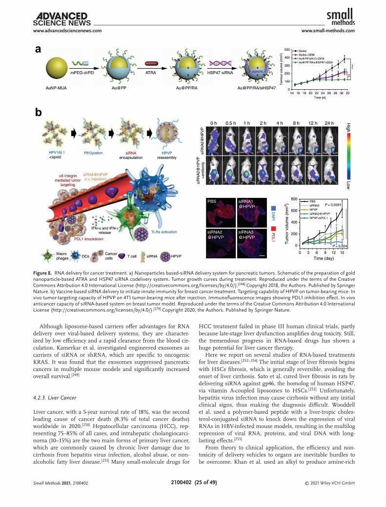
© 2021 Wiley-VCH GmbH2100402 (25 of 49)
www.advancedsciencenews.com www.small-methods.com
Although liposome-based carriers offer advantages for RNA delivery over viral-based delivery systems, they are character-ized by low efficiency and a rapid clearance from the blood cir-culation. Kamerkar et al. investigated engineered exosomes as carriers of siRNA or shRNA, which are specific to oncogenic KRAS. It was found that the exosomes suppressed pancreatic cancers in multiple mouse models and significantly increased overall survival.[249]
4.2.3. Liver Cancer
Liver cancer, with a 5-year survival rate of 18%, was the second leading cause of cancer death (8.3% of total cancer deaths) worldwide in 2020.[250] Hepatocellular carcinoma (HCC), rep-resenting 75–85% of all cases, and intrahepatic cholangiocarci-noma (10–15%) are the two main forms of primary liver cancer, which are commonly caused by chronic liver damage due to cirrhosis from hepatitis virus infection, alcohol abuse, or non-alcoholic fatty liver disease.[251] Many small-molecule drugs for
HCC treatment failed in phase III human clinical trials, partly because late-stage liver dysfunction amplifies drug toxicity. Still, the tremendous progress in RNA-based drugs has shown a huge potential for liver cancer therapy.
Here we report on several studies of RNA-based treatments for liver diseases.[252–254] The initial stage of liver fibrosis begins with HSCs fibrosis, which is generally reversible, avoiding the onset of liver cirrhosis. Sato et al. cured liver fibrosis in rats by delivering siRNA against gp46, the homolog of human HSP47, via vitamin A-coupled liposomes to HSCs.[252] Unfortunately, hepatitis virus infection may cause cirrhosis without any initial clinical signs, thus making the diagnosis difficult. Wooddell et al. used a polymer-based peptide with a liver-tropic choles-terol-conjugated siRNA to knock down the expression of viral RNAs in HBV-infected mouse models, resulting in the multilog repression of viral RNA, proteins, and viral DNA with long-lasting effects.[255]
From theory to clinical application, the efficiency and non-toxicity of delivery vehicles to organs are inevitable hurdles to be overcome. Khan et al. used an alkyl to produce amine-rich
Figure 8. RNA delivery for cancer treatment. a) Nanoparticles based-siRNA delivery system for pancreatic tumors. Schematic of the preparation of gold nanoparticle-based ATRA and HSP47 siRNA codelivery system. Tumor growth curves during treatment. Reproduced under the terms of the Creative Commons Attribution 4.0 International License (http://creativecommons.org/licenses/by/4.0/).[248] Copyright 2018, the Authors. Published by Springer Nature. b) Vaccine-based siRNA delivery to initiate innate immunity for breast cancer treatment. Targeting capability of HPVP on tumor-bearing mice. In vivo tumor-targeting capacity of HPVP on 4T1 tumor-bearing mice after injection. Immunofluorescence images showing PDL1 inhibition effect. In vivo anticancer capacity of siRNA-based system on breast tumor model. Reproduced under the terms of the Creative Commons Attribution 4.0 International License (http://creativecommons.org/licenses/by/4.0/).[279] Copyright 2020, the Authors. Published by Springer Nature.
Small Methods 2021, 2100402

© 2021 Wiley-VCH GmbH2100402 (26 of 49)
www.advancedsciencenews.com www.small-methods.com
ionizable dendrimer cores, which enhanced the efficient com-plexation with negatively charged siRNA under an acidic micro-environment, thus demonstrating that the target gene in siRNA could target specific cells, such as ECs and hepato-cytes.[254] Dendrimers are other powerful delivery vectors. The main challenge is how to design dendrimers with low toxicity and high potency to reduce tissue damage. Zhou et al. reported that modular degradable dendrimers with small RNAs showed a pronounced survival benefit in an aggressive genetic cancer model, owing to a high anti-tumor potency and the low hepa-totoxicity of dendrimers.[256] Some RNA-based drugs or delivery vehicles were studied in clinical trials. As an example, Voutila et al. developed small activating RNAs to upregulate the tran-scription factor CCATT/enhance binding protein alpha. This drug has been undergoing a phase I clinical trial for patients with liver cancer.[257] RNA-based chemotherapy has shown an extremely high value in the treatment of liver cancer. One of the future goals is to reduce the side effects produced by the degradation of drug-carriers.
4.2.4. Prostate Cancer
In men all over the world, prostate cancer is the second most common cause of cancer-related mortality after lung cancer.[250] Common risk factors for developing prostate cancer include age, race, and family history, mostly related to genetic factors. Because of the relatively limited understanding of these genes, their clin-ical management is difficult. Although conventional therapies—including the surgical removal of the prostate, radiation therapy, and hormone therapy—have shown to be useful for prostate cancer, the life quality of patients can be seriously impacted by sur-gical or chemical castration.[258] Thanks to its ability to specifically silence the target gene expression, RNAi technology is emerging as a promising therapeutic procedure for prostate cancer.
With the development of nanotechnology, various efforts have been devoted to developing RNA nanocarriers for prostate cancer therapy. Hasan et al. reported on PLGA/siRNA nano-particles prepared via a unique soft lithography, leading to a high siRNA encapsulation efficiency of 32–46%.[259] As another example, Chen et al. synthesized tertiary amine-functionalized cationic polylactides nanoplexes with a remarkable hydrolytic degradability, while interleukin-8 siRNA can be released by thiol-ene click functionalization.[258] In addition, researchers have moved a step forward to design RNA carriers with a stimulus-responsive function for cancer therapy. For instance, Xu et al. proposed a multifunctional envelope-type RNA-nano-particle, obtained by the modification of ACUPA, a small molecular ligand specifically recognizing the prostate-specific membrane antigen receptor, resulting in an efficient silencing of the prohibitin1 expression.[260] Further research should focus on the study of specific genes for prostate cancer, to improve the effectiveness of targeted therapy.
4.2.5. Lung Cancer
Lung cancer is the leading cause of cancer death and ranks second in incidence (11.4% of new cases), its limited successful
treatment can be attributed to its heterogeneity and adapta-bility.[250,261] Lung cancers can be broken down into two classes: i) non-small-cell lung carcinoma, accounting for ≈85% of all lung cancer cases, and ii) small-cell lung carcinoma. RNA-based therapeutics have recently come into focus as an emerging therapeutic class with great potential for fighting cancer.
It is worth noting that nanoparticles represent an advanced delivery platform for RNA therapeutics due to their high surface areas and easy processability. As spherical vesicles, liposomes have been widely used in RNA-based delivery sys-tems.[186,262,263] As the earliest liposomal delivery system, Zhao et al. developed a lipid-polycation-HA nanoparticle for vascular endothelial growth factor (VEGF) siRNA delivery for VEGF knockdown in a human lung cancer xenograft.[262] Nanoparti-cles were found to induce antitumor efficacy through the activa-tion of the AMP-activated protein kinase and inhibition of the rapamycin; their function was equivalent to that of metformin, an anticancer drug. Another powerful delivery system is rep-resented by MSNPs, thanks to good biocompatibility, tunable pore size, and customizable properties. For instance, Dilnawaz et al. explored the efficacy of the codelivery of a complex with an anticancer drug—such as etoposide or docetaxel-loaded MSNPs and surviving siRNA for lung cancer. This delivery system dem-onstrated pronounced apoptosis effects with a high-dose drug in vitro.[264] In another study, siRNA/MSNP-PEI immobilized on electrospun nanofibers—prepared by Nascimento et al.—provided a longer release period, which exhibited a great poten-tial to achieve a local and sustained release of cancer therapeu-tics.[149] Another strategy for the suppression of cancer is to disrupt the proliferation process of cancer cells. Although the growth of cancer cells can be tuned by different siRNA, adverse side effects usually increase, as happens with normal cells. Therefore, developing siRNA delivery systems with low adverse side effects is one of the further lines of research.
4.2.6. Breast Cancer
Breast cancer is one of the most frequently occurring cancers, with an estimated 2.3 million new cases (11.7% of the new cases) in 2020, and the leading cause of cancer-related deaths among women worldwide.[250] Many factors are associated with an increased risk of breast cancer, such as obesity, use of estrogen and progestin, advanced maternal age at first birth and alcohol consumption. In addition, genetic mutations and epigenetic mechanisms are closely related to the tumorigenesis of breast cancer.[265,266] Therefore, there is still a long way to go for the treatment of breast cancer.
Fortunately, the past few years have witnessed great strides in breast cancer treatment. One example is triple-negative breast cancer: it was defined as a type of breast cancer with negative expression of estrogen, progesterone, and human epi-dermal growth factor receptor-2, and its mortality rate is 40% within the first 5 years after diagnosis.[266] More efforts based on conventional chemotherapy were proven to be effective in reducing side effects, including drug resistance and organ dys-function. Novel nanocomplex carriers and oncogenic miRNA have been widely used for a specific and efficient delivery in cancer treatment to reduce collateral damage to healthy cells
Small Methods 2021, 2100402

© 2021 Wiley-VCH GmbH2100402 (27 of 49)
www.advancedsciencenews.com www.small-methods.com
or organs.[267–269] The combination of multimodal therapeutics shows a good performance in terms of inhibiting breast cancer growth. Juneja et al. developed a porphyrin-based polysilsesqui-oxane platform to deliver RNA, and this platform was proven to affect silencing when tested in MDAMB-231/GFP cells.[270] In another study, paclitaxel, an anti-cancer drug, can be solubilized and co-loaded with RNA into nanoparticles, which showed an ultra-thermodynamic stability for targeted cancer therapy.[269] The treatment of cancers involves the combination of multiple technologies, through which some potential complications could be resolved.
Advanced antiangiogenic therapy has gradually become a new means of preventing and treating cancer metastasis, and it has been successfully used clinically in the treatment of various cancers. Both anti-VEGF siRNA and Nogo-B receptor siRNA are reported to be effective for suppressing EC migra-tion and tubule formation.[271,272] In the future, more research should focus on how to balance the effectiveness and biosafety of RNA delivery systems. Also, it has become increasingly clear that the treatment of cancer, a multifactorial disease, cannot be entrusted to a single molecule or gene editing. Therefore, future research may focus on developing combined strategies including chemotherapy/cancer gene therapy, chemotherapy, RT and immunotherapy.
4.2.7. Cancer Immunotherapy
In addition to surgery, targeted pathway inhibition, chemo-therapy, radiation therapy, and immunotherapy have emerged as alternative modalities of cancer treatment since they can fight aggressive diseases relying on the body’s own immune system. Nowadays, cancer immunotherapy comes in a variety of forms, including cancer vaccines, cell therapy, tumor-infecting viruses, immune checkpoint, cytokines, targeted antibodies, immunogenic cell death, and adjuvants.[273] Impressively, RNA-based therapy has made a huge stride forward for cancer treat-ment since many kinds of RNAi drugs (Patisiran, phase III; ENVISION, phase III) entered clinical trials.[274,275] As a typical immunotherapy method, RNA-based immunotherapy has been increasing interest in elucidating the function of RNA in the regulation of anticancer immune responses and different cancer therapeutics. Therefore, we will discuss and summarize the RNA-based immunotherapy currently applied in the cancer therapy field.
Vaccines as an attractive and effective option have been widely used in treating many infectious diseases, such as polio, measles, and even COVID-19. Nowadays, there are four kinds of vaccines, including viral vector, tumor/immune cell, peptides, and nucleic acid, among which RNA-based vaccines are usually designed to translate tumor-associated antigens in antigen-pre-senting cells and trigger antigen-specific cells.[276] In 1999, Ying et al. utilized self-replicating RNA vectors to enhance the immu-nogenicity of nucleic vaccines. The experimental mice survived from tumor under the self-replicating RNA protection, success-fully demonstrating that RNA may be an excellent candidate for the development of new cancer vaccines.[277] The RNA-based vaccine field is developing extremely rapidly in recent years; Sahin et al. adopted individualized mutanome vaccines and
implemented an RNA-based poly-neoepitope approach to mobi-lize immunity against a spectrum of cancer mutations, and the results indicated that the personal RNA vaccine has a positive effect on melanoma.[34,278] In another study, it has been proven that human papillomavirus pseudovirus (HPVP) nanoparticles loaded with siRNA, forming a human papillomavirus (HPV) vaccine, have effective immunotherapy effects, a high response rate, and good biological safety (Figure 8b).[279] The mRNA vaccine is the highest promising candidate in cancer immu-notherapy as it can encode tumor-associated antigens without potential dangers. In order to deliver mRNA to antigen-pre-senting cells effectively, the lipopolyplex mRNA vaccine incor-porated with a lipid shell or polymer hydrogel has been studied, which demonstrated that the antigen-specific cells increased and tumor growth was inhibited relatively.[280,281] The optimal mRNA stability, cytosolic delivery, and mRNA expression are needed for the efficient mRNA vaccine. At the end of this part, we summarized the significance and challenges of cancer immunotherapy based on the following three therapy methods.
Immune checkpoint blockade (ICB) therapies can induce durable tumor control and extend the survival time of cancer patients by reactivating tumor-associated cells. Recently, long noncoding RNAs show a vital function of immune response, which can predicate survival and immune checkpoint blockade in hepatocellular carcinoma.[282,283] However, the promising results were difficult to be demonstrated due to the complex immune system. In order to enhance efficiency of immune checkpoint therapy, a more powerful combination with RNA has been studied. Immune checkpoint inhibitors, such as T lymphocyte-associated protein 4, programmed cell death pro-tein, and programmed cell death ligand 1, have been applied in many malignant cancers.[284] In the typical study, Bialkowski et al. has demonstrated that ICB combined with antibodies targeting (IL-6 and TGF-β) inhibitions could prolong the sur-vival of treated mice.[285] Due to multiple immunosuppressive mechanisms in the tumor microenvironment, the dysfunctional immune system was the barrier to cancer treatment. Sheng et al. studied ablation of the histone demethylase LSD1 in cancer cells, and the results demonstrated that it led to double-stranded RNA stress and activation of type 1 interferon, which stimulates anti-tumor cells immunity and restrains tumor growth.
The cell’s molecular composition, signalling activity, and metabolic determine the cell fate and function, making RNA-sequencing an effective method for cancer diagnosis. Recently, more advanced technologies were utilized to characterize com-plex cell responses. Katzenelenbogen et al. studied INs-seq for recording scRNA-Seq and intracellular protein activity. Genetic ablation in mice models showed a marked decrease in dysfunc-tion CD8+ cells and reduced tumor growth.[286] Nissim et al. designed an RNA-based AND gate with de novo synthetic pro-moters to enhance specificity. It showed a huge potential for killing cancer cells and significant tumor reduction, and pro-longed mouse survival in vitro and in vivo.[287] The delivery of mitotic checkpoint siRNA-loaded nanoparticles reported in some recent studies showed that the essential mitotic check-point gene’s silencing could induce cell death.[288,289]
To some degree, a collaboration between traditional and novel therapies is crucial to achieving further advances in cancer treatment. Except for the three immune therapies mentioned
Small Methods 2021, 2100402

© 2021 Wiley-VCH GmbH2100402 (28 of 49)
www.advancedsciencenews.com www.small-methods.com
Figure 9. RNA-based treatment for neural tissue applications. a) Flowchart for preparation of electrospun PCL nanofibers functionalized with polydo-pamine and polyDOPA-melanin mussel-derived coatings and Lipofectamine RNAiMAX and TransIT complexed with siRNA, as transfection reagents and b) integration with transected spinal cord tissue of polyDOPA-melanin coated scaffold functionalized with RNAiMAX Lipofectamine-siREST. a,b) Reproduced with permission.[295] Copyright 2018, Royal Society of Chemistry. c,d) SEM images of PCL fiber—collagen hydrogel system.
Small Methods 2021, 2100402

© 2021 Wiley-VCH GmbH2100402 (29 of 49)
www.advancedsciencenews.com www.small-methods.com
above, the other methods, like cytokines and viruses, are key components of RNA-based therapies.[255,290] How to develop immunotherapies with safe and effective antitumor immunity is unsettled. Based on previous research, RNA has been applied as clinical biomarkers for cancer prognosis, diagnosis, and treatment response. RNA-based therapeutics for cancer treat-ment have great potential to enhance immunotherapy by com-bining the current treatment methods.
4.3. Nervous System Treatments
4.3.1. Nervous System Regeneration
An acute trauma affecting the central nervous system often results in an irreversible loss of neural functions. In this con-text, over the years, several approaches have been experimented to replace damaged neurons and foster axonal regeneration. Thanks to their relative abundance and easy accessibility, mes-enchymal stem cells (MSCs) are a clinically viable cell source, whose potential for cross-lineage neuronal differentiation in the presence of proteins and biochemical cocktails is widely reported in the literature.[291,292] Within this framework, Low et al.[293] proposed the absorption of siRNAs onto the surface of DOPA-melanin (DM) coated electrospun nanofibrous scaf-folds with the aim of obtaining materials characterized by i) a prolonged silencing of inhibitory factors against the desired lineage commitment, and ii) a fibrous morphology with a high surface-to-volume ratio in order to maximize the exposure of siRNAs to MSCs, thus making it possible to efficiently induce the MSC differentiation to neural cells.[293] DM-coated random and aligned ε-PCL electrospun nanofibers were subjected to the adsorption of siRNA complexed with the transfection reagent TransIT-TKO (siRNA/TKO), in the same way as in the previ-ously reported procedure published by the same authors.[294] The studies performed on release kinetics highlighted that the presence of DM significantly reduces the initial siRNA burst (from 9.7 ± 0.5% to 6.6 ± 0.2% for random nanofibers and from 16.6 ± 1.5% to 12.3 ± 0.8% for the aligned ones) and increases its maximum cumulative release (from 59.4 ± 3.6% to 30.1 ± 3.1% for random nanofibers and from 57.3 ± 3.7% to 43.3 ± 3.2% for aligned nanofibers). The reduction of the burst release can be ascribed to electrostatic attractions between the positively charged siRNA/TKO complexes and the negatively
charged DM layer. The enhanced siRNA release in the presence of DM coating might be explained by taking into account i) the non-covalent and non-specific interactions between siRNAs and TransIT-TKO molecules, and ii) the fact that the highly nega-tively charged surface of DM coated fibers promotes the release of free siRNA into the external aqueous environment. Under non-specific differentiation conditions, at day 7 a significant REST knockdown was observed only in the presence of DM-coated aligned PCL scaffolds.
In a different study, the same authors employed functional-ized electrospun PCL nanofibers properly optimized to induce the REST knockdown in human induced pluripotent stem cell-derived neural progenitor cells (hiPSC-NPCs) in order to enhance their neuronal differentiation.[295] In this one, two distinct mussel-derived coatings, that is, polydopamine and polyDOPA-melanin, Lipofectamine RNAiMAX and TransIT-TKO, complexed with siRNA, as transfection reagents, were tested (Figure 9a). With the aim of assessing the interactions between the scaffolds seeded with cells and the damaged tis-sues in terms of implant integration, an organotypic spinal cord slice culture, considered a bridge between the in vitro and the in vivo experiments, was used. Interestingly, unlike MSCs, with the same drug dosage TransIT-TKO proved to be cytotoxic for hiPSC-NPCs. Despite a similar siRNA loading efficiency, a lower siRNA release was observed from polyDOPA-melanin-coated nanofibers than from polydopamine-coated ones: in the former, this was probably due to the greater amount of carboxyl groups interacting with positively-charged siRNA complexes. In light of these results, cellular tests were performed only for polyDOPA-melanin-coated scaffolds functionalized with RNAiMAX Lipofectamine-siREST. These results documented: i) the successful integration of the scaffold with the transected spinal cord tissues after 7 days of culturing (Figure 9b); ii) the ability of siREST-absorbed scaffolds to provide differentiation signals to direct cell fate in post-injured tissues; iii) the ability of the scaffold to enable neurite extension across the 2 mm injury gap, without relevant differences the response of the glial cells. The lack of glial response led to the conclusion that the scaffolds did not aggravate spinal cord injuries after in vivo transplantation.
One of the possible obstacles to the spontaneous recovery of traumatic nerve injuries is the lack of surviving oligoden-drocytes (OLs) after the injury, often resulting in an inefficient myelin regeneration and, therefore, in the insurgency of defects
c,d) Reproduced under the terms of the Creative Commons Attribution 4.0 International License (http://creativecommons.org/licenses/by/4.0/).[297] Copyright 2019, Nanyang Technological University. Published by Wiley-VCH. e) Effect of injection of lentiviral vectors encoding Nogo-66 receptor 1 (NgR1)-short hairpin RNA (shRNA) in three groups of rats: LN (LV-NgR1 shRNA injection), LC (LV-control shRNA injection) and Sham (laminectomy only); marker CNPase was used to detect oligodendrocytes, MBP immunostaining and LFB staining were used to detect myelinated fibers. Reproduced under the terms of the Creative Commons Attribution 4.0 International License (http://creativecommons.org/licenses/by/4.0/).[298] Copyright 2018, the Authors. Published by Springer Nature. f) Magnetofection of striatal cells by fluorescein isothiocyanate (FITC) oligonucleotides complexed with Neuromag, magnetic polymeric nanoparticles. Reproduced under the terms of the Creative Commons Attribution 4.0 International License (http://creativecommons.org/licenses/by/4.0/).[324] Copyright 2020, the Authors. Published by Multidisciplinary Digital Publishing Institute. g) Composition and physical properties of PLGA nanoparticles (NPs) containing perfluoro-1,5-crown ether, a fluorine compound trackable non-invasively by fluorine (19F)-magnetic resonance imaging, and coated with protamine sulfate in order to complex miR-12. h) Photomicrographs of SVZ stem/progenitor cells (scale bar: 10 µm). g,h) Reproduced with permission.[325] Copyright 2016, Elsevier. i) Scheme for the fabrication of Gal-NP@siRNA. j) Transmission elec-tron micrographs of Gal-NP@siRNA. k) In vitro gene silencing effects of Gal-NP@siBACE1 and controls at day 3 post-transfection (n = 3, ***p < 0.001). l) Representative image for Cy5 signal in the brain of NP@siRNA and Gal-NP@siRNA groups 1 h after injection. m) Behavioral evaluation of Gal-NP@siBACE1 nanomedicine therapy in APP/PS1 mice: representative swimming track for probe test in the MWM. i–m) Reproduced under the terms of the Creative Commons Attribution 4.0 International License (http://creativecommons.org/licenses/by/4.0/).[332] Copyright 2020, the Authors, some rights reserved. Published by American Association for the Advancement of Science.
Small Methods 2021, 2100402

© 2021 Wiley-VCH GmbH2100402 (30 of 49)
www.advancedsciencenews.com www.small-methods.com
in the neuronal signal transduction. Among micro-RNA, miR-219 and miR-338 have been recognized as playing a pivotal role in the regulation of OL development, being able to silence the expression of negative regulators for OL differentiation. Within this framework, Diao et al.[296] designed a fiber-mediated strategy for the delivery of miR-219 and miR-338 to control OPC development. Similarly to the above-described strategies, DOPA-coated PCL nanofibers were used for miRNA absorp-tion. The authors carefully investigated the effect of both fiber topography and their coupling with miR on the gene silencing outcome. Fiber topography was studied in terms of orienta-tion (random and aligned fibrous scaffolds were taken into account) and diameter (aligned fibers with a diameter varying from 200 nm to 2 µm were produced). Fiber topography (with scrambled miR) resulted in a downregulation of the expression of inhibitory regulators with respect to 2D culture. More specif-ically, the smaller aligned fibers (200 nm in diameter) induced the poorer PDGFR-α knockdown, while 2 µm diameter random fibers inhibited gene expression more than 2 µm diameter aligned fibers did. When coupled with miR-219 or miR-219/miR-338, 2 µm diameter random fibers continued to induce the lowest expression of inhibitory regulators. Although the highest expression of RIP (the early-stage marker of OPC differen-tiation) was registered for 2 µm diameter random fibers, the coupling with miR-219 and the miR-219/miR-338 cocktail had more influence on aligned fibers, with a significant increase in the RIP signal, while the fiber diameter decreased. With regard to the expression of MBP (a late myelin marker of OL matu-ration), independently from the coupled miR, large-diameter fibers induced more MBP expression than 200 nm diameter fibers. This study made it possible to conclude that fiber topog-raphy significantly affects the gene expression and, when cou-pled with miR, both random and aligned large-diameter fibers promoted OPC differentiation, although the small-diameter aligned fibers seemed to be the most promising substrate; on the other hand, the large-diameter aligned fibers seemed more conducive to OL maturation.
In a more recent study, the authors translated the fibrous 2D substrate into a 3D system for direct in vivo micro-RNA screening. Four micro-RNAs (miR-21, miR-222, miR-132, and miR-431) and their cocktails, selected for their ability to regulate neurogenesis and axon growth, were systemati-cally screened.[297] A properly designed fiber-hydrogel system (Figure 9c) was used to enable the implantation of the aligned nanofibers (Figure 9d) into the damaged spinal cord tissue. In the system employed, fibers were immersed in a collagen matrix, enabling the delivery of multiple biochemical factors, while supporting and retaining the aligned fibers in a 3D con-figuration, in order to guide the direction of axon growth as in the in vitro situation. Furthermore, according to the in vitro results, miR-132/miR-222/miR-431 were shown to provide the best regeneration outcomes with respect to miR-21, miR-222/miR-431, and Neg-miR treatments.
Regeneration of Injured Spinal Cord Tissue: Zhao et al.[298] exploited the local injection of lentiviral vectors encoding Nogo-66 receptor 1 (NgR1)-shRNA to induce nerve regenera-tion and functional recovery after spinal cord injury. A lentiviral vector was used due to its documented high transfection rate and long-term and stable gene modifications;[299] NgR1 shRNA
was chosen because of its capability in knocking down the expression of NgR1 gene, which is responsible for suppressing neurogenesis due to its high affinity with myelin-associated inhibitors. The results, achieved from in vivo experiments on rat models, demonstrated that the local injection of lentiviral vectors encoding NgR1 shRNA (LV-NGR1 shRNA) was able to promote nerve regeneration and functional recovery after spinal cord injury[298] Furthermore, studies performed with CNPase and MDB markers to assess, respectively, the presence of oligo-dendrocytes and remyelination at the injury site demonstrated both the survival of more oligodendrocytes and the presence of a higher number of myelinated fibers in the rats subjected to LV-NgR1 shRNA injection, compared to those in the LV-control shRNA group, as reported in Figure 9e.
Another strategy for inducing the regeneration of injured spinal cord tissue by exploiting siRNA delivery is based on silencing nischarin, a protein that inhibits neurite outgrowth and neural cell regeneration, in order to increase the pro-tein expression of the growth-associated protein-43 (GAP-43).[300] Nischarin-targeted siRNA (Nis-siRNA) was delivered by poly(ethylenimine)-alginate (PEI-ALG) nanoparticles to the spinal cord tissue with the aim of promoting motor function recovery in rats. PEI-ALG nanoparticles were selected as car-riers since they had been demonstrated to effectively deliver siRNAs, while preventing enzymatic hydrolysis and facilitating the entry of nucleic acids into cells.[301] As expected, 3 weeks after the injection, a dramatic decrease in nischarin expres-sion was registered in the lesion site for the rats treated with Nis-siRNA, whereas in the region 1 cm distal from the lesion, no significant differences were observed compared to the Ctl-siRNA group. Furthermore, a motor function recovery, in terms of right hindlimb movement, was observed in spinal cord-injured rats treated with PEI-ALG/Nis-siRNA.[300]
In addition to the approaches based on nervous tissue regen-eration after a traumatic injury, strategies focused on blocking or decreasing inflammasome activation after an injury have also been investigated. De Rivero Vaccari et al.,[302] in an attempt to achieve this aim, exploited neuronal-derived exosomes, loaded with siRNA-GFP against apoptosis-associated speck-like protein containing a caspase recruitment domain (ASC), as a thera-peutic vector to block inflammasome activation after a spinal cord injury. In vivo tests were mainly devoted to assessing the ability of exosomes, injected into the femoral vein, to cross the blood-spinal cord barrier, penetrate the spinal cord paren-chyma, and deliver their cargo. The results showed that cells in the epicenter of the lesion were positive to GFP, while they were negative to GFP in the regions distant from the epicenter, thus indicating an effective delivery of the cargo.
4.3.2. Neurodegenerative Diseases
The potential of RNA for the treatment of neurodegenerative diseases is under constant investigation, as demonstrated by very recent studies in this field.[303] Positive outcomes have motivated researchers to identify strategies for aiding the delivery of RNA, injected locally into the brain or at a systemic level, for the treatment of these diseases.[304–306] To be suitable for this specific application, a proper design of delivery systems
Small Methods 2021, 2100402

© 2021 Wiley-VCH GmbH2100402 (31 of 49)
www.advancedsciencenews.com www.small-methods.com
is required to provide them in vivo stability and transcytotic potential.
In this context, Haroon et al. developed a strategy in which a peptide known to target specific gangliosides was fused to a double-stranded RNA binding protein to deliver siRNA to the brain parenchyma of rat models.[307] The resulting protein was labeled as TARBP-BTP and it was able to enter Neuro 2a, IMR32, and HepG2 cells in the presence of monosialoganglio-side GM1. The delivery of TARBP-BTP bound to siRNA led to a significant knockdown in the brain of BACE1, whose activity is associated with neurodegeneration and accretion of amyloid precursor protein products.[308]
Another approach exploiting peptides is based on the pro-duction of peptide nanofibers (PNFs), intended as the assembly of amphiphilic peptides in a single-dimension cylindrical geom-etry. These PNFs can be engineered with amino acids showing a positive charge in order to electrostatically link and deliver the negatively charged siRNA. Mazza et al. realized a surfactant-like peptide (palmitoyl-GGGAAAKRK) showing the capability to self-assemble into PNFs. The authors demonstrated that the complex, administrated intracranially, was uptaken intracel-lularly and promoted the increase in siRNA’s residence time (from 48 h for siRNA to 7 days for PNF-siRNA) in the brain.[309] More specifically, the complex was explored as a nanovector to the target and silenced BCL2, responsible for inhibiting apop-tosis in the neuronal population, in the subthalamic nucleus. Results demonstrated that the complex remained localized in close proximity to the injection site, without migrating into the brain’s distal region even 1 week after the administration. Furthermore, the complex turned out to successfully silence the expression of BCL2 in the subthalamic nucleus compared to the noninjected hemisphere. According to this result, a significant loss of Nissl-stained cells was registered only in the hemisphere subjected to the treatment, thus highlighting the localization of neuronal tissue ablation.
Peptides were also used to produce polymer-peptide nano-particles capable of linking siRNA to be released into Neuro 2a neuronal cells. Nanoparticles were obtained by chemose-lectively conjugating PEG to the chitosan’s C2 hydroxyl group and to a cell-penetrating TAT peptide.[310] The domain of the selected TAT peptide was composed of residues of arginine and lysine amino acids that played a relevant role in translocation across the biological membrane. The potential of chitosan-PEG-TAT nanoparticles complexed with siRNA to silence ataxin-1 protein was assessed by using an in vitro model of spinocer-ebellar ataxia, a neurodegenerative disease. Results indicated a suppression of the ataxin-1 protein after 48 h of transfection with negligible toxicity effects.
A different approach was adopted by Dai et al., who pro-posed a library of multiblock copolymer structures for nucleic acid delivery.[311] The obtained copolymers, produced from PEG, poly(propylene glycol) (PPG), and PLL, were the following: PEG-PLL (P1), PLL-PEG-PLL (P2), and PLL-PPG-PEG-PPG-PLL (P3). The resulting particles, obtained from these block copoly-mers, showed an amphiphilic nature which enabled the ready formation of micelle-complex with siRNA. The block copol-ymer P2 was the largest in size and showed the highest cation charge, proving to induce a great gene knockdown but also the highest cytotoxicity. Compared to P1 and P2, P3 can induce an
effective gene knockdown due to the presence of PPG units in its structure. Furthermore, while P1 and P2 copolymers could complex siRNA, forming micelle-like aggregates of large size and poor density, P3-siRNA complex, with a smaller volume and compact micelle-architecture, could enter the cells more easily. In light of these considerations, P3 complex turned out to be the most efficient among the tested complexes for siRNA delivery and gene silencing, permitting achieving an in vitro decrease in GFP expression in GFP-expressing Neuro 2a cells of around 28%.
Polymeric nanoparticles, made of gelatin and cross-linked with glutaraldehyde, were tested for the intranasal delivery of encapsulated siRNA silencing iNOS in post-ischemic rat brain.[312] iNOS-RNA was selected for its ability in silencing iNOs-derived NO, which contributes to neurotoxicity after an ischemic stroke,[313,314] since iNOS inhibition could reduce the infarct volume. Gelatin was selected for its biocompatibility and the non-toxicity of its degradation products. Moreover, the drug release can be controlled by manipulating the kinetics of gelatin degradation, that is, by controlling the crosslinking degree. As for the intranasal administration, this was preferred over systemic injection in order to bypass the BBB. The delivery of gelatin nanoparticles incorporating iNOS-siRNA 6 h after middle cerebral artery occlusion resulted in suppressing the infarct volumes, with a maximum reduction of around 42%.[312]
Very recently, Krienke et al. proposed an approach based on the delivery of mRNA for the treatment of neurodegenerative diseases.[315] The study suggested a non-inflammatory mRNA vaccine for the treatment of autoimmune encephalomyelitis. In particular, the authors showed that the delivery at the systemic level of nanoparticle-formulated 1-methylpseudouridine-mod-ified messenger RNA coding for disease-related autoantigens resulted in the presentation of the antigen on splenic CD11c+ antigen-presenting cells in absence of costimulatory signals. In vivo tests performed on multiple sclerosis mouse models highlighted suppression of the disease by treatment with this mRNA; this result was ascribed to both the decrease of effector T cells and the development of the regulatory T cell populations.
Parkinson’s Disease: Alpha-synuclein (α-syn) deposition in Lewy bodies (LB) is one of the most characteristic neuropatho-logical hallmarks of Parkinson’s disease (PD). Schlich et al. investigated anionic liposomes, functionalized with rabies virus glycoprotein (RVG) as a target agent, loaded with a siRNA-protamine complex for the silencing of α-syn gene.[316] The potential of the proposed approach was tested in terms of in vitro effects on primary cortical and hippocampal cells. The achieved results on primary cortical neurons showed a signifi-cant decrease in the α-syn immunopositive signal in cells sub-jected to decorated liposomes containing siRNA, compared to those of controls and RVG liposomes not containing siRNA. Data were in line with those obtained on cultures of primary hippocampal neuronal cells, selected as usually affected by α-syn deposition in dementia LB. Also in this case, a reduced α-syn-immunopositive signal in neurons subjected to decorated liposomes containing siRNA was registered, with respect to the other two groups of cells.
As documented in the above-mentioned study, siRNA can be successfully applied for the treatment of PD; however, the inef-ficient delivery of siRNA into neurons hampers its in vivo use.
Small Methods 2021, 2100402
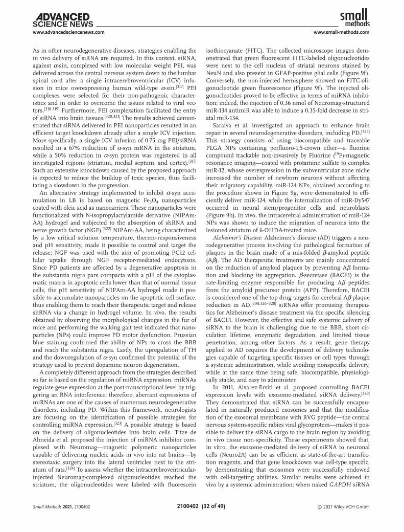
© 2021 Wiley-VCH GmbH2100402 (32 of 49)
www.advancedsciencenews.com www.small-methods.com
As in other neurodegenerative diseases, strategies enabling the in vivo delivery of siRNA are required. In this context, siRNA, against α-sin, complexed with low molecular weight PEI, was delivered across the central nervous system down to the lumbar spinal cord after a single intracerebroventricular (ICV) infu-sion in mice overexpressing human wild-type α-sin.[317] PEI complexes were selected for their non-pathogenic character-istics and in order to overcome the issues related to viral vec-tors.[318,319] Furthermore, PEI complexation facilitated the entry of siRNA into brain tissues.[320,321] The results achieved demon-strated that siRNA delivered in PEI nanoparticles resulted in an efficient target knockdown already after a single ICV injection. More specifically, a single ICV infusion of 0.75 mg PEI/siRNA resulted in a 67% reduction of α-syn mRNA in the striatum, while a 50% reduction in α-syn protein was registered in all investigated regions (striatum, medial septum, and cortex).[317] Such an extensive knockdown caused by the proposed approach is expected to reduce the buildup of toxic species, thus facili-tating a slowdown in the progression.
An alternative strategy implemented to inhibit α-syn accu-mulation in LB is based on magnetic Fe3O4 nanoparticles coated with oleic acid as nanocarriers. These nanoparticles were functionalized with N-isopropylacrylamide derivative (NIPAm-AA) hydrogel and subjected to the absorption of shRNA and nerve growth factor (NGF).[322] NIPAm-AA, being characterized by a low critical solution temperature, thermo-responsiveness and pH sensitivity, made it possible to control and target the release; NGF was used with the aim of promoting PC12 cel-lular uptake through NGF receptor-mediated endocytosis. Since PD patients are affected by a degenerative apoptosis in the substantia nigra pars compacta with a pH of the cytoplas-matic matrix in apoptotic cells lower than that of normal tissue cells, the pH sensitivity of NIPAm-AA hydrogel made it pos-sible to accumulate nanoparticles on the apoptotic cell surface, thus enabling them to reach their therapeutic target and release shRNA via a change in hydrogel volume. In vivo, the results obtained by observing the morphological changes in the fur of mice and performing the walking gait test indicated that nano-particles (NPs) could improve PD motor dysfunction. Prussian blue staining confirmed the ability of NPs to cross the BBB and reach the substantia nigra. Lastly, the upregulation of TH and the downregulation of α-syn confirmed the potential of the strategy used to prevent dopamine neuron degeneration.
A completely different approach from the strategies described so far is based on the regulation of miRNA expression. miRNAs regulate gene expression at the post-transcriptional level by trig-gering an RNA interference; therefore, aberrant expressions of miRNAs are one of the causes of numerous neurodegenerative disorders, including PD. Within this framework, neurologists are focusing on the identification of possible strategies for controlling miRNA expression.[323] A possible strategy is based on the delivery of oligonucleotides into brain cells. Titze de Almeida et al. proposed the injection of miRNA inhibitor com-plexed with Neuromag—magnetic polymeric nanoparticles capable of delivering nucleic acids in vivo into rat brains—by stereotaxic surgery into the lateral ventricles next to the stri-atum of rats.[324] To assess whether the intracerebroventricular-injected Neuromag-complexed oligonucleotides reached the striatum, the oligonucleotides were labeled with fluorescein
isothiocyanate (FITC). The collected microscope images dem-onstrated that green fluorescent FITC-labeled oligonucleotides were next to the cell nucleus of striatal neurons stained by NeuN and also present in GFAP-positive glial cells (Figure 9f). Conversely, the non-injected hemisphere showed no FITC-oli-gonucleotide green fluorescence (Figure 9f). The injected oli-gonucleotides proved to be effective in terms of miRNA inhibi-tion; indeed, the injection of 0.36 nmol of Neuromag-structured miR-134 antimiR was able to induce a 0.35-fold decrease in stri-atal miR-134.
Saraiva et al. investigated an approach to enhance brain repair in several neurodegenerative disorders, including PD.[325] This strategy consists of using biocompatible and traceable PLGA NPs containing perfluoro-1,5-crown ether—a fluorine compound trackable non-invasively by Fluorine (19F)-magnetic resonance imaging—coated with protamine sulfate to complex miR-12, whose overexpression in the subventricular zone niche increased the number of newborn neurons without affecting their migratory capability. miR-124 NPs, obtained according to the procedure shown in Figure 9g, were demonstrated to effi-ciently deliver miR-124, while the internalization of miR-Dy547 occurred in neural stem/progenitor cells and neuroblasts (Figure 9h). In vivo, the intracerebral administration of miR-124 NPs was shown to induce the migration of neurons into the lesioned striatum of 6-OHDA-treated mice.
Alzheimer’s Disease: Alzheimer’s disease (AD) triggers a neu-rodegenerative process involving the pathological formation of plaques in the brain made of a mis-folded β-amyloid peptide (Aβ). The AD therapeutic treatments are mainly concentrated on the reduction of amyloid plaques by preventing Aβ forma-tion and blocking its aggregation. β-secretase (BACE1) is the rate-limiting enzyme responsible for producing Aβ peptides from the amyloid precursor protein (APP). Therefore, BACE1 is considered one of the top drug targets for cerebral Aβ plaque reduction in AD.[308,326–328] siRNAs offer promising therapeu-tics for Alzheimer’s disease treatment via the specific silencing of BACE1. However, the effective and safe systemic delivery of siRNA to the brain is challenging due to the BBB, short cir-culation lifetime, enzymatic degradation, and limited tissue penetration, among other factors. As a result, gene therapy applied to AD requires the development of delivery technolo-gies capable of targeting specific tissues or cell types through a systemic administration, while avoiding nonspecific delivery, while at the same time being safe, biocompatible, physiologi-cally stable, and easy to administer.
In 2011, Alvarez-Erviti et al. proposed controlling BACE1 expression levels with exosome-mediated siRNA delivery.[329] They demonstrated that siRNA can be successfully encapsu-lated in naturally produced exosomes and that the modifica-tion of the exosomal membrane with RVG peptide—the central nervous system-specific rabies viral glycoprotein—makes it pos-sible to deliver the siRNA cargo to the brain region by avoiding in vivo tissue non-specificity. These experiments showed that, in vitro, the exosome-mediated delivery of siRNA to neuronal cells (Neuro2A) can be as efficient as state-of-the-art transfec-tion reagents, and that gene knockdown was cell-type specific, by demonstrating that exosomes were successfully endowed with cell-targeting abilities. Similar results were achieved in vivo by a systemic administration: when naked GAPDH siRNA
Small Methods 2021, 2100402
© 2021 Wiley-VCH GmbH2100402 (33 of 49)
www.advancedsciencenews.com www.small-methods.com
was injected, GAPDH silencing was detected in the spleen, liver, and kidneys; conversely, exosome-encapsulated siRNAs were resistant to nonspecific uptake, but resulted in a signifi-cant knockdown of GAPDH mRNA in several brain regions. The authors then focused on BACE1. Two validated BACE1 siRNAs were applied in vitro to Neuro2A cells in increasing doses, resulting in a dose-dependent knockdown, whereas when the acetylcholine receptor (the cellular target of RVG-peptide) was blocked by α-bungarotoxin, a reduced BACE1 knockdown occurred. In vivo, the administration to normal C57BL/6 mice of BACE1 siRNA encapsulated in RVG exosomes resulted in a significant protein knockdown. Lastly, the authors demonstrated a significant decrease in the total β-amyloid 1–42 levels—a main component of the amyloid plaques in Alzhei-mer’s disease—which was greater than that reported after the intraventricular injection of BACE1 inhibitors in normal mice.
Guo et al. used nanocomplexes made of cationic polymers, PEGylated poly(2-(N,N-dimethylamino) ethylmethacrylate) (PEG-PDMAEMA), loaded with siRNAs against BACE1 and modified on the surface by CGN and QSH peptides for tar-geting BBB and amyloid plaques, respectively.[330] They dem-onstrated that nanocomplexes (diameter of 70 nm) efficiently cross the BBB monolayer without being damaged and that they can be internalized by neuronal cells (Neuro2A) in vitro, while in vivo, they are mainly concentrated on the neurons sur-rounding amyloid plaques, and are useful for increasing gene accumulation in the AD lesion, consequently achieving a better therapeutic effect.[331] Moreover, the presence of siRNA in nano-complexes efficiently inhibited the expression of BACE1 in vivo, with a consequent reduction in the amyloid plaque formation, an increased synaptophysin level, and an improvement in the cognitive performance of AD transgenic mice.
Zhou et al. developed a glycosylated polymeric nano-medicine containing siRNA stabilized by a triple-interaction (Gal-NP@siRNA) to target BACE1 in transgenic AD mouse models (Figure 9i).[332] The triple interaction strategy consisted of using a salt of guanidinium-phosphate (Gu + /PO 3 4−) to stabilize the electrostatic and hydrogen bond interactions, and a hydrophobic interaction due to the complexation between siRNA and the galactose-modified fluorinated polymer.[240] This strategy determines an effective encapsulation of siRNA and a higher stability performance in blood circulation than with nanomedicines based on cationic polymers, where only elec-trostatic interactions stabilize siRNA. Moreover, the Gal-NP@siRNA (diameter of 118 nm, Figure 9j) can effectively penetrate the BBB via glycemia-controlled glucose transporter-1 (Glut1)–mediated transport. After verifying that in Neuro2A cells the Gal-NP@siRNA determines BACE1 gene silencing in vitro (Figure 9k), the authors investigated in vivo pharmacokinetics. They found that Gal-NP@siRNA has a longer blood circulation time than in controls, with an elimination half-lifetime (t1/2) of 39.2 min; the brain targeting by the glycemia-controlled Glut1-mediated transport was verified (Figure 9l). The therapeutic effect of Gal-NP@siBACE1 was investigated in the APP/PS1 double transgenic mouse model with several behavioral tests, such as the novel object recognition (NOR) and the Morris water maze (MWM) tests (Figure 9m), thus confirming that i) the Gal-NP@siRNA significantly improves cognitive perfor-mances in APP/PS1 mice, and ii) both hippocampal and cor-
tical BACE1 protein levels were relevantly reduced with respect to other APP/PS1 control groups.
A different strategy for the treatment of Alzheimer’s dis-ease consists of regulating miR132, which plays a key role in the neural development and regulation of neuronal activity.[333] In recent years, a strategy based on the nose-to-brain delivery of NPs complexed with miR132 to prevent the decrease of miR132 levels was experimented. This intranasal injection was preferred over systemic injection with the aim of inducing a faster transport of the drug to the brain, while minimizing sys-temic exposure and bypassing the BBB. Samaridou et al. pro-posed the formation of electrostatically driven nanocomplexes between a lauric acid chemically conjugated octa-arginine and the miRNA.[334] The resulting nanocomplexes (ENCPs) were enveloped with different protective polymers, that is, PEG-poly-glutamic acid (PEG-PGA) or HA, in order to enhance their sta-bility across the olfactory nasal mucosa. The results, obtained in vivo on an AppNL−G-F knock-in mouse model of Alzheimer’s disease, demonstrated the suitability of the designed NPs in reaching the hippocampus area—which is involved in memory formation and one of the first areas affected in Alzheimer’s disease[335]—while leading to increased miR-132 endogenous levels after 24 h from the administration. Furthermore, addi-tional studies in vivo demonstrated that thanks to the presence of ENCPs, miR-132 in its active form was successfully delivered to the hippocampus, as demonstrated by the reduced levels of two miRNA targets, GATA2 and Rb1.[334]
In order to prevent the decrease of miR132 levels in the blood, Su et al. delivered wheat germ agglutinin (WGA)-NPs-miR132 intranasally in mouse and rat models of Alzheimer’s disease with ischemic brain injury, to treat neurological damages after cerebral ischemia.[336] WGA-NPs were selected as effective car-riers for the intranasal delivery to the central nervous system of the brain, as demonstrated in a previous study.[337] The potential of intranasally delivered WGA-NPs-miR132 was evalu-ated through in vivo analyses on wild-type C57BL/6 mice, by investigating the Aβ plaques and synaptic-related protein (SYN and PSD95) levels. The results demonstrated that Aβ protein expression, upregulated in AD mice, significantly decreases after WGA-NPs-miR132 treatment. Conversely, a significantly increased expression of SYN and PSD-95 was detected after the inhalation.[336] In MCAO model rats, the authors evaluated the effect of NP inhalation in terms of apoptosis around brain lesions after cerebral ischemia. The results demonstrated that WGA-NPs (not containing miRNA) had no protective effect against ischemic brain damages; miRNA delivered alone was easily degraded and/or cleared by nasal cilia; WGA-NPs-miR132 proved to be effective in reducing the area of cerebral infarc-tion, the number of microglia, and the number of apoptotic cells after cerebral hemorrhage.
4.4. COVID-19 Vaccines
4.4.1. Epidemiology: Origin, Features, and Transmission
The emergence of global disease outbreaks has been one of the most significant challenges to be faced for the human society. At irregular intervals, novel flu viruses against which most people
Small Methods 2021, 2100402

© 2021 Wiley-VCH GmbH2100402 (34 of 49)
www.advancedsciencenews.com www.small-methods.com
were not immune have naturally appeared, thus resulting in global pandemics. Moreover, increased international traveling, trading, and ethnic integration, together with a greater urbani-zation and massive exploitation of environmental resources, have sustained the spread of infections worldwide.[338]
Since early in the 21st century, three viruses—specifically coronaviruses—have crossed the species barrier, causing severe pneumonia disease in humans: i) the Severe Acute Respira-tory Syndrome CoronaVirus (SARS-CoV) broke out in 2002 in Guangdong Province (China); ii) the Middle-East Respiratory Syndrome CoronaVirus (MERS-CoV) was discovered in 2012 on the Arabian Peninsula; iii) the SARS-CoV-2, a novel corona-virus, broke out in December 2019 in Wuhan (Hubei Province, China). SARS-CoV-2 was associated with an outbreak of atypical pneumonia, named COronaVIrus Disease 2019 (COVID-19), that rapidly spread from China to all continents, unleashing a global infection.[339,340] Coronaviruses consist of a positive-sense, non-segmented, single-stranded mRNA [(+)ssRNA] of ≈30 Kb, enveloped in a helical nucleocapsid. The viral RNA encodes a set of 28 specific proteins grouped into three main categories: i) non-structural proteins (Nsp1 to Nsp16); ii) struc-tural spike (S), nucleocapsid (N), membrane (M), and enve-lope (E) proteins; and iii) the accessory proteins (orf3a, orf6, orf7a, orf7b, orf8, orf9b, orf9c, and orf10). Nsp proteins lead to the formation of the replication-transcription complex (RTC). This complex promotes the synthesis of a set of nested sub-genomic minus-strands of RNA [(–)sgRNA] in specific double-membrane vesicles. These [(–)sgRNA] serve as templates and encode subgenomic mRNAs from which all the structural and accessory proteins are synthesized.[341] Bioinformatic analyses showed that SARS-Cov-2 belongs to the Coronaviridae family (order Nidovirales, subfamily Orthocoronaviridae). Further-more, SARS-CoV-2 is a beta-coronavirus (2B lineage) showing a relationship with the SARS-like coronavirus strain in bats, BatCov RaTG13 (96% homologies), which can effectively infect a wide range of vertebrates, including bats, pangolins, and humans. Therefore, at the present time, SARS-CoV-2 seems to have a zoonotic origin and to have acquired human-to-human spreading capacity.[342] Several studies suggest that horseshoe bats (Rhinolophus affinis) seem to be a natural reservoir of both SARS-CoV and SARS-CoV-2, while pangolins (Manis javanica) and American minks (Neovison vison) appear to be the interme-diate hosts of SARS-CoV-2 for animal-to-human infection. In a recent study, it has been posited that the beta-coronavirus iso-lated from pangolins (Pangolin-CoV) has a 100%, 98.6%, 97.8%, and 90.7% amino acid similarity with human SARS-CoV-2 E, M, N, and S proteins, respectively. Furthermore, the receptor-binding domain (RBD) of the S protein of Pangolin-CoV practi-cally matches the RBD of SARS-CoV-2, with only one difference in a non-critical amino acid. Unfortunately, humans remain the principal source of transmission for other humans and domestic, farm, and zoo animals. Interestingly, dogs, pigs, and poultry have shown resistance to SARS-CoV-2.[343,344]
4.4.2. Pathogenesis
The primary access mode of SARS-CoV-2 is through the upper respiratory tract (via droplets or aerosol suspension), where the
nasal and bronchial epithelial cells and pneumocytes are its tar-gets. Coronavirus recognizes the angiotensin-converting enzyme 2 (ACE2), a cell membrane enzyme present in epithelial cells of the airway, lungs, heart, blood vessels, kidneys, liver, and intes-tine (Figure 10a). In detail, the S1 transmembrane subunit of S protein promotes the virus attachment to host ACE2 proteins, while the S2 subunit is responsible for the fusion with the host cell membrane. ACE2 is a crucial factor in the renin-angiotensin-aldosterone system (RAAS) pathway, since it regulates processes such as blood pressure, wound healing, and inflammation.[345,346] Furthermore, ACE2, by counteracting the angiotensin II (ANG II) activity and reducing the pathological effects associated with its high levels, plays a crucial role in preventing tissue damage. Therefore, the binding of SARS-CoV-2 with ACE2 hinders ANG II downregulation and sustains tissue injuries.[339]
COVID-19 presents a wide range of symptoms, including fever (88.7%)—more commonly in adults than in children—dry cough (67.8%), fatigue/tiredness (38.1%), sputum production (33.4%), dyspnea (18.6%), sore throat (13.9%), and headache (13.6%). So far, more than 110 million confirmed COVID-19 cases and 2.4 million deaths have been reported.[347] It is impor-tant to stress that most adult patients have had an asympto-matic infection or mild flu-like symptoms, while 14% of COVID patients have suffered severe health issues that require hospi-talization and/or oxygen support, and 5% have been treated in intensive care units.[348,349]
4.4.3. Management Strategies
While several treatments have been adopted to deal with the impact of the disease, vaccination represents the primary means to effectively control this pandemic. To stimulate the human immune response against a virus, a vaccine usually contains an antigen made from its weakened or killed forms, toxins, or one of its surface proteins. Most COVID-19 vaccine candidates are in the development stages, and the so-called S spike protein of SARS-CoV-2 is their main target.
Multiple approaches are being deployed to identify a safe and effective vaccine. Among them, those based on nucleic acids represent the most important contemporary drug discovery platform, since they enable rapid vaccine development, authori-zation, and manufacturing. This method permits the release of small RNA fragments that encode specific viral proteins into the host cells; then they will be internally processed and loaded onto the host cell membrane. Immune cells can recognize them and lead to the synthesis of antibodies. Remarkably, these vaccines are self-adjuvant and promote a humoral and cellular immune response against the engendered proteins; they can deliver multiple antigens with one immunization. Furthermore, their safety, tolerance, and high potency have been demon-strated in numerous studies.[350] Karikó et al. laid the founda-tion for RNA-based vaccines in 2008.[351] In their research, the authors demonstrated that the nucleoside-modified mRNA (modRNA) containing pseudouridines is translated more effi-ciently than unmodified mRNA in vitro, particularly in primary mammalian dendritic cells and in vivo in mice. Moreover, the modRNA effectively improved RNA biological stability, while reducing its immunogenicity in vivo. Several other
Small Methods 2021, 2100402

© 2021 Wiley-VCH GmbH2100402 (35 of 49)
www.advancedsciencenews.com www.small-methods.com
advantages have been demonstrated, including: i) improved safety (modRNA cannot be integrated into chromatin), ii) lack of host inflammatory response; iii) efficient transduction and rapid protein expression using the host ribosomal machinery; iv) unlimited engineered nucleotide-sequence size; v) closely controllable half-life; and vi) high manufacturability for large-scale continuous production.
As discussed earlier, LNP represents a robust strategy for delivering the modRNA to host cells (Figure 10b). The LNP system is usually made of cholesterol, which plays various struc-tural roles, including filling gaps in the particle, preventing LNP–protein interactions, and promoting fusion with the cell membrane. In addition, the ionizable lipids and functional lipids—which play a crucial role in maintaining the physiolog-ical pH and promote endosomal release—are essential compo-nents of the modRNA vaccine.[352] Currently, there are two kinds of mRNA-based vaccines: non-amplifying mRNA-based, and self-amplifying mRNA-based. To date, there are 181 candidates in the pre-clinical evaluation stage and 69 candidates in the clin-ical stage as COVID-19 vaccines. Among them, 30 are based on RNA technologies. So far, the FDA and the European Medicines Agency (EMA) have granted the emergency use and marketing
of two mRNA-based vaccines: Comirnaty (Tozinameran or BNT162-02), produced by Pfizer-BioNTech, and the mRNA-1273 developed by Moderna.[353] Remarkably, a research group of the Departments of Pathology, Genetics, Pediatrics, and Medicine at Stanford University has recently provided the putative sequence information for the two synthetic RNA molecules of Pfizer/BioNTech and Moderna COVID-19 vaccines and has released them online on GitHub. The RNAs samples were obtained from vaccine drops that remained in vials—and destined to be other-wise discarded—after patients immunization and were analyzed under FDA authorization for research use.[354]
Pfizer-BioNTech COVID-19 Vaccine: Comirnaty vaccine, based on a cell-free in vitro transcription approach, was designed from SARS-CoV-2 DNA templates. It contains a modRNA encoding the SARS-CoV-2 full-length viral spike (S) protein, in which two proline mutations have been introduced to lock it in the prefusion conformation (Figure 10c). The exact formulation of the Pfizer-BioNTech LNP was recently disclosed. This con-tains DSPC, cholesterol, and two novel functional lipid excipi-ents, ALC-0315 and ALC-0159. ALC-0315 is a physiological pH cationic synthetic lipid ((4-hydroxybutyl)azanediyl)bis(hexane-6,1-diyl)bis(2-hexyldecanoate) essential for the efficient
Figure 10. COVID-19 RNA vaccine. a) Side view of sarbecovirus S glycoproteins plotted on the SARS-CoV-2 S structure. Reproduced with permission.[339] Copyright 2020, Elsevier. b) modRNA-LNP structure in which mRNA is bound to the ionizable lipids (central core) while PEG lipids and DSPC form the surface of the lipid nanoparticle. Reproduced under the terms of the Creative Commons Attribution 4.0 International License (http://creativecom-mons.org/licenses/by/4.0/).[352] Copyright 2021, the Authors. Published by Multidisciplinary Digital Publishing Institute. c) RNA vaccine vectors that include a cap, 5′ UTR, 3′ UTR, and poly(A) tail of variable length, and modRNA target sequence. Reproduced under the terms of the Creative Commons Attribution 4.0 International License (http://creativecommons.org/licenses/by/4.0/).[340] Copyright 2021, the Authors. Published by Multidisciplinary Digital Publishing Institute.
Small Methods 2021, 2100402

© 2021 Wiley-VCH GmbH2100402 (36 of 49)
www.advancedsciencenews.com www.small-methods.com
self-assembly and encapsulation of the modRNA within the LNP and its delivery into host cells. ALC-0159 is a PEGylated lipid designed to exchange out of the LNPs.[355] Polack et al. reported that two 30 µg doses of vaccine (given 3 weeks apart) into the muscle of the upper arm promote the development of high SARS-CoV-2 antibody titers and robust antigen-specific CD8+ and TH1-CD4+ T cell responses with an overall efficacy of 95%.[181] The first benefit has been observed from day 12 after the first injection, indicating an early onset of immunization, reaching a 52% efficacy in the interval between the two injec-tions. Furthermore, complete protection against COVID-19 was observed 7 days after the second dose. Adverse reactions were classified as mild or moderate in particular in the oldest adults, and interestingly, the reactogenicity after both injections was similar. Unfortunately, despite this promising data, the study is affected by several limitations, including i) the low cold tem-peratures (−80 °C) required for shipping and storage, ii) the low number of participants for each of the selected groups, iii) the limited follow-up time (approx. 2 months) after the second dose, and iv) the long-term monitoring that remains to be deter-mined. Regarding the last item, ethical barriers can give rise to a heated debate. Over the next 2 years, the planned follow-up should also monitor the placebo recipients to collect long-term data, but on the other hand, it is not ethical to deprive these people from receiving the COVID-19 vaccine before the conclu-sion of the trial.[356,357]
Moderna COVID-19 Vaccine: Moderna COVID-19 Vaccine (mRNA-1273) encodes for the full-length SARS-CoV-2 spike protein modified with two proline substitutions, K986P and V987P, within the heptad repeat 1 domain (S-2P), to block the spike protein into a prefusion conformation. In detail, the mRNA sequence includes a 5“ cap, the 5” untranslated region (UTR), the Open Reading Frame (ORF), the 3“UTR, and the 3” polyA tail. The modRNA is encapsulated into LNPs, and the exact formulation of the Moderna mRNA-1273-LNP was publicly released recently. It is made of DSPC, cholesterol, and two innovative excipients, PEG2000-DMG, and the ionisable lipid heptadecan-9-yl-8-((2-hydroxyethyl)(6-oxo-6-(undecyloxy)-hexyl) amino)-octanoate (SM-102). SM-102, a molecule highly soluble in several organic solvents and insol-uble in water, was selected for its vaccine potency, tolerability, and biodegradability. Interestingly, the storage temperature of the finished product is −20 °C, since only a slight degradation was observed.[358]
Baden et al. reported that two 100 µg doses of the mRNA-1273 vaccine, given 28 days apart into the deltoid muscle, lead to a 94.1% efficacy in COVID-19 illness prevention, including its severe forms.[184] Moreover, the local adverse vaccination reactions were mild. However, moderate-to-severe systemic side effects (fatigue, myalgia, arthralgia, and headache) were observed in around 50% of the participants in the mRNA-1273 group after the second dose. Additionally, Anderson et al. showed that the mRNA-1273 vaccine elicited a remarkable CD4 cytokine response, including TH1 cells.[359,360] Moreover, the tumor necrosis factor α (TNFα) responses were more significant than the interleukin-2 (IL2) ones, which in turn were greater than the interferon-γ (INFγ) ones. Unfortunately, only after the second dose low levels of cytotoxic CD8 T cell responses were observed.
4.4.4. Artificial Intelligence to Face with COVID-19 and Its Variants
Automatic feature learning, supported by the increased com-putational capabilities, has contributed immensely to the suc-cessful application of machine learning (ML)—a branch of the broader artificial intelligence (AI) sector in almost every research field. Among the outstanding examples demonstrated so far, one of the most impactful areas consists of vaccine and drug discovery, in which ML has offered several advantages, including compound, activity and reactivity prediction, and ligand-protein interaction. An example is “Reverse Vaccinology” (RV), a genome-based vaccine design approach that has revolu-tionized vaccine research, introducing a very efficient method for identifying the target. VaxiJen, the first integrated ML and RV approach, has improved antigen prediction for vaccine development, while Vaxign-ML, a web-based RV program, ena-bles bacterial antigen prediction. Additionally, artificial intelli-gence has led to the development of tools such as the Vaccine Adverse Event Reporting System (VAERS) and Vaccine Safety Databank (VSD), for the prediction of safety and reliability.
At present, the most powerful AI tools used to counteract COVID-19 provide for the virtual screening of vaccine candi-dates, investigate the biological pathways involved, predict the off-targets, and discover new chemical compounds. Two of the most promising AI-based approaches are MARIA and NetM-HCPan4, which are supervised neural network-driven tools capable of finding putative T-cell epitopes for the SARS-CoV-2 spike receptor-binding domain (RBD).[361] Furthermore, in a recent study, Malone et al., using the Long Short-Term Memory (LSTM), iNeo, NEC Immune Profiler, IEDB, and BepiPred tools, provided a comprehensive vaccine design blueprint for SARS-CoV-2.[362] These tools can predict simulated sequences which offer a useful guideline for further vaccine discoveries to fight COVID-19 and novel zoonoses that may arise in the future. Malone et al. presented the first computational approach that uses a large-scale epitope database of SARS-CoV-2 to improve T-cell mediated immune responses, thus leading to more effec-tive and comprehensive vaccine blueprints. This two-step anal-ysis first provides the identification of statistically significant epitope hotspot regions by Monte Carlo simulations. Afterward, “digital twin” person-specific simulation ranks these epitope hotspots to identify the candidate that best promotes a robust T cell immune response on a global scale.[362]
Lastly, several mutations that lead to a genetic drift and escape from immune control were recently recognized in the coronavirus genome.[363] AI is a suitable tool for facing the spreading of SARS-CoV-2 variants, detecting its mutations, pre-dicting the efficacy of the vaccines approved, and developing new candidates. Notably, among the mutations discovered, the most worrisome ones are the United Kingdom and South Africa variants, which share a common mutation, the N50Y, in the spike domain. This mutation is found in the viral receptor binding site for cell entry, resulting in an increased affinity for the ACE2. Leung et al. discovered that the 501Y Variant 2 (also named B.1.1.7, 20B/501Y.V1 and VOC-202012/01 and assigned clade GR) is 75% more transmissible than the 501N strain.[364] In conclusion, AI can contribute to designing a “one size fits all” universal vaccine to protect against SARS-CoV-2 mutations and future coronavirus variants.
Small Methods 2021, 2100402
© 2021 Wiley-VCH GmbH2100402 (37 of 49)
www.advancedsciencenews.com www.small-methods.com
5. New Trends
5.1. Plasmonic-Based Nanostructures for Spatiotemporal Con-trolled Release of RNA
Current progresses in nanoparticle design and synthesis open up unprecedented opportunities for the development of stim-uli-responsive, on-demand delivery systems that provide great spatiotemporal control over therapeutics release.[365–367]
Stimuli-responsive nanosystems are nanostructured mate-rials that produce a precise behavior in response to a specific instruction incoming from a chemical or a physical stimulus such as pH, redox potential, temperature variation, light, mag-netic field, radiofrequency, or ultrasounds.[368] In particular, once a stimulus (instruction) is received, smart nanosystems can modify the surrounding environments, change wettability, convert chemical and biochemical signals into optical, elec-trical, thermal, and mechanical signals, and control the trans-port of ions and molecules of different species.[365]
Light is one the most valuable of physical stimuli, since it is a safe and useful energy source that can be controlled with great precision and a high degree of freedom; indeed, light can be controlled in terms of wavelength, intensity, and light-spot dimensions.[365] An excellent overview of photoac-tive nanocarriers for controlled delivery is reported in refer-ence.[368] In particular, applications in the biomedical field require preferably near-infrared light (NIR) in order to be beneficial and safe. Indeed, electromagnetic radiation ranges from 700 to 900 nm and from 1000 to 1300 nm correspond, respectively, to the first and second biological windows, where the living tissue absorption is low. Therefore, the penetra-tion depth of light is high: a light source emitting at 800 nm can penetrate tissues up to 3 mm in depth.[369] Furthermore, NIR causes less damage to cells than UV and visible light because hemoglobin, water, and lipids barely absorb light in this region.[365] For this reason, NIR is often used to trigger the selective release of oligonucleotides and molecules from suitably functionalized photoactive nanoparticles, keeping the cargo inactive until its release is triggered on-demand, and providing a high local concentration of therapeutic mole-cules in a specific site, while maintaining the overall systemic dosage relatively low.[367,369]
The two main mechanisms involved in controlled NIR-trig-gered payload release entail the activation of photochemical reactions and the conversion of light into heat, respectively. The former mechanism relies on using upconversion nano-particles (UCNPs), and on promoting non-linear photoconver-sion by switching NIR light into visible light or UV light. At the same time, the latter makes use of nanoparticles capable of absorbing NIR-light and efficiently converting it into local-ized heat, thus inducing the localized and controlled release of the cargo molecules. Among the huge number of nanoparticle-based photothermally induced delivery systems, this section is mainly focused on RNA delivery systems triggered by thermo-plasmonic-assisted gold-based nanostructures, selected because of their unique properties in terms of low toxicity, in vivo sta-bility, and enhanced tumor uptake, and for their ability to con-vert NIR light into heat,[367] making them extremely promising in forefront nanomedicine fields.
Nanoconstructs based on RNA functionalized gold nano-particles can be effectively used for promoting on-demand gene regulation in several applications, including basic cell research, regenerative medicine, and cancer therapy.[112] The NIR-triggered release of RNA mediated by gold nanoparticles relies on the efficiency of gold nanoparticles of a suitable mor-phology to absorb NIR light and convert it into thermal energy, highly localized in the proximity of the nanoparticles.[369] When such a phenomenon, known as thermoplasmonic heating, is promoted by RNA functionalized gold nanoparticles internal-ized in cells and located in the endocytic vesicles, the heating of water surrounding the nanoparticles generates localized nanobubbles that disrupt the endosomal barrier, promoting the release of RNA without damaging the cell.[370]
Importantly, as the optical properties of gold nanoparticles are strongly related to their morphology, the NIR-light-triggered release of RNA can obviously occur only by irradiating gold nanoparticles capable of effectively absorbing NIR light lying in the first and the second biological water windows, such as gold nanorods, nanoshells, and hollow nanoshells.[369]
This issue has been tackled by Morgan et al., who prepared a set of NIR-absorbing gold nanoparticles differing in shape, but with a similar average size of 45 nm, including hollow gold nanoshells (HGNs), hollow gold nanocages (HGNCs), and gold nanorods (GNRs).[112] The authors have evaluated siRNA release ability under an 800 nm laser irradiation, demonstrating how RNA release ability is affected by nanoparticle morphology, irre-spective of their specific spectroscopic features. The amount of released RNA is independent of the laser power for GNRs and HGNCs, while the release ability of HGNs is affected by the laser power. Moreover, provided that the RNA release is nor-malized to the RNA loading for each nanoparticle morphology, the RNA release rate from HGNs appears relatively low if com-pared to the HGN loading capability. However, the three gold nanoparticle morphologies have shown a comparable relative RNA release rate when a laser power of 900 mW has been used for an irradiation time of 3 s. It is important to point out that the efficient protein knockdown effect is linked not only to nan-oparticle photoinduced release ability but also to nanoparticle internalization effectiveness.
From a chemical-physical standpoint, thermal energy pro-duced by RNA-functionalized gold nanoparticles upon NIR irradiation can trigger RNA release by inducing the dehy-bridization of double-stranded nucleic acids on nanocarriers, by dissociating the gold-thiol bond (responsible for the RNA linkage to gold nanocarriers), or by altering the thermally sensi-tive moiety involved in the assembly of the nanoconstruct.
Importantly, photo-thermal-assisted RNA delivery is not strictly related to a temperature increase. Wang et al. have reported HGNs conjugated with a siRNA against Hsp70 (siHsp70) as a thermoplasmonic platform able to generate heat and at the same time inhibit Hsp70: a heat shock pro-tein (Hsp), expressed under hyperthermia, that produces a cytoprotective effect that contrasts PTT-induced apoptosis.[236] The nanoconstruct promotes a 90% siRNA release (estimated based on the siHsp70 loading) when it is irradiated with a con-tinuous wave (CW) laser emitting at the resonant wavelength of 765 nm for 90 s, at a power density greater than 4 W cm−2, resulting in a temperature increase of up to 48 °C. In particular,
Small Methods 2021, 2100402

© 2021 Wiley-VCH GmbH2100402 (38 of 49)
www.advancedsciencenews.com www.small-methods.com
the nanoconstruct proposed by Wang et al. can release 80% of siHsp70 when the NIR irradiation produces a temperature of 35 °C. In contrast, when the temperature increase is obtained without NIR irradiation (using a water bath), no obvious siHsp70 release is observed. Therefore, NIR irradiation’s ther-moplasmonic heating, interacting with siHsp70-functionalized HGNs, is sufficient to promote the Au-S bond cleavage, being extremely localized in the proximity of the nanocarrier. In par-ticular, irradiation of 90 s is sufficient to trigger siHsp70 release in cells. Still, an irradiation time of 6 min is required to induce a temperature increase suitable for PTT’s tumor ablation, with an inhibited Hsp70 expression in the treated cells. This result demonstrates the possibility to achieve a precise gene strategy without affecting unirradiated cells.
Irradiation conditions are a crucial parameter governing the RNA release efficiency. Indeed, irradiation with a continuous wave (CW) laser produces a different effect than irradiation with a femtosecond (fs) pulsed laser. As shown in Figure 11a, a CW laser produces a temperature increase and a temperature-dependent release of both single-strand RNA and dsRNA. In contrast, the fs pulsed laser does not produce a temperature
increase because of the extremely short duration of the pulsed irradiation, but promotes an efficient release of duplex RNA. Such a feature is relevant, as the duplex RNA can enable on-demand gene regulation, recognized by the RNA induced silencing complex. Furthermore, for gene regulation-related applications, a temperature increase is not desirable, as it can induce nucleic acid denaturation, and therefore the loss of the protein knockdown effect. A fs pulsed laser, irradiating siRNA functionalized silica-gold (SiO2@Au) nanoparticles in a core@shell geometry, can induce a 72% release of duplex siRNA, which can mediate on-demand gene silencing, demonstrated by the measurement of a 33% decrease in green fluorescence protein expression, as shown in the histogram of Figure 11a.[371]
However, suitable plasmonic nanocarriers can be created to promote both a gene silencing effect (by siRNA) and PTT, as per the paper by Zhang et al. which proposes a nanocomposite consisting of a biodegradable and thermosensitive polymer folate/PEI-conjugated poly(organophosphazene) polymer nano-capsule, containing both siRNA and Au-Fe3O4 nanoparticles. As shown in Figure 11b, this nanocapsule provides triple tumor targeting (active, passive, and magnetic), resulting in a 12%
Figure 11. Nanostructures for the spatiotemporal controlled NIR-triggered release of RNA. a) Gold nanoshells functionalized with siRNA can induce an efficient release of siRNA duplex and an effective gene silencing activity without a temperature increase under irradiation with femtosecond pulsed laser. Reproduced with permission.[371] Copyright 2018, American Chemical Society. b) Intravenous injections can administer hydrogel nanocapsule containing plasmonic and magnetic nanoparticles for the siRNA release under NIR light irradiation. Reproduced with permission.[372] Copyright 2019, American Chemical Society. c) Gold nanoshells loaded with miR-34a for triple-negative breast cancer (TNBC) treatment can effectively suppress TNBC cells under irradiation with nanosecond pulsed laser miR-34. Reproduced with permission.[373] Copyright 2021, American Chemical Society. d) Upcon-version nanoparticles functionalized with a siRNA for silencing the Polo-like kinase 1 (Plk1) and the photosensitizer Chlorin e6 for the combinatorial cancer treatment through gene and photodynamic therapy. Reproduced with permission.[375] Copyright 2014, Royal Society of Chemistry.
Small Methods 2021, 2100402

© 2021 Wiley-VCH GmbH2100402 (39 of 49)
www.advancedsciencenews.com www.small-methods.com
intravenous delivery in vivo, with a consequent tumor elimina-tion in vivo via the combination of gene therapy and PTT.[372]
NIR-light-controlled RNA release, mediated by plasmonic nanoparticles, has also been successfully used to suppress triple-negative breast cancer (TNBC) cells. In this case, the nanoformulation consists of SiO2@Au conjugated with miR-34a, that is, a tumor-suppressive microRNA which is extremely promising in the treatment of TNBC (Figure 11c). The authors have proposed the irradiation of this nanoformulation with an 810 nm-emitting CW laser and a nanosecond (ns)pulsed laser, using a power density of less than 0.4 W cm−2 to prevent the cell damage caused by consequent thermoplasmonic heating. The miRNA rate linked to the SiO2@Au, as depicted in the histogram of Figure 11c, is higher when CW laser irradiation is applied. Therefore, miR-34a-conjugated SiO2@Au releases a greater rate of miRNA under ns pulsed laser irradiation, irre-spective of the power density, preserving the miRNA function-ality (see the gel electrophoresis of the released miRNA shown in Figure 11c). Such an efficient release is associated with a greater amount of localized heating and higher energy available to cleave the Au-Thiol bond, responsible for nanoparticle conju-gation with the miRNA.[373]
NIR-induced controlled RNA release can also be achieved by plasmonic heterostructures such as gold@silver@gold nanoparticles in a core@shell@shell array[374] and UPCNs (NaGdF4:YbEb). The latter was used by Wang et al. to build a multifunctional nanocomplex that assembles a photosensitizer (Chlorin e6, Ce6) suitable for photodynamic therapy (PDT) and a siRNA for silencing the Polo-like kinase 1 (Plk1) to trigger apop-tosis in treated cells.[375] As shown in Figure 11d, the nanocom-plex has been prepared by a multistep approach that involves the surface functionalization by a polymer layer-by-layer assembly to promote the loading of both the Ce6 and the Plk1siRNA. When UCNPs are irradiated with a laser emitting at 980 nm for 20 min (0.8 W cm−2), the occurrence of a resonance energy transfer trig-gers the excitation of the Ce6, which generates singlet oxygen (cytotoxic to cancer cells) and the release of the Plk1siRNA, whose silencing activity is not hindered by the PDT.
Overall, the spatiotemporal controlled release of RNA trig-gered by NIR light and mediated by plasmonic nanoparticles can be regarded as a powerful tool for developing effective pro-tocols for combinatorial biomedical treatments. This is due to the multifold properties of plasmonic nanoparticles which, according to the nanoconstruct design, can merge stimuli-responsive nanocarrier properties for combined gene therapy, photothermal, and/or photodynamic therapy.
5.2. Pluripotent Stem Cell Differentiation and Reprogramming
Among other types of cells, pluripotent stem cells (PSCs) have an interesting unlimited self-renewal capacity and can differ-entiate into multiple cell lineages. The fate of PSCs, including PSC renewal and PSC differentiation, is strongly regulated and modulated by RNAs exploiting the expression or inhibition of several transcription factors.[67,376] Indeed, RNAs influence sev-eral biological processes and functions, including cell embry-ogenesis, metabolism, division, proliferation, self-renewal, differentiation, organ development, and apoptosis.[68,377]
Due to the high efficiency of transfection, RNA therapeu-tics are considered one of the most promising approaches for directing the fate of PSCs in many tissue engineering and regenerative medicine applications. Within this framework, the ability of RNAs to induce PSC differentiation into lineage-spe-cific cells, cell reprogramming into PSCs, and transdifferentia-tion have gained the attention of scientific researchers.[378]
Several studies have demonstrated that RNA-based therapies can effectively direct the PSC differentiation into fully differen-tiated myocytes, cardiomyocytes, neurons as well as epithelial and renal cells.[25,379] More specifically, PSCs have been success-fully induced to differentiate into lineage-specific neurons by introducing RNA therapeutics that act by upregulating a spe-cific cell transcription factor (Figure 12a,b).[380] In another study, differentiated neurons are obtained from PSCs by the RNA coding of a specific factor that knocks down the expression of a targeted gene (Figure 12c).[378] In the same manner, a study conducted by Hiratsuka et al. on the RNA coding of transcrip-tional factors reports the successful induction of PSC differen-tiation into nephron-like organoids belonging to kidney tissue (Figure 12d).[381] Differentiation protocols have distinguished a high efficiency (>90%) of differentiation in a relatively short time, revealing the higher potential of RNA therapeutics com-pared to growth factor-based treatments.[380,382]
On the other hand, as a differentiation reverse process, cell reprogramming is considered the most promising approach in the PSC field for the regeneration of tissues and the treat-ment of diseases.[383] Indeed, this process aims to reprogram differentiated cells into PSCs, regaining a self-renewal ability and the capacity to differentiate into multiple cell lines. Within this framework, the research on RNA coding reprogramming factors has highlighted several valuable results, including the reprogramming of differentiated cells into PSCs. Zhao et al. performed the reprogramming of epiblast stem cells by using RNA transcripts to silence a specific key gene. The same method has also evidenced the self-renewal of mouse embry-onic stem cells.[376] In addition, the successful reprogramming of human fibroblasts into PSCs has been reported in a study by Kogut et al. The authors established a very efficient (approxi-mately 90.7%) protocol by the introduction of combined RNA encoding reprogramming factors and silencing RNAs.[383]
However, it is worth mentioning that each cell type requires the establishment of a specific protocol for the successful reprogramming process. For these reasons, scientists are con-tinuously involved in the challenging development of efficient methods for reprogramming differentiated cells into PSCs.
6. Conclusion and Perspective
Recent years have seen a vast and rapid development of novel RNA-based technologies in biomedical treatments. The appli-cability of different nanostructured materials used as nanoplat-forms for biological drug delivery has been greatly improved, paving the way for the clinical translation of RNA therapeutics. The achievement of effective RNA targeting has been possible thanks to some crucial advantages given by novel nanocar-riers, including their ability to overcome biological barriers, prevent bioactive molecule degradation, and avoid unwanted
Small Methods 2021, 2100402

© 2021 Wiley-VCH GmbH2100402 (40 of 49)
www.advancedsciencenews.com www.small-methods.com
inflammatory response of the body tissues. This enormous pro-gress has confirmed the bright future for these nanotechnology-assisted therapies, thus inducing the pharmaceutical industry to move toward scaling up and marketing the next generation of drugs based on RNA molecules. Therefore, significant effort has been devoted recently by researchers working in different fields—including chemists, physicists, biologists, material engi-neers, and medical doctors—to rationally designing, developing and testing nanoplatforms with exceptional features for tar-geting RNA to specific cells and tissues.
Even though tremendous progress and outstanding achieve-ments have been recently reported, major challenges in devel-oping ideal therapeutic agents still remain. In this frame, we believe that the unique characteristics of mRNA therapeutics will be continuously explored in the near future for vaccine production, while miRNA-based therapies will be researched and developed for wound healing applications. On the other hand, much work is still necessary to reduce nanocarrier bio-accumulation, aggregation in the body fluids, and non-specific adsorption, to optimize these nanovehicles and achieve the desired goal. For instance, though considerable developments have been made on carbonaceous nanomaterials and inorganic nanoparticles, several issues such as potential toxicity, carcino-genicity, and DNA damage still hinder the broad applications of these vectors as carriers of genetic molecules. Undoubtedly, novel nanovehicles with enhanced biocompatibility, especially
LNPs, represent the most advanced and promising nanocar-riers for the effective delivery of RNA molecules. Despite the already described new trends, future directions of research on RNA-based healthcare materials are continuously explored. Within this framework, the use of fascinating AI tools has been recently considered for investigating the potential of RNA as a therapeutic agent capable of revolutionizing the current phar-macological therapies and opening up many new biomedical applications. AI approaches aim at significantly enhancing the timing and accuracy of medical diagnostics and therapeutics, while predicting the results of potential treatments. It consists of enormous, organized, structured data sets and algorithms. Its power lies in the capacity to process, integrate, interpret, and find patterns in extremely large volumes of data with high accuracy, sensitivity, and specificity, and within a relatively fast timeline. The possibility of including additional complexities and several variabilities also helps guarantee reliable results and problem solutions. For these reasons, artificial intelligence tools are particularly suitable for analyzing and predicting RNA-related mechanisms and alterations, such as RNA struc-ture and folding.[384] This permits a better and faster recogni-tion of complex gene expression patterns, thus permitting the detection of gene disorders or mutations, as well as infectious disease characteristics. Hence, AI can be useful for designing RNAs therapeutics for silencing, correcting, and inhibiting patient-specific gene mutations as well as genome editing.[385]
Figure 12. Differentiation and reprogramming of PSCs by RNAs-based therapies. a) Induction of PSC differentiation into neurons: immunostaining microscope images and b) quantitative real-time PCR analysis of neuronal specific markers, showing the efficient neural differentiation of PSCs (scale bar: 50 µm). a,b) Reproduced under the terms of the Creative Commons Attribution 4.0 International License (http://creativecommons.org/licenses/by/4.0/).[380] Copyright 2020, the Authors. Published by John Wiley and Sons. c) Brightfield microscopic images of PSCs during the culture time, illus-trating the change in cell morphology due to the induction of PSC differentiation into neuron-specific lineages (scale bar: 100 µm). Reproduced with per-mission.[378] Copyright 2018, John Wiley and Sons. d) Induction of PSC differentiation into nephron progenitor cells: analysis of expression of nephron specific markers, showing the efficient differentiation of PSCs into renal cells (scale bar: 100 µm). Reproduced under the terms of the Creative Commons Attribution 4.0 International License (http://creativecommons.org/licenses/by/4.0/).[381] Copyright 2019, the Authors. Published by Springer Nature.
Small Methods 2021, 2100402

© 2021 Wiley-VCH GmbH2100402 (41 of 49)
www.advancedsciencenews.com www.small-methods.com
Additionally, artificial intelligence tools are also quite powerful within the framework of RNA-based cancer treatments.[386]
During the past few years, the RNA therapeutics field has, without a doubt, experienced an enormous growth. Today, novel requests for the development of RNA-based vaccines emerged during the COVID-19 pandemic have brought this technology into the spotlight. It follows that today’s tremendous interest in RNA will ensure an explosive growth of RNA therapeutics over the next few decades. Overall, there is still plenty of room in nanotechnology-assisted RNA delivery. We believe that this review will play a crucial role in the ongoing revolution in patients’ pharmacological treatment and personalized medi-cine, inspiring and guiding a large audience of scientists.
AcknowledgementsC.R. and S.S.Z. contributed equally to this work. This work was supported by the First TEAM grant number POIR.04.04.00-00-5ED7/18-00, within the framework of the First TEAM programme of the Foundation for Polish Science (FNP), co-financed by the European Union under the European Regional Development Fund. The authors are grateful for the support to this work also provided by the National Agency for Academic Exchange (NAWA) grant no. PPI/APM/2018/1/00045/U/001. C.R., P.N., M.C., and F.P. acknowledge the financial support from the Polish Ministry of Science and Higher Education through scholarships for outstanding young scientists. Figure 4 is partly created with BioRender.com. This work was also supported by the National Science Centre Poland (NCN) within SONATA 14 Project No. 2018/31/D/ST8/03647 to M.C. The authors would also like to acknowledge Prof. T. A. Kowalewski for his valuable and constructive suggestions.
Conflict of InterestThe authors declare no conflict of interest.
KeywordsCOVID-19, drug delivery, nanostructured biomaterials, personalized therapy, ribonucleic acids
Received: April 14, 2021Revised: July 4, 2021
Published online:
[1] L. Partridge, M. Fuentealba, B. K. Kennedy, Nat. Rev. Drug Discovery 2020, 19, 513.
[2] D. E. Scott, A. R. Bayly, C. Abell, J. Skidmore, Nat. Rev. Drug Discovery 2016, 15, 533.
[3] J. Mosquera, I. García, L. M. Liz-Marzán, Acc. Chem. Res. 2018, 51, 2305.
[4] S. Holohan, Nat. Rev. Cancer 2013.[5] H. X. Ngo, S. Garneau-Tsodikova, MedChemComm 2018, 9, 757.[6] “What Are ‘Biologics’ Questions and Answers | FDA,” can be
found under https://www.fda.gov/about-fda/center-biologics-evaluation-and-research-cber/what-are-biologics-questions-and-answers, n.d.
[7] G. Walsh, Nat. Biotechnol. 2018, 36, 1136.[8] S. Schleidgen, C. Klingler, T. Bertram, W. H. Rogowski,
G. Marckmann, BMC Med. Ethics 2013, 14, 55.
[9] A. N. Zelikin, C. Ehrhardt, A. M. Healy, Nat. Chem. 2016, 8, 997.[10] C. W. Lindsley, ACS Chem. Neurosci. 2019, 10, 1115.[11] S. Yu, V. N. Kim, Nat. Rev. Mol. Cell Biol. 2020, 21, 542.[12] K. S. Manning, T. A. Cooper, Nat. Rev. Mol. Cell Biol. 2017, 18, 102.[13] A. Rich, D. R. Davies, J. Am. Chem. Soc. 1956, 78, 3548.[14] L.-L. Chen, Nat. Rev. Mol. Cell Biol. 2020, 21, 475.[15] K. D. Warner, C. E. Hajdin, K. M. Weeks, Nat. Rev. Drug Discovery
2018, 17, 547.[16] S. F. Dowdy, Nat. Biotechnol. 2017, 35, 222.[17] M. Sedic, J. J. Senn, A. Lynn, M. Laska, M. Smith, S. J. Platz,
J. Bolen, S. Hoge, A. Bulychev, E. Jacquinet, V. Bartlett, P. F. Smith, Vet. Pathol. 2018, 55, 341.
[18] T. C. Roberts, R. Langer, M. J. A. Wood, Nat. Rev. Drug Discovery 2020, 19, 673.
[19] M. J. Mitchell, M. M. Billingsley, R. M. Haley, M. E. Wechsler, N. A. Peppas, R. Langer, Nat. Rev. Drug Discoveryn.d., 20, 101.
[20] H. Shin, S.-J. Park, Y. Yim, J. Kim, C. Choi, C. Won, D.-H. Min, Adv. Therap. 2018, 1, 1800065.
[21] R. Acharya, Mater. Sci. Eng. C Mater. Biol. Appl. 2019, 104, 109928.[22] A.-M. Yu, C. Jian, A. H. Yu, M.-J. Tu, Pharmacol. Ther. 2019, 196, 91.[23] W. Ho, M. Gao, F. Li, Z. Li, X.-Q. Zhang, X. Xu, Adv. Healthcare
Mater. 2021, 10, 2001812.[24] B. A. Sullenger, S. Nair, Science 2016, 352, 1417.[25] Y. Weng, C. Li, T. Yang, B. Hu, M. Zhang, S. Guo, H. Xiao,
X.-J. Liang, Y. Huang, Biotechnol. Adv. 2020, 40, 107534.[26] B. Hu, L. Zhong, Y. Weng, L. Peng, Y. Huang, Y. Zhao, X.-J. Liang,
Signal Transduction Targeted Ther. 2020, 5, 101.[27] T. M. Carlile, N. M. Martinez, C. Schaening, A. Su, T. A. Bell,
B. Zinshteyn, W. V. Gilbert, Nat. Chem. Biol. 2019, 15, 966.[28] D. M. Mauger, B. J. Cabral, V. Presnyak, S. V. Su, D. W. Reid,
B. Goodman, K. Link, N. Khatwani, J. Reynders, M. J. Moore, I. J. McFadyen, Proc. Natl. Acad. Sci. USA 2019, 116, 24075.
[29] S. Uchida, F. Perche, C. Pichon, H. Cabral, Mol. Pharmaceutics 2020, 17, 3654.
[30] I. Barbieri, T. Kouzarides, Nat. Rev. Cancer 2020, 20, 303.[31] G. Maruggi, C. Zhang, J. Li, J. B. Ulmer, D. Yu, Mol. Ther. 2019, 27,
757.[32] C. Weiss, M. Carriere, L. Fusco, I. Capua, J. A. Regla-Nava,
M. Pasquali, J. A. Scott, F. Vitale, M. A. Unal, C. Mattevi, D. Bedognetti, A. Merkoçi, E. Tasciotti, A. Yilmazer, Y. Gogotsi, F. Stellacci, L. G. Delogu, ACS Nano 2020, 14, 6383.
[33] X. Zhu, L. Yin, M. Theisen, J. Zhuo, S. Siddiqui, B. Levy, V. Presnyak, A. Frassetto, J. Milton, T. Salerno, K. E. Benenato, J. Milano, A. Lynn, S. Sabnis, K. Burke, G. Besin, C. M. Lukacs, L. T. Guey, P. F. Finn, P. G. V. Martini, Am. J. Hum. Genet. 2019, 104, 625.
[34] U. Sahin, E. Derhovanessian, M. Miller, B.-P. Kloke, P. Simon, M. Löwer, V. Bukur, A. D. Tadmor, U. Luxemburger, B. Schrörs, T. Omokoko, M. Vormehr, C. Albrecht, A. Paruzynski, A. N. Kuhn, J. Buck, S. Heesch, K. H. Schreeb, F. Müller, I. Ortseifer, I. Vogler, E. Godehardt, S. Attig, R. Rae, A. Breitkreuz, C. Tolliver, M. Suchan, G. Martic, A. Hohberger, P. Sorn, et al., Nature 2017, 547, 222.
[35] C. Zeng, X. Hou, J. Yan, C. Zhang, W. Li, W. Zhao, S. Du, Y. Dong, Adv. Mater. 2020, 32, 2004452.
[36] K. S. Corbett, D. K. Edwards, S. R. Leist, O. M. Abiona, S. Boyoglu-Barnum, R. A. Gillespie, S. Himansu, A. Schäfer, C. T. Ziwawo, A. T. DiPiazza, K. H. Dinnon, S. M. Elbashir, C. A. Shaw, A. Woods, E. J. Fritch, D. R. Martinez, K. W. Bock, M. Minai, B. M. Nagata, G. B. Hutchinson, K. Wu, C. Henry, K. Bahl, D. Garcia-Dominguez, L. Ma, I. Renzi, W.-P. Kong, S. D. Schmidt, L. Wang, Y. Zhang, et al., Nature 2020, 586, 567.
[37] F. Rizvi, E. Everton, A. R. Smith, H. Liu, E. Osota, M. Beattie, Y. Tam, N. Pardi, D. Weissman, V. Gouon-Evans, Nat. Commun. 2021, 12, 613.
[38] L.-M. Gan, M. Lagerström-Fermér, L. G. Carlsson, C. Arfvidsson, A.-C. Egnell, A. Rudvik, M. Kjaer, A. Collén, J. D. Thompson,
Small Methods 2021, 2100402

© 2021 Wiley-VCH GmbH2100402 (42 of 49)
www.advancedsciencenews.com www.small-methods.com
J. Joyal, L. Chialda, T. Koernicke, R. Fuhr, K. R. Chien, R. Fritsche-Danielson, Nat. Commun. 2019, 10, 871.
[39] Y. Dong, D. J. Siegwart, D. G. Anderson, Adv. Drug Delivery Rev. 2019, 144, 133.
[40] J. K. W. Lam, M. Y. T. Chow, Y. Zhang, S. W. S. Leung, Mol. Ther. Nucleic Acids 2015, 4, e252.
[41] B. Kim, J.-H. Park, M. J. Sailor, Adv. Mater. 2019, 31, 1903637.[42] R. L. Setten, J. J. Rossi, S.-P. Han, Nat. Rev. Drug Discovery 2019,
18, 421.[43] N. S. Petrova, M. A. Zenkova, E. L. Chernolovskaya, in Practical
Applications in Biomedical Engineering (Eds: A.O. Andrade, A. Alves Pereira, E.L.M. Naves, A.B. Soares), InTechOpen, London 2012, pp. 187–228.
[44] M. K. Nguyen, C. T. Huynh, A. Gilewski, S. E. Wilner, K. E. Maier, N. Kwon, M. Levy, E. Alsberg, Sci. Adv. 2019, 5, eaax0801.
[45] A. Gamboa, S. F. Urfano, K. Hernandez, D. A. Fraser, L. Ayalew, K. Slowinska, Sci. Rep. 2019, 9, 16875.
[46] C. R. Brown, S. Gupta, J. Qin, T. Racie, G. He, S. Lentini, R. Malone, M. Yu, S. Matsuda, S. Shulga-Morskaya, A. V. Nair, C. S. Theile, K. Schmidt, A. Shahraz, V. Goel, R. G. Parmar, I. Zlatev, M. K. Schlegel, J. K. Nair, M. Jayaraman, M. Manoharan, D. Brown, M. A. Maier, V. Jadhav, Nucleic Acids Res. 2020, 48, 11827.
[47] J. K. Nair, J. L. S. Willoughby, A. Chan, K. Charisse, M. R. Alam, Q. Wang, M. Hoekstra, P. Kandasamy, A. V. Kel’in, S. Milstein, N. Taneja, J. O’Shea, S. Shaikh, L. Zhang, R. J. van der Sluis, M. E. Jung, A. Akinc, R. Hutabarat, S. Kuchimanchi, K. Fitzgerald, T. Zimmermann, T. J. C. van Berkel, M. A. Maier, K. G. Rajeev, M. Manoharan, J. Am. Chem. Soc. 2014, 136, 16958.
[48] A. Weingärtner, L. Bethge, L. Weiss, M. Sternberger, M. W. Lindholm, Mol. Ther. Nucleic Acids 2020, 21, 242.
[49] J. Wang, X. He, S. Shen, Z. Cao, X. Yang, ACS Appl. Mater. Inter-faces 2019, 11, 1855.
[50] P. Michael, Y. T. Lam, E. C. Filipe, R. P. Tan, A. H. P. Chan, B. S. L. Lee, N. Feng, J. Hung, T. R. Cox, M. Santos, S. G. Wise, Sci. Rep. 2020, 10, 12836.
[51] A. A. Khan, A. M. Alanazi, M. Jabeen, A. Chauhan, M. A. Ansari, Sci. Rep. 2019, 9, 15825.
[52] H. Liu, L. Huang, M. Mao, J. Ding, G. Wu, W. Fan, T. Yang, M. Zhang, Y. Huang, H. Xie, Adv. Funct. Mater. 2020, 30, 2006515.
[53] B. Javan, F. Atyabi, M. Shahbazi, Life Sci. 2018, 202, 140.[54] J. F. Alterman, B. M. D. C. Godinho, M. R. Hassler, C. M. Ferguson,
D. Echeverria, E. Sapp, R. A. Haraszti, A. H. Coles, F. Conroy, R. Miller, L. Roux, P. Yan, E. G. Knox, A. A. Turanov, R. M. King, G. Gernoux, C. Mueller, H. L. Gray-Edwards, R. P. Moser, N. C. Bishop, S. M. Jaber, M. J. Gounis, M. Sena-Esteves, A. A. Pai, M. DiFiglia, N. Aronin, A. Khvorova, Nat. Biotechnol. 2019, 37, 884.
[55] W. Li, J. Qiu, X.-L. Li, S. Aday, J. Zhang, G. Conley, J. Xu, R. Langer, R. Mannix, J. M. Karp, N. Joshi, BioRxiv 2020, https://doi.org/10.1101/2020.06.26.173393.
[56] A. D. Springer, S. F. Dowdy, Nucleic Acid Ther. 2018, 28, 109.[57] M. Krohn-Grimberghe, M. J. Mitchell, M. J. Schloss,
O. F. Khan, G. Courties, P. P. G. Guimaraes, D. Rohde, S. Cremer, P. S. Kowalski, Y. Sun, M. Tan, J. Webster, K. Wang, Y. Iwamoto, S. P. Schmidt, G. R. Wojtkiewicz, R. Nayar, V. Frodermann, M. Hulsmans, A. Chung, F. F. Hoyer, F. K. Swirski, R. Langer, D. G. Anderson, M. Nahrendorf, Nat. Biomed. Eng. 2020, 4, 1076.
[58] S. Guda, C. Brendel, R. Renella, P. Du, D. E. Bauer, M. C. Canver, J. K. Grenier, A. W. Grimson, S. C. Kamran, J. Thornton, H. de Boer, D. E. Root, M. D. Milsom, S. H. Orkin, R. I. Gregory, D. A. Williams, Mol. Ther. 2015, 23, 1465.
[59] D. D. Rao, J. S. Vorhies, N. Senzer, J. Nemunaitis, Adv. Drug Delivery Rev. 2009, 61, 746.
[60] A. J. Cupido, J. J. P. Kastelein, Cardiovasc. Res. 2020, 116, e136.
[61] Z. Wu, C. Chen, J. Luo, J. R. J. Davis, B. Zhang, L. Tang, W. Shi, D. Liao, Sci. Rep. 2020, 10, 19636.
[62] X. Bofill-De Ros, S. Gu, Methods 2016, 103, 157.[63] K. Lundstrom, Viruses 2020, 12, 924.[64] A. Rodriguez, A. del, M. Angeles, in Gene Therapy—Tools and
Potential Applications (Ed: F. Martin), InTech, London 2013.[65] J. Zhang, M. Ding, K. Xu, L. Mao, J. Zheng, Oncotarget 2016, 7,
29824.[66] P. Chen, W. L. Gu, M. Z. Gong, J. Wang, D. Q. Li, Cancer Gene
Ther. 2017, 24, 325.[67] Z. Asadzadeh, B. Mansoori, A. Mohammadi, M. Aghajani,
K. Haji-Asgarzadeh, E. Safarzadeh, A. Mokhtarzadeh, P. H. G. Duijf, B. Baradaran, J. Cell Physiol. 2019, 234, 10002.
[68] K. Saliminejad, H. R. Khorram Khorshid, S. Soleymani Fard, S. H. Ghaffari, J. Cell Physiol. 2019, 234, 5451.
[69] A. Bitetti, A. C. Mallory, E. Golini, C. Carrieri, H. Carreño Gutiérrez, E. Perlas, Y. A. Pérez-Rico, G. P. Tocchini-Valentini, A. J. Enright, W. H. J. Norton, S. Mandillo, D. O’Carroll, A. Shkumatava, Nat. Struct. Mol. Biol. 2018, 25, 244.
[70] R. W. Carthew, E. J. Sontheimer, Cell 2009, 136, 642.[71] G. Riolo, S. Cantara, C. Marzocchi, C. Ricci, Methods Protoc. 2020,
4, 1.[72] A. Valinezhad Orang, R. Safaralizadeh, M. Kazemzadeh-Bavili, Int.
J. Genomics 2014, 2014, 970607.[73] S. E. McGeary, K. S. Lin, C. Y. Shi, T. M. Pham, N. Bisaria,
G. M. Kelley, D. P. Bartel, Science 2019, 366, eaav1741.[74] M. G. Stanton, Nucleic Acid Ther. 2018, 28, 158.[75] N. Theobald, Drug Discovery Today 2020, 25, 1556.[76] B. Li, X. Zhang, Y. Dong, Wiley Interdiscip. Rev. Nanomed. Nanobio-
technol. 2019, 11, e1530.[77] A. Mokhtarzadeh, A. Alibakhshi, M. Hashemi, M. Hejazi,
V. Hosseini, M. de la Guardia, M. Ramezani, J. Controlled Release 2017, 245, 116.
[78] S. Yonezawa, H. Koide, T. Asai, Adv. Drug Delivery Rev. 2020, 154–155, 64.
[79] S. M. Moghimi, A. C. Hunter, J. C. Murray, Pharmacol. Rev. 2001, 53, 283.
[80] A. Dadwal, A. Baldi, R. Kumar Narang, Artif. Cells Nanomed. Bio-technol. 2018, 46, 295.
[81] J. M. Harris, N. E. Martin, M. Modi, Clin. Pharmacokinet. 2001, 40, 539.[82] K. A. Hajj, K. A. Whitehead, Nat. Rev. Mater. 2017, 2, 17056.[83] J. J. Rennick, A. P. R. Johnston, R. G. Parton, Nat. Nanotechnol.
n.d., 16, 266.[84] W. Fan, D. Xia, Q. Zhu, L. Hu, Y. Gan, Drug Discovery Today 2016,
21, 856.[85] M. Jovic, M. Sharma, J. Rahajeng, S. Caplan, Histol. Histopathol.
2010, 25, 99.[86] S. Kumari, S. Mg, S. Mayor, Cell Res. 2010, 20, 256.[87] M. L. Guevara, F. Persano, S. Persano, Front. Chem. 2020, 8, 589959.[88] T.-G. Iversen, T. Skotland, K. Sandvig, Nano Today 2011, 6, 176.[89] M. Faria, M. Björnmalm, K. J. Thurecht, S. J. Kent, R. G. Parton,
M. Kavallaris, A. P. R. Johnston, J. J. Gooding, S. R. Corrie, B. J. Boyd, P. Thordarson, A. K. Whittaker, M. M. Stevens, C. A. Prestidge, C. J. H. Porter, W. J. Parak, T. P. Davis, E. J. Crampin, F. Caruso, Nat. Nanotechnol. 2018, 13, 777.
[90] Y. Jang, S. M. Kim, G. M. Spinks, S. J. Kim, Adv. Mater. 2020, 32, 1902670.
[91] X. Min, M. Zhu, Y. He, Y. Wang, H. Deng, S. Wang, L. Jin, H. Wang, L. Zhang, L. Chai, Chemosphere 2020, 251, 126319.
[92] F. Yin, B. Gu, Y. Lin, N. Panwar, S. C. Tjin, J. Qu, S. P. Lau, K.-T. Yong, Coord. Chem. Rev. 2017, 347, 77.
[93] Y. Cao, H.-Y. Huang, L.-Q. Chen, H.-H. Du, J.-H. Cui, L. W. Zhang, B.-J. Lee, Q.-R. Cao, ACS Appl. Mater. Interfaces 2019, 11, 9763.
[94] C. H. Edwards, C. R. Christie, A. Masotti, A. Celluzzi, A. Caporali, E. M. Campbell, Sci. Rep. 2020, 10, 12422.
Small Methods 2021, 2100402

© 2021 Wiley-VCH GmbH2100402 (43 of 49)
www.advancedsciencenews.com www.small-methods.com
[95] R. Di Santo, L. Digiacomo, S. Palchetti, V. Palmieri, G. Perini, D. Pozzi, M. Papi, G. Caracciolo, Nanoscale 2019, 11, 2733.
[96] I. de Lázaro, S. Vranic, D. Marson, A. F. Rodrigues, M. Buggio, A. Esteban-Arranz, M. Mazza, P. Posocco, K. Kostarelos, Nanoscale 2019, 11, 13863.
[97] Z. Yang, D. Yang, K. Zeng, D. Li, L. Qin, Y. Cai, J. Jin, ACS Omega 2020, 5, 14437.
[98] D. Li, L. Wu, F. Qu, M. C. Ribadeneyra, G. Tu, J. Gautrot, Chem. Commun. 2019, 55, 14166.
[99] Y. Qu, F. Sun, F. He, C. Yu, J. Lv, Q. Zhang, D. Liang, C. Yu, J. Wang, X. Zhang, A. Xu, J. Wu, Eur. J. Pharm. Sci. 2019, 139, 105036.
[100] S. S. Saravanabhavan, M. Rethinasabapathy, S. Zsolt, A. B. Kalambettu, S. Elumalai, M. Janakiraman, Y. S. Huh, B. Natesan, Mater. Sci. Eng. C Mater. Biol. Appl. 2019, 99, 1459.
[101] H. N. Abdelhamid, M. Dowaidar, M. Hällbrink, Ü. Langel, Micropo-rous Mesoporous Mater. 2020, 300, 110173.
[102] R. Madannejad, N. Shoaie, F. Jahanpeyma, M. H. Darvishi, M. Azimzadeh, H. Javadi, Chem. Biol. Interact. 2019, 307, 206.
[103] F. Pierini, P. Nakielski, O. Urbanek, S. Pawłowska, M. Lanzi, L. De Sio, T. A. Kowalewski, Biomacromolecules 2018, 19, 4147.
[104] Y. Liu, Y. Zhao, B. Sun, C. Chen, Acc. Chem. Res. 2013, 46, 702.[105] I. Hansjosten, J. Rapp, L. Reiner, R. Vatter, S. Fritsch-Decker,
R. Peravali, T. Palosaari, E. Joossens, K. Gerloff, P. Macko, M. Whelan, D. Gilliland, I. Ojea-Jimenez, M. P. Monopoli, L. Rocks, D. Garry, K. Dawson, P. J. F. Röttgermann, A. Murschhauser, J. O. Rädler, S. V. Y. Tang, P. Gooden, M.-F. A. Belinga-Desaunay, A. O. Khan, S. Briffa, E. Guggenheim, A. Papadiamantis, I. Lynch, E. Valsami-Jones, S. Diabaté, C. Weiss, Arch. Toxicol. 2018, 92, 633.
[106] P. Ganguly, A. Breen, S. C. Pillai, ACS Biomater. Sci. Eng. 2018, 4, 2237.[107] Y. Wu, W. Feng, R. Liu, T. Xia, S. Liu, ACS Nano 2020, 14, 877.[108] D. A. Heller, P. V. Jena, M. Pasquali, K. Kostarelos, L. G. Delogu,
R. E. Meidl, S. V. Rotkin, D. A. Scheinberg, R. E. Schwartz, M. Terrones, Y. Wang, A. Bianco, A. A. Boghossian, S. Cambré, L. Cognet, S. R. Corrie, P. Demokritou, S. Giordani, T. Hertel, T. Ignatova, M. F. Islam, N. M. Iverson, A. Jagota, D. Janas, J. Kono, S. Kruss, M. P. Landry, Y. Li, R. Martel, S. Maruyama, A. V. Naumov, M. Prato, S. J. Quinn, D. Roxbury, M. S. Strano, J. M. Tour, R. B. Weisman, W. Wenseleers, M. Yudasaka, Nat. Nanotechnol. 2020, 15, 164.
[109] X. Han, M. J. Mitchell, G. Nie, Matter 2020, 3, 1948.[110] Á. Artiga, I. Serrano-Sevilla, L. De Matteis, S. G. Mitchell,
J. M. de la Fuente, J. Mater. Chem. B, Mater. Biol. Med. 2019, 7, 876.[111] C. Yang, Y. Gao, Y. Fan, L. Cao, J. Li, Y. Ge, W. Tu, Y. Liu, X. Cao,
X. Shi, Theranostics 2021, 11, 1721.[112] E. Morgan, D. Wupperfeld, D. Morales, N. Reich, Bioconjugate
Chem. 2019, 30, 853.[113] E. Aali, A. Shokuhi Rad, M. Esfahanian, Appl. Organomet. Chem.
2020, 34, https://doi.org/10.1002/aoc.5690.[114] R. P. Ahwazi, M. Kiani, M. Dinarvand, A. Assali, F. S. M. Tekie,
R. Dinarvand, F. Atyabi, J. Cell Physiol. 2020, 235, 2049.[115] Y. Yi, H. J. Kim, M. Zheng, P. Mi, M. Naito, B. S. Kim,
H. S. Min, K. Hayashi, F. Perche, K. Toh, X. Liu, Y. Mochida, H. Kinoh, H. Cabral, K. Miyata, K. Kataoka, J. Controlled Release 2019, 295, 268.
[116] E. Shaabani, M. Sharifiaghdam, H. De Keersmaecker, R. De Rycke, S. De Smedt, R. Faridi-Majidi, K. Braeckmans, J. C. Fraire, Int. J. Mol. Sci. 2021, 22, 831.
[117] Z. Chen, B. Krishnamachary, J. Pachecho-Torres, M.-F. Penet, Z. M. Bhujwalla, Wiley Interdiscip. Rev. Nanomed. Nanobiotechnol. 2020, 12, e1595.
[118] R. A. Revia, Z. R. Stephen, M. Zhang, Acc. Chem. Res. 2019, 52, 1496.
[119] T. Cristofolini, M. Dalmina, J. A. Sierra, A. H. Silva, A. A. Pasa, F. Pittella, T. B. Creczynski-Pasa, Mater. Sci. Eng. C Mater. Biol. Appl. 2020, 109, 110555.
[120] Y. Xia, G. Tang, Y. Chen, C. Wang, M. Guo, T. Xu, M. Zhao, Y. Zhou, Bioact. Mater. 2021, 6, 1330.
[121] C. Wang, Y. Xia, S. Huo, D. Shou, Q. Mei, W. Tang, Y. Li, H. Liu, Y. Zhou, B. Zhu, Int. J. Nanomed. 2020, 15, 9759.
[122] T. J. Levingstone, S. Herbaj, J. Redmond, H. O. McCarthy, N. J. Dunne, Nanomaterials 2020, 10, 146.
[123] J. E. Lee, Y. Yin, S. Y. Lim, E. S. Kim, J. Jung, D. Kim, J. W. Park, M. S. Lee, J. H. Jeong, Polymers 2019, 11, 798.
[124] H. Fan, L. Zhang, X. Hu, Z. Zhao, H. Bai, X. Fu, G. Yan, L.-H. Liang, X.-B. Zhang, W. Tan, Chem. Commun. 2018, 54, 4310.
[125] X.-L. Shi, Y. Li, L.-M. Zhao, L.-W. Su, G. Ding, Colloids Surf. B 2019, 173, 599.[126] M. A. Khan, M. R. Kiser, M. Moradipour, E. A. Nadeau,
R. W. Ghanim, B. A. Webb, S. E. Rankin, B. L. Knutson, J. Phys. Chem. B 2020, 124, 8549.
[127] Y. Wang, Y. Xie, K. V. Kilchrist, J. Li, C. L. Duvall, D. Oupický, ACS Appl. Mater. Interfaces 2020, 12, 4308.
[128] R. Medhi, P. Srinoi, N. Ngo, H.-V. Tran, T. R. Lee, ACS Appl. Nano Mater. 2020, 3, 8557.
[129] C. H. Kapadia, B. Luo, M. N. Dang, N. Irvin-Choy, D. M. Valcourt, E. S. Day, J. Appl. Polym. Sci. 2020, 137, 48651.
[130] T. Miyazaki, S. Uchida, S. Nagatoishi, K. Koji, T. Hong, S. Fukushima, K. Tsumoto, K. Ishihara, K. Kataoka, H. Cabral, Adv. Healthcare Mater. 2020, 9, 2000538.
[131] V. Francisco, C. Rebelo, A. F. Rodrigues, J. Blersch, H. Fernandes, L. Ferreira, Methods 2020, 190, 13.
[132] D. Ulkoski, M. J. Munson, M. E. Jacobson, C. R. Palmer, C. S. Carson, A. Sabirsh, J. T. Wilson, V. R. Krishnamurthy, ACS Appl. Bio Mater. 2021, 4, 1640.
[133] A. K. Blakney, Y. Zhu, P. F. McKay, C. R. Bouton, J. Yeow, J. Tang, K. Hu, K. Samnuan, C. L. Grigsby, R. J. Shattock, M. M. Stevens, ACS Nano 2020, 14, 5711.
[134] J. Lin, I. Mohamed, P. H. Lin, H. Shirahama, U. Milbreta, J. L. Sieow, Y. Peng, M. Bugiani, S. C. Wong, H. Levinson, S. Y. Chew, Adv. Healthcare Mater. 2020, 9, 1901257.
[135] K. Zhang, W. H. Chooi, S. Liu, J. S. Chin, A. Murray, D. Nizetic, D. Cheng, S. Y. Chew, Biomaterials 2020, 256, 120225.
[136] W. Shen, R. Wang, Q. Fan, Y. Li, Y. Cheng, Gene Ther. 2020, 27, 383.[137] I. Lostalé-Seijo, J. Montenegro, Nat. Rev. Chem. 2018, 2, 258.[138] X. Ke, L. Shelton, Y. Hu, Y. Zhu, E. Chow, H. Tang, J. L. Santos,
H.-Q. Mao, ACS Appl. Mater. Interfaces 2020, 12, 35835.[139] J. Oh, S.-M. Kim, E.-H. Lee, M. Kim, Y. Lee, S. H. Ko, J. H. Jeong,
C.-H. Park, M. Lee, Biomater. Sci. 2020, 8, 3063.[140] J. R. Halman, K.-T. Kim, S.-J. Gwak, R. Pace, M. B. Johnson,
M. R. Chandler, L. Rackley, M. Viard, I. Marriott, J. S. Lee, K. A. Afonin, Nanomedicine 2020, 23, 102094.
[141] Q. Zhang, G. Kuang, D. Zhou, Y. Qi, M. Wang, X. Li, Y. Huang, J. Mater. Chem. B, Mater. Biol. Med. 2020, 8, 5903.
[142] Q. Zhang, G. Kuang, S. He, H. Lu, Y. Cheng, D. Zhou, Y. Huang, Nano Lett. 2020, 20, 3039.
[143] C. He, H. Yue, L. Xu, Y. Liu, Y. Song, C. Tang, C. Yin, Acta Biomater. 2020, 103, 213.
[144] E. Lallana, J. M. Rios de la Rosa, A. Tirella, M. Pelliccia, A. Gennari, I. J. Stratford, S. Puri, M. Ashford, N. Tirelli, Mol. Pharmaceutics 2017, 14, 2422.
[145] W. E. Rudzinski, A. Palacios, A. Ahmed, M. A. Lane, T. M. Aminabhavi, Carbohydr. Polym. 2016, 147, 323.
[146] Y. Liang, Y. Wang, L. Wang, Z. Liang, D. Li, X. Xu, Y. Chen, X. Yang, H. Zhang, H. Niu, Bioact. Mater. 2021, 6, 433.
[147] L. Yan, S. Gao, S. Shui, S. Liu, H. Qu, C. Liu, L. Zheng, Int. J. Biol. Macromol. 2020, 162, 1303.
[148] T. Kim, H. N. Hyun, R. Heo, K. Nam, K. Yang, Y. M. Kim, Y. S. Lee, J. Y. An, J. H. Park, K. Y. Choi, Y. H. Roh, Chem. Commun. 2020, 56, 6624.
[149] C. Pinese, J. Lin, U. Milbreta, M. Li, Y. Wang, K. W. Leong, S. Y. Chew, Acta Biomater. 2018, 76, 164.
Small Methods 2021, 2100402

© 2021 Wiley-VCH GmbH2100402 (44 of 49)
www.advancedsciencenews.com www.small-methods.com
[150] A. Tahmasebi, S. E. Enderami, E. Saburi, M. Islami, S. Yaslianifard, J. A. Mahabadi, A. Ardeshirylajimi, F. Soleimanifar, A. S. Moghadam, J. Biomed. Mater. Res., Part A 2020, 108, 377.
[151] J. Blersch, V. Francisco, C. Rebelo, A. Jiménez-Balsa, H. Antunes, C. Gonzato, S. Pinto, S. Simões, K. Liedl, K. Haupt, L. Ferreira, Angew. Chem. Int. Ed. Engl. 2020, 59, 1985.
[152] E. C. Gale, G. A. Roth, A. A. A. Smith, M. Alcántara-Hernández, J. Idoyaga, E. A. Appel, Adv. Therap. 2020, 3, 1900174.
[153] P. Dosta, I. Tamargo, V. Ramos, S. Kumar, D. W. Kang, S. Borrós, H. Jo, Adv. Healthcare Mater. 2021, 2001894.
[154] P. Gurnani, A. K. Blakney, R. Terracciano, J. E. Petch, A. J. Blok, C. R. Bouton, P. F. McKay, R. J. Shattock, C. Alexander, Biomacro-molecules 2020, 21, 3242.
[155] D.-C. Yang, A. C. Eldredge, J. C. Hickey, H. Muradyan, Z. Guan, Biomacromolecules 2020, 21, 1613.
[156] Y. Zhang, J. Ge, Gene Ther. 2020, 27, 535.[157] M. O. Mohsen, G. Augusto, M. F. Bachmann, Immunol. Rev. 2020,
296, 155.[158] P. Lyu, L. Wang, B. Lu, Life 2020, 10, 366.[159] X. Sun, Z. Cui, Adv. Ther. 2020, 3, 1900194.[160] E. J. Hartzell, R. M. Lieser, M. O. Sullivan, W. Chen, ACS Nano
2020, 14, 12642.[161] M. A. Akwiditya, C. Y. Yong, M. T. Yusof, A. R. Mariatulqabtiah,
K. L. Ho, W. S. Tan, Int. J. Mol. Sci. 2021, 22, 2320.[162] M. M. Alam, C. M. Jarvis, R. Hincapie, C. S. McKay, J. Schimer,
C. A. Sanhueza, K. Xu, R. C. Diehl, M. G. Finn, L. L. Kiessling, ACS Nano 2021, 15, 309.
[163] S. A. Robinson, E. C. Hartman, B. C. Ikwuagwu, M. B. Francis, D. Tullman-Ercek, Biomacromolecules 2020, 21, 4194.
[164] S. K. Chan, P. Du, C. Ignacio, S. Mehta, I. G. Newton, N. F. Steinmetz, ACS Nano 2021, 15, 1259.
[165] C. Wang, B. E. Fernández de Ávila, R. Mundaca-Uribe, M. A. Lopez-Ramirez, D. E. Ramírez-Herrera, S. Shukla, N. F. Steinmetz, J. Wang, Small 2020, 16, 1907150.
[166] E. P. Rybicki, Wiley Interdiscip. Rev. Nanomed. Nanobiotechnol. 2020, 12, e1587.
[167] A. Biddlecome, H. H. Habte, K. M. McGrath, S. Sambanthamoorthy, M. Wurm, M. M. Sykora, C. M. Knobler, I. C. Lorenz, M. Lasaro, K. Elbers, W. M. Gelbart, PLoS One 2019, 14, e0215031.
[168] A. Nuñez-Rivera, P. G. J. Fournier, D. L. Arellano, A. G. Rodriguez-Hernandez, R. Vazquez-Duhalt, R. D. Cadena-Nava, Beilstein J. Nanotechnol. 2020, 11, 372.
[169] H. Cai, S. Shukla, N. F. Steinmetz, Adv. Funct. Mater. 2020, 30, 1908743.
[170] B. Martin, M. Sainlos, A. Aissaoui, N. Oudrhiri, M. Hauchecorne, J. P. Vigneron, J. M. Lehn, P. Lehn, Curr. Pharm. Des. 2005, 11, 375.
[171] K. Hadinoto, A. Sundaresan, W. S. Cheow, Eur. J. Pharm. Biop-harm. 2013, 85, 427.
[172] L. Wu, L.-P. Wu, J. Wu, J. Sun, Z. He, C. Rodríguez-Rodríguez, K. Saatchi, L. A. Dailey, U. O. Häfeli, D. Cun, M. Yang, ACS Appl. Mater. Interfaces 2021, 13, 3722.
[173] N. Veiga, Y. Diesendruck, D. Peer, Adv. Drug Delivery Rev. 2020, 159, 364.
[174] Y. Li, J. Lin, Z. Cai, P. Wang, Q. Luo, C. Yao, Y. Zhang, Z. Hou, J. Liu, X. Liu, J. Controlled Release 2020, 321, 222.
[175] S. S. Nogueira, A. Schlegel, K. Maxeiner, B. Weber, M. Barz, M. A. Schroer, C. E. Blanchet, D. I. Svergun, S. Ramishetti, D. Peer, P. Langguth, U. Sahin, H. Haas, ACS Appl. Nano Mater. 2020, 3, 10634.
[176] C. Costa, I. S. Oliveira, J. P. N. Silva, S. G. Silva, C. Botelho, M. L. C. do Vale, M. E. C. D. Real Oliveira, A. C. Gomes, E. F. Marques, J. Colloid Interface Sci. 2021, 584, 34.
[177] K. Zhou, L. T. Johnson, H. Xiong, S. Barrios, J. T. Minnig, Y. Yan, B. Abram, X. Yu, D. J. Siegwart, Mol. Pharmaceutics 2020, 17, 1575.
[178] J. Liu, J. Chang, Y. Jiang, X. Meng, T. Sun, L. Mao, Q. Xu, M. Wang, Adv. Mater. 2019, 31, 1902575.
[179] S. Ramishetti, I. Hazan-Halevy, R. Palakuri, S. Chatterjee, S. Naidu Gonna, N. Dammes, I. Freilich, L. Kolik Shmuel, D. Danino, D. Peer, Adv. Mater. 2020, 32, 1906128.
[180] K. A. Hajj, J. R. Melamed, N. Chaudhary, N. G. Lamson, R. L. Ball, S. S. Yerneni, K. A. Whitehead, Nano Lett. 2020, 20, 5167.
[181] S. Patel, N. Ashwanikumar, E. Robinson, Y. Xia, C. Mihai, J. P. Griffith, S. Hou, A. A. Esposito, T. Ketova, K. Welsher, J. L. Joyal, Ö. Almarsson, G. Sahay, Nat. Commun. 2020, 11, 983.
[182] Y. Eygeris, S. Patel, A. Jozic, G. Sahay, Nano Lett. 2020, 20, 4543.[183] Y. Sato, N. Okabe, Y. Note, K. Hashiba, M. Maeki, M. Tokeshi,
H. Harashima, Acta Biomater. 2020, 102, 341.[184] H. Tanaka, Y. Sakurai, J. Anindita, H. Akita, Adv. Drug Delivery Rev.
2020, 154–155, 210.[185] G. Anderluzzi, G. Lou, S. Gallorini, M. Brazzoli, R. Johnson,
D. T. O’Hagan, B. C. Baudner, Y. Perrie, Vaccines 2020, 8, 212.[186] Q. Cheng, T. Wei, L. Farbiak, L. T. Johnson, S. A. Dilliard,
D. J. Siegwart, Nat. Nanotechnol. 2020, 15, 313.[187] J. Kim, A. Jozic, G. Sahay, Cell Mol. Bioeng. 2020, 13, 463.[188] M. M. Billingsley, N. Singh, P. Ravikumar, R. Zhang, C. H. June,
M. J. Mitchell, Nano Lett. 2020, 20, 1578.[189] C.-Y. Chen, D. M. Tran, A. Cavedon, X. Cai, R. Rajendran, M. J. Lyle,
P. G. V. Martini, C. H. Miao, Mol. Ther. Nucleic Acids 2020, 20, 534.[190] P. Zhao, X. Hou, J. Yan, S. Du, Y. Xue, W. Li, G. Xiang, Y. Dong,
Bioact. Mater. 2020, 5, 358.[191] H. Pratsinis, E. Mavrogonatou, D. Kletsas, Adv. Drug Delivery Rev.
2019, 146, 325.[192] J. L. Burns, J. S. Mancoll, L. G. Phillips, Clin. Plast. Surg. 2003, 30,
47.[193] A. J. Singer, R. A. Clark, N. Engl. J. Med. 1999, 341, 738.[194] H. Sorg, D. J. Tilkorn, S. Hager, J. Hauser, U. Mirastschijski, Eur.
Surg. Res. 2017, 58, 81.[195] E. Gianino, C. Miller, J. Gilmore, Bioengineering 2018, 5, 51.[196] S. A. Eming, T. Krieg, J. M. Davidson, Clin. Dermatol. 2007, 25, 79.[197] E. Hanna, C. Rémuzat, P. Auquier, M. Toumi, J. Mark. Access
Health Policy 2017, 5, 1265293.[198] M. Bhaskaran, M. Mohan, Vet. Pathol. 2014, 51, 759.[199] Y.-K. Kim, Chonnam Med. J. 2020, 56, 87.[200] P. Wang, S. Huang, Z. Hu, W. Yang, Y. Lan, J. Zhu, A. Hancharou,
R. Guo, B. Tang, Acta Biomater. 2019, 100, 191.[201] V. Dharamdasani, A. Mandal, Q. M. Qi, I. Suzuki,
M. V. L. B. Bentley, S. Mitragotri, J. Controlled Release 2020, 323, 475.
[202] A. Bulysheva, J. Hornef, C. Edelblute, C. Jiang, K. Schoenbach, C. Lundberg, M. A. Malik, R. Heller, Bioelectrochemistry 2019, 125, 127.
[203] A. Kuehbacher, C. Urbich, A. M. Zeiher, S. Dimmeler, Circ. Res. 2007, 101, 59.
[204] E. J. Mulholland, N. Dunne, H. O. McCarthy, Mol. Ther. Nucleic Acids 2017, 8, 46.
[205] S. Roy, C. K. Sen, Microcirculation 2012, 19, 224.[206] T. Wang, Y. Feng, H. Sun, L. Zhang, L. Hao, C. Shi, J. Wang, R. Li,
X. Ran, Y. Su, Z. Zou, Am. J. Pathol. 2012, 181, 1911.[207] A. M. Soliman, S. Das, N. Abd Ghafar, S. L. Teoh, Front. Genet.
2018, 9, 38.[208] Z. Meng, D. Zhou, Y. Gao, M. Zeng, W. Wang, Adv. Drug Delivery
Rev. 2018, 129, 308.[209] H. Park, X. Huang, C. Lu, M. S. Cairo, X. Zhou, J. Biol. Chem. 2015,
290, 2831.[210] J. Xu, W. Wu, L. Zhang, W. Dorset-Martin, M. W. Morris,
M. E. Mitchell, K. W. Liechty, Diabetes 2012, 61, 2906.[211] C. Zgheib, S. A. Hilton, L. C. Dewberry, M. M. Hodges, S. Ghatak,
J. Xu, S. Singh, S. Roy, C. K. Sen, S. Seal, K. W. Liechty, J. Am. Coll. Surg. 2019, 228, 107.
Small Methods 2021, 2100402

© 2021 Wiley-VCH GmbH2100402 (45 of 49)
www.advancedsciencenews.com www.small-methods.com
[212] D. Li, H. Peng, L. Qu, P. Sommar, A. Wang, T. Chu, X. Li, X. Bi, Q. Liu, I. G. Sérézal, O. Rollman, W. Lohcharoenkal, X. Zheng, S. E. Angelstig, J. Grünler, A. Pivarcsi, E. Sonkoly, S.-B. Catrina, C. Xiao, M. Ståhle, Q.-S. Mi, L. Zhou, N. X. Landén, J. Invest. Der-matol. 2021, 141, P659.
[213] S. Li, J. Ren, N. Xu, J. Zhang, Q. Geng, C. Cao, C. Lee, J. Song, J. Li, H. Chen, J. Mol. Cell Cardiol. 2014, 75, 49.
[214] Y. C. Chan, J. Banerjee, S. Y. Choi, C. K. Sen, Microcirculation 2012, 19, 215.
[215] S. Biswas, S. Roy, J. Banerjee, S.-R. A. Hussain, S. Khanna, G. Meenakshisundaram, P. Kuppusamy, A. Friedman, C. K. Sen, Proc. Natl. Acad. Sci. USA 2010, 107, 6976.
[216] P. Fasanaro, Y. D’Alessandra, V. Di Stefano, R. Melchionna, S. Romani, G. Pompilio, M. C. Capogrossi, F. Martelli, J. Biol. Chem. 2008, 283, 15878.
[217] S. Ghatak, J. Li, Y. C. Chan, S. C. Gnyawali, E. Steen, B. C. Yung, S. Khanna, S. Roy, R. J. Lee, C. K. Sen, Nanomedicine 2016, 12, 1827.
[218] H. Tang, X. Wang, M. Zhang, Y. Yan, S. Huang, J. Ji, J. Xu, Y. Zhang, Y. Cai, B. Yang, W. Lan, M. Huang, L. Zhang, Cell Death Dis. 2020, 11, 931.
[219] L. K. Branski, C. T. Pereira, D. N. Herndon, M. G. Jeschke, Gene Ther. 2007, 14, 1.
[220] V. Patrulea, V. Ostafe, G. Borchard, O. Jordan, Eur. J. Pharm. Biop-harm. 2015, 97, 417.
[221] S. A. Castleberry, B. D. Almquist, W. Li, T. Reis, J. Chow, S. Mayner, P. T. Hammond, Adv. Mater. 2016, 28, 1809.
[222] L. L. Wang, J. A. Burdick, Adv. Healthcare Mater. 2017, 6, 1601041.[223] M. Oba, Y. Ito, T. Umeno, T. Kato, M. Tanaka, ACS Biomater. Sci.
Eng. 2019, 5, 5660.[224] S. Hyun, Y. Choi, H. N. Lee, C. Lee, D. Oh, D.-K. Lee, C. Lee,
Y. Lee, J. Yu, Chem. Sci. 2018, 9, 3820.[225] A.-L. Schachner-Nedherer, O. Werzer, K. Kornmueller, R. Prassl,
A. Zimmer, Int. J. Nanomed. 2019, 14, 7795.[226] L.-P. Yan, I. M. Castaño, R. Sridharan, D. Kelly, M. Lemoine,
B. L. Cavanagh, N. J. Dunne, H. O. McCarthy, F. J. O’Brien, Mater. Sci. Eng. C Mater. Biol. Appl. 2020, 114, 111022.
[227] G. Carreras-Badosa, J. Maslovskaja, K. Periyasamy, E. Urgard, K. Padari, H. Vaher, L. Tserel, M. Gestin, K. Kisand, P. Arukuusk, C. Lou, Ü. Langel, J. Wengel, M. Pooga, A. Rebane, Biomaterials 2020, 262, 120316.
[228] O. M. Elsharkasy, J. Z. Nordin, D. W. Hagey, O. G. de Jong, R. M. Schiffelers, S. E. Andaloussi, P. Vader, Adv. Drug Delivery Rev. 2020, 159, 332.
[229] P. Lou, S. Liu, X. Xu, C. Pan, Y. Lu, J. Liu, Acta Biomater. 2021, 119, 42.[230] Q. Lv, J. Deng, Y. Chen, Y. Wang, B. Liu, J. Liu, Mol. Pharmaceutics
2020, 17, 1723.[231] Q. Li, H. Zhao, W. Chen, P. Huang, J. Bi, Int. J. Biochem. Cell Biol.
2019, 114, 105570.[232] R. L. Siegel, K. D. Miller, A. Jemal, CA Cancer J. Clin. 2019, 69, 7.[233] R. Baskar, K. A. Lee, R. Yeo, K.-W. Yeoh, Int. J. Med. Sci. 2012, 9,
193.[234] T. A. Ahles, A. J. Saykin, Nat. Rev. Cancer 2007, 7, 192.[235] J. I. Cutler, E. Auyeung, C. A. Mirkin, J. Am. Chem. Soc. 2012, 134,
1376.[236] Z. Wang, S. Li, M. Zhang, Y. Ma, Y. Liu, W. Gao, J. Zhang, Y. Gu,
Adv. Sci. 2017, 4, 1600327.[237] E. G. Van Meir, C. G. Hadjipanayis, A. D. Norden, H.-K. Shu,
P. Y. Wen, J. J. Olson, CA Cancer J. Clin. 2010, 60, 166.[238] A. C. Tan, D. M. Ashley, G. Y. López, M. Malinzak, H. S. Friedman,
M. Khasraw, CA Cancer J. Clin. 2020, 70, 299.[239] L. Kong, J. Qiu, W. Sun, J. Yang, M. Shen, L. Wang, X. Shi, Bio-
mater. Sci. 2017, 5, 258.[240] M. Zheng, Y. Liu, Y. Wang, D. Zhang, Y. Zou, W. Ruan, J. Yin,
W. Tao, J. B. Park, B. Shi, Adv. Mater. 2019, 31, 1903277.
[241] D. Yu, O. F. Khan, M. L. Suvà, B. Dong, W. K. Panek, T. Xiao, M. Wu, Y. Han, A. U. Ahmed, I. V. Balyasnikova, H. F. Zhang, C. Sun, R. Langer, D. G. Anderson, M. S. Lesniak, Proc. Natl. Acad. Sci. USA 2017, 114, E6147.
[242] U. K. Sukumar, R. J. C. Bose, M. Malhotra, H. A. Babikir, R. Afjei, E. Robinson, Y. Zeng, E. Chang, F. Habte, R. Sinclair, S. S. Gambhir, T. F. Massoud, R. Paulmurugan, Biomaterials 2019, 218, 119342.
[243] E. P. Balaban, P. B. Mangu, A. A. Khorana, M. A. Shah, S. Mukherjee, C. H. Crane, M. M. Javle, J. R. Eads, P. Allen, A. H. Ko, A. Engebretson, J. M. Herman, J. H. Strickler, A. B. Benson, S. Urba, N. S. Yee, J. Clin. Oncol. 2016, 34, 2654.
[244] H. Yoshimura, Y. Matsuda, M. Yamamoto, M. Michishita, K. Takahashi, N. Sasaki, N. Ishikawa, J. Aida, K. Takubo, T. Arai, T. Ishiwata, Lab. Invest. 2018, 98, 814.
[245] L. Zeng, J. Li, Y. Wang, C. Qian, Y. Chen, Q. Zhang, W. Wu, Z. Lin, J. Liang, X. Shuai, K. Huang, Nanomedicine 2014, 10, 463.
[246] J. P. Morris, S. C. Wang, M. Hebrok, Nat. Rev. Cancer 2010, 10, 683.[247] S. Uchida, H. Kinoh, T. Ishii, A. Matsui, T. A. Tockary, K. M. Takeda,
H. Uchida, K. Osada, K. Itaka, K. Kataoka, Biomaterials 2016, 82, 221.
[248] X. Han, Y. Li, Y. Xu, X. Zhao, Y. Zhang, X. Yang, Y. Wang, R. Zhao, G. J. Anderson, Y. Zhao, G. Nie, Nat. Commun. 2018, 9, 3390.
[249] S. Kamerkar, V. S. LeBleu, H. Sugimoto, S. Yang, C. F. Ruivo, S. A. Melo, J. J. Lee, R. Kalluri, Nature 2017, 546, 498.
[250] H. Sung, J. Ferlay, R. L. Siegel, M. Laversanne, I. Soerjomataram, A. Jemal, F. Bray, CA Cancer J. Clin. 2021, 71, 209.
[251] A. J. Craig, J. von Felden, T. Garcia-Lezana, S. Sarcognato, A. Villanueva, Nat. Rev. Gastroenterol. Hepatol. 2020, 17, 139.
[252] Y. Sato, K. Murase, J. Kato, M. Kobune, T. Sato, Y. Kawano, R. Takimoto, K. Takada, K. Miyanishi, T. Matsunaga, T. Takayama, Y. Niitsu, Nat. Biotechnol. 2008, 26, 431.
[253] S. T. Crowley, J. A. Poliskey, N. J. Baumhover, K. G. Rice, Gene Ther. 2015, 22, 993.
[254] O. F. Khan, E. W. Zaia, H. Yin, R. L. Bogorad, J. M. Pelet, M. J. Webber, I. Zhuang, J. E. Dahlman, R. Langer, D. G. Anderson, Angew. Chem. Int. Ed. Engl. 2014, 53, 14397.
[255] C. I. Wooddell, D. B. Rozema, M. Hossbach, M. John, H. L. Hamilton, Q. Chu, J. O. Hegge, J. J. Klein, D. H. Wakefield, C. E. Oropeza, J. Deckert, I. Roehl, K. Jahn-Hofmann, P. Hadwiger, H.-P. Vornlocher, A. McLachlan, D. L. Lewis, Mol. Ther. 2013, 21, 973.
[256] K. Zhou, L. H. Nguyen, J. B. Miller, Y. Yan, P. Kos, H. Xiong, L. Li, J. Hao, J. T. Minnig, H. Zhu, D. J. Siegwart, Proc. Natl. Acad. Sci. USA 2016, 113, 520.
[257] J. Voutila, V. Reebye, T. C. Roberts, P. Protopapa, P. Andrikakou, D. C. Blakey, R. Habib, H. Huber, P. Saetrom, J. J. Rossi, N. A. Habib, Mol. Ther. 2017, 25, 2705.
[258] C.-K. Chen, W.-C. Law, R. Aalinkeel, B. Nair, A. Kopwitthaya, S. D. Mahajan, J. L. Reynolds, J. Zou, S. A. Schwartz, P. N. Prasad, C. Cheng, Adv. Healthcare Mater. 2012, 1, 751.
[259] W. Hasan, K. Chu, A. Gullapalli, S. S. Dunn, E. M. Enlow, J. C. Luft, S. Tian, M. E. Napier, P. D. Pohlhaus, J. P. Rolland, J. M. DeSimone, Nano Lett. 2012, 12, 287.
[260] X. Xu, J. Wu, Y. Liu, P. E. Saw, W. Tao, M. Yu, H. Zope, M. Si, A. Victorious, J. Rasmussen, D. Ayyash, O. C. Farokhzad, J. Shi, ACS Nano 2017, 11, 2618.
[261] A. Maynard, C. E. McCoach, J. K. Rotow, L. Harris, F. Haderk, D. L. Kerr, E. A. Yu, E. L. Schenk, W. Tan, A. Zee, M. Tan, P. Gui, T. Lea, W. Wu, A. Urisman, K. Jones, R. Sit, P. K. Kolli, E. Seeley, Y. Gesthalter, D. D. Le, K. A. Yamauchi, D. M. Naeger, S. Bandyopadhyay, K. Shah, L. Cech, N. J. Thomas, A. Gupta, M. Gonzalez, H. Do, L. Tan, B. Bacaltos, R. Gomez-Sjoberg, M. Gubens, T. Jahan, J. R. Kratz, D. Jablons, N. Neff, R. C. Doebele, J. Weissman, C. M. Blakely, S. Darmanis, T. G. Bivona, Cell 2020, 182, 1232.
Small Methods 2021, 2100402

© 2021 Wiley-VCH GmbH2100402 (46 of 49)
www.advancedsciencenews.com www.small-methods.com
[262] Y. Zhao, W. Wang, S. Guo, Y. Wang, L. Miao, Y. Xiong, L. Huang, Nat. Commun. 2016, 7, 11822.
[263] A. Jarzębińska, T. Pasewald, J. Lambrecht, O. Mykhaylyk, L. Kümmerling, P. Beck, G. Hasenpusch, C. Rudolph, C. Plank, C. Dohmen, Angew. Chem. Int. Ed. Engl. 2016, 55, 9591.
[264] F. Dilnawaz, S. K. Sahoo, ACS Appl. Nano Mater. 2018, 1, 730.[265] X. Wang, L. Fang, J. Exp. Clin. Cancer Res. 2018, 37, 206.[266] W. D. Foulkes, I. E. Smith, J. S. Reis-Filho, N. Engl. J. Med. 2010,
363, 1938.[267] M. Ahir, P. Upadhyay, A. Ghosh, S. Sarker, S. Bhattacharya,
P. Gupta, S. Ghosh, S. Chattopadhyay, A. Adhikary, Biomater. Sci. 2020, 8, 2939.
[268] D. Shu, H. Li, Y. Shu, G. Xiong, W. E. Carson, F. Haque, R. Xu, P. Guo, ACS Nano 2015, 9, 9731.
[269] S. Guo, M. Vieweger, K. Zhang, H. Yin, H. Wang, X. Li, S. Li, S. Hu, A. Sparreboom, B. M. Evers, Y. Dong, W. Chiu, P. Guo, Nat. Commun. 2020, 11, 972.
[270] R. Juneja, Z. Lyles, H. Vadarevu, K. A. Afonin, J. L. Vivero-Escoto, ACS Appl. Mater. Interfaces 2019, 11, 12308.
[271] Y. Zhou, F. Yu, F. Zhang, G. Chen, K. Wang, M. Sun, J. Li, D. Oupický, Biomacromolecules 2018, 19, 392.
[272] B. Wang, Y. Ding, X. Zhao, X. Han, N. Yang, Y. Zhang, Y. Zhao, X. Zhao, M. Taleb, Q. R. Miao, G. Nie, Biomaterials 2018, 175, 110.
[273] M. S. Goldberg, Nat. Rev. Cancer 2019, 19, 587.[274] D. Adams, A. Gonzalez-Duarte, W. D. O’Riordan, C. C. Yang,
M. Ueda, A. V. Kristen, I. Tournev, H. H. Schmidt, T. Coelho, J. L. Berk, K. P. Lin, G. Vita, S. Attarian, V. Planté-Bordeneuve, M. M. Mezei, J. M. Campistol, J. Buades, T. H. Brannagan, B. J. Kim, J. Oh, Y. Parman, Y. Sekijima, P. N. Hawkins, S. D. Solomon, M. Polydefkis, P. J. Dyck, P. J. Gandhi, S. Goyal, J. Chen, A. L. Strahs, S. V. Nochur, M. T. Sweetser, P. P. Garg, A. K. Vaishnaw, J. A. Gollob, O. B. Suhr, N. Engl. J. Med. 2018, 379, 11.
[275] Y. Y. Syed, Drugs 2021, 81, 841.[276] E. Faghfuri, F. Pourfarzi, A. H. Faghfouri, M. Abdoli Shadbad,
K. Hajiasgharzadeh, B. Baradaran, Expert Opin. Biol. Ther. 2021, 21, 201.[277] H. Ying, T. Z. Zaks, R. F. Wang, K. R. Irvine, U. S. Kammula,
F. M. Marincola, W. W. Leitner, N. P. Restifo, Nat. Med. 1999, 5, 823.[278] U. Sahin, P. Oehm, E. Derhovanessian, R. A. Jabulowsky,
M. Vormehr, M. Gold, D. Maurus, D. Schwarck-Kokarakis, A. N. Kuhn, T. Omokoko, L. M. Kranz, M. Diken, S. Kreiter, H. Haas, S. Attig, R. Rae, K. Cuk, A. Kemmer-Brück, A. Breitkreuz, C. Tolliver, J. Caspar, J. Quinkhardt, L. Hebich, M. Stein, A. Hohberger, I. Vogler, I. Liebig, S. Renken, J. Sikorski, M. Leierer, V. Müller, H. Mitzel-Rink, M. Miederer, C. Huber, S. Grabbe, J. Utikal, A. Pinter, R. Kaufmann, J. C. Hassel, C. Loquai, Ö. Türeci, Nature 2020, 585, 107.
[279] D.-W. Zheng, F. Gao, Q. Cheng, P. Bao, X. Dong, J.-X. Fan, W. Song, X. Zeng, S.-X. Cheng, X.-Z. Zhang, Nat. Commun. 2020, 11, 1985.
[280] Y. Yin, X. Li, H. Ma, J. Zhang, D. Yu, R. Zhao, S. Yu, G. Nie, H. Wang, Nano Lett. 2021, 21, 2224.
[281] S. Persano, M. L. Guevara, Z. Li, J. Mai, M. Ferrari, P. P. Pompa, H. Shen, Biomaterials 2017, 125, 81.
[282] D. Lau, A. M. Bobe, A. A. Khan, Trends Cancer 2019, 5, 149.[283] Y. Zhang, L. Zhang, Y. Xu, X. Wu, Y. Zhou, J. Mo, J. Cell Physiol.
2020, 235, 9304.[284] Y. R. Murciano-Goroff, A. B. Warner, J. D. Wolchok, Cell Res. 2020,
30, 507.[285] L. Bialkowski, K. Van der Jeught, S. Bevers, P. Tjok Joe,
D. Renmans, C. Heirman, J. L. Aerts, K. Thielemans, Int. J. Cancer 2018, 143, 686.
[286] Y. Katzenelenbogen, F. Sheban, A. Yalin, I. Yofe, D. Svetlichnyy, D. A. Jaitin, C. Bornstein, A. Moshe, H. Keren-Shaul, M. Cohen, S.-Y. Wang, B. Li, E. David, T.-M. Salame, A. Weiner, I. Amit, Cell 2020, 182, 872.
[287] L. Nissim, M.-R. Wu, E. Pery, A. Binder-Nissim, H. I. Suzuki, D. Stupp, C. Wehrspaun, Y. Tabach, P. A. Sharp, T. K. Lu, Cell 2017, 171, 1138.
[288] A. V. Nascimento, A. Singh, H. Bousbaa, D. Ferreira, B. Sarmento, M. M. Amiji, Acta Biomater. 2017, 47, 71.
[289] Y. Yan, L. Liu, H. Xiong, J. B. Miller, K. Zhou, P. Kos, K. E. Huffman, S. Elkassih, J. W. Norman, R. Carstens, J. Kim, J. D. Minna, D. J. Siegwart, Proc. Natl. Acad. Sci. USA 2016, 113, E5702.
[290] T. E. Wagner, J. R. Becraft, K. Bodner, B. Teague, X. Zhang, A. Woo, E. Porter, B. Alburquerque, B. Dobosh, O. Andries, N. N. Sanders, J. Beal, D. Densmore, T. Kitada, R. Weiss, Nat. Chem. Biol. 2018, 14, 1043.
[291] J. Yuan, G. Huang, Z. Xiao, L. Lin, T. Han, Mol. Cell. Biochem. 2013, 383, 201.
[292] G. J. Her, H.-C. Wu, M.-H. Chen, M.-Y. Chen, S.-C. Chang, T.-W. Wang, Acta Biomater. 2013, 9, 5170.
[293] W. C. Low, P.-O. Rujitanaroj, D.-K. Lee, J. Kuang, P. B. Messersmith, J. K. Y. Chan, S. Y. Chew, Macromol. Biosci. 2015, 15, 1457.
[294] W. C. Low, P.-O. Rujitanaroj, D.-K. Lee, P. B. Messersmith, L. W. Stanton, E. Goh, S. Y. Chew, Biomaterials 2013, 34, 3581.
[295] W. H. Chooi, W. Ong, A. Murray, J. Lin, D. Nizetic, S. Y. Chew, Bio-mater. Sci. 2018, 6, 3019.
[296] H. J. Diao, W. C. Low, Q. R. Lu, S. Y. Chew, Biomaterials 2015, 70, 105.
[297] N. Zhang, U. Milbreta, J. S. Chin, C. Pinese, J. Lin, H. Shirahama, W. Jiang, H. Liu, R. Mi, A. Hoke, W. Wu, S. Y. Chew, Adv. Sci. 2019, 6, 1800808.
[298] X. Zhao, Z. Peng, L. Long, N. Chen, H. Zheng, D. Y. B. Deng, Y. Wan, Sci. Rep. 2018, 8, 5447.
[299] S. Gascón, J. A. Paez-Gomez, M. Díaz-Guerra, P. Scheiffele, F. G. Scholl, J. Neurosci. Methods 2008, 168, 104.
[300] Y.-M. Ding, Y.-Y. Li, C. Wang, H. Huang, C.-C. Zheng, S.-H. Huang, Y. Xuan, X.-Y. Sun, X. Zhang, Neural Regener. Res. 2017, 12, 1687.
[301] S. Patnaik, M. Arif, A. Pathak, N. Singh, K. C. Gupta, Int. J. Pharm. 2010, 385, 194.
[302] J. P. de Rivero Vaccari, F. Brand, S. Adamczak, S. W. Lee, J. Perez-Barcena, M. Y. Wang, M. R. Bullock, W. D. Dietrich, R. W. Keane, J. Neurochem. 2016, 136, 39.
[303] A. Gomes-Duarte, S. Bauer, M. T. Venø, B. A. Norwood, D. C. Henshall, J. Kjems, F. Rosenow, V. R. Vangoor, R. J. Pasterkamp, Front. Genet. 2021, 12, 627907.
[304] N. Seyfizadeh, N. Seyfizadeh, S. Borzouisileh, F. Elahimanesh, V. Hosseini, M. Nouri, Process Biochem. 2019, 85, 164.
[305] D. K. Sarko, C. E. McKinney, Front. Neurosci. 2017, 11, 82.[306] M. M. Wen, Front. Mol. Neurosci. 2016, 9, 129.[307] M. M. Haroon, G. H. Dar, D. Jeyalakshmi, U. Venkatraman,
K. Saba, N. Rangaraj, A. B. Patel, V. Gopal, J. Controlled Release 2016, 228, 120.
[308] O. Singer, R. A. Marr, E. Rockenstein, L. Crews, N. G. Coufal, F. H. Gage, I. M. Verma, E. Masliah, Nat. Neurosci. 2005, 8, 1343.
[309] M. Mazza, M. Hadjidemetriou, I. de Lázaro, C. Bussy, K. Kostarelos, ACS Nano 2015, 9, 1137.
[310] M. Malhotra, C. Tomaro-Duchesneau, S. Prakash, Biomaterials 2013, 34, 1270.
[311] Z. Dai, M. T. Arévalo, J. Li, M. Zeng, Bioengineered 2014, 5, 30.[312] I.-D. Kim, E. Sawicki, H.-K. Lee, E.-H. Lee, H. J. Park, P.-L. Han,
K. K. Kim, H. Choi, J.-K. Lee, Nanomedicine 2016, 12, 1219.[313] Z. Huang, P. L. Huang, N. Panahian, T. Dalkara, M. C. Fishman,
M. A. Moskowitz, Science 1994, 265, 1883.[314] X. Zhao, C. Haensel, E. Araki, M. E. Ross, C. Iadecola, Brain Res.
2000, 872, 215.[315] C. Krienke, L. Kolb, E. Diken, M. Streuber, S. Kirchhoff, T. Bukur,
Ö. Akilli-Öztürk, L. M. Kranz, H. Berger, J. Petschenka, M. Diken, S. Kreiter, N. Yogev, A. Waisman, K. Karikó, Ö. Türeci, U. Sahin, Science 2021, 371, 145.
Small Methods 2021, 2100402

© 2021 Wiley-VCH GmbH2100402 (47 of 49)
www.advancedsciencenews.com www.small-methods.com
[316] M. Schlich, F. Longhena, G. Faustini, C. M. O’Driscoll, C. Sinico, A. M. Fadda, A. Bellucci, F. Lai, Nano Res. 2017, 10, 3496.
[317] C. Helmschrodt, S. Höbel, S. Schöniger, A. Bauer, J. Bonicelli, M. Gringmuth, S. A. Fietz, A. Aigner, A. Richter, F. Richter, Mol. Ther. Nucleic Acids 2017, 9, 57.
[318] C. E. Khodr, A. Becerra, Y. Han, M. C. Bohn, Brain Res. 2014, 1550, 47.[319] C. E. Khodr, M. K. Sapru, J. Pedapati, Y. Han, N. C. West,
A. P. Kells, K. S. Bankiewicz, M. C. Bohn, Brain Res. 2011, 1395, 94.[320] A. Ewe, S. Höbel, C. Heine, L. Merz, S. Kallendrusch, I. Bechmann,
F. Merz, H. Franke, A. Aigner, Drug Delivery Transl. Res. 2017, 7, 206.[321] L. Merz, S. Höbel, S. Kallendrusch, A. Ewe, I. Bechmann, H. Franke,
F. Merz, A. Aigner, Eur. J. Pharm. Biopharm. 2017, 112, 45.[322] S. Niu, L.-K. Zhang, L. Zhang, S. Zhuang, X. Zhan, W.-Y. Chen,
S. Du, L. Yin, R. You, C.-H. Li, Y.-Q. Guan, Theranostics 2017, 7, 344.[323] C. Soto-Sánchez, G. Martínez-Navarrete, L. Humphreys, G. Puras,
J. Zarate, J. L. Pedraz, E. Fernández, Nanomedicine 2015, 11, 835.[324] S. S. Titze de Almeida, C. H. Horst, C. Soto-Sánchez, E. Fernandez,
R. Titze de Almeida, Molecules 2018, 23, 1825.[325] C. Saraiva, J. Paiva, T. Santos, L. Ferreira, L. Bernardino, J. Con-
trolled Release 2016, 235, 291.[326] F. M. Laird, A. V. Savonenko, T. Melnikova, H. Cai, D. R. Borchelt,
D. L. Price, P. C. Wong, Neurobiol. Aging 2004, 25, S146.[327] K. Nishitomi, G. Sakaguchi, Y. Horikoshi, A. J. Gray, M. Maeda,
C. Hirata-Fukae, A. G. Becker, M. Hosono, I. Sakaguchi, S. S. Minami, Y. Nakajima, H.-F. Li, C. Takeyama, T. Kihara, A. Ota, P. C. Wong, P. S. Aisen, A. Kato, N. Kinoshita, Y. Matsuoka, J. Neu-rochem. 2006, 99, 1555.
[328] R. Li, K. Lindholm, L.-B. Yang, X. Yue, M. Citron, R. Yan, T. Beach, L. Sue, M. Sabbagh, H. Cai, P. Wong, D. Price, Y. Shen, Proc. Natl. Acad. Sci. USA 2004, 101, 3632.
[329] L. Alvarez-Erviti, Y. Seow, H. Yin, C. Betts, S. Lakhal, M. J. A. Wood, Nat. Biotechnol. 2011, 29, 341.
[330] Q. Guo, X. Zheng, P. Yang, X. Pang, K. Qian, P. Wang, S. Xu, D. Sheng, L. Wang, J. Cao, W. Lu, Q. Zhang, X. Jiang, Acta Pharm. Sin. B 2019, 9, 590.
[331] X. Zheng, X. Pang, P. Yang, X. Wan, Y. Wei, Q. Guo, Q. Zhang, X. Jiang, Acta Biomater. 2017, 49, 388.
[332] Y. Zhou, F. Zhu, Y. Liu, M. Zheng, Y. Wang, D. Zhang, Y. Anraku, Y. Zou, J. Li, H. Wu, X. Pang, W. Tao, O. Shimoni, A. I. Bush, X. Xue, B. Shi, Sci. Adv. 2020, 6, eabc7031.
[333] D. Edbauer, J. R. Neilson, K. A. Foster, C.-F. Wang, D. P. Seeburg, M. N. Batterton, T. Tada, B. M. Dolan, P. A. Sharp, M. Sheng, Neuron 2010, 65, 373.
[334] E. Samaridou, H. Walgrave, E. Salta, D. M. Álvarez, V. Castro-López, M. Loza, M. J. Alonso, Biomaterials 2020, 230, 119657.
[335] G. Halliday, Lancet Neurol. 2017, 16, 862.[336] Y. Su, B. Sun, X. Gao, X. Dong, L. Fu, Y. Zhang, Z. Li, Y. Wang,
H. Jiang, B. Han, Front. Pharmacol. 2020, 11, 1165.[337] R. Li, Y. Huang, L. Chen, H. Zhou, M. Zhang, L. Chang, H. Shen,
M. Zhou, P. Su, D. Zhu, Nanomedicine 2019, 18, 380.[338] V. K. Graversen, S. E. Hamichi, A. Gold, T. G. Murray, Curr. Opin.
Ophthalmol. 2020, 31, p538.[339] A. C. Walls, Y.-J. Park, M. A. Tortorici, A. Wall, A. T. McGuire,
D. Veesler, Cell 2020, 181, 281.[340] A. K. Blakney, S. Ip, A. J. Geall, Vaccines 2021, 9, 97.[341] J. Díaz, Front. Physiol. 2020, 11, 870.[342] T. Asselah, D. Durantel, E. Pasmant, G. Lau, R. F. Schinazi,
J. Hepatol. 2021, 74, 168.[343] K. Xiao, J. Zhai, Y. Feng, N. Zhou, X. Zhang, J.-J. Zou, N. Li,
Y. Guo, X. Li, X. Shen, Z. Zhang, F. Shu, W. Huang, Y. Li, Z. Zhang, R.-A. Chen, Y.-J. Wu, S.-M. Peng, M. Huang, W.-J. Xie, Q.-H. Cai, F.-H. Hou, W. Chen, L. Xiao, Y. Shen, Nature 2020, 583, 286.
[344] B. do Vale, A. P. Lopes, M. da C. Fontes, M. Silvestre, L. Cardoso, A. C. Coelho, Vet. Res. Commun. 2021, 45, 1.
[345] S. Satarker, M. Nampoothiri, Arch. Med. Res. 2020, 51, 482.
[346] G. P. Rossi, V. Sanga, M. Barton, Elife 2020, 9, e57278.[347] WHO Coronavirus (COVID-19) Dashboard, WHO Corona-
virus Disease (COVID-19) Dashboard, https://covid19.who.int/ (accessed: July 2021).
[348] O. Sharma, A. A. Sultan, H. Ding, C. R. Triggle, Front. Immunol. 2020, 11, 585354.
[349] A. Krishnan, J. P. Hamilton, S. A. Alqahtani, T. A. Woreta, World J. Clin. Cases 2021, 9, 8.
[350] T. K. Le, C. Paris, K. S. Khan, F. Robson, W.-L. Ng, P. Rocchi, Trends Biochem. Sci. 2020, 351, https://doi.org/10.1016/j.tibs.2020.11.010.
[351] K. Karikó, H. Muramatsu, F. A. Welsh, J. Ludwig, H. Kato, S. Akira, D. Weissman, Mol. Ther. 2008, 16, 1833.
[352] M. D. Buschmann, M. J. Carrasco, S. Alishetty, M. Paige, M. G. Alameh, D. Weissman, Vaccines 2021, 9, 65.
[353] Draft landscape and tracker of COVID-19 candidate vaccines, https://www.who.int/publications/m/item/draft-landscape-of-covid-19-candidate-vaccines, n.d.
[354] Assemblies of putative SARS-CoV2-spike-encoding mRNA sequences for vaccines BNT162b2 and mRNA-1273, https://github.com/NAalytics/Assemblies-of-putative-SARS-CoV2-spike-encoding-mRNA-sequences-for-vaccines-BNT-162b2-and-mRNA-1273/blob/main/Assemblies%20of%20putative%20SARS-CoV2-spike-encoding%20mRNA%20sequences%20for%20vaccines%20BNT-162b2%20and%20mRNA-1273.docx.pdf (accessed: July 2021).
[355] Committee for Medicinal Products for Human Use Assessment report on COVID-19 mRNA vaccine (nucleoside-modified) Pro-cedure No. EMEA/H/C/005735/0000, https://www.ema.europa.eu/documents/assessment-report/comirnaty-epar-public-assess-ment-report_en.pdf,(accessed: July 2021).
[356] F. P. Polack, S. J. Thomas, N. Kitchin, J. Absalon, A. Gurtman, S. Lockhart, J. L. Perez, G. Pérez Marc, E. D. Moreira, C. Zerbini, R. Bailey, K. A. Swanson, S. Roychoudhury, K. Koury, P. Li, W. V. Kalina, D. Cooper, R. W. Frenck, L. L. Hammitt, Ö. Türeci, H. Nell, A. Schaefer, S. Ünal, D. B. Tresnan, S. Mather, P. R. Dormitzer, U. Şahin, K. U. Jansen, W. C. Gruber, C4591001 Clinical Trial Group, N. Engl. J. Med. 2020, 383, 2603.
[357] E. E. Walsh, R. W. Frenck, A. R. Falsey, N. Kitchin, J. Absalon, A. Gurtman, S. Lockhart, K. Neuzil, M. J. Mulligan, R. Bailey, K. A. Swanson, P. Li, K. Koury, W. Kalina, D. Cooper, C. Fontes-Garfias, P.-Y. Shi, Ö. Türeci, K. R. Tompkins, K. E. Lyke, V. Raabe, P. R. Dormitzer, K. U. Jansen, U. Şahin, W. C. Gruber, N. Engl. J. Med. 2020, 383, 2439.
[358] Committee for Medicinal Products for Human Use Assess-ment report on COVID-19 Vaccine Moderna Procedure No. EMEA/H/C/005791/0000, https://www.ema.europa.eu/docu-ments/assessment-report/covid-19-vaccine-moderna-epar-public-assessment-report_en.pdf (accessed: July 2021).
[359] L. R. Baden, H. M. El Sahly, B. Essink, K. Kotloff, S. Frey, R. Novak, D. Diemert, S. A. Spector, N. Rouphael, C. B. Creech, J. McGettigan, S. Khetan, N. Segall, J. Solis, A. Brosz, C. Fierro, H. Schwartz, K. Neuzil, L. Corey, P. Gilbert, H. Janes, D. Follmann, M. Marovich, J. Mascola, L. Polakowski, J. Ledgerwood, B. S. Graham, H. Bennett, R. Pajon, C. Knightly, B. Leav, W. Deng, H. Zhou, S. Han, M. Ivarsson, J. Miller, T. Zaks, COVE Study Group, N. Engl. J. Med. 2021, 384, 403.
[360] E. J. Anderson, N. G. Rouphael, A. T. Widge, L. A. Jackson, P. C. Roberts, M. Makhene, J. D. Chappell, M. R. Denison, L. J. Stevens, A. J. Pruijssers, A. B. McDermott, B. Flach, B. C. Lin, N. A. Doria-Rose, S. O’Dell, S. D. Schmidt, K. S. Corbett, P. A. Swanson, M. Padilla, K. M. Neuzil, H. Bennett, B. Leav, M. Makowski, J. Albert, K. Cross, V. V. Edara, K. Floyd, M. S. Suthar, D. R. Martinez, mRNA-1273 Study Group, et al. N. Engl. J. Med. 2020, 383, 2427.
[361] A. Keshavarzi Arshadi, J. Webb, M. Salem, E. Cruz, S. Calad-Thomson, N. Ghadirian, J. Collins, E. Diez-Cecilia, B. Kelly, H. Goodarzi, J. S. Yuan, Front. Artif. Intell. 2020, 3, 65.
Small Methods 2021, 2100402
https://www.ema.europa.eu/documents/assessment-report/comirnaty-epar-public-assessment-report_en.pdf
https://www.ema.europa.eu/documents/assessment-report/comirnaty-epar-public-assessment-report_en.pdf

© 2021 Wiley-VCH GmbH2100402 (48 of 49)
www.advancedsciencenews.com www.small-methods.com
[362] B. Malone, B. Simovski, C. Moline, J. Cheng, M. Gheorghe, H. Fontenelle, I. Vardaxis, S. Tennoe, J.-A. Malmberg, R. Stratford, T. Clancy, BioRxiv 2020, https://doi.org/10.1101/2020.04.21.052084. bioRxiv
[363] W. Na, H. Moon, D. Song, J. Microbiol. 2021, 59, 332.[364] K. Leung, M. H. Shum, G. M. Leung, T. T. Lam, J. T. Wu, Euro Sur-
veill. 2021, 26, 2002106.[365] D.-K. Ji, C. Ménard-Moyon, A. Bianco, Adv. Drug Delivery Rev. 2019,
138, 211.[366] J. Liao, Y. Jia, Y. Wu, K. Shi, D. Yang, P. Li, Z. Qian, Wiley Interdiscip.
Rev. Nanomed. Nanobiotechnol. 2020, 12, e1581.[367] A. M. Goodman, O. Neumann, K. Nørregaard, L. Henderson,
M.-R. Choi, S. E. Clare, N. J. Halas, Proc. Natl. Acad. Sci. USA 2017, 114, 12419.
[368] Q. Xiong, Y. Lim, D. Li, K. Pu, L. Liang, H. Duan, Adv. Funct. Mater. 2020, 30, 1903896.
[369] B. De Angelis, N. Depalo, F. Petronella, C. Quintarelli, M. L. Curri, R. Pani, A. Calogero, F. Locatelli, L. De Sio, J. Mater. Chem. B, Mater. Biol. Med. 2020, 8, 1823.
[370] W. Lu, G. Zhang, R. Zhang, L. G. Flores, Q. Huang, J. G. Gelovani, C. Li, Cancer Res. 2010, 70, 3177.
[371] R. S. Riley, M. N. Dang, M. M. Billingsley, B. Abraham, L. Gundlach, E. S. Day, Nano Lett. 2018, 18, 3565.
[372] Z.-Q. Zhang, Y.-M. Kim, S.-C. Song, ACS Appl. Mater. Interfaces 2019, 11, 34634.
[373] M. N. Dang, C. Gomez Casas, E. S. Day, Nano Lett. 2021, 21, 68.[374] R. R. Kumal, M. Abu-Laban, P. Hamal, B. Kruger, H. T. Smith,
D. J. Hayes, L. H. Haber, J. Phys. Chem. C, Nanomater. Interfaces 2018, 122, 19699.
[375] X. Wang, K. Liu, G. Yang, L. Cheng, L. He, Y. Liu, Y. Li, L. Guo, Z. Liu, Nanoscale 2014, 6, 9198.
[376] B. S. Zhao, I. A. Roundtree, C. He, Nat. Rev. Mol. Cell Biol. 2017, 18, 31.
[377] D. P. Bartel, Cell 2018, 173, 20.[378] Y. Xue, X. Zhan, S. Sun, S. S. Karuppagounder, S. Xia,
V. L. Dawson, T. M. Dawson, J. Laterra, J. Zhang, M. Ying, Stem Cells Transl. Med. 2019, 8, 112.
[379] Y. Long, T. Hwang, A. R. Gooding, K. J. Goodrich, J. L. Rinn, T. R. Cech, Nat. Genet. 2020, 52, 931.
[380] T. Akiyama, S. Sato, S. B. H. Ko, O. Sano, S. Sato, M. Saito, H. Nagai, M. S. H. Ko, H. Iwata, Stem Cells Transl. Med. 2020, 10, 572.
[381] K. Hiratsuka, T. Monkawa, T. Akiyama, Y. Nakatake, M. Oda, S. K. Goparaju, H. Kimura, N. Chikazawa-Nohtomi, S. Sato, K. Ishiguro, S. Yamaguchi, S. Suzuki, R. Morizane, S. B. H. Ko, H. Itoh, M. S. H. Ko, Sci. Rep. 2019, 9, 913.
[382] K. Wang, R.-Z. Lin, X. Hong, A. H. Ng, C. N. Lee, J. Neumeyer, G. Wang, X. Wang, M. Ma, W. T. Pu, G. M. Church, J. M. Melero-Martin, BioRxiv 2020, https://doi.org/10.1101/2020.03.02.973289. bioRxiv
[383] I. Kogut, S. M. McCarthy, M. Pavlova, D. P. Astling, X. Chen, A. Jakimenko, K. L. Jones, A. Getahun, J. C. Cambier, A. M. G. Pasmooij, M. F. Jonkman, D. R. Roop, G. Bilousova, Nat. Commun. 2018, 9, 745.
[384] D. Zhang, S.-J. Chen, J. Chem. Theory Comput. 2018, 14, 2230.[385] B. Schmauch, A. Romagnoni, E. Pronier, C. Saillard, P. Maillé,
J. Calderaro, A. Kamoun, M. Sefta, S. Toldo, M. Zaslavskiy, T. Clozel, M. Moarii, P. Courtiol, G. Wainrib, Nat. Commun. 2020, 11, 3877.
[386] O. Adir, M. Poley, G. Chen, S. Froim, N. Krinsky, J. Shklover, J. Shainsky-Roitman, T. Lammers, A. Schroeder, Adv. Mater. 2020, 32, 1901989.
Marco Costantini is currently a principal investigator at the Institute of Physical Chemistry—Polish Academy of Sciences, Warsaw, Poland. In 2015, he defended a Ph.D. thesis on the synthesis of regular polymeric biomaterials using microfluidics at La Sapienza University, Rome, Italy. He then went for a post-doctoral stay at the Warsaw University of Technology and at Campus Bio-Medico University of Rome, developing new bioprinting strategies. His current interests span from micro-fluidics and biofabrication to digital manufacturing and artificial intelligence.
Luciano De Sio is an assistant professor at the Sapienza University of Rome. He graduated in Physics (2003) and received his Ph.D. in Materials Science (2006) from the University of Calabria. Luciano spent more than 7 years working in different international scientific organizations such as EPFL, Philips Research, and Beam Engineering. Nowadays, Luciano is the group leader of a young and highly motivated research group working in the field of thermo-plasmonics, nanotechnology, optics, and bio-photonics.
Small Methods 2021, 2100402

© 2021 Wiley-VCH GmbH2100402 (49 of 49)
www.advancedsciencenews.com www.small-methods.com
Chiara Gualandi received her Ph.D. degree in Industrial Chemistry in 2010 from the University of Bologna (Italy). Afterward, she joined the Advanced Mechanics and Materials Interdepartmental Center for Industrial Research (CIRI MAM) of the University of Bologna. Currently, she is an assistant professor at the Chemistry Department (G. Ciamician), Bologna, Italy. Her research interests span from polymers for biomedical applications to stimuli-responsive and functional polymers, including mechano-responsive materials, thermo-reversible polymeric systems, and shape-memory polymers.
Bin Ding received his B.Sc. from Northeast Normal University, China, in 1998 and his M.Sc. from Chonbuk National University, Korea, in 2003. After that, he earned his Ph.D. degree at Keio University, Japan, in 2005. Currently, he is a full professor at the Innovation Center for Textile Science and Technology at Donghua University, China. His research mainly focuses on biomate-rials, drug delivery, waterproof and breathable materials, flexible ceramic nanofibers, nanofibrous aerogels, and regenerative medicine.
Filippo Pierini is a professor and the head of the Pierini Research Group at the Institute of Fundamental Technological Research (IPPT PAN). He received his M.Sc. in Advanced Chemical Methodologies with the highest grades and honors (110/110 summa cum laude) in 2009, and his Ph.D. in Chemical Sciences at the University of Bologna (Italy) in 2013. His research interests include biomaterials, drug delivery, light-matter interaction, and the development of functional fibrous nanomaterials.
Small Methods 2021, 2100402