B Vitamins and n–3 Fatty Acids for Brain Development and Function: Review of Human Studies

ORIGINAL PAPER
Supplementation with vitamins E plus C or soyisoflavones in ovariectomized rats: effecton the activities of Na+, K+-ATPase and cholinesterases
Siomara C. Monteiro & Cristiane B. Mattos &
Emilene B. S. Scherer & Angela T. S. Wyse
Received: 12 October 2006 /Accepted: 12 December 2006 / Published online: 19 May 2007# Springer Science + Business Media, LLC 2007
Abstract Since a previous study demonstrated that ovariectomized rats present anactivation of Na+, K+-ATPase and acetylcholinesterase (AChE) activities, in thepresent study we investigated the influence of vitamins E plus C or soy isoflavones onthe effects elicited by ovariectomy on the activities of these enzyme in hippocampus ofovariectomized rats. We also determined the effect of the same compounds on thereduction of serum butyrylcholinesterase (BuChE) activity caused by ovariectomy.Female adult Wistar rats were assigned to one of the following groups: sham(submitted to surgery without removal of the ovaries) and ovariectomized. Seven daysafter surgery, animals were treated for 30 days with a single daily intraperitoneousinjection of vitamins E (40 mg/kg) plus C (100 mg/kg) or saline (control). In anotherset of experiments, the rats were fed for 30 days on a special diet with soy protein or astandard diet with casein (control). Rats were sacrificed after treatments and thehippocampus was dissected and serum was separated. Data demonstrate that vitaminsE plus C reversed the activation of Na+, K+-ATPase and AChE in hippocampus ofovariectomized rats. Conversely, soy protein supplementation reversed the increase ofAChE activity, but not of Na+, K+-ATPase activity, caused by ovariectomized group.Neither treatment was able to reverse the reduction of serum BuChE activity.Furthermore, treatments with vitamins E plus C or soy were unable to reverse thedecrease in estradiol levels caused by ovariectomy. Our findings show that thetreatment with vitamins E plus C significantly reversed the effect of ovariectomy onhippocampal Na+, K+-ATPase and AChE activities. However, a soy diet that was richin isoflavones was able to reverse just the increase of AChE. Neither treatment alteredthe reduction in serum BuChE activity. Taken together, these vitamins and soy mayhave a protective role against the possible brain dysfunction observed in some
Metab Brain Dis (2007) 22:156–171DOI 10.1007/s11011-007-9051-8
S. C. Monteiro : C. B. Mattos : E. B. S. Scherer : A. T. S. Wyse (*)Departamento de Bioquímica, Instituto de Ciências Básicas da Saúde,Universidade Federal do Rio Grande do Sul, Rua Ramiro Barcellos,2600-Anexo, CEP 90035-003 Porto Alegre, Rio Grande do Sul, Brazile-mail: [email protected]
menopause women. Vitamins E plus C and soy isoflavones may be a good alternativeas a novel therapeutic strategy.
Keywords Ovariectomy . Vitamins E plus C . Soy isoflavones . Na+, K+-ATPase .
Acetylcholinesterase . Butyrylcholinesterase
Introduction
Adult women with normal reproductive cycles secrete a great quantity of estrogeniccompounds, mainly from the ovaries, with 17β estradiol being the dominant form ofestrogen in the body (Rodrigues et al. 1999). Estrogen also exerts diverse non-reproductive actions on multiple organs, including the brain (Wise 2002), and it hasbeen shown that estrogen deprivation is implicated in the pathogenesis ofneurodegenerative conditions, such as Alzheimer’s disease and cerebral ischemia(Tang et al. 1996, Van Duijn 1999; Zhang et al. 1998). Consistently, there is a largebody of literature to suggest that post-menopausal women are more vulnerable thanyounger women to such diseases and to cognitive deficits (Green and Simpkins2000; Wise et al. 2001a, b).
Hormone replacement therapy (HRT), in the form of estrogen and progesterone orestrogen alone, has been used to treat menopause symptoms and other similarconditions. However, due to the possible side effects of HRT, such as breast cancerand increased risk of thromboembolic accidents, there is a growing demand foralternatives for the treatment of pathological processes and symptoms associatedwith menopause (Miquel et al. 2006). The first Women’s Health Initiative Study(WHI 2002) showed an increase in cardiovascular disease and breast cancer inwomen treated with equine estrogens and medroxyprogesterone acetate. In order toprotect against injurious effects, nutritional supplements have been studied tosubstitute for HRT. In this context, there is evidence showing that antioxidants, suchas vitamins E plus C, and phytoestrogens like isoflavones, could be a goodalternative to the synthetic estrogens (Miquel et al. 2006).
Vitamin E (α-tocopherol) has been considered a lipophilic antioxidant in humansand it is important for a normal brain function (Vatassery 1998). Vitamin C(ascorbate), soluble in the aqueous phase, plays an important role for regeneratingthe vitamin E back to the reduced tocopherol. Evidence shows that α-tocopherol has,in addition to antioxidant properties (McCay 1985; Carr and Frei 1999) also anti-inflammatory actions (Upritchard et al. 2000). It has been suggested that age-relatedestrogen loss results in a deficit of the antioxidant protection (Arteaga et al. 1998). Inthis context, recent results from our group have shown that the impairment of spatialmemory, caused by ovariectomy, in female adult rats was prevented by treatmentwith vitamins E plus C (Monteiro et al. 2005a).
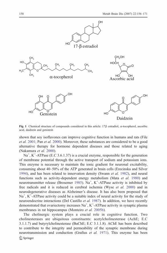
Isoflavones are compounds with estrogenic activity (phytoestrogens) found almostexclusively in soybeans and in a few other legumes. The principal isoflavones aregenistein, daidzein and glycitein (Sirtori et al. 2005). The phytoestrogens interactwith the estrogen receptors, ERα and ERβ, due to their structural similarity to βestradiol (Fig. 1). These substances act as antioxidants, directly or indirectly, byenhancing the enzyme activities of catalase, superoxide dismutase, glutathioneperoxidase and glutathione reductase (Kurzer and Xu 1997). Some studies have
Metab Brain Dis (2007) 22:156–171 157
shown that soy isoflavones can improve cognitive function in humans and rats (Fileet al. 2001; Pan et al. 2000). Moreover, these substances are considered to be a goodalternative therapy for hormone dependent diseases and those related to aging(Nakamura et al. 2000).
Na+, K+-ATPase (E.C 3.6.1.37) is a crucial enzyme, responsible for the generationof membrane potential through the active transport of sodium and potassium ions.This enzyme is necessary to maintain the ionic gradient for neuronal excitability,consuming about 40–50% of the ATP generated in brain cells (Erecinska and Silver1994), and has been related to innervation density (Swann et al. 1982), and neuralfunctions such as activity-dependent energy metabolism (Mata et al. 1980) andneurotransmitter release (Brosemer 1985). Na+, K+-ATPase activity is inhibited byfree radicals and it is reduced in cerebral ischemia (Wyse et al. 2000) and inneurodegenerative diseases as Alzheimer’s disease. It has also been proposed thatNa+, K+-ATPase activity could be a suitable index of neural activity for the study ofneuroendocrine interactions (Del Castillo et al. 1987). In addition, we have recentlydemonstrated that ovariectomy increases Na+, K+-ATPase activity in synaptic plasmamembranes in rat hippocampus (Monteiro et al. 2005b).
The cholinergic system plays a crucial role in cognitive function. Twocholinesterases are ubiquitous constituents: acetylcholinesterase (AchE; E.C3.1.1.7) and butyrylcholinesterase (BuChE; E.C 3.1.1.8). AChE has been describedto contribute to the integrity and permeability of the synaptic membrane duringneurotransmission and conduction (Grafius et al. 1971). This enzyme has been
OH
OH17-β estradiol
O
CH3OH
CH3CH3
CH3
CH3CH3 CH3 CH3
α -tocopherol
O O
OHOH
HOCH
CH2OH
Ascorbic acid
OH O
OHOOH
Genistein OH O
OHO
Daidzein
Fig. 1 Chemical structure of compounds considered in this article: 17β estradiol, α-tocopherol, ascorbicacid, daidzein and genistein
158 Metab Brain Dis (2007) 22:156–171
implicated in cholinergic and non-cholinergic actions, which may play a role inneurodegenerative diseases (Henderson et al. 1996, Cummings 2000, Arendt et al.1992). Recent evidence suggests that, in addition to AChE, BuChE catalyses thehydrolysis of the neurotransmitter, acetylcholine, and serves as a co-regulator ofcholinergic transmission (Geula and Darvesh 2004). In addition, associations ofserum BuChE with alterations in lipid metabolism (Magarian and Dietz 1987) andcoronary artery disease have been suggested (Alcantara et al. 2002). We have shownthat ovariectomized rats present an increase of hippocampal AChE activity(Monteiro et al. 2005b) and a reduction of serum BuChE (Monteiro et al. 2005c)activity in female adult rats.
Considering that: (a) previous studies show that ovariectomy increases Na+, K+-ATPase and AChE activities in hippocampus of female adult rats (Monteiro et al.2005b), (b) the administration of vitamins E plus C prevents the memory deficitcaused by ovariectomy (Monteiro et al. 2005a), (c) soy isoflavones have beenproposed as a good alternative to substitute the HRT (Miquel et al. 2006); wedecided to investigate the influence of vitamins E plus C or soy isoflavones on theeffects elicited by ovariectomy on Na+, K+-ATPase and AChE activities inhippocampus of ovariectomized rats. We also determined the actions of vitaminsor soy isoflavones on serum BuChE activity of ovariectomized rats. The workinghypothesis is that vitamins E plus C and isoflavones could reverse the alteration ofthese enzymes caused by ovariectomy.
Materials and methods
Animals and reagents
Female adult Wistar rats (3 months, 180–210 g BW) were obtained from the CentralAnimal House of the Department of Biochemistry, Instituto de Ciências Básicas daSaúde, Universidade Federal do Rio Grande do Sul, Porto Alegre, RS, Brazil.Animals were maintained on a 12/12 h light/dark cycle in an air-conditionedconstant temperature (22±1°C) colony room, with free access to water. Animal carefollowed the official governmental guidelines in compliance with the Federation ofBrazilian Societies for Experimental Biology and was approved by the EthicsCommittee of the Federal Rio Grande do Sul, Brazil.
Casein, 87% purity, was from Farmaquímica, Porto Alegre, Brazil, supplementedwith 0.15% L-methionine (from Merk, Rio de Janeiro, Brazil), a mixture of mineralsand vitamins (from Roche, São Paulo, Brazil) and Samprosoy 90 LH (generouslysupplied by EMBRAPA, Brazil). All other chemicals were purchased from SigmaChemical Co., St. Louis, MO, USA.
Experimental treatment
Eighty-day-old female rats were randomly assigned to one of the following groups:sham (only submitted to surgery, without removal of the ovaries) and ovariecto-mized. The stage of the estrous cycle was determined by vaginal swabs for 10 daysprior to ovariectomy, to ensure that animals were cycling normally (Baker et al.1979). Animals were ovariectomized by the surgical removal of both ovaries under
Metab Brain Dis (2007) 22:156–171 159
ketamine anesthesia (90 mg/kg) and xylazine (10 mg/kg) intraperitoneous (i.p.) toeliminate endogenous ovarian steroids (Waynforth and Flecknell 1992).
In the first set of experiments, 7 days after surgery, animals were treated for30 days with a single daily i.p. injection of saline (control) or vitamins E (40 mg/kg)plus C (100 mg/kg; Wyse et al. 2002). These dosing regimes have proved effectivefor preventing biochemical and behavioral effects in experimental models ofmetabolic diseases and ovariectomized rats (Reis et al. 2002; Wyse et al. 2002;Delwing et al. 2005; Monteiro et al. 2005a).
In other set of experiments, 7 days after surgery, animals were fed for 30 days ona standard diet with casein (control) or a special diet with soy isolated protein—Samprosoy 90 LH (1.89 mg isoflavones/g soy protein). The diet contained anisoflavone mixture that included Daidzein (0.25 mg/g soy protein) and Genistein(0.23 mg/g soy protein). The soy protein dose was chosen according to a protocolestablish by Reeves et al. (1993). Food intake and body weight were examinedweekly. Both diets were isocaloric.
Tissue preparation
On the 31st day of treatment, rats were killed by decapitation without anesthesia.Brains were quickly removed and the hippocampus dissected. All sham rats werekilled on the afternoon of proestrus.
For preparation of synaptic plasma membrane and determination of Na+, K+-ATPase activity, the hippocampus was homogenized in 10 vol. 0.32 mM sucrosesolution containing 5.0 mM HEPES and 1.0 mM EDTA, pH 7.4.
For the AChE assay, the same structure was homogenized in 10 volumes 0.1 mMpotassium phosphate buffer, pH 7.5, and centrifuged for 10 min at 1,000×g. Thesupernatant was used for the enzymatic AChE analyses.
For the BuChE assay and the evaluation of estrogen levels, the blood was rapidlycollected, centrifuged at 1,000×g for 10 min and the serum was separated.
Preparation of synaptic plasma membrane from hippocampus
Synaptic plasma membranes from hippocampus were prepared according to themethod of Jones and Matus (1974) with some modifications (Wyse et al. 1995). Thehomogenate was centrifuged at 1,000×g for 20 min and the supernatant removed andcentrifuged at 12,000×g for a further 20 min. The pellet was then resuspended inhypotonic buffer (5.0 mM Tris–HCl buffer, pH 8.1), at 0°C for 30 min, and appliedon a discontinuous sucrose density gradient consisting of successive layers of 0.3,0.8 and 1.0 mM. After centrifugation at 69,000×g for 2 h, the fraction between 0.8and 1.0 mM sucrose interface was taken as the membrane enzyme preparation.
Na+, K+-ATPase activity assay
The reaction mixture for Na+, K+-ATPase activity assay contained 5.0 mM MgCl2,80.0 mM NaCl, 20.0 mM KCl and 40.0 mM Tris–HCl, pH 7.4, in a final volume of200 μL. The reaction was initiated by the addition of ATP. Controls were carried outunder the same conditions with the addition of 1.0 mM ouabain. Na+, K+-ATPase
160 Metab Brain Dis (2007) 22:156–171
activity was calculated by the difference between the two assays, as described byWyse et al. (2000). Released inorganic phosphate (Pi) was measured by the methodof Chan et al. (1986). Specific activity of the enzyme was expressed as nmol Pireleased per min per mg of protein. All samples were run in duplicate.
AChE activity assay
Acetylcholinesterase activity was determined according to Ellman et al. (1961), withsome modifications (Villescas et al. 1981). Hydrolysis rates v were measured atacetylthiocholine (S) concentrations of 0.8 mM in 1 mL assay solutions with 30 mMphosphate buffer, pH 7.5, and 1.0 mM 5,5′-Dithiobis-(2-nitrobenzoic Acid) (DTNB)at 25°C. Fifty microliters of rat hippocampus supernatant was added to the reactionmixture and preincubated for 3 min. The hydrolysis was monitored by the formationof the thiolate dianion of DTNB at 412 nm for 2–3 min (intervals of 30 s). Specificenzyme activity was expressed as μmol ASCh per hour per milligram of protein. Allsamples were run in duplicate.
BuChE activity assay
Butyrylcholinesterase activity was determined by the method of Ellman et al. (1961)with some modifications. Hydrolysis rate v was measured at acetylthiocholine (S)concentrations of 0.8 mM in 1 mL assay solutions with 100 mM phosphate buffer,pH 7.5, and 1.0 mM DTNB. Fifty microliters of rat serum was added to the reactionmixture and preincubated for 3 min. The hydrolysis was monitored by formation ofthe thiolate dianion of DTNB at 412 nm for 2–3 min (intervals of 30 s) at 25°C.Specific enzyme activity was expressed as μmol ASCh per hour per milligram ofprotein. All samples were run in duplicate.
Estradiol measurement
Serum concentration of estradiol was carried out in microplates by enzymeimmuno-assay (EIA) using a Biomedical kit (BioCheck, Inc., USA). This assay uses thequantitative sandwich enzyme immunoassay technique that involves the simulta-neous reaction of the measured molecules to two monoclonal antibodies. The assaysensitivity is 1 pg/mL and, to eliminate interassay variation, all samples wereassayed in a single run.
Protein determination
Protein was measured by the method of Bradford (1976) using bovine serumalbumin as standard.
Statistical analysis
All assays were performed in duplicate and the mean was used for statisticalanalysis. Data were analyzed by one way ANOVA followed by the Duncan multipletest when F-test was significant. All analyses were performed using the Statistical
Metab Brain Dis (2007) 22:156–171 161
Package for the Social Sciences (SPSS) software using a PC-compatible computer.Values of p<0.05 were considered to be significant.
Results
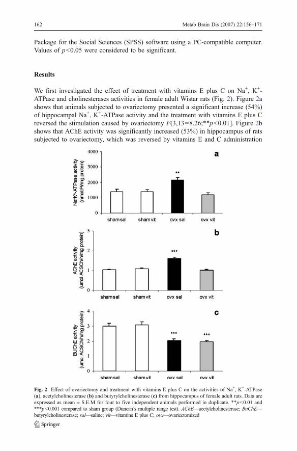
We first investigated the effect of treatment with vitamins E plus C on Na+, K+-ATPase and cholinesterases activities in female adult Wistar rats (Fig. 2). Figure 2ashows that animals subjected to ovariectomy presented a significant increase (54%)of hippocampal Na+, K+-ATPase activity and the treatment with vitamins E plus Creversed the stimulation caused by ovariectomy F(3,13=8.26;**p<0.01]. Figure 2bshows that AChE activity was significantly increased (53%) in hippocampus of ratssubjected to ovariectomy, which was reversed by vitamins E and C administration
Fig. 2 Effect of ovariectomy and treatment with vitamins E plus C on the activities of Na+, K+-ATPase(a), acetylcholinesterase (b) and butyrylcholinesterase (c) from hippocampus of female adult rats. Data areexpressed as mean ± S.E.M for four to five independent animals performed in duplicate. **p<0.01 and***p<0.001 compared to sham group (Duncan’s multiple range test). AChE—acetylcholinesterase; BuChE—butyrylcholinesterase; sal—saline; vit—vitamins E plus C; ovx—ovariectomized
162 Metab Brain Dis (2007) 22:156–171

[F(3,16)=43.44;***p<0.001]. As can be observed in Fig. 2c ovariectomized ratspresented a reduction (33%) in serum BuChE activity and vitamins E plus Ctreatment did not reverse such effect [F(3,16)=14.31; ***p<0.001]. Vitamins E plusC administration per se did not alter Na+, K+-ATPase and cholinesterases activities.
Next, we investigated the effect of the soy diet, rich in isoflavones on theactivities of Na+, K+-ATPase and cholinesterases in hippocampus and BuChe inserum of ovariectomized female adult Wistar rats (Fig. 3). As can be observed inFig. 3a, Na+, K+-ATPase activity was significantly increased (48%) in hippocampusof ovariectomized rats and soy treatment did not reverse such effect [F(3,12=5.04;*p<0.05]. Figure 3b shows that ovariectomy significantly increased (38%) AChEactivity and soy treatment reversed this effect [F(3,16=9.30; **p<0.01]. BuChEactivity (Fig. 3c) was decreased (45%) in serum of ovariectomized rats [F(3,16)=17.86; ***p<0.001] and soy diet did not reverse the effect of ovariectomy on
Fig. 3 Effect of ovariectomy and the soy diet rich in isoflavones on the activities of Na+, K+-ATPase(a), acetylcholinesterase (b) and butyrylcholinesterase (c) from hippocampus of female adult rats. Dataare expressed as mean ± S.E.M. for four to five independent animals performed in duplicate. *p<0.05,**p<0.01 and ***p<0.001 and compared to sham group (Duncan’s multiple range test). AChE—acetylcholinesterase; BuChE—butyrylcholinesterase; cas—casein; isofl—soy rich on isoflavones; ovx—ovariectomized
Metab Brain Dis (2007) 22:156–171 163
reduction of this enzyme activity. Soy diet per se did not alter Na+, K+-ATPase andcholinesterases activities.
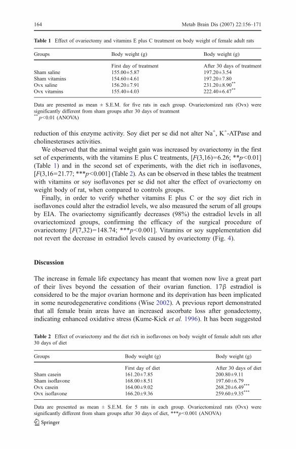
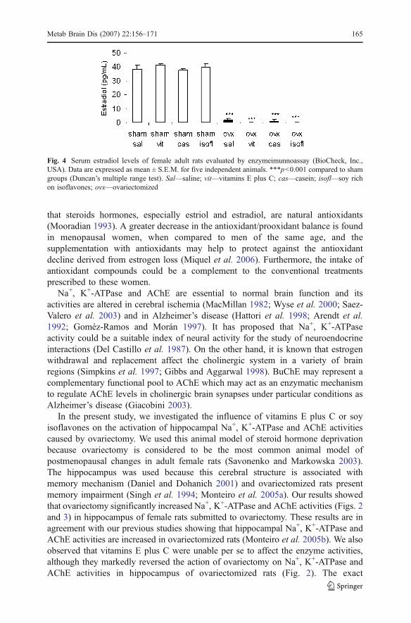
We observed that the animal weight gain was increased by ovariectomy in the firstset of experiments, with the vitamins E plus C treatments, [F(3,16)=6.26; **p<0.01](Table 1) and in the second set of experiments, with the diet rich in isoflavones,[F(3,16=21.77; ***p<0.001] (Table 2). As can be observed in these tables the treatmentwith vitamins or soy isoflavones per se did not alter the effect of ovariectomy onweight body of rat, when compared to controls groups.
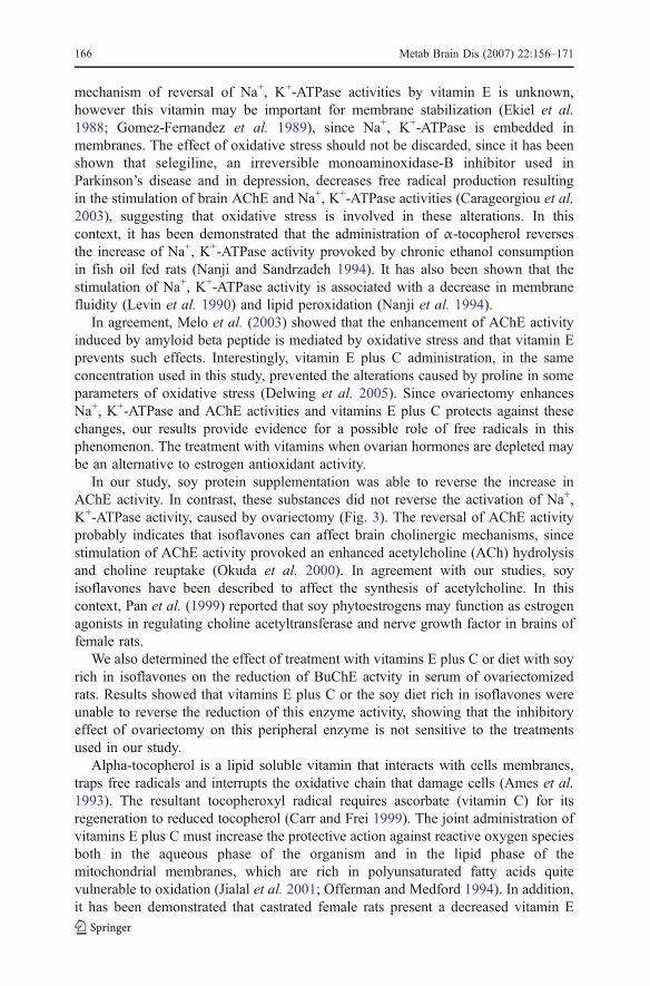
Finally, in order to verify whether vitamins E plus C or the soy diet rich inisoflavones could alter the estradiol levels, we also measured the serum of all groupsby EIA. The ovariectomy significantly decreases (98%) the estradiol levels in allovariectomized groups, confirming the efficacy of the surgical procedure ofovariectomy [F(7,32)=148.74; ***p<0.001]. Vitamins or soy supplementation didnot revert the decrease in estradiol levels caused by ovariectomy (Fig. 4).
Discussion
The increase in female life expectancy has meant that women now live a great partof their lives beyond the cessation of their ovarian function. 17β estradiol isconsidered to be the major ovarian hormone and its deprivation has been implicatedin some neurodegenerative conditions (Wise 2002). A previous report demonstratedthat all female brain areas have an increased ascorbate loss after gonadectomy,indicating enhanced oxidative stress (Kume-Kick et al. 1996). It has been suggested
Table 1 Effect of ovariectomy and vitamins E plus C treatment on body weight of female adult rats
Groups Body weight (g) Body weight (g)
First day of treatment After 30 days of treatmentSham saline 155.00±5.87 197.20±3.54Sham vitamins 154.60±4.61 197.20±7.80Ovx saline 156.20±7.91 231.20±8.90**
Ovx vitamins 155.40±4.03 222.40±6.47**
Data are presented as mean ± S.E.M. for five rats in each group. Ovariectomized rats (Ovx) weresignificantly different from sham groups after 30 days of treatment** p<0.01 (ANOVA)
Table 2 Effect of ovariectomy and the diet rich in isoflavones on body weight of female adult rats after30 days of diet
Groups Body weight (g) Body weight (g)
First day of diet After 30 days of dietSham casein 161.20±7.85 200.80±9.11Sham isoflavone 168.00±8.51 197.60±6.79Ovx casein 164.00±9.02 268.20±6.49***
Ovx isoflavone 166.20±9.36 259.60±9.35***
Data are presented as mean ± S.E.M. for 5 rats in each group. Ovariectomized rats (Ovx) weresignificantly different from sham groups after 30 days of diet, ***p<0.001 (ANOVA)
164 Metab Brain Dis (2007) 22:156–171
that steroids hormones, especially estriol and estradiol, are natural antioxidants(Mooradian 1993). A greater decrease in the antioxidant/prooxidant balance is foundin menopausal women, when compared to men of the same age, and thesupplementation with antioxidants may help to protect against the antioxidantdecline derived from estrogen loss (Miquel et al. 2006). Furthermore, the intake ofantioxidant compounds could be a complement to the conventional treatmentsprescribed to these women.
Na+, K+-ATPase and AChE are essential to normal brain function and itsactivities are altered in cerebral ischemia (MacMillan 1982; Wyse et al. 2000; Saez-Valero et al. 2003) and in Alzheimer’s disease (Hattori et al. 1998; Arendt et al.1992; Goméz-Ramos and Morán 1997). It has proposed that Na+, K+-ATPaseactivity could be a suitable index of neural activity for the study of neuroendocrineinteractions (Del Castillo et al. 1987). On the other hand, it is known that estrogenwithdrawal and replacement affect the cholinergic system in a variety of brainregions (Simpkins et al. 1997; Gibbs and Aggarwal 1998). BuChE may represent acomplementary functional pool to AChE which may act as an enzymatic mechanismto regulate AChE levels in cholinergic brain synapses under particular conditions asAlzheimer’s disease (Giacobini 2003).
In the present study, we investigated the influence of vitamins E plus C or soyisoflavones on the activation of hippocampal Na+, K+-ATPase and AChE activitiescaused by ovariectomy. We used this animal model of steroid hormone deprivationbecause ovariectomy is considered to be the most common animal model ofpostmenopausal changes in adult female rats (Savonenko and Markowska 2003).The hippocampus was used because this cerebral structure is associated withmemory mechanism (Daniel and Dohanich 2001) and ovariectomized rats presentmemory impairment (Singh et al. 1994; Monteiro et al. 2005a). Our results showedthat ovariectomy significantly increased Na+, K+-ATPase and AChE activities (Figs. 2and 3) in hippocampus of female rats submitted to ovariectomy. These results are inagreement with our previous studies showing that hippocampal Na+, K+-ATPase andAChE activities are increased in ovariectomized rats (Monteiro et al. 2005b). We alsoobserved that vitamins E plus C were unable per se to affect the enzyme activities,although they markedly reversed the action of ovariectomy on Na+, K+-ATPase andAChE activities in hippocampus of ovariectomized rats (Fig. 2). The exact
Fig. 4 Serum estradiol levels of female adult rats evaluated by enzymeimunnoassay (BioCheck, Inc.,USA). Data are expressed as mean ± S.E.M. for five independent animals. ***p<0.001 compared to shamgroups (Duncan’s multiple range test). Sal—saline; vit—vitamins E plus C; cas—casein; isofl—soy richon isoflavones; ovx—ovariectomized
Metab Brain Dis (2007) 22:156–171 165
mechanism of reversal of Na+, K+-ATPase activities by vitamin E is unknown,however this vitamin may be important for membrane stabilization (Ekiel et al.1988; Gomez-Fernandez et al. 1989), since Na+, K+-ATPase is embedded inmembranes. The effect of oxidative stress should not be discarded, since it has beenshown that selegiline, an irreversible monoaminoxidase-B inhibitor used inParkinson’s disease and in depression, decreases free radical production resultingin the stimulation of brain AChE and Na+, K+-ATPase activities (Carageorgiou et al.2003), suggesting that oxidative stress is involved in these alterations. In thiscontext, it has been demonstrated that the administration of α-tocopherol reversesthe increase of Na+, K+-ATPase activity provoked by chronic ethanol consumptionin fish oil fed rats (Nanji and Sandrzadeh 1994). It has also been shown that thestimulation of Na+, K+-ATPase activity is associated with a decrease in membranefluidity (Levin et al. 1990) and lipid peroxidation (Nanji et al. 1994).
In agreement, Melo et al. (2003) showed that the enhancement of AChE activityinduced by amyloid beta peptide is mediated by oxidative stress and that vitamin Eprevents such effects. Interestingly, vitamin E plus C administration, in the sameconcentration used in this study, prevented the alterations caused by proline in someparameters of oxidative stress (Delwing et al. 2005). Since ovariectomy enhancesNa+, K+-ATPase and AChE activities and vitamins E plus C protects against thesechanges, our results provide evidence for a possible role of free radicals in thisphenomenon. The treatment with vitamins when ovarian hormones are depleted maybe an alternative to estrogen antioxidant activity.
In our study, soy protein supplementation was able to reverse the increase inAChE activity. In contrast, these substances did not reverse the activation of Na+,K+-ATPase activity, caused by ovariectomy (Fig. 3). The reversal of AChE activityprobably indicates that isoflavones can affect brain cholinergic mechanisms, sincestimulation of AChE activity provoked an enhanced acetylcholine (ACh) hydrolysisand choline reuptake (Okuda et al. 2000). In agreement with our studies, soyisoflavones have been described to affect the synthesis of acetylcholine. In thiscontext, Pan et al. (1999) reported that soy phytoestrogens may function as estrogenagonists in regulating choline acetyltransferase and nerve growth factor in brains offemale rats.
We also determined the effect of treatment with vitamins E plus C or diet with soyrich in isoflavones on the reduction of BuChE actvity in serum of ovariectomizedrats. Results showed that vitamins E plus C or the soy diet rich in isoflavones wereunable to reverse the reduction of this enzyme activity, showing that the inhibitoryeffect of ovariectomy on this peripheral enzyme is not sensitive to the treatmentsused in our study.
Alpha-tocopherol is a lipid soluble vitamin that interacts with cells membranes,traps free radicals and interrupts the oxidative chain that damage cells (Ames et al.1993). The resultant tocopheroxyl radical requires ascorbate (vitamin C) for itsregeneration to reduced tocopherol (Carr and Frei 1999). The joint administration ofvitamins E plus C must increase the protective action against reactive oxygen speciesboth in the aqueous phase of the organism and in the lipid phase of themitochondrial membranes, which are rich in polyunsaturated fatty acids quitevulnerable to oxidation (Jialal et al. 2001; Offerman and Medford 1994). In addition,it has been demonstrated that castrated female rats present a decreased vitamin E
166 Metab Brain Dis (2007) 22:156–171
concentration in serum and liver (Feingold et al. 1993). The antioxidant activity ofestradiol is attributed to its phenolic hydroxyl (−OH) group (Fig. 1), which iscapable of reducing peroxyl radicals. The regeneration of tocopherols fromtocopheroxyl radicals by estradiol has been observed under in vitro conditions(Mukai et al. 1990). It has been demonstrated that ovariectomy decreases, andestradiol replacement elevates, the tissue concentration of α-tocopherol inovariectomized rats (Feingold et al. 1993). We have recently reported that animpairment of spatial navigation, caused by ovariectomy, was prevented by vitaminsE plus C administration (Monteiro et al. 2005a). In agreement with these data, Socciet al. (1995) showed that chronic antioxidant treatment enhances cognitiveperformance of aged rats in the same behavior task.
Soy isoflavones are referred to as phytoestrogens because they bind to estrogenreceptors and can exert both agonistic and antagonistic estrogenic effects (Miksicek1995). The phytoestrogens interact with the estrogen receptors ERα and ERβ, due totheir diphenolic structural similarity to 17β estradiol (Fig. 1; Setchell 2001). Thesesubstances may improve cognitive functions by mimicking estrogen effects in thebrain. Phytoestrogens have gained recognition as protective agents against diseasesrelated to age and hormone dependent cancers (Cornwell et al. 2004; Aldlercreutz1998). It has been observed that a prolonged treatment with estradiol reduced thefrequency of spontaneous oscillations and the expression/activity of Na+, K+-ATPasein rat uteri, indicating that this enzyme could be an important target for estrogens andestrogen-like molecules (Tsai et al. 2003). However, the effect of these compoundson brain function remains to be elucidated. Lephart et al. (2003) showed that theconsumption of dietary phytoestrogen can alter hormone-sensitive hypothalamic brainvolumes in rodents during adulthood. In addition, ovariectomized rats that consumeddiets with soy isoflavones demonstrated a dose-dependent improvement in theirperformance in radial arm maze tests (Pan et al. 2000).
Since reports show that estrogens may be regenerated by endogenous antioxidants(Gridley et al. 1997), in the present study we also verified whether vitamins E plus Cor the soy diet rich in isoflavones could alter estradiol levels. We measured theserum estradiol in all groups by EIA and the results showed that ovariectomysignificantly decreases (98%) the estradiol levels (Fig. 4) in all ovariectomizedgroups, confirming the efficacy of the surgical procedure of ovariectomy. Nodifference in estradiol levels was observed in the groups treated with vitamins orsubmitted to the soy diet rich in isoflavones.
In summary, in the present study we demonstrate that the treatment with vitaminsE plus C significantly reverses the action of ovariectomy on Na+, K+-ATPase andAChE activities in hippocampus of female adult rats. Conversely, a diet of soy richin isoflavones was able to reverse only the increased AChE activity. Taken together,these compounds may have a protective role against the damage brain caused by theloss of estrogen during menopause. These data are very encouraging, since vitaminsE plus C and soy isoflavones may constitute a good alternative to a novel therapeuticstrategy. However, further studies are required to elucidate the exact mechanism oftheir action.
Acknowledgements This workwas supported in part by grants fromConselhoNacional de DesenvolvimentoCientífico e Tecnológico (CNPq—Brazil).
Metab Brain Dis (2007) 22:156–171 167

References
Alcantara VM, Chautard-Freire-Maia EA, Scartezini M, Cerci MS, Braun-Prado K, Picheth G (2002)Butyrylcholinesterase activity and risk factors for coronary artery disease. Scand J Clin Lab Invest62:399–404
Aldlercreutz H (1998) Epidemiology of phytoestrogens. Bailliéres Clin Endocrinol Metab 12:605–623Ames BN, Shigenara MK, Hagen TM (1993) Oxidants, antioxidants and the degenerative diseases of
aging. Proc Natl Acad Sci USA 90:7915–7922Arendt T, Bruckner MK, Lange M, Bigl V (1992) Changes in acetylcholinesterase and butyrylcholines-
terase in Alzeimer’s disease resemble embryonic development—a study of molecular forms.Neurochem Int 21:381–396
Arteaga E, Rojas A, Villaseca P, Bianchi M, Arteaga A, Durán D (1998) In vitro effect of oestradiol,progesterone, testosterone, and of combined estradiol/progestins on low density lipoprotein (LDL)oxidation in postmenopausal women. Menopause 6:16–23
Baker HJ, Lindsey JR, Weisbroth SH (1979) The laboratory rat. In: Biology and diseases (vol 1),Academic, New York
Bradford MM (1976) A rapid and sensitive method for the quantification of micrograms quantities ofprotein utilizing the principle of protein-die-binding. Anal Biochem 72:248–254
Brosemer RW (1985) Effects of inhibitors of Na+,K+-ATPase on the membrane potentials andneurotransmitter efflux in rat brain slices. Brain Res 334:125–137
Carageorgiou H, Zarros A, Tsakiris S (2003) Selegiline long-term effects on brain acetylcholinesterase,(Na+, K+)-ATPase activities, antioxidant status and learning performance of aged rats. Pharmacol Res48:245–251
Carr A, Frei B (1999) Does vitamin C act as a pro-oxidant under physiological conditions? FASEB J13:1007–1024
Chan KM, Delfer D, Junger KD (1986) A direct colorimetric assay for Ca2+-stimulated ATPase activity.Anal Biochem 157:375–380
Cornwell T, Cohick W, Raskin I (2004) Dietary phytoestrogens and health. Phytochemistry 65:995–1016Cummings JL (2000) The role of cholinergic agents in the management of behavioral disturbances in
Alzheimer’s disease. Int J Neuropsychopharmacol 3:21–29Daniel JM, Dohanich GP (2001) Acetylcholine mediates the estrogen-induced increase in NMDA receptor
binding in CA1 of the hippocampus and the associated improvement in working memory. J Neurosci21:6949–6956
Del Castillo AR, Battaner E, Guerra M, Alonso T, Mas M (1987) Regional changes of brain Na+, K+-transporting adenosine triphosphatase related to ovarian function. Brain Res 416:113–118
Delwing D, Chiarani F, Bavaresco CS, Wannmacher CM, Wajner M, Dutra-Filho CS, Wyse ATS (2005)Protective effect of antioxidants on brain oxidative damage caused by proline administration.Neurosci Res 52:69–74
Ekiel IH, Hughes L, Burton GW, Jovall PA, Ingold KU, Smith IC (1988) Structure and dynamics of alpha-tocopherol in model membranes and in solution: a broad-line and high-resolution NMR study.Biochemistry 27:1432–1440
Ellman GL, Courtney KD, Andres V Jr, Feather-Stone RM (1961) A new and rapid determination ofacetylcholinesterase activity. Biochem Pharmacol 7:88–95
Erecinska M, Silver IA (1994) Ions and energy in mammalian brain. Prog Neurobiol 43:37–71Feingold IB, Longhurst PA, Colby HD (1993) Regulation of adrenal and hepatic α-tocopherol content by
androgens and estrogens. Biochim Biophys Acta 1176:192–196File SE, Jarrett N, Fluck E, Duffy R, Casey K, Wiseman H (2001) Soy phytoestrogens improves human
memory. Psychopharmacology 157:430–436Geula C, Darvesh S (2004) Butyrylcholinesterase, cholinergic neurotransmission and the pathology of
Alzheimer’s disease. Drugs Today 40:711–721Giacobini E (2003) Cholinesterases: new roles in brain function and in Alzheimer’s disease. Neurochem
Res 28:515–522Gibbs RB, Aggarwal P (1998) Estrogen and basal forebrain cholinergic neurons: implications for brain
aging and Alzheimer’s disease-related cognitive decline. Horm Behav 34:98–111Gomez-Fernandez JC, Villalain J, Aranda FJ, Ortiz A, Micol V, Coutinho A, Berberan-Santos MN, Prieto
MJ (1989) Localization of alpha-tocopherol in membranes. Ann NY Acad Sci 570:109–120Gómez-Ramos P, Morán MA (1997) Ultra structural localization of butyrylcholinesterase in senile plaques
in the brains of aged and Alzheimer’s disease patients. Mol Chem Neuropathol 30:161–173
168 Metab Brain Dis (2007) 22:156–171
Grafius MA, Bond HE, Millar DB (1971) Acetylcholinesterase interaction with a lipoprotein matrix. Eur JBiochem 22:382–390
Green PS, Simpkins JW (2000) Neuroprotective effects of estrogens: potential mechanisms of action. Int JDev Neurosci 18:347–358
Gridley KE, Green PS, Simpkins JW (1997) Low concentrations of estradiol reduce beta-amyloid (25–35)-induced toxicity, lipid peroxidation and glucose utilization in human SK-N-SH neuroblastoma cells.Brain Res 778:158–165
Hattori N, Kitagawa K, Higashida T, Yagyu K, Shimohama S, Wataya T, Perry G, Smith MA, Inagaki C(1998) Cl−-ATPase and Na+/K+-ATPase activities in Alzheimer’s disease brains. Neurosci Lett254:141–144
Henderson VW, Watt L, Buckwalter JG (1996) Cognitive skills associated with estrogen replacement inwomen with Alzeimer’s disease. Psychoneuroendocrinology 21:421–430
Jialal L, Devaraj S, Kaul N (2001) The effect of alpha-tocopherol on monocyte proatherogenic activity.Am J Clin Nutr 131:389S–394S
Jones DH, Matus AI (1974) Isolation of plasma synaptic membrane from brain by combination flotation-sedimentation density gradient centrifugation. Biochim Biophys Acta 356:276–287
Kume-kick J, Ferris DC, Russo-Menna I, Rice ME (1996) Enhanced oxidative stress in female rat brainafter gonadectomy. Brain Res 738:8–14
Kurzer MS, Xu X (1997) Dietary phytoestrogens. Annu Rev Nutr 17:353–381Law A, Gauthier S, Quirion R (2001) Say NO Alzheimer’s disease: the putative links between nitric oxide
and dementia of the Alzheimer’s type. Brain Res Rev 35:73–96Lephart ED, Rhees RW, Setchell KD, Bu LH, Lund TD (2003) Estrogens and phytoestrogens: brain
plasticity of sexually dimorphic brain volumes. J Steroid Biochem Mol Biol 85:299–309Levin G, Cogan U, Levy Y, Mokady S (1990) Riboflavin deficiency and the function and fluidity of rat
erythrocyte membranes. J Nutr 120:857–861MacMillan V (1982) Cerebral Na+/K+-ATPase activity during exposure to and recovery from acute
ischemia. J Cereb Blood Flow Metab 2:457–465Magarian EO, Dietz AJ (1987) Correlation of cholinesterase with serum lipids and lipoproteins. J Clin
Pharmacol 27:819–820Mata M, Fink DJ, Gainer H, Smith CB, Davidsen L, Savaki H, Schwartz WJ, Sokoloff L (1980) Activity-
dependent energy metabolism rat posterior pituitary primarily reflects sodium pump activity.J Neurochem 34:213–215
McCay PB (1985) Vitamin E: interactions with free radical and ascorbate. Annu Rev Nutr 5:323–340Melo JB, Agostinho P, Oliveira CR (2003) Involvement of oxidative stress in the enhancement of
acetylcholinesterase activity induced by amyloid beta-peptide. Neurosci Res 45:117–127Miksicek RJ (1995) Estrogenic flavonoids: structural requirements for biological activity. Proc Soc Exp
Biol Med 208:44–50Miquel J, Ramírez-Boscá A, Ramírez-Boscá JV, Alperi JD (2006). Menopause: a review on the role of
oxygen stress and favorable effects of dietary antioxidants. Arch Gerontol Geriatr 42:289–306Monteiro SC, Matté C, Bavaresco CS, Netto CA, Wyse ATS (2005a) Vitamins E and C pretreatment
prevents ovariectomy-induced memory deficits in water maze. Neurobiol Learn Mem 84:192–199Monteiro SC, Matté C, Delwing D, Wyse ATS (2005b) Ovariectomy increases Na+,K+-ATPase,
acetylcholinesterase and catalase in rat hippocampus. Mol Cell Endocrinol 236:9–16Monteiro SC, Stefanello FM, Vianna LP, Matté C, Barp J, Belló-Klein A, Trindade VMT, Wyse ATS
(2005c). Ovariectomy enhances acetylcholinesterase activity but does not alter ganglioside content incerebral cortex of female adult rats. Metab Brain Dis 20:35–44
Mooradian AD (1993) Antioxidant properties of steroids. J Steroid Biochem Mol Biol 45:509–511Mukai K, Daifuku K, Yokoyama S, Nakano M (1990) Stopped-flow investigation of antioxidant activity
of estrogens in solution. Biochim Biophys Acta 1035:348–352Nakamura Y, Tsuji S, Tonogai Y (2000) Determination of the levels of isoflavonoids in soybeans and
soy-derived foods and estimation of isoflavonoids in the Japanese daily intake. J AOAC Int 83:635–650Nanji AA, Sadrzadeh SM (1994) Effect of fish oil and vitamin E on ethanol-induced changes in membrane
ATPases. Life Sci 55:245–249Nanji AA, Sadrzadeh SM, Dannenberg AJ (1994) Liver microsomal fatty acid composition in ethanol-fed rats:
effect of different dietary fats and relationship to liver injury. Alcohol Clin Exp Res 18:1024–1028Offerman MK, Medford RM (1994) Antioxidants and atherosclerosis, a molecular perspective. Heart Dis
Stroke 3:52–57
Metab Brain Dis (2007) 22:156–171 169
Okuda T, Haga T, Kanai Y, Endou H, Ishihara T, Katsura I (2000) Identification and characterization of thehigh-affinity choline transporter. Nat Neurosci 3:120–125
Pan Y, Anthony M, Clarkson TB (1999) Effect of estradiol and soy phytoestrogens on cholineacetyltransferase and nerve growth factor mRNAs in the frontal cortex and hippocampus of femalerats. Pro Soc Exp Biol Med 221:118–125
Pan Y, Anthony M, Watson S, Clarkson TB (2000) Soy phytoestrogens improve radial arm mazeperformance in ovariectomized retired breeder rats and do not attenuate benefits of 17β-estradioltreatment. Menopause 7:230–235
Reeves PG, Nielsen FH, Fabey Jr. (1993) GC. AIN-93 purified diets for laboratory rodents: final report ofthe American Institute of Nutrition ad hoc writing committee on the reformulation of the AIN-76Arodent diet. J Nutr 123:1939–1951
Reis EA, Zugno AI, Franzon R, Tagliari B, Matté C, Lammers ML, Netto CA, Wyse ATS (2002)Pretreatment with vitamins E and C prevent the impairment of memory caused by homocysteineadministration in rats. Metab Brain Dis 17(3):211–217
Rodrigues HD, Kinder JE, Fitzpatrick LA (1999). Treatment with 17 beta-estradiol does not influence ageand weight at puberty in Bos indicus heifers. Anim Reprod Sci 56:1–10
Saez-Valero J, Gonzales-Garcia C, Cena V (2003) Acetylcholinesterase activation in organotypic rathippocampal slice cultures deprived of oxygen and glucose. Neurosci Lett 348(2):123–125
Savonenko AV, Markowska AL (2003) The cognitive effects of ovariectomy and estrogen replacement aremodulated by aging. Neurosci 119:821–830
Setchell KD (2001) Soy isoflavones—benefits and risks from nature’s selective estrogen receptormodulators (SERMs). J Am Coll Nutr 20:354–362
Simpkins JW, Rajakumar G, Zhang YQ, Simpkins CE, Greenwald D, Yu CJ, Bodor N, Day AL (1997)Estrogens may reduce mortality and ischemic damage caused by middle cerebral artery occlusion inthe female rats. J Neurosurg 87:724–730
Singh M, Meyer EM, Millard WJ, Simpkins JW (1994) Ovarian steroid deprivation results in a reversiblelearning impairment and compromised cholinergic function in female Sprague–Dawley rats. BrainRes 644:305–312
Sirtori CR, Arnoldi A, Johnson SK (2005) Phytoestrogens: end of a tale? Ann Med 37:423–438Socci DJ, Crandall BM, Arendash GW (1995) Chronic antioxidants treatment improves the cognitive
performance of aged rats. Brain Res 693:88–94Swann AC, Grant SJ, Maas JW (1982) Brain (Na+, K+)-ATPase and noradrenergic activity: effects of
hyperinnervation and denervation on high-affinity ouabain binding. J Neurochem 38:836–839Tang MX, Jacobs D, Stern Y, Marder K, Schofield P, Gurland B, Andrews, H, Mayeux R (1996) Effect of
oestrogen during menopause on risk and age at onset of Alzeimer’s disease. Lancet 348:429–432Tsai ML, Lee CL, Tang MJ, Liu MY (2000) Preferential reduction of Na+/K+ ATPase alpha 3 by 17 beta-
estradiol influences contraction frequency in rat uteri. Chin J Physiol 43:1–8Tsai ML, Chang CC, Lee CL, Huang BY (2003) The differential effects of tamoxifen and ICI 182, 780 on the
reduction of Na+/K+ ATPase and spontaneous oscillations by 17 beta-estradiol. Chin J Physiol 46:55–62Upritchard JE, Sutherland WHE, Mann JI (2000) Effect of supplementation with tomato juice, vitamin E,
and vitamin C on LDL oxidation and products of inflammation activity in type 2 diabetes. DiabetesCare 23:733–738
Van Duijn CM (1999) Hormone replacement therapy and Alzeimer’s disease. Maturitas 31:201–205Vatassery GT (1998) Vitamin E and other endogeneous antioxidants in the central nervous system.
Geriatrics 53:25–27Villescas R, Ostwald R, Morimoto HD, Bennett EL (1981) Effects of neonatal undernutrition and cold
stress on behavior and biochemical brain parameters in rats. J Nutr 111:1103–1110Waynforth HB, Flecknell PA (1992) Experimental and surgical technique. In: The rat, 2nd edn, Academic,
London, pp 276–278Wise PM (2002) Estrogens and neuroprotection. Trends Endocrinol Metab 13:229–230WHI (2002) Risk and benefits of estrogen plus progestin in healthy postmenopausal women. J Am Med
Assoc 288:321–333Wise PM, Dubal DB, Wilson ME, Rau SW, Liu Y (2001a). Estrogens: trophic and protective factors in the
adult brain. Front Neuroendocrinol 22:33–66Wise MP, Dubal DB, Wilson ME, Rau SW, Böttner M, Rosewell KL (2001b) Estradiol is a protective
factor in the adult and aging brain: understanding of mechanism derived from in vivo and in vitrostudies. Brain Res Brain Res Rev 37:313–319
170 Metab Brain Dis (2007) 22:156–171
Wyse ATS, Bolognesi G, Brusque AM, Wajner M, Wannmaker CMD (1995) Na+, K+-ATPase activity inthe synaptic plasma membrane from the central cerebral cortex of rats subjected to chemically inducedphenylketonuria. Med Sci Res 23:261–262
Wyse ATS, Streck EL, Worm P, Wajner A, Ritter F, Netto CA (2000) Preconditioning prevents theinhibition of Na+,K+-ATPase activity after brain ischemia. Neurochem Res 25:969–973
Wyse ATS, Zugno AI, Streck EL, Matte C, Calcagnotto T, Wannmacher CM, Wajner M (2002) Inhibitionof Na+,K+-ATPase activity in hippocampus of rats subjected to acute administration of homocysteineis prevented by vitamins E and C treatment. Neurochem Res 27:1685–1689
Zhang YQ, Shi J, Rajakumar G, Day AL, Simpkins JW (1998) Effects of gender and estradiol treatmenton focal brain ischemia. Brain Res 784:321–324
Metab Brain Dis (2007) 22:156–171 171
Copyright © 2022 FDOKUMEN