Characterization of a Novel Stress-Response Member of the MAPK Family in Malus hupehensis Rehd
Upload
independentCategory
view
3download
0
Journal of CellularBiochemistry
ARTICLEJournal of Cellular Biochemistry 108:388–399 (2009)
Methoxylated Isoflavones, Cajanin and Isoformononetin,Have Non-Estrogenic Bone Forming Effect Via DifferentialMitogen Activated Protein Kinase (MAPK) Signaling
B
A
A
GGB
*E
R
P
Biju Bhargavan,1 Abnish Kumar Gautam,1 Divya Singh,1* Amit Kumar,2 Sumit Chaurasia,2
Abdul Malik Tyagi,1 Dinesh Kumar Yadav,2 Jay Sharan Mishra,3 Amar Bahadur Singh,3
Sabyasachi Sanyal,3 Atul Goel,2 Rakesh Maurya,2 and Naibedya Chattopadhyay1
1Division of Endocrinology, Central Drug Research Institute (Council of Scientific and Industrial Research), ChattarManzil, P.O. Box 173, Lucknow, Uttar Pradesh, India
2Division of Medicinal & Process Chemistry, Central Drug Research Institute (Council of Scientific and IndustrialResearch), Chattar Manzil, P.O. Box 173, Lucknow, Uttar Pradesh, India
3Drug Target Discovery and Development, Central Drug Research Institute (Council of Scientific and IndustrialResearch), Chattar Manzil, P.O. Box 173, Lucknow, Uttar Pradesh, India
ABSTRACTFollowing a lead obtained from stem-bark extract of Butea monosperma, two structurally related methoxyisoflavones; cajanin and
isoformononetin were studied for their effects in osteoblasts. Cajanin had strong mitogenic as well as differentiation-promoting effects
on osteoblasts that involved subsequent activation of MEK-Erk and Akt pathways. On the other hand, isoformononetin exhibited potent anti-
apoptotic effect in addition to promoting osteoblast differentiation that involved parallel activation of MEK-Erk and Akt pathways. Unlike
genistein or daidzein, none of these two compounds appear to act via estrogen receptors in osteoblast. Once daily oral (by gavage) treatment
for 30 consecutive days was given to recently weaned female Sprague–Dawley rats with each of these compounds at 10.0 mg kg�1 day�1 dose.
Cajanin increased bone mineral density (BMD) at all skeletal sites studied, bone biomechanical strength, mineral apposition rate (MAR) and
bone formation rate (BFR), compared with control. BMD levels at various anatomic positions were also increased with isoformononetin
compared with control however, its effect was less potent than cajanin. Isoformononetin had no effect on the parameters of bone
biomechanical strength although it enhanced MAR and BFR compared with control. Isoformononetin had very mild uterotrophic effect,
whereas cajanin was devoid of any such effect. Our data suggest that cajanin is more potent than isoformononetin in accelerating peak bone
mass achievement. To the best of our knowledge, this work represents the first attempt to elucidate structure-activity relationship
between the two methoxylated isoflavones regarding their effects in osteoblasts and bone formation. J. Cell. Biochem. 108: 388–399,
2009. � 2009 Wiley-Liss, Inc.
KEY WORDS: OSTEOGENIC; APOPTOSIS; CAJANIN; ISOFORMONONETIN; MAPK SIGNALING; PEAK BONE MASS
H igh dietary intake of isoflavones has been reported to
increase BMD levels in lumbar spine in Japanese [Somekawa
et al., 2001], Chinese [Mei et al., 2001], and US [Kritz-Silverstein and
Goodman-Gruen, 2002] postmenopausal women than the control
iju Bhargavan, Abnish Kumar Gautam, and Divya Singh contributed equ
uthors have no conflict of interest.
dditional Supporting Information may be found in the online version of
rant sponsor: Ministry of Heath and Family Welfare; Grant sponsor: Courant sponsor: University Grants Commission; Grant sponsor: Governmeniotechnology (DBT).
Correspondence to: Dr. Divya Singh, PhD, Chattar Manzil, P.O. Box 173,-mail: [email protected]
eceived 6 May 2009; Accepted 3 June 2009 � DOI 10.1002/jcb.22264 �ublished online 13 July 2009 in Wiley InterScience (www.interscience.w
group. Soy isoflavones including genistein and daidzein have been
widely investigated for having beneficial effects on bone [Setchell
and Lydeking-Olsen, 2003]. At micromolar concentrations in vitro,
genistein and daidzein promote osteoblast functions via estrogen
388
ally to this work.
this article.
ncil of Scientific and Industrial Research;t of India; Grant sponsor: Department of
Lucknow, Uttar Pradesh 226001, India.
� 2009 Wiley-Liss, Inc.
iley.com).
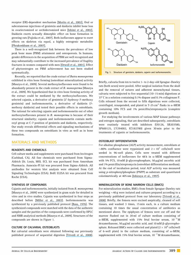
Fig. 1. Structure of genistein, daidzein, cajanin and isoformononetin.
receptor (ER)-dependent mechanism [Morito et al., 2002]. Oral or
subcutaneous injections of genistein and daidzein inhibit bone loss
in ovariectomized or orchidectomized mice [Ishimi et al., 2002].
Daidzein exerts sexually dimorphic effect on bone formation in
growing rats [Fujioka et al., 2007]. Both isoflavones appear to exert
effects on skeleton via equol, a highly estrogenic metabolite
[Phrakonkham et al., 2007].
There is a well-recognized link between the prevalence of low
peak bone mass (PBM) attainment and osteoporosis. In humans,
gender differences in the acquisition of PBM are well recognized and
may substantially contribute to the increased prevalence of fragility
fractures in women compared with men [Orwoll et al., 2001]. Effect
of phytoestrogen on PBM achievement has not been studied
systematically.
Recently, we reported that the crude extract of Butea monosperma
exhibited in vitro bone forming (osteoblast mineralization) activity
[Maurya et al., 2009]. Several methoxyisoflavones were found to be
abundantly present in the crude extract of B. monosperma [Maurya
et al., 2009]. We hypothesized that in vitro bone forming activity of
the extract could be mediated by the methoxyisoflavones. We
selected cajanin, a derivative of genistein (20-hydroxy-7-methoxy
genistein) and isoformononetin, a derivative of daidzein (7-
methoxy daidzein) and tested their possible effects in osteoblasts.
The rational for selecting cajanin and isoformononetin out of other
methoxyisoflavones present in B. monosperma is because of their
structural similarity; cajanin and isoformononetin contain meth-
oxyl group at C-7 position of genistein and daidzein, respectively.
Our study reveals differential effects and signaling mechanisms of
these two compounds on osteoblasts in vitro as well as in bone
formation.
MATERIALS AND METHODS
REAGENTS AND CHEMICALS
Cell culture media and supplements were purchased from Invitrogen
(Carlsbad, CA). All fine chemicals were purchased from Sigma-
Aldrich (St. Louis, MO). ECL kit was purchased from Amersham
Pharmacia. Annexin-PI kit was procured from Sigma-Aldrich. All
antibodies for western blot analysis were obtained from Cell
Signaling Technologies (USA). BrdU ELISA kit was procured from
Roche (USA).
SYNTHESIS OF COMPOUNDS
Cajanin and isoformononetin, initially isolated from B. monosperma
[Maurya et al., 2009] were synthesized in gram scale for detailed in
vitro and in vivo studies. Cajanin was synthesized in four steps as
described before [Miller et al., 2003]. Isoformononetin was
synthesized by a previously published protocol [Bass, 1976]. The
synthesized compounds were matched with the data of the authentic
samples and the purities of the compounds were confirmed by HPLC
and NMR analytical methods [Maurya et al., 2009]. Structures of the
compounds are shown in Figure 1.
CULTURE OF CALVARIAL OSTEOBLASTS
Rat calvarial osteoblasts were obtained following our previously
published protocol of sequential digestion [Trivedi et al., 2008].
JOURNAL OF CELLULAR BIOCHEMISTRY
Briefly, calvaria from ten to twelve 1- to 2-day-old Sprague–Dawley
rats (both sexes) were pooled. After surgical isolation from the skull
and the removal of sutures and adherent mesenchymal tissues,
calvaria were subjected to five sequential (10–15 min) digestions at
378C in a solution containing 0.1% dispase and 0.1% collagenase P.
Cells released from the second to fifth digestions were collected,
centrifuged, resuspended, and plated in T-25 cm2 flasks in a-MEM
containing 10% FCS and 1% penicillin/streptomycin (complete
growth medium).
For studying the involvements of various MAP kinase pathways
and estrogen signaling, that are described subsequently, osteoblasts
were routinely treated with inhibitors (U0126, SB203580,
SP600125, LY294002, ICI182780) given 30 min prior to the
treatments of cajanin or isoformononetin.
OSTEOBLAST DIFFERENTIATION
For alkaline phosphatase (ALP) activity measurement, osteoblasts at
�80% confluence were trypsinized and 2� 103 cells/well were
seeded in 96-well plates. Cells were treated with different
concentrations of isoflavones for 48 h in a-MEM supplemented
with 5% FCS, 10 mM b-glycerophosphate, 50mg/ml ascorbic acid
and 1% penicillin/streptomycin (osteoblast differentiation medium).
At the end of incubation period, total ALP activity was measured
using p-nitrophenylphosphate (PNPP) as substrate and quantitated
colorimetrically at 405 nm [Ishizuya et al., 1997].
MINERALIZATION OF BONE MARROW CELLS (BMCS)
For mineralization studies, BMCs from female Sprague–Dawley rats
weighing �40 g were isolated and cultures prepared according to a
previously published protocol from our laboratory [Trivedi et al.,
2008]. Briefly, the femora were excised aseptically, cleaned of soft
tissues, and washed 3 times, 15 min each, in a culture medium
containing 10 times the usual concentration of antibiotics as
mentioned above. The epiphyses of femora were cut off and the
marrow flushed out in 20 ml of culture medium consisting of
a-MEM, supplemented with 15% fetal bovine serum, 10�7 M
dexamethasone, 50mg/ml ascorbic acid, and 10 mM b-glyceropho-
sphate. Released BMCs were collected and plated (2� 106 cells/well
of 6-well plate) in the culture medium, consisting of a-MEM,
supplemented with 15% fetal bovine serum, 10�7 M dexamethasone,
ANABOLIC ACTIONS OF CAJANIN AND ISOFORMONONETIN 389
50mg/ml ascorbic acid, and 10 mM b-glycerophosphate. Cells were
cultured with and without isoflavones for 21 days at 378C in a
humidified atmosphere of 5% CO2 and 95% air, and the medium was
changed every 48 h. After 21 days, the attached cells were fixed in
4% formaldehyde for 20 min at room temperature and rinsed once in
PBS. After fixation, the specimens were processed for staining with
40 mM Alizarin Red-S, which stains areas rich in nascent calcium.
For quantification of staining, 800ml of 10% (v/v) acetic acid was
added to each well, and plates were incubated at room temperature
for 30 min with shaking. The monolayer, now loosely attached to the
plate, was then scraped from the plate with a cell scraper and
transferred with 10% (v/v) acetic acid to a 1.5-ml tube. After
vortexing for 30 s, the slurry was overlaid with 500ml mineral oil
(Sigma-Aldrich), heated to exactly 858C for 10 min, and transferred
to ice for 5 min. The slurry was then centrifuged at 20,000g for
15 min and 500ml of the supernatant was removed to a new tube.
Then 200ml of 10% (v/v) ammonium hydroxide was added to
neutralize the acid. In some cases, the pH was measured at this point
to ensure that it was between 4.1 and 4.5. OD (405 nm) of 150ml
aliquots of the supernatant were measured in 96-well format using
opaque-walled, transparent-bottomed plates [Gregory et al., 2004;
Maurya et al., 2009].
CELL PROLIFERATION AND CELL CYCLE ANALYSIS
Bromodeoxy uridine (BrdU) cell proliferation assay was performed
as per the protocol of kit manufacturer’s at 50–60% confluency. For
cell cycle analysis, osteoblast cells were cultured in T75 culture
flasks in complete growth media. When the cells attained 50–60%
confluency, isoflavone treatments was given for 24 h. On termina-
tion cells were harvested in PBS and stained with propidium iodide
(PI) stain and cell cycle analysis was carried out in a FACS machine.
ANALYSIS OF APOPTOSIS BY ANNEXIN-PI STAINING
For apoptosis study, osteoblast cells were grown to �50–60%
confluency, serum was withdrawn from the culture for 2 h and then
exposed to isoflavones at effective dose (ED) for 24 h in a-MEM
containing 0.5% FCS. Annexin-PI staining for FACS analyses was
carried out according to kit manufacturer’s instructions.
TRANSFECTION ASSAY
In order to validate whether the two methoxyisoflavones were able
to activate ER-mediated transcription, a mammalian two hybrid
assay was performed. Huh7 (Kind gift from Dr. Iannis Talianidis,
Alexander Fleming Biomedical Sciences Research Center, Greece)
cells were maintained in DMEM (high glucose) plus 10% FBS.
Twenty-four hours before transfection, cells were seeded into 24-
well plates and transfections with indicated DNAs were carried out
with lipofectamine 2000 (Invitrogen) according to manufacturer’s
instructions. Sixteen hours after transfection, cells were treated with
indicated ligands for 24 h, following which cells were lysed and
luciferase and GFP (internal control) were measured. In all wells,
total DNA was kept at 700 ng (including empty vectors). The data
represent mean� SEM of three independent experiments performed
in duplicates.
390 ANABOLIC ACTIONS OF CAJANIN AND ISOFORMONONETIN
WESTERN BLOTTING
Cells were grown to 60–70% confluence following which they were
exposed to isoflavones for different time intervals. The cells were
then homogenized with lysis buffer (50 mM Tris–HCl, pH 8
containing 150 mM NaCl, 1% Triton X-100, 0.02% sodium azide,
10 mM EDTA, 10mg/ml aprotinin, and 1mg/ml aminoethylbenze-
nesulfonyl fluoride). Protein samples were loaded onto 10% SDS–
PAGE gel. After electrophoresis proteins were transferred to a PVDF
membrane. The membranes were incubated with phospho and non-
phospho Erk1/2 and phospho and non-phospho Akt antibodies. The
bands were developed using ECL kit.
IN VIVO EXPERIMENTS
The study was conducted in accordance with current legislation on
animal experiments [Institutional Animal Ethical Committee (IAEC)]
at C.D.R.I. 21-day immature female Sprague–Dawley rats were used
for the study [Fujioka et al., 2007]. All rats were housed at 218C, in
12-h light/12-h dark cycles. Normal chow diet and water were
provided ad libitum.
Rats were treated with 10.0 mg kg�1 body weight doses of
individual compound or vehicle (gum acacia in distilled water) once
daily for 30 consecutive days by oral gavage. Each animal received
intra-peritoneal injection of fluorochromes tetracycline (20 mg kg�1
body weight dose) and calcein (20 mg kg�1 body weight dose) on
days 15 and 28 of treatment, respectively. At autopsy lumbar
vertebrae, femur and tibia were dissected and separated from
adjacent tissue, cleaned, fixed in 70% ethanol and stored at 48C until
mechanical testing and bone mineral density (BMD) measurement.
Initial and final body weight and uterine weight were recorded. Uteri
were carefully excised, gently blotted, weighed, and fixed for
histology and histomorphometry as we reported earlier [Trivedi
et al., 2008].
BMD measurements of regions of interest were performed using
a bone densitometer (Model 4500 Elite, Hologic) fitted with
commercially available software (QDR 4500 ACCLAIM series). After
BMD measurement, the bones were embedded in an acrylic material
for bone formation rate (BFR), mineral appositional rate (MAR), and
mineralization surface (MS) measurements. Fifty micron sections
were cut using Isomet Bone cutter and photograph was taken under
fluorescent microscope aided with appropriate filters. The calcula-
tions were done according to previous report [Hara et al., 2002].
Measurement of bone strength was done with 3 point bending
strength of femur using Bone strength tester Model TK 252C as we
reported earlier [Trivedi et al., 2008]. Following endplate removal,
the third lumbar vertebrae (LV3) from each rat was isolated for
compression testing [Trivedi et al., 2008]. Estrogen agonistic and
antagonistic activities were evaluated as reported earlier from our
laboratory [Srivastava et al., 2007].
STATISTICS
Data are expressed as mean� SEM. The data obtained in
experiments with multiple treatments were subjected to one-way
ANOVA followed by post hoc Tukey’ test of significance using
MINITAB 13.1 software. Student’s t-test was used to study statistical
significance in experiments with only two treatments.
JOURNAL OF CELLULAR BIOCHEMISTRY

RESULTS
STRUCTURE OF THE COMPOUNDS
As described in the Materials and Methods Section, both cajanin and
isoformononetin were synthesized for this study. Figure 1 shows
structures of cajanin (20,40,5-trihydroxy-7-methoxy isoflavone or
20-hydroxy-7-methoxy genistein), a methoxy-derivative of genis-
tein and isoformononetin (40-hydroxy-7-methoxy isoflavone or
7-methoxy daidzein), a methoxy-derivative of daidzein.
CAJANIN STIMULATED OSTEOBLAST PROLIFERATION AND
DIFFERENTIATION: SUBSEQUENT ACTIVATION OF MEK-ERK AND
AKT PATHWAYS
At concentrations ranging from 10�12 to 10�10 M, cajanin
stimulated osteoblast proliferation, as assessed by BrdU incorpora-
tion (Fig. 2A). In subsequent experiments, we used 10�11 M cajanin.
Fig. 2. Effects of cajanin on osteoblast proliferation, differentiation, and mineralization
concentrations of cajanin (Caj) for 24 h and proliferation was determined by BrdU ELISA.
10% FCSþ cajanin (10�11 M) for 24 h and stained with propidium iodide (PI) for FAC
determined as described in the Materials and Methods Section. D: Bone marrow cells (25,0
treated with cajanin (10�11 M) for 21 days (as described in the Materials and Methods S
extracted and OD measured colorimetrically. Data shown as mean� SEM; n¼ 3; P< 0
www.interscience.wiley.com.]
JOURNAL OF CELLULAR BIOCHEMISTRY
Cell cycle analysis shows reduced population of osteoblasts in G0/G1
phase due to cajanin treatment; 78.34� 1.45% in control (10%
FCSþ vehicle) versus 64� 1.73% in the presence of 10�11 M cajanin
(Fig. 2B). On the other hand, cajanin treatment resulted in 3.0-fold
increase in osteoblast population at G2/M phase; 1.5� 0.26% in
control (10% FCSþ vehicle) versus 5.07� 0.13% in the presence of
cajanin (Fig. 2B). Osteoblasts at S phase were marginally increased
(27.35� 1.16%) by cajanin treatment compared with cells treated
with 10% FCS (20.16� 1.22%) (Fig. 2B). Together, these data
suggested that cajanin stimulated osteoblast proliferation by
enhancing osteoblasts mitosis G2/M- and S phase of the cell cycle.
In addition, cajanin treatment led to increased osteoblast
differentiation as assessed by osteoblast ALP production
at 10�11 M (Fig. 2C). At the same concentration, cajanin increased
mRNA levels of ALP in osteoblasts (Supplementary Fig. 1).
Furthermore, we observed that cajanin, in addition to calvarial
of bone marrow osteoprogenitor cells. A: Calvarial osteoblasts were exposed to various
Data are mean� SEM; n¼ 3; P< 0.001. B: Osteoblasts were treated with 10% FCS or
S analysis. C: Cells were exposed to cajanin (10�11 M) for 48 h and ALP activity was
00 cells/well) from rats were seeded into 12-well plates in differentiation medium and
ection). At the end of the incubation, cells were stained with alizarin red-S. Stain was
.01, P< 0.001. [Color figure can be viewed in the online issue, which is available at
ANABOLIC ACTIONS OF CAJANIN AND ISOFORMONONETIN 391
osteoblasts, promoted differentiation of BMCs. To this end, rat BMCs
were induced for mineralization for 21 days under osteoblast
differentiation condition, whereby cajanin was found to stimulate
increased formation of mineralized nodules of BMCs compared with
control cells (Fig. 2D).
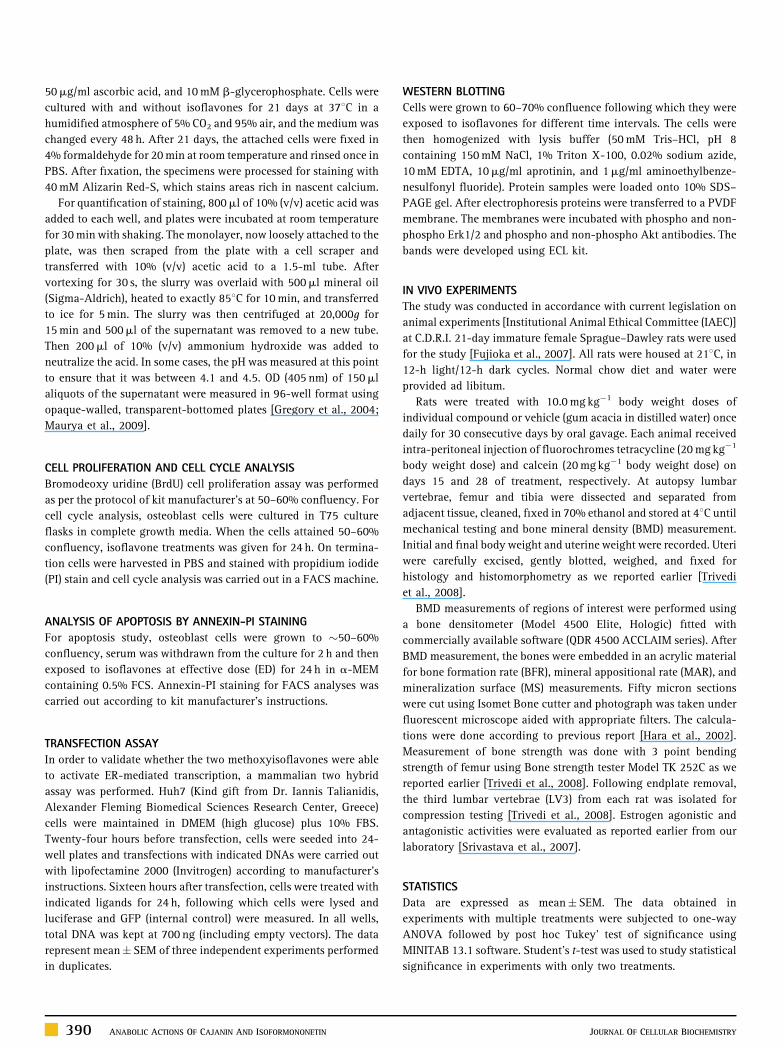
The impact of cajanin on the stimulation of osteoblast
proliferation, assessed by BrdU incorporation, was abolished by
U0126, a MEK1/2 inhibitor (Fig. 3A). In addition, Figure 3A shows
that osteoblast proliferation was also dependent on Akt signaling, a
cellular survival pathway, as the specific inhibitor, LY-294002
(10.0mM) completely abrogated cajanin-stimulated BrdU incor-
poration to osteoblasts. Together, these data suggested activation of
MEK-Erk and subsequent Akt pathways in regulating cajanin-
stimulated osteoblast proliferation. In case of stimulation of
osteoblast differentiation by cajanin, it was however, observed
that U0126 completely inhibited ALP production by osteoblast, in
support of above data, suggesting involvement of MEK-Erk pathway
in this process (Fig. 3B).
Fig. 3. Stimulation of proliferation and differentiation of osteoblasts by cajanin are me
LY294002 (10mM) significantly abolished cajanin (Caj)-induced osteoblast proliferati
significant differences amongst individual mean at P< 0.001. B: Osteoblasts were incub
U0126, SB203580 and SP600125. ALP production from osteoblasts was measured as d
differences amongst individual mean at P< 0.001. C: Activation of Erk and Akt pathways
collected at different time points. Lysates were resolved on SDS–PAGE and the blots w
LY294002 had no effect on Erk1/2 phosphorylation, while pre-treatment with U0126 (E)
gels of three independent experiments with similar results.
392 ANABOLIC ACTIONS OF CAJANIN AND ISOFORMONONETIN
Western blot data were in conformity with the inhibitor data as
cajanin-induced phosphorylation of both Erk1/2 and Akt (Fig. 3C).
Pre-treatment with LY294002 had no effect on the phosphorylation
of Erk1/2 by cajanin (Fig. 3D), whereas presence of U0126
completely abolished phosphorylation of Akt induced by cajanin
(Fig. 3E), suggesting sequential activation of MEK-Erk and Akt
pathways by cajanin in promoting osteoblast functions.
ISOFORMONONETIN STIMULATED OSTEOBLAST SURVIVAL AND
DIFFERENTIATION: SIMULTANEOUS ACTIVATION OF MEK-ERK AND
AKT PATHWAYS
In pilot study we observed that unlike cajanin, isoformononetin
(10�9–10�7 M) had no effect on BrdU incorporation (data not
shown). Next, calvarial osteoblasts induced for apoptosis by serum
deprivation was tested for possible protection by cajanin or
isoformononetin, assessed by FACS using annexin-PI staining.
We observed that cajanin failed to afford any protection to
osteoblast apoptosis under serum deprivation (data not shown). In
diated by MEK/Erk-Akt pathway. A: Treatment of osteoblasts with U0126 (10mM) and
on assessed by BrdU ELISA. Data are mean� SEM; n¼ 3. Different alphabets denote
ated for 48 h with cajanin in the presence or absence of various MAPK inhibitors, viz.
escribed before. Data are mean� SEM; n¼ 3. Different alphabets denote significant
by cajanin in osteoblasts. Cells were treated with 10�11 M cajanin and cell lysates were
ere probed with specific antibodies as indicated in the figure. D: Pre-treatment with
inhibited the phosphorylation of Akt induced by cajanin. Panels C–E are representative
JOURNAL OF CELLULAR BIOCHEMISTRY
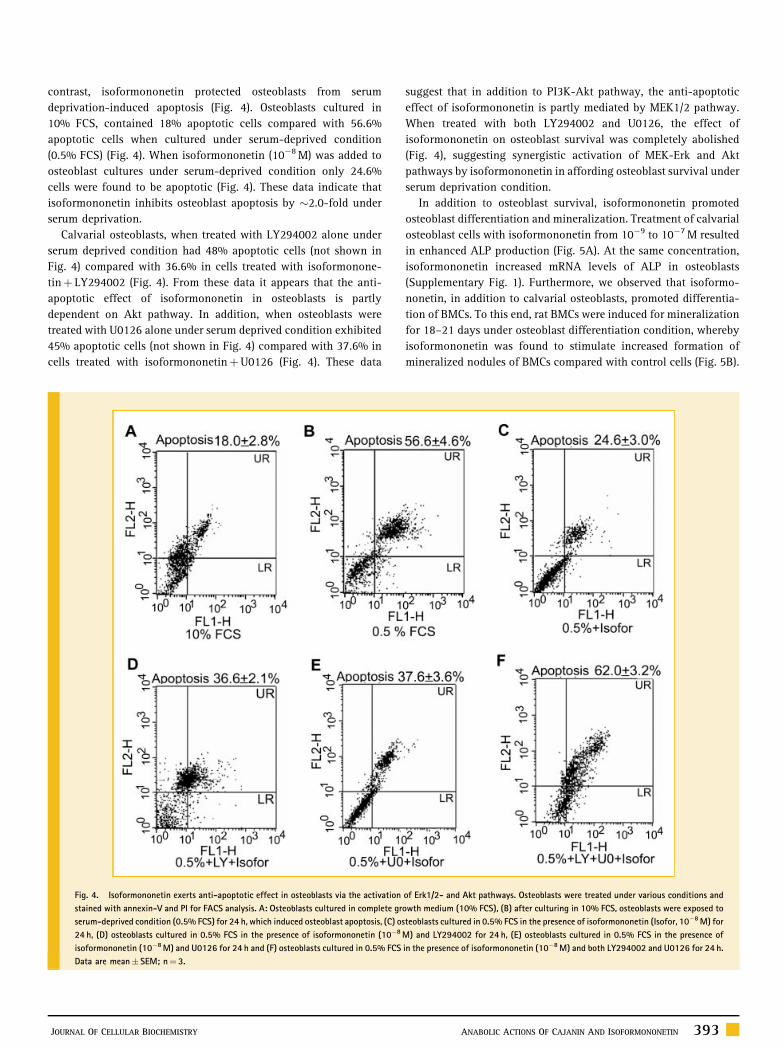
contrast, isoformononetin protected osteoblasts from serum
deprivation-induced apoptosis (Fig. 4). Osteoblasts cultured in
10% FCS, contained 18% apoptotic cells compared with 56.6%
apoptotic cells when cultured under serum-deprived condition
(0.5% FCS) (Fig. 4). When isoformononetin (10�8 M) was added to
osteoblast cultures under serum-deprived condition only 24.6%
cells were found to be apoptotic (Fig. 4). These data indicate that
isoformononetin inhibits osteoblast apoptosis by �2.0-fold under
serum deprivation.
Calvarial osteoblasts, when treated with LY294002 alone under
serum deprived condition had 48% apoptotic cells (not shown in
Fig. 4) compared with 36.6% in cells treated with isoformonone-
tinþ LY294002 (Fig. 4). From these data it appears that the anti-
apoptotic effect of isoformononetin in osteoblasts is partly
dependent on Akt pathway. In addition, when osteoblasts were
treated with U0126 alone under serum deprived condition exhibited
45% apoptotic cells (not shown in Fig. 4) compared with 37.6% in
cells treated with isoformononetinþU0126 (Fig. 4). These data
Fig. 4. Isoformononetin exerts anti-apoptotic effect in osteoblasts via the activation
stained with annexin-V and PI for FACS analysis. A: Osteoblasts cultured in complete gr
serum-deprived condition (0.5% FCS) for 24 h, which induced osteoblast apoptosis, (C) os
24 h, (D) osteoblasts cultured in 0.5% FCS in the presence of isoformononetin (10�8
isoformononetin (10�8 M) and U0126 for 24 h and (F) osteoblasts cultured in 0.5% FCS
Data are mean� SEM; n¼ 3.
JOURNAL OF CELLULAR BIOCHEMISTRY
suggest that in addition to PI3K-Akt pathway, the anti-apoptotic
effect of isoformononetin is partly mediated by MEK1/2 pathway.
When treated with both LY294002 and U0126, the effect of
isoformononetin on osteoblast survival was completely abolished
(Fig. 4), suggesting synergistic activation of MEK-Erk and Akt
pathways by isoformononetin in affording osteoblast survival under
serum deprivation condition.
In addition to osteoblast survival, isoformononetin promoted
osteoblast differentiation and mineralization. Treatment of calvarial
osteoblast cells with isoformononetin from 10�9 to 10�7 M resulted
in enhanced ALP production (Fig. 5A). At the same concentration,
isoformononetin increased mRNA levels of ALP in osteoblasts
(Supplementary Fig. 1). Furthermore, we observed that isoformo-
nonetin, in addition to calvarial osteoblasts, promoted differentia-
tion of BMCs. To this end, rat BMCs were induced for mineralization
for 18–21 days under osteoblast differentiation condition, whereby
isoformononetin was found to stimulate increased formation of
mineralized nodules of BMCs compared with control cells (Fig. 5B).
of Erk1/2- and Akt pathways. Osteoblasts were treated under various conditions and
owth medium (10% FCS), (B) after culturing in 10% FCS, osteoblasts were exposed to
teoblasts cultured in 0.5% FCS in the presence of isoformononetin (Isofor, 10�8 M) for
M) and LY294002 for 24 h, (E) osteoblasts cultured in 0.5% FCS in the presence of
in the presence of isoformononetin (10�8 M) and both LY294002 and U0126 for 24 h.
ANABOLIC ACTIONS OF CAJANIN AND ISOFORMONONETIN 393

Fig. 5. Effect of isoformononetin on differentiation of calvarial osteoblasts and mineralization of bone marrow osteoprogenitor cells. A: Osteoblasts were exposed to various
concentrations of isoformononetin (Isofor) for 48 h and ALP activity was determined as described in the Materials and Methods Section. Data are mean� SEM; n¼ 3;P< 0.01, P< 0.001. B: Bone marrow cells (25,000 cells/well) from rats were seeded into 12-well plates in differentiation medium and treated with isoformononetin
(10�8 M) for 21 days (as described in the Materials and Methods Section). At the end of the incubation, cells were stained with alizarin red-S. Stain was extracted and OD
measured colorimetrically. Data are mean� SEM; n¼ 3; P< 0.001. C: Osteoblasts were incubated for 48 h with isoformononetin (10�8 M) in the presence or absence of
U0126 or LY294002. Data are mean� SEM; n¼ 3. Different alphabets denote significant differences amongst individual mean at P< 0.001. D: Time course of activation of Erk
and Akt by isoformononetin. The figure is a representative gel of three independent experiments with similar results. [Color figure can be viewed in the online issue, which is
available at www.interscience.wiley.com.]
Effect of isoformononetin on osteoblast ALP production was
inhibited by U0126 or LY-294002, albeit both partially (Fig. 5C).
Since, co-treatment of U0126 and LY-294002 completely abolished
ALP production stimulated by isoformononetin, suggested parallel
activation of MEK-Erk and Akt pathways by isoformononetin in
stimulating osteoblast ALP production (Fig. 5C). These results were
confirmed by western blotting which show that isoformononetin
stimulated phosphorylation of both Erk1/2 and Akt in osteoblasts
(Fig. 5D).
CAJANIN AND ISOFORMONONETIN ACT INDEPENDENT OF
ESTROGEN RECEPTOR (ER)
Reports show that isoflavones such as genistein and daidzein act via
the ER in osteoblasts [Choi et al., 2008]. Hence, in order to test
whether cajanin or isoformononetin mediated their actions in
osteoblasts through ER, we used anti-estrogen, ICI-182780. Our data
show that ICI-182780 had no effect on cajanin- or isoformononetin-
induced osteoblast functions such as proliferation and differentia-
tion (Fig. 6A,B). Furthermore, in Huh7 cells transfected with human
394 ANABOLIC ACTIONS OF CAJANIN AND ISOFORMONONETIN
ERa and ERb neither cajanin nor isoformononetin transactivated
these reporter gene constructs (Fig. 6C,D), suggesting lack of ER-
mediated signaling by these compounds.
CAJANIN AND ISOFORMONONETIN DIFFERENTIALLY PROMOTE
ACHIEVEMENT OF PBM
We next assessed the in vivo effects of cajanin and isoformononetin
in growing female rats where bone formation is the dominant
event. Female Sprague–Dawley rats at weaning were given
individual treatment of either cajanin or isoformononetin at
10.0 mg kg�1 day�1 dose by oral gavage for 30 consecutive days.
Since, pilot studies at lower doses than 10.0 mg kg�1 day�1 dose had
no effect, we selected this dose. Gum acacia was used as vehicle
(control group).
Figure 7 shows the effects of these isoflavones on BMD levels.
Cajanin robustly increased BMD levels in all anatomical regions of
the skeleton studied when compared with control. Isoformononetin
treated rats, when compared with control, also exhibited significant
increases in BMD levels at all anatomical regions of skeleton except
JOURNAL OF CELLULAR BIOCHEMISTRY

Fig. 6. Cajanin and isoformononetin do not signal via ER. A: Effect of ICI-182780 on osteoblast proliferation induced by cajanin (Caj). Cells were pre-treated with ICI 182780
(1 nM) for 30 min with or without various test compounds indicated and subsequently cultured for 24 h. Data show that ICI 182780 failed to abolish osteoblast proliferation
induced by cajanin. Data are mean� SEM; n¼ 3. Different alphabets denote significant differences amongst individual mean at P< 0.001. E2 was taken as a positive control.
B: Effect of ICI-182780 on osteoblast differentiation induced by cajanin and isoformononetin (Isofor). Cells were pre-treated with ICI 182780 (1 nM) for 30 min with or
without the test compounds indicated and subsequently cultured for 48 h. Data show that ICI-182780 failed to abolish osteoblast differentiation induced by cajanin or
isoformononetin. E2 was taken as a positive control. Data are mean� SEM; n¼ 3. Different alphabets denote significant differences amongst individual mean at P< 0.001. C: A
mammalian two hybrid assay was performed where, Huh7 cells in 24-well plates were co-transfected with 200 ng GAL4-UAS-Luc, 100 ng EGFPC1, 100 ng pMTIF-2 and 100 ng
VP16-ER (a or b) and treated with indicated compounds as described in Materials and Methods Section. Luciferase values were normalized with GFP values and are expressed as
fold activity over vehicle treated cells. D: Huh7 cells were transfected with 200 ng ERE-Luc, 100 ng EGFPC1 and 40 ng pcDNA3 ER (a or b) and treated with indicated
compounds. Luciferase values were normalized with GFP values and are expressed as fold activity over vehicle treated cells.
femur neck, however, the extent of increase was less robust than that
obtained with cajanin treatment.
Bone strength is the measure of the quality of bone. Table I shows
that force and stiffness of femur were significantly higher in rats
treated with cajanin compared with controls, correlating with
femoral BMD data. Isoformononetin had no effect on femoral force
or stiffness, although it significantly increased BMD of midshaft. Our
data suggest that a P-value less than 0.001 in BMD levels of femoral
midshaft, as in the case of cajanin correlates with increased femoral
biomechanical strength. Compressive energy measurement of
lumbar vertebra 3 (LV3) also was in agreement with the BMD data.
Cajanin treatment but not isoformononetin showed a higher
compressive energy for breaking LV3 when compared with vehicle
group. Thus, cajanin treatment not only increased the BMD but also
improved the quality of bone.
Dynamic histomorphometric studies by double fluorochrome
(tetracycline–calcein) labeling experiment allowed determination of
new bone formation during the period of administration of a given
agent. Figure 8A show that both cajanin and isoformononetin
JOURNAL OF CELLULAR BIOCHEMISTRY
significantly increased mineral apposition rate (MAR) and BFR
compared with control.
ASSESSMENT OF UTERINE ESTROGEN AGONISTIC/ANTAGONISTIC
ACTIVITY OF CAJANIN AND ISOFORMONONETIN
Isoflavones are known to possess varied degrees of estrogenic or
anti-estrogenic effects [Hopert et al., 1998]. We observed that in
vitro, none of these compounds appear to signal via ERs. Here, we
studied whether cajanin and isoformononetin have estrogen
agonistic or antagonistic effects in uterus. Weaning (21 days old)
Sprague–Dawley rats were ovariectomized (OVx) and oral admin-
istration of cajanin or isoformononetin were given at
10 mg kg�1 day�1 dose for 3 consecutive days with or without
17b-E2. Table II shows that while cajanin did not exhibit neither
estrogenic nor anti-estrogenic effects as assessed by uterine wet
weight, isoformononetin was mildly uterotropic as it increased
uterine weight by �88% in OVx rats over the control. As expected,
17b-E2 treatment (0.01 mg kg�1 day�1) to OVx rats exhibited �5.7-
fold increase in uterine wet weight compared with controls (Table II).
ANABOLIC ACTIONS OF CAJANIN AND ISOFORMONONETIN 395
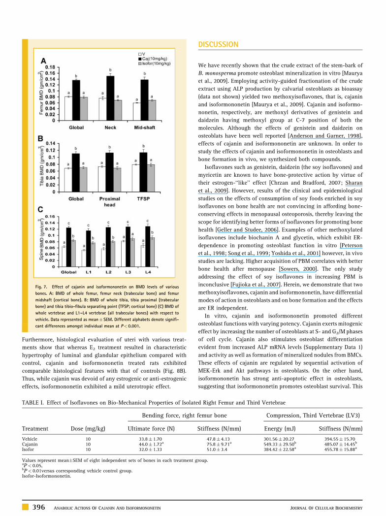
Fig. 7. Effect of cajanin and isoformononetin on BMD levels of various
bones. A: BMD of whole femur, femur neck (trabecular bone) and femur
midshaft (cortical bone). B: BMD of whole tibia, tibia proximal (trabecular
bone) and tibia tibio-fibula separating point (TFSP; cortical bone) (C) BMD of
whole vertebrae and L1–L4 vertebrae (all trabecular bones) with respect to
vehicle. Data represented as mean� SEM. Different alphabets denote signifi-
cant differences amongst individual mean at P< 0.001.
Furthermore, histological evaluation of uteri with various treat-
ments show that whereas E2 treatment resulted in characteristic
hypertrophy of luminal and glandular epithelium compared with
control, cajanin and isoformononetin treated rats exhibited
comparable histological features with that of controls (Fig. 8B).
Thus, while cajanin was devoid of any estrogenic or anti-estrogenic
effects, isoformononetin exhibited a mild uterotropic effect.
TABLE I. Effect of Isoflavones on Bio-Mechanical Properties of Isolate
Treatment Dose (mg/kg)
Bending force, right
Ultimate force (N)
Vehicle 10 33.8� 1.70Cajanin 10 44.0� 1.72a
Isofor 10 32.0� 1.33
Values represent mean�SEM of eight independent sets of bones in each treatment gaP< 0.05,bP< 0.01versus corresponding vehicle control group.Isofor-Isoformononetin.
396 ANABOLIC ACTIONS OF CAJANIN AND ISOFORMONONETIN
DISCUSSION
We have recently shown that the crude extract of the stem-bark of
B. monosperma promote osteoblast mineralization in vitro [Maurya
et al., 2009]. Employing activity-guided fractionation of the crude
extract using ALP production by calvarial osteoblasts as bioassay
(data not shown) yielded two methoxyisoflavones, that is, cajanin
and isoformononetin [Maurya et al., 2009]. Cajanin and isoformo-
nonetin, respectively, are methoxyl derivatives of genistein and
daidzein having methoxyl group at C-7 position of both the
molecules. Although the effects of genistein and daidzein on
osteoblats have been well reported [Anderson and Garner, 1998],
effects of cajanin and isoformononetin are unknown. In order to
study the effects of cajanin and isoformononetin in osteoblasts and
bone formation in vivo, we synthesized both compounds.
Isoflavones such as genistein, daidzein (the soy isoflavones) and
myricetin are known to have bone-protective action by virtue of
their estrogen-‘‘like’’ effect [Chrzan and Bradford, 2007; Sharan
et al., 2009]. However, results of the clinical and epidemiological
studies on the effects of consumption of soy foods enriched in soy
isoflavones on bone health are not convincing in affording bone-
conserving effects in menopausal osteoporosis, thereby leaving the
scope for identifying better forms of isoflavones for promoting bone
health [Geller and Studee, 2006]. Examples of other methoxylated
isoflavones include biochanin A and glycetin, which exhibit ER-
dependence in promoting osteoblast function in vitro [Peterson
et al., 1998; Song et al., 1999; Yoshida et al., 2001] however, in vivo
studies are lacking. Higher acquisition of PBM correlates with better
bone health after menopause [Sowers, 2000]. The only study
addressing the effect of soy isoflavones in increasing PBM is
inconclusive [Fujioka et al., 2007]. Herein, we demonstrate that two
methoxyisoflavones, cajanin and isoformononetin, have differential
modes of action in osteoblasts and on bone formation and the effects
are ER independent.
In vitro, cajanin and isoformononetin promoted different
osteoblast functions with varying potency. Cajanin exerts mitogenic
effect by increasing the number of osteoblasts at S- and G2/M phases
of cell cycle. Cajanin also stimulates osteoblast differentiation
evident from increased ALP mRNA levels (Supplementary Data 1)
and activity as well as formation of mineralized nodules from BMCs.
These effects of cajanin are regulated by sequential activation of
MEK-Erk and Akt pathways in osteoblasts. On the other hand,
isoformononetin has strong anti-apoptotic effect in osteoblasts,
suggesting that isoformononetin promotes osteoblast survival. This
d Right Femur and Third Vertebrae
femur bone Compression, Third Vertebrae (LV3)
Stiffness (N/mm) Energy (mJ) Stiffness (N/mm)
47.8� 4.13 301.56� 20.27 394.55� 15.7075.8� 9.71a 549.33� 29.50b 485.07� 14.45b
51.0� 3.4 384.42� 22.58a 455.78� 15.88a
roup.
JOURNAL OF CELLULAR BIOCHEMISTRY

Fig. 8. Cajanin and isoformononetin promote bone formation and do not exhibit uterine estrogenicity. A: Representative images of transverse sections of tetracycline and
calcein labeled femur diaphyses from rats after 30 days treatment with vehicle or cajanin (Caj) at 10.0 mg kg�1 day�1 dose or isoformononetin (isofor) at 10.0 mg kg�1 day�1
dose. Tertracyclin (UV filter) and calcein (orange) labeling are shown. Arrow head—calcein, arrow—tetracyclin. Bone histomorphometric parameters calculated from these
labeling experiments are shown in lower panel. Different alphabets denote significant differences amongst individual mean at P< 0.001. B: Uteri were harvested from rats
treated with vehicle or E2 or cajanin (Caj) or isoformononetin (Isofor). Cross-sections of uterine tissues were stained with H&E. Much thicker endometrium and greater epithelial
cell heights were observed in the E2 treated group while cajanin or isoformononetin exhibited comparable histological features with that of vehicle. [Color figure can be viewed
in the online issue, which is available at www.interscience.wiley.com.]
effect of isoformononetin is partially mediated by MEK-Erk and Akt
pathways. In addition, both MEK-Erk and Akt pathways regulate
isoformononetin-induced differentiation of osteoblast. Finally,
cajanin at 0.01 nM stimulated formation of mineralized nodules
from the BMCs compared to 10 nM required for isoformononetin for
the same effect. As the concentration of cajanin required for
stimulating various osteoblast functions in vitro was less than that
required by isoformononetin, it appears that cajanin is more potent
JOURNAL OF CELLULAR BIOCHEMISTRY
than isoformononetin. Sequential activation of MEK-Erk and Akt
pathways by cajanin compared with simultaneous activation of both
these pathways by isoformononetin may be attributed to more
potent effect of cajanin over isoformononetin. It should be noted
that the effects of cajanin and isoformononetin on osteoblasts are
observed at concentrations that are 100- to 1,000-fold less than that
reported for genistein and daidzein [Yamaguchi and Sugimoto,
2000]. Therefore, it appears that methoxylation enhances activities
ANABOLIC ACTIONS OF CAJANIN AND ISOFORMONONETIN 397
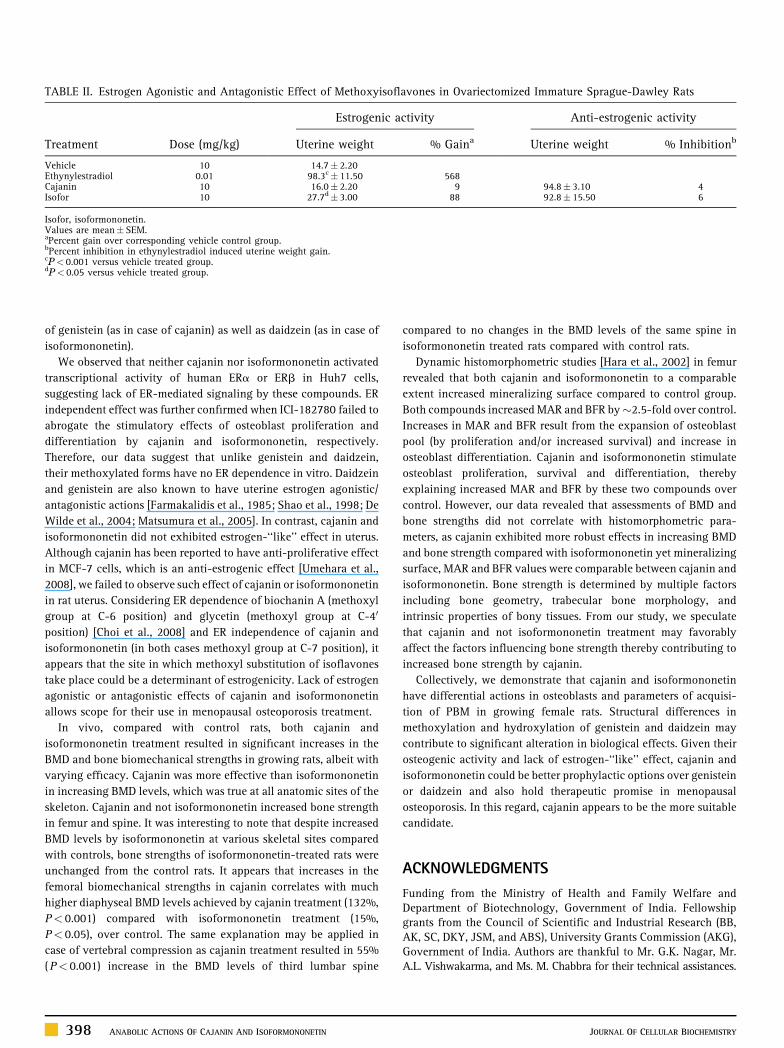
TABLE II. Estrogen Agonistic and Antagonistic Effect of Methoxyisoflavones in Ovariectomized Immature Sprague-Dawley Rats
Treatment Dose (mg/kg)
Estrogenic activity Anti-estrogenic activity
Uterine weight % Gaina Uterine weight % Inhibitionb
Vehicle 10 14.7� 2.20Ethynylestradiol 0.01 98.3c� 11.50 568Cajanin 10 16.0� 2.20 9 94.8� 3.10 4Isofor 10 27.7d� 3.00 88 92.8� 15.50 6
Isofor, isoformononetin.Values are mean� SEM.aPercent gain over corresponding vehicle control group.bPercent inhibition in ethynylestradiol induced uterine weight gain.cP< 0.001 versus vehicle treated group.dP< 0.05 versus vehicle treated group.
of genistein (as in case of cajanin) as well as daidzein (as in case of
isoformononetin).
We observed that neither cajanin nor isoformononetin activated
transcriptional activity of human ERa or ERb in Huh7 cells,
suggesting lack of ER-mediated signaling by these compounds. ER
independent effect was further confirmed when ICI-182780 failed to
abrogate the stimulatory effects of osteoblast proliferation and
differentiation by cajanin and isoformononetin, respectively.
Therefore, our data suggest that unlike genistein and daidzein,
their methoxylated forms have no ER dependence in vitro. Daidzein
and genistein are also known to have uterine estrogen agonistic/
antagonistic actions [Farmakalidis et al., 1985; Shao et al., 1998; De
Wilde et al., 2004; Matsumura et al., 2005]. In contrast, cajanin and
isoformononetin did not exhibited estrogen-‘‘like’’ effect in uterus.
Although cajanin has been reported to have anti-proliferative effect
in MCF-7 cells, which is an anti-estrogenic effect [Umehara et al.,
2008], we failed to observe such effect of cajanin or isoformononetin
in rat uterus. Considering ER dependence of biochanin A (methoxyl
group at C-6 position) and glycetin (methoxyl group at C-40
position) [Choi et al., 2008] and ER independence of cajanin and
isoformononetin (in both cases methoxyl group at C-7 position), it
appears that the site in which methoxyl substitution of isoflavones
take place could be a determinant of estrogenicity. Lack of estrogen
agonistic or antagonistic effects of cajanin and isoformononetin
allows scope for their use in menopausal osteoporosis treatment.
In vivo, compared with control rats, both cajanin and
isoformononetin treatment resulted in significant increases in the
BMD and bone biomechanical strengths in growing rats, albeit with
varying efficacy. Cajanin was more effective than isoformononetin
in increasing BMD levels, which was true at all anatomic sites of the
skeleton. Cajanin and not isoformononetin increased bone strength
in femur and spine. It was interesting to note that despite increased
BMD levels by isoformononetin at various skeletal sites compared
with controls, bone strengths of isoformononetin-treated rats were
unchanged from the control rats. It appears that increases in the
femoral biomechanical strengths in cajanin correlates with much
higher diaphyseal BMD levels achieved by cajanin treatment (132%,
P< 0.001) compared with isoformononetin treatment (15%,
P< 0.05), over control. The same explanation may be applied in
case of vertebral compression as cajanin treatment resulted in 55%
(P< 0.001) increase in the BMD levels of third lumbar spine
398 ANABOLIC ACTIONS OF CAJANIN AND ISOFORMONONETIN
compared to no changes in the BMD levels of the same spine in
isoformononetin treated rats compared with control rats.
Dynamic histomorphometric studies [Hara et al., 2002] in femur
revealed that both cajanin and isoformononetin to a comparable
extent increased mineralizing surface compared to control group.
Both compounds increased MAR and BFR by�2.5-fold over control.
Increases in MAR and BFR result from the expansion of osteoblast
pool (by proliferation and/or increased survival) and increase in
osteoblast differentiation. Cajanin and isoformononetin stimulate
osteoblast proliferation, survival and differentiation, thereby
explaining increased MAR and BFR by these two compounds over
control. However, our data revealed that assessments of BMD and
bone strengths did not correlate with histomorphometric para-
meters, as cajanin exhibited more robust effects in increasing BMD
and bone strength compared with isoformononetin yet mineralizing
surface, MAR and BFR values were comparable between cajanin and
isoformononetin. Bone strength is determined by multiple factors
including bone geometry, trabecular bone morphology, and
intrinsic properties of bony tissues. From our study, we speculate
that cajanin and not isoformononetin treatment may favorably
affect the factors influencing bone strength thereby contributing to
increased bone strength by cajanin.
Collectively, we demonstrate that cajanin and isoformononetin
have differential actions in osteoblasts and parameters of acquisi-
tion of PBM in growing female rats. Structural differences in
methoxylation and hydroxylation of genistein and daidzein may
contribute to significant alteration in biological effects. Given their
osteogenic activity and lack of estrogen-‘‘like’’ effect, cajanin and
isoformononetin could be better prophylactic options over genistein
or daidzein and also hold therapeutic promise in menopausal
osteoporosis. In this regard, cajanin appears to be the more suitable
candidate.
ACKNOWLEDGMENTS
Funding from the Ministry of Health and Family Welfare andDepartment of Biotechnology, Government of India. Fellowshipgrants from the Council of Scientific and Industrial Research (BB,AK, SC, DKY, JSM, and ABS), University Grants Commission (AKG),Government of India. Authors are thankful to Mr. G.K. Nagar, Mr.A.L. Vishwakarma, and Ms. M. Chabbra for their technical assistances.
JOURNAL OF CELLULAR BIOCHEMISTRY

REFERENCES
Anderson JJ, Garner SC. 1998. Phytoestrogens and bone. Baillieres ClinEndocrinol Metab 12:543–557.
Bass RJ. 1976. Synthesis of chromones by cyclization of 2-hydroxyphenylketones with boron trifluoride–diethyl ether and methanesulphonyl chloride.J Chem Soc Chem Commun 2:78–79.
Choi SY, Ha TY, Ahn JY, Kim SR, Kang KS, Hwang IK, Kim S. 2008. Estrogenicactivities of isoflavones and flavones and their structure-activity relation-ships. Planta Med 74:25–32.
Chrzan BG, Bradford PG. 2007. Phytoestrogens activate estrogen receptorbeta1 and estrogenic responses in human breast and bone cancer cell lines.Mol Nutr Food Res 51:171–177.
De Wilde A, Lieberherr M, Colin C, Pointillart A. 2004. A low dose of daidzeinacts as an ERbeta-selective agonist in trabecular osteoblasts of young femalepiglets. J Cell Physiol 200:253–262.
Farmakalidis E, Hathcock JN, Murphy PA. 1985. Oestrogenic potency ofgenistin and daidzin in mice. Food Chem Toxicol 23:741–745.
Fujioka M, Sudo Y, Okumura M, Wu J, Uehara M, Takeda K, Hosokawa Y,Yamada K, Ikegami S, Ishimi Y. 2007. Differential effects of isoflavones onbone formation in growing male and female mice. Metabolism 56:1142–1148.
Geller SE, Studee L. 2006. Soy and red clover for mid-life and aging.Climacteric 9:245–263.
Gregory CA, Gunn WG, Peister A, Prockop DJ. 2004. An Alizarin red-basedassay of mineralization by adherent cells in culture: Comparison withcetylpyridinium chloride extraction. Anal Biochem 329:77–84.
Hara K, Kobayashi M, Akiyama Y. 2002. Vitamin K2 (menatetrenone) inhibitsbone loss induced by prednisolone partly through enhancement of boneformation in rats. Bone 31:575–581.
Hopert AC, Beyer A, Frank K, Strunck E, Wunsche W, Vollmer G. 1998.Characterization of estrogenicity of phytoestrogens in an endometrial-derived experimental model. Environ Health Perspect 106:581–586.
Ishimi Y, Yoshida M, Wakimoto S, Wu J, Chiba H, Wang X, Takeda K,Miyaura C. 2002. Genistein, a soybean isoflavone, affects bone marrowlymphopoiesis and prevents bone loss in castrated male mice. Bone 31:180–185.
Ishizuya T, Yokose S, Hori M, Noda T, Suda T, Yoshiki S, Yamaguchi A. 1997.Parathyroid hormone exerts disparate effects on osteoblast differentiationdepending on exposure time in rat osteoblastic cells. J Clin Invest 99:2961–2970.
Kritz-Silverstein D, Goodman-Gruen DL. 2002. Usual dietary isoflavoneintake, bone mineral density, and bone metabolism in postmenopausalwomen. J Womens Health Gend Based Med 11:69–78.
Matsumura A, Ghosh A, Pope GS, Darbre PD. 2005. Comparative study ofoestrogenic properties of eight phytoestrogens in MCF7 human breast cancercells. J Steroid Biochem Mol Biol 94:431–443.
Maurya R, Yadav DK, Singh G, Bhargavan B, Narayana Murthy PS, Sahai M,Singh MM. 2009. Osteogenic activity of constituents from Butea mono-sperma. Bioorg Med Chem Lett 19:610–613.
Mei J, Yeung SS, Kung AW. 2001. High dietary phytoestrogen intake isassociated with higher bone mineral density in postmenopausal but notpremenopausal women. J Clin Endocrinol Metab 86:5217–5221.
JOURNAL OF CELLULAR BIOCHEMISTRY
Miller CP, Collini MD, Harris HA. 2003. Constrained phytoestrogensand analogues as ERb selective ligands. Bioorg Med Chem Lett 13:2399–2403.
Morito K, Aomori T, Hirose T, Kinjo J, Hasegawa J, Ogawa S, InoueS, Muramatsu M, Masamune Y. 2002. Interaction of phytoestrogenswith estrogen receptors alpha and beta (II). Biol Pharm Bull 25:48–52.
Orwoll ES, Belknap JK, Klein RF. 2001. Gender specificity in the geneticdeterminants of peak bone mass. J Bone Miner Res 16:1962–1971.
Peterson TG, Ji GP, Kirk M, Coward L, Falany CN, Barnes S. 1998. Metabolismof the isoflavones genistein and biochanin A in human breast cancer celllines. Am J Clin Nutr 68:1505S–1511S.
Phrakonkham P, Chevalier J, Desmetz C, Pinnert MF, Berges R, Jover E,Davicco MJ, Bennetau-Pelissero C, Coxam V, Artur Y, Canivenc-Lavier MC.2007. Isoflavonoid-based bone-sparing treatments exert a low activity onreproductive organs and on hepatic metabolism of estradiol in ovariecto-mized rats. Toxicol Appl Pharmacol 224:105–115.
Setchell KD, Lydeking-Olsen E. 2003. Dietary phytoestrogens and their effecton bone: Evidence from in vitro and in vivo, human observational, anddietary intervention studies. Am J Clin Nutr 78:593S–609S.
Shao ZM, Alpaugh ML, Fontana JA, Barsky SH. 1998. Genistein inhibitsproliferation similarly in estrogen receptor-positive and negative humanbreast carcinoma cell lines characterized by P21WAF1/CIP1 induction, G2/Marrest, and apoptosis. J Cell Biochem 69:44–54.
Sharan K, Siddiqui JA, Swarnkar G, Maurya R, Chattopadhyay N. 2009. Roleof phytochemicals in the prevention of menopausal bone loss: Evidence fromin vitro and in vivo, human interventional and pharmacokinetic studies. CurrMed Chem 16:1138–1157.
Somekawa Y, Chiguchi M, Ishibashi T, Aso T. 2001. Soy intake related tomenopausal symptoms, serum lipids, and bone mineral density in postme-nopausal Japanese women. Obstet Gynecol 97:109–115.
Song TT, Hendrich S, Murphy PA. 1999. Estrogenic activity of glycitein, a soyisoflavone. J Agric Food Chem 47:1607–1610.
Sowers MF. 2000. Lower peak bone mass and its decline. Baillieres Best PractRes Clin Endocrinol Metab 14:317–329.
Srivastava SR, Keshri G, Bhargavan B, Singh C, Singh MM. 2007. Pregnancyinterceptive activity of the roots of Calotropis gigantea Linn in rats. Contra-ception 75:318–322.
Trivedi R, Kumar S, Kumar A, Siddiqui JA, Swarnkar G, Gupta V, KendurkerA, Dwivedi AK, Romero JR, Chattopadhyay N. 2008. Kaempferol has osteo-genic effect in ovariectomized adult Sprague-Dawley rats. Mol Cell Endo-crinol 289:85–93.
Umehara K, Nemoto K, Kimijima K, Matsushita A, Terada E, MonthakantiratO, De-Eknamkul W, Miyase T, Warashina T, Degawa M, Noguchi H. 2008.Estrogenic constituents of the heartwood of Dalbergia parviflora. Phyto-chemistry 69:546–552.
Yamaguchi M, Sugimoto E. 2000. Stimulatory effect of genistein anddaidzein on protein synthesis in osteoblastic MC3T3-E1 cells: Activationof aminoacyl-tRNA synthetase. Mol Cell Biochem 214:97–102.
Yoshida H, Teramoto T, Ikeda K, Yamori Y. 2001. Glycitein effecton suppressing the proliferation and stimulating the differentiation ofosteoblastic MC3T3-E1 cells. Biosci Biotechnol Biochem 65:1211–1213.
ANABOLIC ACTIONS OF CAJANIN AND ISOFORMONONETIN 399
Copyright © 2022 FDOKUMEN