
Non-covalent routes to tune the optical properties of molecular materials
Upload
independentCategory
view
1download
0
Journal of Steroid Biochemistry & Molecular Biology 98 (2006) 236–247
Mass spectrometry identification of covalent attachment sites of tworelated estrogenic ligands on human estrogen receptor �
Helene Mattras a, Sigrid Aliau b, Emmanuelle Demey c,Joel Poncet c, Jean-Louis Borgna b,∗
a INSERM U554, 29 rue de Navacelles, 34090 Montpellier Cedex 5, Franceb INSERM U540, 60 rue de Navacelles, 34090 Montpellier Cedex 5, France
c CNRS UPR 2580, 141 rue de la Cardonille, 34094 Montpellier Cedex 5, France
Received 6 May 2005; accepted 7 October 2005
Abstract
A purified preparation of human estrogen receptor � (hER�) ligand-binding domain (LBD) involving mainly the Ser309Ala569 (∼30%) andSer309Ala571 (∼63%) ER portions was used to identify the covalent attachment sites of two closely related estrogenic ER affinity labels 17�-
bromoacetamidopropylestradiol (17BAPE2) and 17�-bromoacetamidomethylestradiol (17BAME2). To identify and quantify the electrophilecovalent attachment sites, [14C]17BAPE2- and [14C]17BAME2-alkylated hLBD preparations were trypsinized and submitted to HPLC. Ineach case, two radioactive fractions were obtained. Mass spectrometry analyses of the two fractions showed signals, which closely matchedthe molecular masses of alkylated Cys530Lys531 and Cys417Arg434 hLBD tryptic peptides. The covalent attachment of the two electrophileson hLBD was assigned to the S atoms of Cys530 and Cys417. However, the balance between Cys530 and Cys417 labeling markedly differedaccording to the affinity label used, with the Cys530/Cys417 ratio being 2.1 for 17BAPE2, and 20 for 17BAME2. We attempted to interpretthe covalent attachment of electrophiles by molecular modeling using the crystallographic structure of LBD bound to E2. In agreement withthe different levels of Cys417 alkylation, the LBD model with unchanged helices could not easily account for Cys417 labeling by 17BAME2,whereas favorable results were obtained through 17BAPE2 docking. Moreover, labeling at Cys530 by the two electrophiles could not beinterpreted using the LBD model. This indicates that some states of solute LBD bound to the estrogenic E2 17�-derivatives differ from thestructure of crystallized LBD bound to E2.© 2006 Elsevier Ltd. All rights reserved.Keywords: Estrogen receptor; Ligand-binding domain; Estradiol 17�-derivatives; Affinity labeling; Mass spectrometry
Abbreviations: ER, estrogen receptor; hER� and mER�, humanand mouse ER�s; NR, nuclear receptor; LBD, ligand-binding domain;hLBD and mLBD, human and mouse ER� portions involvingLBD and adjacent domain F; LBP, ligand-binding pocket; GST,glutathione-S-transferase; E2, estradiol; OHT, 4-hydroxytamoxifen; Ral,raloxifene; 17BAPE2, 17�-bromoacetamidopropylE2; 17BAME2, 17�-bromoacetamidomethylE2; 17APE2, 17�-acetamidopropylE2; 17AME2,17�-acetamidomethylE2; �-carbon, terminal electrophilic carbon in the 17�-substituent of 17BAPE2 and 17BAME2; T20, 20 mM Tris–HCl buffer; PBS,phosphate-buffered saline; RAC, binding affinity constant relative to thatof E2; MS, mass spectrometry; ESI, electrospray ionization; MALDI-TOF,matrix-assisted laser desorption ionization time-of-flight; �, symbolizes anundefined atom or group of atoms displaying a monoisotopic mass very closeto 16
∗ Corresponding author. Tel.: +33 4 67 33578.E-mail address: [email protected] (J.-L. Borgna).
1. Introduction
Two estrogen receptors (ER� and ER�) which mediatethe effects of estrogens at the transcriptional level havebeen identified in mammals [1–4]. Like most nuclearreceptor (NR) superfamily members [5], these two proteinsdisplay a modular structure involving six domains [3,6]. Theligand-binding domain (LBD) made of ∼250 amino acids(Ser301Pro552 sequence in human ER� (hER�)) constitutesthe largest domain in ERs. This domain is involved in therecruitment of coactivators [7] and corepressors [8] andtherefore plays an essential role in the NR transcriptionalfunction. Crystal structures of hER� and hER� LBDsbound to agonist or antagonist ligands have been recentlyestablished [9–15]. These structures show the predominantly
0960-0760/$ – see front matter © 2006 Elsevier Ltd. All rights reserved.doi:10.1016/j.jsbmb.2005.10.006

H. Mattras et al. / Journal of Steroid Biochemistry & Molecular Biology 98 (2006) 236–247 237
�-helical fold characteristically observed with other crys-tallized NR LBDs [16]. Interestingly, one major differencebetween estrogen–LBD and antiestrogen–LBD complexesrelated to the orientation of the C-terminal helix 12 (whichharbors the AF-2 activation domain [17,18]) was firstreported [9,11]. Although the description of these LBDstructures was a milestone in the NR research field, it affordsa static view of LBD–ligand complexes, which may notfully reflect LBD structures of NR complexes in the cellularcontext, as there could be equilibria between multipleconformational states in these complexes. This hypothesis issupported by recent reports [12,15,19,20], which indicatedthat it is difficult to account for the estrogenic/antiestrogenicactivity of an ER ligand on the basis of the orientation ofhelix 12 in crystallized ER LBD–ligand complexes. Toenhance the overall understanding of the molecular action ofestrogens and antiestrogens, we sought to directly identify,within ER� LBD, the covalent attachment sites of estrogenicestradiol (E2) 17�-derivatives [21] and antiestrogenic E211�-derivatives [22] through mass spectrometry (MS) anal-ysis and then to interpret the biochemical data by molecularmodeling.
In a recent work [23], using a C-terminal portion ofmouse ER� (mER�, Ser313Ile599 sequence, correspondingto the LBD and the adjacent C-terminal domain F, anddesignated mLBD), the combined use of HPLC and MSaCabvi
Fltat8a
Fig. 2. Structure of the GST–hLBD chimera protein. The GST–hLBDchimera (exact counterpart of the previously studied GST–mLBD chimera[23]) encoded by the pGEX-2TK expression vector is schematically rep-resented. Its hLBD moiety constituted by the C-terminal half of hER�
(Ser309-Val595) involves the actual LBD (ending at Pro552) and the adjacentterminal F domain (Thr553-Val595). LBD is linked to GST by a 18 aminoacid sequence enclosing a thrombin cleavage site. The hLBD forms obtainedby thrombin cleavage of the chimera and identified by ESI MS involved twotruncated major forms, ending at Ala569 (30%) and Ala571 (63%), and twominor forms, the Ser575-truncated form (4%) and the full-length LBD (3%)(cf. text).
E2 [9] or 4-hydroxytamoxifen (OHT, a non-steroidal estrogenantagonist) [11] could account for the covalent attachmentof 17BAPE2 to Cys421 and Cys534, respectively, whereasthat derived from LBD bound to raloxifene (Ral, anothernon-steroidal estrogen antagonist) [9] could account for thecovalent attachment of the affinity labeling agent to bothcysteines.
17�-BromoacetamidomethylE2 (17BAME2, Fig. 1), onepreviously tested ER� affinity label [21], directly derivesfrom 17BAPE2 by removal of the (CH2)2 module fromthe 17�-(CH2)3 group. In 17BAME2 the maximal dis-tance between the steroidal C-17 and the electrophilic car-bon (�-carbon) is ∼35% lower than that of the homol-ogous distance in 17BAPE2 and the degree of freedomof the �-carbon is two instead of four. The markeddecrease in the mobility of the �-carbon (relative to theER-recognized E2 moiety) from 17BAPE2 to 17BAME2,might qualitatively or quantitatively alter the results ofLBD labeling by 17BAPE2. This makes 17BAME2 aninteresting tool for probing the ER� ligand-binding pocket(LBP). Since human but not mouse ER� LBD modelsare presently available, we decided to use human LBDrather than mouse LBD to get a more valid interpreta-tion of ER� LBD affinity labeling by E2 17�-derivatives.Accordingly, we constructed an expression vector encod-ing glutathione-S-transferase (GST) fused with the exacthaofiyEGi[o(cio
nalysis showed that Cys421 and Cys534 (homologues ofys417 and Cys530 in hER�) were the mLBD alternativend quantitatively equivalent attachment sites of 17�-romoacetamidopropylE2 (17BAPE2, Fig. 1). We usedarious X-ray crystallographic structures of hER� LBD tonterpret these biochemical data. Structures of LBD bound to
ig. 1. Structures of [14C]17BAPE2 and [14C]17BAME2. The ER� affinityabeling agents are derived from E2 by 17�-substitution. They differ only athe alkyl moiety (either propyl or methyl) of the substituent. They possessn electrophilic carbon (terminal bromine-bearing carbon, �-carbon) and,o facilitate characterization of the affinity labeled hLBD and fragments, a8% (17BAPE2) or 91% (17BAME2) 14C-enriched carbon (denoted by ansterisk).
uman counterpart (Ser309-Val595 sequence involving LBDnd adjacent domain F, and designated hLBD) (Fig. 2)f the previously studied portion of mER� [23]. Werst identified by electrospray ionization (ESI) MS anal-sis, molecular forms of hLBD extracted from induced. coli cells and characterized the GST–hLBD/E2 andST–hLBD/17BAME2 or 17BAPE2 interactions. We then
dentified and quantified the covalent attachment sites of14C]17BAPE2 and [14C]17BAME2 on hLBD by meansf matrix-assisted laser desorption ionization time-of-flightMALDI-TOF) MS and HPLC analyses. Finally, using therystal structure of LBD bound to estrogen, we try tonterpret by molecular modeling the biochemical resultsbtained.

238 H. Mattras et al. / Journal of Steroid Biochemistry & Molecular Biology 98 (2006) 236–247
2. Materials and methods
2.1. Materials
1-Isopropylthio-�-d-galactopyranoside, protease inhibi-tor cocktail for bacterial cell extracts (containing 4-(2-aminoethyl)benzenesulfonylfluoride, pepstatin A, trans-epoxysuccinyl-l-leucylamido(4-guanidino)butane, bestatin,and EDTA) and �-cyano-4-hydroxycinnamic acid werefrom Sigma–Aldrich. Glutathione-Sepharose 4B andthrombin were purchased from Amersham Biosciences.Trypsin (sequencing grade) was from Promega. Solvents andreagents used for HPLC and MALDI-TOF MS analyses wereHPLC grade (for HPLC, Milli-Q grade water was passedthrough a C18 preparative column and then sterilized).[6,7-3H]E2 (specific activity 1.6 PBq/mol, radiochemicalpurity >98%) was purchased from New England Nuclear,and bromo[1-14C]acetic acid (specific activity 2.1 TBq/mol,radiochemical purity >97%) from Amersham Biosciences.[NHCO-14C]17BAPE2 (specific activity, 2.0 TBq/mol)[23], 17BAME2, 17BAPE2, 17AME2, and 17APE2 [21]had been synthesized previously in the laboratory. Labeledligands, i.e. [3H]E2, [14C]17BAPE2 and synthesized[NHCO-14C]17BAME2 (specific activity, 2.1 TBq/mol), andunlabeled ligands, i.e. E2, 17BAME2, 17BAPE2, 17AME2,17APE , were solubilized in absolute ethanol. Solutionswc
2
taTc>
2
(cuGncehCaCactc
Clones expressing the correct sized plasmid were selectedand the accuracy of the construct was checked by sequenc-ing. PCMV5 plasmids encoding wild-type hER� and theHis524 → Ala mutant were given by B.S. Katzenellenbogen;preparation of these plasmids has been described previously[26].
2.4. GST–hLBD expression and extraction
Expression of GST–hLBD in E. coli BL21 cells andcell pellet preparation was performed as described previ-ously for GST–mLBD [23], except that instead of 2 h (at37 ◦C) we used 15 h (at 15 ◦C) induction of the cells with1-isopropylthio-�-d-galactopyranoside (0.6 mM). For fusionprotein extraction, one or two cell pellets (0.5 mL; storedat −70 ◦C) were thawed and slowly resuspended in 3.5 mLof ice-cooled 20 mM Tris–HCl (T20, pH 8.0), containing100 mM NaCl, 1 mM EDTA, 10 mM dithiothreitol, 0.2%Nonidet P-40 and the protease inhibitor cocktail. The cell sus-pension was incubated for 40 min at 0 ◦C and then submittedto sonication at 0 ◦C using a Bioblock-Scientific Vibra-cell72405 (five series of 200 pulses for 0.5 s, amplitude 60, over4 min, at 1 min intervals). The sonicate was centrifuged for30 min at 13,000 × g (4 ◦C) to obtain the crude bacteriallysate.
2e
mc[
2
[pffa
2f
owT(w(cl9
2ere stored at −20 ◦C in the dark. The purity of solubilized
ompounds was checked before use by TLC.
.2. Synthesis and characterisation of [14C]17BAME2
[14C]17BAME2 was prepared from 3-(tert-butyldime-hylsiloxy)-17�-aminomethylE2 and bromo[1-14C]aceticcid, as described previously [21] for unlabeled compound.he labeled compound was purified on silica gel and thenharacterized by MS. Its purity (assessed by HPLC) was95%.
.3. GST–hLBD and hERα expression vectors
A pGEX-2TK vector encoding GST fused with mLBDSer313-Ile599) [24], via a sequence enclosing a thrombinleavage site located 14 amino acids ahead of Ser313, wassed to prepare an homologous expression vector coding forST fused with hLBD (Ser309Val595). DNA encoding theine amino acids upstream of mLBD in the GST–mLBDhimera and the hLBD amino acids 309–595 was gen-rated by PCR using pSG5-HEGO (involving completeER� DNA) [25], and two primers, 5′TCTGTTGGATCCC-GGGAATTTCGACTTCCCTGACGGCCGACCAGATG3′nd 5′CCCCGGGGATCCTCAGACTGTGGCAGGGAAC-3′ enclosing a BamH1 cleavage site. Amplified DNAnd the pGEX-2TK plasmid encoding GST–mLBD wereleaved by BamH1 and purified. The amplified DNA washen ligated to the dephosphorylated pGEX-2TK plasmid. E.oli BL21 cells were transformed with the ligation products.
.5. Wild-type and mutant (His524Ala) hERαsxpression and extraction
Expression of the wild-type hER� and the His524 → Alautant in COS cells, then extraction of ERs from the
ells by sonication were carried out as described previously27].
.6. Purification and affinity labeling of hLBD
Affinity labeling of GST–hLBD with [14C]17BAPE2 or14C]17BAME2 was performed at 2 ◦C, pH 8.0, as describedreviously for GST–mLBD [23] using 3.8 mL bacterial lysateor 100 �L glutathione-Sepharose 4B. hLBD was releasedrom matrix-bound GST–hLBD by the action of thrombinnd then glutathione; it was purified by HPLC [23].
.7. Proteolysis and purification of hLBD trypticragments
The radioactive fractions obtained by HPLC purificationf hLBD labeled with [14C]17BAPE2 or [14C]17BAME2ere assembled and partially dried to evaporate CH3CN.he pH of the resulting sample was adjusted to 7.5; trypsin
enzyme to substrate ratio: 1/40) was added and the sampleas incubated for 4 h at 37 ◦C, and then submitted to HPLC
C4 reversed-phase column, 3.2 mm × 250 mm, 5-�m parti-les, 300-A pore size). Peptides were eluted with a CH3CNinear gradient (10–95%) at 500 �L/min flow rate over a0 min course.

H. Mattras et al. / Journal of Steroid Biochemistry & Molecular Biology 98 (2006) 236–247 239
2.8. Protein assay and radioactivity determination
The protein concentration was determined by the methodof Bradford [28] using bovine serum albumin as standard.The radioactivity of the various samples (10–200 �L) wascounted in 4 mL of Emulsifier Safe (Packard) using anLS1801 Beckman scintillation counter.
2.9. SDS-PAGE and autoradiography
Cell lysate fractions and purified hLBD preparations wereanalyzed by SDS-PAGE and autoradiography as describedpreviously [23].
2.10. Binding assays
[3H]E2 binding to the wild-type hER� or the His524 → Alamutant [27] and to GST–hLBD or hLBD [23] were per-formed using charcoal assays as previously described. TheGST–hLBD concentrations in E. coli extracts were deter-mined using 800-fold diluted extracts. Competitive bind-ing assays between [3H]E2 and E2 17�-derivatives usingthe wild-type hER� or the His524 → Ala mutant (≤ 2 nM,expressed in COS cells), or purified GST–hLBD (≤ 2 nM)supplemented with 2 mg ovalbumin/mL, were carried outi ◦Hu[iAtusa
R
H
r[b
2
itiw(liii
holder. Prior to analysis, the sample (5 �L) was desaltedby ion exchange chromatography on Poros 20 R2 (1 �L)packed in a gel-loader pipette tip and proteins wereeluted with methanol/water/formic acid: 80/20/1 (2 �L)according to an already described procedure [30]. MALDImass spectra of hLBD tryptic peptides were obtainedon a Bruker time-of-flight mass spectrometer (Bruker-Franzen, Bremen, Germany) equipped with a SCOUT sourceand Xmass software for data collection and analysis, aspreviously described [23]. The matrices were preparedfrom a saturated solution of �-cyano-4-hydroxycinnamicacid.
2.12. Ligand docking
The X-ray crystallographic structure of the human ER�LBD bound to E2 (PDB entry 2ERE, monomer A) was usedto account for the covalent attachment of 17BAME2 and17BAPE2 to Cys417 and Cys530, while firmly bound to LBD.Docking of 17BAME2 and 17BAPE2 into the LBD structurewas carried out as previously decribed for 17BAPE2 [23].17BAME2 or 17BAPE2 was superimposed over LBD-boundE2 by aligning the two A-ring. This resulted in almost mergedsteroid nuclei, with very close locations of the two 17�-OHfunctions. The flexibility of the 17�-substituent was then usedto bring the �-carbon into the vicinity of the S-atom of Cys417
oatb
3
3
EtwGd(c(oteoatfisPmt
n T20 (pH 8.0) at 20 C with 20 h (wild-type ER andis524 → Ala mutant) or 2 h (GST–hLBD) incubation timessing 5 nM [3H]E2 (wild-type ER and GST–hLBD) or 20 nM3H]E2 (His524 → Ala mutant ER) as a tracer ligand andncreasing concentrations of E2 and E2 17�-derivatives.pparent equilibrium constants of competitors (KC
a ) relativeo that of E2 (KH
a ) were calculated from the concentration ofnlabeled E2 (H) and competitor (C) which inhibited [3H]E2pecific binding to the same extent (50% inhibition was used),ccording to Korenman [29]
AC = KCa
KHa
= [(Ht − Hb)/H∗sb] × H/C
[(H∗t − H∗
b )/H∗sb] + 1 − H/C
∗t is the total concentration of [3H]E2; H∗
b and H∗sb are the
espective concentrations of bound and specifically bound3H]E2 at 50% binding inhibition. H∗
t − H∗b was assumed to
e the unbound [3H]E2 concentration.
.11. Mass spectrometry
ESI mass spectra of hLBD preparations were obtainedn a QSTAR-Pulsar-i mass spectrometer (Applied Biosys-ems, Foster City, CA, USA) fitted with a Protana nanospraynlet system (Protana, Odense, Denmark). Compoundsere analyzed using the Analyst QS software package
Applied Biosystems). Parameters were adjusted as fol-ows: ion spray voltage, 900 V; curtain gas, 25; decluster-ng potential, 45–75 V; focusing potential, 265 V; decluster-ng potential, 2–15 V. Capillaries were loaded with approx-mately 2 �L of sample and placed in the source tip
r Cys530. Steric clashes between 17BAME2 or 17BAPE2nd the LBD were manually suppressed by appropriate rota-ions of some amino acid side chains and single chemicalonds of the 17�-substituent.
. Results
.1. hLBD expression and purification
The expression of the GST–hLBD chimera (Fig. 2) in. coli using a pGEX-2TK expression vector, the extrac-
ion from cells and the purification on glutathione-Sepharoseere similar to the procedures previously described for theST–mLBD chimera [23]. To improve GST–hLBD pro-uction, cell induction was performed for 15 h at 15 ◦Cinstead of 2 h at 37 ◦C for the mouse chimera); the con-entration of E2 binding species in the bacterial extractabout 3 �M) was ∼3–4-fold higher than those previouslybtained using the homologous GST–mLBD expression vec-or. To assess the molecular forms of hLBD in chimeraxtracted from E. coli cells, GST–hLBD was absorbedn glutathione-Sepharose, and hLBD was released by thection of thrombin; it was further purified by HPLC andhen analyzed by SDS-PAGE and ESI MS. The puri-ed hLBD preparation showed a very prominent ∼31 kDapecies and minor ∼34 and ∼28 kDa species in SDS-AGE (Fig. 3). ESI MS analysis (Fig. 4) evidenced fiveultiply charged ion series. From various m/z values of
he ion series (cf. Table 1), four species of 33634.6 ± 1.7,

240 H. Mattras et al. / Journal of Steroid Biochemistry & Molecular Biology 98 (2006) 236–247
Fig. 3. SDS-PAGE analysis of hLBD. Cell extract (3.8 mL) from E. coliinduced to express the GST–hLBD chimera was rotated for 15 h at 4 ◦C withglutathione-Sepharose (100 �L). The gel was transferred into a column andwashed with 6 mL of phosphate buffered saline (PBS) (pH 7.3). Thrombinwas added to the gel resuspended in 1 mL PBS and the mixture was rotatedfor 15 h at 4 ◦C. The gel was drained and washed twice with 50 �L PBS,then the various eluates were pooled. The eluted species were submitted toreverse-phase HPLC. The bacterial extract before (lane 1) and after (lane2) incubation with glutathione-Sepharose, the proteins released by thrombin(lane 3) and the related HPLC fractions containing the last major 220-nmabsorbing species were analyzed on a 12% polyacrylamide gel. Standardmolecular mass markers (kDa) are in lane 5.
31454.2 ± 0.6, 31121.3 ± 0.3, and 30949.3 ± 0.3 Da (whoserelative signal intensities were 1, 1.3, 21 and 10, respec-tively) were identified and corresponded exactly to thefull-length hLBD (GSR· · ·ISTS309LT· · ·PATV595 sequence,33634.0 Da) and to hLBD truncated at Ser575 (31453.6 Da),Ala571 (31121.3 Da) and Ala569 (30949.1 Da), respectively(Table 1), whereas the last species (31318.6 ± 0.3 Da), whichdid not correspond to any truncated form of hLBD, could bea contaminant in the preparation. This suggested that the full-length hLBD constituted ∼3% of the hLBD forms, whereastruncated forms ending at Ala571, Ala569 and Ser575 consti-tuted ∼63%, ∼30% and ∼4% of the hLBD forms, respec-tively. These interpretations were subsequently confirmedby MALDI-TOF MS analysis of hLBD tryptic fragments:signals corresponding to Gly556-Ala569, Gly556-Ala571 and
Gly556-Ser575 peptides, resulting from trypsin cleavage justafter Arg555 of the various hLBD forms, were routinelyobserved (not shown). These results indicated that in contrastto mLBD used in a previous study [23], which involved∼75%of the full-length form and ∼25% of the Ala573-truncatedform, the purified hLBD essentially involved two truncatedforms, a minor form (∼30%) ending at Ala569 (human homo-logue of mER� Ala573) and a major form (∼63%) ending atAla571. These latter results agreed with those reported bySeielstad et al. [31] concerning the expression of isolatedhLBD in E. coli.
3.2. Binding of 17α-derivatives to hLBD
The binding properties of GST–hLBD and hLBD werecharacterized by determining the dissociation rate of E2from these species purified via absorption on glutathione-Sepharose. Very similar kinetics were obtained with thechimera and isolated hLBD. E2 dissociation occurred at 0and 20 ◦C according to biphasic processes with mean half-dissociation times of 18–21 h and 17–18 min, respectively(not shown).
We then tried to assess the affinity of 17BAPE2 and17BAME2 (Fig. 1) for hLBD. For a conventional (chemicallyinert) ligand, the binding affinity constant relative to that ofEivRteR1wt[hftwfdL[
Table 1Major hLBD forms observed in ESI MS analysis of hLBD preparation
Sequence Predicted mass (Da) Obse
GS· · ·S309LT· · ·LA569 30949.1 3094GS· · ·S309LT· · ·LATA571 31121.3 3112GS· · ·S309LT· · ·LATAGSTS575 31453.6 3145GS· · ·S309LT· · ·LA· · ·PATV595 33634.0 3363
Four species corresponding to intact and three truncated hLBD forms were identified(mean ± S.D.) was determined from eight m/z values (z = 28–35) of signals belonginconsidered to account for the abundance of the corresponding species. The predicteS
wissProt/TrEMBL data bank.2 (RAC) could be determined by competitive binding exper-ments. For a ligand which covalently binds to LBD, the irre-ersible binding precludes determination of the compoundAC by such an approach. However, the RAC of a conven-
ional ligand, whose structure is very close to that of thelectrophile, should enable a good estimate of the electrophileAC. We thus used 17�-acetamidopropylE2 (17APE2) and7�-acetamidomethylE2 (17AME2) [21] two inert analogueshich derive from the two electrophilic ligands by substi-
uting H for Br. Competitive binding experiments between3H]E2 and the electrophiles or their inert analogues usingER� (crude) or GST–hLBD (purified) gave similar valuesor the compound RACs (Table 2). RACs of the inert competi-ors for hER� or GST–hLBD were in the 0.05–0.12% range,hereas the apparent RACs of the two electrophiles were
rom 20–70-fold higher. This discrepancy was most probablyue to covalent binding of the electrophiles within the hER�BP, resulting in irreversible inactivation of the latter for
3H]E2 binding. Taking into account the affinity constant
rved mass (Da) �M (Da) Relative abundance (%)
9.3 ± 0.3 0.2 301.3 ± 0.3 0.0 634.2 ± 0.6 0.6 44.6 ± 1.7 0.6 3
by ESI MS analysis of purified hLBD (cf. Fig. 4). Each experimental massg to a same family. The mean of relative heights for each signal family was
d masses of the various hLBD forms were obtained using ExPASy from the

H. Mattras et al. / Journal of Steroid Biochemistry & Molecular Biology 98 (2006) 236–247 241
Fig. 4. ESI MS analysis of hLBD. The species released from glutathione-Sepharose by thrombin were submitted to reverse-phase HPLC. Fractions containingthe last major 220-nm absorbing species were analyzed by ESI MS. (A) Whole spectrum; five series of signals were detected. The m/z values of the peaksfrom the major series corresponding to multicharged ([M + nH]n+) ions of the Ala571-truncated form are given. (B) Enlargement of the spectrum in the m/z910–965 region; the various signals denoted An, Bn, Cn, Dn, En and corresponding m/z values are related to [M+nH]n+ ions of the Ala569-, Ala571-, andSer575-truncated hLBD forms, the full-length hLBD form and the unidentified (31318.6 Da) species, respectively. Signals A′
n, B′n, A′′
n, B′′n correspond to
alternative [M + (n − 1)H + Na]n+ and [M + (n − 2)H + 2Na]n+ ions, respectively related to the Ala569- and Ala571-truncated hLBD major forms.

242 H. Mattras et al. / Journal of Steroid Biochemistry & Molecular Biology 98 (2006) 236–247
Table 2RACs of E2 17�-derivatives for binding GST–hLBD and wild-type andH524A hER�s
Compound GST–hLBD hER� H524A hER�
E2 100 100 10017AME2 0.12 ± 0.07 0.094 ± 0.030 0.43 ± 0.0617BAME2 5.6 ± 0.9 6.5 ± 0.1 14 ± 417APE2 0.054 ± 0.003 0.070 ± 0.035 0.71 ± 0.1117BAPE2 1.2 ± 0.4 1.9 ± 0.4 20 ± 5
Apparent RACs of E2 17�-derivatives for GST−hLBD, the wild-type hER�
and corresponding His524 → Ala mutant were determined as described inSection 2. The data are means ± S.D. of two independent determinations.
determined for the ER/E2 interaction [32,33], the resultsobtained for the inert E2 derivatives suggested that the stud-ied E2 17�-derivatives, including the two electrophiles, haveaffinity constants close to 109 M−1 for hER�. We previouslyobserved that the 17�-OH/His524 interaction plays an impor-tant role in E2 binding to hER�, since the compound affinityconstant (relative to that of OHT) and dissociation rate con-stant related to the His524 → Ala mutant were, respectively24-fold lower and 250-fold higher than the corresponding val-ues determined with wild-type hER� [27]. RACs determinedfor inert E2 17�-derivatives moderately increased from thewild-type hER� to the His524 → Ala mutant, suggestingthat the 17�-OH/His524 interaction still operates for thesecompounds, although less intensively than with E2 (Table 2).
3.3. Identification of covalent attachment sites of17BAPE2 and 17BAME2 on hLBD
After in situ exposure of glutathione-Sepharose-boundmaterial to [14C]17BAPE2 or [14C]17BAME2, the speciesreleased by the action of thrombin and then of glutathionewere submitted to HPLC. In both cases the 220 nm absorptionprofile was very similar to that obtained in the HPLC purifi-cation of mLBD generated from GST–mLBD [23]. Threecomponents were resolved containing GST, putative dnaKand hLBD, the latter made of the 34 kDa form, the three3GocGwldp
[mitoHet
Fig. 5. HPLC of [14C]17BAPE2- and [14C]17BAME2-labeled hLBD tryp-tic fragments. hLBD covalently labeled by [14C]17BAME2 was purifiedby HPLC. The main radioactive fractions were concentrated, treated bytrypsin and then submitted to reverse-phase HPLC. Column elution was per-formed with a linear CH3CN gradient. The curve represents recorded 220 nmabsorption according to the elution time, whereas green bars indicate the 14Ccontents of the various fractions (counts per minute per milliliter). In a par-allel experiment performed with [14C]17BAPE2 instead of [14C]17BAME2,the HPLC separation of hLBD tryptic fragments afforded a very similar220 nm absorption profile (not represented for figure clarity) and a differentprofile for the 14C-content of the various fractions, shown by red bars.
related species, and (ii) the proportion of the first species tothe second species in the chromatogram was ∼68/32 with17BAPE2 and ∼95/5 with 17BAME2.
For each electrophile, the two 14C-labeled species wereanalyzed by MALDI-TOF MS to identify the ligand-modifiedhLBD tryptic peptides. Concerning [14C]17BAPE2 label-ing, the first and second species gave signals typical oflabeled Cys530Lys531 and labeled Cys417-Arg434 hLBD tryp-tic peptides, respectively (Table 3), since these signals wereidentical to those attributed to labeled mLBD tryptic pep-tides Cys534Lys535 and Cys421-Arg438 (displaying the samesequences as their hLBD homologues), in our previous studyof mLBD labeling by [14C]17BAPE2 [23]. Briefly, these twolabeled species (whose respective molecular masses are thesum of that of the peptide and that of dehydrobrominated[14C]17BAPE2) were characterized by the signal of the pseu-domolecular [M + H]+ ion and by signals of alternative ionsresulting from (i) elimination of H2O ([M + H–H2O]+) or ofan unascribed group � ([M + H–�]+) displaying a monoiso-topic mass very close to 16 [23] (labeled Cys530Lys531),(ii) monooxidation ([M + H + O]+) of methionyl residues(labeled Cys417-Arg434), and (iii) substitution of Na for H([M + Na]+, labeled Cys530Lys531).
The first species related to [14C]17BAME2 labeling gavesignals which were the exact counterparts of signals obtainedfor [14C]17BAPE -alkylated Cys530Lys531 peptide (i.e. dif-ft
1 kDa forms and the 28 kDa contaminant (cf. Fig. 3). WhenST–hLBD labeling was performed with [14C]17BAPE2,nly the LBD-containing fractions included 14C. Whenhimera labeling was carried out with [14C]17BAME2, bothST- and hLBD-containing fractions included 14C; however,hen GST–hLBD was preincubated with excess E2 before
abeling, the 14C concentration in hLBD fractions was veryrastically decreased, whereas that in GST fractions wasractically unchanged (not shown).
The HPLC-purified hLBD labeled with [14C]17BAPE2 or14C]17BAME2 was then trypsinized and submitted to chro-atography (Fig. 5). Determination of the 14C concentration
n the various fractions indicated that, for each electrophile,wo 14C-labeled species were present, a situation alreadybserved in the study of mLBD labeling by [14C]17BAPE2.owever (i) each of the two 17BAME2-related species was
luted from the column at a CH3CN percentage slightly lowerhan that required for elution of the corresponding 17BAPE2-
2ering from the latter signals by a �m/z increment very closeo the isotopic mass of the (CH2)2 group: 28.03); these sig-

H. Mattras et al. / Journal of Steroid Biochemistry & Molecular Biology 98 (2006) 236–247 243
Table 3MS characterisitic of 14C-labeled hLBD tryptic fragments
(Cys530Lys531)–APE2
(m/z)(Cys417-Arg434)–APE2
(m/z)(Cys530Lys531)–AME2
(m/z)(Cys417-Arg434)–AME2
(m/z)
ion Expt Calcd Expt Calcd Expt Calcd Expt Calcd
[M + H]+ 621.41 621.36 2372.28 2372.18 593.30, 593.03c 593.32 2343.86c 2344.15[M + H–H2O]+ 603.43 603.35 – – 575.43, 575.02c 575.31 – –[M + H–�]+ 605.55a 605.36b – – 577.63a, 577.04c 577.32b – –[M + H + O]+ – – 2388.26 2388.18 – – – –[M + Na]+ 643.43 643.34 – – 615.38, 614.99c 615.31 – –
The m/z values of signals obtained in the MALDI-TOF MS analysis of tryptic [14C]peptides generated from [14C]17BAPE2- or [14C]17BAME2-alkylatedhLBD, which matched the values calculated for pseudomolecular [M + H]+ ions and alternative ions, are given. In the third ion species, � symbolizes anundefined atom or group of atoms displaying a monoisotopic mass very close to 16.
a Slightly inaccurate value (due to the presence of an interfering signal).b Value calculated assuming a monoisotopic mass of 16 for �.c Value obtained from [14C]17BAME2-alkylated mLBD.
nals were related to the [M + H]+ ion and to [M + H–H2O]+,[M+H–�]+ and [M + Na]+ ions (Fig. 6A and B, Table 3).Probably due to its low abundance in HPLC fractions, byMALDI-TOF MS analysis it was not easy to identify thesecond species related to [14C]17BAME2 labeling. However,HPLC of tryptic peptides obtained from mLBD alkylated by[14C]17BAME2, afforded a more abundant species elutedat the same CH3CN percentage as the hLBD-related secondspecies (not shown); MALDI-TOF MS analysis of themouse species gave a faint signal at m/z 2343.86 (Fig. 6C),which matched the mass of [14C]17BAME2-alkylatedCys421-Arg438 peptide (Table 3). This strongly suggestedthat alkylated Cys417-Arg434 peptide constituted the secondlabeled species detected in the HPLC of tryptic peptidesobtained from [14C]17BAME2-labeled hLBD.
After identifying the electrophile-labeled hLBD trypticpeptides, the next point was to specify the covalent attachmentof 17BAPE2 and 17BAME2 in Cys530Lys531 and Cys417-Arg434 peptides. As discussed previously [23], only the Satom of the cysteinyl residue displays a nucleophilic charac-ter in the Cys530Lys531 dipeptide (especially at pH 8). Thecovalent attachment site of 17BAPE2 and 17BAME2 in thathLBD portion could then be unambigously assigned to theCys530 S atom. The demonstration, through a synthetic hep-tapeptide (displaying the mLBD Cys421-Glu427 sequence),that Cys421 was the covalent attachment site of 17BAPEohaapfoCtChlo
We concluded that Cys417 and Cys530 were two alterna-tive, but quantitatively different, covalent attachment sitesof the two electrophiles on hER� LBD; the involvement ofCys530, greater than that of Cys417 with 17BAPE2, was muchmore pronounced with 17BAME2.
4. Discussion
In this study we produced high amounts of hER� LBDthrough the expression of GST–LBD chimera in BL21 E.coli cells and then identified and quantified, by means ofMS and HPLC analyses, the covalent attachment sites of17BAPE2 and 17BAME2, two closely related estrogenic lig-ands derived from E2 by 17�-substitution. ESI MS analysisrevealed that the hLBD preparation involved four differentforms, sharing a common N terminus and having various Ctermini: the full-length hLBD ending at Val595 (∼3%), andthree fragments truncated at Ser575 (∼4%), Ala571 (∼63%)and Ala569 (∼30%). The hLBD preparation used for cova-lent labeling studies was, however, rather homogeneous sincethe two major forms which only differ by the Thr570Ala571
dipeptidyl group, accounted for >90% of the various hLBDforms. It is noteworthy that these results closely agreed withthose obtained by Seielstad et al. concerning the expressiono
Gpb(awptlfhs
2n the mLBD Cys421-Arg438 peptide [23] still applied to theLBD Cys417-Arg434 peptide, since the latter two peptidesre identical. Then the S atom of Cys417 was the covalentttachment site of 17BAPE2 in the Cys417-Arg434 hLBDortion. We did not carry out a similar thorough study toormally specify the covalent attachment site of 17BAME2n Cys417-Arg434 peptide, but the fact that in hER� theys417 → Ala or Cys530 → Ala single change did not abolish
he ability of hER� to be labeled by 17BAME2, whereas theys417 → Ala, Cys530 → Ala double change abolished theER� labeling ability [34], strongly suggested that hLBDabeling in its Cys417-Arg434 portion occurred at the S atomf Cys417.
f another recombinant hER� LBD in E. coli [31].The results we previously obtained [23] with a
ST–mLBD expression vector encoding the exact counter-art of hLBD were strikingly different since, as revealedy MALDI-TOF MS analysis, only the full-length mLBD∼70%) ending at Ile599 and a fragment (∼30%) truncatedt Ala573 (the exact counterpart of Ala569-truncated hLBD)ere detected in the mLBD preparations. Note that the pro-ortion of these two forms was very close to that of thewo hLBD major forms. ESI MS analysis of mLBD (unpub-ished results) confirmed the MALDI-TOF MS data: no signalor the counterpart of the Ala571- or the Ser575-truncatedLBD form was detected in the mLBD preparations. Theequence variation between the F domains of mLBD and

244 H. Mattras et al. / Journal of Steroid Biochemistry & Molecular Biology 98 (2006) 236–247
Fig. 6. MALDI-TOF MS analysis of 14C-labeled species obtained from trypsin hydrolysis of [14C]17BAME2-alkylated hLBD and mLBD. The two radioactivefractions (27 and 58 in Fig. 5) obtained in the HPLC separation of labeled hLBD tryptic fragments (or homologous mLBD tryptic fragments) were analyzedby MALDI-TOF MS. For each fraction, the obtained signals compatible with [14C]17BAME2-alkylated forms of a hLBD (or mLBD) tryptic peptide areshown. (A) and (B): analysis of the first radioactive fraction related to hLBD (A) or mLBD (B). Signals at m/z 593.30 (A) and 593.03 (B) accurately matchthe monoisotopic mass of molecular [M + H]+ ion of human Cys530Lys531 or mouse Cys534Lys535 tryptic peptide alkylated by [14C]17BAME2 ([M + H]+,calcd value of m/z 593.32 for the dehydrobrominated form). Other signals closely agree with the masses of alternative ions deriving from the latter by: (i)dehydratation ([M + H–H2O]+, m/z 575.43 (A) and 575.02 (B), calcd 575.31), (ii) elimination of an unascribed group � displaying a monoisotopic mass veryclose to 16 ([M + H–�]+, m/z 577.63 (A) and 577.04 (B), calcd 577.32), or (iii) Na for H substitution ([M + Na]+, m/z 615.38 (A) and 614.99 (B), calcd 615.31).(C): analysis of the second radioactive fraction related to mLBD. The signal at m/z 2343.86 closely matchs the monoisotopic mass of the Cys421-Arg438 mLBDtryptic peptide alkylated by [14C]17BAME2 ([M + H]+, calcd value of m/z 2244.15). Note that in (A)–(C) panels: (i) the companion signals observed just aheadof the above mentioned signals (at �m/z −2.00) correspond to tryptic peptides alkylated by non-radioactive 17BAME2 molecules; their intensities, relative tothose of the main signals, reflect the proportion of [NHCO-12C] and [NHCO-14C] in the affinity probe (1/9.9) and, (ii) the fine structures of signals characterizedby the relative intensities of [M + H]+, [M + H + 1]+, [M + H + 2]+. . .peaks (due to the presence of 13C in natural C), reflect both the relative abundance of 13Cand the total carbon number in alkylated human Cys530Lys531 and mouse Cys534Lys535(one 14C and 29 unlabeled carbons) and alkylated mouse Cys421-Arg438
(one 14C and 105 unlabeled carbons).
hLBD seemed to be the cause of the observed discrep-ancy. As previously stated [31], the presence of the threetruncated forms in hLBD preparations was probably theresult of hLBD cleavage at three different sites (indicatedby a colon) 568LA:TA:GSTS:SH577, by an E. coli-specificpeptidase. The fact that the mLBD counterpart sequence572LA:TTSSTSAH581 displays three changed amino acids(underlined), which directly affect two cleavage sites, could
explain why only one truncated form was present in themLBD preparation.
This study allowed the identification of Cys417 and Cys530
as alternative (but quantitatively non-equivalent) covalentattachment sites of 17BAPE2 and 17BAME2 on hLBD. Astriking result was the change in the Cys530/Cys417 balance,which increased from ∼2.1 to ∼20 when hLBD labelingwas performed with 17BAME2 instead of 17BAPE2, despite
H. Mattras et al. / Journal of Steroid Biochemistry & Molecular Biology 98 (2006) 236–247 245
the small difference (methyl versus propyl in the alkyl moi-ety of the 17�-substituent) between the two electrophilesand the fact that the two compounds displayed very sim-ilar ER binding affinities and estrogenic properties [21].The covalent attachment of the two electrophiles to Cys417
and Cys530 indicates that the non-covalent LBD–17BAME2and LBD–17BAPE2 complexes display states in which theCH2Br group of the ligand and the Cys417 or Cys530 S atomare in close proximity, with opposite locations of the Br andS atoms, relative to the electrophilic center.
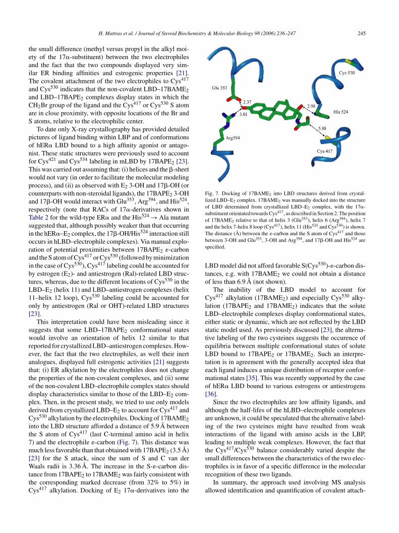
To date only X-ray crystallography has provided detailedpictures of ligand binding within LBP and of conformationsof hER� LBD bound to a high affinity agonist or antago-nist. These static structures were previously used to accountfor Cys421 and Cys534 labeling in mLBD by 17BAPE2 [23].This was carried out assuming that: (i) helices and the �-sheetwould not vary (in order to facilitate the molecular modelingprocess), and (ii) as observed with E2 3-OH and 17�-OH (orcounterparts with non-steroidal ligands), the 17BAPE2 3-OHand 17�-OH would interact with Glu353, Arg394, and His524,respectively (note that RACs of 17�-derivatives shown inTable 2 for the wild-type ER� and the His524 → Ala mutantsuggested that, although possibly weaker than that occurringin the hER�–E2 complex, the 17�-OH/His524 interaction stilloccurs in hLBD–electrophile complexes). Via manual explo-ration of potential proximities between 17BAPE �-carbonaibtL1o[
swreattodpdCit7m[WttC
Fig. 7. Docking of 17BAME2 into LBD structures derived from crystal-lized LBD–E2 complex. 17BAME2 was manually docked into the structureof LBD determined from crystallized LBD–E2 complex, with the 17�-substituent orientated towards Cys417, as described in Section 2. The positionof 17BAME2 relative to that of helix 3 (Glu353), helix 6 (Arg394), helix 7and the helix 7-helix 8 loop (Cys417), helix 11 (His524 and Cys530) is shown.The distance (A) between the �-carbon and the S atom of Cys417 and thosebetween 3-OH and Glu353, 3-OH and Arg394, and 17�-OH and His524 arespecified.
LBD model did not afford favorable S(Cys530)-�-carbon dis-tances, e.g. with 17BAME2 we could not obtain a distanceof less than 6.9 A (not shown).
The inability of the LBD model to account forCys417 alkylation (17BAME2) and especially Cys530 alky-lation (17BAPE2 and 17BAME2) indicates that the soluteLBD–electrophile complexes display conformational states,either static or dynamic, which are not reflected by the LBDstatic model used. As previously discussed [23], the alterna-tive labeling of the two cysteines suggests the occurence ofequilibria between multiple conformational states of soluteLBD bound to 17BAPE2 or 17BAME2. Such an interpre-tation is in agreement with the generally accepted idea thateach ligand induces a unique distribution of receptor confor-mational states [35]. This was recently supported by the caseof hER� LBD bound to various estrogens or antiestrogens[36].
Since the two electrophiles are low affinity ligands, andalthough the half-lifes of the hLBD–electrophile complexesare unknown, it could be speculated that the alternative label-ing of the two cysteines might have resulted from weakinteractions of the ligand with amino acids in the LBP,leading to multiple weak complexes. However, the fact thatthe Cys417/Cys530 balance considerably varied despite thesmall differences between the characteristics of the two elec-tr
a
2nd the S atom of Cys417 or Cys530 (followed by minimizationn the case of Cys530), Cys417 labeling could be accounted fory estrogen (E2)- and antiestrogen (Ral)-related LBD struc-ures, whereas, due to the different locations of Cys530 in theBD–E2 (helix 11) and LBD–antiestrogen complexes (helix1–helix 12 loop), Cys530 labeling could be accounted fornly by antiestrogen (Ral or OHT)-related LBD structures23].
This interpretation could have been misleading since ituggests that some LBD–17BAPE2 conformational statesould involve an orientation of helix 12 similar to that
eported for crystallized LBD–antiestrogen complexes. How-ver, the fact that the two electrophiles, as well their inertnalogues, displayed full estrogenic activities [21] suggestshat: (i) ER alkylation by the electrophiles does not changehe properties of the non-covalent complexes, and (ii) somef the non-covalent LBD–electrophile complex states shouldisplay characteristics similar to those of the LBD–E2 com-lex. Then, in the present study, we tried to use only modelserived from crystallized LBD–E2 to account for Cys417 andys530 alkylation by the electrophiles. Docking of 17BAME2
nto the LBD structure afforded a distance of 5.9 A betweenhe S atom of Cys417 (last C-terminal amino acid in helix) and the electrophile �-carbon (Fig. 7). This distance wasuch less favorable than that obtained with 17BAPE2 (3.5 A)
23] for the S attack, since the sum of S and C van deraals radii is 3.36 A. The increase in the S-�-carbon dis-
ance from 17BAPE2 to 17BAME2 was fairly consistent withhe corresponding marked decrease (from 32% to 5%) inys417 alkylation. Docking of E2 17�-derivatives into the
rophiles is in favor of a specific difference in the molecularecognition of these two ligands.
In summary, the approach used involving MS analysisllowed identification and quantification of covalent attach-

246 H. Mattras et al. / Journal of Steroid Biochemistry & Molecular Biology 98 (2006) 236–247
ment sites of E2 17�-derivatives to hER� LBD; it revealedthat minor changes in the electrophilic ligand markedlyaffected the LBD affinity labeling process and that an LBDmodel very close to the crystallized LBD–E2 complex couldnot easily account for all our results. Further studies involvinghigh affinity electrophiles [37], other LBD specific probes,such as those recently used in the Katzenellenbogen’s group[36], or high resolution NMR, could be useful to gain furtherinsight into ER dynamics and ligand-induced ER conforma-tional changes.
Acknowledgement
This work was supported by the “Institut National de laSante et de la Recherche Medicale” and the “Association pourle Recherche sur le Cancer”.
References
[1] S. Green, P. Walter, V. Kumar, A. Krust, J.M. Bornert, P. Argos, P.Chambon, Human oestrogen receptor cDNA: sequence, expressionand homology to v-erb A, Nature 320 (1986) 134–139.
[2] G.L. Greene, P. Gilna, M. Waterfield, A. Baker, Y. Hort, J. Shine,Sequence and expression of human estrogen receptor complementaryDNA, Science 231 (1986) 1150–1154.
[
[
[
the presence of partial agonist and a full antagonist, EMBO J. 18(1999) 4608–4618.
[13] A.C.W. Pike, A.M. Brzozowski, J. Walton, R.E. Hubbard, A.G.Thorsell, Y.L. Li, J.A. Gustafsson, M. Carlquist, Structural insightsinto the mode of action of a pure antiestrogen, Structure 9 (2001)145–153.
[14] S. Eiler, M. Gangloff, S. Duclaud, D. Moras, M. Ruff, Overexpres-sion, purification, and crystal structure of native ER� LBD, ProteinExpress. Purif. 22 (2001) 165–173.
[15] A.K. Shiau, D. Barstad, J.T. Radek, M.J. Meyers, K.W. Nettles, B.S.Katzenellenbogen, J.A. Katzenellenbogen, D.A. Agard, G.L. Greene,Structural characterization of a subtype-selective ligand reveals anovel mode of estrogen receptor antagonism, Nat. Struct. Biol. 9(2002) 359–364.
[16] J.M. Wurtz, W. Bourguet, J.P. Renaud, V. Vivat, P. Chambon, D.Moras, H. Gronemeyer, A canonical structure for the ligand-bindingdomain of nuclear receptors, Nat. Struct. Biol. 3 (1996) 87–94.
[17] A. Lees, S.E. Fawell, M.G. Parker, Identification of two transacti-vation domains in the mouse oestrogen receptor, Nucleic Acid Res.17 (1989) 5477–5488.
[18] L. Tora, J. White, C. Brou, D. Tasset, N. Webster, E. Scheer, P.Chambon, The human estrogen receptor has two independent non-acidic transcriptional activation functions, Cell 59 (1989) 477–487.
[19] W. Bourguet, P. Germain, H. Gronemeyer, Nuclear receptor ligand-binding domains: three-dimensional structures, molecular interac-tions and pharmacological implications, Trends Pharm. Sci. 21(2000) 381–388.
[20] M. Gangloff, M. Ruff, S. Eiler, S. Duclaud, J.M. Wurtz, D. Moras,Crystal structure of a mutant hER� ligand-binding domain revealskey structural features for the mechanism of partial agonism, J. Biol.Chem. 27 (2001) 15059–15065.
[
[
[
[
[
[
[
[
[
[
[
[3] S. Mosselman, J. Polman, R. Dijkema, ER�: identification and char-acterization of a novel human estrogen receptor, FEBS Lett. 392(1996) 49–53.
[4] G.G.J.M. Kuiper, E. Enmark, M. Pelto-Huikko, S. Nilsson, J.A.Gustafsson, Cloning of a novel estrogen receptor expressed inrat prostate and ovary, Proc. Natl. Acad. Sci. U.S.A. 93 (1996)5925–5930.
[5] M.J. Tsai, B.W. O’Malley, Molecular mechanisms of action ofsteroid/thyroid receptor superfamily members, Annu. Rev. Biochem.63 (1994) 451–486.
[6] A. Krust, S. Green, P. Argos, V. Kumar, P. Walter, J.M. Bornert,P. Chambon, The chicken oestrogen receptor sequence: homologywith v-erb A and the human oestrogen and glucocorticoid receptors,EMBO J. 5 (1986) 891–897.
[7] W. Feng, R.C. Ribiero, R.L. Wagner, H. Nguyen, J.W. Apriletti, R.J.Fletterick, J.D. Baxter, P.J. Kushner, B.L. West, Hormone-dependentcoactivator binding to a hydrophobic cleft on nuclear receptors, Sci-ence 280 (1998) 1747–1749.
[8] A. Moraitis, V. Giguere, C.C. Thompson, Novel mechanism ofnuclear receptor corepressor interaction dictated by activation func-tion 2 helix determinants, Mol. Cell. Biol. 22 (2002) 6831–6841.
[9] A.M. Brzozowski, A.C.W. Pike, Z. Dauter, R.E. Hubbard, T. Bonn,O. Engstrom, L. Ohman, G.L. Greene, J.A. Gustafsson, M. Car-lquist, Molecular basis of agonism and antagonism in the oestrogenreceptor, Nature 389 (1997) 753–758.
10] D.M. Tanenbaum, Y. Wang, S.P. Williams, P.B. Sigler, Crystal-lographic comparison of the estrogen and progesterone receptor’sligand binding domains, Proc. Natl. Acad. Sci. U.S.A. 95 (1998)5998–6003.
11] A.K. Shiau, D. Barstad, P.M. Loria, L. Cheng, P.J. Kushner,D.A. Agard, G.L. Greene, The structural basis of estrogen recep-tor/coactivator recognition and the antagonism of this interaction bytamoxifen, Cell 95 (1998) 927–937.
12] A.C.W. Pike, A.M. Brzozowski, R.E. Hubbard, T. Bonn, A.G.Thorsell, O. Engstrom, J. Ljunggren, J.A. Gustafsson, M. Carlquist,Structure of the ligand-binding domain of œstrogen receptor beta in
21] D. El Garrouj, S. Aliau, A. Aumelas, J.L. Borgna, Steroidal affin-ity labels of the estrogen receptor. 2. 17�-(haloacetamidoalkyl)estradiols, J. Med. Chem. 38 (1995) 2339–2348.
22] S. Aliau, G. Delettre, H. Mattras, D. El Garrouj, F. Nique, G.Teutsch, J.L. Borgna, Steroidal affinity labels of the estrogen receptor�. 4. Electrophilic 11�-aryl derivatives of estradiol, J. Med. Chem.43 (2000) 613–628.
23] H. Mattras, S. Aliau, E. Richard, J.C. Bonnafous, P. Jouin, J.L.Borgna, Identification by MALDI-TOF mass spectrometry of 17�-bromoacetamidopropylestradiol covalent attachment sites on estrogenreceptor �, Biochemistry 41 (2002) 15713–15727.
24] V. Cavailles, S. Dauvois, P.S. Danielan, M.G. Parker, Interactions ofproteins with transcriptionally active estrogen receptors, Proc. Natl.Acad. Sci. U.S.A. 91 (1994) 10009–10013.
25] L. Tora, A. Mullick, D. Metzger, M. Ponglikitmongkol, I. Park, P.Chambon, The cloned human oestrogen receptor contains a mutationwhich alters its hormone binding properties, EMBO J. 8 (1989)1981–1986.
26] K. Ekena, K.E. Weis, J.A. Katzenellenbogen, B.S. Katzenellenbogen,Identification of amino acids in the hormone binding domain ofthe human estrogen receptor important in estrogen binding, J. Biol.Chem. 27 (1996) 20053–20059.
27] S. Aliau, H. Mattras, E. Richard, J.C. Bonnafous, J.L. Borgna, Differ-ential interactions of estrogens and antiestrogens at the 17�-hydroxylor counterpart hydroxyl with histidine 524 of the human estrogenreceptor �, Biochemistry 41 (2002) 7979–7988.
28] M.M. Bradford, A rapid and sensitive method for the quantitationof microgram quantities of protein utilizing the principle of proteindye binding, Anal. Biochem. 72 (1976) 248–254.
29] S.G. Korenman, Relation between estrogen receptor inhibitory activ-ity and binding to cytosol of rabbit and human uterus, Endocrinology87 (1970) 1119–1123.
30] M. Wilm, M. Mann, Analytical properties of the nanoelectrospraysource, Anal. Chem. 68 (1996) 1–8.
31] D.A. Seielstad, K.E. Carlson, J.A. Katzenellenbogen, P.J. Kush-ner, G.L. Greene, Molecular characterization by mass spec-

H. Mattras et al. / Journal of Steroid Biochemistry & Molecular Biology 98 (2006) 236–247 247
trometry of the human estrogen receptor ligand-binding domainexpressed in Escherichia coli, Mol. Endocrinol. 9 (1995) 647–658.
[32] M. Salomonsson, B. Carlsson, J. Haggblad, Equilibrium hormonebinding to human estrogen receptors in highly diluted cell extractsis non-cooperative and has a Kd of approximately 10 pM, J. SteroidBiochem. Mol. Biol. 503 (1994) 313–318.
[33] J.L. Borgna, H. Rochefort, High affinity binding to the estrogenreceptor of [3H]4-hydroxytamoxifen, an active antiestrogen metabo-lite, Mol. Cell. Endocrinol. 20 (1980) 71–85.
[34] S. Aliau, D. El Garrouj, A. Yasri, B.S. Katzenellenbogen, J.L.Borgna, 17�-(Haloacetamidoalkyl)estradiols alkylate the human
estrogen receptor at cysteine residues 417 and 530, Biochemistry36 (1997) 5861–5867.
[35] H.O. Onaran, T. Costa, Agonist efficacy and allosteric models ofreceptor action, Ann. NY Acad. Sci. 812 (1997) 98–115.
[36] K.M. Hurth, M.J. Nilges, K.E. Carlson, A. Tamrazi, R.L. Belford,J.A. Katzenellenbogen, Ligand-induced changes in estrogen receptorconformation as measured by site-directed spin labeling, Biochem-istry 43 (2004) 1891–1907.
[37] S. Aliau, H. Mattras, J.L. Borgna, Identification of covalent attach-ment site of antiestrogenic estradiol 11�-derivatives on human estro-gen receptor ligand-binding domain, J. Steroid Biochem. Mol. Biol.,in press.
Copyright © 2022 FDOKUMEN