Non-covalent routes to tune the optical properties of molecular materials
Upload
khangminh22Category
view
0download
0
ChemicalScience
EDGE ARTICLE
Ope
n A
cces
s A
rtic
le. P
ublis
hed
on 2
8 Ja
nuar
y 20
21. D
ownl
oade
d on
5/2
6/20
22 6
:59:
40 A
M.
Thi
s ar
ticle
is li
cens
ed u
nder
a C
reat
ive
Com
mon
s A
ttrib
utio
n 3.
0 U
npor
ted
Lic
ence
.
View Article OnlineView Journal | View Issue
Mechanism of co
aCentre for Computational Chemistry, Sch
Cantock's Close, Bristol BS8 1TS, UK. E-mabComputational Chemistry, Janssen
Pharmaceutica N. V., Turnhoutseweg 30, B-
† Electronic supplementary informa10.1039/d0sc06122k
Cite this: Chem. Sci., 2021, 12, 5511
All publication charges for this articlehave been paid for by the Royal Societyof Chemistry
Received 6th November 2020Accepted 17th January 2021
DOI: 10.1039/d0sc06122k
rsc.li/chemical-science
© 2021 The Author(s). Published by
valent binding of ibrutinib toBruton's tyrosine kinase revealed by QM/MMcalculations†
Angus T. Voice,a Gary Tresadern, b Rebecca M. Twidale,a Herman van Vlijmenb
and Adrian J. Mulholland *a
Ibrutinib is the first covalent inhibitor of Bruton's tyrosine kinase (BTK) to be used in the treatment of B-cell
cancers. Understanding the mechanism of covalent inhibition will aid in the design of safer and more
selective covalent inhibitors that target BTK. The mechanism of covalent inhibition in BTK has been
uncertain because there is no appropriate residue nearby that can act as a base to deprotonate the
cysteine thiol prior to covalent bond formation. We investigate several mechanisms of covalent
modification of C481 in BTK by ibrutinib using combined quantum mechanics/molecular mechanics
(QM/MM) molecular dynamics reaction simulations. The lowest energy pathway involves direct proton
transfer from C481 to the acrylamide warhead in ibrutinib, followed by covalent bond formation to form
an enol intermediate. There is a subsequent rate-limiting keto–enol tautomerisation step (DG‡ ¼10.5 kcal mol�1) to reach the inactivated BTK/ibrutinib complex. Our results represent the first
mechanistic study of BTK inactivation by ibrutinib to consider multiple mechanistic pathways. These
findings should aid in the design of covalent drugs that target BTK and other similar targets.
Introduction
Covalent inhibitor drug discovery has re-emerged because ofadvantages compared with conventional non-covalent revers-ible binding that can include complete target blockage,increased selectivity and longer duration of action.1–3 Recentyears have seen the approval of several new marketed covalentdrugs targeting protein kinases.4,5 In particular, inhibition ofBruton's tyrosine kinase (BTK) is an attractive target for bloodcancers and autoimmune diseases, due to its function in signaltransduction in the B-cell antigen receptor (BCR) pathway.6,7
BTK inhibitors have also been explored as possible inhibitorsagainst the SARS-CoV-2 coronavirus in drug repurposingstudies.8 Ibrutinib and acalabrutinib are two BTK inhibitorsthat are approved for the treatment of B-cell cancers includingmantle cell lymphoma (MCL) and chronic lymphocyticleukaemia (CLL).9 Both drugs contain electrophilic Michaelacceptor warheads that covalently modify a cysteine residue(C481) in the kinase domain of BTK. Utilising warheads of thistype to target poorly conserved cysteine residues is a commontechnique to developing covalent inhibitors in drug discovery.10
ool of Chemistry, University of Bristol,
Research & Development, Janssen
2340 Beerse, Belgium
tion (ESI) available. See DOI:
the Royal Society of Chemistry
Despite the massive investments made to discover and developthese drugs, the detailedmechanism of covalent binding to BTKis unknown. Understanding the precise mechanism will help inthe design of improved covalent inhibitors targeting BTK, andalso other covalent drug targets. The ability to rationally tunecovalent reactivity should lead to safer, more selective covalentdrugs that have fewer side effects.11,12
The reaction of a Michael acceptor warhead such as anacrylamide group to a thiol side chain is typically modelled inthree steps. First, deprotonation of the cysteine thiol occurs toform a thiolate anion, followed by nucleophilic attack of thethiolate on to the electrophile to form an enolate intermediate,and nally re-protonation of the enolate to form a covalentthiol-adduct.13 Sulfur reactivity in proteins has been studiedpreviously using combined quantum mechanics/molecularmechanics (QM/MM) approaches.1,14 Effort has focused oncysteine proteases and protein kinases, given their roles in somedisease processes. For example, covalent nitrile inhibitors of thecysteine protease rhodesain have been investigated using QM/MM reaction simulations at the semi-empirical PM6 level.15
The protocol was found to be a useful predictor of reversiblecovalent binding affinity, in good agreement with experimentaldata. The mechanism of covalent modication of C797 by anacrylamide warhead in the protein kinase EGFR has beenelucidated by QM/MM modelling at the self-consistent-chargedensity-functional-based tight-binding (SCC-DFTB) level.16,17
These results identied a neighbouring aspartate residue, D800
Chem. Sci., 2021, 12, 5511–5516 | 5511

Chemical Science Edge Article
Ope
n A
cces
s A
rtic
le. P
ublis
hed
on 2
8 Ja
nuar
y 20
21. D
ownl
oade
d on
5/2
6/20
22 6
:59:
40 A
M.
Thi
s ar
ticle
is li
cens
ed u
nder
a C
reat
ive
Com
mon
s A
ttrib
utio
n 3.
0 U
npor
ted
Lic
ence
.View Article Online
in the i+3 position relative to C797, as the catalytic base todeprotonate the cysteine thiol.
Comparing kinases that contain cysteine at the equivalentposition to C481 in BTK, different amino acids are found at thei+3 position: the majority are either aspartate (Asp) or aspara-gine (Asn).18 BTK contains an Asn residue, N484, rather than anAsp residue or other good proton acceptor in the i+3 position(Fig. 1). Asparagine is a very weak base, suggesting amechanismin BTK different to the EGFR Asp-catalysed mechanism.
Fig. 1 (A) Crystal structure of the BTK kinase domain with ibrutinibbound in the ATP binding pocket (PDB 5P9I). (B) Binding mode ofibrutinib in the active site of BTK, in a reactive conformation observedin MM molecular dynamics simulations. The acrylamide warhead ispositioned in close proximity to C481. The S–C distance is 3.7 A. Theasparagine residue found in the i+3 position, N484, is also shown.Important active site distances are reported in A.
5512 | Chem. Sci., 2021, 12, 5511–5516
Residues in the surrounding microenvironment can modulatecysteine pKa, depending on their properties.19 Although there isno experimentally determined C481 pKa available, the pKa ofa free cysteine thiol in solution is 8.6. The cysteine pKa calcu-lated for EGFR in a recent computational study is 11.1.20 Incomparison, with the i+3 residue in BTK being Asn rather thanAsp, a slightly more acidic cysteine pKa of 10.4 was predicted inthe same study. A pKa of around 10 would result in a 1 : 1000ratio of ionised to neutral cysteine at pH 7, with a free energydifference of 4.1 kcal mol�1 between the ionised and neutralstates. Thus, C481 is predominantly neutral at physiological pH.This suggests that a nearby proton acceptor may be required fordeprotonation of the thiol group, either prior to or concertedwith the reaction with the electrophile. Although asparagineperforms catalytic roles in some enzymes, for example asa nucleophile in some protein splicing reactions,21,22 the amidesidechain of Asn is weakly basic and has only been reported toact as a base when it is activated by metal ions.23 It is thereforeunlikely to accept a proton from C481 in BTK indicating thata different mechanism of inhibition operates in BTK than thatwhich has been established for EGFR.
The lack of an obvious amino acid to act as acid or baseleaves several other possible mechanisms for covalent inhibi-tion of C481 in BTK by Michael acceptors. These includemechanisms that proceed via direct addition between C481 andthe Michael acceptor (Fig. 2), such as a direct transfer of thethiol proton to the a-carbon of the acrylamide warhead toproduce the covalently bound keto adduct in a single step.Alternatively, the thiol proton could transfer to the carbonyloxygen atom of the acrylamide inhibitor to form an enol inter-mediate, followed by a tautomerization step to form the cova-lently bound keto product. There is also the possibility thatwater could be involved in the reaction, e.g. assisting in protontransfer between the cysteine thiol and acrylamide warhead.Here, we assess each of these pathways using a QM/MMumbrella sampling approach. The results identify the prob-able mechanism of covalent binding between the acrylamidewarhead in ibrutinib and BTK.
Methods
QM/MM umbrella sampling molecular dynamics (MD) simula-tions were used to explore each mechanistic pathway in thekinase domain of BTK. The QM region included all of theibrutinib ligand and the side chain of C481. The effects ofincluding N484 in the QM region were also tested; this did notsignicantly change the calculated energetics, showing thechoice of QM region to be reasonable24 (Fig. S8, ESI†). For thewater-mediated pathways, an adjacent crystallographic watermolecule hydrogen bonded to the carbonyl oxygen of theacrylamide was also included in the QM region. The umbrellasampling protocol consisted of generating a free energy surface(FES) using the density-functional tight-binding (DFTB3) QMmethod, with 20 ps of MD sampling per reaction coordinate(RC) window to get an approximate minimum energy pathway(MEP) for the reaction. This was followed by running an addi-tional 10 ps of sampling along the minimum energy path at the
© 2021 The Author(s). Published by the Royal Society of Chemistry

Fig. 2 Lowest energy pathway of BTK inhibition by covalent inhibitor ibrutinib from QM/MM umbrella sampling simulations at the DFTB3/MMlevel. Other mechanisms were also explored (see ESI†) but found to have significantly higher barriers to reaction.
Edge Article Chemical Science
Ope
n A
cces
s A
rtic
le. P
ublis
hed
on 2
8 Ja
nuar
y 20
21. D
ownl
oade
d on
5/2
6/20
22 6
:59:
40 A
M.
Thi
s ar
ticle
is li
cens
ed u
nder
a C
reat
ive
Com
mon
s A
ttrib
utio
n 3.
0 U
npor
ted
Lic
ence
.View Article Online
same level of theory. Previous studies have compared the use ofDFTB, PM3 and PM6 methods for modelling the reactivity ofthiol compounds.13,25 Our extensive benchmarking (ESI, Fig. S1–S7†) showed that PM3/6/7 performed poorly for modelling thioladdition mechanisms, whereas DFTB3 gave geometries in goodagreement with higher level ab intio (MP2) and DFT methods.In all cases, tests showed that the FESs were converged withrespect to simulation time, with good overlap between neigh-bouring simulation windows (ESI, Fig. S8 and S9†).
Results and discussion
QM/MM umbrella sampling MD simulations indicate that thelowest energy pathway of C481 modication by ibrutinibproceeds by a direct proton transfer (PT) from the C481 thiolgroup to the carbonyl oxygen atom of ibrutinib, resulting ina Cys-S�/C]OH+ ion pair (E-I1, Fig. 2). This is followed by C–Sbond formation to form a covalent enol complex (E-I2, Fig. 2).Finally, a solvent-assisted keto–enol tautomerization step formsthe covalent keto product (E-P, Fig. 2). This pathway is lower inenergy than the three alternative pathways we investigated (seeESI for details†), including solvent-assisted PT resulting in enolformation; a direct 1,2-olen addition pathway; and a solvent-assisted variant of the 1,2-olen addition pathway. The mech-anism that we identify is consistent with experimental kinetics(see below).
The free energy surface for the lowest energy pathway haslow free energy barriers of 3.1 kcal mol�1, 2.6 kcal mol�1, and10.5 kcal mol�1 for the initial PT step, S–C bond formation andsolvent-assisted tautomerization steps, respectively. Invokinga water molecule to assist in the initial proton transfer step isentropically unfavorable, resulting in a higher barrier thana direct PT (8.4 kcal mol�1 vs. 3.1 kcal mol�1). An equivalentsolvent-assisted PT and enol formation has been modelled forthe modication of cysteine residues by microcystins, wherea high barrier of 21.9 kcal mol�1 was calculated for the reactionpathway involving water.26 The 1,2-addition pathway, consisting
© 2021 The Author(s). Published by the Royal Society of Chemistry
of a direct PT from the thiol to the a-carbon of the acrylamidewarhead and simultaneous S–C formation has been reported asa high energy pathway by Rowley et al. who calculated thebarrier of the 1,2-s addition of methylvinyl ketone and methylthiolate to be 65.2 kcal mol�1 at the CCSD(T)//uB97X-D level.27
QM/MM simulations of 1,2-addition in BTK suggested a veryhigh barrier (approximately 47.7 kcal mol�1), and we thereforediscounted it as a feasible mechanism based on our simulationsand the ndings of Rowley et al. We also investigated thesolvent-assisted variant of this pathway, but the free energysurface did not represent a feasible reaction pathway: weobserved additional PTs that indicated a strong preference forthe reaction to proceed via an enol intermediate (see ESI fordetails†).
Our QM/MM simulations also show the importance of thei+3 asparagine residue. Although the N484 sidechain does notdirectly participate as a proton acceptor in the reaction (unlikethe i+3 aspartate residue in EGFR),16,17 it has an essential role instabilising the TS and intermediates along the reaction pathwayin BTK (Fig. 4). This is particularly evident in the initial protontransfer step, in which this asparagine side chain interactsstrongly with the developing negative charge on the C481 sulfuratom as the proton transfer occurs. Without the N484/C481-S�
interaction, no stable intermediate was found for E-I1 (seeESI†). To investigate the possibility that the protonated acryl-amide group (E-I1, Fig. 2) might be favoured by the umbrellasampling restraints, 25 ps of unrestrained QM/MMMD was runon the E-I1 structure at the DFTB3/MM level. Over the course ofthe trajectory, no proton transfers occurred, and the structuredid not collapse, indicating that it is indeed a stable interme-diate. The stabilizing N484/C481-S� interaction continues untilTS2 is reached, at which point the N484/C481-S� interactionbreaks, as the S–C bond begins to form between the thiol groupand the inhibitor (Fig. 3). The stabilization of the thiolate byN484 and water molecules, in combination with a more favor-able TS geometry for the stepwise pathway, helps to explain whythe barriers for the PT step and S–C formation steps are low
Chem. Sci., 2021, 12, 5511–5516 | 5513

Fig. 3 (A) Free energy profile for reaction of BTK with ibrutinibcalculated at the DFTB3/MM level. The lowest energy pathway consistsof a direct proton transfer step, followed by S–C bond formation. (B)The final step is a rate-limiting solvent-assisted keto–enol tautome-rization with a free energy barrier of 10.5 kcal mol�1 for this step.
Chemical Science Edge Article
Ope
n A
cces
s A
rtic
le. P
ublis
hed
on 2
8 Ja
nuar
y 20
21. D
ownl
oade
d on
5/2
6/20
22 6
:59:
40 A
M.
Thi
s ar
ticle
is li
cens
ed u
nder
a C
reat
ive
Com
mon
s A
ttrib
utio
n 3.
0 U
npor
ted
Lic
ence
.View Article Online
(Fig. 4). In EGFR, a low barrier of 8.6 kcal mol�1 has beencalculated for S–C bond formation between C797 and anacrylamide inhibitor at the DFTB3/MM level.16 In that EGFRstudy, desolvation of the thiolate anion prior to nucleophilicattack was found to be an important reactivity determinant. InBTK, the negatively charged thiolate is stabilized by only twowater molecules, compared to three in EGFR, possiblycontributing to the lower reaction barrier in BTK.
There are ten kinases that contain a cysteine residue in theequivalent position to BTK.28 Of these, TEC kinase contains ani+3 asparagine residue analogous to BTK. Ibrutinib exhibits verysimilar inactivation kinetics with TEC and BTK.29 Comparisonof the experimental kinetics of ibrutinib with kinases thatcontain an i+3 aspartate residue, such as EGFR and ITK, showsa very different kinetic prole. This indicates an alternative
5514 | Chem. Sci., 2021, 12, 5511–5516
mechanism of action in kinases with Asn at the i+3 positioncould occur. Covalent binding of ibrutinib to kinases thatcontain i+3 aspartate residues apparently proceed via a base-catalysed mechanism, where proton transfer occurs via theaspartate residue as previously shown e.g. by a similar detailedQM/MM studies for EGFR.16,17 The base-catalysed mechanismthat operates in EGFR is expected to have a higher kinact.17
However, IC50 measurements show that ibrutinib is a betterinhibitor of BTK than of EGFR.30 Overall inhibition will bedetermined by the kinact/Ki ratio, which is two orders ofmagnitude higher for BTK than EGFR.30 This implies that theinhibition of BTK depends on a high non-covalent bindingaffinity, which should also be considered in inhibitordesign.17,31 Although S–C formation is the rate-limiting step inkinases with an i+3 aspartate residue, its replacement byasparagine in BTK results in a different mechanism in whichS–C bond formation is no longer the rate limiting step. The i+3residue is crucial to dictate the mechanism of reaction. Site-directed mutagenesis of the i+3 residue may provide a tool totest this hypothesis.
Keto–enol tautomerization steps have been previously re-ported to be high energy pathways and thus unlikely to occur inthio-Michael addition reactions in protein active sites.32
However, our QM/MM simulations show that this is a feasiblereaction step in BTK covalent inhibition due to the low freeenergy barrier and the solvent-exposed nature of the edge of theATP binding pocket. Both of the crystal structures available forBTK complexed with ibrutinib (PDB codes: 5P9I and 5P9J33)contain a water molecule positioned above the carbonyl oxygenatom (d[Oibrutinib–Owat] ¼ 2.9 A) and the a-carbon (d[Owat–
Caibrutinib] ¼ 3.6 A) ideally placed for a solvent-assisted tauto-merization. QM/MM umbrella sampling MD simulations alsoindicate that the enol intermediate E-I2 forms a hydrogen bondwith a water molecule (Fig. 4). The FES at the DFTB3/MM levelfor this step (Fig. 3) shows a reaction barrier of 10.5 kcal mol�1.This is in good agreement with a previous study that investi-gated the energetics of solvent assisted keto–enol tautomeriza-tion in a substituted triazolone compound, and founda reaction barrier of 10.6 kcal mol�1 at the B3LYP level.34 Thecovalent keto adduct lies 36.8 kcal mol�1 lower in energy thanthe enol intermediate, consistent with irreversible inhibition.
Experimental studies of BTK inhibition kinetics are availablethat provide inactivation rates (kinact) of several covalent BTKinhibitors.35 These are summarized in Table 1, along with thecorresponding DG‡ values, calculated using the Eyring equa-tion. The similarity of kinact for inhibitors with different chem-ical scaffolds and reactive warheads suggests little dependenceon the ligand structure. For ibrutinib, the inactivation ratecorresponds to a DG‡ value of 19.6 kcal mol�1. This is higherthan the calculated barrier heights from our QM/MM simula-tions, due to two main factors. First, comparisons with higherlevel calculations shows that DFTB3 QM method underesti-mates the barrier to reaction. For S–C bond formation, DFTB3gives barriers �5 kcal mol�1 lower than those predicted byhigher level (u-B97-XD/6-31G(d)) QM/MM umbrella samplingMD simulations (Fig. S14, ESI†). Second, there is likely to bea free energy cost associated with forming a reactive
© 2021 The Author(s). Published by the Royal Society of Chemistry

Table 1 Inactivation rates of 5 covalent BTK inhibitors.35 The corre-sponding free energy of activation DG‡ values (calculated using tran-sition state theory) are shown for comparison
InhibitorBTK inactivationrate, kinact (s
�1)Free energy of inactivationDG‡ (kcal mol�1)
Ibrutinib 2.66 � 10�2 19.6Acalabrutinib 5.59 � 10�3 20.5Zanubrutinib 3.33 � 10�2 19.5Spebrutinib 1.36 � 10�2 20.0Tirabrutinib 9.72 � 10�2 20.2
Fig. 4 Representative structures of TS1, EI1, TS2 and EI2 from QM/MM umbrella sampling MD at the DFTB3/MM level from the lowest energypathway. Transition state distances are reported in A. The stabilising interaction between the C481 thiolate and the N484 side chain is clearlyvisible in EI1 and TS1.
Edge Article Chemical Science
Ope
n A
cces
s A
rtic
le. P
ublis
hed
on 2
8 Ja
nuar
y 20
21. D
ownl
oade
d on
5/2
6/20
22 6
:59:
40 A
M.
Thi
s ar
ticle
is li
cens
ed u
nder
a C
reat
ive
Com
mon
s A
ttrib
utio
n 3.
0 U
npor
ted
Lic
ence
.View Article Online
conformation.36,37 The DG‡ value derived from experimentalkinetics will include any free energy penalty for adoptinga reactive conformation. This potentially includes rotation ofthe amide side chain of N484, and also rotation of the thiol sidechain of C481 to form a reactive conformation. Taking these twofactors into account, the calculated energetics are consistentwith experimental kinetics.
Our tests of QM methods (see ESI†) show that DFTB3underestimates the reaction barrier for C–S bond formationcompared to higher levels of QM theory such as u-B97-XD andMP2. However, DFTB3 predicts reaction pathways that arestructurally in close agreement with higher level methods (e.g.in intrinsic reaction coordinate calculations (Fig. S4†)). DFTB3provides a reasonably accurate description of the energetics ofthe rate-limiting solvent-assisted keto–enol tautomerizationstep (Fig. S7†). The balance of speed and accuracy afforded byDFTB3 therefore make it an appropriate method for theassessment of possible reaction pathways, while using a rela-tively large QM region (including the drug molecule). Duringthe nal preparation of this work, a preprint appeared thatincludes modelling of the mechanism of covalent binding ofa cyanoacrylamide inhibitor to BTK.31 That work studieda mechanism involving direct attack of the (deprotonated) Cysthiolate on the ligand electrophile. The barrier to S–C bond
© 2021 The Author(s). Published by the Royal Society of Chemistry
formation was predicted to be 3.4 kcal mol�1, and is consistentwith our barrier for this step in the reaction.
Conclusions
Our results indicate that the most probable mechanism for BTKinhibition by ibrutinib involves three steps. An initial protontransfer occurs from the thiol group to the carbonyl oxygenatom of the acrylamide group of ibrutinib (DG‡ ¼3.1 kcal mol�1). This is followed by S–C bond formation to forman enol intermediate (DG‡¼ 2.6 kcal mol�1). A rate-determiningsolvent-assisted tautomerization step then occurs to form thecovalently bound BTK/ibrutinib complex (DG‡ ¼10.5 kcal mol�1). This pathway was lower in energy than all theother pathways that were investigated (see ESI† for full details ofconsideration of alternative mechanisms).
Understanding the precise mechanism by which C481 inBTK is covalently modied by ibrutinib should help in thedesign of safer, more selective, covalent drugs. To our knowl-edge, there are currently no other studies that have investigatedthe covalent mechanism of action of ibrutinib at the atomiclevel. Insights from this work should help to rationally tune thereactivity of acrylamide (and potentially other types of) covalentinhibitors of BTK. Our simulations highlight the importance ofinhibitor conformation, thiol reactivity, and the hydration ofthe binding site. Commonmedicinal chemistry strategies couldbe employed to enhance or attenuate covalent reactivity. Theseinclude the use of substituted acrylamides with different elec-tronic properties, alternative linker groups to attach the elec-trophilic warhead to the main drug scaffold, or using differentcovalent reactive groups/warheads.38 However, the kinetic datafor ve covalent BTK inhibitors shown in Table 1 suggest thateven changes in the linker and/or different warheads havevirtually no effect on the observed inactivation rates. This raisesthe possibility that reactivity is inuenced by the protein itself,e.g. affecting the orientation and pKa of the cysteine residue.Designing new inhibitors that modulate cysteine pKa in situ, or
Chem. Sci., 2021, 12, 5511–5516 | 5515
Chemical Science Edge Article
Ope
n A
cces
s A
rtic
le. P
ublis
hed
on 2
8 Ja
nuar
y 20
21. D
ownl
oade
d on
5/2
6/20
22 6
:59:
40 A
M.
Thi
s ar
ticle
is li
cens
ed u
nder
a C
reat
ive
Com
mon
s A
ttrib
utio
n 3.
0 U
npor
ted
Lic
ence
.View Article Online
affect it is conformational behaviour, could therefore be usefulfor tuning covalent reactivity and designing specic inhibitors.
Conflicts of interest
There are no conicts to declare.
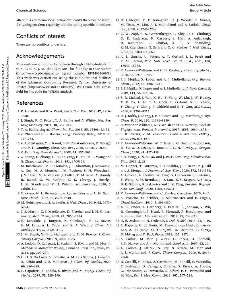
Acknowledgements
This work was supported by Janssen through a PhD studentshipto A. T. V. A. J. M. thanks EPSRC for funding to CCP-BioSim(http://www.ccpbiosim.ac.uk) [grant number EP/M022609/1].This work was carried out using the computational facilitiesof the Advanced Computing Research Centre, University ofBristol (http://www.bristol.ac.uk/acrc/). We thank Alan Gross-eld for his code for WHAM analysis.
References
1 R. Lonsdale and R. A. Ward, Chem. Soc. Rev., 2018, 47, 3816–3830.
2 J. Singh, R. C. Petter, T. A. Baillie and A. Whitty, Nat. Rev.Drug Discovery, 2011, 10, 307–317.
3 T. A. Baillie, Angew. Chem., Int. Ed., 2016, 55, 13408–13421.4 Z. Zhao and P. E. Bourne, Drug Discovery Today, 2018, 23,727–735.
5 A. Abdeldayem, Y. S. Raouf, S. N. Constantinescu, R. Moriggland P. T. Gunning, Chem. Soc. Rev., 2020, 49, 2617–2687.
6 Z. Pan, Drug News Perspect., 2008, 21, 357–362.7 Z. Zhang, D. Zhang, Y. Liu, D. Yang, F. Ran, M. L. Wang andG. Zhao, Arch. Pharm., 2018, 351, 1700369.
8 M. Roschewski, M. S. Lionakis, J. P. Sharman, J. Roswarski,A. Goy, M. A. Monticelli, M. Roshon, S. H. Wrzesinski,J. V. Desai, M. A. Zarakas, J. Collen, K. M. Rose, A. Hamdy,R. Izumi, G. W. Wright, K. K. Chung, J. Baselga,L. M. Staudt and W. H. Wilson, Sci. Immunol., 2020, 5,eabd0110.
9 C. Owen, N. L. Berinstein, A. Christodes and L. H. Sehn,Curr. Oncol., 2019, 26, e233–e240.
10 M. Gehringer and S. A. Laufer, J. Med. Chem., 2019, 62, 5673–5724.
11 J. S. Martin, C. J. MacKenzie, D. Fletcher and I. H. Gilbert,Bioorg. Med. Chem., 2019, 27, 2066–2074.
12 R. Lonsdale, J. Burgess, N. Colclough, N. L. Davies,E. M. Lenz, A. L. Orton and R. A. Ward, J. Chem. Inf.Model., 2017, 57, 3124–3137.
13 J. M. Smith, Y. Jami Alahmadi and C. N. Rowley, J. Chem.Theory Comput., 2013, 9, 4860–4865.
14 A. Lodola, D. Callegari, L. Scalvini, S. Rivara and M. Mor, inMethods in Molecular Biology, Humana Press Inc., 2020, vol.2114, pp. 307–337.
15 C. H. S. Da Costa, V. Bonatto, A. M. Dos Santos, J. Lameira,A. Leitao and C. A. Montanari, J. Chem. Inf. Model., 2020,60, 880–889.
16 L. Capoferri, A. Lodola, S. Rivara and M. Mor, J. Chem. Inf.Model., 2015, 55, 589–599.
5516 | Chem. Sci., 2021, 12, 5511–5516
17 D. Callegari, K. E. Ranaghan, C. J. Woods, R. Minari,M. Tiseo, M. Mor, A. J. Mulholland and A. Lodola, Chem.Sci., 2018, 9, 2740–2749.
18 C. W. Zapf, B. S. Gerstenberger, L. Xing, D. C. Limburg,D. R. Anderson, N. Caspers, S. Han, A. Aulabaugh,R. Kurumbail, S. Shakya, X. Li, V. Spaulding,R. M. Czerwinski, N. Seth and Q. G. Medley, J. Med. Chem.,2012, 55, 10047–10063.
19 A. L. Garske, U. Peters, A. T. Cortesi, J. L. Perez andK. M. Shokat, Proc. Natl. Acad. Sci. U. S. A., 2011, 108,15046–15052.
20 E. Awoonor-Williams and C. N. Rowley, J. Chem. Inf. Model.,2018, 58, 1935–1946.
21 J. I. Mujika, X. Lopez and A. J. Mulholland, Org. Biomol.Chem., 2012, 10, 1207–1218.
22 J. I. Mujika, X. Lopez and A. J. Mulholland, J. Phys. Chem. B,2009, 113, 5607–5616.
23 S. R. Malwal, J. Gao, X. Hu, Y. Yang, W. Liu, J. W. Huang,T. P. Ko, L. Li, C. C. Chen, B. O'Dowd, R. L. Khade,Y. Zhang, Y. Zhang, E. Oldeld and R. T. Guo, ACS Catal.,2018, 8, 4299–4312.
24 H. J. Kulik, J. Zhang, J. P. Klinman and T. J. Martınez, J. Phys.Chem. B, 2016, 120, 11381–11394.
25 E. Awoonor-Williams, A. G. Walsh and C. N. Rowley, Biochim.Biophys. Acta, Proteins Proteomics, 2017, 1865, 1664–1675.
26 S. R. Pereira, V. M. Vasconcelos and A. Antunes, FEBS J.,2013, 280, 674–680.
27 E. Awoonor-Williams, W. C. Isley, S. G. Dale, E. R. Johnson,H. Yu, A. D. Becke, B. Roux and C. N. Rowley, J. Comput.Chem., 2020, 41, 427–438.
28 S.-T. Bong, L. N.-S. Law and J. W.-F. Law, Prog. Microbes Mol.Biol., 2019, 2, 28.
29 M. Hopper, T. Gururaja, T. Kinoshita, J. P. Dean, R. J. Hilland A. Mongan, J. Pharmacol. Exp. Ther., 2020, 372, 331–338.
30 A. Liclican, L. Serani, W. Xing, G. Czerwieniec, B. Steiner,T. Wang, K. M. Brendza, J. D. Lutz, K. S. Keegan, A. S. Ray,B. E. Schultz, R. Sakowicz and J. Y. Feng, Biochim. Biophys.Acta, Gen. Subj., 2020, 1864, 129531.
31 E. Awoonor-Williams and C. Rowley, ChemRxiv, 2020, 1–11.32 A. Paasche, M. Schiller, T. Schirmeister and B. Engels,
ChemMedChem, 2010, 5, 869–880.33 A. T. Bender, A. Gardberg, A. Pereira, T. Johnson, Y. Wu,
R. Grenningloh, J. Head, F. Morandi, P. Haselmayer andL. Liu-Bujalski, Mol. Pharmacol., 2017, 91, 208–219.
34 N. B. Arslan and N. Ozdemir, J. Mol. Model., 2015, 21, 1–10.35 A. Kaptein, G. de Bruin, M. Emmelot-van Hoek, B. van de
Kar, A. de Jong, M. Gulrajani, D. Demont, T. Covey,D. Mittag and T. Barf, Blood, 2018, 132, 1871.
36 A. Lodola, M. Mor, J. Zurek, G. Tarzia, D. Piomelli,J. N. Harvey and A. J. Mulholland, Biophys. J., 2007, 92, 20.
37 A. Lodola, J. Sirirak, N. Fey, S. Rivara, M. Mor andA. J. Mulholland, J. Chem. Theory Comput., 2010, 6, 2948–2960.
38 R. Castelli, N. Bozza, A. Cavazzoni, M. Bonelli, F. Vacondio,F. Ferlenghi, D. Callegari, C. Silva, S. Rivara, A. Lodola,G. Digiacomo, C. Fumarola, R. Aleri, P. G. Petronini andM. Mor, Eur. J. Med. Chem., 2019, 162, 507–524.
© 2021 The Author(s). Published by the Royal Society of Chemistry
Copyright © 2022 FDOKUMEN