CNTF, a pleiotropic cytokine: emphasis on its myotrophic role
58 � 2009 The Authors • Journal compilation � 2009 Blackwell Munksgaard • Immunological Reviews 228/2009
Abdalla J. Mohamed
Liang Yu
Carl-Magnus Backesjo
Leonardo Vargas
Rani Faryal
Alar Aints
Birger Christensson
Anna Berglof
Mauno Vihinen
Beston F. Nore
C. I. Edvard Smith
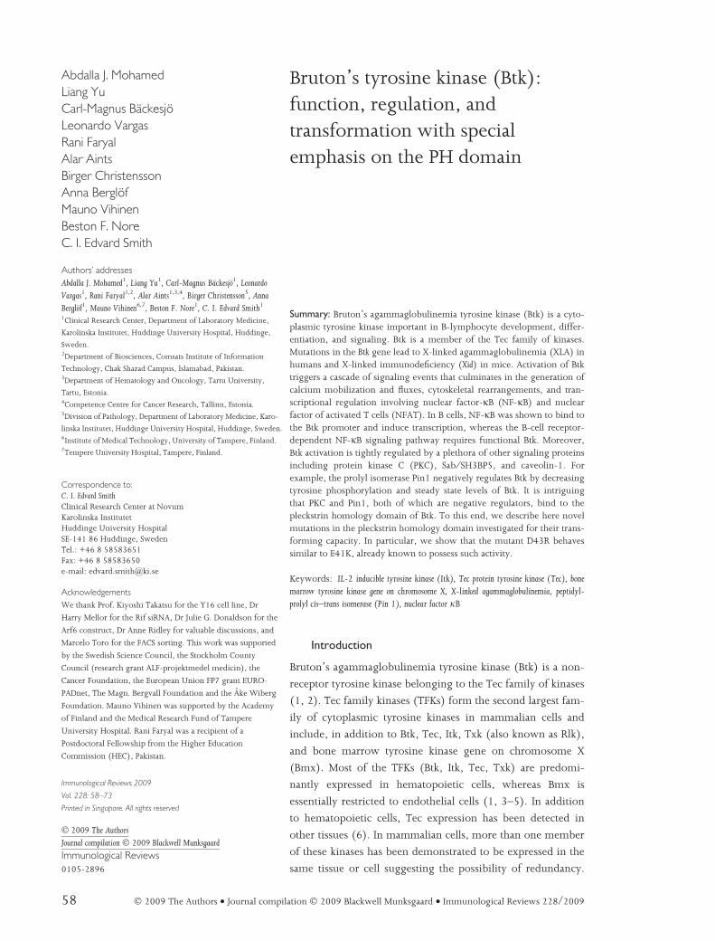
Bruton’s tyrosine kinase (Btk):function, regulation, andtransformation with specialemphasis on the PH domain
Authors’ addresses
Abdalla J. Mohamed1, Liang Yu1, Carl-Magnus Backesjo1, Leonardo
Vargas1, Rani Faryal1,2, Alar Aints1,3,4, Birger Christensson5, AnnaBerglof1, Mauno Vihinen6,7, Beston F. Nore1, C. I. Edvard Smith1
1Clinical Research Center, Department of Laboratory Medicine,
Karolinska Institutet, Huddinge University Hospital, Huddinge,
Sweden.2Department of Biosciences, Comsats Institute of Information
Technology, Chak Shazad Campus, Islamabad, Pakistan.3Department of Hematology and Oncology, Tartu University,
Tartu, Estonia.4Competence Centre for Cancer Research, Tallinn, Estonia.5Division of Pathology, Department of Laboratory Medicine, Karo-
linska Institutet, Huddinge University Hospital, Huddinge, Sweden.6Institute of Medical Technology, University of Tampere, Finland.7Tempere University Hospital, Tampere, Finland.
Correspondence to:
C. I. Edvard SmithClinical Research Center at NovumKarolinska InstitutetHuddinge University HospitalSE-141 86 Huddinge, SwedenTel.: +46 8 58583651Fax: +46 8 58583650e-mail: [email protected]
Acknowledgements
We thank Prof. Kiyoshi Takatsu for the Y16 cell line, Dr
Harry Mellor for the Rif siRNA, Dr Julie G. Donaldson for the
Arf6 construct, Dr Anne Ridley for valuable discussions, and
Marcelo Toro for the FACS sorting. This work was supported
by the Swedish Science Council, the Stockholm County
Council (research grant ALF-projektmedel medicin), the
Cancer Foundation, the European Union FP7 grant EURO-
PADnet, The Magn. Bergvall Foundation and the Ake Wiberg
Foundation. Mauno Vihinen was supported by the Academy
of Finland and the Medical Research Fund of Tampere
University Hospital. Rani Faryal was a recipient of a
Postdoctoral Fellowship from the Higher Education
Commission (HEC), Pakistan.
Immunological Reviews 2009
Vol. 228: 58–73
Printed in Singapore. All rights reserved
� 2009 The Authors
Journal compilation � 2009 Blackwell Munksgaard
Immunological Reviews
0105-2896
Summary: Bruton’s agammaglobulinemia tyrosine kinase (Btk) is a cyto-plasmic tyrosine kinase important in B-lymphocyte development, differ-entiation, and signaling. Btk is a member of the Tec family of kinases.Mutations in the Btk gene lead to X-linked agammaglobulinemia (XLA) inhumans and X-linked immunodeficiency (Xid) in mice. Activation of Btktriggers a cascade of signaling events that culminates in the generation ofcalcium mobilization and fluxes, cytoskeletal rearrangements, and tran-scriptional regulation involving nuclear factor-jB (NF-jB) and nuclearfactor of activated T cells (NFAT). In B cells, NF-jB was shown to bind tothe Btk promoter and induce transcription, whereas the B-cell receptor-dependent NF-jB signaling pathway requires functional Btk. Moreover,Btk activation is tightly regulated by a plethora of other signaling proteinsincluding protein kinase C (PKC), Sab ⁄ SH3BP5, and caveolin-1. Forexample, the prolyl isomerase Pin1 negatively regulates Btk by decreasingtyrosine phosphorylation and steady state levels of Btk. It is intriguingthat PKC and Pin1, both of which are negative regulators, bind to thepleckstrin homology domain of Btk. To this end, we describe here novelmutations in the pleckstrin homology domain investigated for their trans-forming capacity. In particular, we show that the mutant D43R behavessimilar to E41K, already known to possess such activity.
Keywords: IL-2 inducible tyrosine kinase (Itk), Tec protein tyrosine kinase (Tec), bonemarrow tyrosine kinase gene on chromosome X, X-linked agammaglobulinemia, peptidyl-prolyl cis–trans isomerase (Pin 1), nuclear factor jB
Introduction
Bruton’s agammaglobulinemia tyrosine kinase (Btk) is a non-
receptor tyrosine kinase belonging to the Tec family of kinases
(1, 2). Tec family kinases (TFKs) form the second largest fam-
ily of cytoplasmic tyrosine kinases in mammalian cells and
include, in addition to Btk, Tec, Itk, Txk (also known as Rlk),
and bone marrow tyrosine kinase gene on chromosome X
(Bmx). Most of the TFKs (Btk, Itk, Tec, Txk) are predomi-
nantly expressed in hematopoietic cells, whereas Bmx is
essentially restricted to endothelial cells (1, 3–5). In addition
to hematopoietic cells, Tec expression has been detected in
other tissues (6). In mammalian cells, more than one member
of these kinases has been demonstrated to be expressed in the
same tissue or cell suggesting the possibility of redundancy.

For example, T cells express Tec, Itk, and Txk, while B cells
use Btk and Tec. In addition, expression of Itk and Btk is much
higher than Tec in T and B lymphocytes, respectively. The
biological function and intracellular signaling of Tec family
kinases have been described in several reports (3, 7–11).
Gene-targeted animals deficient for Btk or Itk have defined
phenotypes, while single knockouts of Tec or Txk do not
show overt changes (12–18). By contrast, certain knockouts
involving combinations of two members (Btk ⁄Tec and
Itk ⁄Txk) have been shown to display profound phenotypic
changes (16, 19). TFKs have been reported to signal through
several cell surface receptors. A common cellular response
triggered by TFK signaling is a dramatic cytoskeletal remodel-
ing involving actin polymerization (2, 11, 20).
Btk is critical for B-cell development, differentiation,
and signaling (21–23). Moreover, Btk expression is
assumed to be a prerequisite for B-cell proliferation and
survival. Consequently, Btk-deficient B lymphocytes fail to
reach the mature state and are presumably doomed to
premature death. Btk is the only member of the TFKs
reported to be associated with human disease. Thus, indi-
viduals harboring loss-of-function mutations in the gene
encoding Btk virtually lack circulating B lymphocytes, are
unable to generate immunoglobulins of all classes, and
therefore cannot mount humoral immune responses. This
primary immunodeficiency is named X-linked agamma-
globulinemia (XLA). Similarly, a spontaneous mutation in
the mouse-Btk gene (23) leads to X-linked immunodefi-
ciency (Xid), a considerably milder condition than XLA,
while the very same mutation in humans causes classical
XLA, suggesting species differences (23, 24).
In this review, we focus on the function and regulation of
Btk, and in particular, discuss the achievements made over the
past decade with respect to the biochemistry of this remark-
able protein. We also include primary data regarding several
aspects of Btk biology, including the existence of a truncated
form of Btk, as well as novel, site-directed mutants with trans-
forming capability.
Genetics and expression of Btk
The BTK gene consists of 19 exons and spans approximately
37.5 kb on the human X chromosome (25). Scientists on both
sides of the Atlantic, including our laboratory, independently
cloned the gene in 1992 (21, 22). Over 800 mutations affect-
ing Btk have been reported to the international mutation data-
base designated BTKbase (http://bioinf.uta.fi/BTKbase),
including deletions encompassing also three additional genes
(26). Similar to other genetic diseases, the disease-causing
mutations that occur in Btk include missense, nonsense, splice
site, deletions ⁄ insertions and are literally spread over the
entire gene (27–34). However, no disease-related missense
mutation in the Src homology 3 (SH3) domain has been
reported.
Btk is predominantly expressed in B lymphocytes but not in
plasma cells (35). Btk expression in the B-cell lineage is also
developmentally regulated, with marrow-derived hematopoi-
etic stem cells, common lymphoid progenitor cells, develop-
ing B and myeloid lineages showing the highest levels,
whereas resting mature cells prior to activation have reduced
cellular Btk (36). Moreover, with the exception of T lympho-
cytes, all other hematopoietic lineages have been shown to
express Btk. As B lymphocytes are the only cells known to be
affected in XLA, the physiological significance of Btk expres-
sion in other cell types remains to be established. Nonetheless,
there are multiple reports describing defects in other cell types
such as platelets and macrophages (37–39). Recent studies
show that osteoclast development is impaired in Btk-deficient
mice (40, 41). Although changes have been reported to occur
in different cell types of the hematopoietic system, as men-
tioned, the severity in B lymphocytes is far more pronounced.
Thus, in humans Btk is indispensable for B-cell development.
Second, the molecular mechanism(s) underlying the differ-
ence in the degree of severity in phenotype between XLA and
Xid is not known. It is known, however, that Tec partially can
replace Btk in mice (16), while in humans Btk deficiency
alone causes severe disease in spite of the presence of Tec in
human B lymphocytes. In transfection experiments involving
a Btk-deficient chicken B-cell line (DT40-derived), Tec was
demonstrated to be capable of restoring activity and trigger
downstream signaling (42). This cell line is deficient in
calcium signaling (43, 44) and also displays cytoskeletal
features characterized by formation of shorter membrane
protrusions and the degree of aggregation is much less than in
wildtype cells (20).
Btk is the only member in the Tec family reported to be
associated with pathogenesis in humans. In the absence of
Btk, B-cell development is impaired due to a block
between the pro- and pre-B-cell stages leading to severe
reduction of mature B lymphocytes. Accordingly, individu-
als who have loss-of-function mutations in the Btk gene
develop XLA, the hallmark of which is an essentially
complete absence of circulating B cells. These patients are
incapable of producing antibodies and are subject to
recurrent bacterial and enteroviral infections (45). There
are also mouse models of this disease, one of which is Xid.
Mohamed et al Æ Bruton’s tyrosine kinase (Btk)
� 2009 The Authors • Journal compilation � 2009 Blackwell Munksgaard • Immunological Reviews 228/2009 59

Xid manifests as a result of a spontaneous mutation of a
CpG site in the Btk gene changing residue 28 from an argi-
nine to a cysteine (R28C) in the pleckstrin homology (PH)
domain (23). While the identical mutation leads to classi-
cal XLA in humans (24), Xid mice maintain about half the
number of splenic B lymphocytes. The second model,
genetically modified Btk knockout mice, has a similar
phenotype as Xid mice (17, 18). When female mice
heterozygous for the R28C missense mutation and the null
allele were analyzed, we found that none of the alleles
conferred any B-cell survival advantage over the other,
suggesting that this missense mutation functions as a bona
fide null allele (46).
Biochemistry of Btk
It is generally considered that mammals and birds have all five
TFK family members, although only a single avian genome
has been sequenced. Using bioinformatics tools, we recently
demonstrated that a TFK ancestor exists in the unicellular
choanoflagellate Monosiga brevicollis, which is the closest known
relative to metazoans with a sequenced genome (47). The
analysis of the genomes of sponges, insects, hagfish, and frogs
suggests that these species encode a single TFK. TFKs are
non-receptor kinases containing several unique domains char-
acteristic for signaling proteins (1). Btk comprises of several
domains from the N-terminus: the PH, Tec homology (TH),
SH2, SH3, and kinase (SH1) domains (Fig. 1). Each of these
domains has the potential to interact with a plethora of
proteins critical for intracellular signaling. Moreover,
functional association of Btk with many of its partners is
crucial for its activation and regulation. Here we want to
highlight some of the key proteins that execute this.
Btk is a metalloprotein enzyme requiring Zn2+ for optimal
activity and stability. Binding and coordination of Btk to the
Zn2+ion is mediated by a highly conserved zinc finger motif,
also called the Btk motif, located in the TH domain (89, 90).
Mutations affecting Zn2+binding lead to the generation of
extremely unstable protein (31, 61).
Fig. 1. Schematic representation of the Bruton’s tyrosine kinase (Btk) promoter and Btk domains with all known interacting/regulatingpartners. Domains shown: pleckstrin homology (PH); Tec homology (TH), [Btk homology region (BH) and polyproline region (PPR)]; Src-homology3 (SH3); Src-homology 2 (SH2); Kinase (SH1), tyrosine kinase domain. Regulatory phosphorylation sites are indicated by arrows, phosphotyrosinespY223 and pY551 are in red color, and phosphoserines pS21, pS115, and pS180 in blue. The black box contains putative partners for which the siteof interaction has not been identified (20, 31, 48–60, 62–88).
Mohamed et al Æ Bruton’s tyrosine kinase (Btk)
60 � 2009 The Authors • Journal compilation � 2009 Blackwell Munksgaard • Immunological Reviews 228/2009

Btk is transcribed from a single gene, and there is no
evidence of alternative splicing. Yet, a shorter, presumably
truncated isoform of the protein is often observed in sodium
dodecyl sulfate polyacrylamide gel electrophoresis (SDS-PAGE)
when an SH3 or C-terminus specific antibody is utilized (Abdalla
J. Mohamed, Liang Yu, C. I. Edvard Smith, unpublished data).
This shorter isoform of Btk is expressed both in endogenous
and in transfected cell lines and seems to be highly unstable.
In fact, in some cell lines including primary B lymphocytes, it
is virtually undetectable. Although the nature and origin of
this faster migrating band is not known, we think that it is
generated as a result of alternative translation initiation or pro-
teolysis. In addition to the first methionine (ATG), which as
customary is often utilized, Btk contains several additional
in-frame translation initiation codons that may be responsi-
ble for producing the truncated form. Second, missense muta-
tions that modify the first ATG codon have been reported.
Although patients harboring these types of mutations present
classical XLA, full-length Btk protein, albeit at much lower
levels, could readily be detected in peripheral blood mono-
nuclear cells (PBMCs) (91). This result is consistent with our
own findings, whereby mutating the first ATG to TTG leads to
the production of not only the shorter truncated protein
but also full-length Btk (Abdalla J. Mohamed, Liang Yu,
C. I. Edvard Smith, unpublished data). It has been reported
that the translation machinery of eukaryotic genes can some
times utilize a non-ATG codon but with much less efficiency
(92–94).
Btk activation
The molecular mechanisms that underlie activation of Btk are
currently not fully understood. However, it is generally
believed that post-translational modifications as well as sub-
cellular localization could play key roles. In steady state, Btk is
predominantly cytosolic. Following B-cell receptor (BCR)
engagement, Btk translocates to the plasma membrane where
it gets phosphorylated at Y551 by a Src family kinase (SFK),
presumably Lyn. Phosphorylation of Btk at this particular tyro-
sine, which is in the catalytic domain, is followed by an auto-
phosphorylation event involving another tyrosine in the SH3
domain, Y223. Notably, the initial phosphorylation at Y551
primes the protein for activation, while the role of phosphory-
lation at Y223 is less clear (95). In close cooperation with the
adapter protein B-cell linker protein (BLNK), also known as
SH2 domain-containing leukocyte protein of 65 kDa (SLP-
65), Btk subsequently phosphorylates phospholipase Cc2
(PLCc2), igniting the downstream signaling pathway. Since
Btk is a multidomain protein, it has also the ability to bring
together a diverse array of signaling proteins into a single
platform. In primary B lymphocytes, Btk is almost always
non-phosphorylated and is thus catalytically inactive. How-
ever, many of the experiments that address phosphorylation
of Btk and other tyrosine kinases have been conducted on
heterologous cell types, such as COS-7 and HEK 293T, as well
as transformed B cells. Although some important information
relevant to these questions can be obtained using these cells,
there are two fundamental problems associated with both of
these systems. First, heterologous cell lines, though easily
tranfectable, do not endogenously express Btk or other B-cell-
specific proteins required for activation and signaling. Second,
as most utilized B-cell lines are transformed, they may not
represent the genuine milieu that prevails in primary cells.
Nonetheless, COS-7 and HEK 293T have been invaluable in
the biochemical and functional characterization of cloned
genes.
In spite of the accumulation of a great amount of data in
recent years, the precise mechanism(s) underlying phosphor-
ylation and activation of Btk has not been established. How
does Btk get phosphorylated in the first place? Why does it get
phosphorylated in some cells but not in others? We need to
understand these and similar questions to get deeper insight
into the dynamics of Btk activation. For instance, whereas Btk
and Tec are robustly phosphorylated in the COS-7 cell line, Itk
is not (Abdalla J. Mohamed, Liang Yu, C. I. Edvard Smith,
unpublished data). This situation is reminiscent to that of Syk
and f-associated protein of 70 kDa (ZAP-70) (96). It is
intriguing that these structurally similar kinases (Btk ⁄ Itk,Syk ⁄ZAP-70) behave so differently in the same cellular
environment. It is conceivable that a specific kinase or a phos-
phatase can differentiate between Btk and Itk in the COS-7 cell
line. Second, it is also possible that the two proteins localize
differently within the cell, leading to phosphorylation of one
but not the other. Third, as Btk and Itk differ in their primary
structure (approximately 50% sequence identity), post-
translational modification may be an important process in reg-
ulating their activity. Last but not least, although similar in
structure, the two proteins may still adopt different conforma-
tions in the same milieu.
Btk and Toll-like receptor signaling
The ability of Btk to interact with members of Toll-like recep-
tor (TLR) family (namely TLR4, TLR6, TLR8, and TLR9) and
key proteins from TLR signaling pathways, such as myeloid
differentiation protein 88 (Myd88), MyD88 adapter-like
protein (Mal), and interleukin-1 receptor (IL-1R)-associated
kinase-1 (IRAK-1), suggest the importance of Btk as a compo-
Mohamed et al Æ Bruton’s tyrosine kinase (Btk)
� 2009 The Authors • Journal compilation � 2009 Blackwell Munksgaard • Immunological Reviews 228/2009 61

nent of TLR pathways and that this kinase therefore plays an
important role in the function of immune cells of innate as
well as adaptive immunity (97–99).
We have investigated the potential role of Btk in the TLR9
activation and the subsequent production of pro-inflamma-
tory cytokines such as IL-6, tumor necrosis factor-a (TNF-a)
and IL-12p40 (13). Our data show that Btk-deficient B cells
respond more efficiently to CpG-DNA stimulation, producing
significantly higher levels of pro-inflammatory cytokines
when compared to wildtype cells but less of the inhibitory
cytokine IL-10. Quantitative reverse transcriptase polymerase
chain reaction (RT-PCR) analysis presented in our previous
work shows that IL-27 mRNA was significantly increased in
Btk-deficient B cells after CpG-DNA stimulation. In this study,
we also demonstrated important differences in CpG respon-
siveness between transitional 1 (T1) and T2 B-cell survival
and maturation. Interestingly, CpG-DNA stimulation induced
a time-dependent accumulation of transitional T2 B cells in
both Btk-deficient and normal B cells.
Furthermore, stimulation of B cells with TLR9 ligand
CpG leads to transient phosphorylation of Btk, and in the
absence of Btk, TLR9-induced proliferation of B cells is
impaired (100). Collectively, these data suggest that Btk is
a critical molecule in regulating TLR9 activation in splenic
B cells. Finally, elucidation of the physiological role of
TLRs in B cells may provide important clues for under-
standing the molecular mechanisms behind inflammation
and immunity.
Subcellular localization of Btk
Plasma membrane localization of Btk is a critical step in the
phosphorylation and activation of Btk. Following BCR engage-
ment by antigen, phosphatidylinositol-3-kinase (PI3K) is
activated, leading to the generation of the phosphoinositide
phosphatidylinositol-3,4,5-trisphosphate (PIP3). Accumula-
tion of PIP3 in the inner surface of the plasma membrane
results in the recruitment of various signaling proteins includ-
ing Btk. Subsequent binding of the PH domain of Btk to PIP3
is a prerequisite for its activation. Although the exact molecu-
lar mechanisms responsible for targeting Btk to the cell surface
are not well known, the PH domain is thought to play a key
role (20, 101). Deletion of the PH domain renders Btk mainly
cytoplasmic, although a substantial amount translocates to the
nucleus (102). Also, for reasons that are not clear to us, steady
state levels of this form of the protein are higher than wildtype
Btk. Further, though such a PH domain-less version of Btk is
robustly phosphorylated, it cannot properly signal. Even a
subtle alteration in the PH domain could have disastrous
effects on the overall function of the Btk protein. This is sub-
stantiated by the fact that the R28C mutation in the PH
domain (Fig. 2) leads to classical XLA, and, moreover, there
are additional missense mutations in this domain that cause
disease (28, 103).
Although predominantly cytoplasmic, a small fraction of
Btk can at any given time be detected in the nucleus, suggest-
ing that it is a nucleocytoplasmic shuttling protein (102).
Thus, it is becoming increasingly clear that Btk not only trans-
locates to the nucleus but also that it seems to be rapidly
exported. In some B cells, the fraction of Btk that is inside the
nucleus can be as high as 50% (Abdalla J. Mohamed, Liang
Yu, C. I. Edvard Smith, unpublished data). The molecular
mechanism(s) responsible for nuclear localization and ⁄or
export of Btk as well as its potential role in the nucleus remain
to be established. Although motifs resembling classical nuclear
localization signals (NLS) appear in the PH domain, they do
not seem to play a role in the nuclear translocation process.
Similarly, nuclear shuttling of Btk is independent of phos-
phorylation, since a kinase-defective mutant can also translo-
cate into the nucleus. Of note, deletion of the PH–TH domain
doublet or a truncation extending into the N-terminal part of
the SH3 domain renders Btk slightly more nuclear (102). In
Fig. 2. The structure of the pleckstrin homology (PH) domain andthe Btk motif corresponding to residues 1–170 of the humanBruton’s tyrosine kinase (Btk). The figure is based on the experimentalstructure in Protein Data Bank (entry 1bwn). Cyan ribbon runs alongbackbone of the molecule, except for orange for the C-terminal a-helix,and blue for the Btk motif of TH domain. Serines 21 (top) and 115(bottom), which can be phosphorylated, are in magenta. The yellow sidechains represent residues glutamate 41 (right), aspartate 43 (middle),and glutamate 45 (left), respectively. The arginine 28 residue is in pink.IP4 molecules are green. The long Btk-specific loop is not visible in thisstructure. The blue sphere denotes the Zn2+ion in the Btk motif.
Mohamed et al Æ Bruton’s tyrosine kinase (Btk)
62 � 2009 The Authors • Journal compilation � 2009 Blackwell Munksgaard • Immunological Reviews 228/2009

addition, mutation of Y223, but not Y551, increases the frac-
tion of Btk in the nucleus (Abdalla J. Mohamed, Liang Yu,
C. I. Edvard Smith, unpublished data).
Nuclear export of Btk has been shown to be mediated by
the nuclear export receptor CRM1 (exportin), since treatment
of cells with the antibiotic leptomycin B leads to nuclear arrest
of Btk (102). Thus, these data suggest that Btk is continuously
shuttling between the cytosol and the nucleus. Furthermore,
we have conclusive evidence that Btk interacts with CRM1
both in endogenous and in transfected settings (Abdalla J.
Mohamed, Liang Yu, C. I. Edvard Smith, unpublished data).
At present, we do not know the nuclear export signals that Btk
utilizes to exit from the nucleus. Very little is known about
the function of Btk inside this compartment. However, as
nuclear Btk is less phosphorylated as compared with the form
residing in the cytosol, it is possible that a nuclear phosphatase
is responsible and that this could be of functional importance
for the regulation of this kinase. Alternatively, Btk may have
unknown nuclear substrates. As described in the section ‘Role
of Btk in transcription’, there are reports suggesting a role for
Btk in the activation of the transcription factor Bright. This
activity is independent of Btk’s catalytic function, as Bright
does not seem to be phosphorylated by Btk (75).
For signaling proteins in particular, nucleocytoplasmic shut-
tling could be one way of regulating and ⁄ or fine-tuning their
activity. It creates a forum for compartmentalization. In the
case of kinases like Btk, activation could be elicited following
signals at the plasma membrane, whereas inactivation and
termination of signaling may occur in the nucleus.
Plasma membrane localization of Btk
As discussed in the previous section, plasma membrane associ-
ation of Btk is a prerequisite for its phosphorylation and
subsequent activation. Thus, to properly signal during BCR
engagement, Btk has to traffic first to the cell surface. As the
PH domain is thought to mediate plasma membrane targeting
of Btk, it is not surprising that mutations in this domain have
profound effects on this activity. For instance, a great majority
of the disease-causing mutations in the PH domain of Btk
frequently affect arginine 28 (32). This alters the positive
charge on the surface of the PH domain (Fig. 2), abolishing its
binding to membrane-resident lipid ligands and consequently
the recruitment to the plasma membrane after stimulation of
the cell (61, 104).
By contrast, when the glutamate at position 41 in the PH
domain of Btk is substituted by lysine (E41K), Btk achieves a
gain-of-function state with increased membrane targeting and
tyrosine phosphorylation level (105). In addition, this
gain-of-function mutation has been shown to display robust
transformation potential in the anchorage-dependent soft agar
assay. According to the structure of the PH domain, the E41K
mutant binds to two PtdIns(3, 4, 5)P3 molecules compared
with the wildtype that binds to only one (104). Therefore, it
is possible that this type of mutation enables Btk to spend
more time at the inner surface of the plasma membrane, lead-
ing to sustained activation of the protein. To determine
whether creation of additional positively charged amino acids
in the PH domain leads to increased activity of Btk, we
mutated asparagine 43 (D43) and glutamate 45 (E45) to
either lysine (K) or arginine (R) and tested them, as described
in the next sections. The E41K mutant was also combined
with E45K or E45R. According to the X-ray crystal structure of
Btk, these residues are all located on the surface of the PH
domain, within the region that binds to the cell membrane
(Fig. 2).
Using a Btk–green fluorescence protein (GFP) fusion
construct, subcellular localization of the mutants as well as
wildtype and the kinase inactive form of Btk were character-
ized in HeLa cells. As shown in Fig. 3, wildtype Btk displays
typical cytoplasmic distribution in these cells. However, as
expected, a small fraction of Btk is also detected in the
nucleus. We had shown previously that overexpression of the
Btk mutant E41K in COS-7 cells results in a pronounced
induction of extensive lamellipodia and some filopodia forma-
tions (20). In sharp contrast, HEK 293 cells transfected with
the same construct predominantly display filopodia formation
(Abdalla J. Mohamed, Liang Yu, C. I. Edvard Smith, unpub-
lished data). In HeLa cells, formation of ‘hairy’ actin filament
bundles or microspike structures were the most pronounced
form of cytoskeletal alterations found, following expression of
the E41K mutant in these cells. The nature of the cytoskeletal
structures formed in the different cell lines depends largely on
the endogenous expression of the small GTPases Rac, Rho,
and Cdc 42. For example, Rac is probably the major small
GTPase expressed in COS-7, which could explain the forma-
tion of lamellipodia in this cell line. In fact, lamellipodia for-
mations in COS-7 were successfully blocked with dominant
negative Rac1 (20).
With the exception of E45K and E45R, all other PH domain
mutants displayed a pattern similar to E41K, and this pattern
was more pronounced after serum stimulation (Fig. 3, data
not shown). The cytoskeletal changes in the form of protru-
sions induced by expressing E45K and E45R were much less
pronounced. The most striking difference, however, was seen
after introducing the D43R mutant. The cells get rounder and
smaller following serum stimulation but to some degree keep
Mohamed et al Æ Bruton’s tyrosine kinase (Btk)
� 2009 The Authors • Journal compilation � 2009 Blackwell Munksgaard • Immunological Reviews 228/2009 63

their microspike-like structures. As mentioned earlier, in
steady state, Btk is predominantly cytoplasmic, although
sometimes it can also show a punctuate staining all over the
cell. In serum-stimulated cells, Btk translocates mainly to a
perinuclear compartment, seemingly residing on filaments
reaching to the plasma membrane. The kinase-inactive form
of Btk failed to induce the microspike formations, indicating
that both membrane association and kinase activity are impor-
tant for this phenotype (Abdalla J. Mohamed, Liang Yu, C. I.
Edvard Smith, unpublished data). In addition, membrane tar-
geting was not clearly altered for the PH domain mutants
D43K and D43R compared with E41K, whereas membrane
tethering of the E45 mutants was reduced (Abdalla J.
Mohamed, Liang Yu, C. I. Edvard Smith, unpublished data).
Table 1 summarizes the cell biological and functional charac-
terization of the different PH domain mutants.
The structures formed by the expression of E41K in COS-7
cells show similarities to the cytoskeletal structures appearing
when a constitutively active form of Rif or Arf6 is expressed
in cells (106, 107). Co-transfection of siRNA targeting Rif or
dominant negative form of Arf6 with Btk E41K did not block,
however, the microspike formation (Abdalla J. Mohamed,
Liang Yu, C. I. Edvard Smith, unpublished data).
Transforming potential of Btk
An important question is whether Btk is oncogenic or not.
This issue has been debated since the cloning of the BTK gene
and has not been conclusively resolved. There are numerous
reports implicating a number of tyrosine kinases in cancer
(108–110). Most of these concern receptor tyrosine kinases
belonging to the growth factor receptor family. However,
there are also non-receptor tyrosine kinases that are critical for
cancer development, the most well-known being Abl. As
many of the tyrosine kinases are important in cell proliferation
and survival, they are also prone to support tumor growth. In
addition, some tyrosine kinases are important in cell migra-
tion, a property of cancer cells. For example, SFKs are believed
to play a key role in growth and metastasis of many types of
human malignancies. Although Btk is known to be crucial for
important biological process in B-cell development, such as
proliferation and survival, its relation to cancer is complex.
Thus, there are at least five rather different scenarios where
Btk has been implicated in tumor formation: (i) inactivating
mutations of Btk found in lymphoid tumors, (ii) inactivating
mutations of Btk as a cause of colorectal cancer, (iii) a role for
splice variants of Btk in leukemia, especially chronic myeloge-
nous leukemia, (iv) Btk-defective mice are resistant to certain
experimentally induced tumors, such as the pristine-induced
plasmacytomagenesis (111), suggesting that Btk-deficient B
cells are less likely to get transformed, and (v) activating
mutations causing a transforming phenotype in experimental
Steady state
Btk
D43R
E41K
Stimulated
Fig. 3. Confocal images of HeLa cells transfected with expressionconstructs for Bruton’s tyrosine kinase (Btk)–green fluorescenceprotein (GFP). Wildtype Btk (upper panel); D43R (middle panel); E41K (lowerpanel). The left panels show serum-starved cells and the panels to the rightafter serum stimulation. Red indicates F-actin stained with phalloidinconjugated to rhodamine, and blue indicates nucleic acid, stained withDAPI.
Table 1. Characteristics of transforming Btk mutants
Btk mutant Btk localization* Microspikes*Transformingcapacity�
WT Cytoplasmic ) )E41K Cytoplasmic ⁄ membrane ++ +E41R Cytoplasmic ⁄ membrane ++ )D43K Cytoplasmic ⁄ membrane ++ )D43R Cytoplasmic + ⁄ ) +E45K Cytoplasmic ⁄ membrane + )E45R Cytoplasmic ) )E41K + E45K Cytoplasmic ⁄ membrane ++ +E41K + E45R Cytoplasmic + +
*Data are from HeLa cells transfected with the various Bruton’s tyrosinekinase (Btk)–green fluorescence protein (GFP) constructs. Forty hourslater, subcellular localization and microspike formation was determinedusing confocal microscopy. Perinuclear localization was normally seen.�The transforming potential of the mutants was investigated in the IL-5-dependent Y16 mouse B-cell line. Transformation of these cells rendersthem IL-5 independent.
Mohamed et al Æ Bruton’s tyrosine kinase (Btk)
64 � 2009 The Authors • Journal compilation � 2009 Blackwell Munksgaard • Immunological Reviews 228/2009

settings, as already discussed, where Btk achieves a
gain-of-function state with increased membrane targeting and
tyrosine phosphorylation (105).
Concerning (i) the inactivating mutations of Btk found in
lymphoid tumors, there are both reports claiming such an
association in human leukemia (112) and others that did not
find evidence for this association (113). The strongest argu-
ment in support comes from knockout mice with inactivated
BLNK. Here, it is very clear that combining this null mutation
with Btk deficiency potentiates tumor development (114,
115). Regarding (ii) inactivating mutations of Btk as a cause
of colorectal cancer, there are several such reports (reviewed
in 45, 116), and very recently two additional patients were
described (117). Owing to that Btk is normally not expressed
in colon-derived cells, it is likely that any association is indi-
rect and caused for example by an altered bacterial flora,
which subsequently predisposes for cancer.
With regard to (ii) a role for splice variants of Btk in leuke-
mia, especially chronic myelogenous leukemia, Btk has been
implicated as tumor-inducing through an effect dependent on
different splice variants induced by the fusion protein Bcr–Abl
(118, 119). The interpretation of these findings was that lack
of Btk expression, or expression of dominant-negative splice
variants, in B-cell precursor leukemia cells can inhibit differ-
entiation beyond the pre-B-cell stage and protect from radia-
tion-induced apoptosis. However, while the SH3 domain of
Btk may serve as a substrate for the Abl cytoplasmic tyrosine
kinase (120, 121), a recent study found no evidence for a
connection (121). These authors reported that inhibition of
Btk did not affect the ability of Bcr–Abl to transform primary
murine hematopoietic cells in colony-forming and B-cell
transformation assays.
Owing to the remarkable effects of the Btk PH domain
mutants in HeLa cells, the interest was to further characterize
the mutants and determine how they behave in B cells. We
therefore chose to use the mouse B-cell line Y16. This cell line
strictly requires the cytokine IL-5 for optimal growth. Thus,
in the absence of IL-5, the cells cannot proliferate and eventu-
ally go into apoptosis. Next, we stably expressed the different
mutants in the form of Btk–GFP fusions in Y16 cells. GFP+
cells were then sorted from the GFP) cells by fluorescence
activated cell sorting (FACS) and were studied in a digital con-
focal microscopy. In the Y16 cells stably expressing the Btk
mutant E41K, we could readily detect membrane protrusions
(Abdalla J. Mohamed, Liang Yu, C. I. Edvard Smith, unpub-
lished data). By contrast, there were no visible protrusions in
cells expressing the other mutants. Interestingly, the E41R
mutant showed clear accumulation in one or two cytoplasmic
spots located close to the nucleus, which could indicate co-
localization with centriols in the microtubule organizing cen-
ters (MTOCs) (Abdalla J. Mohamed, Liang Yu, C. I. Edvard
Smith, unpublished data). Similar observations were found in
the rat basophilic cell line RBL 2H3 (Abdalla J. Mohamed,
Liang Yu, C. I. Edvard Smith, unpublished data). In addition,
we noticed an enhanced proliferation of some of the mutants,
as indicated by the rapid acidification of cell culture medium.
Analysis by cell count and the colorimetric cell proliferation
reagent WST-1 showed a higher proliferation rate of E41K,
D43R, and the double mutants E41K + E45K or E45R com-
pared with the wildtype Btk–GFP (Abdalla J. Mohamed, Liang
Yu, C. I. Edvard Smith, unpublished data). This result is con-
sistent with previous findings showing that the E41K mutant
can support growth of Y16 cells in the absence of IL-5 (105).
To further determine whether these mutants could also
support IL-5-independent growth of the cells, we propagated
the cells in the presence or absence of IL-5. The data clearly
show proliferation of cells expressing E41K, D43R, and
E41K +E45R, even though IL-5 was excluded from the
growth medium, indicating that these mutants have a trans-
forming potential (Fig. 4, data not shown). Owing to that
E41K induces an increased proliferation rate, it is likely that
this is the dominating feature also of the double mutants, as
neither of the E45 mutations further alters the effect. By con-
trast, cell proliferation following introduction of the E45
mutants alone was more similar to cells expressing wildtype
Btk. Interestingly, the D43R mutant induced a pronounced
cell-proliferative effect. This finding may suggest that the E45
Fig. 4. Growth curve of Y16 cell lines expressing the different formsof Bruton’s tyrosine kinase (Btk)–green fluorescence protein (GFP).Y16 cells stably expressing the different forms of Btk–GFP were culturedin the absence of IL-5 over 6 days. The OD values obtained using theWST-1 reagent, during 1 h of incubation, are proportional to the num-ber of living cells as detected by a colorimetric cell proliferation assay.
Mohamed et al Æ Bruton’s tyrosine kinase (Btk)
� 2009 The Authors • Journal compilation � 2009 Blackwell Munksgaard • Immunological Reviews 228/2009 65

mutants are located too far away from the active membrane-
binding region of the PH domain and therefore do not con-
tribute to enhanced activation, in spite of changing the net
surface charge of Btk similar to the E41 and D43 mutants.
Moreover, as the E41 + E45 double mutants have an
increased effect whereas the E45 single mutants do not, the
overall structural conformation of the PH domain with an E45
mutation is probably not altered. The results are summarized
in Table 1.
Enhanced electrostatic polarization seems to increase the
affinity for PtdIns(4,5)P2 and PtdIns(3, 4, 5)P3 (101, 122). In
the crystal structure of the Btk PH domain with the E41K
mutation, soluble Ins(1, 3, 4, 5)P4 was used to mimic the
PtdIns(3, 4, 5)P3. A second PtdIns(3, 4, 5)P3 bound to the
E41K mutant, indicating a role of positively charged regions
in binding phosphoinositides. E41K has also been shown to
transform fibroblasts in vitro (105, 123). When overexpressed
in B lymphocytes, it disrupts the architecture of lymphatic
tissues (124). Thus, the extension of electrostatic polarization
with positively charged amino acids in transgenic mice over-
expressing the Btk E41K results in a more severe B-cell defect
than the Xid phenotype (18). This mutant not only blocks the
development of follicular recirculating B cells but also causes
an enhanced blast formation of splenic B cells in vitro (124).
Recent findings show an increased pre-B cell tumor incidence
in mice defective in both Btk and BLNK and that Btk E41K
could substitute for SLP-65 as a tumor suppressor (114).
Regulation of Btk function – an overview
Much of the regulation of Btk function remains elusive. More-
over, information about structure–function relationship of
this cytoplasmic tyrosine kinase is scanty. Although a com-
plete and thorough understanding of the three-dimensional
structure of Btk is not available, experiments indicate that the
molecule is extended under certain conditions and in steady
state exists as a monomer (125, Abdalla J. Mohamed, Liang
Yu, C. I. Edvard Smith, unpublished data). However, crystal
structures formed using the PH domain of Btk have suggested
that dimers could be formed in the membrane (61, 126).
Unlike SFKs, Btk does not possess a negative regulatory
C-terminal tyrosine to control its activity. Also there is no
evidence of intra or intermolecular interaction(s) involved
in the regulation of full-length Btk, while there is ample
evidence for such interactions when various forms of TH–SH3
domain doublets are studied (127–129).
Thus, it may well be that Btk predominantly relies on pro-
tein–protein interaction mechanism(s) to regulate its own
kinase activity. Interestingly, most of the identified Btk-inter-
acting proteins seem to be negative regulators. One of the ear-
liest described negative regulators of Btk was protein kinase C
(PKC) (67, 69, 70, 130–132). By phosphorylating a key
serine in the TH domain (S180), PKCb has been shown to
interfere with the plasma membrane targeting and subsequent
activation of Btk (68, 131). In stark contrast, PKCh activates
Btk, while Btk downregulates the activity of PKCh (70, 132).
Remarkably, PKCb-deficient mice display the same phenotype
as Xid mice (133). Recently, another protein termed inhibitor
of Btk (IBtk) has been shown to physically associate with Btk
and downregulate its kinase activity (82). However, little is
known about how this happens, although the same group
recently reported that IBtk specifically binds to the PH domain
of Btk (134). Also, Sab (synonymous SH3BP5) has been
shown to inhibit the autophosphorylation and transphosph-
orylation activity of Btk and thus negatively regulates BCR sig-
naling (62, 63), as also shown in genetic studies in Drosophila
melanogaster (135).
It has been demonstrated that caveolin-1 is expressed in B
lymphocytes and functionally interacts with the kinase
domain of Btk (64, 136). Interaction of Btk with caveolin-1
led to dramatic downregulation of the kinase activity of Btk.
We envisage that following BCR stimulation, activated Btk
translocates to lipid rafts and caveolae. Lipid rafts and caveolae
are lipid-rich plasma membrane compartments known to
function as signaling platforms for regulating the activity of
intracellular signaling molecules. Thus, caveolae may function
as a temporary station for deactivating Btk and other signaling
proteins. Caveolae-resident Btk may have two fates: (i) it is
either degraded or (ii) it may be reactivated upon BCR signal-
ing. Subsequently, activated Btk exits the caveolae compart-
ment and translocates to the cytosol to be reused.
Pin1 negatively regulates Btk
Btk is a remarkably stable protein with a half-life of more than
20 h in the mouse B-lymphocyte cell line A20 (Abdalla J.
Mohamed, Liang Yu, C. I. Edvard Smith, unpublished data).
In stark contrast, the half-life is considerably reduced when
heterologous systems for over expressing Btk are used. The
reason for this discrepancy is not clear. Our laboratory
recently reported that Pin1 (protein interacting with NIMA1)
regulates phosphorylation and steady state levels of Btk (65)
(Fig. 5). Pin1 is a peptidyl–prolyl cis–trans isomerase [prolyl
isomerase (PPIase)] involved in controlling a diverse array of
proteins ranging from enzymes to transcription factors. Pin1
is a mitotic regulator containing two functional domains: an
N-terminal, substrate-binding WW domain and a C-terminal
PPase domain responsible for isomerase activity. Pin1 is a
Mohamed et al Æ Bruton’s tyrosine kinase (Btk)
66 � 2009 The Authors • Journal compilation � 2009 Blackwell Munksgaard • Immunological Reviews 228/2009

master regulator in key biological processes important in
health and disease, and various studies have shown altered
expression of Pin1 in a number of pathological conditions
including cancer and neurodegenerative diseases (137). By
binding to phosphorylated serines or threonines located N-
terminally of a proline residue, Pin1 changes the conforma-
tion of protein substrates influencing their function or stabil-
ity. Initially, we became interested to find out whether Pin1
could also regulate Btk. Visual inspection of the amino acid
sequence of Btk shows five potential Pin1-binding sites that
are spread over the entire protein.
It is becoming increasingly clear that serines 21 and 115,
both conserved residues, are bona fide Pin1-binding sites that
are differentially regulated during the cell cycle. S21 mediates
binding to Pin1 during mitosis while S115 is used during
interface (Fig. 6). By alternately binding to these phosphory-
lated sites, Pin1 can modulate the kinase activity of Btk in a
cell cycle-dependent manner. Interestingly, Pin1-binding
leads also to destabilization of Btk. Pin1-mediated regulation
of Btk requires an intact PH domain and a catalytically active
protein, as a PH domain-less and a kinase-deficient mutant are
not affected (65). How does interaction with Pin1 lead to
dephosphorylation of Btk? As Pin1 usually changes conforma-
tion of its substrates, it is conceivable that Btk is made
conformationally available to the action of a neighboring
phosphatase. Alternatively, Pin1 may itself recruit a specific
phosphatase and bring it in close proximity to Btk, making
dephosphorylation possible. In fact, we have preliminary
evidence suggesting that Btk can associate with one of the sub-
units of the serine threonine phosphatase PP2A (Abdalla J.
Mohamed, Liang Yu, C. I. Edvard Smith, unpublished data).
PP2A is a conformation-specific phosphatase (dephosphory-
lates only the trans pSer ⁄Thr–Pro isomer) that cooperates with
Pin1 to effectively dephosphorylate substrates (138). It has
also been reported that the catalytic activity of Itk is inhibited
by cyclophilin A (CypA), another prolyl isomerase (139,
140). Although the molecular mechanism(s) responsible for
the regulation of Btk activity by Pin1 is speculative, those
underlying the destabilization of Btk are even less clear. Our
previous work on this disqualifies both the proteasome and
the lysosome as the end station for Pin1-mediated Btk degra-
dation.
Our working hypothesis is the following (Fig. 6): upon BCR
stimulation, activated PI3K causes the accumulation of
increased levels of PIP3 serving as a tether for Btk, with the net
result that there is an increase of membrane attached Btk. Sub-
sequently, a membrane-bound SFK transphosphorylates Btk at
Y551, a highly conserved site in the activation loop of cyto-
plasmic PTKs. This post-translational modification is followed
by autophosphorylation at Y223 in the SH3 domain, the role
of which remains somewhat elusive (95). Functionally active,
membrane-bound Btk is now ready to phosphorylate down-
stream substrates including PLCc2. Meanwhile, additional
phosphorylation events are known to occur at serine and ⁄ or
threonine sites. Serine ⁄ threonine phosphorylation of Btk
could be critical for Btk regulation, and as mentioned, phos-
phorylation of S21 and S115 in the PH domain leads to the
recruitment of the PPase Pin1. The protein kinase(s) responsi-
ble for phosphorylation at these sites is unknown. Regarding
the outcome of this event, we think that Pin1-binding to Btk
can attenuate the signaling activity of Btk in two ways:
dephosphorylation and destabilization of Btk protein.
Role of Btk in transcription
Although the exact role of Btk in the nucleus is not clear,
increasing evidence suggests that it is involved in transcrip-
tional regulation. Btk interacts with several regulators of tran-
scription and phosphorylates some of these, thereby inducing
the activity of transcription factors. One such factor is nuclear
factor jB (NF-jB), which is involved in regulating the expres-
Fig. 5. Pin1 negatively regulates Bruton’s tyrosine kinase (Btk). In thepresence of Pin1, Btk is dephosphorylated and subsequently degraded.Immunoprecipitation analysis of COS-7 cells transfected with constructsencoding Btk and Pin1. The upper panel represents phosphorylated Btk(decorated with an anti-phosphotyrosine antibody), and the lower panelis stained with an anti-Btk antibody. Open arrowhead represents post-translationally modified Btk with increased molecular weight (M.W).Filled arrowhead represents Btk with normal M.W. *Represents truncatedform of Btk (only shown in the upper panel, see text for details).
Mohamed et al Æ Bruton’s tyrosine kinase (Btk)
� 2009 The Authors • Journal compilation � 2009 Blackwell Munksgaard • Immunological Reviews 228/2009 67

sion of hundreds of genes, and in Btk-deficient B cells the
activity of NF-jB is profoundly impaired (141, 142). Another
transcriptional target of Btk is nuclear factor of activated T cells
(NFAT) (143), which is more known for its role in T-lym-
phocyte transcription, with the prototype target being the
gene encoding IL-2. Following BCR ligation, Btk also binds
and phosphorylates the transcription factor BAP-135 ⁄TFII-I
(53). Phosphorylated TFII-I translocates to the nucleus and
binds regulatory enhancer elements to modulate gene expres-
sion (144, 145). Btk has been shown to associate with another
transcription factor, Bright (75). Although Bright is not a
direct substrate for Btk, its activity was shown to be dependent
on functionally active Btk. Further, interaction of Btk and
Bright is thought to take place in the nucleus (in contrast to
most other effects of Btk on transcriptional regulators). More
recently, it has been demonstrated that in addition to Btk,
Bright can associate with TFII-I to induce immunoglobulin
heavy chain transcription (76, 146). Btk induces also the tran-
scriptional co-activation activity of the Btk-associated protein
BAM 11 (147). Finally, Btk has been shown to phosphorylate
signal transducer and activator of transcription 5A (STAT5A)
in the chicken B-cell line DT-40 (148). However, we have
recently found that different sublines of DT-40 are highly
divergent, suggesting that such interactions must be verified
in other cells (149). Although Btk physically interacts with a
number of transcription factors, there is no evidence that it is
capable of binding directly to DNA.
Work from our laboratory demonstrated recently that
transcription of Btk is suppressed following inhibition of the
NF-jB signaling pathway (85). By contrast, overexpression of
the NF-jB subunit p65 led to a dramatic increase of the pro-
moter activity of Btk. Notably, visual inspection of the Btk
promoter region was sufficient to identify two tandem
repeats, which turned out to be bona fide NF-jB-binding ele-
ments. Both of these putative NF-jB-binding elements were
shown to be functionally active both in vitro and in vivo. More-
over, both of the NF-jB subunits p50 and p65 were found to
bind to the Btk promoter (85). In Xid mice as well as in Btk-
deficient cells, the NF-jB signaling pathway is known to be
out of order. As expected, we also found that Btk is required
for the activation of the NF-jB signaling pathway. Collec-
tively, these findings show that Btk can positively regulate its
Fig. 6. Schematic model showing Bruton’s tyrosine kinase (Btk) regulation by the prolyl isomerase Pin1. B-cell receptor (BCR) ligation activatesphosphatidylinositol-3-kinase (PI3K) resulting in the production of the phosphoinositide, phosphatidylinositol-3,4,5-trisphosphate (PIP3). PIP3 gen-eration leads to recruitment of several intracellular signaling proteins including Btk. Once present at the cytoplasmic surface of the plasma membrane,Btk gets phosphorylated and becomes fully active. Functionally competent Btk executes downstream signaling duties, such as phosphorylation of phos-pholipase Cc2 (PLCc2). At the same time, the enzymatic activity of Btk has to be controlled. Thus, active Btk readily interacts with Pin1 using thePin1-binding motifs S21 ⁄ Pro and S115 ⁄ Pro in the pleckstrin homology (PH) domain. Pin1 hypothetically cooperates with the serine threonine phos-phatase PP2A to facilitate dephosphorylation of Btk. Finally, dephosphorylated Btk is degraded.
Mohamed et al Æ Bruton’s tyrosine kinase (Btk)
68 � 2009 The Authors • Journal compilation � 2009 Blackwell Munksgaard • Immunological Reviews 228/2009

own promoter via NF-jB signaling (Fig. 7). Moreover, Btk
and NF-jB form an autoregulatory network, where Btk not
only regulates its own transcription but also that of NF-jB
(Fig. 8).
We have conducted microarray analysis over the last few
years to study the effect of Btk deficiency (46, 150–153).
Expression profiling provides a complete picture of the tran-
scriptome and, among other things, can identify crucial regu-
latory factors. These studies revealed that Id2, a negative
regulator of B-lymphocyte development (154), is upregulated
in Btk deficiency. Conversely, a positive regulator of B1a cell
development (155), NFATc1, was found to be 3.8-fold
downregulated in primary, unstimulated Btk-deficient splenic
B cells as well as in transitional type 1 (T1) B cells (151, 152).
Interestingly, NFATc1 is also known to be a downstream tar-
get of the Itk kinase, suggesting that this transcription factor
can be a common denominator for the signaling of at least
certain TFKs (156, 157). To this end, we have recently com-
pared expression profiles in Itk-deficient primary cells with
normal and cyclosporin A-treated cells, and several interesting
observations have been made (158). Importantly, regarding
NFATc1, Btk may thus have a dual role, both as an upstream
activator of the protein and additionally in positively regulat-
ing transcript levels of NFATc1 (Fig. 8). Thus, the effects
caused by NFATc1 alone could be key for the phenotype
observed in Btk deficiency.
Role of Btk in survival ⁄ apoptosis
As discussed in the previous section, Btk is critical for B-cell
proliferation and differentiation. While normal primary B
lymphocytes start cycling following BCR cross-linking, those
that are deficient in Btk stay quiescent and fail to expand.
Whether this is merely due to aberrant cell cycle, an increase
in cell death, and ⁄or senescence is a subject of intense debate.
However, an interesting point regarding this issue is the pre-
sumed role of Btk in apoptosis. Numerous reports have
described possible involvement of Btk in programmed cell
death. Although there are conflicting reports in the literature
that address this particular question, it is becoming increas-
ingly clear that Btk could be at the crossroads of cell survival,
proliferation, and apoptosis (159, 160). Under normal physi-
ological conditions, Btk is required for cell proliferation, dif-
ferentiation, and signaling. However, it is conceivable that Btk
could be assigned for a special mission related to apoptosis
when cells are subjected to extreme growth conditions or an
exogenous insult. The fact that heterologous cell lines stably
overexpressing Btk, or B lymphocytes expressing an extra
copy of Btk, could readily be derived is a clear indication that
in steady state, Btk does not induce apoptosis. However,
expression of Btk in these cell lines is not stable, instead Btk is
robustly downregulated. In conclusion, over the years, XLA
and Btk have been instrumental for the analysis of many
aspects of B-cell biology. As indicated in this article, while
much knowledge has been gained, many mysteries remain
unresolved, making Btk a highly interesting and challenging
molecule for future studies.
Fig. 7. Schematic model showing reciprocal regulation of Bruton’styrosine kinase (Btk) and nuclear factor jB (NF-jB) signaling. B-cellreceptor (BCR)-dependent activation of Btk induces signaling pathwaysthat converge on the transcription factors NF-jB and nuclear factor ofactivated T cells (NFAT). In both cases it is mediated by protein kinase C(PKC). NF-jB subsequently induces transcription of the Btk gene(depicted) as well as of other NF-jB-dependent genes, including IjB(not shown).
Gene Protein
NF-κBNF-κB
NFAT NFAT
Btk Btk
Fig. 8. Schematic representation of a transcriptional regulatorynetwork involving Bruton’s tyrosine kinase (Btk). Nuclear factor jB(NF-jB) positively autoregulates its transcription and also induces tran-scription of Btk. The Btk protein positively regulates both NF-jB- andnuclear factor of activated T cells (NFAT)-mediated signaling. NFATforms an autoregulatory transcriptional unit. Negative feedback loops arealso involved for NF-jB, which induces inhibitor of NF-jB (IjB), butthis is not included in the scheme for simplicity.
Mohamed et al Æ Bruton’s tyrosine kinase (Btk)
� 2009 The Authors • Journal compilation � 2009 Blackwell Munksgaard • Immunological Reviews 228/2009 69

References
1. Smith CIE, Islam TC, Mattsson PT, Moham-
ed AJ, Nore BF, Vihinen M. The Tec familyof cytoplasmic tyrosine kinases: mammalian
Btk, Bmx, Itk, Tec, Txk and homologs inother species. Bioessays 2001;23:436–446.
2. Gomez-Rodriguez J, Readinger JA, ViorrittoIC, Mueller KL, Houghtling RA, Schwartz-
berg PL. Tec kinases, actin, and cell adhe-sion. Immunol Rev 2007;218:45–64.
3. Berg LJ, Finkelstein LD, Lucas JA, Schwartz-berg PL. Tec family kinases in T lymphocyte
development and function. Annu Rev
Immunol 2005;23:549–600.4. Hu Q, et al. Identification of Rlk, a novel
protein tyrosine kinase with predominantexpression in the T cell lineage. J Biol Chem
1995;270:1928–1934.5. Ekman N, et al. The Bmx tyrosine kinase is
activated by IL-3 and G-CSF in a PI-3Kdependent manner. Oncogene
2000;19:4151–4158.6. Mano H, Ishikawa F, Nishida J, Hirai H,
Takaku F. A novel protein-tyrosine kinase,tec, is preferentially expressed in liver.
Oncogene 1990;5:1781–1786.7. Lewis CM, Broussard C, Czar MJ, Schwartz-
berg PL. Tec kinases: modulators of lym-phocyte signaling and development. Curr
Opin Immunol 2001;13:317–325.8. Schmidt U, Boucheron N, Unger B, Ellmeier
W. The role of Tec family kinases in mye-loid cells. Int Arch Allergy Immunol
2004;134:65–78.9. Felices M, Falk M, Kosaka Y, Berg LJ. Tec
kinases in T cell and mast cell signaling. AdvImmunol 2007;93:145–184.
10. Felices M, Berg LJ. The Tec kinases Itk andRlk regulate NKT cell maturation, cytokine
production, and survival. J Immunol2008;180:3007–3018.
11. Finkelstein LD, Schwartzberg PL. Tec kinas-es: shaping T-cell activation through actin.
Trends Cell Biol 2004;14:443–451.12. Brunner C, Muller B, Wirth T. Bruton’s
tyrosine kinase is involved in innate and
adaptive immunity. Histol Histopathol2005;20:945–955.
13. Hasan M, et al. Defective Toll-like receptor9-mediated cytokine production in B cells
from Bruton’s tyrosine kinase-deficientmice. Immunology 2008;123:239–249.
14. Broussard C, et al. Altered development ofCD8+ T cell lineages in mice deficient for
the Tec kinases Itk and Rlk. Immunity2006;25:93–104.
15. Liao XC, Littman DR. Altered T cell receptorsignaling and disrupted T cell development
in mice lacking Itk. Immunity 1995;3:757–769.
16. Ellmeier W, et al. Severe B cell deficiency inmice lacking the tec kinase family members
Tec and Btk. J Exp Med 2000;192:
1611–1624.17. Khan WN, et al. Defective B cell develop-
ment and function in Btk-deficient mice.Immunity 1995;3:283–299.
18. Hendriks RW, de Bruijn MF, Maas A,Dingjan GM, Karis A, Grosveld F. Inacti-
vation of Btk by insertion of lacZ revealsdefects in B cell development only past
the pre-B cell stage. EMBO J 1996;15:4862–4872.
19. Schaeffer EM, et al. Requirement for Tec
kinases Rlk and Itk in T cell receptor signal-ing and immunity. Science 1999;284:
638–641.20. Nore BF, et al. Redistribution of Bruton’s
tyrosine kinase by activation of phosphati-dylinositol 3-kinase and Rho-family GTPas-
es. Eur J Immunol 2000;30:145–154.21. Vetrie D, et al. The gene involved in
X-linked agammaglobulinaemia is a mem-ber of the src family of protein-tyrosine kin-
ases. Nature 1993;361:226–233.22. Tsukada S, et al. Deficient expression of a B
cell cytoplasmic tyrosine kinase in humanX-linked agammaglobulinemia. Cell
1993;72:279–290.23. Thomas JD, Sideras P, Smith CIE, Vorechov-
sky I, Chapman V, Paul WE. Colocalizationof X-linked agammaglobulinemia and
X-linked immunodeficiency genes. Science1993;261:355–358.
24. Vihinen M, et al. BTKbase, mutation data-base for X-linked agammaglobulinemia
(XLA). Nucleic Acids Res 1997;25:166–171.
25. Sideras P, et al. Genomic organization ofmouse and human Bruton’s agammaglobu-
linemia tyrosine kinase (Btk) loci. J Immu-nol 1994;153:5607–5617.
26. Sediva A, et al. Contiguous X-chromosomedeletion syndrome encompassing the BTK,
TIMM8A, TAF7L, and DRP2 genes. J ClinImmunol 2007;27:640–646.
27. Lindvall JM, et al. Bruton’s tyrosine kinase:
cell biology, sequence conservation, muta-tion spectrum, siRNA modifications, and
expression profiling. Immunol Rev 2005;203:200–215.
28. Valiaho J, Smith CI, Vihinen M. BTKbase:the mutation database for X-linked agam-
maglobulinemia. Hum Mutat 2006;27:1209–1217.
29. Vihinen M, Nilsson L, Smith CI. Structuralbasis of SH2 domain mutations in X-linked
agammaglobulinemia. Biochem BiophysRes Commun 1994;205:1270–1277.
30. Vihinen M, et al. Structural basis for chro-mosome X-linked agammaglobulinemia: a
tyrosine kinase disease. Proc Natl Acad SciUSA 1994;91:12803–12807.
31. Vihinen M, et al. Missense mutations affect-
ing a conserved cysteine pair in the THdomain of Btk. FEBS Lett 1997;413:
205–210.32. Vihinen M, et al. Mutations of the human
BTK gene coding for bruton tyrosine kinasein X-linked agammaglobulinemia. Hum
Mutat 1999;13:280–285.33. Conley ME, Mathias D, Treadaway J,
Minegishi Y, Rohrer J. Mutations in Btk inpatients with presumed X-linked agamma-
globulinemia. Am J Hum Genet
1998;62:1034–1043.34. Winkelstein JA, et al. X-linked agamma-
globulinemia: report on a United Statesregistry of 201 patients. Medicine 2006;85:
193–202.35. Smith CIE, et al. Expression of Bruton’s
agammaglobulinemia tyrosine kinase gene,BTK, is selectively down-regulated in T
lymphocytes and plasma cells. J Immunol1994;152:557–565.
36. Nisitani S, Satterthwaite AB, Akashi K,Weissman IL, Witte ON, Wahl MI. Posttran-
scriptional regulation of Bruton’s tyrosinekinase expression in antigen receptor-stimu-
lated splenic B cells. Proc Natl Acad Sci USA2000;97:2737–2742.
37. Melcher M, Unger B, Schmidt U, RajantieIA, Alitalo K, Ellmeier W. Essential roles for
the Tec family kinases Tec and Btk in M-CSFreceptor signaling pathways that regulate
macrophage survival. J Immunol 2008;180:8048–8056.
38. Quek LS, Bolen J, Watson SP. A role forBruton’s tyrosine kinase (Btk) in platelet
activation by collagen. Curr Biol 1998;8:1137–1140.
39. Jongstra-Bilen J, Puig Cano A, Hasija M,Xiao H, Smith CIE, Cybulsky MI. Dual func-
tions of Bruton’s tyrosine kinase and Teckinase during Fcgamma receptor-induced
signaling and phagocytosis. J Immunol2008;181:288–298.
40. Lee SH, Kim T, Jeong D, Kim N, Choi Y.
The tec family tyrosine kinase Btk RegulatesRANKL-induced osteoclast maturation.
J Biol Chem 2008;283:11526–11534.41. Shinohara M, et al. Tyrosine kinases Btk and
Tec regulate osteoclast differentiation bylinking RANK and ITAM signals. Cell 2008;
132:794–806.42. Tomlinson MG, Kane LP, Su J, Kadlecek TA,
Mollenauer MN, Weiss A. Expression andfunction of Tec, Itk, and Btk in
lymphocytes: evidence for a unique rolefor Tec. Mol Cell Biol 2004;24:
2455–2466.43. Uckun FM, et al. BTK as a mediator of radia-
tion-induced apoptosis in DT-40 lymphomaB cells. Science 1996;273:1096–1100.
Mohamed et al Æ Bruton’s tyrosine kinase (Btk)
70 � 2009 The Authors • Journal compilation � 2009 Blackwell Munksgaard • Immunological Reviews 228/2009

44. Takata M, Kurosaki T. A role for Bruton’styrosine kinase in B cell antigen receptor-
mediated activation of phospholipaseC-gamma 2. J Exp Med 1996;184:
31–40.45. Ochs HD, Smith CI. X-linked agammaglob-
ulinemia. A clinical and molecular analysis.Medicine 1996;75:287–299.
46. Lindvall JM, Blomberg KE, Wennborg A,Smith CI. Differential expression and molec-
ular characterisation of Lmo7, Myo1e,Sash1, and Mcoln2 genes in Btk-defective
B-cells. Cell Immunol 2005;235:46–55.47. Ortutay C NB, Vihinen M, Smith CIE. Phy-
logeny of Tec family kinases: identification
of pre-metzoan origin of Btk, Bmx, Itk, Tec,Txk and the Btk regulator SH3BP5. Adv
Genet 2008;64:51–80.48. Fruman DA, et al. Impaired B cell develop-
ment and proliferation in absence of phos-phoinositide 3-kinase p85alpha. Science
1999;283:393–397.49. Suzuki H, et al. PI3K and Btk differentially
regulate B cell antigen receptor-mediatedsignal transduction. Nat Immunol 2003;4:
280–286.50. Alexandropoulos K, Cheng G, Baltimore D.
Proline-rich sequences that bind to Srchomology 3 domains with individual speci-
ficities. Proc Natl Acad Sci USA 1995;92:3110–3114.
51. Morrogh LM, Hinshelwood S, Costello P,Cory GO, Kinnon C. The SH3 domain of
Bruton’s tyrosine kinase displays alteredligand binding properties when auto-phos-
phorylated in vitro. Eur J Immunol 1999;29:2269–2279.
52. Hashimoto S, et al. Identification of the SH2domain binding protein of Bruton’s tyro-
sine kinase as BLNK–functional significanceof Btk-SH2 domain in B-cell antigen recep-
tor-coupled calcium signaling. Blood 1999;94:2357–2364.
53. Yang W, Desiderio S. BAP-135, a target forBruton’s tyrosine kinase in response to B cell
receptor engagement. Proc Natl Acad SciUSA 1997;94:604–609.
54. Egloff AM, Desiderio S. Identification of
phosphorylation sites for Bruton’s tyrosinekinase within the transcriptional regulator
BAP ⁄ TFII-I. J Biol Chem 2001;276:27806–27815.
55. Guinamard R, Aspenstrom P, Fougereau M,Chavrier P, Guillemot JC. Tyrosine
phosphorylation of the Wiskott–Aldrichsyndrome protein by Lyn and Btk is regu-
lated by CDC42. FEBS Lett 1998;434:431–436.
56. Kikuchi Y, Hirano M, Seto M, Takatsu K.Identification and characterization of a mol-
ecule, BAM11, that associates with thepleckstrin homology domain of mouse Btk.
Int Immunol 2000;12:1397–1408.
57. Vassilev A, Ozer Z, Navara C, Mahajan S,Uckun FM. Bruton’s tyrosine kinase as an
inhibitor of the Fas ⁄ CD95 death-inducingsignaling complex. J Biol Chem 1999;274:
1646–1656.58. Tumang JR, et al. BCR engagement induces
Fas resistance in primary B cells in theabsence of functional Bruton’s tyrosine
kinase. J Immunol 2002;168:2712–2719.59. Patel HV, Tzeng SR, Liao CY, Chen SH,
Cheng JW. SH3 domain of Bruton’s tyrosinekinase can bind to proline-rich peptides of
TH domain of the kinase and p120cbl.Proteins 1997;29:545–552.
60. Yao L, et al. Pleckstrin homology domains
interact with filamentous actin. J Biol Chem1999;274:19752–19761.
61. Hyvonen M, Saraste M. Structure of the PHdomain and Btk motif from Bruton’s
tyrosine kinase: molecular explanations forX-linked agammaglobulinaemia. EMBO J
1997;16:3396–3404.62. Matsushita M, et al. Identification and char-
acterization of a novel SH3-domain bindingprotein, Sab, which preferentially associates
with Bruton’s tyrosine kinase (BtK). Bio-chem Biophys Res Commun 1998;245:
337–343.63. Yamadori T, et al. Bruton’s tyrosine kinase
activity is negatively regulated by Sab, theBtk-SH3 domain-binding protein. Proc Natl
Acad Sci USA 1999;96:6341–6346.64. Vargas L, et al. Functional interaction of
caveolin-1 with Bruton’s tyrosine kinaseand Bmx. J Biol Chem 2002;277:
9351–9357.65. Yu L, et al. Regulation of Bruton tyro-
sine kinase by the peptidylprolyl isomer-ase Pin1. J Biol Chem 2006;281:
18201–18207.66. Ma YC, Huang XY. Identification of the
binding site for Gqalpha on its effector Bru-ton’s tyrosine kinase. Proc Natl Acad Sci
USA 1998;95:12197–12201.67. Yao L, Kawakami Y, Kawakami T. The
pleckstrin homology domain of Brutontyrosine kinase interacts with protein kinase
C. Proc Natl Acad Sci USA 1994;91:
9175–9179.68. Kang SW, et al. PKCbeta modulates antigen
receptor signaling via regulation of Btkmembrane localization. EMBO J 2001;
20:5692–5702.69. Johannes FJ, et al. Bruton’s tyrosine kinase
(Btk) associates with protein kinase C mu.FEBS Lett 1999;461:68–72.
70. Crosby D, Poole AW. Interaction of Bruton’styrosine kinase and protein kinase Ctheta in
platelets. Cross-talk between tyrosine andserine ⁄ threonine kinases. J Biol Chem
2002;277:9958–9965.71. Saito K, et al. BTK regulates PtdIns-4,5-P2
synthesis: importance for calcium signaling
and PI3K activity. Immunity 2003;19:669–678.
72. Jiang Y, Ma W, Wan Y, Kozasa T, Hattori S,Huang XY. The G protein G alpha12 stimu-
lates Bruton’s tyrosine kinase and a rasGAPthrough a conserved PH ⁄ BM domain.
Nature 1998;395:808–813.73. Tsukada S, Simon MI, Witte ON, Katz A.
Binding of beta gamma subunits of hetero-trimeric G proteins to the PH domain of
Bruton tyrosine kinase. Proc Natl Acad SciUSA 1994;91:11256–11260.
74. Okada T, Maeda A, Iwamatsu A, Gotoh K,Kurosaki T. BCAP: the tyrosine kinase
substrate that connects B cell receptor to
phosphoinositide 3-kinase activation.Immunity 2000;13:817–827.
75. Webb CF, et al. The transcription factorBright associates with Bruton’s tyrosine
kinase, the defective protein in immunode-ficiency disease. J Immunol 2000;165:
6956–6965.76. Rajaiya J, Nixon JC, Ayers N, Desgranges
ZP, Roy AL, Webb CF. Induction ofimmunoglobulin heavy-chain transcription
through the transcription factor Brightrequires TFII-I. Mol Cell Biol 2006;26:
4758–4768.77. Fluckiger AC, et al. Btk ⁄ Tec kinases regulate
sustained increases in intracellular Ca2+following B-cell receptor activation. EMBO J
1998;17:1973–1985.78. Rodriguez R, et al. Tyrosine residues in
phospholipase Cgamma 2 essential for theenzyme function in B-cell signaling. J Biol
Chem 2001;276:47982–47992.79. Humphries LA, et al. Tec kinases mediate
sustained calcium influx via site-specifictyrosine phosphorylation of the phospholi-
pase Cgamma Src homology 2-Src homol-ogy 3 linker. J Biol Chem 2004;279:
37651–37661.80. Jefferies CA, et al. Bruton’s tyrosine kinase
is a Toll ⁄ interleukin-1 receptor domain-binding protein that participates in nuclear
factor kappaB activation by Toll-likereceptor 4. J Biol Chem 2003;278:
26258–26264.
81. Gray P, Dunne A, Brikos C, Jefferies CA,Doyle SL, O’Neill LA. MyD88 adapter-like
(Mal) is phosphorylated by Bruton’s tyro-sine kinase during TLR2 and TLR4 signal
transduction. J Biol Chem2006;281:10489–10495.
82. Liu W, et al. Direct inhibition of Bruton’styrosine kinase by IBtk, a Btk-binding
protein. Nat Immunol 2001;2:939–946.
83. Guinamard R, Fougereau M, Seckinger P.The SH3 domain of Bruton’s tyrosine
kinase interacts with Vav, Sam68 andEWS. Scand J Immunol 1997;45:
587–595.
Mohamed et al Æ Bruton’s tyrosine kinase (Btk)
� 2009 The Authors • Journal compilation � 2009 Blackwell Munksgaard • Immunological Reviews 228/2009 71

84. Brunner C, Wirth T. Btk expression is con-trolled by Oct and BOB.1 ⁄ OBF.1. Nucleic
Acids Res 2006;34:1807–1815.85. Yu L, et al. Proteasome-dependent autore-
gulation of Bruton tyrosine kinase (Btk)promoter via NF-kappaB. Blood 2008;111:
4617–4626.86. Muller S, et al. Synergistic activation of the
human Btk promoter by transcription fac-tors Sp1 ⁄ 3 and PU.1. Biochem Biophys Res
Commun 1999;259:364–369.87. Muller S, Sideras P, Smith CI, Xanthopoulos
KG. Cell specific expression of human Bru-ton’s agammaglobulinemia tyrosine kinase
gene (Btk) is regulated by Sp1- and Spi-
1 ⁄ PU.1-family members. Oncogene 1996;13:1955–1964.
88. Himmelmann A, Thevenin C, Harrison K,Kehrl JH. Analysis of the Bruton’s tyrosine
kinase gene promoter reveals critical PU.1and SP1 sites. Blood 1996;87:1036–1044.
89. Smith CIE, et al. X-linked agammaglobulin-emia and other immunoglobulin deficien-
cies. Immunol Rev 1994;138:159–183.90. Vihinen M, Nilsson L, Smith CIE. Tec
homology (TH) adjacent to the PH domain.FEBS Lett 1994;350:263–265.
91. Bykowsky MJ, et al. Discordant phenotypein siblings with X-linked agammaglobulin-
emia. Am J Hum Genet 1996;58:477–483.92. Kochetov AV. Alternative translation start
sites and hidden coding potential of eukary-otic mRNAs. Bioessays 2008;30:683–691.
93. Kevil C, Carter P, Hu B, DeBenedetti A.Translational enhancement of FGF-2 by eIF-
4 factors, and alternate utilization of CUGand AUG codons for translation initiation.
Oncogene 1995;11:2339–2348.94. Fouillot N, Tlouzeau S, Rossignol JM, Jean-
Jean O. Translation of the hepatitis B virus Pgene by ribosomal scanning as an alternative
to internal initiation. J Virol 1993;67:4886–4895.
95. Park H, et al. Regulation of Btk functionby a major autophosphorylation site
within the SH3 domain. Immunity1996;4:515–525.
96. Latour S, Chow LM, Veillette A. Differential
intrinsic enzymatic activity of Syk and Zap-70 protein-tyrosine kinases. J Biol Chem
1996;271:22782–22790.97. Horwood NJ, et al. Bruton’s tyrosine kinase
is required for TLR2 and TLR4-inducedTNF, but not IL-6, production. J Immunol
2006;176:3635–3641.98. Mansell A, et al. Suppressor of cytokine sig-
naling 1 negatively regulates Toll-like recep-tor signaling by mediating Mal degradation.
Nat Immunol 2006;7:148–155.99. Doyle SL, Jefferies CA, Feighery C, O’Neill
LA. Signaling by Toll-like receptors 8 and9 requires Bruton’s tyrosine kinase. J Biol
Chem 2007;282:36953–36960.
100. Lee KG, Xu S, Wong ET, Tergaonkar V, LamKP. Bruton’s tyrosine kinase separately regu-
lates NFkappaB p65RelA activation andcytokine interleukin (IL)-10 ⁄ IL-12 produc-
tion in TLR9-stimulated B Cells. J Biol Chem2008;283:11189–11198.
101. Varnai P, Rother KI, Balla T. Phosphatidyl-inositol 3-kinase-dependent membrane
association of the Bruton’s tyrosine kinasepleckstrin homology domain visualized in
single living cells. J Biol Chem 1999;274:10983–10989.
102. Mohamed AJ, Vargas L, Nore BF, BackesjoCM, Christensson B, Smith CIE. Nucleocyto-
plasmic shuttling of Bruton’s tyrosine kinase.
J Biol Chem 2000;275:40614–40619.103. Vihinen M, et al. Structural basis for pleck-
strin homology domain mutations inX-linked agammaglobulinemia. Biochemis-
try 1995;34:1475–1481.104. Baraldi E, et al. Structure of the PH domain
from Bruton’s tyrosine kinase in complexwith inositol 1,3,4,5-tetrakisphosphate.
Structure 1999;7:449–460.105. Li T, et al. Activation of Bruton’s tyrosine
kinase (BTK) by a point mutation in itspleckstrin homology (PH) domain. Immu-
nity 1995;2:451–460.106. Ellis S, Mellor H. The novel Rho-family
GTPase rif regulates coordinated actin-based membrane rearrangements.
Curr Biol 2000;10:1387–1390.107. Jackson TR, et al. ACAPs are arf6 GTPase-
activating proteins that function in the cellperiphery. J Cell Biol 2000;151:627–638.
108. Lappalainen I, Thusberg J, Shen B, VihinenM. Genome wide analysis of pathogenic
SH2 domain mutations. Proteins2008;72:779–792.
109. Martin GS. The road to Src. Oncogene2004;23:7910–7917.
110. Ortutay C, Valiaho J, Stenberg K, VihinenM. KinMutBase: a registry of disease-causing
mutations in protein kinase domains. HumMutat 2005;25:435–442.
111. Potter M, Wax JS, Hansen CT, Kenny JJ.BALB ⁄ c.CBA ⁄ N mice carrying the defective
Btk(xid) gene are resistant to pristane-
induced plasmacytomagenesis. Int Immunol1999;11:1059–1064.
112. Goodman PA, Wood CM, Vassilev AO, MaoC, Uckun FM. Defective expression of Bru-
ton’s tyrosine kinase in acute lymphoblasticleukemia. Leuk Lymphoma 2003;44:
1011–1018.113. Ritis K, et al. Absence of Bruton’s tyrosine
kinase (Btk) mutations in patients withacute myeloid leukaemia. Br J Haematol
1998;102:1241–1248.114. Kersseboom R, et al. Bruton’s tyrosine
kinase cooperates with the B cell linker pro-tein SLP-65 as a tumor suppressor in Pre-B
cells. J Exp Med 2003;198:91–98.
115. Middendorp S, Zijlstra AJ, Kersseboom R,Dingjan GM, Jumaa H, Hendriks RW.
Tumor suppressor function of Bruton tyro-sine kinase is independent of its catalytic
activity. Blood 2005;105:259–265.116. Sideras P, Smith CI. Molecular and cellular
aspects of X-linked agammaglobulinemia.Adv Immunol 1995;59:135–223.
117. Brosens LA, et al. Multiple colorectal neo-plasms in X-linked agammaglobulinemia.
Clin Gastroenterol Hepatol 2008;6:115–119.
118. Feldhahn N, et al. Mimicry of a constitu-tively active pre-B cell receptor in acute
lymphoblastic leukemia cells. J Exp Med
2005;201:1837–1852.119. Feldhahn N, et al. Deficiency of Bruton’s
tyrosine kinase in B cell precursor leukemiacells. Proc Natl Acad Sci USA
2005;102:13266–13271.120. Backesjo CM, Vargas L, Superti-Furga G,
Smith CI. Phosphorylation of Bruton’s tyro-sine kinase by c-Abl. Biochem Biophys Res
Commun 2002;299:510–515.121. MacPartlin M, Smith AM, Druker BJ, Honig-
berg LA, Deininger MW. Bruton’s tyrosinekinase is not essential for Bcr-Abl-mediated
transformation of lymphoid or myeloidcells. Leukemia 2008;22:1354–1360.
122. Baraldi E, et al. Structure of the PH domainfrom Bruton’s tyrosine kinase in complex
with inositol 1,3,4,5-tetrakisphosphate.Structure Fold Des 1999;7:449–460.
123. Li T, Rawlings DJ, Park H, Kato RM, WitteON, Satterthwaite AB. Constitutive mem-
brane association potentiates activation ofBruton tyrosine kinase. Oncogene
1997;15:1375–1383.124. Dingjan GM, et al. Severe B cell deficiency
and disrupted splenic architecture in trans-genic mice expressing the E41K mutated
form of Bruton’s tyrosine kinase. EMBO J1998;17:5309–5320.
125. Marquez JA, et al. Conformation of full-length Bruton tyrosine kinase (Btk) from
synchrotron X-ray solution scattering.EMBO J 2003;22:4616–4624.
126. Murayama K K-MM, Mishima C, Akasaka R,
Shirouzu M, Fukui Y, Yokoyama S. Crys-tal structure of the Bruton’s tyrosine kinase
PH domain with phosphatidylinositol.Biochem Biophys Res Commun 2008;
377:23–28.127. Hansson H, Okoh MP, Smith CI, Vihinen M,
Hard T. Intermolecular interactions betweenthe SH3 domain and the proline-rich TH
region of Bruton’s tyrosine kinase. FEBS Lett2001;489:67–70.
128. Hansson H, Smith CI, Hard T. Both proline-rich sequences in the TH region of Bruton’s
tyrosine kinase stabilize intermolecularinteractions with the SH3 domain. FEBS Lett
2001;508:11–15.
Mohamed et al Æ Bruton’s tyrosine kinase (Btk)
72 � 2009 The Authors • Journal compilation � 2009 Blackwell Munksgaard • Immunological Reviews 228/2009

129. Laederach A, et al. Competing modes ofself-association in the regulatory domains of
Bruton’s tyrosine kinase: intramolecularcontact versus asymmetric homodimeriza-
tion. Protein Sci 2002;11:36–45.130. Kawakami Y, Kitaura J, Hartman SE, Lowell
CA, Siraganian RP, Kawakami T. Regulationof protein kinase CbetaI by two protein-
tyrosine kinases, Btk and Syk. Proc Natl AcadSci USA 2000;97:7423–7428.
131. Venkataraman C, Chen XC, Na S, Lee L,Neote K, Tan SL. Selective role of PKCbeta
enzymatic function in regulating cell sur-vival mediated by B cell antigen receptor
cross-linking. Immunol Lett 2006;105:
83–89.132. Soriani A, et al. A role for PKCtheta in out-
side-in alpha(IIb)beta3 signaling. J ThrombHaemost 2006;4:648–655.
133. Leitges M, et al. Immunodeficiency in pro-tein kinase cbeta-deficient mice. Science
1996;273:788–791.134. Spatuzza C, et al. Physical and functional
characterization of the genetic locus ofIBtk, an inhibitor of Bruton’s tyrosine
kinase: evidence for three protein isoformsof IBtk. Nucleic Acids Res 2008;36:
4402–4416.135. Hamada N, Backesjo CM, Smith CI, Yamam-
oto D. Functional replacement of DrosophilaBtk29A with human Btk in male genital
development and survival. FEBS Lett2005;579:4131–4137.
136. Medina FA, Williams TM, Sotgia F, Tano-witz HB, Lisanti MP. A novel role for caveo-
lin-1 in B lymphocyte function and thedevelopment of thymus-independent
immune responses. Cell Cycle2006;5:1865–1871.
137. Lu KP, Zhou XZ. The prolyl isomerase PIN1:a pivotal new twist in phosphorylation sig-
nalling and disease. Nat Rev Mol Cell Biol2007;8:904–916.
138. Zhou XZ, et al. Pin1-dependent prolylisomerization regulates dephosphorylation
of Cdc25C and tau proteins. Mol Cell2000;6:873–883.
139. Brazin KN, Fulton DB, Andreotti AH. A spe-
cific intermolecular association between the
regulatory domains of a Tec family kinase.J Mol Biol 2000;302:607–623.
140. Colgan J, et al. Cyclophilin A regulates TCRsignal strength in CD4+ T cells via a pro-
line-directed conformational switch in Itk.Immunity 2004;21:189–201.
141. Bajpai UD, Zhang K, Teutsch M, Sen R,Wortis HH. Bruton’s tyrosine kinase links the
B cell receptor to nuclear factor kappaB acti-vation. J Exp Med 2000;191:1735–1744.
142. Petro JB, Rahman SM, Ballard DW, KhanWN. Bruton’s tyrosine kinase is required for
activation of IkappaB kinase and nuclear fac-tor kappaB in response to B cell receptor
engagement. J Exp Med 2000;191:
1745–1754.143. Hao S, Kurosaki T, August A. Differential
regulation of NFAT and SRF by the B cellreceptor via a PLCgamma-Ca(2+)-depen-
dent pathway. EMBO J 2003;22:4166–4177.
144. Novina CD, et al. Regulation of nuclearlocalization and transcriptional activity of
TFII-I by Bruton’s tyrosine kinase. Mol CellBiol 1999;19:5014–5024.
145. Sacristan C, Tussie-Luna MI, Logan SM, RoyAL. Mechanism of Bruton’s tyrosine kinase-
mediated recruitment and regulation ofTFII-I. J Biol Chem 2004;279:7147–7158.
146. Rajaiya J, Hatfield M, Nixon JC, RawlingsDJ, Webb CF. Bruton’s tyrosine kinase regu-
lates immunoglobulin promoter activationin association with the transcription factor
Bright. Mol Cell Biol 2005;25:2073–2084.147. Hirano M, et al. Bruton’s tyrosine kinase
(Btk) enhances transcriptional co-activationactivity of BAM11, a Btk-associated mole-
cule of a subunit of SWI ⁄ SNF complexes.Int Immunol 2004;16:747–757.
148. Mahajan S, Vassilev A, SunN,Ozer Z,Mao C,Uckun FM. Transcription factor STAT5A is a
substrate of Bruton’s tyrosine kinase in Bcells. J Biol Chem2001;276:31216–31228.
149. Nawaz HM, Blomberg KE, Lindvall JM,Kurosaki T, Smith CIE. Expression profiling
of chicken DT40 lymphoma cells indicatesclonal selection of knockout and gene
reconstituted cells. Biochem Biophys Res
Commun 2008;377:584–588.
150. Blomberg KE, Smith CI, Lindvall JM. Geneexpression signatures in primary immuno-
deficiencies: the experience from humandisease and mouse models. Curr Mol Med
2007;7:555–566.151. Islam TC, Lindvall J, Wennborg A, Branden
LJ, Rabbani H, Smith CI. Expression profil-ing in transformed human B cells: influence
of Btk mutations and comparison to B celllymphomas using filter and oligonucleotide
arrays. Eur J Immunol 2002;32:982–993.152. Lindvall JM, Blomberg KE, Berglof A, Yang
Q, Smith CI, Islam TC. Gene expression pro-file of B cells from Xid mice and Btk knock-
out mice. Eur J Immunol 2004;34:
1981–1991.153. Lindvall JM, Blomberg KE, Berglof A, Smith
CIE. Distinct gene expression signature inBtk-defective T1 B-cells. Biochem Biophys
Res Commun 2006;346:461–469.154. Ji M, Li H, Suh HC, Klarmann KD, Yokota
Y, Keller JR. Id2 intrinsically regulates lym-phoid and erythroid development via inter-
action with different target proteins. Blood2008;112:1068–1077.
155. Berland R, Wortis HH. Normal B-1a celldevelopment requires B cell-intrinsic
NFATc1 activity. Proc Natl Acad Sci USA2003;100:13459–13464.
156. Schaeffer EM, et al. Mutation of Tec familykinases alters T helper cell differentiation.
Nat Immunol 2001;2:1183–118.157. Tanaka N, Abe H, Yagita H, Okumura K,
Nakamura M, Sugamura K. Itk, a T cell-spe-cific tyrosine kinase, is required for CD2-
mediated interleukin-2 promoter activationin the human T cell line Jurkat. Eur J Immu-
nol 1997;27:834–841.158. Raberger J, et al. The transcriptional regu-
lator PLZF induces the development ofCD44 high memory phenotype T cells.
Proc Natl Acad Sci USA 2008;105:17919–17924.
159. Uckun FM. Bruton’s tyrosine kinase (BTK)as a dual-function regulator of apoptosis.
Biochem Pharmacol 1998;56:683–691.160. Islam TC, Smith CI. The cellular phenotype
conditions Btk for cell survival or apoptosis
signaling. Immunol Rev 2000;178:49–63.
Mohamed et al Æ Bruton’s tyrosine kinase (Btk)
� 2009 The Authors • Journal compilation � 2009 Blackwell Munksgaard • Immunological Reviews 228/2009 73
Copyright © 2022 FDOKUMEN