Btk regulates localization, in vivo activation, and class switching of anti-DNA B cells
9
Molecular Immunology 46 (2008) 233–241 Contents lists available at ScienceDirect Molecular Immunology journal homepage: www.elsevier.com/locate/molimm Btk regulates localization, in vivo activation, and class switching of anti-DNA B cells Kristina E. Halcomb a , Sandirai Musuka a , Toni Gutierrez a , Heather L. Wright a , Anne B. Satterthwaite a,b,∗ a Department of Internal Medicine, University of Texas Southwestern Medical Center at Dallas, Dallas, TX 75390, United States b Department of Immunology, University of Texas Southwestern Medical Center at Dallas, Dallas, TX 75390, United States article info Article history: Received 10 July 2008 Received in revised form 25 August 2008 Accepted 27 August 2008 Available online 11 October 2008 Keywords: B cells Systemic lupus erythematosus Autoantibodies Transgenic/Knockout mice Protein kinases abstract The autoimmune disease systemic lupus erythematosus (SLE) is characterized by loss of tolerance to nuclear antigens such as chromatin, DNA, and RNA. This focused autoreactivity is thought to arise from the ability of DNA or RNA specific B cells to receive dual signals from the BCR and TLR9 or TLR7, respectively. The Tec kinase Btk is necessary for the production of anti-DNA antibodies in several murine models of SLE. To assess the role of Btk in the fate of DNA reactive B cells, we generated Btk−/− mice carrying the 56R anti- DNA Ig transgene on the C57BL/6 background. dsDNA specific B cells were present in 56R.Btk−/− mice, although they were not preferentially localized to the marginal zone. These cells were able to proliferate in response to large CpG DNA containing fragments that require BCR-induced internalization to access TLR9. However, anti-DNA antibodies were not observed in the serum of 56R.Btk−/− mice. A transgene expressing a low level of Btk in B cells (Btk lo ) restored anti-DNA IgM in these mice. This correlated with partial rescue of proliferative response to BCR engagement and TLR9-induced IL-10 secretion in Btk lo B cells. anti-DNA IgG was not observed in 56R.Btk lo mice, however. This was likely due, at least in part, to a role for Btk in controlling the expression of T-bet and AID in cells stimulated with CpG DNA. Thus, Btk is required for the initial loss of tolerance to DNA and the subsequent production of pathogenic autoantibodies once tolerance is breached. © 2008 Elsevier Ltd. All rights reserved. 1. Introduction The autoimmune disease systemic lupus erythematosus (SLE) is characterized by loss of tolerance to nuclear antigens such as chromatin, DNA, and RNA (Plotz, 2003). This results in autoanti- body production, immune complex deposition, inflammation, and end organ damage. Current therapy for SLE involves relatively non- specific immunosuppression with undesirable side effects. Thus, a thorough understanding of the mechanisms controlling the devel- opment and activation of nucleic acid reactive B cells may lead to the identification of novel therapeutic targets for SLE. Abbreviations: AID, activation induced cytidine deaminase; BCR, B cell antigen receptor; Btk, Bruton’s tyrosine kinase; ODN, oligodeoxynucleotide; SLE, Systemic lupus erythematosus; TLR, Toll-like receptor. ∗ Corresponding author at: Department of Internal Medicine, University of Texas Southwestern Medical Center at Dallas, 5323 Harry Hines Boulevard, Dallas, TX 75390-8884, United States. E-mail address: [email protected] (A.B. Satterthwaite). The focused autoreactivity towards nuclear antigens in SLE is likely explained by the recent observation that B cells specific for DNA or RNA containing antigens can be activated by signals from both the BCR and TLR9 or TLR7, respectively (Leadbetter et al., 2002; Viglianti et al., 2003; Marshak-Rothstein et al., 2004; Lau et al., 2005). In addition to directly activating anti-DNA or anti-RNA B cells, the binding of DNA or RNA containing antigen to the BCR leads to receptor internalization and delivery of antigen to intra- cellular compartments containing TLR9 and TLR7. TLR signaling in B cells induces proliferation, differentiation into plasma cells, and the secretion of cytokines (Peng, 2005). In addition, dual BCR/TLR9 engagement promotes events such as production of the growth factor IL-2 that do not occur when either receptor signals alone (Busconi et al., 2007). Two related site-directed anti-DNA IgH transgenes have been widely used to generate DNA-reactive B cells in mice and study their development and regulation. The 3H9 transgene can contribute to anti-dsDNA, anti-ssDNA, and non-auto antibodies when paired with the appropriate light chains (Radic et al., 1991). A second trans- gene, 56R, is a mutated version of 3H9 that has a stronger affinity for DNA and produces antibodies against dsDNA more frequently 0161-5890/$ – see front matter © 2008 Elsevier Ltd. All rights reserved. doi:10.1016/j.molimm.2008.08.278
-
Upload
independent -
Category
Documents
-
view
5 -
download
0
Transcript of Btk regulates localization, in vivo activation, and class switching of anti-DNA B cells
Molecular Immunology 46 (2008) 233–241
Contents lists available at ScienceDirect
Molecular Immunology
journa l homepage: www.e lsev ier .com/ locate /mol imm
Btk regulates localization, in vivo activation, and classswitching of anti-DNA B cells
Kristina E. Halcomba, Sandirai Musukaa, Toni Gutierreza,Heather L. Wrighta, Anne B. Satterthwaitea,b,∗
a Department of Internal Medicine, University of Texas Southwestern Medical Center at Dallas, Dallas, TX 75390, United Statesb Department of Immunology, University of Texas Southwestern Medical Center at Dallas, Dallas, TX 75390, United States
a r t i c l e i n f o
Article history:Received 10 July 2008Received in revised form 25 August 2008Accepted 27 August 2008Available online 11 October 2008
Keywords:B cellsSystemic lupus erythematosusAutoantibodiesTransgenic/Knockout miceProtein kinases
a b s t r a c t
The autoimmune disease systemic lupus erythematosus (SLE) is characterized by loss of tolerance tonuclear antigens such as chromatin, DNA, and RNA. This focused autoreactivity is thought to arise fromthe ability of DNA or RNA specific B cells to receive dual signals from the BCR and TLR9 or TLR7, respectively.The Tec kinase Btk is necessary for the production of anti-DNA antibodies in several murine models of SLE.To assess the role of Btk in the fate of DNA reactive B cells, we generated Btk−/− mice carrying the 56R anti-DNA Ig transgene on the C57BL/6 background. dsDNA specific B cells were present in 56R.Btk−/− mice,although they were not preferentially localized to the marginal zone. These cells were able to proliferate inresponse to large CpG DNA containing fragments that require BCR-induced internalization to access TLR9.However, anti-DNA antibodies were not observed in the serum of 56R.Btk−/− mice. A transgene expressinga low level of Btk in B cells (Btklo) restored anti-DNA IgM in these mice. This correlated with partial rescueof proliferative response to BCR engagement and TLR9-induced IL-10 secretion in Btklo B cells. anti-DNAIgG was not observed in 56R.Btklo mice, however. This was likely due, at least in part, to a role for Btk
in controlling the expression of T-bet and AID in cells stimulated with CpG DNA. Thus, Btk is requiredfor the initial loss of tolerance to DNA and the subsequent production of pathogenic autoantibodies oncetolerance is breached.1
icbestot
rl
S7
lDbV2cl
0d
. Introduction
The autoimmune disease systemic lupus erythematosus (SLE)s characterized by loss of tolerance to nuclear antigens such ashromatin, DNA, and RNA (Plotz, 2003). This results in autoanti-ody production, immune complex deposition, inflammation, andnd organ damage. Current therapy for SLE involves relatively non-pecific immunosuppression with undesirable side effects. Thus, a
horough understanding of the mechanisms controlling the devel-pment and activation of nucleic acid reactive B cells may lead tohe identification of novel therapeutic targets for SLE.Abbreviations: AID, activation induced cytidine deaminase; BCR, B cell antigeneceptor; Btk, Bruton’s tyrosine kinase; ODN, oligodeoxynucleotide; SLE, Systemicupus erythematosus; TLR, Toll-like receptor.∗ Corresponding author at: Department of Internal Medicine, University of Texas
outhwestern Medical Center at Dallas, 5323 Harry Hines Boulevard, Dallas, TX5390-8884, United States.
E-mail address: [email protected] (A.B. Satterthwaite).
cBtef(
wdtwgf
161-5890/$ – see front matter © 2008 Elsevier Ltd. All rights reserved.oi:10.1016/j.molimm.2008.08.278
© 2008 Elsevier Ltd. All rights reserved.
The focused autoreactivity towards nuclear antigens in SLE isikely explained by the recent observation that B cells specific forNA or RNA containing antigens can be activated by signals fromoth the BCR and TLR9 or TLR7, respectively (Leadbetter et al., 2002;iglianti et al., 2003; Marshak-Rothstein et al., 2004; Lau et al.,005). In addition to directly activating anti-DNA or anti-RNA Bells, the binding of DNA or RNA containing antigen to the BCReads to receptor internalization and delivery of antigen to intra-ellular compartments containing TLR9 and TLR7. TLR signaling incells induces proliferation, differentiation into plasma cells, and
he secretion of cytokines (Peng, 2005). In addition, dual BCR/TLR9ngagement promotes events such as production of the growthactor IL-2 that do not occur when either receptor signals aloneBusconi et al., 2007).
Two related site-directed anti-DNA IgH transgenes have beenidely used to generate DNA-reactive B cells in mice and study their
evelopment and regulation. The 3H9 transgene can contributeo anti-dsDNA, anti-ssDNA, and non-auto antibodies when pairedith the appropriate light chains (Radic et al., 1991). A second trans-ene, 56R, is a mutated version of 3H9 that has a stronger affinityor DNA and produces antibodies against dsDNA more frequently
2 r Imm
t3DldS
miHeeDpabr
nIDe1aBsteh
ao5ttaarsb
2
2
iWaghed3tswa
2
tt
bt
2
sC(EsgTwo((
2
R21Hi
2
ctE
2
c1kBtftu
2
oooPCBos
2.8. Hybridoma production
34 K.E. Halcomb et al. / Molecula
han 3H9 (Chen et al., 1994). Tolerance to DNA is maintained inH9 transgenic mice on a Balb/c background such that no anti-NA antibodies are produced. In contrast, Balb/c 56R mice generate
ow levels of anti-DNA IgM, while C57BL/6 56R (B6.56R) mice pro-uce both IgM and IgG against ssDNA and dsDNA (Li et al., 2002b;ekiguchi et al., 2003, 2006; Fukuyama et al., 2005).
Anti-DNA B cells in 56R mice are localized preferentially to thearginal zone (Li et al., 2002a,b; Sekiguchi et al., 2006). This was
nitially proposed as a mechanism of tolerance (Li et al., 2002a,b).owever, recent reports demonstrating rapid activation and differ-ntiation of marginal zone B cells in response to TLR ligands (Fairfaxt al., 2007; Genestier et al., 2007) suggest that localization of anti-NA B cells to this compartment may actually lead to autoantibodyroduction in B6.56R mice. Once tolerance to DNA is lost in thesenimals, the generation of pathogenic anti-DNA IgG is promotedy TLR9 signaling (Ehlers et al., 2006) and limited by the inhibitoryeceptor Fc�RIIb (Fukuyama et al., 2005).
The Tec family kinase Btk is an important component of BCR sig-aling pathways (Khan et al., 1995; Satterthwaite and Witte, 2000).
t is necessary for the production of autoantibodies, including anti-NA antibodies, in a number of murine models of SLE (Steinbergt al., 1982; Golding et al., 1983; Scribner et al., 1987; Seldin et al.,987; Satterthwaite et al., 1998; Takeshita et al., 1998; Whyburn etl., 2003). We have shown that at least some of the contribution oftk to autoimmunity is independent of its role in transmitting BCRignals (Whyburn et al., 2003). While studies in humans indicatehat Btk is not required for the development of anti-DNA B cells (Ngt al., 2004), the role of Btk in determining the fate of these cellsas not been examined in detail.
To study the contribution of Btk to the development, activation,nd localization of anti-DNA B cells, we generated 56R.Btk−/− micen a C57BL/6 background. dsDNA-specific B cells were present in6R.Btk−/− mice, although they were not preferentially localizedo the marginal zone. anti-DNA antibodies were not observed inhe serum of 56R.Btk−/− mice, however. A transgene expressinglow level of Btk in B cells (Satterthwaite et al., 1997) restored
nti-DNA IgM, but not anti-DNA IgG, in these mice. Thus, Btk isequired for both the initial loss of tolerance to DNA and the sub-equent production of pathogenic autoantibodies once tolerance isreached.
. Materials and methods
.1. Mice
Mice were on the C57BL/6 background. 56R anti-DNA IgH knock-n mice (Li et al., 2001, Sekiguchi et al., 2006), a gift of Dr. Martin
eigert via Dr. Chandra Mohan, were crossed to Btk−/− (Khan etl., 1995) and Btklo (Satterthwaite et al., 1997) mice. Mice wereenotyped by PCR. Anti-DNA antibodies were measured in andybridomas generated from 6 to 12 month old mice in order tonsure that a lack of autoantibodies in 56R.Btk−/− mice was notue simply to a delay in loss of tolerance. All other experiments used–6 month old mice to identify intrinsic differences in responseshat might lead to or affect subsequent loss of tolerance and thushould precede the appearance of autoantibodies. All proceduresere approved by the UT Southwestern Institutional Animal Care
nd Use Committee.
.2. B cell purification
Splenocytes were depleted of red blood cells by a 5 min incuba-ion in 0.15 M NH4Cl, 1 mM KHCO3, 0.1 mM Na2EDTA. B cells werehen purified using negative selection with anti-CD43 magnetic
fis
unology 46 (2008) 233–241
eads (Miltenyi Biotec) according to the manufacturer’s instruc-ions.
.3. Reagents for B cell activation
Goat-anti-mouse IgM F(ab)′2 fragments (Jackson Immunore-
earch) were used to crosslink the BCR. Plasmids containing theG50 and HIV(CG+) fragments were a gift of Dr. Greg VigliantiViglianti et al., 2003). They were amplified in the dam−/dcm−
. coli strain GM2163 (New England Biolabs) to ensure that CpGequences were hypomethylated. DNA was prepared using Qia-en Endo-Free Maxi kit to eliminate endotoxin contamination.he CG50 and HIV(CG+) fragments were excised by digestingith BamHI and EcoRI. Phosphorothiorate-modified stimulatory
ligodeoxynucleotide (ODN) 1826 5′ TCCATGACGTTCCTGACGTT 3′
Yi et al., 1998) and inhibitory ODN 2088 5′ TCCTGGCGGGGAAGT 3′
Lenert et al., 2001) were obtained from Oligo’s, etc.
.4. B cell proliferation
Purified B cells were plated in 96 well plates at 106 ml−1 inPMI + 10% fetal calf serum. Cells were incubated with media alone,or 20 �g/ml anti-IgM F(ab)′
2 fragments, 0.1 or 1 �g/ml ODN826, 0.1 �g/ml CG50 fragment ± 4 �g/ml ODN 2088, or 0.1 �g/mlIV (CG+) fragment. Proliferation was measured by 3H-thymidine
ncorporation during the final 6–12 h of a 36–48 h culture.
.5. B cell differentiation
Purified B cells were cultured at 5 × 10⁵ ml−1 in RPMI + 10% fetalalf serum with or without 1 �g/ml ODN 1826 for 72 or 96 h. Cul-ured cells and supernatants were harvested for flow cytometry andLISA as described below.
.6. Real time PCR
Purified B cells were cultured at 106 ml−1 in RPMI + 10% fetalalf serum with 15 �g/ml anti-IgM F(ab)′
2 fragments, 1 �g/ml ODN826, or both for 6 or 24 h. Total RNA was prepared using the RNeasyit (Qiagen). cDNA was generated with a cDNA Archive Kit (Appliediosystems). Real-time PCR was preformed in an Applied Biosys-ems 7300 Real Time PCR system using TaqMan reagents specificor mouse IL-2, Tbx21 (T-bet), Aicda (AID) and the internal con-rol GAPDH (Applied Biosystems). Data were normalized to GAPDHsing the delta comparative threshold cycle (Ct) method.
.7. Flow cytometry
Single cell suspensions of red-blood cell depleted splenocytesr cultured cells were incubated in PBS + 2% FCS with vari-us combinations of antibodies against the following moleculesr isotype controls: B220-PerCP, CD21-FITC, CD23-PE, IgMa-E, Ig�-biotin + streptavidin APC, CD138-biotin + streptavidin APC,D86-biotin + streptavidin APC. All antibodies were obtained fromD Pharmingen except streptavidin APC (Caltag). Samples were runn a Becton-Dickinson FACS Calibur and analyzed using Cellquestoftware. Live cells were gated based on FSC and SSC.
Total splenocytes were stimulated with 20 �g/ml LPS (Sigma)or 1 to 5 days and then fused to SP2/0 myeloma cells as describedn (Fukuyama et al., 2005; Sekiguchi et al., 2006). Hybridomas wereelected via growth in HAT media.
r Imm
2
as
(s
osa
2
wFwrvrI
r(
2
3
loomoe2B
FoBc
K.E. Halcomb et al. / Molecula
.9. ELISA
Autoantibodies: anti-ssDNA and dsDNA ELISAs were performeds described in (Whyburn et al., 2003) using serial dilutions oferum or hybridoma culture supernatant.
Total Ig: Total IgM and IgG were measured as described inWhyburn et al., 2003) using serial dilutions of culture media fromtimulated B cells or hybridoma supernatants.
IL-10: Purified splenic B cells were stimulated with media aloner 1 �g/ml ODN 1826 (Oligo’s Etc.) for 24 h. IL-10 levels were mea-ured in culture supernatants using a commercially available ELISAssay (Becton Dickinson).
.10. BCR internalization
Single cell suspensions of red-blood cell depleted splenocytesere stained on ice with FITC-labeled donkey anti-mouse Ig (H + L)
(ab)′2 fragments in PBS + 5% fetal calf serum. Stained cells were
armed to 37◦ to induce BCR signaling and internalization. Theeaction was stopped at various time points by the addition of 20olumes of ice cold PBS + 5% fetal calf serum + azide. The mean fluo-escence intensity of FITC+ cells was determined by flow cytometry.nternalization of the BCR is accompanied by a reduction in fluo-
mfiKD
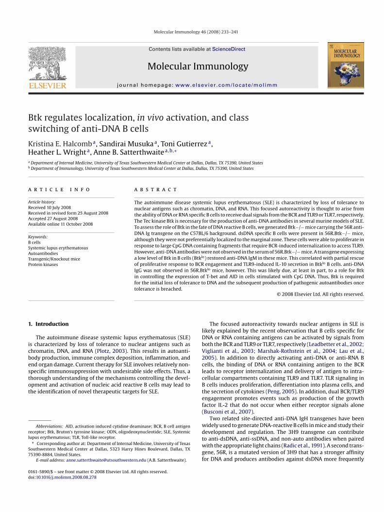
ig. 1. B cells with potential for anti-DNA reactivity are present in 56R.Btk−/− mice but ald mice were stained with antibodies against (A) B220 and IgMa and (B and C) B220, C220+Ig�+ cells (C). The frequency of gated cells in the marginal zone (CD23−CD21+) poells among B220+ cells in mice without the 56R transgene and among B220+ and B220+Ig
unology 46 (2008) 233–241 235
escence due to entry of the FITC label into acidic compartmentsAluvihare et al., 1997).
.11. Statistics
Groups were compared by Student’s t-test.
. Results
To study the role of Btk in the development, activation, andocalization of anti-DNA B cells, we generated 56R.Btk−/− micen a C57BL/6 background. A similar number of B220+ cells wasbserved in the spleens of 3–6 month old 56R and 56R.Btk−/−ice (1.47 × 10⁷ vs. 1.09 × 10⁷, p = 0.49), and a similar frequency
f B cells expressed the 56R transgene (Fig. 1a). Skewing of 56Rxpressing B cells to the marginal zone compartment (Li et al.,002a,b; Sekiguchi et al., 2006) was not observed in the absence oftk (Fig. 1b and d), however. This was not due to a general require-
ent for Btk in the development of marginal zone B cells, as therequency of cells in this compartment was normal in Btk−/− micen the absence of the 56R transgene (Fig. 1b and d) (Martin andearney, 2000; Cariappa et al., 2001). Nor is it due to a lack of anti-NA B cells in Btk−/− spleens, as Btk is required for the preferential
re not preferentially localized to the marginal zone. Splenocytes from 3 to 6 monthD21, CD23, and Ig�. Dot plots are gated on all splenocytes (A), B220+ cells (B) orpulation is indicated in B and C. (D) The mean ± S.D. percentage of marginal zone�+ cells in mice with the 56R transgene (D) is shown. n = 3. *p < 0.05, wt vs. Btk−/−.
2 r Imm
lt(
wLocafSaIms
matealfirea1iwre
g1B
mTtaptiaraacti
mdanmiree
lcfna
FspDC1aOc
36 K.E. Halcomb et al. / Molecula
ocalization to the marginal zone of cells expressing both the 56Rransgene and Ig�, a combination known to confer DNA-reactivityFig. 1c and d) (Chen et al., 1994; Li et al., 2002a,b).
The presence of DNA reactive cells in 56R.Btk−/− miceas directly demonstrated by generating hybridomas from
PS-stimulated splenocytes. 74 IgM secreting hybridomas werebtained from a total of two 56R mice, of which 66 (89%) were spe-ific for dsDNA. 2 of 19 (11%) IgG secreting hybridomas producednti-dsDNA antibodies. This is consistent with previous resultsrom 56R mice on the B6 background (Fukuyama et al., 2005;ekiguchi et al., 2006). Anti-DNA B cells were also present in thebsence of Btk, although at a reduced frequency. 41% (27 of 65) ofgM secreting hybridomas obtained from a total of 3 56R.Btk−/−
ice produced antibodies against dsDNA, while none of the 10 IgGecreting hybridomas were dsDNA-specific.
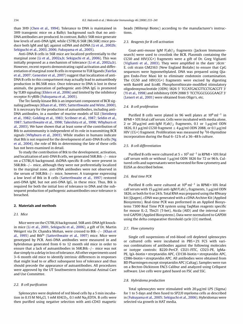
Btk is required for proliferation in response to antigen engage-ent (Khan et al., 1995). However, BCR-mediated shuttling of
ntigen to TLR9 is sufficient to induce TLR9-dependent prolifera-ion of B cells in the absence of apparent BCR crosslinking (Vigliantit al., 2003). This suggests that DNA containing antigens might beble to activate Btk-deficient anti-DNA B cells via TLR9 despite theack of a mitogenic signal from the BCR. To address this issue, werst used the diffusible CpG containing ODN 1826 to assess theole of Btk in responses to TLR9 (Leadbetter et al., 2002; Vigliantit al., 2003; Marshak-Rothstein et al., 2004). Btk−/− B cells wereble to proliferate, upregulate CD86, and differentiate in response to�g/ml ODN 1826 (Fig. 2a-d, Fig. 3a). However, a six-fold reduction
n the secretion of IL-10 by B cells in response to TLR9 engagementas observed in the absence of Btk (Fig. 2e), consistent with recent
eports published during the preparation of this manuscript (Hasan
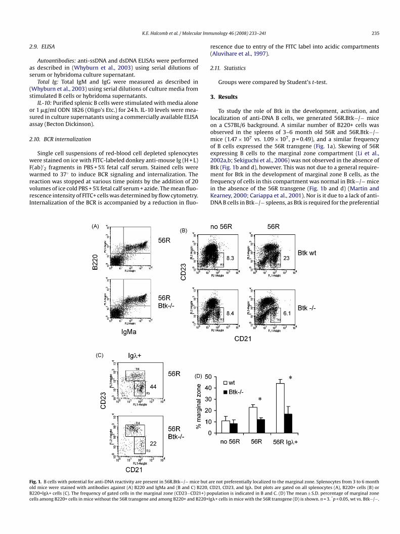
t al., 2008; Lee et al., 2008).We next asked whether Btk promotes interactions between anti-en and CpG DNA induced signals. Low doses of anti-IgM and ODN826 were unable to synergize to promote proliferation of Btk−/−cells as they did in wild type cells (Fig. 3a). In addition to pro-
lteDC
ig. 2. Btk−/− B cells proliferate and differentiate in response to CpG DNA but demonstratetimulated with 1 �g/ml ODN 1826. Proliferation was measured by 3H-thymidine incorpo= 0.5 (not significant). (B) Purified B cells were stimulated with media alone (thin line) oata are representative of 3 independent experiments. (C) Purified B cells were stimulatD138, a plasma cell marker. The frequency of CD138hi cells is indicated. Data are represen�g/ml ODN 1826 for 72 or 96 h. Total IgM levels in the culture supernatants were measund each time point represents the mean ± S.D. of 2 mice per genotype, for a total of 4 micDN 1826 for 24 h. Culture supernatants were analyzed for IL-10 by ELISA. Data representells stimulated with media alone.
unology 46 (2008) 233–241
oting proliferation, however, engagement of both the BCR andLR9 results in functional consequences distinct from the sum ofhose elicited by stimulation of either receptor alone (Busconi etl., 2007). Of particular interest is the upregulation of IL-2, whichromotes the growth of both B and T cells and may serve to amplifyhe immune response to DNA containing antigens. Btk−/− cellsncubated with anti-IgM plus ODN 1826 had IL-2 mRNA levelspproaching those seen in wild type cells stimulated with botheagents (Fig. 3b). Synergy between BCR and TLR9 signaling waslso observed visually in Btk−/− B cells. Together, but not alone,nti-IgM and ODN 1826 induced homotypic adhesion of Btk−/− Bells (Fig. 3c). Thus, despite the lack of a mitogenic response fromhe BCR, Btk−/− cells are able to transmit some BCR signals thatnteract with TLR9 signals.
Although ODN 1826 can access TLR9 directly, the larger frag-ents of DNA likely to activate anti-DNA B cells in vivo must be
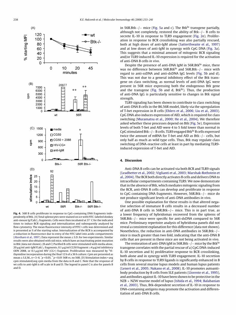
elivered to TLR9 by the BCR (Leadbetter et al., 2002; Viglianti etl., 2003). To determine whether Btk−/− B cells are able to inter-alize the BCR, FITC-labeled anti-IgM was used to both induce andeasure BCR internalization. Over time, the mean fluorescence
ntensity of IgM+ cells from both wild type and Btk−/− B cells waseduced (Fig. 4a), indicating quenching of fluorescence upon deliv-ry of FITC to intracellular compartments with low pH (Aluviharet al., 1997).
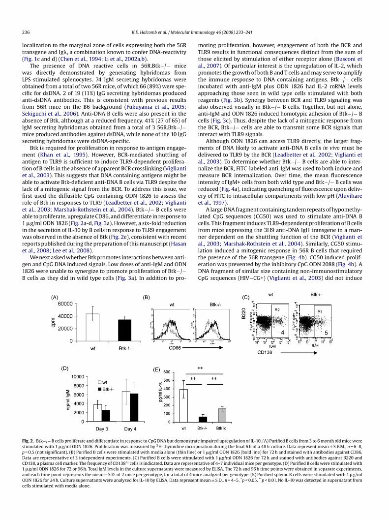
A large DNA fragment containing tandem repeats of hypomethy-ated CpG sequences (CG50) was used to stimulate anti-DNA Bells. This fragment induces TLR9-dependent proliferation of B cellsrom mice expressing the 3H9 anti-DNA IgH transgene in a man-er dependent on the shuttling function of the BCR (Viglianti etl., 2003; Marshak-Rothstein et al., 2004). Similarly, CG50 stimu-
ation induced a mitogenic response in 56R B cells that requiredhe presence of the 56R transgene (Fig. 4b). CG50 induced prolif-ration was prevented by the inhibitory CpG ODN 2088 (Fig. 4b). ANA fragment of similar size containing non-immunostimulatorypG sequences (HIV−CG+) (Viglianti et al., 2003) did not induceimpaired upregulation of IL-10. (A) Purified B cells from 3 to 6 month old mice wereration during the final 6 h of a 48 h culture. Data represent mean ± S.E.M., n = 6–8,r 1 �g/ml ODN 1826 (bold line) for 72 h and stained with antibodies against CD86.ed with 1 �g/ml ODN 1826 for 72 h and stained with antibodies against B220 andtative of 4–7 individual mice per genotype. (D) Purified B cells were stimulated withred by ELISA. The 72 h and 96 h time points were obtained in separate experiments,e analyzed per genotype. (E) Purified splenic B cells were stimulated with 1 �g/mlmean ± S.D., n = 4–5. *p < 0.05, **p < 0.01. No IL-10 was detected in supernatant from
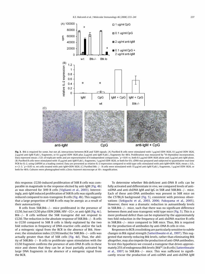
K.E. Halcomb et al. / Molecular Immunology 46 (2008) 233–241 237
Fig. 3. Btk is required for some, but not all, interactions between BCR and TLR9 signals. (A) Purified B cells were stimulated with 1 �g/ml ODN 1826, 0.1 �g/ml ODN 1826,2 �g/ml anti-IgM F(ab)′
2 fragments, or 0.1 �g/ml ODN 1826 plus 2 �g/ml anti-IgM F(ab)′2 fragments for 48 h. Proliferation was measured by 3H-thymidine incorporation.
Data represent mean ± S.D. of triplicate wells and are representative of 4 independent comparisons. *p < 0.01 vs. both 0.1 �g/ml ODN 1826 alone and 2 �g/ml anti-IgM alone.(B) Purified B cells were stimulated with 15 �g/ml anti-IgM F(ab)′
2 fragments, 1 �g/ml ODN 1826, or both for 6 h. cDNA was prepared and subjected to quantitative real timePCR for IL-2, using GAPDH as a loading control. Data are presented as relative IL-2 expression compared to wild type cells stimulated with anti-IgM+ODN 1826, mean ± S.D.,n cellsb magn
tpairtt
CBCtfoeaiCmlt
fsEtvHibmtif
cg
= 2–3. *p < 0.05 vs. wt cells treated with anti-IgM+ODN 1826. (C) Purified Btk−/− Both for 48 h. Cultures were photographed with a Zeiss Axiovert microscope at 10×
his response. CG50-induced proliferation of 56R B cells was com-arable in magnitude to the response elicited by anti-IgM (Fig. 4b)s was observed for 3H9 B cells (Viglianti et al., 2003). Interest-ngly, anti-IgM induced proliferation of 56R B cells was significantlyeduced compared to non-transgenic B cells (Fig. 4b). This suggestshat a large proportion of 56R B cells may be anergic as a result ofheir autoreactivity.
B cells from 56R.Btk−/− mice proliferated in the presence ofG50, but not CG50 plus ODN 2088, HIV−CG+, or anti-IgM (Fig. 4c).tk−/− B cells without the 56R transgene did not respond toG50. The reduction in the absolute response of 56R.Btk−/− B cellso CG50 compared to 56R B cells is likely explained by the twoold reduction in frequency of DNA-reactive cells and/or the lackf a mitogenic signal from the BCR in the absence of Btk. How-ver, the stimulation index (CG50/media) for 56R.Btk−/− cells wasctually greater than that of 56R cells (Fig. 4d). Thus, the abil-
ty of 56R.Btk−/− B cells to proliferate upon stimulation with theG50 fragment confirms the presence of anti-DNA B cells in theseice and shows that they can be at least partially activated byarge DNA fragments in the absence of a mitogenic signal fromhe BCR.
aTmec
were stimulated with 15 �g/ml anti-IgM F(ab)′2 fragments, 1 �g/ml ODN 1826, or
ification.
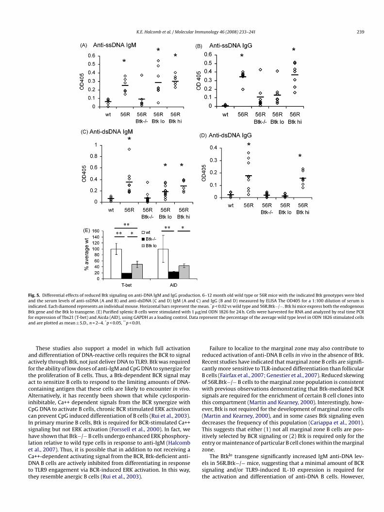
To determine whether Btk-deficient anti-DNA B cells can beully activated and differentiate in vivo, we compared levels of anti-sDNA and anti-dsDNA IgM and IgG in 56R and 56R.Btk−/− mice.ach of these anti-DNA antibodies was present in 56R mice onhe C57BL/6 background (Fig. 5), consistent with previous obser-ations (Sekiguchi et al., 2003, 2006; Fukuyama et al., 2005).owever, there was a dramatic reduction in autoantibody levels
n 56R.Btk−/− mice, such that there was no significant differenceetween them and non-transgenic wild type mice (Fig. 5). This is aore profound defect than can be explained by the approximately
wo fold reduction in the frequency of anti-dsDNA reactive B cellsn 56R.Btk−/− mice compared to 56R mice. Thus, Btk is requiredor the production of antibodies by anti-DNA B cells in vivo.
Responses to BCR crosslinking are particularly sensitive to subtlehanges in Btk signal strength (Satterthwaite et al., 1997). This sug-ested that merely reducing Btk levels, rather than eliminating Btk
ltogether, may also impede the production of anti-DNA antibodies.o test this hypothesis we crossed a transgene that drives approxi-ately 25% of endogenous Btk levels (Btklo) in B cells (Satterthwaitet al., 1997) to 56R.Btk−/− mice. This was sufficient to signifi-antly rescue the production of anti-ssDNA and anti-dsDNA IgM
238 K.E. Halcomb et al. / Molecular Imm
Fig. 4. 56R B cells proliferate in response to CpG-containing DNA fragments inde-pendently of Btk. (A) Total splenocytes were stained on ice with FITC-labeled donkeyanti-mouse Ig F(ab)′
2 fragments. Cells were then incubated at 37 ◦C for the indicatedtimes to induce BCR signaling and internalization and subsequently analyzed byflow cytometry. The mean fluorescence intensity of FITC+ cells was determined andis presented as % of the starting value. Internalization of the BCR is accompanied bya reduction in fluorescence due to entry of the FITC label into acidic compartments(Aluvihare et al., 1997). Data represent the mean ± S.D. for two experiments. Similarresults were also obtained with xid mice, which have an inactivating point mutationin Btk (data not shown). (B and C) Purified B cells were stimulated with media alone,20 �g/ml anti-IgM F(ab)′
2 fragments, 0.1 �g/ml CG50 fragment ±4 �g/ml inhibitoryODN 2088, or 0.1 �g/ml HIV (CG+) fragment. Proliferation was measured by 3H-thymidine incorporation during the final 12 h of a 36 h culture. Cpm are presented asmean ± S.E.M., n = 3–6. *p < 0.05, **p < 0.01 56R vs. no 56R. (D) Stimulation index = avgcpm stimulated/avg cpm media from the data in B and C. Note that the response ofwt cells to anti-IgM is off scale in B and D. The legend in panel C is also for panels Band D.
iasabaTao
wrTgpaos
ooCsalCtosi
4
(aitttn
toa5mrNmc
tIbbc(baNeDt
unology 46 (2008) 233–241
n 56R.Btk−/− mice (Fig. 5a and c). The Btklo transgene partially,lthough not completely, restored the ability of Btk−/− B cells toecrete IL-10 in response to TLR9 engagement (Fig. 2e). Prolifer-tion in response to BCR crosslinking was also partially rescued,oth at high doses of anti-IgM alone (Satterthwaite et al., 1997)nd at low doses of anti-IgM in synergy with CpG DNA (Fig. 3a).his suggests that a minimal amount of mitogenic BCR signalingnd/or TLR9 induced IL-10 expression is required for the activationf anti-DNA B cells in vivo.
Despite the presence of anti-DNA IgM in 56R.Btklo mice, thereas no difference between 56R.Btklo and 56R.Btk−/− mice with
egard to anti-ssDNA and anti-dsDNA IgG levels (Fig. 5b and d).his was not due to a general inhibitory effect of the Btk trans-ene on class switching, as normal levels of anti-DNA IgG wereresent in 56R mice expressing both the endogenous Btk genend the transgene (Fig. 5b and d, Btkhi). Thus, the productionf anti-DNA IgG is particularly sensitive to changes in Btk signaltrength.
TLR9 signaling has been shown to contribute to class switchingf anti-DNA B cells in the B6.56R model, likely via the upregulationf T-bet expression in B cells (Ehlers et al., 2006; Liu et al., 2003).pG DNA also induces expression of AID, which is required for classwitching (Muramatsu et al., 2000; He et al., 2004). We thereforesked whether these processes depend on Btk (Fig. 5e). Expressionevels of both T-bet and AID were 4 to 5 fold lower than normal inpG stimulated Btk−/− B cells. TLR9 engaged Btklo B cells expressedwice the amount of mRNA for T-bet and AID as Btk−/− cells, butnly half as much as wild type cells. Thus, Btk may regulate classwitching of DNA-reactive cells at least in part by mediating TLR9-nduced expression of T-bet and AID.
. Discussion
Anti-DNA B cells can be activated via both BCR and TLR9 signalsLeadbetter et al., 2002; Viglianti et al., 2003; Marshak-Rothstein etl., 2004). The BCR both directly activates B cells and delivers DNA tontracellular compartments containing TLR9. We now demonstratehat in the absence of Btk, which mediates mitogenic signaling fromhe BCR, anti-DNA B cells can develop and proliferate in responseo CpG containing DNA fragments. However, 56R.Btk−/− mice doot produce significant levels of anti-DNA antibodies in vivo.
One possible explanation for these results is that altered nega-ive selection of immature B cells results in a decreased numberf anti-DNA B cells in 56R.Btk−/− mice. This is in part true, aslower frequency of hybridomas recovered from the spleens of
6R.Btk−/− mice were specific for anti-dsDNA compared to 56Rice. Preliminary repertoire analysis of hybridoma panels did not
eveal a consistent explanation for this difference (data not shown).onetheless, the reduction in anti-DNA antibodies in 56R.Btk−/−ice is much greater than two fold, indicating that the anti-DNA B
ells that are present in these mice are not being activated in vivo.The restoration of anti-DNA IgM in 56R.Btk−/− mice by the Btklo
ransgene correlates with the partial rescue of a) CpG DNA-inducedL-10 secretion and b) proliferative response to BCR crosslinking,oth alone and in synergy with TLR9 engagement. IL-10 secretiony B cells in response to TLR9 ligands is significantly enhanced in Bells from several murine lupus models and human lupus patientsLenert et al., 2005; Nakano et al., 2008). IL-10 promotes autoanti-ody production by B cells from SLE patients (Llorente et al., 1995),
nd antibodies against IL-10 have been shown to be protective in theZB × NZW murine model of lupus (Ishida et al., 1994; Balabaniant al., 2003). Thus, Btk-dependent secretion of IL-10 in response toNA-containing antigens may promote the activation and differen-iation of anti-DNA B cells.
K.E. Halcomb et al. / Molecular Immunology 46 (2008) 233–241 239
Fig. 5. Differential effects of reduced Btk signaling on anti-DNA IgM and IgG production. 6–12 month old wild type or 56R mice with the indicated Btk genotypes were bledand the serum levels of anti-ssDNA (A and B) and anti-dsDNA (C and D) IgM (A and C) and IgG (B and D) measured by ELISA The OD405 for a 1:100 dilution of serum isindicated. Each diamond represents an individual mouse. Horizontal bars represent the mean. *p < 0.02 vs wild type and 56R.Btk−/−. Btk hi mice express both the endogenousBtk gene and the Btk lo transgene. (E) Purified splenic B cells were stimulated with 1 �g/ml ODN 1826 for 24 h. Cells were harvested for RNA and analyzed by real time PCRf ata rea
aaftacAiCcIshleCDtt
rRcBowste(dTie
or expression of Tbx21 (T-bet) and Aicda (AID), using GAPDH as a loading control. Dnd are plotted as mean ± S.D., n = 2–4. *p < 0.05, **p < 0.01.
These studies also support a model in which full activationnd differentiation of DNA-reactive cells requires the BCR to signalctively through Btk, not just deliver DNA to TLR9. Btk was requiredor the ability of low doses of anti-IgM and CpG DNA to synergize forhe proliferation of B cells. Thus, a Btk-dependent BCR signal mayct to sensitize B cells to respond to the limiting amounts of DNA-ontaining antigen that these cells are likely to encounter in vivo.lternatively, it has recently been shown that while cyclosporin-
nhibitable, Ca++ dependent signals from the BCR synergize withpG DNA to activate B cells, chronic BCR stimulated ERK activationan prevent CpG induced differentiation of B cells (Rui et al., 2003).n primary murine B cells, Btk is required for BCR-stimulated Ca++ignaling but not ERK activation (Forssell et al., 2000). In fact, weave shown that Btk−/− B cells undergo enhanced ERK phosphory-
ation relative to wild type cells in response to anti-IgM (Halcomb
t al., 2007). Thus, it is possible that in addition to not receiving aa++-dependent activating signal from the BCR, Btk-deficient anti-NA B cells are actively inhibited from differentiating in responseo TLR9 engagement via BCR-induced ERK activation. In this way,hey resemble anergic B cells (Rui et al., 2003).
z
est
present the percentage of the average wild type level in ODN 1826 stimulated cells
Failure to localize to the marginal zone may also contribute toeduced activation of anti-DNA B cells in vivo in the absence of Btk.ecent studies have indicated that marginal zone B cells are signifi-antly more sensitive to TLR-induced differentiation than follicularcells (Fairfax et al., 2007; Genestier et al., 2007). Reduced skewingf 56R.Btk−/− B cells to the marginal zone population is consistentith previous observations demonstrating that Btk-mediated BCR
ignals are required for the enrichment of certain B cell clones intohis compartment (Martin and Kearney, 2000). Interestingly, how-ver, Btk is not required for the development of marginal zone cellsMartin and Kearney, 2000), and in some cases Btk signaling evenecreases the frequency of this population (Cariappa et al., 2001).his suggests that either (1) not all marginal zone B cells are pos-tively selected by BCR signaling or (2) Btk is required only for thentry or maintenance of particular B cell clones within the marginal
one.The Btklo transgene significantly increased IgM anti-DNA lev-ls in 56R.Btk−/− mice, suggesting that a minimal amount of BCRignaling and/or TLR9-induced IL-10 expression is required forhe activation and differentiation of anti-DNA B cells. However,
2 r Imm
twbaBeoacdt
CrdaetaauAaeId(gp
diIav
A
laaWcSCd
R
A
B
B
B
C
C
E
F
F
F
G
G
H
H
H
I
K
L
L
L
L
L
L
L
L
L
L
M
M
M
N
40 K.E. Halcomb et al. / Molecula
here was no difference between 56R.Btklo and 56R.Btk−/− miceith respect to anti-DNA IgG levels. 56R.Btklo mice thus resem-
le 56R.Balb/c mice in the production of intermediate levels ofnti-DNA IgM and no anti-DNA IgG (Fukuyama et al., 2005). Thetklo transgene did not impair anti-DNA IgG production in the pres-nce of a normal endogenous Btk gene, however, ruling out linkagef the Btklo transgene to a Balb/c-like allele of a gene controllingnti-DNA IgG levels. We did not observe significant impairment oflass switching in Btklo mice in response to T-independent or T-ependent viral antigens (Pinschewer et al., 1999), suggesting thathe current observation may be specific for anti-DNA B cells.
Consistent with this idea, we demonstrate that Btk mediatespG DNA induced expression of AID and T-bet in B cells. AID isequired for class switching (Muramatsu et al., 2000), and T-beteficient MRL.lpr mice have dramatic reductions in pathogenicutoantibody levels (Peng et al., 2002). While AID and T-bet lev-ls were partially restored in Btklo B cells, they remained lowerhan normal and may be insufficient to promote class switching ofnti-DNA B cells. Additionally, the balance between Btk-dependentctivating signals and Fc�RIIb-mediated inhibitory signals may reg-late the terminal differentiation of switched anti-DNA B cells.major function of Fc�RIIb is to inhibit BCR-induced membrane
ssociation and activation of Btk (Bolland et al., 1998; Scharenbergt al., 1998). 56R.Fc�RIIb−/− mice have similar levels of anti-DNAgM to 56R mice but significantly increased levels of anti-DNA IgGue to an increase in the frequency of IgG secreting plasma cellsFukuyama et al., 2005). Taken together, these observations sug-est that Btk signal strength is a critical determinant of whetherathogenic IgG autoantibodies will be produced.
In summary, Btk plays an important role at multiple checkpointsuring the development, localization, activation, and class switch-
ng of DNA-reactive B cells. The particular sensitivity of anti-DNAgG to subtle changes in Btk signal strength suggests that Btk, itsctivators or effectors may be points of potential therapeutic inter-ention to prevent pathogenic autoimmunity in SLE.
cknowledgements
We thank Cristina Contreras and Rochelle Hinman for excel-ent technical assistance. We are grateful to Drs. Martin Weigertnd Chandra Mohan for providing 56R mice and Drs. Greg Vigliantind Ann Marshak-Rothstein for the CG50 and HIV(CG+) plasmids.e also thank Rochelle Hinman and Dr. Chandra Mohan for criti-
al review of the manuscript. This work was supported by the UTouthwestern Endowment for Scholars in Biomedical Research, ahapter Grant from the North Texas Chapter of the Arthritis Foun-ation, and NIH grants AI039824 and GM076982.
eferences
luvihare, V.R., Khamlichi, A.A., Williams, G.T., Adorini, L., Neuberger, M.S., 1997.Acceleration of intracellular targeting of antigen by the B-cell antigen receptor:importance depends on the nature of the antigen–antibody interaction. EMBOJ. 16, 3553S–3562S.
alabanian, K., Couderc, J., Bouchet-Delbos, L., Amara, A., Berrebi, D., Foussat, A.,Baleux, F., Portier, A., Durand-Gasselin, I., Coffman, R.L., Galanaud, P., Peuch-maur, M., Emilie, D., 2003. Role of the chemokine stromal cell-derived factor1 in autoantibody production and nephritis in murine lupus. J. Immunol. 170,3392–3400.
olland, S., Pearse, R.N., Kurosaki, T., Ravetch, J.V., 1998. SHIP modulates immunereceptor responses by regulating membrane association of Btk. Immunity 8,509–516.
usconi, L., Bauer, J.W., Tumang, J.R., Laws, A., Perkins-Mesires, K., Tabor, A.S., Lau,
C., Corley, R.B., Rothstein, T.L., Lund, F.E., Behrens, T.W., Marshak-Rothstein, A.,2007. Functional outcome of B cell activation by chromatin immune complexengagement of the B cell receptor and TLR9. J. Immunol. 179, 7397–7405.ariappa, A., Tang, M., Parng, C., Nebelitskiy, E., Carroll, M., Georgopoulos, K., Pillai,S., 2001. The follicular versus marginal zone B lymphocyte cell fate decision isregulated by Aiolos, Btk, and CD21. Immunity 14, 603–615.
N
P
unology 46 (2008) 233–241
hen, C., Radic, M.Z., Erikson, J., Camper, S.A., Litwin, S., Hardy, R.R., Weigert, M.,1994. Deletion and editing of B cells that express antibodies to DNA. J. Immunol.152, 1970–1982.
hlers, M., Fukuyama, H., McGaha, T.L., Aderem, A., Ravetch, J.V., 2006. TLR9/MyD88signaling is required for class switching to pathogenic IgG2a and 2b autoanti-bodies in SLE. J. Exp. Med. 203, 553–561.
airfax, K.A., Corcoran, L.M., Pridans, C., Huntington, N.D., Kallies, A., Nutt, S.L., Tarlin-ton, D.M., 2007. Different kinetics of blimp-1 induction in B cell subsets revealedby reporter gene. J. Immunol. 178, 4104–4111.
orssell, J., Nilsson, A., Sideras, P., 2000. Reduced formation of phosphatidic acid uponB-cell receptor triggering of mouse B-lymphocytes lacking Bruton’s tyrosinekinase. Scand. J. Immunol. 52, 30–38.
ukuyama, H., Nimmerjahn, F., Ravetch, J.V., 2005. The inhibitory Fcgamma receptormodulates autoimmunity by limiting the accumulation of immunoglobulin G+anti-DNA plasma cells. Nat. Immunol. 6, 99–106.
enestier, L., Taillardet, M., Mondiere, P., Gheit, H., Bella, C., Defrance, T., 2007. TLRagonists selectively promote terminal plasma cell differentiation of B cell subsetsspecialized in thymus-independent responses. J. Immunol. 178, 7779–7786.
olding, B., Golding, H., Foiles, P.G., Morton, J.I., 1983. CBA/N X-linked defect delaysexpression of the Y-linked accelerated autoimmune disease in BXSB mice. J.Immunol. 130, 1043–1046.
alcomb, K.E., Contreras, C.M., Hinman, R.M., Coursey, T.G., Wright, H.L., Satterth-waite, A.B., 2007. Btk and phospholipase C gamma 2 can function independentlyduring B cell development. Eur. J. Immunol. 37, 1033–1042.
asan, M., Lopez-Herrera, G., Blomberg, K.E., Lindvall, J.M., Berglöf, A., Smith,C.I., Vargas, L., 2008. Defective Toll-like receptor 9-mediated cytokine produc-tion in B cells from Bruton’s tyrosine kinase-deficient mice. Immunology 123,239–249.
e, B., Qiao, X., Cerutti, A., 2004. CpG DNA induces IgG class switch DNA recombina-tion by activating human B cells through an innate pathway that requires TLR9and cooperates with IL-10. J. Immunol. 173, 4479–4491.
shida, H., Muchamuel, T., Sakaguchi, S., Andrade, S., Menon, S., Howard, M., 1994.Continuous administration of anti-interleukin 10 antibodies delays onset ofautoimmunity in NZB/W F1 mice. J. Exp. Med. 179, 305–310.
han, W.N., Alt, F.W., Gerstein, R.M., Malynn, B.A., Larsson, I., Rathbun, G., Davidson,L., Muller, S., Kantor, A.B., Herzenberg, L.A., Rosen, F.S., Sideras, P., 1995. DefectiveB cell development and function in Btk-deficient mice. Immunity 3, 283–299.
au, C.M., Broughton, C., Tabor, A.S., Akira, S., Flavell, R.A., Mamula, M.J., Chris-tensen, S.R., Shlomchik, M.J., Viglianti, G.A., Rifkin, I.R., Marshak-Rothstein, A.,2005. RNA-associated autoantigens activate B cells by combined B cell antigenreceptor/Toll-like receptor 7 engagement. J. Exp. Med. 202, 1171–1177.
eadbetter, E.A., Rifkin, I.R., Hohlbaum, A.M., Beaudette, B.C., Shlomchik, M.J.,Marshak-Rothstein, A., 2002. Chromatin-IgG complexes activate B cells by dualengagement of IgM and Toll-like receptors. Nature 416, 603–607.
ee, K.G., Xu, S., Wong, E.T., Tergaonkar, V., Lam, K.P., 2008. Bruton’s tyrosine kinaseseparately regulates NFkappaB p65RelA activation and cytokine interleukin (IL)-10/IL-12 production in TLR9-stimulated B Cells. J. Biol. Chem. 283, 11189–11198.
enert, P., Stunz, L., Yi, A.K., Krieg, A.M., Ashman, R.F., 2001. CpG stimulation of pri-mary mouse B cells is blocked by inhibitory oligodeoxyribonucleotides at a siteproximal to NF-�B activation. Antisense Nucleic Acid Drug Dev. 4, 247–256.
enert, P., Brummel, R., Field, E.H., Ashman, R.F., 2005. TLR-9 activation of marginalzone B cells in lupus mice regulates immunity through increased IL-10 produc-tion. J. Clin. Immunol. 25, 29–40.
i, H., Jiang, Y., Prak, E.L., Radic, M., Weigert, M., 2001. Editors and editing of anti-DNAreceptors. Immunity 15, 947–957.
i, Y., Li, H., Weigert, M., 2002a. Autoreactive B cells in the marginal zone that expressdual receptors. J. Exp. Med. 195, 181–188.
i, Y., Li, H., Ni, D., Weigert, M., 2002b. Anti-DNA B cells in MRL/lpr mice show altereddifferentiation and editing pattern. J. Exp. Med. 196, 1543–1552.
iu, N., Ohnishi, N., Ni, L., Akira, S., Bacon, K.B., 2003. CpG directly induces T-betexpression and inhibits IgG1 and IgE switching in B cells. Nat. Immunol. 4,687–693.
lorente, L., Zou, W., Levy, Y., Richaud-Patin, Y., Wijdenes, J., Alcocer-Varela, J., Morel-Fourrier, B., Brouet, J.C., Alarcon-Segovia, D., Galanaud, P., Emilie, D., 1995. Role ofinterleukin 10 in the B lymphocyte hyperactivity and autoantibody productionof human systemic lupus erythematosus. J. Exp. Med. 181, 839–844.
arshak-Rothstein, A., Busconi, L., Lau, C.M., Tabor, A.S., Leadbetter, E.A., Akira, S.,Krieg, A.M., Lipford, G.B., Viglianti, G.A., Rifkin, I.R., 2004. Comparison of CpG s-ODNs, chromatin immune complexes, and dsDNA fragment immune complexesin the TLR9-dependent activation of rheumatoid factor B cells. J. Endotoxin. Res.10, 247–251.
artin, F., Kearney, J.F., 2000. Positive selection from newly formed to marginal zoneB cells depends on the rate of clonal production, CD19, and btk. Immunity 12,39–49.
uramatsu, M., Kinoshita, K., Fagarasan, S., Yamada, S., Shinkai, Y., Honjo, T., 2000.Class switch recombination and hypermutation require activation-induced cyti-dine deaminase (AID), a potential RNA editing enzyme. Cell 102, 553–563.
akano, S., Morimoto, S., Suzuki, J., Nozawa, K., Amano, H., Tokano, Y., Takasaki, Y.,2008. Role of pathogenic auto-antibody production by Toll-like receptor 9 of B
cells in active systemic lupus erythematosus. Rheumatology 47, 145–149.g, Y.S., Wardemann, H., Chelnis, J., Cunningham-Rundles, C., Meffre, E., 2004. Bru-ton’s tyrosine kinase is essential for human B cell tolerance. J. Exp. Med. 200,927–934.
eng, S.L., Szabo, S.J., Glimcher, L.H., 2002. T-bet regulates IgG class switching andpathogenic autoantibody production. Proc. Natl. Acad. Sci. U. S. A. 99, 5545–5550.
r Imm
P
P
P
R
R
S
S
S
S
S
S
S
S
S
T
V
K.E. Halcomb et al. / Molecula
eng, S.L., 2005. Signaling in B cells via Toll-like receptors. Curr. Opin. Immunol. 17,230–236.
inschewer, D.D., Ochsenbein, A.F., Satterthwaite, A.B., Witte, O.N., Hengartner,H., Zinkernagel, R.M., 1999. A Btk transgene restores the antiviral TI-2 anti-body responses of xid mice in a dose-dependent fashion. Eur. J. Immunol. 29,2981–2987.
lotz, P.H., 2003. The autoantibody repertoire: searching for order. Nat. Rev.Immunol. 3, 73–78.
adic, M.Z., Mascelli, M.A., Erikson, J., Shan, H., Weigert, M., 1991. Ig H and L chaincontributions to autoimmune specificities. J. Immunol. 146, 176–182.
ui, L., Vinuesa, C.G., Blasioli, J., Goodnow, C.C., 2003. Resistance to CpG DNA-inducedautoimmunity through tolerogenic B cell antigen receptor ERK signaling. Nat.Immunol. 4, 594–600.
atterthwaite, A.B., Cheroutre, H., Khan, W.N., Sideras, P., Witte, O.N., 1997. Btk dosagedetermines sensitivity to B cell antigen receptor cross-linking. Proc. Natl. Acad.Sci. U. S. A. 94, 13152–13157.
atterthwaite, A.B., Lowell, C.A., Khan, W.N., Sideras, P., Alt, F.W., Witte, O.N., 1998.Independent and opposing roles for Btk and lyn in B and myeloid signalingpathways. J. Exp. Med. 188, 833–844.
atterthwaite, A.B., Witte, O.N., 2000. The role of Bruton’s tyrosine kinase in
B cell development and function; a genetic perspective. Immunol. Rev. 175,120–127.charenberg, A.M., El-Hillal, O., Fruman, D.A., Beitz, L.O., Li, Z., Lin, S., Gout, I., Cant-ley, L.C., Rawlings, D.J., Kinet, J.P., 1998. Phosphatidylinositol-3,4,5-trisphosphate(PtdIns-3,4,5-P3)/Tec kinase-dependent calcium signaling pathway: a target forSHIP-mediated inhibitory signals. EMBO J. 17, 1961–1972.
W
Y
unology 46 (2008) 233–241 241
cribner, C.L., Hansen, C.T., Klinman, D.M., Steinberg, A.D., 1987. The interaction ofthe xid and me genes. J. Immunol. 138, 3611–3617.
ekiguchi, D.R., Eisenberg, R.A., Weigert, M., 2003. Secondary heavy chain rearrange-ment: a mechanism for generating anti-double-stranded DNA B cells. J. Exp. Med.197, 27–39.
ekiguchi, D.R., Yunk, L., Gary, D., Charan, D., Srivastava, B., Allman, D., Weigert, M.G.,Prak, E.T., 2006. Development and selection of edited B cells in B6.56R mice. J.Immunol. 176, 6879–6887.
eldin, M.F., Reeves, J.P., Scribner, C.L., Roths, J.B., Davidson, W.F., Morse III, H.C., Stein-berg, A.D., 1987. Effect of xid on autoimmune C3H-gld/gld mice. Cell. Immunol.107, 249–255.
teinberg, B.J., Smathers, P.A., Frederiksen, K., Steinberg, A.D., 1982. Ability of thexid gene to prevent autoimmunity in (NZB × NZW)F1 mice during the courseof their natural history, after polyclonal stimulation, or following immunizationwith DNA. J. Clin. Invest. 70, 587–597.
akeshita, H., Taniuchi, I., Kato, J., Watanabe, T., 1998. Abrogation of autoimmunedisease in Lyn-deficient mice by the mutation of the Btk gene. Int. Immunol. 10,435–444.
iglianti, G.A., Lau, C.M., Hanley, T.M., Miko, B.A., Shlomchik, M.J., Marshak-Rothstein,A., 2003. Activation of autoreactive B cells by CpG dsDNA. Immunity 19, 837–847.
hyburn, L.R., Halcomb, K.E., Contreras, C.M., Lowell, C.A., Witte, O.N., Satterthwaite,A.B., 2003. Reduced dosage of Bruton’s tyrosine kinase uncouples B cell hyperre-sponsiveness from autoimmunity in lyn−/− mice. J. Immunol. 171, 1850–1858.
i, A.K., Tuetken, R., Redford, T., Waldschmidt, M., Kirsch, J., Krieg, A.M., 1998. CpGmotifs in bacterial DNA activate leukocytes through the pH-dependent genera-tion of reactive oxygen species. J. Immunol. 160, 4755–4761.