Large Effects from Small Exposures. I. Mechanisms for Endocrinedisrupting Chemicals with Estrogenic...
13
994 VOLUME 111 | NUMBER 8 | June 2003 • Environmental Health Perspectives During the past decade a number of pesticides, industrial by-products, manufac- tured products such as plastics, and natural chemicals have been shown to disrupt the endocrine system. These chemicals are referred to as endocrine-disrupting chemicals (EDCs). These chemicals have received con- siderable attention, in part because endocrine disruption is a relatively unstudied area in tox- icology and is only recently being taken into account in risk assessment. The focus here is on EDCs with estrogenic activity (EEDCs), which are chemicals that act as hormone mimics via estrogen receptor mechanisms; this is currently the largest group of known endocrine disruptors. The main purpose of this article is to present an overview of the mechanisms of hormone action that provide the basis for understanding how EEDCs have the potential to be biologically active at low, environmentally relevant doses. Our strategy is to discuss the receptor mechanisms mediat- ing responses to a natural hormone, 17β-estra- diol (E 2 ), and then to use this information as the basis for describing the low-dose effects of chemicals that disrupt the normal functioning of this hormonal system, either by mimicking, modulating, or antagonizing the activity of the hormone. We have chosen to use estrogen as our example because there is more known about the biology of estrogens and xenoestro- gens than other components of the endocrine system for which there is evidence for disrup- tion by environmental chemicals; however, the information presented here is applicable to endocrine disruptors that interfere with other hormonal systems. We will begin by briefly reviewing information concerning the relationship between dose, receptor occupancy, and responses (such as cell proliferation) after binding of E 2 to estrogen receptors (ER-α) in cultured human MCF-7 breast cancer cells. A number of specific factors influence the dose of an EEDC that reaches the target cells to produce a response. These factors include route of administration, absorption, distribu- tion, metabolism, rate of clearance, plasma transport, cell uptake, affinity for estrogen receptor subtype in the cell, and the interac- tion of the ligand–receptor complex with tissue-specific factors comprising the tran- scriptional apparatus. This mechanistic infor- mation provides the basis for establishing the dose at the target site in cells (nuclear recep- tors associated with DNA or more recently identified receptors associated with the cell membrane) for an EEDC required to elicit a biological response similar to that produced by a dose of E 2 with equal estrogenic activity. Modeling that takes into account each of these factors would encompass physiologically based pharmacokinetic information (1), as well as quantitative structure-activity relation- ships (QSAR) (2,3). We have previously dis- cussed the factors that influence access of E 2 and EEDCs from blood to estrogen receptors in cells elsewhere (4–6). Our primary focus in this review is on the latter part of the overall process that occurs once an estrogenic chemi- cal has reached the nuclear estrogen receptor. Dose ranges. We have separated dose- specific effects into three general categories: the physiological dose range for estrogenic activity, the toxicological dose range for acute toxicity, and the environmentally relevant dose range related to current exposures. The physiological dose range (of estrogenic activ- ity, whatever the source) is defined by the normal concentration range of an endoge- nous hormone. More specifically, with regard to steroid hormones, the physiological con- centration refers to the amount of free (unbound to plasma proteins and unconju- gated) endogenous hormone that the EEDC is mimicking or antagonizing. The free hor- mone concentration is generally considered to be the biologically active portion of total hor- mone concentration in blood (7,8) and most accurately predicts biological activity (for example, free triiodothyronine and free thy- roxine, as opposed to total hormone concen- tration, are routinely used for clinical diagnosis). The toxicological dose range is identified by some measure of toxicity, such Large Effects from Small Exposures. I. Mechanisms for Endocrine-Disrupting Chemicals with Estrogenic Activity Wade V. Welshons, 1 Kristina A. Thayer, 2 Barbara M. Judy, 1 Julia A. Taylor, 1 Edward M. Curran, 1 and Frederick S. vom Saal 2 1 Department of Veterinary Biomedical Sciences and 2 Division of Biological Sciences, University of Missouri-Columbia, Columbia, Missouri, USA Address correspondence to W.V. Welshons, Dept. of Veterinary Biomedical Sciences, E102 Veterinary Medicine, University of Missouri-Columbia, Columbia, MO 65211 USA. Telephone: (573) 882- 3347. Fax: (573) 884-6890. E-mail: welshonsw@ missouri.edu Support during the preparation of this manuscript was provided by the W. Alton Jones Foundation to K.A.T, as well as by grants from the National Institutes of Health (NIH) (CA50354) and the University of Missouri (VMFC0018) to W.V.W. and NIH (ES08293 and ES11283), U.S. Environmental Protection Agency (U914991), and University of Missouri Research Board to F.v.S. The authors declare they have no conflict of interest. Received 8 January 2002; accepted 20 February 2003. Information concerning the fundamental mechanisms of action of both natural and environmental hormones, combined with information concerning endogenous hormone concen- trations, reveals how endocrine-disrupting chemicals with estrogenic activity (EEDCs) can be active at concentrations far below those currently being tested in toxicological studies. Using only very high doses in toxicological studies of EEDCs thus can dramatically underestimate bioactivity. Specifically: a) The hormonal action mechanisms and the physiology of delivery of EEDCs predict with accuracy the low-dose ranges of biological activity, which have been missed by traditional toxicological testing. b) Toxicology assumes that it is valid to extrapolate linearly from high doses over a very wide dose range to predict responses at doses within the physiological range of receptor occupancy for an EEDC; however, because receptor-mediated responses saturate, this assumption is invalid. c) Furthermore, receptor-mediated responses can first increase and then decrease as dose increases, contradicting the assumption that dose–response relationships are monotonic. d) Exogenous estrogens modulate a system that is physiologically active and thus is already above threshold, contradicting the traditional toxicological assumption of thresholds for endocrine responses to EEDCs. These four fundamental issues are problematic for risk assessment methods used by regulatory agencies, because they challenge the traditional use of extrapolation from high- dose testing to predict responses at the much lower environmentally relevant doses. These doses are within the range of current exposures to numerous chemicals in wildlife and humans. These problems are exacerbated by the fact that the type of positive and negative controls appropriate to the study of endocrine responses are not part of traditional toxicological testing and are frequently omitted, or when present, have been misinterpreted. Key words: dose response, endocrine disrup- tors, estrogen action, estrogen receptors, fetal development, inverted U, MCF-7 cells. Environ Health Perspect 111:994–1006 (2003). doi:10.1289/ehp.5494 available via http://dx.doi.org/ [Online 2 February 2003] Research Review
-
Upload
texaswomans -
Category
Documents
-
view
0 -
download
0
Transcript of Large Effects from Small Exposures. I. Mechanisms for Endocrinedisrupting Chemicals with Estrogenic...
994 VOLUME 111 | NUMBER 8 | June 2003 • Environmental Health Perspectives
During the past decade a number ofpesticides, industrial by-products, manufac-tured products such as plastics, and naturalchemicals have been shown to disrupt theendocrine system. These chemicals arereferred to as endocrine-disrupting chemicals(EDCs). These chemicals have received con-siderable attention, in part because endocrinedisruption is a relatively unstudied area in tox-icology and is only recently being taken intoaccount in risk assessment. The focus here ison EDCs with estrogenic activity (EEDCs),which are chemicals that act as hormonemimics via estrogen receptor mechanisms; thisis currently the largest group of knownendocrine disruptors. The main purpose ofthis article is to present an overview of themechanisms of hormone action that providethe basis for understanding how EEDCs havethe potential to be biologically active at low,environmentally relevant doses. Our strategyis to discuss the receptor mechanisms mediat-ing responses to a natural hormone, 17β-estra-diol (E2), and then to use this information asthe basis for describing the low-dose effects ofchemicals that disrupt the normal functioningof this hormonal system, either by mimicking,modulating, or antagonizing the activity of
the hormone. We have chosen to use estrogenas our example because there is more knownabout the biology of estrogens and xenoestro-gens than other components of the endocrinesystem for which there is evidence for disrup-tion by environmental chemicals; however,the information presented here is applicable toendocrine disruptors that interfere with otherhormonal systems.
We will begin by briefly reviewinginformation concerning the relationshipbetween dose, receptor occupancy, andresponses (such as cell proliferation) afterbinding of E2 to estrogen receptors (ER-α) incultured human MCF-7 breast cancer cells. Anumber of specific factors influence the doseof an EEDC that reaches the target cells toproduce a response. These factors includeroute of administration, absorption, distribu-tion, metabolism, rate of clearance, plasmatransport, cell uptake, affinity for estrogenreceptor subtype in the cell, and the interac-tion of the ligand–receptor complex withtissue-specific factors comprising the tran-scriptional apparatus. This mechanistic infor-mation provides the basis for establishing thedose at the target site in cells (nuclear recep-tors associated with DNA or more recently
identified receptors associated with the cellmembrane) for an EEDC required to elicit abiological response similar to that producedby a dose of E2 with equal estrogenic activity.Modeling that takes into account each ofthese factors would encompass physiologicallybased pharmacokinetic information (1), aswell as quantitative structure-activity relation-ships (QSAR) (2,3). We have previously dis-cussed the factors that influence access of E2and EEDCs from blood to estrogen receptorsin cells elsewhere (4–6). Our primary focus inthis review is on the latter part of the overallprocess that occurs once an estrogenic chemi-cal has reached the nuclear estrogen receptor.
Dose ranges. We have separated dose-specific effects into three general categories:the physiological dose range for estrogenicactivity, the toxicological dose range for acutetoxicity, and the environmentally relevantdose range related to current exposures. Thephysiological dose range (of estrogenic activ-ity, whatever the source) is defined by thenormal concentration range of an endoge-nous hormone. More specifically, with regardto steroid hormones, the physiological con-centration refers to the amount of free(unbound to plasma proteins and unconju-gated) endogenous hormone that the EEDCis mimicking or antagonizing. The free hor-mone concentration is generally considered tobe the biologically active portion of total hor-mone concentration in blood (7,8) and mostaccurately predicts biological activity (forexample, free triiodothyronine and free thy-roxine, as opposed to total hormone concen-tration, are routinely used for clinicaldiagnosis). The toxicological dose range isidentified by some measure of toxicity, such
Large Effects from Small Exposures. I. Mechanisms for Endocrine-DisruptingChemicals with Estrogenic Activity
Wade V. Welshons,1 Kristina A. Thayer,2 Barbara M. Judy,1 Julia A. Taylor,1 Edward M. Curran,1 and Frederick S.vom Saal2
1Department of Veterinary Biomedical Sciences and 2Division of Biological Sciences, University of Missouri-Columbia, Columbia,Missouri, USA
Address correspondence to W.V. Welshons, Dept. ofVeterinary Biomedical Sciences, E102 VeterinaryMedicine, University of Missouri-Columbia,Columbia, MO 65211 USA. Telephone: (573) 882-3347. Fax: (573) 884-6890. E-mail: [email protected]
Support during the preparation of this manuscriptwas provided by the W. Alton Jones Foundation toK.A.T, as well as by grants from the NationalInstitutes of Health (NIH) (CA50354) and theUniversity of Missouri (VMFC0018) to W.V.W.and NIH (ES08293 and ES11283), U.S.Environmental Protection Agency (U914991), andUniversity of Missouri Research Board to F.v.S.
The authors declare they have no conflict of interest.Received 8 January 2002; accepted 20 February
2003.
Information concerning the fundamental mechanisms of action of both natural andenvironmental hormones, combined with information concerning endogenous hormone concen-trations, reveals how endocrine-disrupting chemicals with estrogenic activity (EEDCs) can beactive at concentrations far below those currently being tested in toxicological studies. Using onlyvery high doses in toxicological studies of EEDCs thus can dramatically underestimate bioactivity.Specifically: a) The hormonal action mechanisms and the physiology of delivery of EEDCs predictwith accuracy the low-dose ranges of biological activity, which have been missed by traditionaltoxicological testing. b) Toxicology assumes that it is valid to extrapolate linearly from high dosesover a very wide dose range to predict responses at doses within the physiological range of receptoroccupancy for an EEDC; however, because receptor-mediated responses saturate, this assumptionis invalid. c) Furthermore, receptor-mediated responses can first increase and then decrease as doseincreases, contradicting the assumption that dose–response relationships are monotonic. d) Exogenous estrogens modulate a system that is physiologically active and thus is already abovethreshold, contradicting the traditional toxicological assumption of thresholds for endocrineresponses to EEDCs. These four fundamental issues are problematic for risk assessment methodsused by regulatory agencies, because they challenge the traditional use of extrapolation from high-dose testing to predict responses at the much lower environmentally relevant doses. These dosesare within the range of current exposures to numerous chemicals in wildlife and humans. Theseproblems are exacerbated by the fact that the type of positive and negative controls appropriate tothe study of endocrine responses are not part of traditional toxicological testing and are frequentlyomitted, or when present, have been misinterpreted. Key words: dose response, endocrine disrup-tors, estrogen action, estrogen receptors, fetal development, inverted U, MCF-7 cells. EnvironHealth Perspect 111:994–1006 (2003). doi:10.1289/ehp.5494 available via http://dx.doi.org/[Online 2 February 2003]
Research Review
as death in the extreme case, a decrease inbody weight, or malformations in a develop-mental study. The environmentally relevantdose range can be established for chemicalswhere there is information concerning levelsmonitored in air, food, or water or, less com-monly, if there is information based on moni-toring of biological tissues in wildlife orhuman populations.
It is important to note that during fetaland early postnatal life, the pharmacokineticsof chemicals and drugs are markedly differentrelative to adulthood, and pregnant and non-pregnant females also differ in this regard.Therefore, dose ranges in pregnant femalesand fetuses cannot be assumed to be thesame as in adults and should be evaluatedseparately.
Low-dose range. The physiological andthe environmentally relevant dose ranges typi-cally fall well below the toxicological doserange based on using established protocols forexamining acute toxic effects of chemicals.Exceptions would be instances of industrialaccidents or workplace exposure, such as theYu-Cheng incident in Taiwan involving acci-dental exposure to acutely toxic doses of poly-chlorinated biphenyls (PCBs) (9) or exposureto synthetic estrogens by workers in pharma-ceutical plants (10).
At a meeting hosted by the NationalInstitutes of Health (NIH) at the request ofthe U.S. Environmental Protection Agency(U.S. EPA), devoted to the low-dose issue(11), low dose was defined as doses below therange typically used in toxicological studies,where the dose range seldom extends morethan 50-fold below the maximum tolerateddose (MTD) in an animal (12,13). The phys-iological and the environmentally relevantranges we describe here fall within this low-dose range defined at the NIH meeting. Forexample, the MTD for the plastic monomerbisphenol A is 1,000 mg/kg/day (14). TheU.S. EPA calculated a reference dose (RfD)based on a LOEL (lowest-observed-effectlevel) of 50 mg/kg/day; this was because a no-observed-adverse-effect level had not beendetermined, and adverse responses occurred atthe lowest dose tested. The RfD of bisphenolA based on application of a safety factor of1,000 was calculated to be 50 µg/kg/day (15).
The environmentally relevant amount ofbisphenol A, however, has recently been deter-mined on the basis of direct measurement inthe blood of human fetuses at term. Parent(unconjugated, aglycone) bisphenol A concen-trations ranged from 0.2 to 9.2 ng/mL, with amean ± SD of 2.9 ± 2.5 ng/mL (16).
Developmental exposures. Although theissues discussed in this review apply to expo-sure to endocrine disruptors at any time inlife, it is generally accepted that EDCs havethe greatest impact when exposure occurs
during development (17,18). In describingthe in vivo effects of EDCs, we will emphasizeeffects of endocrine disruptors on fetal devel-opment. During fetal life, endogenous hor-mones regulate the differentiation and growthof cells, and developmental processes appearto have evolved to be exquisitely sensitive tochanges in hormone concentrations. A conse-quence of this evolved strategy of develop-ment being epigenetic (that is, based onsignals that cells are exposed to rather thandue to a fixed genetic program) is that even inanimals that are genetically identical, smallfluctuations in endogenous hormonal signalsduring development provide the basis for sig-nificant variability in phenotype (19). Thisprovides the mechanism via which even slightalterations in hormonal activity due to expo-sure to EDCs during very brief critical devel-opmental periods in fetal life can potentiallylead to irreversible changes in the course ofdifferentiation of cells. These cellular changesare associated with permanent alterations ingene activity and organ function (20,21).
Implications. We will review mechanisticinformation showing that failure to apply fun-damental principles of hormone receptor biol-ogy to dose selection in toxicological studiescan potentially lead to a huge error in estimat-ing risk associated with exposure to dosesbelow the NOEL (no-observed-effect level)determined in traditional toxicological studies.These issues are problematic for toxicology,because they challenge the traditional use ofextrapolation from high-dose testing to predictresponses at much lower environmentally rele-vant doses. Additionally, these data also pro-vide evidence that some traditionalassumptions used in risk assessment for sys-temic (noncarcinogenic) toxicants, such as theassumption of a threshold (22) and a monoto-nic dose–response relationship (23), cannot beuniformly applied to EDCs (24,25). We willrelate our findings regarding effects of very lowdoses (within the range of human exposure) ofbisphenol A (the monomer used to manufac-ture resins and polycarbonate plastic and usedas an additive in many other products) andmethoxychlor (a currently used insecticide) tocurrent methods of risk assessment for sys-temic toxicants. The classification of EDCs assystemic toxicants is due to an absence of dataand is not based on findings of no genotoxiceffects, particularly for estrogenic EDCs (26).Because estrogen is implicated in a number ofcancers, both as an initiator and promoter,environmental chemicals that mimic estrogencannot be ruled out as carcinogens. In particu-lar, research is needed to determine whetherexposure to EDCs during early life is relatedto the development of cancer later in life(26,27). A recent example of a relevant findingis that at very low doses (0.1–10 nM,0.023–2.3 ng/mL), bisphenol A induces pro-
liferation of human prostate cancer cells viabinding to a mutant form of the androgenreceptor found in some prostate tumors (28).
It has been known for decades that someenvironmental chemicals mimic the activityof endogenous hormones. However, themechanistic information we provide here con-cerning the functioning of the hormonal sys-tems being disrupted by these chemicals was,in general, not considered in designing toxi-cological studies conducted to assess safety.This is especially true with regard to dosesadministered, long-term consequences ofexposure during sensitive periods in develop-ment, and types of end points examined.With regard to dose, if the mechanistic infor-mation concerning hormone action that wereview here had been considered, the cur-rently accepted practice of only testing veryhigh doses to predict effects of doses thou-sands or even millions of times lower wouldhave been recognized as inappropriate. Theresult would have been that doses of EDCssuch as methoxychlor and bisphenol A farbelow those currently being described as safewould, in fact, have been predicted to pro-duce biological responses, and much lowerdoses would have been tested. A recent doserange–finding study of the dietary estrogengenistein (29) has used a wide range of multi-ple doses including a low-dose range, andthese studies illustrate the importance of thisapproach (29,30). On the basis of the infor-mation provided here, we propose that toxi-cological testing procedures incorporate amuch wider dose range, take into account theheightened sensitivity and unique effects(some of which may not be apparent untiladulthood) that can occur as a result ofendocrine disruption in the fetus, and shift tomeasuring functional changes in organs(focusing on continuous variables), ratherthan low-frequency dichotomous variablessuch as malformations associated with acutetoxicity.
Mechanisms of EstrogenAction Predict Low-DoseEffects of EEDCsAlthough the mechanism of action of mosttoxicants is unknown, the mechanism ofaction for estrogens, including EEDCs, isalready known in substantial detail; however,much remains to be learned. For an EEDC toexert a direct estrogenic effect in a cell, thecell must have estrogen receptors (whether thereceptors are located in the nucleus, cyto-plasm, or cell membrane). With regard tonuclear receptors, the most critical piece ofinformation regarding the mechanism ofaction of an EEDC is defined by its bindingaffinity for the subtype of estrogen receptor(alpha or beta) present in the cell. Onceaffinity for the receptor is estimated, one can
Review | Mechanisms of large effects from small exposures
Environmental Health Perspectives • VOLUME 111 | NUMBER 8 | June 2003 995
immediately apply information from a vastliterature concerning the interaction of estro-genic chemicals with receptors to understanda considerable amount about the mechanismsof action of the chemical. Understanding themechanism of action for a toxicant allows theincorporation of this information into pre-dicting appropriate doses to use in toxicologi-cal studies (11). In this section we willdescribe the relationship between dose, recep-tor occupancy, and responses, such as cellproliferation, after binding of E2 to estrogenreceptors (specifically, ER-α) in culturedhuman MCF-7 breast cancer cells. In a subse-quent article (31), we will relate this informa-tion to the results of in vivo experimentsshowing that the bioactive concentration ofE2 in serum during development in mice andrats is very similar to the bioactive concentra-tion that stimulates cell proliferation inhuman MCF-7 cells. This information willprovide the basis for determining doses ofEEDCs that produce effects similar to thosecaused by an increase in E2 during develop-ment in mice, as well as effects caused by lowdoses of EEDCs administered at other timesin life.
Lipophilic and hydrophilic hormones.Hormones do not act directly, but rather indi-rectly, through binding to specific receptorproteins. When these receptor proteins areoccupied by hormone, they become the signaltransduction system for inducing the hor-monal response. Two basic transduction sys-tems for hormones have been identified.Hydrophilic hormones, such as the hypothala-mic and pituitary hormones, do not easilycross cell membranes, but instead bind to theextracellular domain of transmembrane recep-tors; binding of the hydrophilic hormone tothe membrane-bound receptor results in acti-vation of complex intracellular signaling path-ways that can lead to rapid changes (inseconds) in cell function (32). The secondtransduction system is used by lipophilic hor-mones, including the sex steroids such as E2,which are small (molecular weight of a fewhundred daltons) lipophilic molecules that candiffuse into cells. These hormones bind tointracellular receptors and induce transcriptionof specific genes (a much slower process).These intracellular receptors act as ligand-dependent transcription factors and belong tothe nuclear receptor superfamily that, inaddition to estrogen receptors, includes recep-tors for triiodothyronine, retinoic acid, vita-min D3, cortisol, androgens, progesterone,and aldosterone (33–35). In addition to actingvia binding to nuclear receptors, there is nowconsiderable evidence that estradiol interactswith transmembrane receptors to stimulaterapid responses in some cells (36–39).
Although hydrophilic and lipophilichormones act through different receptor
systems, both require receptor occupancy as aprecursor to produce a response in target cells.There is a critical aspect of this issue withregard to the potential for species differencesin the response to EEDCs. It is well knownthat the gene structure and ligand-bindingproperties of the classical estrogen receptor(ER-α) have been highly conserved (that is,have experienced relatively little change)among vertebrates separated for up to 300million years of evolution. Thus, the bindingof an estrogenic chemical to ER-α in fish,amphibians, reptiles, birds, and mammals(including humans) shows relatively little dif-ference (40–42). Binding to the receptor is theinitiating step in endocrine disruption byestrogenic chemicals. It is during events priorto and subsequent to receptor binding thatspecies and tissue differences emerge in termsof differences in absorption and metabolism,as well as specific genes regulated by estrogen.There are also tissue-specific components ofthe transcriptional apparatus (receptor coregu-lators) involved in determining which genesare regulated by ligand-activated receptors(43,44).
Even within a specific tissue in a singleorganism, there are developmental changesin the genes regulated by specific hormones(45). In addition, with regard to uniquedevelopmental effects of EEDCs, there isevidence that the functioning of enzymesystems involved in metabolizing endoge-nous steroids, drugs, and EDCs differs dur-ing fetal life and in adulthood (46,47).Regardless of these species, tissue, and lifestage differences, if a chemical can bind toestrogen receptors in fish, the evidence isthat it will also bind to estrogen receptors inhumans and other vertebrates. Until thereare data to the contrary, one would expectthat the possibility of endocrine disruptionoccurring in humans can be predicted byassessing binding of an estrogenic chemicalto estrogen receptors in any vertebrate.With regard to estrogenic EDCs and theirpotential for disrupting embryonic develop-ment, the similarity between vertebrateswith regard to the mechanism of action ofestrogenic chemicals that act via binding toestrogen receptors argues strongly for thecontinued use of animal models to assesshuman risk (40–42). Within the field ofcomparative endocrinology, the finding ofhighly conserved molecules such as estradioland the estrogen-receptor complex has ledto the general assumption that it is the spe-cific uses to which hormones and theirreceptors have been put that has changedthroughout the evolution of multicellularorganisms, not the hormones and receptorsthemselves (48).
Relationship between hormone concentra-tion and receptor occupancy. There are four
properties of receptors that predict responsesto estrogen and other hormones. The firstproperty is affinity of the ligand for the recep-tor, which must be high enough for a suffi-cient number of receptors to be occupied atthe concentrations at which the natural ormanmade estrogen is present. The secondproperty is saturability. As binding of the hor-mone to its receptor shows the property ofsaturation, there is no further increase innumber of occupied receptors as a function ofincrease in dose once all receptors are occu-pied. Likewise, biological responses to hor-mones saturate; interestingly, saturation ofresponse frequently occurs considerably below100% receptor occupancy in what has beentraditionally termed “spare receptor” observa-tions (we cover this in more detail below).The third property is ligand specificity, as allcompounds that show hormonal activity (orreceptor-mediated antihormonal activity)must bind to the hormone receptor, whereascompounds that at a given concentration donot have hormonal activity (or antihormonalactivity) do not bind to the receptor. Thefourth property is tissue specificity of receptordistribution. Tissues that respond to the pres-ence of a hormone must have receptors forthe hormone. If a given cell does not havereceptors for the hormone, that hormone is“invisible” to that cell, and the cell can showno primary response to the hormone,although indirect (secondary) effects may beobserved. At concentrations above thosewithin a normal physiological range, hor-mones may bind to receptors for other hor-mones. For example, E2 binds to androgenreceptors at concentrations approximately100 times higher than the concentrationsrequired to occupy estrogen receptors andinduce responses (49). The biological conse-quences of “cross-talk” with other receptors athigh doses of a ligand have not been wellcharacterized for most systems, but this likelycontributes to qualitatively different effects atlow (physiological) and high (toxicological)doses. We discuss dose–response issues inmore detail below.
Receptor occupancy is directly linked toresponses, and responses to either a naturalestrogen or an EEDC are brought about inrelation to the number of occupied recep-tors. Above 10% receptor occupancy, andparticularly above 50% receptor occupancy,which mathematically defines the Kd (the dis-sociation constant from the law of mass actionapplied to receptor–ligand binding kinetics) ofthe binding of hormone and receptor, receptoroccupancy is never determined to be linear inrelation to hormone concentration. Using aless stringent definition of linearity, propor-tionality between receptor occupancy and hor-mone concentration is observed below 10%receptor occupancy, and the relationship
Review | Welshons et al.
996 VOLUME 111 | NUMBER 8 | June 2003 • Environmental Health Perspectives
between receptor occupancy and response(such as cell proliferation) is also only propor-tional below 10% receptor occupancy. Wewill thus consider that the relationshipbetween receptor occupancy and hormoneconcentration, as well as between receptoroccupancy and response, are approximatelylinear up to 10% receptor occupancy. At con-centrations above the Kd, saturation ofresponse occurs first, and then at higher con-centrations, saturation of receptors is observed.
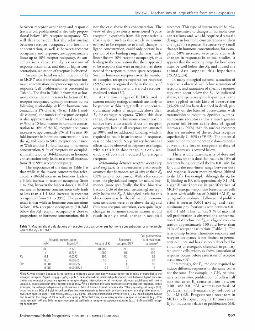
An example based on administration of E2to MCF-7 cells of the relationship between hor-mone concentration, receptor occupancy, and aresponse (cell proliferation) is presented inTable 1. The data in Table 1 show that as hor-mone concentration increases by factors of 10,receptor occupancy typically increases by thefollowing relationship: a) If the hormone con-centration is 1% of its Kd (% Kd: Table 1, mid-dle column), the number of receptors occupiedis also approximately 1% of total receptors.b) With a 10-fold increase in hormone concen-tration to 10% of the Kd, receptor occupancyincreases to approximately 9%. c) The next 10-fold increase in hormone concentration is tothe Kd and leads to 50% receptor occupancy.d) With another 10-fold increase in hormoneconcentration, 91% of receptors are occupied.e) Finally, another 10-fold increase in hormoneconcentration only leads to a small increase,from 91 to 99% receptor occupancy.
The importance of the data in Table 1 isthat while at the lowest concentration refer-enced, a 10-fold increase in hormone leads toa 9-fold increase in receptor occupancy (from1 to 9%), between the highest doses, a 10-foldincrease in hormone concentration only leadsto less than a 1.1-fold increase in receptoroccupancy (from 91 to 99%). The practicalresult is that while at hormone concentrationsbelow 10% receptor occupancy (10-foldbelow the Kd) receptor occupancy is close toproportional to hormone concentration, this is
not the case above this concentration. Theview of the previously mentioned “sparereceptor” hypothesis from this perspective isthat a system such as this, which we assumeevolved to be responsive to small changes inligand concentration, could only operate in aportion of the binding range that was nearlylinear (below 10% receptor occupancy), thusleading to the observation that there appearedto be receptors that were in surplus over thoseneeded for responses, hence spare receptors.Surplus hormone receptors over the numberof occupied receptors required for response(50,51) was recognized early in the study ofthe steroid receptors and steroid receptor-mediated action (52).
At the dose ranges of EEDCs used incurrent toxicity testing, chemicals are likely tobe present within target cells at concentra-tions many orders of magnitude above theirKd for estrogen receptors. Within this doserange, changes in hormone concentrationcannot have a detectable effect on receptoroccupancy, because all receptors are saturatedat 100% and no additional binding, which isrequired to result in an increase in response,can be observed. No primary hormonaleffects can be observed in response to changeswithin this high-dose range, but only sec-ondary effects not mediated by estrogenreceptors.
Relationship between receptor occupancyand response. It is sometimes erroneouslyassumed that hormones act in vivo at their Kd(50% receptor occupancy). With a few excep-tions, the physiological ranges for natural hor-mones (more specifically, the free, bioactivefraction (7,8) of the total circulating) are typi-cally below the Kd. A biological basis for thisobservation may be that if natural hormoneconcentrations were at or above the Kd andthus near receptor saturation, even quite largechanges in hormone concentrations wouldresult in only a small change in occupied
receptors. This type of system would be rela-tively insensitive to changes in hormone con-centrations and would require dramaticchanges in hormone concentrations to elicitchanges in response. Because very smallchanges in hormone concentrations, for exam-ple, a 50% increase, were associated withchanges in responses in animal studies, itappears that the working range for hormonesmust be well below the Kd, and indeed theanimal data support this hypothesis(19,23,53,54).
In many biological systems, saturation ofresponse is observed well below saturation ofreceptors, and saturation of specific responsesmay even occur below the Kd. As indicatedabove, the spare receptor hypothesis is theterm applied to this kind of observation(55–58) and has been described in detail, par-ticularly on the basis of observations withtransmembrane receptors. Specifically, trans-membrane receptors show a much greaterpercent inhibition as the dose of ligandincreases (~ 90%) than do nuclear receptorsthat are members of the nuclear receptorsuperfamily (~ 50%) (59,60). The potentialcontribution to nonmonotonic dose–responsecurves of the loss of receptors as dose ofligand increases is covered below.
There is only near-linearity of dose andoccupancy up to a dose that results in 10% ofreceptors being occupied (below 0.01 nM forE2), and the near-linear range between doseand response is even more restricted (shiftedto the left). For example, although the Kd forE2 binding to ER-α is approximately 0.1 nM,a significant increase in proliferation ofMCF-7 estrogen-responsive breast cancer cellsis seen with addition of 0.0004 nM E2 toestrogen-free medium. Half-maximal prolifer-ation is seen at 0.001 nM E2, and near-maximum proliferation is seen between 0.01and 0.1 nM. Thus, almost 91% of maximalcell proliferation is observed at a concentra-tion 10-fold below the Kd, at a ligand concen-tration approximately 100-fold lower than91% of receptor saturation (Table 1). Therelationship between hormone response andreceptor occupancy is not limited to perma-nent cell lines and has also been described fora number of estrogenic chemicals in primaryrat uterine cells, where, as above, saturation ofresponse occurs before saturation of receptoroccupancy (61).
Interestingly, for E2, the dose required toinduce different responses in the same cell isnot the same. For example, in GH3 rat pitu-itary cells in vitro, proliferation of cells is halfmaximal at an E2 concentration between0.001 and 0.01 nM, whereas synthesis ofprolactin is half-maximally induced at0.1 nM (62). Progesterone receptors inMCF-7 cells require roughly 10 times moreE2 for induction relative to proliferation (63),
Review | Mechanisms of large effects from small exposures
Environmental Health Perspectives • VOLUME 111 | NUMBER 8 | June 2003 997
Table 1. Mathematical calculations of receptor occupancy versus hormone concentration for an examplewhere the Kd = 0.1 nM.a
Cell proliferation,Estradiol concentration Receptors (% of maximum
(nM) (ng/mL)b Percent of Kd occupied (%)c response)d
10 2.72 10,000 99 1001 0.272 1,000 91 100
Kd 0.1 0.0272 100 50 990.01 0.00272 10 9 91
PRe 0.001 0.000272 1 1 500.0001 0.0000272 0.1 0.1 9
aThis Kd was chosen because it represents a midrange value commonly measured for the binding of estradiol to theestrogen receptor. bng/mL = ng/g = µg/kg = ppb. cThe mathematical relationship described here between ligand concen-tration and receptor occupancy applies to receptor–ligand interactions for all hormones, although each ligand will have aunique Kd associated with 50% receptor occupancy. dThis column in the table represents a physiological response, in thisexample, the estrogen-dependent proliferation of MCF-7 human breast cancer cells. eThe physiological range (PR),occurring at an EC50 of 1 pM for cell proliferation, was determined from both in vitro stimulation of cell proliferation at 1pM = 0.27 pg/mL (Figure 1) and free E2 at EC50 = 0.2 pg/mL (54), and in vivo studies where free E2 = 0.21 to 0.54 pg/mL (23,53)and is within the range of 1% receptor occupancy. Note that here, as in many systems, response saturates (e.g., 99%response at 0.1 nM and 50% receptor occupancy) well before receptor occupancy saturates (e.g., 10 nM and 99% recep-tor occupancy).
similar to induction of prolactin in GH3cells. This relationship demonstrates that theactivation of different genes requires differentnumbers of receptors to be occupied.Importantly, both of these responses saturateat a percent receptor occupancy far belowreceptor saturation, that is, spare receptorkinetics still apply.
Nonmonotonic Dose Responseto EstrogensNonmonotonic (inverted-U) dose–responserelationships: in vitro effects of low and highdoses of estrogens. Responses to hormones,including estrogens, saturate as does receptoroccupancy, and therefore cannot be linear as afunction of an increase in dose within thehigh-dose range. Further, for many responsesto a wide range of concentrations, acrossmany powers of 10-fold, the dose–responserelationship is nonmonotonic as well, withresponse decreasing at doses above those thatinitially reach a level of saturation. There are anumber of published examples of this in vivoand in vitro. In male mouse fetuses, a verysmall increase in E2 or a physiologicallyequivalent increase in estrogenic activity by anestrogenic chemical such as diethylstilbestrol(DES) resulted in prostate enlargementdetected later in life (23,64–66). In markedcontrast to these findings, consistent withnumerous prior studies, administration ofmuch higher doses of either natural or man-made estrogens during the prenatal or neona-tal period of prostate development caused areduction in prostate size relative to untreatedmales (23,64,66–69).
The lower doses of DES that resulted inan increase in prostate size (23,64,65) werepredicted to increase total serum estrogenicactivity within a physiological range, based onstudies of the free concentration of DES inserum (5) and transplacental transport ofradiolabeled DES in pregnant mice (47).Specifically, a low dose of DES of 0.02µg/kg/day administered to pregnant mice waspredicted to lead to an increase in free,bioavailable DES in the fetus that falls withinthe physiological dose range of free, bioavail-able estrogenic activity during normal fetaldevelopment (54); this exposure led to theprostate enlargement response (23). This doseof DES, in the physiological range of estro-genic activity, falls within the low-dose rangeof exposure. In contrast, in the same studies, a10,000-times higher dose of DES (200µg/kg/day) resulted in gross abnormalities inthe reproductive organs, including a markedreduction in prostate size (23,64). This doseof DES therefore falls within the toxicologicaldose range and represents a high-dose rangeof exposure.
There are many additional examples ofnonmonotonic dose–response relationships.
For example, it has been known for sometime that there are adverse effects at low andhigh doses, on either side of an optimumphysiological range for normal development,for other ligands that bind to receptors in thesteroid receptor superfamily, such as vitaminA and thyroid hormone. It is difficult to com-pile a literature focusing on inverted-Udose–response curves, as these types ofdose–response functions are common inendocrine studies and are often not identifiedin titles or abstracts as a noteworthy finding.Among those that have been reported, non-monotonic dose–response curves can occur atseveral levels of organization, ranging fromthe biochemical based on in vitro studies(28,54,62,70–75) to the organ or system levelbased on in vivo studies (23,60,66,76–82).
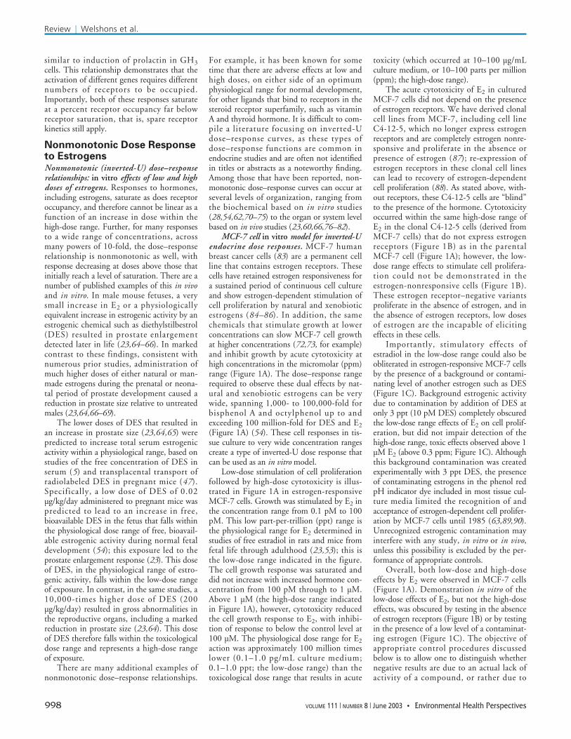
MCF-7 cell in vitro model for inverted-Uendocrine dose responses. MCF-7 humanbreast cancer cells (83) are a permanent cellline that contains estrogen receptors. Thesecells have retained estrogen responsiveness fora sustained period of continuous cell cultureand show estrogen-dependent stimulation ofcell proliferation by natural and xenobioticestrogens (84–86). In addition, the samechemicals that stimulate growth at lowerconcentrations can slow MCF-7 cell growthat higher concentrations (72,73, for example)and inhibit growth by acute cytotoxicity athigh concentrations in the micromolar (ppm)range (Figure 1A). The dose–response rangerequired to observe these dual effects by nat-ural and xenobiotic estrogens can be verywide, spanning 1,000- to 100,000-fold forbisphenol A and octylphenol up to andexceeding 100 million-fold for DES and E2(Figure 1A) (54). These cell responses in tis-sue culture to very wide concentration rangescreate a type of inverted-U dose response thatcan be used as an in vitro model.
Low-dose stimulation of cell proliferationfollowed by high-dose cytotoxicity is illus-trated in Figure 1A in estrogen-responsiveMCF-7 cells. Growth was stimulated by E2 inthe concentration range from 0.1 pM to 100pM. This low part-per-trillion (ppt) range isthe physiological range for E2 determined instudies of free estradiol in rats and mice fromfetal life through adulthood (23,53); this isthe low-dose range indicated in the figure.The cell growth response was saturated anddid not increase with increased hormone con-centration from 100 pM through to 1 µM.Above 1 µM (the high-dose range indicatedin Figure 1A), however, cytotoxicity reducedthe cell growth response to E2, with inhibi-tion of response to below the control level at100 µM. The physiological dose range for E2action was approximately 100 million timeslower (0.1–1.0 pg/mL culture medium;0.1–1.0 ppt; the low-dose range) than thetoxicological dose range that results in acute
toxicity (which occurred at 10–100 µg/mLculture medium, or 10–100 parts per million(ppm); the high-dose range).
The acute cytotoxicity of E2 in culturedMCF-7 cells did not depend on the presenceof estrogen receptors. We have derived clonalcell lines from MCF-7, including cell lineC4-12-5, which no longer express estrogenreceptors and are completely estrogen nonre-sponsive and proliferate in the absence orpresence of estrogen (87); re-expression ofestrogen receptors in these clonal cell linescan lead to recovery of estrogen-dependentcell proliferation (88). As stated above, with-out receptors, these C4-12-5 cells are “blind”to the presence of the hormone. Cytotoxicityoccurred within the same high-dose range ofE2 in the clonal C4-12-5 cells (derived fromMCF-7 cells) that do not express estrogenreceptors (Figure 1B) as in the parentalMCF-7 cell (Figure 1A); however, the low-dose range effects to stimulate cell prolifera-tion could not be demonstrated in theestrogen-nonresponsive cells (Figure 1B).These estrogen receptor–negative variantsproliferate in the absence of estrogen, and inthe absence of estrogen receptors, low dosesof estrogen are the incapable of elicitingeffects in these cells.
Importantly, stimulatory effects ofestradiol in the low-dose range could also beobliterated in estrogen-responsive MCF-7 cellsby the presence of a background or contami-nating level of another estrogen such as DES(Figure 1C). Background estrogenic activitydue to contamination by addition of DES atonly 3 ppt (10 pM DES) completely obscuredthe low-dose range effects of E2 on cell prolif-eration, but did not impair detection of thehigh-dose range, toxic effects observed above 1µM E2 (above 0.3 ppm; Figure 1C). Althoughthis background contamination was createdexperimentally with 3 ppt DES, the presenceof contaminating estrogens in the phenol redpH indicator dye included in most tissue cul-ture media limited the recognition of andacceptance of estrogen-dependent cell prolifer-ation by MCF-7 cells until 1985 (63,89,90).Unrecognized estrogenic contamination mayinterfere with any study, in vitro or in vivo,unless this possibility is excluded by the per-formance of appropriate controls.
Overall, both low-dose and high-doseeffects by E2 were observed in MCF-7 cells(Figure 1A). Demonstration in vitro of thelow-dose effects of E2, but not the high-doseeffects, was obscured by testing in the absenceof estrogen receptors (Figure 1B) or by testingin the presence of a low level of a contaminat-ing estrogen (Figure 1C). The objective ofappropriate control procedures discussedbelow is to allow one to distinguish whethernegative results are due to an actual lack ofactivity of a compound, or rather due to
Review | Welshons et al.
998 VOLUME 111 | NUMBER 8 | June 2003 • Environmental Health Perspectives
unresponsiveness of a tissue, or contamina-tion that is obscuring all responses.
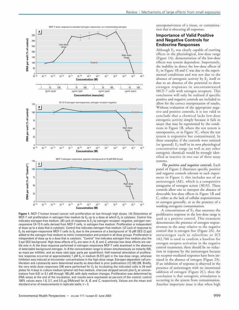
Importance of Valid Positiveand Negative Controls forEndocrine ResponsesAlthough E2 was clearly capable of exertingeffects in the physiological, low-dose range(Figure 1A), demonstration of the low-doseeffects was system dependent. Importantly,the inability to detect the low-dose effects ofE2 in Figure 1B and C was due to the experi-mental conditions and was not due to theabsence of estrogenic activity by E2 itself ordue to an absence of the potential to showestrogen responses in uncontaminatedMCF-7 cells with estrogen receptors. Thisconclusion will only be realized if specificpositive and negative controls are included toallow for the correct interpretation of results.Without evaluation of the appropriate nega-tive and positive controls, it is not valid toconclude that a chemical lacks low-doseestrogenic activity simply because it fails inassays that may be represented by the condi-tions in Figure 1B, where the test system isunresponsive, or in Figure 1C, where the testsystem is responsive but contaminated. Inthese examples, if the controls were omitted(or ignored), E2 itself in its own physiologicalconcentration range (as well as any otherestrogenic chemical) would be wrongly iden-tified as inactive in two out of three assaysystems.
The positive and negative controls. Eachpanel of Figure 2 illustrates specific positiveand negative controls relevant to each experi-ment in Figure 1; this includes use of anantiestrogen (AE), which is a competitiveantagonist of estrogen action (90,91). Thesecontrols allow one to interpret the absence ofdetectable low-dose effects in Figure 1B andC, either as the lack of cellular responsivenessto estrogen generally, or as the presence of amasking estrogenic contamination.
A concentration of E2 that saturates theproliferative response in the low-dose range isused as a positive control. This treatmentdemonstrates the presence of estrogen respon-siveness in the assay relative to the negativecontrol that is estrogen free (Figure 2A). Anantiestrogen such as raloxifene or ICI182,780 is used to confirm a baseline forestrogen receptor activation in the negativecontrol treatments; there should be no reduc-tion in response by the antiestrogen becauseno receptor-mediated responses have been ini-tiated in the absence of estrogen (Figure 2A).If an inhibition of response is observed in thepresence of antiestrogen with no intentionaladdition of estrogen (Figure 2C), then theconclusion is that estrogenic stimulation isoccurring in the system from contamination.Another important issue is that when high
Review | Mechanisms of large effects from small exposures
Environmental Health Perspectives • VOLUME 111 | NUMBER 8 | June 2003 999
Figure 1. MCF-7 human breast cancer cell proliferation at low through high doses. (A) Stimulation ofMCF-7 cell proliferation in estrogen-free medium by E2 up to a dose at which E2 is cytotoxic. Control lineindicates estrogen-free medium. (B) Lack of response to E2 by estrogen receptor–negative, estrogen-non-responsive C4-12-5 cells derived from MCF-7 cells, in estrogen-free medium. Proliferation is independentof dose up to a dose that is cytotoxic. Control line indicates estrogen-free medium. (C) Lack of response toE2 by estrogen-responsive MCF-7 cells to E2 due to the presence of a background of 10 pM DES (3 ppt)added to the estrogen-free medium to mimic contamination and present in all dose groups. Proliferation isindependent of dose up to a dose that is cytotoxic. “Control” line indicates estrogen-free medium plus the3 ppt DES background. High-dose effects of E2 are seen in A, B, and C, whereas low-dose effects are visi-ble only in A, the dose response performed in estrogen-responsive MCF-7 cells examined in the absenceof detectable background estrogen. In A the concentration range is shown simultaneously as molarity (M),as mass per milliliter, and as mass ratio (ppq: parts per quadrillion). Half-maximal stimulation of prolifera-tive response occurred at approximately 1 pM E2 in medium (0.272 ppt) in the low-dose range, whereasinhibition was induced at micromolar concentrations in the high-dose range. Estrogen-dependent cell pro-liferation and cytotoxicity were determined exactly as described in prior publications (72,138,139). Briefly,the very wide dose responses (54) were performed for E2 by incubating the indicated cells in 24-wellplates for 4 days in culture medium (phenol red-free medium, charcoal-stripped serum) plus E2 at concen-trations from 0.01 or 0.1 pM through 100 µM, with daily medium changes. Proliferation was determined byDNA assay at the end of the incubation, and results were expressed as percent of the control; control100% values were 1.0, 3.7, and 5.5 µg DNA/well for A, B, and C, respectively. Values are the mean andstandard error of measurements in replicate wells; n = 3.
A
C
B
MCF-7 dose-response to estradiol estrogen-responsive, no contaminating estrogen
C4-12-5 estrogen-nonresponsive, estrogen receptor-negative
10–7 10–6 10–5 10–4 10–3
Concentration (M)10–15 10–14 10–13 10–12 10–11 10–10 10–9 10–8 10–7 10–6 10–5 10–4 10–3
100 1 10 100 1 10 100fg/mL (ppq) pg/mL (ppt) ng/mL (ppb) µg/mL (ppm)
Concentration (mass/mL)
350
300
250
200
150
100
50
0
Control
Low-doserange
DN
A p
er w
ell (
% o
f con
trol
)
High-doserange
1 101 10 100
Concentration (M)10–15 10–14 10–13 10–12 10–11 10–10 10–9 10–8
125
100
75
50
25
0
Control
High-doserange
DN
A p
er w
ell (
% o
f con
trol
)
Concentration (M)10–15 10–14 10–13 10–12 10–11 10–10 10–9 10–8 10–7 10–6 10–5 10–4 10–3
125
100
75
50
25
0
“Control”
High-doserange
DN
A p
er w
ell (
% o
f “Co
ntro
l”)
MCF-7 estrogen-responsive, against a background of 10 pM DES (3 ppt)
doses of a chemical are being examined forestrogenic activity, after demonstrating thataddition of antiestrogen inhibits the response,competitive reversal of this inhibition ofresponse by co-incubation with an excess ofestrogen (for example, 10 nM E2) (Figure 2C)added with the antiestrogen is in turn used todistinguish antiestrogenic activity from toxic-ity due to the combined action of the testchemical and antiestrogen. This last step isthe final element in discriminating betweenantiestrogenic activity of a compound andacute toxicity (91).
Interpretation of the controls. In Figure2A, the positive control E2 at 100 pM stimu-lated response, and of equal importance,exposure to an antiestrogen at 100 nM (AE)in the absence of any E2 did not reduce theproliferative response below the control levelof growth. The interpretation drawn fromthe controls in Figure 2A is that a) the MCF-7 cell system was estrogen responsive, andimportantly, b) under the negative controlgrowth conditions, there was no detectablebackground estrogenic contamination. Inthis system, both low- and high-dose effectsof E2 were observed (Figure 1A).
Figure 2B shows the same controlsapplied to C4-12-5 cells, a clonal variant ofMCF-7 cells that lacks estrogen receptors.Positive control E2 did not stimulate cell pro-liferation, and furthermore, the antiestrogendid not inhibit proliferation of the C4-12-5cells (Figure 2B). The interpretation of thesecontrols is that the C4-12-5 cells are estrogennonresponsive, showing responses neither tolow-dose estrogen nor to antiestrogen.Importantly, even though the cells were notresponsive in the low-dose range of exposure,the proliferation of the estrogen receptor–negative C4-12-5 cells was still inhibited byE2 in the same high-dose range that inhibitedproliferation of the estrogen-responsive
MCF-7 cells (Figure 1B); only high-dosetoxic effects of E2 were observed, and theseare clearly not mediated by nuclear estrogenreceptors.
Finally, as can be seen in Figure 2C, evenin the same MCF-7 cells that were responsivewithin the low-dose range in the full doseresponse (Figure 1A), a very slight back-ground level (contamination) of an estrogenicchemical was sufficient to eliminate detectionof the low-dose stimulating effect of estradiol,if treatments are compared only with a nega-tive control that is presumed, without testing,to be estrogen-free. In Figure 2C, it can beseen that the positive control E2 added to the“Control” medium did not stimulate furthergrowth, and without further information, thesystem would be incorrectly interpreted asnonresponsive in the low-dose range (Figure1C). Incubating cells in the “Control”medium plus antiestrogen, however, inhibitedcell proliferation, indicating the potential foran estrogen receptor–driven stimulation ofcell growth. Competitive reversal of the antie-strogen effect with a surplus of E2, indicatedby the light blue bar in Figure 2C, confirmedthat the inhibition was antiestrogenic and notdue to nonspecific toxicity.
The interpretation of the dose–responseexperiment (Figure 1C) is now that theMCF-7 cells were fully responsive to E2 in thelow-dose range but were already maximallystimulated by background estrogenic contam-ination in the presumed negative control.DES at only 3 ppt was sufficient to fully maskthe low-dose effects of E2; only high-dose,toxic effects of E2 could be observed(Figure 1C). In the absence of the appropriatecontrols, or if the controls were misinter-preted or ignored, E2 itself, an unquestionedestrogen, would be incorrectly identified fromFigure 1B or C as an inactive chemical in thelow-dose range (its physiological range), but
not in the high-dose range, with respect toestrogen-dependent cell proliferation.
Implications. Positive and negativecontrols such as those described above areneeded for adequate interpretation of EEDCsin the context of low-dose effects, nonlinearsaturation of response, and reversal ofresponse that can generate a nonmonotonicdose–response relationship. Of great impor-tance, research on low-dose effects requires anew level of understanding of ambient estro-genic activities, and controls are absolutelyrequired to assess these activities experimen-tally. Ambient estrogenic activities for in vitrostudies consist of contaminants in air, media,or plastic, whereas in vivo, ambient estrogenicactivities could include variable backgroundlevels of endogenous hormone as well as activ-ity from a variety of external sources such asfeeds. Appropriate controls are not typicallyincluded in toxicological tests conducted forregulatory purposes.
Relevant to this discussion are findingsthat the concentration of E2 in cell culturemedium that results in proliferation atapproximately 50% of maximum is very closeto the concentrations of free serum E2 duringdevelopment in mouse and rat fetuses(0.2–0.3 pg/mL) (23,53). Even slight varia-tions in the levels of estradiol have beenrelated to differences in the course of develop-ment in mice, rats, and gerbils (19,23,92–94).For example, we experimentally increased thefree serum estradiol concentration in malemouse fetuses from the control level of0.2–0.3 pg/mL (via a Silastic capsule contain-ing estradiol implanted in the pregnant dam).This 0.1 pg/mL increase in free serum estra-diol resulted in a marked change in develop-ment of the urogenital system in the malefetuses (23).
Taken together, these findings indicate avery high degree of sensitivity (well below apart per trillion) of both human and rodenttissues to E2 both in vitro and in vivo. Thishigh degree of sensitivity to very small pertur-bations in E2 provides the basis for concernabout the use of appropriate controls to testfor background contamination by estrogenicchemicals in studies with animals. Estrogeniccontamination can occur via the food(95,96), caging (97), or bedding (98), as wellas in studies with cultured tissue via compo-nents of media (63), or plastic tubes andcultureware (99,100). Although there havebeen studies that have examined the effects ofcomponents of diets on steroid synthesis inhumans (101), this issue has not been a focusof toxicological studies involving EEDCs.Our recent findings show that in mice main-tained on different types of commercial ani-mal feeds during pregnancy, serum estradiollevels in fetuses are markedly different(unpublished observation).
Review | Welshons et al.
1000 VOLUME 111 | NUMBER 8 | June 2003 • Environmental Health Perspectives
DN
A p
er w
ell (
% o
f con
trol
)
DN
A p
er w
ell (
% o
f con
trol
)
DN
A p
er w
ell (
% o
f “Co
ntro
l”)Estrogen-responsive
MCF-7 cells400
350
300
250
200
150
100
50
0C E2 AE
A Nonresponsive C4-12-5 cellsB
C E2 AE
120
100
80
60
40
20
0“C” E2 AE AE +E2
10–8 M
MCF-7 cells + DES BackgroundC120
100
80
60
40
20
0
Figure 2. The relevant controls for the dose responses of Figure 1A–C. (A) Estrogen-responsive MCF-7cells in estrogen-free medium. (B) Estrogen receptor–negative, estrogen-nonresponsive C4-12-5 cellsderived from MCF-7 cells, in estrogen-free medium. (C) Controls. Estrogen-responsive MCF-7 cells in thepresence of a background of 10 pM DES (3 ppt) added to the estrogen-free medium and present in allmedia and treatments including controls. Abbreviations: AE, 100 nM antiestrogen (raloxifene or ICI182,780); AE + E2 10–8 M, 100 nM antiestrogen (raloxifene or ICI 182,780) plus E2 at 10–8 M; C, control estro-gen-free medium; “C”, estrogen-free medium plus 3 ppt DES; E2, 100 pM E2. Values are the mean and stan-dard error of measurements in replicate wells; n = 3.
Endocrine MechanismsMediating Errors in EstimatingLow-Dose Responses fromHigh-Dose Studies
The default risk assessment assumes linearityof dose response. Major errors in assessing riskcan be made when linearity of response andthe preceding receptor occupancy is assumedacross the entire dose range, which is the cur-rent assumption used in risk assessment.Although almost everyone involved in riskassessment recognizes that the assumption oflinearity is invalid (even for cancer) (102), theapplication of safety factors that results in lin-ear extrapolation across a wide dose rangeremains the default for current risk assess-ment. For example, safety factors (used to cal-culate a “safe” dose for human exposure) of10-fold each are often used to estimate eachof the following: human risk from animalstudies, to account for variability within thehuman population, when the lowest dosetested results in an adverse response (termedthe LOEL), and most recently, as an addedsafety factor for protecting children.Application of these 10-fold safety factorsresults in linear extrapolation from a LOEL orNOEL (determined by testing a few very high
doses) to arrive at a safe dose. Thus, inpractice, the model upon which risk assess-ment is practiced assumes that this linearextrapolation procedure is valid and will resultin calculation of a dose that is safe for humansexposure.
Error of a linear estimate relative toactual receptor occupancy. When a linearextrapolation model is applied to a saturating,receptor-mediated response to estimate therisk of an adverse response, this linear esti-mate results in a false assumption concerningthe actual reduction in response (and thusrisk) that occurs with decreasing dose. Theerror we refer to is illustrated in the simplifiedgraphic example in Figure 3. The use of 10-fold safety factors to estimate occupancy ofreceptors (and subsequent responses) on thebasis of results from animal studies assumes alinear relationship between dose and response,even though this may not be overtly acknowl-edged. We will initially discuss the theorybehind the error that occurs on the basis ofextrapolation from very high to very lowdoses assuming a linear function and thenprovide examples from actual data for DES,genistein, and bisphenol A obtained from invitro studies using MCF-7 cells. The error werefer to here based on receptor occupancy isin reality lower than the error based on actualresponses, as responses can saturate at lowerconcentrations than those required to achievereceptor saturation (Table 1). Therefore, ourcalculations of error in Table 2 are, in fact,conservative.
For simplicity here, in the discussionbelow we will not discriminate between doseadministered and dose at the estrogen recep-tor in target cells and will simply refer here toa test dose. The reason for this is that for invitro studies conducted in serum-freemedium, the administered dose and the doseavailable to bind to estrogen receptors are verysimilar (4). In vivo this is obviously not thecase due to absorption, metabolism, clear-ance, plasma binding, etc., all of which are farmore complicated to study in developingfetuses than in adults (54). It is nonethelessthe basis of modern endocrinology that a doseat target does exist, whether or not it can beeasily determined, and that this dose deter-mines the response and its magnitude relativeto the receptor occupancy it can generate.Our discussion here is meant to apply to thedose at target.
It is important to note that during fetaland early postnatal life, the pharmacokineticsof chemicals and drugs are markedly differentrelative to adulthood. In addition, pregnantand nonpregnant females also differ in thisregard. Data from studies with adult animalsthus cannot be used to predict the pharmaco-kinetics of chemicals in pregnant females andfetuses (16,103,104). Thus, evidence that a
particular chemical is cleared rapidly in anonpregnant adult cannot be used to dis-count the possibility of achieving a muchhigher dose at target in fetuses and neonates(46). Unfortunately, for most chemicals,there are no pharmacokinetic data and thusno basis for predicting dose at target for themost susceptible subpopulation: pregnantfemales and their fetuses.
The test dose for purposes of ourdiscussion here is a high dose administered intoxicological experiments that is used to pre-dict responses at much lower doses. As shownin Table 1 and Figure 3, the relationshipbetween hormone concentration and receptoroccupancy is approximately linear at lowreceptor occupancy (Figure 3, test dose exam-ple at 1/4 Kd). As the test dose exceeds therange of approximate linearity, for example, atest dose at 80% receptor occupancy (Figure3 at 4 × Kd), the linear model (linear extrapo-lation from test dose to zero dose) will clearlyunderestimate actual receptor occupancy andwill thus underestimate the actual responsesthat would occur at lower doses (Figure 3,arrow labeled “error of the linear estimate”).This deviation from linearity has great impor-tance with regard to the strategy of using veryhigh doses of EEDCs in toxicological studiesand extrapolating to predict responses atmuch lower doses.
Table 2 presents specific quantitativeinformation for a number of chemicals. Withregard to understanding the error that canoccur in estimating the potential for low-doseresponses on the basis of extrapolating fromhigh to low doses across a wide dose range, wewill describe an in vitro experiment in whichbisphenol A was examined in MCF-7 cells asan example. For our example here, the testdose for bisphenol A (shown in Table 2,row 1) is 844,000 ppb (844 mg/kg), chosenfor its relation to Kd for ER-α and for prox-imity to test doses administered in prior invivo toxicological studies of bisphenol A(again, using this as the dose at target) (14).Under the assumption that the test dose of844,000 ppb is within a linear response rangeand therefore within a linear receptor occu-pancy range for direct hormonal effects,reducing the dose by 50% (to a dose of422,000 ppb) would lead to the predictionthat receptor occupancy would also drop by50% (Table 2, row 2). In fact, because thetest concentration is so much higher than theKd, virtually no actual change in receptoroccupancy occurs (the actual change in recep-tor binding in MCF-7 cells would be from99.99 to 99.98% with this 50% reduction indose), and no change in response mediated bythese receptors would be detected.
When one administers a dose ofbisphenol A that is 10-fold lower than the testdose (84,400 ppb or 84.4 mg/kg), receptor
Review | Mechanisms of large effects from small exposures
Environmental Health Perspectives • VOLUME 111 | NUMBER 8 | June 2003 1001
Kd = 0.1 nM
1/4 Kd
4 x Kd
Occ
upan
cy (%
of m
axim
um) 100
80
60
40
20
0
Concentration (nM), linear scale0 0.1 0.2 0.3 0.4 0.5
Error of thelinear estimate
Figure 3. Error in predicting actual receptor occu-pancy based on linear estimation applied to a satu-rating test dose. Receptor occupancy (solid line) isgraphed against a linear scale of ligand concentra-tion from 0 to 0.5 nM, where the Kd for ligandbinding = 0.1 nM. Linear estimations to zeroconcentration (dotted lines) are shown originatingfrom single measurements at two test doses, onebelow the Kd (square point of origin, at 1/4 Kd =0.025 nM) and one above the Kd (round point of ori-gin, at 4 × Kd = 0.4 nM). This assumes no back-ground-contaminating estrogenic activity fromeither endogenous or exogenous sources otherthan the chemical being tested. Where the testdose used as the origin of the linear estimation isbelow the Kd, the linear estimation is very close toactual occupancy. Where the lowest test doseused as the origin of the linear estimation to zerodose is above the Kd, the linear estimation deviatessubstantially from actual receptor occupancy, indi-cated as “Error of the linear estimate.” The fold-underestimate of occupancy, and thereforeunderestimate of response for receptor-mediatedevents, increases as the origin of measurementincreases above the Kd and is calculated in Table 2for a number of EEDCs where the origin is 10,000-fold above the Kd, which could not be shown toscale on this figure.
occupancy still only drops from 99.98% to99.90% in MCF-7 cells (Table 2, row 3), andagain, this change is not likely to be adetectable decrease in binding. This decreasein dose also would thus not be likely to leadto a detectable decrease in response mediatedby these receptors. Even at a dose of 844 ppb,which is a dose 1,000 times lower than thetest dose of 844,000 ppb, 90.91% of recep-tors will still be occupied in MCF-7 cells. Onthe basis of the information presented inTable 1, one would not expect to approachthe region of maximum detectability for achange in response until doses that resulted inless than 50% receptor occupancy (the Kd)were reached. In addition, on the basis ofresults in Table 1, it is apparent that responsescan occur at concentrations in the range of1% receptor occupancy. As shown in Table 2,at the concentration of bisphenol A thatresults in approximately 1% receptor occu-pancy (0.844 ppb), or 1 million times lowerthan our initial test dose, the linear extrapola-tion model would have predicted negligiblereceptor binding, and thus no response, basedon a test dose of 844,000 ppb.
Nonmonotonic dose–response curve,response to endogenous hormone, and anassumed threshold dose all increase the mag-nitude of the error of a linear estimate. Ourcalculations are based on receptor occupancy,which is a physical chemical parameter sub-ject to less between-species variation andgreater precision of measurement than is themeasurement of response. Cellular responses,however, occur at doses associated with verylow receptor occupancy: the cell in essenceamplifies the receptor signal. Therefore, useof receptor occupancy is in fact conservativerelative to the ultimate physiologicalresponses on which risk assessment would bebased. For example, if these calculations werebased on the EC50 (effective concentration50%; 50% response) for a specific cellresponse such as cell proliferation that is 10-to 100-fold lower than the Kd (Table 1), thenthe underestimate of the potential for aresponse would be 10- to 100-fold higher, or
up to 1,000,000-fold, instead of the 10,000-fold in this example.
Incorporation of additional features ofreal-world risk assessment will further add tothe error, not reduce it. A nonmonotonicdose response, specifically the inverted U, cansubstantially increase the error of the linearestimate based on a high-dose reference point(that is well below the maximum responsebecause of the inverted-U dose–responsecurve). This is illustrated qualitatively inFigure 4A, where the error of the linear esti-mate for response is compared with that foran inverted-U dose response from a referencepoint above the dose that results in the maxi-mum response. To avoid the possibility ofthis type of error, it is necessary to examine amuch wider range of doses than is typical intoxicological studies involving animals.
Finally, as illustrated in Figure 4B, thedefault risk assessment applied to EEDCsassumes the existence of a threshold. Butwhen xenoestrogen activity is added to a nat-ural system that is already responding toendogenous estrogen such as estradiol, anythreshold in estrogenic response must alreadybe exceeded by the endogenous hormone.This absence of a threshold in response toexogenous estrogen has been experimentallyconfirmed in an experiment concerning theregulation by estrogen of sex determination inreptiles (22). The assumption of no responseup to an assumed threshold above the zeroEEDC dose, when this is not the case, willresult in a great, potentially infinite error iflinear extrapolation is used instead of actuallydetermining the shape of the dose–responsecurve (Figure 4B).
Figure 4B also depicts the error associatedwith examining a test chemical with estrogenicactivity, such as bisphenol A, that adds to anexisting background level of endogenousestradiol, which is variable because of endoge-nous and exogenous factors (19). Variation inendogenous estradiol is related to variation inphenotype in rodents (105), supporting thehypothesis that endogenous estrogen is alreadyabove threshold for estrogen-mediated
responses (22). There can thus be no thresh-old for responses to exogenous EEDCs. Thisfinding is important, as background levels ofendogenous estradiol markedly alter theresponse of fetuses to endocrine disruptorsadministered to pregnant mice and rats,including EEDCs such as bisphenol A(93,94). This issue is also relevant with regardto comparing effects of EEDCs at differentlife stages. During fetal life in males andfemales, pregnancy, or proestrus in females,estradiol levels are significantly higher thanduring postnatal life in males or prior topuberty and during diestrus in females (53).These marked differences in the backgroundlevels of estradiol will obviously influenceresponses to low doses of EEDCs. Theimportance of endogenous estradiol levels inthe response to low doses of EEDCs, whichhas been ignored in toxicological studies andin the models used in risk assessment, iscovered in more detail below.
Implications for current risk assessment.For an EEDC such as bisphenol A, with a rel-ative estrogenic activity approximately10,000-fold less than E2 in MCF-7 cells [butnot necessarily other tissues where it is muchmore active; (64)], the range of estrogenicactivity of this chemical equivalent to that ofphysiological E2 would be approximately0.05–30 ppb (0.05–30 ng/mL) within targetcells. There are now numerous publishedreports that bisphenol A shows estrogenicactivity at and below this concentration in avariety of cell culture systems (4,28,100,106–112). For example, Gupta (64) reportedthat a 50-pg/mL (50 ppt) dose of bisphenol Asignificantly stimulated prostate gland forma-tion and growth of the fetal mouse prostate inprimary culture, similar to a 0.5-pg/mL doseof DES. Bisphenol A stimulated humanprostate cancer cells to proliferate at a dose of1 nM (~ 0.23 ppb) (28).
The currently accepted LOEL dose ofbisphenol A of 50 mg/kg/day (15) wasreported from high-dose toxicological studies(14,113). This study is typical in that it useddoses 50,000–500,000-times higher than the
Review | Welshons et al.
1002 VOLUME 111 | NUMBER 8 | June 2003 • Environmental Health Perspectives
Table 2. Error in estimating responses to low doses, in the physiological range of estrogenic activity, for estradiol, DES, genistein, and bisphenol A as a result ofassuming linearity across the entire dose–response curve with regard to predicted versus actual estrogen receptor occupancy.
Actual Occupied receptors ≈ Fold underestimationEstradiol DES Genistein Bisphenol A receptors predicted by of response by
Row (ppb) (ppb) (ppb) (ppb) occupied (%) linear model (%) linear extrapolationa
1 test doseb 272 568 475,000 844,000 99.99 100 12 136 284 238,000 422,000 99.98 50 23 27.2 56.8 47,500 84,400 99.90 10 104 2.72 5.68 4,750 8,440 99.01 1 1005 0.272 0.568 475 844 90.91 0.1 9006 Kd
c 0.0272 0.0568 47.5 84.4 50 0.01 5,0007 0.00272 0.00568 4.75 8.44 9.09 0.001 9,0008 0.000272 0.000568 0.475 0.844 0.99 0.0001 10,000aFold underestimation of response by linear extrapolation is the actual receptors occupied divided by the predicted receptors occupied. bThe dose in row 1 is referred to in the text asthe "test dose," at a dose 10,000-times higher than each Kd; calculated from Kd values of 0.1 nM (0.0272 ppb) for estradiol (approximate), 0.212 nM (0.0568 ppb) for DES, 176 nM (47.5 ppb)for genistein, and 370 nM (84.4 ppb) for bisphenol A (4,5). cRow contains concentrations at the respective Kd of each compound.
2- and 20-µg/kg/day doses we administered topregnant mice on the basis of our calculationof an amount of bisphenol A that our prelimi-nary findings accurately predicted would bebioactive in male mouse fetuses (4). The
transplacental transport of bisphenol A hasnow been studied in greater detail in rodents(103,114–116), and the doses we used wouldresult in unconjugated bisphenol A levels inmouse fetuses that are within the range meas-ured in human umbilical cord blood (16,103).
Effects using low doses of bisphenol A,which are in the new low-dose range belowthe LOEL based on testing very high doses,have now been reported in rodent studies onmammary gland (117), vagina (118), prostate(4,64,65,119,120), sperm production(121,122), epididymis (64,121), rate ofembryonic development (123,124), pituitaryresponse to E2 (109), and rate of growth andtiming of puberty in females (93,125). Thereare also reports of effects of bisphenol A inmollusks, fish, and frogs at very low concen-trations, including below 1 µg/L (1 ppb)(126–132). Even though a few studies havereported no effects of low doses of bisphenolA, the weight of the evidence now clearlysupports that such effects occur in bothvertebrates and invertebrates.
It is also interesting that in two highlypublicized studies using low doses ofbisphenol A (133,134), no effects ofbisphenol A were found; in addition, noeffects of their positive control chemical,DES, were found. Although DES at the doseused was questioned as a valid positive controlby one of the groups (135), its validity as apositive control estrogen at the low doses usedin these studies was fully endorsed by theNational Institute of Environmental HealthSciences Low-Dose Peer Review Panel (11).In each of the two studies, the control ani-mals were obese (30% over normal bodyweight) relative to mice used in prior studiesthat used the same strain and age and thathad shown effects of fetal exposure to bisphe-nol A and positive control chemicals (4,136),including the same low dose of DES(23,64,82) used by Ashby et al. and Cagenet al. (133,134). The fact that the control ani-mals in both the Ashby and Cagen studieswere obese and had enlarged prostates andthen did not respond to either bisphenol A orthe positive control DES suggests that theinteraction of components of the diet withmanmade chemicals, such as bisphenol A, isan issue that requires further study; our recentstudies have confirmed this prediction(unpublished data). This also serves as anexample of the importance of attending toinformation provided by the appropriatenegative and positive controls (Figure 2),which these authors ignored (11).
Conclusions
Information about the mechanism of actionof EEDCs, together with information con-cerning mechanisms of hormone action, pre-dict that current risk assessment assumptions
can lead to a dramatic underestimation ofresponses (and thus risk) associated withexposure to low doses of EEDCs, particularlyduring development when the effects of verysmall changes in hormonal activity are perma-nent (54,64). The practice of examining onlya few very high doses and then extrapolatingto predict effects of doses thousands ormillions of time below those being studied isespecially problematic for endocrinedisruptors. The necessity for including lowdoses in the physiologically relevant range ofestrogenic activity, as opposed to only veryhigh doses, when testing for effects ofendocrine disruptors is dictated by a) evi-dence that estrogenic chemicals (as well asother hormone mimics or chemicals that oth-erwise interfere with endocrine function) canproduce nonmonotonic dose–response curveswhere responses both increase and decreaseacross the dose range, and b) the theoreticalabsence of a threshold for environmentalchemicals that operate via receptors (such asthe estrogen receptor) for endogenous ligands,such as E2; the threshold issue is covered inmore detail elsewhere (13,22). In addition,controls valid for the positive determinationof endocrine responsiveness must be included,and when included, interpreted appropriately,particularly when results that are apparentlynegative are obtained. The potential for errorinherent in drawing strong positive conclu-sions from purely negative data has clearly notbeen appreciated by some toxicologists(133,134), as well as regulators responsible forassessing this information.
Taken together, the above in vitrofindings show the substantial error that occursas a result of extrapolating on the basis offindings using very high doses to predicteffects at environmentally relevant doses,which are often thousands or millions oftimes lower than doses being tested.Responses to low doses of EEDCs should bedetermined by testing a much wider range ofdoses than the 50-fold range common in toxi-cological studies today (13), including dosesin the environmentally relevant range, and byaccounting for all sources of estrogenicactivity (endogenous and exogenous) andtheir interactive effect (137).
REFERENCES AND NOTES
1. Sheehan DM, Branham WS. Dissociation of estrogen-induced uterine growth and ornithine decarboxylaseactivity in the postnatal rat. Teratog Carcinog Mutagen7:411–422 (1987).
2. Waller CL, Oprea TI, Chae K, Park HK, Korach KS, LawsSC, Wiese TE, Kelce WR, Gray LE Jr. Ligand-based identi-fication of environmental estrogens. Chem Res Toxicol9:1240–1248 (1996).
3. Hong H, Tong W, Fang H, Shi L, Xie Q, Wu J, Perkins R,Walker JD, Branham W, Sheehan DM. Prediction of estro-gen receptor binding for 58,000 chemicals using an inte-grated system of a tree-based model with structuralalerts. Environ Health Perspect 110:29–36 (2002).
Review | Mechanisms of large effects from small exposures
Environmental Health Perspectives • VOLUME 111 | NUMBER 8 | June 2003 1003
Figure 4. (A) The error due to assuming that thedose–response curve is linear (dotted line) when, infact, the dose–response curve is nonmonotonic andforms an inverted U (solid blue line). The error inestimating actual responses that will occur atdoses below the test dose in a toxicological studyincreases as the concentration of the test doseincreases relative to a test dose that would result ina maximum response. This figure shows that themagnitude of the error in estimating responses atdoses below the test dose for an EEDC (using linearextrapolation) is greater when the dose–responsecurve is nonmonotonic relative to the error whenthe dose–response curve is monotonic (Figure 3).(B) This figure depicts the error associated withexamining a single dose of a test chemical (trian-gle) with estrogenic activity, such as bisphenol A,that adds to an existing background level ofendogenous estradiol, which is variable because ofendogenous and exogenous factors. In the currentmodel used in risk assessment, a linear extrapola-tion (dotted line) from the test dose (triangle) to anassumed threshold dose (circle) is used, based onthe assumption there will be an absence ofresponse at this assumed threshold dose. In thisfigure, the assumption is that endogenous estrogenis already above threshold for the estrogen recep-tor–mediated response to the EEDC (verticaldashed line). There can thus be no threshold for theresponse to the exogenous EEDC. The assumptionof no response at the assumed threshold EEDCdose, when this is not the case, will result in a greaterror, potentially infinite, in estimating the responseat this dose, if linear extrapolation from a high testdose is used instead of actually determining theshape of the dose–response curve.
0
100
Concentration (arbitrary units), linear scale
Inverted-U
Resp
onse
(% o
f max
imum
)
Error of thelinear estimate
0
100
Total concentration of estrogenic activityas E2 equivalent (arbitrary units)
Error of thelinear estimate
Threshold
Endogenous hormone
Assumed threshold
0 Endogenous E20 Added test xenoestrogen
A
B
Resp
onse
(% o
f max
imum
)
0 2 3 4 51
0 2 3 4 51
Response toEndogenous E2Test xenoestrogen
Inverted-U dose-response
Inverted-U dose-response, endogenous hormone, and threshold
Review | Welshons et al.
1004 VOLUME 111 | NUMBER 8 | June 2003 • Environmental Health Perspectives
4. Nagel SC, vom Saal FS, Thayer KA, Dhar MG, Boechler M,Welshons WV. Relative binding affinity-serum modifiedaccess (RBA-SMA) assay predicts the relative in vivobioactivity of the xenoestrogens bisphenol A andoctylphenol. Environ Health Perspect 105:70–76 (1997).
5. Nagel SC, vom Saal FS, Welshons WV. The effective freefraction of estradiol and xenoestrogens in human serummeasured by whole cell uptake assays: physiology ofdelivery modifies estrogenic activity. Proc Soc Exp BiolMed 217:300–309 (1998).
6. Nagel SC, vom Saal FS, Welshons WV. Developmentaleffects of estrogenic chemicals are predicted by an invitro assay incorporating modification of cell uptake byserum. J Steroid Biochem Mol Biol 69:343–357 (1999).
7. Ekins R, Edwards R, Newman B. Free Hormones in Blood,vol 3. New York:Elsevier Biomedical Press, 1982.
8. Mendel CM. The free hormone hypothesis: a physiologi-cally based mathematical model. Endocr Rev 10:232–274(1989).
9. Hsu ST, Ma CI, Hsu SK, Wu SS, Hsu NH, Yeh CC, Wu SB.Discovery and epidemiology of PCB poisoning in Taiwan: afour-year followup. Environ Health Perspect 59:5–10 (1985).
10. Quinn MM, Wegman DH, Greaves IA, Hammond SK,Ellenbecker MJ, Spark RF, Smith ER. Investigation ofreports of sexual dysfunction among male chemical work-ers manufacturing stilbene derivatives. Am J Ind Med18:55–68 (1990).
11. NTP. Final Report of the Endocrine Disruptors Low-Dose PeerReview Panel. In: Endocrine Disruptors Low-Dose Peer Review.Research Triangle Park, NC:National Toxicology Program, 2001.Available: http://ntp-server.niehs.nih.gov/htdocs/liason/LowDoseWebPage.html [accessed 28 May 2003].
12. Calabrese EJ, Baldwin LA. The dose determines the stimu-lation (and poison): development of a chemical hormesisdatabase. Int J Toxicol 16:545–559 (1997).
13. vom Saal FS, Sheehan DM. Challenging risk assessment.Forum Appl Res Public Policy:11–18 (1998).
14. Morrissey RE, George JD, Price CJ, Tyl RW, Marr MC,Kimmel CA. The developmental toxicity of bisphenol A inrats and mice. Fundam Appl Toxicol 8:571–582 (1987).
15. Integrated Risk Information System (IRIS). Bisphenol A.(CASRN 80-05-7), Vol 2002. US-EPA IRIS Substance File.Available: http://www.epa.gov/iris/subst/0356.htm[accessed 28 May 2003].
16. Schönfelder G, Wittfoht W, Hopp H, Talsness CE, Paul M,Chahoud I. Parent bisphenol A accumulation in humanmaternal-fetal-placental unit. Environ Health Perspect110:A703–A707 (2002).
17. Bern HA. The fragile fetus. In: Chemically-InducedAlterations in Sexual and Functional Development: TheWildlife/Human Connection (Colborn T, Clement C, eds).Vol 21. Princeton, NJ:Princeton Scientific Publishing,1992;9–15.
18. Colborn T, Clement C. Chemically-induced alterations insexual and functional development: the wildlife/humanconnection. In: Advances in Modern EnvironmentalToxicology (Mehlman MA, ed). Vol 21. Princeton,NJ:Princeton Scientific Publishing, 1992;403.
19. vom Saal FS. Sexual differentiation in litter-bearing mam-mals: influence of sex of adjacent fetuses in utero. J AnimSci 67:1824–1840 (1989).
20. Newbold R. Cellular and molecular effects of developmen-tal exposure to diethylstilbestrol: implications for otherenvironmental estrogens. Environ Health Perspect103:83–87 (1995).
21. Li S, Washburn KA, Moore R, Uno T, Teng C, Newbold RR,McLachlan JA, Negishi M. Developmental exposure todiethylstilbestrol elicits demethylation of estrogen-respon-sive lactoferrin gene in mouse uterus. Cancer Res57:4356–4359 (1997).
22. Sheehan DM, Willingham E, Gaylor D, Bergeron JM,Crews D. No threshold dose for estradiol-induced sexreversal of turtle embryos: how little is too much? EnvironHealth Perspect 107:155–159 (1999).
23. vom Saal FS, Timms BG, Montano MM, Palanza P, ThayerKA, Nagel SC, Dhar MD, Ganjam VK, Parmigiani S,Welshons WV. Prostate enlargement in mice due to fetalexposure to low doses of estradiol or diethylstilbestrol andopposite effects at high doses. Proc Natl Acad Sci USA94:2056–2061 (1997).
24. Sheehan DM, vom Saal FS. Low dose effects of hormones:a challenge for risk assessment. Risk Policy Rep 4:31–39(1997).
25. Crews D, Willingham E, Skipper JK. Endocrine disruptors:present issues, future directions. Q Rev Biol 75:243–260(2000).
26. National Research Council. Hormonally Active Agents inthe Environment. Washington, DC:National AcademyPress, 1999.
27. Newbold RR, Banks EP, Bullock B, Jefferson WN. Uterineadenocarcinoma in mice treated neonatally with genis-tein. Cancer Res 61:4325–4328 (2001).
28. Wetherill YB, Petre CE, Monk KR, Puga A, Knudsen KE.The xenoestrogen bisphenol A induces inappropriateandrogen receptor activation and mitogenesis in prostaticadenocarcinoma cells. Mol Cancer Ther 1:515–524 (2002).
29. Delclos KB, Bucci TJ, Lomax LG, Latendresse JR,Warbritton A, Weis CC, Newbold RR. Effects of dietarygenistein exposure during development on male andfemale CD (Sprague-Dawley) rats. Reprod Toxicol15:647–663 (2001).
30. Guo TL, White KL, Brown RD, Delclos KB, Newbold RR,Weis C, Germolec DR, McCay JA. Genistein modulatessplenic natural killer cell activity, antibody-forming cellresponse, and phenotypic marker expression in F-0 andF-1 generations of Sprague-Dawley rats. Toxicol ApplPharmacol 181:219–227 (2002).
31. vom Saal FS, Sheehan DM, Welshons WV. Unpublisheddata.
32. Greenspan FS, Strewler GJ. Basic and ClinicalEndocrinology. Stamford, CT:Appleton & Lange, 1997.
33. Tsai M, Clark JH, Schrader WT, O’Malley BW.Mechanisms of action of hormones that act as transcrip-tion-regulatory factors. In: Will iams Textbook ofEndocrinology (Wilson JD, Foster DW, Kronenberg HM,Larsen PR, eds). Philadelphia:W.B. Saunders Co.,1998;55–94.
34. Hawkins MB, Thornton JW, Crews D, Skipper JK, Dotte A,Thomas P. Identification of a third distinct estrogen recep-tor and reclassification of estrogen receptors in teleosts.Proc Natl Acad Sci USA 97:10751–10756 (2000).
35. Thornton JW. Evolution of vertebrate steroid receptorsfrom an ancestral estrogen receptor by ligand exploitationand serial genome expansions. Proc Natl Acad Sci USA98:5671–5676 (2001).
36. Judy BM, Welshons WV. Cellular localization of receptorsmediating actions of steroid hormones. In: Handbook ofPhysiology. Section 7: The Endocrine System. Vol I:Cellular Endocrinology (Conn PM, ed). New York:OxfordUniversity Press, 1998;437–460.
37. Falkenstein E, Wehling M. Nongenomically initiatedsteroid actions. Eur J Clin Invest 30 (suppl 3):51–54 (2000).
38. Levin ER. Cell localization, physiology, and nongenomicactions of estrogen receptors. J Appl Physiol 91:1860–1867(2001).
39. Mendelsohn ME. Genomic and nongenomic effects ofestrogen in the vasculature. Am J Cardiol 90:3F–6F (2002).
40. Katzenellenbogen BS, Katzenellenbogen JA, Mordecai D.Zearalenones: characterization of the estrogenic poten-cies and receptor interactions of a series of fungal beta-resorcylic acid lactones. Endocrinology 105:33–40 (1979).
41. Pakdel F, Le Guellec C, Vaillant C, Le Roux MG, Valotaire Y.Identification and estrogen induction of two estrogenreceptors (ER) messenger ribonucleic acids in the rain-bow trout liver: sequence homology with other ERs. MolEndocrinol 3:44–51 (1989).
42. White R, Jobling S, Hoare SA, Sumpter JP, Parker MG.Environmentally persistent alkylphenolic compounds areestrogenic. Endocrinology 135:175–182 (1994).
43. Paige LA, Christensen DJ, Gron H, Norris JD, Gottlin EB,Padilla KM, et al. Estrogen receptor (ER) modulators eachinduce distinct conformational changes in ER alpha andER beta. Proc Natl Acad Sci USA 96:3999–4004 (1999).
44. Shang Y, Brown M. Molecular determinants for the tissuespecificity of SERMs. Science 295:2465–2468 (2002).
45. Favier B, Dolle P. Developmental functions of mammalianHox genes. Mol Hum Reprod 3:115–131 (1997).
46. Fischer LJ, Weissinger JL. Development in the newbornrat of the conjugation and de-conjugation processesinvolved in the enterohepatic circulation of diethylstilboe-strol. Xenobiotica 2:399–412 (1972).
47. Shah HC, McLachlan JA. The fate of diethylstilbestrol inthe pregnant mouse. J Pharmacol Exp Ther 197:687–696(1976).
48. LeRoith D, Delahunty G, Wilson GL, Roberts CT, Shemer J,Hart C, Lesniak MA, Shiloach J, Roth J. Evolutionary
aspects of the endocrine and nervous systems. RecentProg Horm Res 42:549–587 (1986).
49. Fox TO. Androgen and estrogen binding macromoleculesin developing mouse brain: biochemical and genetic evi-dence. Proc Natl Acad Sci USA 72:4303–4307 (1975).
50. Ariens EJ, ed. Molecular Pharmacology, Vol 1. NewYork:Academic Press, 1964.
51. Kier LB. Molecular Orbital Theory in Drug Research. NewYork:Academic Press, 1971.
52. King RJB, Mainwaring WIP. Steroid-Cell Interactions.London:Butterworths, 1974.
53. Montano MM, Welshons WV, vom Saal FS. Free estradiolin serum and brain uptake of estradiol during fetal andneonatal sexual differentiation in female rats. Biol Reprod53:1198–1207 (1995).
54. Welshons WV, Nagel SC, Thayer KA, Judy BM, vom SaalFS. Low-dose bioactivity of xenoestrogens in animals: fetalexposure to low doses of methoxychlor and other xeno-estrogens increases adult prostate weight. Toxicol IndHealth 15:12–25 (1999).
55. Furchgott RF. The pharmacology of vascular smooth mus-cle. Pharmacol Rev 7:183 (1955).
56. Nickerson M. Receptor occupancy and tissue response.Nature 178:697–698 (1955).
57. Stephenson RP. A modification of receptor theory. Br JPharmacol 11:379–393 (1956).
58. Zhu BT. The competitive and noncompetitive antagonismof receptor-mediated drug actions in the presence ofspare receptors. J Pharmacol Toxicol Methods 29:85–91(1993).
59. Horwitz KB, McGuire WL. Nuclear mechanisms of estro-gen action. Effects of estradiol and anti-estrogens onestrogen receptors and nuclear receptor processing. JBiol Chem 253:8185–8191 (1978).
60. Medlock KL, Forrester TM, Sheehan DM. Short-termeffects of physiological and pharmacological doses ofestradiol on estrogen receptor and uterine growth. JRecept Res 11:743–756 (1991).
61. Walent JH, Gorski J. Estrogen binding is a noncooperativeprocess in primary rat uterine cells. Endocrinology126:2383–2391 (1990).
62. Amara JF, Dannies PS. 17beta-Estradiol has a biphasiceffect on GH cell growth. Endocrinology 112:1141–1143(1983).
63. Welshons WV, Jordan VC. Adaptation of estrogen-depen-dent MCF-7 cells to low estrogen (phenol red-free) cul-ture. Eur J Cancer Clin Oncol 23:1935–1939 (1987).
64. Gupta C. Reproductive malformation of the male offspringfollowing maternal exposure to estrogenic chemicals.Proc Soc Exp Biol Med 224:61–68 (2000).
65. Gupta C. The role of estrogen receptor, androgen receptorand growth factors in diethylstilbestrol-induced program-ming of prostate differentiation. Urol Res 28:223–229(2000).
66. Putz O, Schwartz CB, Kim S, LeBlanc GA, Cooper RL, PrinsGS. Neonatal low-and high-dose exposure to estradiolbenzoate in the male rat. 1. Effects on the prostate gland.Biol Reprod 65:1496–1505 (2001).
67. Prins GS, Birch L. The developmental pattern of androgenreceptor expression in rat prostate lobes is altered afterneonatal exposure to estrogen. Endocrinology136:1303–1314 (1995).
68. Santti R, Newbold RR, Makela S, Pylkkanen L, McLachlanJA. Developmental estrogenization and prostatic neopla-sia. Prostate 24:67–78 (1994).
69. vom Saal FS, Finch CE, Nelson JF. Natural history andmechanisms of reproductive aging in humans, laboratoryrodents and other selected vertebrates. In: The Physiologyof Reproduction (Knobil E, Neil JD, eds). Vol 2. NewYork:Raven Press, 1994;1213–1314.
70. Martikainen PM, Makela SI, Santti RS, Harkonen PL,Suominen JJ. Interaction of male and female sex hor-mones in cultured rat prostate. Prostate 11:291–303 (1987).
71. Bigazzi M, Brandi ML, Bani G, Sacchi TB. Relaxin influ-ences the growth of MCF-7 breast cancer cells. Mitogenicand antimitogenic action depends on peptide concentra-tion. Cancer 70:639–643 (1992).
72. Welshons WV, Engler KS, Taylor JA, Grady LH, Curran EM.Lithium-stimulated proliferation and alteration of phospho-inositide metabolites in MCF-7 human breast cancer cells.J Cell Physiol 165:134–144 (1995).
73. Taylor JA, Grady LH, Engler KS, Welshons WV.Relationship of growth stimulated by lithium, estradiol and
EGF to phospholipase C activity in MCF-7 human breastcancer cells. Breast Cancer Res Treat 34:265–277 (1995).
74. Somjen D, Kohen F, Jaffe A, Amir-Zaltsman Y, Knoll E,Stern N. Effects of gonadal steroids and their antagonistson DNA synthesis in human vascular cells. Hypertension32:39–45 (1998).
75. Moosmann B, Behl C. The antioxidant neuroprotectiveeffects of estrogens and phenolic compounds are inde-pendent from their estrogenic properties. Proc Natl AcadSci USA 96:8867–8872 (1999).
76. Davis JM, Svendsgaard DJ. U-shaped dose-responsecurves: their occurrence and implications for risk assess-ment. J Toxicol Environ Health A 30:71–83 (1990).
77. Medlock KL, Lyttle CR, Kelepouris N, Newman ED,Sheehan DM. Estradiol down-regulation of the rat uterineestrogen receptor. Proc Soc Exp Biol Med 196:293–300(1991).
78. vom Saal FS, Nagel SC, Palanza P, Boechler M, ParmigianiS, Welshons WV. Estrogenic pesticides: binding relative toestradiol in MCF-7 cells and effects of exposure duringfetal life on subsequent territorial behaviour in male mice.Toxicol Lett 77:343–350 (1995).
79. Shelby MD, Newbold RR, Tully DB, Chae K, Davis VL.Assessing environmental chemicals for estrogenicityusing a combination of in vitro and in vivo assays. EnvironHealth Perspect 104:1296–1300 (1996).
80. Oberdorster E, Rittschof D, LeBlanc GA. Alteration of [14C]-testosterone metabolism after chronic exposure ofDaphnia magna to tributyltin. Arch Environ Contam Toxicol34:21–25 (1998).
81. Newbold RR, Jefferson WN, Banks EP. Developmentalexposure to low doses of diethylstilbestrol (DES) results inpermanent alteration in the reproductive tract [Abstract].In: Program of the 81st Annual Meeting of the EndocrineSociety, 12–15 June 1999, San Diego, California. Bethesda,MD:The Endocrine Society Press, 1999;261.
82. Alworth LC, Howdeshell KL, Ruhlen RL, Day JK, Huang H-M, Besch-Williford C, Lubahn DB, vom Saal FS. Uterineresponsiveness to estradiol and DNA methylation arealtered by fetal exposure to diethylsti lbestrol andmethoxychlor in CD-1 mice: effects of low versus highdoses. Toxicol Appl Pharmacol 183:10–22 (2002).
83. Soule HD, Vazquez J, Long A, Albert S, Brennan M. Ahuman cell line from a pleural effusion derived from abreast carcinoma. J Natl Cancer Inst 51:1409–1413 (1973).
84. Lippman ME, Bolan G. Estrogen responsive human breastcancer in long-term tissue culture. Nature 256:592–593(1975).
85. Lippman ME, Bolan G, Huff K. The effects of estrogens andantiestrogens on hormone-responsive human breast can-cer in long-term tissue culture. Cancer Res 36:4595–4601(1976).
86. Welshons WV, Murphy CS, Koch R, Calaf G, Jordan VC.Stimulation of breast cancer cells in vitro by the environ-mental estrogen enterolactone and the phytoestrogenequol. Breast Cancer Res Treat 10:169–175 (1987).
87. Curran EM, Welshons WV. Unpublished data.88. Oesterreich S, Zhang P, Guler RL, Sun X, Curran EM,
Welshons WV, Osborne CK, Lee AV. Re-expression ofestrogen receptor α in estrogen receptor α-negativeMCF-7 cells restores both estrogen and insulin-like growthfactor-mediated signaling and growth. Cancer Res61:5771–5777 (2001).
89. Berthois Y, Katzenellenbogen JA, Katzenellenbogen BS.Phenol red in tissue culture media is a weak estrogen:implications concerning the study of estrogen-responsivecells in culture. Proc Natl Acad Sci USA 83:2496–2500(1986).
90. Jordan VC, Murphy CS. Endocrine pharmacology of antie-strogens as antitumor agents. Endocr Rev 11:578–610(1990).
91. Jordan VC. Biochemical pharmacology of antiestrogenaction. Pharmacol Rev 36:245–276 (1984).
92. Clark MM, Galef BG Jr. Effects of intrauterine position onthe behavior and genital morphology of litter-bearingrodents. Dev Neuropsychology 14:197–211 (1998).
93. Howdeshell KL, Hotchkiss AK, Thayer KA, VandenberghJG, vom Saal FS. Exposure to bisphenol A advancespuberty. Nature 401:763–764 (1999).
94. Timms BG, Peterson RE, vom Saal FS. 2,3,7,8-Tetrachloro-dibenzo-p-dioxin interacts with endogenous estradiol to dis-rupt prostate gland morphogenesis in male rat fetuses.Toxicol Sci 67:264–274 (2002).
95. Boettger-Tong H, Murthy L, Chiappetta C, Kirkland JL,Goodwin B, Adlercreutz H. A case of a laboratory animalfeed with high estrogenic activity and its impact on in vivoresponses to exogenously administered estrogens.Environ Health Perspect 106:369–373. (1998).
96. Ruhlen RL, Sandner CM, Howdeshell KL, Taylor JA, BroseJ, Beckwith S, Bronson FH, Welshons WV, vom Saal FS.The effects of soy on obesity and related health issues[Poster Abstract]. In: Program of the EnvironmentalHormones meeting, 18–20 October 2001, New Orleans,Louisiana.New Orleans, LA:Tulane and Xavier Universities,2001.
97. Howdeshell KL, Peterman PH, Judy BM, Taylor JA, OrazioCE, Ruhlen RL, vom Saal FS, Welshons WV. Bisphenol A isreleased from used polycarbonate animal cages intowater at room temperature. Environ Health Perspectdoi:10.1289/ehp.5993 [Online 5 February 2003].
98. Markaverich B, Mani S, Alejandro MA, Mitchell A,Markaverich D, Brown T, Velez-Trippe C, Murchison C,O’Malley B, Faith R. A novel endocrine-disrupting agent incorn with mitogenic activity in human breast and prostaticcancer cells. Environ Health Perspect 110:169–177 (2002).
99. Soto AM, Justicia H, Wray JW, Sonnenschein C. p-Nonyl-phenol: an estrogenic xenobiotic released from “modified”polystyrene. Environ Health Perspect 92:167–173 (1991).
100. Krishnan AV, Stathis P, Permuth SF, Tokes L, Feldman D.Bisphenol-A: an estrogenic substance is released frompolycarbonate flasks during autoclaving. Endocrinology132:2279–2286 (1993).
101. Adlercreutz H, Bannwart C, Wahala K, Makela T, BrunowG, Hase T, Arosemena PJ, Kellis JT, Vickery LE. Inhibitionof human aromatase by mammalian lignans andisoflavonoid phytoestrogens. J Steroid Biochem Mol Biol44:147–153 (1993).
102. Hoel DG, Portier CJ. Nonlinearity of dose-response func-tions for carcinogenicity. Environ Health Perspect102(suppl 1):109–113 (1994).
103. Zalko D, Soto AM, Dolo L, Dorio C, Rathahao E, DebrauwerL, Faure R, Cravedi JP. Biotransformations of bisphenol Ain a mammalian model: answers and new questions raisedby low-dose metabolic fate studies in pregnant CD-1 mice.Environ Health Perspect 111:309–320 (2003).
104. Matsumoto J, Yokota H, Yuasa A. Developmentalincreases in rat hepatic microsomal UDP-glucuronosyl-transferase activities toward xenoestrogens anddecreases during pregnancy. Environ Health Perspect110:193–196 (2002).
105. vom Saal FS, Clark MM, Galef BG Jr, Drickamer LC,Vandenbergh JG. The intrauterine position (IUP) phenom-enon. In: Encyclopedia of Reproduction (Knobil E, Neill J,eds). New York:Academic Press, 1999;893–900.
106. Brotons JA, Olea-Serrano MF, Villalobos M, Pedraza V,Olea N. Xenoestrogens released from lacquer coating infood cans. Environ Health Perspect 103:608–612 (1995).
107. Sonnenschein C, Soto AM, Fernandez MF, Olea N, Olea-Serrano MF, Ruiz-Lopez MD. Development of a marker ofestrogenic exposure in human serum. Clin Chem41:1888–1895 (1995).
108. Olea N, Pulgar R, Perez P, Olea-Serrano F, Rivas A,Novillo-Fertrell A, Pedraza V, Soto AM, Sonnenschein C.Estrogenicity of resin-based composites and sealantsused in dentistry. Environ Health Perspect 104:298–305(1996).
109. Steinmetz R, Brown NG, Allen DL, Bigsby RM, Ben-Jonathan N. The environmental estrogen bisphenol Astimulates prolactin release in vitro and in vivo .Endocrinology 138:1780–1786 (1997).
110. Bolger R, Wiese TE, Ervin K, Nestich S, Checovich W.Rapid screening of environmental chemicals for estrogenreceptor binding capacity. Environ Health Perspect106:551–557 (1998).
111. Gould JC, Leonard LS, Maness SC, Wagner BL, Conner K,Zacharewski T, Safe S, McDonnell DP, Gaido KW.Bisphenol A interacts with the estrogen receptor alpha ina distinct manner from estradiol. Mol Cell Endocrinol142:203–214 (1998).
112. Celius T, Haugen TB, Grotmol T, Walther BT. A sensitivezonagenetic assay for rapid in vitro assessment of estro-genic potency of xenobiotics and mycotoxins. EnvironHealth Perspect 107:63–68 (1999).
113. NTP. Bisphenol A: Reproduction and Fertility in CD-1When Administered in the Feed NTP. Document 85-192.Research Triangle Park, NC:National Toxicology Program.
114. Takahashi O, Oishi S. Disposition of orally administered2,2-bis(4-hydroxyphenyl)propane (bisphenol A) in pregnantrats and the placental transfer to fetuses. Environ HealthPerspect 108:931–935 (2000).
115. Shin BS, Yoo SD, Cho CY, Jung JH, Lee BM, Kim JH, et al.Maternal-fetal disposition of bisphenol a in pregnantSprague-Dawley rats. J Toxicol Environ Health A 65:395–406(2002).
116. Ikezuki Y, Tsutsumi O, Takai Y, Kamei Y, Taketani Y.Determination of bisphenol A concentrations in humanbiological fluids reveals significant early prenatal expo-sure. Human Reprod 17:2839–2841 (2002).
117. Markey CM, Luque EH, Munoz De Toro M, SonnenscheinC, Soto AM. In utero exposure to bisphenol A alters thedevelopment and tissue organization of the mouse mam-mary gland. Biol Reprod 65:1215–1223 (2001).
118. Schönfelder G, Flick B, Mayr E, Talsness C, Paul M,Chahoud I. In utero exposure to low doses of bisphenol Alead to long-term deleterious effects in the vagina.Neoplasia 4:98–102 (2002).
119. Elswick BA, Welsch F, Janszen DB. Effect of differentsampling designs on outcome of endocrine disruptor stud-ies. Reprod Toxicol 14:359–367 (2000).
120. Ramos JG, Varayoud J, Sonnenschein C, Soto AM, MunozDe Toro M, Luque EH. Prenatal exposure to low doses ofbisphenol A alters the periductal stroma and glandular cellfunction in the rat ventral prostate. Biol Reprod65:1271–1277 (2001).
121. vom Saal FS, Cooke PS, Buchanan DL, Palanza P, ThayerKA, Nagel SC, Parmigiani S, Welshons WV. A physiologi-cally based approach to the study of bisphenol A andother estrogenic chemicals on the size of reproductiveorgans, daily sperm production, and behavior. Toxicol IndHealth 14:239–260 (1998).
122. Sakaue M, Ohsako S, Ishimura R, Kurosawa S, KurohmaruM, Hayashi Y, Aoki Y, Yonemoto J, Tohyama C. BisphenolA affects spermatogenesis in the adult rat even at lowdoses. J Occup Health 43:185–190 (2001).
123. Takai Y, Tsutsumi O, Ikezuki Y, Kamei Y, Osuga Y, Yano T,Taketan Y. Preimplantation exposure to bisphenol Aadvances postnatal development. Reprod Toxicol 15:71–74(2000).
124. Takai Y, Tsutsumi O, Ikezuki Y, Hiroi H, Osuga Y, MomoedaM, Yano T, Taketani Y. Estrogen receptor-mediated effectsof a xenoestrogen, bisphenol A, on preimplantation mouseembryos. Biochem Biophys Res Commun 270:918–921(2000).
125. Honma S, Suzuki A, Buchanan DL, Katsu Y, Watanabe H,Iguchi T. Low dose effect of in utero exposure to bisphenolA and diethylstilbestrol on female mouse reproduction.Reprod Toxicol 16:117–122 (2002).
126. Klos W, Lutz I, Einspanier R. Amphibians as a model tostudy endocrine disruptors. II: Estrogenic activity of envi-ronmental chemicals in vitro and in vivo. Sci Total Environ225:59–68 (1999).
127. Haubruge E, Petit F, Gage MJG. Reduced sperm counts inguppies (Poecilia reticulata) following exposure to lowlevels of tributyltin and bisphenol A. Proc R Soc Lond267:2333–2337 (2000).
128. Oehlmann J, Schulte-Oehlmann U, Tillmann M, Markert B.Effects of endocrine disruptors on prosobranch snails(Mollusca: Gastropoda) in the laboratory. Part I: BisphenolA and octylphenol as xeno-estrogens. Ecotoxicology9:383–397 (2000).
129. Metcalfe CD, Metcalfe TL, Kiparissis Y, Koenig BG, KhanC, Hughes RJ, Croley TR, March RE, Potter T. Estrogenicpotency of chemicals detected in sewage treatment planteffluents as determined by in vivo assays with Japanesemedaka (Oryzias latipes). Environ Toxicol Chem 20:297–308(2001).
130. Sohoni P, Tyler CR, Hurd K, Caunter J, Hetheridge M,Williams T, Woods C, Evans M, Toy R, Gargas M, SumpterJP. Reproductive effects of long-term exposure to bisphe-nol A in the fathead minnow (Pimephales promelas).Environ Sci Technol 35:2917–2925 (2001).
131. Tabota A, Kashiwada S, Ohnishi Y, Ishikawa H, MiyamotoN, Itoh M, et al. Estrogenic influences of estradiol-17β,o-nonylphenol, and bisphenol A on Japanese medaka(Oryzias latipes) at detected environmental concentra-tions. Water Sci Technol 43:109–116 (2001).
132. Watts MM, Pascoe D, Carroll K. Chronic exposureto 17alpha-ethinylestradiol and bisphenol A—effectson development and reproduction in the freshwater
Review | Mechanisms of large effects from small exposures
Environmental Health Perspectives • VOLUME 111 | NUMBER 8 | June 2003 1005
Review | Welshons et al.
1006 VOLUME 111 | NUMBER 8 | June 2003 • Environmental Health Perspectives
invertebrate Chironomus riparius (Diptera: Chironomidae).Aquat Toxicol 55:113–124 (2001).
133. Ashby J, Tinwell H, Haseman J. Lack of effects for lowdose levels of bisphenol A (BPA) and diethylstilbestrol(DES) on the prostate gland of CF1 mice exposed in utero.Regul Toxicol Pharmacol 30:156–166 (1999).
134. Cagen SZ, Waechter JM, Dimond SS, Breslin WJ, ButalaJH, Jekat FW, Joiner RL, Shiotsuka RN, Veenstra GE,Harris LR. Normal reproductive organ development in CF-1mice following prenatal exposure to bisphenol A. ToxicolSci 11:15–29 (1999).
135. Ashby J. Dose levels of 0.01-0.2 microg/kg/day diethyl-stilbestrol are not suitable for use as a positive control inendocrine toxicity studies. Regul Toxicol Pharmacol29:235–237 (1999).
136. Thayer KA, Ruhlen RL, Howdeshell KL, Buchanan D, CookePS, Welshons WV, vom Saal FS. Altered reproductive organsin male mice exposed prenatally to sub-clinical doses of17α-ethinyl estradiol. Hum Reprod 16:988–996 (2001).
137. Rajapakse N, Silva E, Kortenkamp A. Combining xeno-estrogens at levels below individual no-observed-effect-concentrations dramatically enhances steroid hormone
action. Environ Health Perspect 110:917–921 (2002).138. Grady LH, Nonneman DJ, Rottinghaus GE, Welshons WV.
pH-Dependent cytotoxicity of contaminants of phenol redfor MCF-7 breast cancer cells. Endocrinology129:3321–3330 (1991).
139. Welshons WV, Rottinghaus GE, Nonneman DJ, Dolan-Timpe M, Ross PF. A sensitive bioassay for detection ofdietary estrogens in animal feeds. J Vet Diagn Invest2:268–273 (1990).