A cembranoid protects acute hippocampal slices against paraoxon neurotoxicity
Upload
independentCategory
view
0download
0
at SciVerse ScienceDirect
European Journal of Medicinal Chemistry 67 (2013) 64e74
Contents lists available
European Journal of Medicinal Chemistry
journal homepage: http: / /www.elsevier .com/locate/ejmech
Short communication
Synthesis, pharmacological assessment, and molecular modelingof 6-chloro-pyridonepezils: New dual AChE inhibitors as potentialdrugs for the treatment of Alzheimer’s disease
Abdelouahid Samadi a,*, Mario de la Fuente Revenga b, Concepción Pérez b, Isabel Iriepa c,Ignacio Moraleda c, María Isabel Rodríguez-Franco b, José Marco-Contelles a,*
a Laboratorio de Química Médica, Instituto de Química Orgánica General (CSIC), C/Juan de la Cierva 3, 28006 Madrid, Spainb Instituto de Química Médica (IQM e CSIC), C/Juan de la Cierva 3, 28006 Madrid, SpaincDepartamento de Química Orgánica, Facultad de Farmacia, Universidad de Alcalá, Ctra. Barcelona, Km. 33.5, 28817 Alcalá de Henares, Spain
a r t i c l e i n f o
Article history:Received 17 April 2013Received in revised form31 May 2013Accepted 4 June 2013Available online 25 June 2013
Keywords:hAChEhBuChEDual AChE inhibitors6-Chloro-pyridonepezilsIn vitro blood brain barrierMolecular modelingADMEAlzheimer’s disease
* Corresponding authors. Fax: þ34 91 5644853.E-mail addresses: [email protected], asamadi1
yahoo.com (A. Samadi), [email protected] (J. Marco
0223-5234/$ e see front matter � 2013 Elsevier Mashttp://dx.doi.org/10.1016/j.ejmech.2013.06.021
a b s t r a c t
6-Chloro-pyridonepezils are chloropyridineedonepezil hybrids designed by combining the N-benzylpi-peridine moiety present in donepezil with the 2-chloropyridine-3,5-dicarbonitrile heterocyclic ringsystem, both connected by an appropriate polymethylene linker. 6-Chloro-pyridonepezils 1e8 wereprepared by reaction of 2,6-dichloro-4-phenylpyridine-3,5-dicarbonitrile (13) [or 2,6-dichloropyridine-3,5-dicarbonitrile (14)] with suitable 2-(1-benzylpiperidin-4-yl)alkylamines (9e12). The biologicalevaluation showed that these new compounds are cholinesterase inhibitors, in the submicromolar range,one of them (6) being a potent hBuChE inhibitor (IC50 ¼ 0.47 � 0.08 mM). 6-Chloro-pyridonepezils 4, 7 and8 are potent hAChE inhibitors showing IC50 in the 0.013e0.054 mM range. Particularly, 6-chloro-pyr-idonepezil 8 is 625-fold more selective for hAChE than for hBuChE and compared to donepezil is equi-potent for the inhibition of hAChE. Molecular modeling investigation on 6-chloro-pyridonepezils 4, 6e8supports its dual AChE inhibitory profile, by binding simultaneously at the catalytic active and at pe-ripheral anionic sites of the enzyme. The in vitro Blood Brain Barrier (BBB) and theoretical ADME analysisof 6-chloro-pyridonepezils 1e8 have been carried out. Overall, compound 8, is a permeable potent andselective dual AChEI that can be considered as a good candidate with potential impact for furtherpharmacological development in Alzheimer’s therapy.
� 2013 Elsevier Masson SAS. All rights reserved.
1. Introduction
Alzheimer’s disease (AD) is the most prevalent neurodegener-ative disorder [1]. AD is characterized by the gradual developmentof forgetfulness, progressing to disturbances in language, disori-entation, and mutism. The symptomatic course of the disease isgenerally five or more years of stepwise decline in memory andattention span [2]. Typical pathological hallmarks are extracellularsenile plaques, consisting principally of amyloid-b (Ab), and intra-cellular neurofibrillary tangles, which are composed of phosphor-ylated tau protein. Moreover, the basal nucleus of Meynertundergoes profound neuron loss, the neocortex exhibits a loss ofcholinergic fibers and receptors, and a decrease of both choline
[email protected], samadi72@-Contelles).
son SAS. All rights reserved.
acetyltransferase and acetylcholinesterase (AChE) enzyme activity[2,3].
Since the symptoms of AD were associated with an alteredcholinergic function, research has been focused on the basal fore-brain cholinergic system [3]. As a result, the cholinergic hypothesiswas developed, which postulated that a loss of cholinergic functionin the central nervous system contributed significantly to thecognitive decline associated with advanced age and AD [4]. Thus,drugs capable of inhibiting AChE might potentiate central cholin-ergic function, therefore improving cognition and perhaps evensome of the behavioral problems experienced by AD patients [5].
AChE inhibitors (AChEI) may inhibit AChE via a competitivemechanism, by interacting with the catalytic active site (CAS) of theenzyme, via a non-competitive mechanism, by binding with theperipheral anionic site (PAS), or via both mechanisms, by exerting adual binding AChE inhibition. For a while the treatment with AChEIwas reported to produce only symptomatic improvement, havingno effect in the course of the disease [6]. However, other studies
A. Samadi et al. / European Journal of Medicinal Chemistry 67 (2013) 64e74 65
indicated that AChE interacts with Ab by a hydrophobic environ-ment close to the PAS, thus promoting Ab fibril formation [7,8].Moreover, AChE-Ab complexes increase Ab-dependent neurotox-icity [9]. These reports arose a new interest in AChEI, demonstratingtheir ability to enhance the release of non-amyloidogenic solublederivatives of amyloid precursor protein (APP) in vitro and in vivo,and possibly to slow down the formation of amyloidogenic com-pounds in the brain [10]. The increase of soluble APP (APPs) wasalso consistent with AChE inhibition [11]. Numerous clinical trialshave shown the safety and efficacy of AChEIs in the treatment ofAD. Besides, there is growing evidence from preclinical studiesindicating that these agents can attenuate neuronal damage anddeath from cytotoxic insults, and therefore might affect AD path-ogenesis [12].
Considering the non-cholinergic aspects of AChE related to thePAS [7e9] in associating with Ab, an attractive target for the designof new antidementia drugs emerged. Peripheral or dual site in-hibitors of AChE may simultaneously alleviate the cognitive deficitin AD patients and prevent the assembly of Ab, which will delay theneurodegenerative process [13]. This strategy was pursued forseveral medicinal chemists who have been developing new com-pounds with dual AChE inhibitory activity [14e17], based on wellknown AChEIs such as tacrine [18], (E)-2-(4-(((7-(diethylamino)-2,4-dioxochroman-3-ylidene)methyl)amino)phenyl)-N0-(1,2,3,4-tetrahydroacridin-9-yl)acetohydrazide, a high-affinity, fluorescenttacrine-derived cholinesterase inhibitor able to bind to amyloidstructures, described by Gütschow and co-workers [19], riva-stigmine [20], donepezil [21] and galanthamine [22]. A recentclinical study with moderate-to-severe Alzheimer’s disease pa-tients determined that continued treatment with donepezil couldimprove cognition and function for even severe patients [23].
Several classes of donepezil hybrids such as donepeziletacrine[24,25], donepezileaminoacids [26], indanone hybrids [27], done-pezileaminothienoquinoline [28], or indanone and aurone de-rivatives [29] have been developed as dual binding site AChEinhibitors. The success of the dual binding site strategy is evidencedby the increased AChE inhibitory potency of these hybridscompared to the parent compounds from which they have beendesigned.
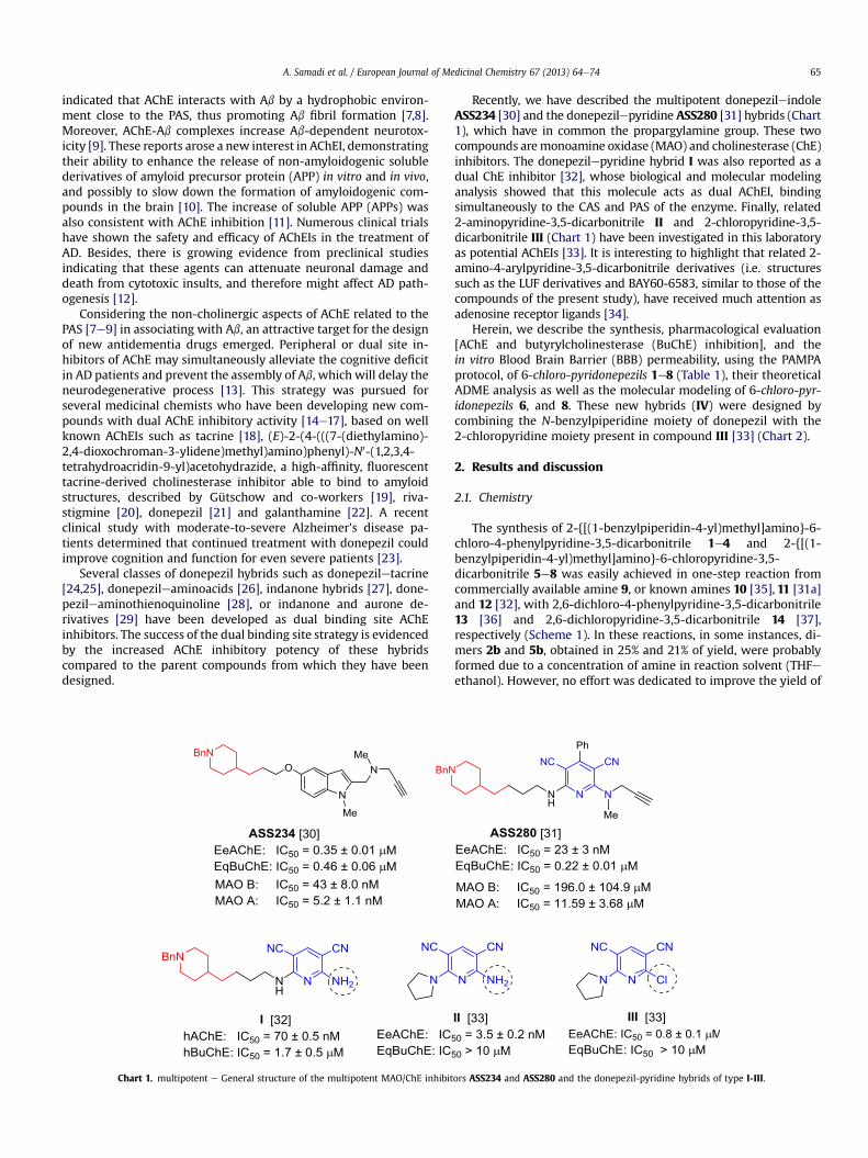
Chart 1. multipotent e General structure of the multipotent MAO/ChE inhibit
Recently, we have described the multipotent donepezileindoleASS234 [30] and the donepezilepyridine ASS280 [31] hybrids (Chart1), which have in common the propargylamine group. These twocompounds aremonoamine oxidase (MAO) and cholinesterase (ChE)inhibitors. The donepezilepyridine hybrid I was also reported as adual ChE inhibitor [32], whose biological and molecular modelinganalysis showed that this molecule acts as dual AChEI, bindingsimultaneously to the CAS and PAS of the enzyme. Finally, related2-aminopyridine-3,5-dicarbonitrile II and 2-chloropyridine-3,5-dicarbonitrile III (Chart 1) have been investigated in this laboratoryas potential AChEIs [33]. It is interesting to highlight that related 2-amino-4-arylpyridine-3,5-dicarbonitrile derivatives (i.e. structuressuch as the LUF derivatives and BAY60-6583, similar to those of thecompounds of the present study), have received much attention asadenosine receptor ligands [34].
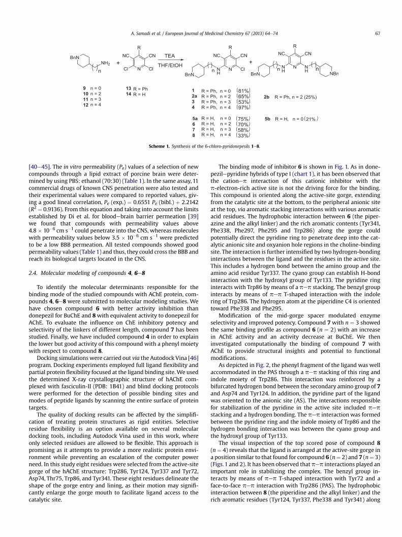
Herein, we describe the synthesis, pharmacological evaluation[AChE and butyrylcholinesterase (BuChE) inhibition], and thein vitro Blood Brain Barrier (BBB) permeability, using the PAMPAprotocol, of 6-chloro-pyridonepezils 1e8 (Table 1), their theoreticalADME analysis as well as the molecular modeling of 6-chloro-pyr-idonepezils 6, and 8. These new hybrids (IV) were designed bycombining the N-benzylpiperidine moiety of donepezil with the2-chloropyridine moiety present in compound III [33] (Chart 2).
2. Results and discussion
2.1. Chemistry
The synthesis of 2-{[(1-benzylpiperidin-4-yl)methyl]amino}-6-chloro-4-phenylpyridine-3,5-dicarbonitrile 1e4 and 2-{[(1-benzylpiperidin-4-yl)methyl]amino}-6-chloropyridine-3,5-dicarbonitrile 5e8 was easily achieved in one-step reaction fromcommercially available amine 9, or known amines 10 [35], 11 [31a]and 12 [32], with 2,6-dichloro-4-phenylpyridine-3,5-dicarbonitrile13 [36] and 2,6-dichloropyridine-3,5-dicarbonitrile 14 [37],respectively (Scheme 1). In these reactions, in some instances, di-mers 2b and 5b, obtained in 25% and 21% of yield, were probablyformed due to a concentration of amine in reaction solvent (THFeethanol). However, no effort was dedicated to improve the yield of
ors ASS234 and ASS280 and the donepezil-pyridine hybrids of type I-III.
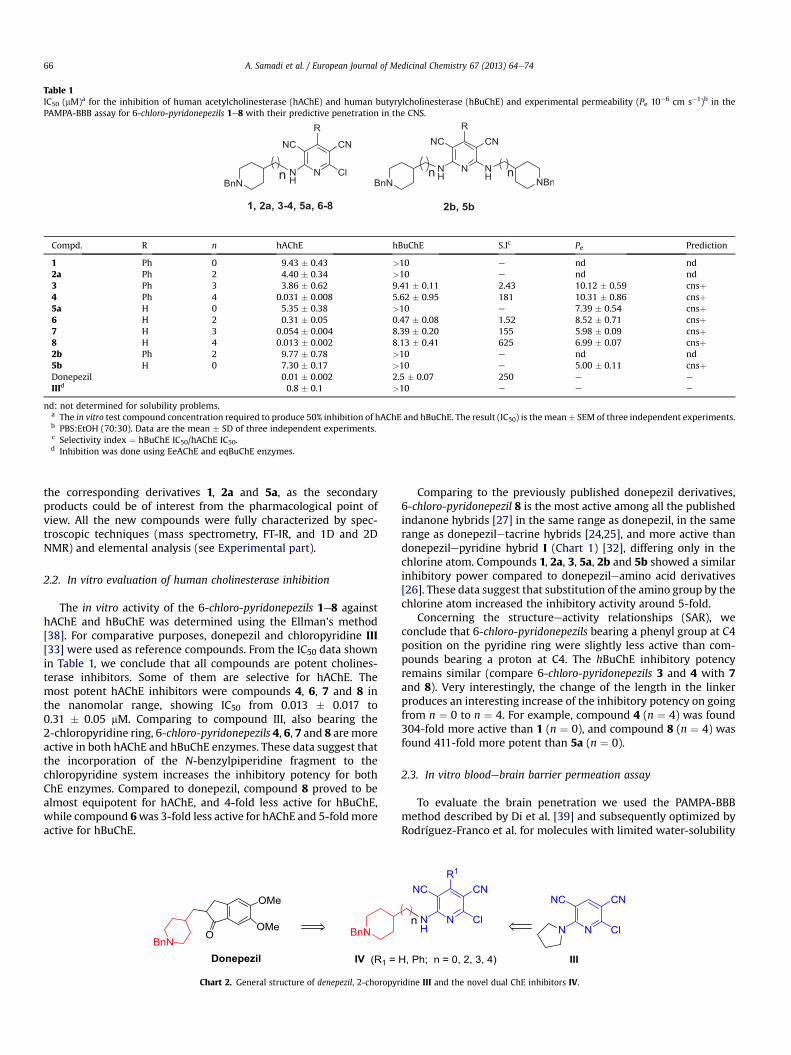
Table 1IC50 (mM)a for the inhibition of human acetylcholinesterase (hAChE) and human butyrylcholinesterase (hBuChE) and experimental permeability (Pe 10�6 cm s�1)b in thePAMPA-BBB assay for 6-chloro-pyridonepezils 1e8 with their predictive penetration in the CNS.
Compd. R n hAChE hBuChE S.Ic Pe Prediction
1 Ph 0 9.43 � 0.43 >10 e nd nd2a Ph 2 4.40 � 0.34 >10 e nd nd3 Ph 3 3.86 � 0.62 9.41 � 0.11 2.43 10.12 � 0.59 cnsþ4 Ph 4 0.031 � 0.008 5.62 � 0.95 181 10.31 � 0.86 cnsþ5a H 0 5.35 � 0.38 >10 e 7.39 � 0.54 cnsþ6 H 2 0.31 � 0.05 0.47 � 0.08 1.52 8.52 � 0.71 cnsþ7 H 3 0.054 � 0.004 8.39 � 0.20 155 5.98 � 0.09 cnsþ8 H 4 0.013 � 0.002 8.13 � 0.41 625 6.99 � 0.07 cnsþ2b Ph 2 9.77 � 0.78 >10 e nd nd5b H 0 7.30 � 0.17 >10 e 5.00 � 0.11 cnsþDonepezil 0.01 � 0.002 2.5 � 0.07 250 e e
IIId 0.8 � 0.1 >10 e e e
nd: not determined for solubility problems.a The in vitro test compound concentration required to produce 50% inhibition of hAChE and hBuChE. The result (IC50) is the mean� SEM of three independent experiments.b PBS:EtOH (70:30). Data are the mean � SD of three independent experiments.c Selectivity index ¼ hBuChE IC50/hAChE IC50.d Inhibition was done using EeAChE and eqBuChE enzymes.
A. Samadi et al. / European Journal of Medicinal Chemistry 67 (2013) 64e7466
the corresponding derivatives 1, 2a and 5a, as the secondaryproducts could be of interest from the pharmacological point ofview. All the new compounds were fully characterized by spec-troscopic techniques (mass spectrometry, FT-IR, and 1D and 2DNMR) and elemental analysis (see Experimental part).
2.2. In vitro evaluation of human cholinesterase inhibition
The in vitro activity of the 6-chloro-pyridonepezils 1e8 againsthAChE and hBuChE was determined using the Ellman’s method[38]. For comparative purposes, donepezil and chloropyridine III[33] were used as reference compounds. From the IC50 data shownin Table 1, we conclude that all compounds are potent cholines-terase inhibitors. Some of them are selective for hAChE. Themost potent hAChE inhibitors were compounds 4, 6, 7 and 8 inthe nanomolar range, showing IC50 from 0.013 � 0.017 to0.31 � 0.05 mM. Comparing to compound III, also bearing the2-chloropyridine ring, 6-chloro-pyridonepezils 4, 6, 7 and 8 aremoreactive in both hAChE and hBuChE enzymes. These data suggest thatthe incorporation of the N-benzylpiperidine fragment to thechloropyridine system increases the inhibitory potency for bothChE enzymes. Compared to donepezil, compound 8 proved to bealmost equipotent for hAChE, and 4-fold less active for hBuChE,while compound 6was 3-fold less active for hAChE and 5-foldmoreactive for hBuChE.
Chart 2. General structure of denepezil, 2-choropyr
Comparing to the previously published donepezil derivatives,6-chloro-pyridonepezil 8 is the most active among all the publishedindanone hybrids [27] in the same range as donepezil, in the samerange as donepeziletacrine hybrids [24,25], and more active thandonepezilepyridine hybrid I (Chart 1) [32], differing only in thechlorine atom. Compounds 1, 2a, 3, 5a, 2b and 5b showed a similarinhibitory power compared to donepezileamino acid derivatives[26]. These data suggest that substitution of the amino group by thechlorine atom increased the inhibitory activity around 5-fold.
Concerning the structureeactivity relationships (SAR), weconclude that 6-chloro-pyridonepezils bearing a phenyl group at C4position on the pyridine ring were slightly less active than com-pounds bearing a proton at C4. The hBuChE inhibitory potencyremains similar (compare 6-chloro-pyridonepezils 3 and 4 with 7and 8). Very interestingly, the change of the length in the linkerproduces an interesting increase of the inhibitory potency on goingfrom n ¼ 0 to n ¼ 4. For example, compound 4 (n ¼ 4) was found304-fold more active than 1 (n ¼ 0), and compound 8 (n ¼ 4) wasfound 411-fold more potent than 5a (n ¼ 0).
2.3. In vitro bloodebrain barrier permeation assay
To evaluate the brain penetration we used the PAMPA-BBBmethod described by Di et al. [39] and subsequently optimized byRodríguez-Franco et al. for molecules with limited water-solubility
idine III and the novel dual ChE inhibitors IV.
Scheme 1. Synthesis of the 6-chloro-pyridonepesils 1e8.
A. Samadi et al. / European Journal of Medicinal Chemistry 67 (2013) 64e74 67
[40e45]. The in vitro permeability (Pe) values of a selection of newcompounds through a lipid extract of porcine brain were deter-mined by using PBS: ethanol (70:30) (Table 1). In the same assay, 11commercial drugs of known CNS penetration were also tested andtheir experimental values were compared to reported values, giv-ing a good lineal correlation, Pe (exp.) ¼ 0.6551 Pe (bibl.) þ 2.2142(R2 ¼ 0.9136). From this equation and taking into account the limitsestablished by Di et al. for bloodebrain barrier permeation [39]we found that compounds with permeability values above4.8 � 10�6 cm s�1 could penetrate into the CNS, whereas moleculeswith permeability values below 3.5 � 10�6 cm s�1 were predictedto be a low BBB permeation. All tested compounds showed goodpermeability values (Table 1) and thus, they could cross the BBB andreach its biological targets located in the CNS.
2.4. Molecular modeling of compounds 4, 6e8
To identify the molecular determinants responsible for thebinding mode of the studied compounds with AChE protein, com-pounds 4, 6e8 were submitted to molecular modeling studies. Wehave chosen compound 6 with better activity inhibition thandonepezil for BuChE and 8with equivalent activity to donepezil forAChE. To evaluate the influence on ChE inhibitory potency andselectivity of the linkers of different length, compound 7 has beenstudied. Finally, we have included compound 4 in order to explainthe lower but good activity of this compound with a phenyl moietywith respect to compound 8.
Docking simulations were carried out via the Autodock Vina [46]program. Docking experiments employed full ligand flexibility andpartial protein flexibility focused at the ligand binding site.We usedthe determined X-ray crystallographic structure of hAChE com-plexed with fasciculin-II (PDB: 1B41) and blind docking protocolswere performed for the detection of possible binding sites andmodes of peptide ligands by scanning the entire surface of proteintargets.
The quality of docking results can be affected by the simplifi-cation of treating protein structures as rigid entities. Selectiveresidue flexibility is an option available on several moleculardocking tools, including Autodock Vina used in this work, whereonly selected residues are allowed to be flexible. This approach ispromising as it attempts to provide a more realistic protein envi-ronment while preventing an escalation of the computer powerneed. In this study eight residues were selected from the active-sitegorge of the hAChE structure: Trp286, Tyr124, Tyr337 and Tyr72,Asp74, Thr75, Trp86, and Tyr341. These eight residues delineate theshape of the gorge entry and lining, as their motion may signifi-cantly enlarge the gorge mouth to facilitate ligand access to thecatalytic site.
The binding mode of inhibitor 6 is shown in Fig. 1. As in done-pezilepyridine hybrids of type I (chart 1), it has been observed thatthe cationep interaction of this cationic inhibitor with thep-electron-rich active site is not the driving force for the binding.This compound is oriented along the active-site gorge, extendingfrom the catalytic site at the bottom, to the peripheral anionic siteat the top, via aromatic stacking interactions with various aromaticacid residues. The hydrophobic interaction between 6 (the piper-azine and the alkyl linker) and the rich aromatic contents (Tyr341,Phe338, Phe297, Phe295 and Trp286) along the gorge couldpotentially direct the pyridine ring to penetrate deep into the cat-alytic anionic site and oxyanion hole regions in the choline-bindingsite. The interaction is further intensified by two hydrogen-bondinginteractions between the ligand and the residues in the active site.This includes a hydrogen bond between the amino group and theamino acid residue Tyr337. The cyano group can establish H-bondinteraction with the hydroxyl group of Tyr133. The pyridine ringinteracts with Trp86 by means of a pep stacking. The benzyl groupinteracts by means of pep T-shaped interaction with the indolering of Trp286. The hydrogen atom at the piperidine C4 is orientedtoward Phe338 and Phe295.
Modification of the mid-gorge spacer modulated enzymeselectivity and improved potency. Compound 7 with n ¼ 3 showedthe same binding profile as compound 6 (n ¼ 2) with an increasein AChE activity and an activity decrease at BuChE. We theninvestigated computationally the binding of compound 7 withAChE to provide structural insights and potential to functionalmodifications.
As depicted in Fig. 2, the phenyl fragment of the ligand was wellaccommodated in the PAS through a pep stacking of this ring andindole moiety of Trp286. This interaction was reinforced by abifurcated hydrogen bond between the secondary amino group of 7and Asp74 and Tyr124. In addition, the pyridine part of the ligandwas oriented to the anionic site (AS). The interactions responsiblefor stabilization of the pyridine in the active site included pepstacking and a hydrogen bonding. The pep interaction was formedbetween the pyridine ring and the indole moiety of Trp86 and thehydrogen bonding interaction was between the cyano group andthe hydroxyl group of Tyr133.
The visual inspection of the top scored pose of compound 8(n¼ 4) reveals that the ligand is arranged at the active-site gorge ina position similar to that found for compound 6 (n¼ 2) and 7 (n¼ 3)(Figs.1 and 2). It has been observed that pep interactions played animportant role in stabilizing the complex. The benzyl group in-teracts by means of pep T-shaped interaction with Tyr72 and aface-to-face pep interaction with Trp286 (PAS). The hydrophobicinteraction between 8 (the piperidine and the alkyl linker) and therich aromatic residues (Tyr124, Tyr337, Phe338 and Tyr341) along
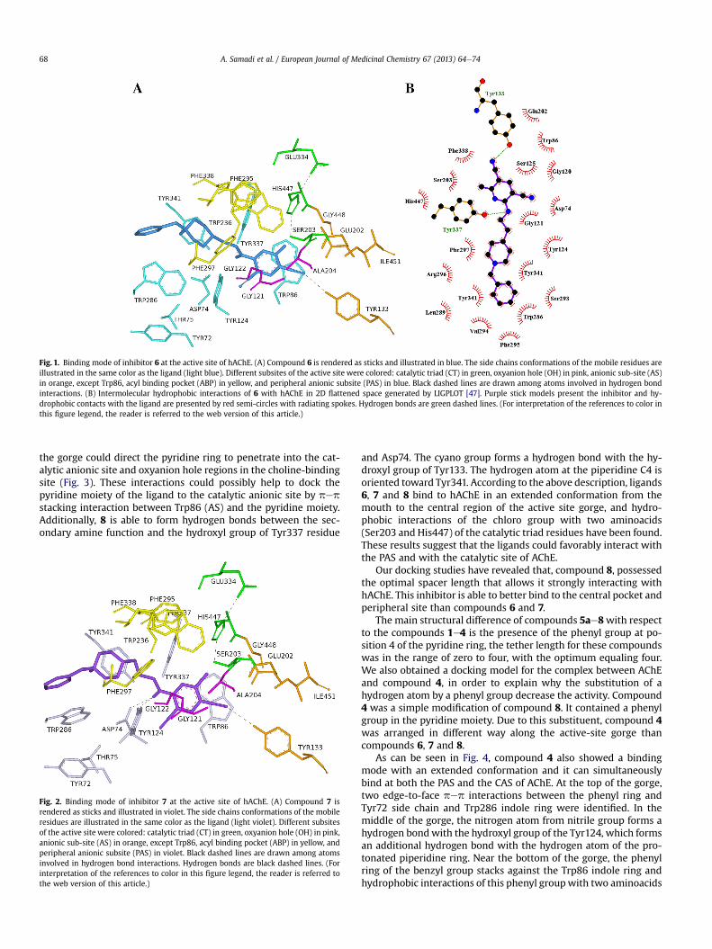
Fig. 1. Binding mode of inhibitor 6 at the active site of hAChE. (A) Compound 6 is rendered as sticks and illustrated in blue. The side chains conformations of the mobile residues areillustrated in the same color as the ligand (light blue). Different subsites of the active site were colored: catalytic triad (CT) in green, oxyanion hole (OH) in pink, anionic sub-site (AS)in orange, except Trp86, acyl binding pocket (ABP) in yellow, and peripheral anionic subsite (PAS) in blue. Black dashed lines are drawn among atoms involved in hydrogen bondinteractions. (B) Intermolecular hydrophobic interactions of 6 with hAChE in 2D flattened space generated by LIGPLOT [47]. Purple stick models present the inhibitor and hy-drophobic contacts with the ligand are presented by red semi-circles with radiating spokes. Hydrogen bonds are green dashed lines. (For interpretation of the references to color inthis figure legend, the reader is referred to the web version of this article.)
A. Samadi et al. / European Journal of Medicinal Chemistry 67 (2013) 64e7468
the gorge could direct the pyridine ring to penetrate into the cat-alytic anionic site and oxyanion hole regions in the choline-bindingsite (Fig. 3). These interactions could possibly help to dock thepyridine moiety of the ligand to the catalytic anionic site by pepstacking interaction between Trp86 (AS) and the pyridine moiety.Additionally, 8 is able to form hydrogen bonds between the sec-ondary amine function and the hydroxyl group of Tyr337 residue
Fig. 2. Binding mode of inhibitor 7 at the active site of hAChE. (A) Compound 7 isrendered as sticks and illustrated in violet. The side chains conformations of the mobileresidues are illustrated in the same color as the ligand (light violet). Different subsitesof the active site were colored: catalytic triad (CT) in green, oxyanion hole (OH) in pink,anionic sub-site (AS) in orange, except Trp86, acyl binding pocket (ABP) in yellow, andperipheral anionic subsite (PAS) in violet. Black dashed lines are drawn among atomsinvolved in hydrogen bond interactions. Hydrogen bonds are black dashed lines. (Forinterpretation of the references to color in this figure legend, the reader is referred tothe web version of this article.)
and Asp74. The cyano group forms a hydrogen bond with the hy-droxyl group of Tyr133. The hydrogen atom at the piperidine C4 isoriented toward Tyr341. According to the above description, ligands6, 7 and 8 bind to hAChE in an extended conformation from themouth to the central region of the active site gorge, and hydro-phobic interactions of the chloro group with two aminoacids(Ser203 and His447) of the catalytic triad residues have been found.These results suggest that the ligands could favorably interact withthe PAS and with the catalytic site of AChE.
Our docking studies have revealed that, compound 8, possessedthe optimal spacer length that allows it strongly interacting withhAChE. This inhibitor is able to better bind to the central pocket andperipheral site than compounds 6 and 7.
The main structural difference of compounds 5ae8with respectto the compounds 1e4 is the presence of the phenyl group at po-sition 4 of the pyridine ring, the tether length for these compoundswas in the range of zero to four, with the optimum equaling four.We also obtained a docking model for the complex between AChEand compound 4, in order to explain why the substitution of ahydrogen atom by a phenyl group decrease the activity. Compound4 was a simple modification of compound 8. It contained a phenylgroup in the pyridine moiety. Due to this substituent, compound 4was arranged in different way along the active-site gorge thancompounds 6, 7 and 8.
As can be seen in Fig. 4, compound 4 also showed a bindingmode with an extended conformation and it can simultaneouslybind at both the PAS and the CAS of AChE. At the top of the gorge,two edge-to-face pep interactions between the phenyl ring andTyr72 side chain and Trp286 indole ring were identified. In themiddle of the gorge, the nitrogen atom from nitrile group forms ahydrogen bondwith the hydroxyl group of the Tyr124, which formsan additional hydrogen bond with the hydrogen atom of the pro-tonated piperidine ring. Near the bottom of the gorge, the phenylring of the benzyl group stacks against the Trp86 indole ring andhydrophobic interactions of this phenyl groupwith two aminoacids
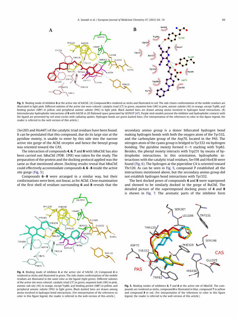
Fig. 3. Binding mode of inhibitor 8 at the active site of hAChE. (A) Compound 8 is rendered as sticks and illustrated in red. The side chains conformations of the mobile residues areillustrated in light pink. Different subsites of the active site were colored: catalytic triad (CT) in green, oxyanion hole (OH) in pink, anionic subsite (AS) in orange, except Trp86, acylbinding pocket (ABP) in yellow, and peripheral anionic subsite (PAS) in light pink. Black dashed lines are drawn among atoms involved in hydrogen bond interactions. (B)Intermolecular hydrophobic interactions of 8 with hAChE in 2D flattened space generated by LIGPLOT [47]. Purple stick models present the inhibitor and hydrophobic contacts withthe ligand are presented by red semi-circles with radiating spokes. Hydrogen bonds are green dashed lines. (For interpretation of the references to color in this figure legend, thereader is referred to the web version of this article.)
A. Samadi et al. / European Journal of Medicinal Chemistry 67 (2013) 64e74 69
(Ser203 and His447) of the catalytic triad residues have been found.It can be postulated that this compound, due do its large size at thepyridine moiety, is unable to enter by this side into the narrowactive site gorge of the AChE receptor and hence the benzyl groupwas oriented toward the CAS.
The interaction of compounds 4, 6, 7 and 8with hBuChE has alsobeen carried out. hBuChE (PDB: 1P0I) was taken for the study. Thepreparation of the protein and the docking protocol appliedwas thesame as that mentioned above. Docking results reveal that hBuChEcould effectively accommodate compounds 4, 6e8 inside the activesite gorge (Fig. 5).
Compounds 6e8 were arranged in a similar way, but theirconformations were bent, not linear, as for AChE. Close examinationof the first shell of residues surrounding 6 and 8 reveals that the
Fig. 4. Binding mode of inhibitor 4 at the active site of hAChE. (A) Compound 4 isrendered as sticks and illustrated in green. The side chains conformations of the mobileresidues are illustrated in the same color as the ligand (light green). Different subsitesof the active site were colored: catalytic triad (CT) in green, oxyanion hole (OH) in pink,anionic sub-site (AS) in orange, except Trp86, acyl binding pocket (ABP) in yellow, andperipheral anionic subsite (PAS) in light green. Black dashed lines are drawn amongatoms involved in hydrogen bond interactions. (For interpretation of the references tocolor in this figure legend, the reader is referred to the web version of this article.)
secondary amino group is a donor bifurcated hydrogen bondmaking hydrogen bonds with both the oxygen atom of the Tyr332,and the carboxylate group of the Asp70, located in the PAS. Thenitrogen atom of the cyano group is bridged toTyr332 via hydrogenbonding. The pyridine moiety formed pep stacking with Trp82.Besides, the phenyl moiety interacts with Trp231 by means of hy-drophobic interactions. In this orientation, hydrophobic in-teractions with the catalytic triad residues, Ser198 and His438 werefound (Fig. 6). The hydrogen at the piperidine C4 is oriented towardThr120. As can be seen in Fig. 5, compound 7 established all theinteractions mentioned above, but the secondary amino group didnot establish hydrogen bond interactions with Tyr332.
The best docked poses of compounds 4 and 8 were superposedand showed to be similarly docked in the gorge of BuChE. Thedetailed picture of the superimposed docking poses of 4 and 8is shown in Fig. 7. The aromatic parts of the inhibitor form
Fig. 5. Binding modes of inhibitors 6, 7 and 8 at the active site of hBuChE. The com-pounds are rendered as sticks, compound 6 is illustrated in blue, compound 7 in yellowand compound 8 in red. (For interpretation of the references to color in this figurelegend, the reader is referred to the web version of this article.)
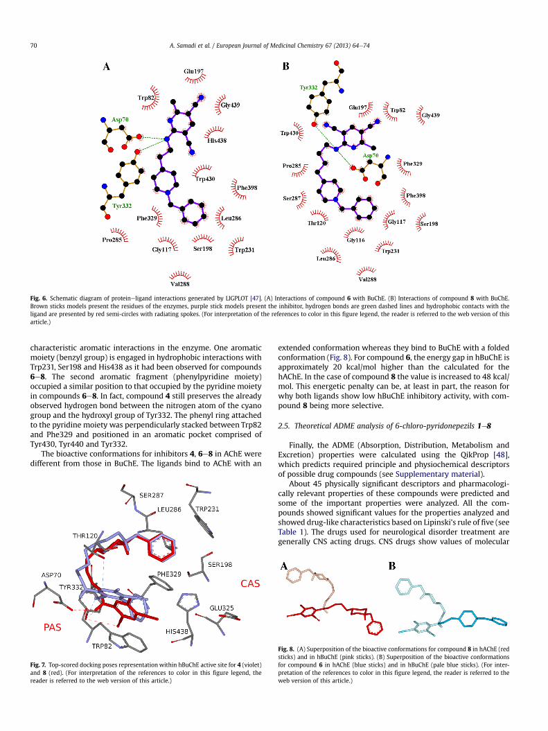
Fig. 6. Schematic diagram of proteineligand interactions generated by LIGPLOT [47]. (A) Interactions of compound 6 with BuChE. (B) Interactions of compound 8 with BuChE.Brown sticks models present the residues of the enzymes, purple stick models present the inhibitor, hydrogen bonds are green dashed lines and hydrophobic contacts with theligand are presented by red semi-circles with radiating spokes. (For interpretation of the references to color in this figure legend, the reader is referred to the web version of thisarticle.)
A. Samadi et al. / European Journal of Medicinal Chemistry 67 (2013) 64e7470
characteristic aromatic interactions in the enzyme. One aromaticmoiety (benzyl group) is engaged in hydrophobic interactions withTrp231, Ser198 and His438 as it had been observed for compounds6e8. The second aromatic fragment (phenylpyridine moiety)occupied a similar position to that occupied by the pyridine moietyin compounds 6e8. In fact, compound 4 still preserves the alreadyobserved hydrogen bond between the nitrogen atom of the cyanogroup and the hydroxyl group of Tyr332. The phenyl ring attachedto the pyridine moiety was perpendicularly stacked between Trp82and Phe329 and positioned in an aromatic pocket comprised ofTyr430, Tyr440 and Tyr332.
The bioactive conformations for inhibitors 4, 6e8 in AChE weredifferent from those in BuChE. The ligands bind to AChE with an
Fig. 7. Top-scored docking poses representation within hBuChE active site for 4 (violet)and 8 (red). (For interpretation of the references to color in this figure legend, thereader is referred to the web version of this article.)
extended conformation whereas they bind to BuChE with a foldedconformation (Fig. 8). For compound 6, the energy gap in hBuChE isapproximately 20 kcal/mol higher than the calculated for thehAChE. In the case of compound 8 the value is increased to 48 kcal/mol. This energetic penalty can be, at least in part, the reason forwhy both ligands show low hBuChE inhibitory activity, with com-pound 8 being more selective.
2.5. Theoretical ADME analysis of 6-chloro-pyridonepezils 1e8
Finally, the ADME (Absorption, Distribution, Metabolism andExcretion) properties were calculated using the QikProp [48],which predicts required principle and physiochemical descriptorsof possible drug compounds (see Supplementary material).
About 45 physically significant descriptors and pharmacologi-cally relevant properties of these compounds were predicted andsome of the important properties were analyzed. All the com-pounds showed significant values for the properties analyzed andshowed drug-like characteristics based on Lipinski’s rule of five (seeTable 1). The drugs used for neurological disorder treatment aregenerally CNS acting drugs. CNS drugs show values of molecular
Fig. 8. (A) Superposition of the bioactive conformations for compound 8 in hAChE (redsticks) and in hBuChE (pink sticks). (B) Superposition of the bioactive conformationsfor compound 6 in hAChE (blue sticks) and in hBuChE (pale blue sticks). (For inter-pretation of the references to color in this figure legend, the reader is referred to theweb version of this article.)
A. Samadi et al. / European Journal of Medicinal Chemistry 67 (2013) 64e74 71
weight, HB donor, acceptors and rotatable bonds, etc., in general, ina smaller range than general therapeutics. Factors that are relevantto the success of CNS drugs were analyzed. CNS drugs show valuesof MW <450, HB donor <3, HB acceptors <7, QPlogPo/w <5, PSA<90, number of rotatable bonds <8 and hydrogen bonds <8 [49].The compounds 1, 5a, and 6e8 satisfied all the characteristic of CNSacting drugs. The QPLogPo/w values and the molecular weight ofcompounds 2a, 3, 4 and 2b exceeded those of CNS drugs. The sol-ubility of organic molecules in water has a significant impact onmany ADME-related properties. Only compounds 1, 5a, and 6e8present solubility values within the limits, while the rest of thecompounds show values between 7.3 and 8.4.
3. Conclusions
To sum up, 6-chloro-pyridonepezils 1e8 were synthesized andevaluated against hAChE and hBuChE, using donepezil as a refer-ence compound. Most of the assayed compounds showed high ChEinhibitory activity, the most potent, 6-chloro-pyridonepezil 8, wasequipotent to donepezil. Compounds 1, 2a, 5a, 2b and 5b wereextremely selective for hAChE, but 6-chloro-pyridonepezils 2e4 and6e8 showed high activity for both hAChE and hBuChE. ConcerningSAR, compound 8 (n ¼ 4), bearing a hydrogen at C4, was moreactive than the analog 4 (n ¼ 4) bearing a phenyl group at C4. TheBBB-PAMPA study indicates that all tested compounds wouldprobably cross the BBB by passive diffusion. Finally, the dockinganalysis confirmed that these new chloro-pyridonepezils behave asdual hAChE inhibitors, binding simultaneously to the CAS and PASof the enzyme, showing thus potential for the treatment of AD.Experiments toward this objective are being now investigated andwill be reported in due course.
4. Experimental part
4.1. General methods
Melting points were determined on a Koffler apparatus, and areuncorrected. 1H NMR and 13C NMR spectra were recorded in CDCl3or DMSO-d6 at 300, 400 or 500 MHz and at 75, 100 or 125 MHz,respectively, using solvent peaks [CDCl3: 7.27 (D), 77.2 (C) ppm;D2O: 4.60 ppm and DMSO-d6: 2.49 (D), 40 (C) ppm] as internalreference. The assignment of chemical shifts was based on standardNMR experiments (1H, 13C-DEPT, 1H, 1HeCOSY, gHSQC, gHMBC).Mass spectra were recorded on a GC/MS spectrometer with an API-ES ionization source. Elemental analyses were performed at CNQO(CSIC, Spain). TLC were performed on silica F254 and detectionby UV light at 254 nm or by charring with either ninhydrin,anisaldehyde or phosphomolybdiceH2SO4 drying reagents. Anhy-drous solvents were used in all experiments. Column chromatog-raphy was performed on silica gel 60 (230 mesh). Chromatotronseparations were performed on a Harrison Research Model 7924Chromatotron equipped with a UV Lamp. The circular disks werecoated with Kieselgel 60 PF254 (E. Merck). All known compoundsshowed NMR data identical to those described in the literature.
4.1.1. General procedure for the synthesis of compounds (1e8)2,6-Dichloro-4-phenylpyridine-3,5-dicarbonitrile 13 or 2,6-
dichloropyridine-3,5-dicarbonitrile 14 was suspended in amixture of THF/EtOH (2:1, v:v). The corresponding amines 9e12,followed by triethylamine, were then added. The mixture was thenheated under reflux. After cooling, the solvent was removed invacuum and the resulting crude submitted to flash column chro-matography, to give compounds 1e8.
4.1.2. 2-((1-Benzylpiperidin-4-yl)amino)-6-chloro-4-phenylpyridine-3,5-dicarbonitrile (1) [31a]
Following the General procedure, reaction of 2,6-dichloro-4-phenylpyridine-3,5-dicarbonitrile 13 (300 g, 1.1 mmol) in a mixtureof THF/EtOH (10/5 mL) was added 4-amine-1-benzylpiperidine 9(0.22 mL, 1.1 mmol) and triethylamine (0.335 mL, 2.2 mmol). Thereaction was heated at reflux for 2 h and 30 min. The reaction wasconcentrated and the residue was purified by column chromatog-raphy (hexane:EtOAc, from 2:1 to 1:1) to gave compounds 1 [31a] asa white solid (339 mg, 81%). Mp 185e187 �C; IR (KBr) n 3436, 3315,2224, 1588, 1557 cm�1; 1H NMR (400 MHz, CDCl3) d 7.61e7.50 (m,5H, Ph), 7.39e7.26 (m, 5H, Bn), 5.76 (d, J¼ 7.6 Hz, 1H, NH), 4.21e4.06(m, H, CH), 3.56 (s, 2H, CH2Ph), 2.89 (d, J ¼ 11.9 Hz, 2H, CH2), 2.24 (t,J¼ 10.6 Hz, 2H, CH2), 2.05 (d, J¼ 10.9 Hz, 2H, CH2), 1.72e1.56 (m, 2H,CH2); 13C NMR (100 MHz, CDCl3) d 159.9 (C4), 157.7 (C6), 157.5 (C2),138.2 (C100), 133.0 (C10), 131.3 (C40), 129.2 (C30’’), 129.2 (C30), 128.5(C20), 128.2 (C200), 127.3 (C400), 114.7, 114.5 (2� CN), 98.0 (C5), 94.4(C3), 63.0 (CH2Ph), 51.9 (2� CH2), 49.5 (CH), 31.9 (2� CH2); MS (ESþ)m/z (%): 428 [M þ H]þ; Anal. Calcd. for C25H22ClN5: C, 70.17; H, 5.18;N, 16.37; Cl, 8.28. Found: C, 69.96; H, 5.31; Cl, 8.30; N, 16.09.
4.1.3. 2-((2-(1-Benzylpiperidin-4-yl)ethyl)amino)-6-chloro-4-phenylpyridine-3,5-dicarbonitrile (2a) and 2,6-bis((2-(1-benzylpiperidin-4-yl)ethyl)amino)-4-phenylpyridine-3,5-dicarbonitrile (2b)
Following the General procedure, reaction of 2,6-dichloro-4-phenylpyridine-3,5-dicarbonitrile 13 (383 mg, 1.398 mmol) in amixture of THF/EtOH (10/5 mL) with 2-(1-benzylpiperidin-4-yl)ethanamine 10 (300 mg, 1.398 mmol) and triethylamine (0.43 mL,2.795 mmol). After 15 h, and column chromatography (2% of MeOHin DCM) afforded compounds 2a and 2b. 2a: white solid (334 mg,65%); Rf¼ 0.45 (10% ofMeOH inDCM);mp 180e3 �C; IR (KBr) n 3336,2933, 2918, 2223, 1586, 1452 cm�1; 1H NMR (400 MHz, CDCl3)d 7.56e7.49 (m, 5H, Ph), 7.40e7.19 (m, 5H, Bn), 5.96 (t, J ¼ 5.3 Hz, 1H,NH), 3.60 (m, 2H, CH2NH), 3.51 (s, CH2Ph), 2.90 (d, J ¼ 11.2 Hz, 2H),1.97 (t, J ¼ 10.6 Hz, 2H), 1.71 (m, 2H), 1.60 (m, 2H), 1.35 (m, 3H); 13CNMR (100 MHz, CDCl3) d 159.6 (C4ePy), 158.3 (C2ePy), 157.4 (C6ePy), 138.2 (C1eBn),132.8 (C10ePh),131.1 (C40ePh),129.1 (2C, 2� CH),129.0 (2C, 2� CH), 128.3 (2C, 2� CH), 128.1 (2C, 2� CH), 126.9 (C4,Bn), 114.5 and 114.4 (2� CN), 97.7 (C5ePy), 90.5 (C3ePy), 63.3(CH2Ph), 53.5 (2C, 2� CH2), 39.8 (CH2NH), 35.7 (CH2(CH2NH)), 33.2(CH), 32.0 (2C, 2� CH2); MS (ES) m/z (%): 456 [M þ H]þ, 478[M þ Na]þ. Anal. Calcd. for C27H26ClN5: C, 71.12; H, 5.75; Cl, 7.78; N,15.36. Found: C, 70.89; H, 6.01; Cl, 7.34; N, 15.33. 2b: white solid(220 mg, 25%); Rf ¼ 0.24 (10% of MeOH in DCM); mp 209e11 �C; IR(KBr) n 3338, 2924, 2203, 1586, 1567, 1536, 736, 698 cm�1; 1H NMR(400 MHz, CDCl3) d 7.49 (s, 5H, Ph), 7.34e7.22 (m, 10H, Bn), 5.66 (t,J¼ 5.6 Hz, 2H, 2� NH), 3.56e5.51 (m, 4H, 2� CH2NH), 3.49 (s, 4H, 2�CH2Ph), 2.88 (d, J ¼ 11.5 Hz, 4H, 2� CH2), 1.95 (t, J ¼ 10.6 Hz, 4H, 2�CH2), 1.68 (d, J ¼ 9.3 Hz, 4H, 2� CH2), 1.60e1.55 (m, 4H, 2�CH2CH2NH), 1.37e1.26 (m, 6H, 2� CH2 and 2� CH); 13C NMR(100MHz, CDCl3) d 159.75 (C2 and C6),158.9 (C4),138.6 (2� C1eBn),134.7 (C10ePh), 130.6 (C, CePh), 129.4 (4C, Bn), 129.0 (2C, CePh),128.5 (2C, Ph), 128.4 (4C, Bn), 127.2 (2C, Bn), 117.2 (2� CN), 80.8 (C3and C5), 63.7 (2� CH2Ph), 53.8 (4C, 4� CH2), 39.5 (2� CH2NH), 36.3(2� CH2(CH2NH)), 33.6 (2� CH), 32.4 (4C, 4� CH2); MS (ESIþ) m/z:638 [M þ H]þ, 660 [M þ Na]þ. Anal. Calcd. for C41H47N7: C, 77.20; H,7.43; N, 15.37. Found: C, 76.97; H, 7.40; N, 15.45.
4.1.4. 2-((3-(1-Benzylpiperidin-4-yl)propyl)amino)-6-chloro-4-phenylpyridine-3,5-dicarbonitrile (3)
Following the General procedure, reaction of 2,6-dichloro-4-phenylpyridine-3,5-dicarbonitrile 13 (61 mg, 0.25 mmol) with 3-(1-benzylpiperidin-4-yl)propan-1-amine 11 (58 mg, 0.25mmol) andtriethylamine (76 mL) in EtOH/THF (2/4, 5 mL), after 1 h, and column
A. Samadi et al. / European Journal of Medicinal Chemistry 67 (2013) 64e7472
chromatography (from 0% to 5% of MeOH in CH2Cl2) gave product 3(55mg, 53%) as a white solid: Rf ¼ 0.42 (CH2C2/MeOH, 20/1, v/v); mp172e4 �C; IR (KBr) n 3436, 2236, 1582, 1561, 1540, 1476, 1385 cm�1;1H NMR (500 MHz, CDCl3) d 7.52e7.42 (m, 5H, Ph), 7.27e7.17 (m, 5H,Ph), 5.83 (t, J ¼ 5.3 Hz, 1H, NH), 3.52e3.48 (m, 2H, CH2eNH), 3.47 (s,2H, CH2Ph), 2.86 (d, J ¼ 9.9 Hz, 2H), 1.94e1.89 (m, 2H), 1.64e1.57 (m,4H),1.29e1.19 (m, 5H); 13C NMR (126MHz, CDCl3) d 159.6 (C4), 158.3(C2), 157. 4 (C6), 138.0 (C100), 132.8 (C10), 131.2 (C40), 129.3 (C300),129.1 (C30), 128.3 (C20), 128.2 (C200), 127.0 (C400), 114.6, 114.5 (2� CN),97.8 (C5), 90.6 (C3), 63.3 (CH2Ph), 53.6 (2� CH2), 42.5 (CH2NH), 35.2(CH), 33.4 (CH2e(CH2)2NH), 32.0 (2� CH2), 26.3 (CH2eCH2NH); MS(IE) m/z (%): 91 (100) [PhCH2]þ, 188 (8) [BnepiperidineeCH2]þ, 202(34) [BnepiperidineeCH2CH2]þ, 216 (8) [Bnepiperidinee(CH2)3]þ,378 (5) [M � Bn]þ, 392 (2) [M � Ph]þ, 468 (6) [M � H]þ; HRMS:Calcd. for C28H29ClN5 (M þ H)þ: 470.2111. Found: 470.2119. Anal.Calcd. for C28H28ClN5: C, 71.55; H, 6.00; Cl, 7.54; N, 14.90. Found: C,71.3; H, 5.87; Cl, 7.83; N, 14.76.
4.1.5. 2-((4-(1-Benzylpiperidin-4-yl)butyl)amino)-6-chloro-4-phenylpyridine-3,5-dicarbonitrile (4)
Following the General procedure, reaction of 2,6-dichloro-4-phenylpyridine-3,5-dicarbonitrile 13 (71.7 mg, 0.261 mmol) with4-(1-benzylpiperidin-4-yl)butan-1-amine 12 (60 mg, 0.243 mmol)and triethylamine (74 mL) in EtOH/THF (2/4, 6 mL), after 7 h, andcolumn chromatography (AcOEt/CH2Cl2 from 1/5 to 1/3, v/v) gaveproduct 4 (155mg, 97%) as awhite solid: Rf ¼ 0.47 (CH2C2/MeOH,10/1, v/v); mp 90e2 �C; IR (KBr) n 3421, 3306, 2930, 2223, 1591, 1552,1449 cm�1; 1H NMR (500MHz, CDCl3) d 7.51e7.43 (m, 5H, Ph), 7.26e7.16 (m, 5H, Ph), 5.80 (t, J ¼ 5.4 Hz, 1H, NH), 3.54e3.50 (m, 2H, CH2e
NH), 3.43 (s, 2H, CH2Ph), 2.82 (d, J¼ 10.98Hz, 2H),1.87 (t, J¼ 10.5 Hz,2H), 1.61e1.55 (m, 4H), 1.36e1.30 (m, 2H), 1.25e1.13 (m, 5H); 13CNMR (126 MHz, CDCl3) d 159.6 (C4), 158.4 (C2), 157.5 (C6), 138.3(C100), 132.8 (C10), 131.2 (C40), 129.2 (C300), 129.1 (C30), 128.3 (C20),128.1 (C200), 126.9 (C400), 114.6 and 114.5 (2� CN), 97.8 (C5), 90.6 (C3),63.4 (CH2Ph), 53.8 (2� CH2), 42.2 (CH2NH), 36.0 (CH2e(CH2)3NH),35.5 (CH), 32.2 (CH2eCH2NH), 29.2 (2� CH2), 23.9 (CH2e(CH2)2NH);MS (IE) m/z (%): 91 (100) [PhCH2]þ, 188 (12) [BnepiperidineeCH2]þ,202 (22) [BnepiperidineeCH2CH2]þ, 216 (75) [Bnepiperidinee(CH2)3]þ, 392 (37) [M� Bn]þ, 406 (19) [M� Ph]þ, 482 (44) [M�H]þ,483 (36), [M]þ. HRMS: Calcd. for C29H31ClN5 (M þ H)þ: 484.2268.Found: 484.2285. Anal. Calcd. for C29H30ClN5: C, 71.96; H, 6.25; Cl,7.32; N, 14.47. Found: C, 71.94; H, 5.94; Cl, 7.10; N, 14.62.
4.1.6. 2-((1-Benzylpiperidin-4-yl)amino)-6-chloropyridine-3,5-dicarbonitrile (5a) [31a] and 2,6-bis((1-benzylpiperidin-4-yl)amino)pyridine-3,5-dicarbonitrile (5b) [31a]
Following the General procedure, reaction of 2,6-dichloro-4-phenylpyridine-3,5-dicarbonitrile 14 (300 mg, 1.51 mmol) in amixture of THF/EtOH (10/5 mL) was added 4-amine-1-benzylpiperidine 9 (0.31 mL, 1.51 mmol) and triethylamine(0.46 mL, 3 mmol). The reaction was stirred overnight at roomtemperature. After completion, the reaction was concentrated andthe residue was purified by column chromatography (hex-ane:EtOAc, 1:1) to afford 5a and 5b. 5a [31a]: white solid (399 mg,75%). Anal. Calcd. for C19H18ClN5: C, 64.86; H, 5.16; Cl, 10.08; N,19.91. Found: C, 64.59; H, 5.45; Cl, 9.40; N, 19.68. 5b [31a]: whitesolid (161 mg, 21%); Anal. Calcd. for C31H35N7: C, 73.63; H, 6.98; N,19.39. Found: C, 73.39; H, 7.11; N, 19.21.
4.1.7. 2-((2-(1-Benzylpiperidin-4-yl)ethyl)amino)-6-chloropyridine-3,5-dicarbonitrile (6) [31a]
A solution of 2,6-dichloropyridine-3,5-dicarbonitrile 14 (300 mg,1.515 mmol) in a mixture of THF/EtOH (10/5 mL) was added 2-(1-benzylpiperidin-4-yl)ethanamine 10 (330 mg, 1.515 mmol) andtriethylamine (0.7 mL, 4.545 mmol). The reaction mixture was
heated at reflux for 1 h and then for overnight at room temperature.The reaction was concentrated and the residue was purified by col-umn chromatography (from 2.5 to 5% of MeOH in DCM). to givecompound 6 [31a] (301 mg, 70%) as a white solid; Anal. Calcd. forC21H22ClN5: C, 66.39; H, 5.84; Cl, 9.33; N, 18.44. Found: C, 66.14; H,5.79; Cl, 9.41; N, 18.31.
4.1.8. 2-((3-(1-Benzylpiperidin-4-yl)propyl)amino)-6-chloropyridine-3,5-dicarbonitrile (7)
Following the General procedure, reaction of 2,6-dichlorop-yridine-3,5-dicarbonitrile 14 (51 mg, 0.258 mmol) with 3-(1-benzylpiperidin-4-yl)propan-1-amine 11 (60 mg, 0.258 mmol) andtriethylamine (78 mL) in EtOH/THF (1/4, 5 mL), after 1 h, and columnchromatography (AcOEt/CH2Cl2 from 1/5 to 1/3, v/v) gave product 7(59mg, 58%) as a white solid: Rf ¼ 0.29 (CH2C2/MeOH, 20/1, v/v); mp143e5 �C; IR (KBr) n 3344, 2927, 2224, 1602, 1590, 11,395 cm�1; 1HNMR (500 MHz, CDCl3) d 7.83 (m, 1H, CH4), 7.39e7.24 (m, 5H, Ph),5.94 (t, J¼ 5.5 Hz,1H, NH), 3.70 (s, 2H, CH2Ph), 3.53 (td, J¼ 7.3, 5.7 Hz,2H, CH2eNH) 3.09 (d, J ¼ 12.2 Hz, 2H), 2.16 (t, J ¼ 11.2 Hz, 2H), 1.79e1.57 (m, 4H), 1.53e1.21 (m, 5H); 13C NMR (126 MHz, CDCl3) d 158.1(C2), 156.4 (C6), 145.9 (C4), 137.7 (C100), 129.4 (C300), 128.2 (C200), 127.1(C400), 114.5 and 114.1 (2� CN), 97.1 (C5), 90.7 (C3), 62.5 (CH2Ph), 53.3(2� CH2), 42.3 (CH2NH), 34.7 (CH), 33.1 (CH2e(CH2)2NH), 31.7 (2�CH2), 26.3 (CH2eCH2NH); MS (IE) m/z (%): 91 (100) [PhCH2]þ, 188(12) [BnepiperidineeCH2]þ, 202 (37) [BnepiperidineeCH2CH2]þ,216 (9) [Bnepiperidinee(CH2)3]þ, 302 (8) [M � Bn]þ, 316 (5)[M � Ph]þ, 392 (5) [M � H]þ, 393 (6), [M]þ. HRMS: Calcd. forC22H25ClN5 (M þ H)þ: 394.1798. Found: 394.1802; Anal. Calcd. forC22H24ClN5: C, 67.08; H, 6.14; Cl, 9.00; N, 17.78. Found: C, 66.79; H,6.22; Cl, 9.23; N, 18.02.
4.1.9. 2-((4-(1-Benzylpiperidin-4-yl)butyl)amino)-6-chloropyridine-3,5-dicarbonitrile (8)
Following the General procedure, reaction of 2,6-dichloro-pyridine-3,5-dicarbonitrile 14 (44 mg, 0.223 mmol) with 4-(1-benzylpiperidin-4-yl)butan-1-amine 12 (55 mg, 0.223 mmol) andtriethylamine (68 mL) in EtOH/THF (2/4, 6 mL), after 1 h, and columnchromatography (from 1% to 5% of MeOH in CH2Cl2) gave product 8(30 mg, 33%) as a white solid: Rf ¼ 0.2 (CH2C2/MeOH, 20/1, v/v); mp140e2 �C; IR (KBr) n 3413, 3329, 2920, 2227, 1599, 158, 1396 cm�1;1H NMR (500 MHz, CDCl3) d 7.83 (m, 1H, CH4), 7.33e7.24 (m, 5H,Ph), 5.81 (t, J ¼ 5.2 Hz, 1H, NH), 3.57e3.53 (m, 4H, CH2e
NH þ CH2Ph), 2.93 (d, J ¼ 10.3 Hz, 2H, CH2epiperidine), 1.98 (t,J ¼ 9.7 Hz, 2H, CH2epiperidine), 1.66e1.60 (m, 4H, CH2CH2e
NH þ CH2epiperidine), 1.41e1.35 (m, 2H), 1.31e1.27 (m, 5H); 13CNMR (126 MHz, CDCl3) d 158.1 (C2), 156.4 (C6), 145.9 (C4), 137.7(C100), 129.4 (C300), 128.2 (C200), 127.1 (C400), 114.5 and 114.1 (2� CN),97.1 (C5), 90.5 (C3), 63.2 (CH2Ph), 53.7 (2� CH2), 42.1 (CH2NH), 35.9(CH2e(CH2)3NH), 35.4 (CH), 32.0 (CH2eCH2NH), 29.1 (2� CH2), 23.8(CH2e(CH2)2NH); MS (IE)m/z (%): 91 (100) [PhCH2]þ, 202 (12) [BnepiperidineeCH2CH2]þ, 216 (25) [Bnepiperidinee(CH2)3]þ, 316 (12)[M � Bn]þ, 330 (5) [M � Ph]þ, 406 (9) [M � H]þ, 407 (5), [M]þ.HRMS: Calcd. for C23H27ClN5 (M þ H)þ: 408.1955. Found: 408.1957.Anal. Calcd. for C23H26ClN5: C, 67.72; H, 6.42; Cl, 8.69; N, 17.17.Found: C, 67.54; H, 6.70; Cl, 9.01; N, 17.08.
4.2. Pharmacology
4.2.1. Inhibition experiments of AChE and BuChETo assess the inhibitory activity of the compounds toward AChE
or BuChE, we followed the spectrophotometric method of Ellman[38], using purified AChE from human erythrocytes (min. 500 units/mg protein in buffered aqueous solution) and BuChE from humanserum (3 units/mg protein, lyophilized powder) (Sigma Aldrich,Madrid, Spain). Compounds were evaluated in 100 mM phosphate
A. Samadi et al. / European Journal of Medicinal Chemistry 67 (2013) 64e74 73
buffer, pH 8.0, at 30 �C using acetylthiocholine and butyrylth-iocholine (0.4 mM) as substrates, respectively. 5,5-Dithio-bis(2-nitrobenzoic) acid (DTNB, Ellman’s reagent, 0.2 mM) was used,and the values of IC50 were calculated by UV spectroscopy from theabsorbance changes at 412 nm as the concentration of compoundthat produces 50% AChE activity inhibition. Data are expressedas means � s.e.m. of at least three different experiments inquadruplicate.
4.2.2. In vitro bloodebrain barrier permeation assayPrediction of the brain penetration was evaluated using a par-
allel artificial membrane permeation assay (PAMPA), in a similarmanner as described previously [39,44,45]. Commercial drugs,phosphate-buffered saline solution at pH 7.4 (PBS), and dodecanewere purchased from Sigma, Aldrich, Acros, and Fluka. Millex filterunits (PVDF membrane, diameter 25 mm, pore size 0.45 mm) wereacquired fromMillipore. The porcine brain lipid (PBL) was obtainedfrom Avanti Polar Lipids. The donor microplate was a 96-well filterplate (PVDF membrane, pore size 0.45 mm), and the acceptormicroplate was an indented 96-well plate, both fromMillipore. Theacceptor 96-well microplate was filled with 180 mL of PBS/ethanol(7:3), and the filter surface of the donor microplate was impreg-nated with 4 mL of porcine brain lipid (PBL) in dodecane(20 mgmL�1). Compounds 1e8were dissolved in PBS/ethanol (7:3)at 1 mg mL-1, filtered through a Millex filter, and then added to thedonor wells (180 mL). The donor filter plate was carefully put on theacceptor plate to form a sandwich, which was left undisturbed for4 h at 25 �C. After incubation, the donor plate is carefully removed,and the concentration of compounds in the acceptor wells wasdetermined by UV spectroscopy. Every sample is analyzed at fivewavelengths, in four wells, and at least in three independent runs,and the results are given as the mean (standard deviation). In eachexperiment, 15 quality control standards of known BBB perme-ability were included to validate the analysis set.
4.2.3. Molecular docking into AChE and BuChECompounds 6 and 8 were assembled within Discovery Studio,
version 2.1, software package, using standard bond lengths andbond angles and with protonation at the piperidine ring. With theCHARMm force field [50] and partial atomic charges, the moleculargeometries of 6 and 8 were energy-minimized using the adopted-based Newton-Rapson algorithm. Structure was considered fullyoptimized when the energy changes between interactions wereless than 0.01 kcal/mol [51].
The coordinates of hAChE (PDB: 1B41), were obtained from theProtein Data Bank (PDB). For docking studies, initial protein wasprepared by removing all water molecules, heteroatoms, any co-crystallized solvent and the ligand. Proper bonds, bond orders,hybridization and charges were assigned using protein model toolin Discovery Studio, version 2.1, software package. CHARMm forcefield was applied using the receptoreligand interactions tool inDiscovery Studio, version 2.1, software package. Docking calcula-tions were performed with the program Autodock Vina [46].AutoDockTools (ADT; version 1.5.4) was used to add hydrogens andpartial charges for proteins and ligands using Gasteiger charges.Flexible torsions in the ligands were assigned with the AutoTorsmodule, and the acyclic dihedral angles were allowed to rotatefreely. Trp286, Tyr124, Tyr337, Tyr72, Asp74, Thr75, Trp86, andTyr341 receptor residues were selected to keep flexible duringdocking simulation using the AutoTors module. Because VINA usesrectangular boxes for the binding site, the box center was definedand the docking box was displayed using ADT. The docking pro-cedure was applied to whole protein target, without imposing thebinding site (“blind docking”). A grid box of 60 � 60 � 72 with gridpoints separated 1 �A, was positioned at the middle of the protein
(x ¼ 116.546; y ¼ 110.33; z ¼ �134.181). Default parameters wereused except num_modes, which was set to 40. The AutoDock Vinadocking procedure used was previously validated [52].
For hBuChE (1P0I) docking calculations were performedfollowing the same protocol described before for hAChE. All dock-ings were performed as blind dockings where a cube of, 75 �A withgrid points separated 1 �A, was positioned at the middle of theprotein (x ¼ 137.985; y ¼ 122.725; z ¼ 38.78). Default parameterswere used except num_modes, which was set to 40. In order to findthe best poses for compounds 6 and 8, the proper energies of thedocked poses along with the analysis of molecular feature of pro-teineligand interactions, were selected as filter criteria to rejectother poses. Finally, the docking results generated were directlyloaded into Discovery Studio, version 2.1.
Acknowledgments
A. Samadi thanks CSIC for JAE-Doc post-doctoral contract.JMC thanks MICINN (SAF2009-07271; SAF2012-33304), and COSTEU (CM1103 Action) for support. M.I.R-F. acknowledges the finan-cial support from the Spanish Ministry of Economy and Competi-tiveness (SAF2012-31035), CSIC (PIE-201280E074), and Fundaciónde Investigación Médica Mutua Madrileña Automovilística(AP103952012). M.F.R. thanks CSIC for a PhD fellowship (JAEProgram).
Appendix A. Supplementary data
Supplementary data related to this article can be found at http://dx.doi.org/10.1016/j.ejmech.2013.06.021.
References
[1] L. Bertram, R.E. Tanzi, Nat. Rev. Neurosci. 9 (2008) 768e778.[2] R.S. Shah, H.-G. Lee, Z. Xiongwei, G. Perry, M.A. Smith, R.J. Castellani, Biomed.
Pharmacother. 62 (2008) 199e207.[3] G.L. Wenk, J. Clin. Psychiatry 64 (2003) 7e10.[4] A.V. Terry, J.J. Buccafusco, J. Pharmacol. Exp. Ther. 306 (2003) 821e827.[5] P.T. Francis, A.M. Palmer, M. Snape, G.K. Wilcock, J. Neurol. Neurosurg. Psy-
chiatry 66 (1999) 137e147.[6] G. Benzi, A. Moretti, Eur. J. Pharmacol. 346 (1998) 1e13.[7] N.C. Inestrosa, A. Alvarez, C.A. Pérez, R.D. Moreno, M. Vicente, C. Linker,
O.I. Casanueva, C. Soto, J. Garrido, Neuron 16 (1996) 881e891.[8] A.E. Reyes, D.R. Perez, A. Alvarez, J. Garrido, M.K. Gentry, B.P. Doctor,
N.C. Inestrosa, Biochem. Biophys. Res. Commun. 232 (1997) 652e655.[9] N.C. Inestrosa, A. Alvarez, J. Godoy, A. Reyes, G.V. De Ferrari, Acta Neurol.
Scand. Suppl. 102 (2000) 53e56.[10] F. Mori, C.C. Lai, F. Fusi, E. Giacobini, Neuroreport 7 (1995) 633e636.[11] E. Giacobini, Neurochem. Res. 28 (2003) 515e522.[12] P.T. Francis, A. Nordberg, S.E. Arnold, Trends Pharmacol. Sci. 26 (2005) 104e111.[13] A. Castro, A. Martinez, Mini Rev. Med. Chem. 1 (2001) 267e272.[14] Y.P. Pang, P. Quiram, T. Jelaçic, F. Hong, S. Brimijoin, J. Biol. Chem. 271 (1996)
23646e23649.[15] P.R. Carlier, Y.F. Han, E.S. Chow, C.P. Li, T.X. Lieu, H.S. Wong, Y.P. Pang, Bioorg.
Med. Chem. 7 (1999) 351e357.[16] A. Mary, D.Z. Renko, C. Guillou, C. Thal, Bioorg. Med. Chem. 6 (1998) 1835e1850.[17] M.L. Bolognesi, V. Andrisano, M. Bartolini, R. Banzi, C. Melchiorre, J. Med.
Chem. 48 (2005) 24e27.[18] A. Romero, R. Cacabelos, M.J. Oset-Gasque, A. Samadi, J. Marco-Contelles,
Bioorg. Med. Chem. Lett. 23 (2013) 1916e1922.[19] P.W. Elsinghorst, W. Härtig, S. Goldhammer, J. Grosche, M. Gütschow, Org.
Biomol. Chem. 7 (2009) 3940e3946.[20] C.M. Spencer, S. Noble, Drugs Aging 13 (1998) 391e400.[21] E.L. Barner, S.L. Gray, Ann. Pharmacother. 32 (1998) 70e77.[22] J.J. Sramek, E.J. Frackiewicz, N.R. Cutler, Expert Opin. Invest. Drugs 9 (2000)
2393e2402.[23] R. Howard, R. McShane, J. Lindesay, C. Ritchie, A. Baldwin, R. Barber, A. Burns,
T. Dening, D. Findlay, C. Holmes, et al., N. Engl. J. Med. 366 (2012) 893e903.[24] P. Camps, X. Formosa, C. Galdeano, T. Gómez, D. Muñoz-Torrero,
M. Scarpellini, E. Viayna, A. Badia, M.V. Clos, A. Camins, M. Pallás, M. Bartolini,F. Mancini, V. Andrisano, J. Estelrich, M. Lizondo, A. Bidon-Chanal, F.J. Luque,J. Med. Chem. 51 (2008) 3588e3598.
[25] D. Alonso, I. Dorronsoro, L. Rubio, P. Muñoz, E. García-Palomero, M. Del Monte,A. Bidon-Chanal, M. Orozco, F.J. Luque, A. Castro, M. Medina, A. Martínez,Bioorg. Med. Chem. 13 (2005) 6588e6597.
A. Samadi et al. / European Journal of Medicinal Chemistry 67 (2013) 64e7474
[26] M.P. Arce, M.I. Rodríguez-Franco, G.C. González-Muñoz, C. Pérez, B. López,M.Villarroya,M.G.López,A.G.García, S.Conde, J.Med.Chem.52(2009)7249e7257.
[27] S. Rizzo, M. Bartolini, L. Ceccarini, L. Piazzi, S. Gobbi, A. Cavalli, M. Recanatini,V. Andrisano, A. Rampa, Bioorg. Med. Chem. 18 (2010) 1749e1760.
[28] Yang-Heon Song, Boung Sun Jo, Bull. Korean Chem. Soc. 30 (2009) 969e971.[29] R. Sheng, Y. Xu, C. Hu, J. Zhang, X. Lin, J. Li, B. Yang, Q. He, Y. Hu, Eur. J. Med.
Chem. 44 (2009) 7e17.[30] I. Bolea, J. Juárez-Jiménez, C. de los Ríos, M. Chioua, R. Pouplana, F.J. Luque,
M. Unzeta, J. Marco-Contelles, A. Samadi, J. Med. Chem. 54 (2011) 8251e8270.[31] (a) A. Samadi, M. Chioua, I. Bolea, C. de los Ríos, I. Iriepa, I. Moraleda,
A. Bastida, G. Esteban, M. Unzeta, E. Gálvez, J. Marco-Contelles, Eur. J. Med.Chem. 46 (2011) 4665e4668. See also;(b) A. Samadi, C. de los Ríos, I. Bolea, M. Chioua, I. Iriepa, I. Moraleda,M. Bartolini, V. Andrisano, E. Gálvez, C. Valderas, M. Unzeta, J. Marco-Con-telles, Eur. J. Med. Chem. 52 (2012) 251e262.
[32] A. Samadi, M. Estrada, C. Pérez, M.I. Rodríguez-Franco, I. Iriepa, I. Moraleda,M. Chioua, J. Marco-Contelles, Eur. J. Med. Chem. 57 (2012) 296e301.
[33] A. Samadi, J. Marco-Contelles, E. Soriano, M. Álvarez-Pérez, M. Chioua,A. Romero, L. González-Lafuente, L. Gandía, J.M. Roda, M.G. López, M. Villarroya,A.G. García, C. de los Ríos, Bioorg. Med. Chem. 18 (2010) 5861e5872.
[34] (a) P.G. Baraldi, M.A. Tabrizi, F. Fruttarolo, R. Romagnoli, D. Preti, PurinergicSignal. 5 (2009) 3e19;(b) L.H. Heitman, Br. J. Pharmacol. 147 (2006) 533e541.
[35] H. Sugimoto, Y. Tsuchiya, H. Sugumi, K. Higurashi, N. Karibe, Y. Iimura,A. Sasaki, Y. Kawakami, T. Nakamura, S. Araki, Y. Yamanishi, K. Yamatsu,J. Med. Chem. 33 (1990) 1880e1887.
[36] (a) C. Peinador, M.C. Veiga, J. Vilar, J.M. Quintela, Heterocycles 38 (1994)1299e1305;(b) E. Patent, A.G. Merck DE1182896, 1963; Chem. Abstr. 62 (1965) 4013a.
[37] (a) A. Duindam, V.L. Lishinsky, D.J. Sikkema, Synth. Commun. 23 (1993) 2605e2609;(b) D.V. Vilarelle, C. Peinador, J.M. Quintela, Tetrahedron 60 (2004) 275e283.
[38] G.L. Ellman, K.D. Courtney, B.J. Andres, R.M. Featherstone, Biochem. Pharma-col. 7 (1961) 88e95.
[39] L. Di, E.H. Kerns, K. Fan, O.J. McConnell, G.T. Carter, Eur. J. Med. Chem. 38(2003) 223e232.
[40] M.I. Rodríguez-Franco, M.I. Fernández-Bachiller, C. Pérez, B. Hernández-Ledesma, B. Bartolomé, J. Med. Chem. 49 (2006) 459e462.
[41] M.I. Fernández-Bachiller, C. Pérez, N.E. Campillo, J.A. Páez, G.C. González-Muñoz, P. Usán, E. García-Palomero, M.G. López, M. Villarroya, A.G. García,A. Martínez, M.I. Rodríguez-Franco, ChemMedChem 4 (2009) 828e841.
[42] M.I. Fernández-Bachiller, C. Pérez, G.C. González-Muñoz, S. Conde, M.G. López,M. Villarroya, A.G. García, M.I. Rodríguez-Franco, J. Med. Chem. 53 (2010)4927e4937.
[43] G.C. González-Muñoz, M.P. Arce, B. López, C. Pérez, M. Villarroya, M.G. López,A.G. García, S. Conde, M.I. Rodríguez-Franco, Eur. J. Med. Chem. 45 (2010)6152e6158.
[44] G.C. González-Muñoz, M.P. Arce, B. López, C. Pérez, A. Romero, L. del Barrio,M.D. Martín-de-Saavedra, J. Egea, R. León, M. Villarroya, M.G. López,A.G. García, S. Conde, M.I. Rodríguez-Franco, Eur. J. Med. Chem. 46 (2011)2224e2235.
[45] M.I. Fernández-Bachiller, C. Pérez, L. Monjas, J. Rademann, M.I. Rodríguez-Franco, J. Med. Chem. 55 (2012) 1303e1317.
[46] O. Trott, A.J. Olson, AutoDock Vina, J. Comput. Chem. 31 (2010) 455e461.[47] A.C. Wallace, R.A. Laskowski, J.M. Thornton, Protein Eng. 8 (1996) 127e134.[48] Rapid ADME. QikProp, Version 3.5, Schrödinger, LLC, New York, NY, 2012.[49] H. Pajouhesh, G.R. Lenz, NeuroRx 2 (2005) 541e553.[50] B.R. Brooks, R.E. Bruccoleri, B.D. Olafson, D.J. States, S. Swaminathan,
M. Karplus, J. Comput. Chem. 4 (1983) 187e217.[51] A. Morreale, F. Maseras, I. Iriepa, E. Gálvez, J. Mol. Graphics Modell. 21 (2002)
111e118.[52] M. Bartolini, M. Pistolozzi, V. Andrisano, J. Egea, M.G. López, I. Iriepa,
I. Moraleda, E. Gálvez, J. Marco-Contelles, A. Samadi, Chem. Med. Chem. 6(2011) 1990e1997.
Copyright © 2022 FDOKUMEN