Effect of Fe 3 O 4 magnetic nanoparticles on lysozyme amyloid aggregation
6
IOP PUBLISHING NANOTECHNOLOGY Nanotechnology 21 (2010) 065103 (6pp) doi:10.1088/0957-4484/21/6/065103 Effect of Fe 3 O 4 magnetic nanoparticles on lysozyme amyloid aggregation Andrea Bellova 1 , Eva Bystrenova 2 , Martina Koneracka 1 , Peter Kopcansky 1 , Francesco Valle 2 , Natalia Tomasovicova 1 , Milan Timko 1 , Jaroslava Bagelova 1 , Fabio Biscarini 2 and Zuzana Gazova 1 1 Department of Biophysics, Department of Magnetism, Institute of Experimental Physics, Slovak Academy of Science, Watsonova 47, 04001 Kosice, Slovakia 2 CNR—Instituto per lo Studio dei Materiali Nanostrutturati, via Gobetti 101, I-40129 Bologna, Italy E-mail: [email protected] Received 7 July 2009, in final form 8 December 2009 Published 11 January 2010 Online at stacks.iop.org/Nano/21/065103 Abstract Peptide amyloid aggregation is a hallmark of several human pathologies termed amyloid diseases. We have investigated the effect of electrostatically stabilized magnetic nanoparticles of Fe 3 O 4 on the amyloid aggregation of lysozyme, as a prototypical amyloidogenic protein. Thioflavin T fluorescence assay and atomic force microscopy were used for monitoring the inhibiting and disassembly activity of magnetic nanoparticles of Fe 3 O 4 . We have found that magnetic Fe 3 O 4 nanoparticles are able to interact with lysozyme amyloids in vitro leading to a reduction of the amyloid aggregates, thus promoting depolymerization; the studied nanoparticles also inhibit lysozyme amyloid aggregation. The ability to inhibit lysozyme amyloid formation and promote lysozyme amyloid disassembly exhibit concentration-dependent characteristics with IC50 = 0.65 mg ml −1 and DC50 = 0.16 mg ml −1 indicating that nanoparticles interfere with lysozyme aggregation already at stoichiometric concentrations. These features make Fe 3 O 4 nanoparticles of potential interest as therapeutic agents against amyloid diseases and their non-risk exploitation in nanomedicine and nanodiagnostics. (Some figures in this article are in colour only in the electronic version) 1. Introduction Peptide amyloid aggregation is a hallmark of several human pathologies termed amyloid diseases including Alzheimer’s, Parkinson’s and Creutzfeldt–Jakob diseases, type II diabetes, bovine spongiform encephalopathy and various forms of systemic amyloidosis. The common feature of amyloid diseases is transformation of proteins from soluble form into β -sheet-rich amyloid aggregates located in a specific part of the body [1]. It is generally accepted that accumulation of amyloid aggregates (oligomers, pore-like structures, filaments, fibrils) has toxic consequences leading to cellular damage by deregulation of cell homeostasis, disruption of the intracellular transport and membrane integrity [1–3]. Recent works have addressed the potential of surfaces to affect amyloid aggregation. It was observed that surface composition, size and structure as well as surface charge can markedly change the conformation of protein molecules, and thus influence their propensity to form amyloid structures. It has been shown that surfaces presented by lipid bilayers, collagen fibers, liquid–air, liquid–solid or liquid– liquid interfaces [4–7] have specific and significant effects in promoting amyloid formation. Nanoparticles possess enormous surface areas and are found to affect the protein amyloid aggregation very controversially. The experimental data showed that copolymer particles, cerium oxide particles, carbon nanotubes and quantum dots [8] enhance the rate of fibril formation from β 2 -microglobulin by decreasing the lag time for nucleation. The observed shorter lag phase depends 0957-4484/10/065103+06$30.00 © 2010 IOP Publishing Ltd Printed in the UK 1
-
Upload
independent -
Category
Documents
-
view
0 -
download
0
Transcript of Effect of Fe 3 O 4 magnetic nanoparticles on lysozyme amyloid aggregation
IOP PUBLISHING NANOTECHNOLOGY
Nanotechnology 21 (2010) 065103 (6pp) doi:10.1088/0957-4484/21/6/065103
Effect of Fe3O4 magnetic nanoparticles onlysozyme amyloid aggregationAndrea Bellova1, Eva Bystrenova2, Martina Koneracka1,Peter Kopcansky1, Francesco Valle2, Natalia Tomasovicova1,Milan Timko1, Jaroslava Bagelova1, Fabio Biscarini2 andZuzana Gazova1
1 Department of Biophysics, Department of Magnetism, Institute of Experimental Physics,Slovak Academy of Science, Watsonova 47, 04001 Kosice, Slovakia2 CNR—Instituto per lo Studio dei Materiali Nanostrutturati, via Gobetti 101,I-40129 Bologna, Italy
E-mail: [email protected]
Received 7 July 2009, in final form 8 December 2009Published 11 January 2010Online at stacks.iop.org/Nano/21/065103
AbstractPeptide amyloid aggregation is a hallmark of several human pathologies termed amyloiddiseases. We have investigated the effect of electrostatically stabilized magnetic nanoparticlesof Fe3O4 on the amyloid aggregation of lysozyme, as a prototypical amyloidogenic protein.Thioflavin T fluorescence assay and atomic force microscopy were used for monitoring theinhibiting and disassembly activity of magnetic nanoparticles of Fe3O4. We have found thatmagnetic Fe3O4 nanoparticles are able to interact with lysozyme amyloids in vitro leading to areduction of the amyloid aggregates, thus promoting depolymerization; the studiednanoparticles also inhibit lysozyme amyloid aggregation. The ability to inhibit lysozymeamyloid formation and promote lysozyme amyloid disassembly exhibitconcentration-dependent characteristics with IC50 = 0.65 mg ml−1 and DC50 = 0.16 mg ml−1
indicating that nanoparticles interfere with lysozyme aggregation already at stoichiometricconcentrations. These features make Fe3O4 nanoparticles of potential interest as therapeuticagents against amyloid diseases and their non-risk exploitation in nanomedicine andnanodiagnostics.
(Some figures in this article are in colour only in the electronic version)
1. Introduction
Peptide amyloid aggregation is a hallmark of several humanpathologies termed amyloid diseases including Alzheimer’s,Parkinson’s and Creutzfeldt–Jakob diseases, type II diabetes,bovine spongiform encephalopathy and various forms ofsystemic amyloidosis. The common feature of amyloiddiseases is transformation of proteins from soluble form intoβ-sheet-rich amyloid aggregates located in a specific part ofthe body [1]. It is generally accepted that accumulation ofamyloid aggregates (oligomers, pore-like structures, filaments,fibrils) has toxic consequences leading to cellular damage byderegulation of cell homeostasis, disruption of the intracellulartransport and membrane integrity [1–3].
Recent works have addressed the potential of surfacesto affect amyloid aggregation. It was observed thatsurface composition, size and structure as well as surfacecharge can markedly change the conformation of proteinmolecules, and thus influence their propensity to form amyloidstructures. It has been shown that surfaces presented by lipidbilayers, collagen fibers, liquid–air, liquid–solid or liquid–liquid interfaces [4–7] have specific and significant effectsin promoting amyloid formation. Nanoparticles possessenormous surface areas and are found to affect the proteinamyloid aggregation very controversially. The experimentaldata showed that copolymer particles, cerium oxide particles,carbon nanotubes and quantum dots [8] enhance the rate offibril formation from β2-microglobulin by decreasing the lagtime for nucleation. The observed shorter lag phase depends
0957-4484/10/065103+06$30.00 © 2010 IOP Publishing Ltd Printed in the UK1

Nanotechnology 21 (2010) 065103 A Bellova et al
on the amount and the nature of the particle surface. Wuet al observed that TiO2 nanoparticles promote Aβ peptideamyloid aggregation [9]. In these cases, the nanoparticles canact as catalysts for protein amyloid assembly, increasing therisk for amyloid diseases. In contrast to previous results, thenegatively charged silica particles had no potential to induceprotein aggregation [9]. Moreover, the significant inhibitionof Aβ amyloid polymerization was observed for fluorinatednanoparticles and hydrophobic teflon nanoparticles [10].
Recently there is increasing interest in developingmagnetic nanoparticles as novel materials for biomedicaluses such as magnetic resonance imaging, hyperthermia fortumor treatment, cell labeling and sorting, DNA separationand drug delivery. However, few studies are related to theeffect of magnetic nanoparticles on amyloid aggregation ofproteins. Skaat et al showed that maghemite nanoparticlesare able to attach selectively to the insulin fibrils leadingto specific magnetization of fibrils, allowing extraction offibril/nanoparticle assemblies from the aqueous phase via amagnetic field [11].
The aim of this study was to investigate the effect ofelectrostatically stabilized magnetic nanoparticles of Fe3O4
on the amyloid aggregation of lysozyme as a prototypicalamyloidogenic protein. We investigate the interaction ofmagnetic nanoparticles with lysozyme by a combination ofspectroscopic and microscopy techniques. We found thatmagnetic Fe3O4 nanoparticles are able to inhibit formation ofamyloid aggregates and have the potential to destroy amyloidfibrils, indicating their possible application for treatment ofamyloid diseases. Our results also suggest their non-riskexploitation in nanomedicine and nanodiagnostics in view oftheir inhibitory effect on amyloid aggregation.
2. Experimental details
2.1. Chemicals
Lysozyme from chicken egg white (CEW lysozyme) (lyophili-zed powder, lot number L 6876, ∼50.000 units mg−1 protein),NaCl, Thioflavin T (ThT) and Congo red (CR) were obtainedfrom Sigma Chemical Company (St Louis, MO). All otherchemicals were purchased from Sigma or Fluka and were ofanalytical reagent grade. The protein concentrations weredetermined spectrophotometrically (Specord S100, AnalytikJena), using an extinction coefficient (at λ = 280 nm) of2.63 Lg−1 cm−1.
2.2. Preparation of magnetic nanoparticles
The ionic stabilized magnetic nanoparticles (NPs) wereprepared by synthesis based on co-precipitation of Fe2+ andFe3+ salts by NH4OH at 60 ◦C [10]. In a typical synthesisto obtain 1 g of Fe3O4 precipitate, 0.86 g of FeCl2·4H2O and2.35 g of FeCl3·6H2O were dissolved in 40 ml of deionizedwater by vigorous stirring, such that the molar ratio ofFe3+/Fe2+ = 2. As the solution was heated to 60 ◦C, 5.6 mlof 25% NH4OH was added. The precipitate was isolated fromthe solution by magnetic decantation with distilled water. Toobtain an acidic sol, the precipitate was mixed with 1 ml
concentrated perchloric acid and peptization was accomplishedby adding water. Finally, the magnetic fluid was centrifuged for30 min at 5000 rpm to remove aggregates. The final samplecontains 38 mg Fe3O4 per ml as determined by chemicalanalysis.
2.3. AFM measurements
Samples for AFM measurements were prepared by spreadingon the surface of freshly cleaved mica and left to adsorb for2 min. After rinsing with ultrapure water added dropwisethe samples were dried under a gentle flow of N2 gas.AFM images were performed by stand-alone SMENA NT-MDT microscopy (NT-MDT, PRA.MA. Sondalo, Italy) insemi-contact mode in air using silicon cantilevers (NSG11,NT-MDT, PRA.MA. Sondalo, Italy) with a typical resonantfrequency 150 kHz and force constant 5.5 N m−1. All imagesare unfiltered. The topographic images were corrected line byline for background trend effect by second-order polynomialfitting (Image Analysis 2.2.0, NT-MDT, PRA.MA. Sondalo,Italy).
2.4. Lysozyme aggregation
The lysozyme amyloids were achieved by 2 h incubation ofthe soluble protein (10 μM) in 70 mM glycine containing80 mM NaCl, pH 2.7 at 65 ◦C and constant stirringof the solution. Lysozyme from chicken egg white(CEW lysozyme, lyophilized powder, 50 000 units mg−1
protein Sigma Chemical Company) was used in ourexperiments. Formation of amyloid aggregates was monitoredby characteristic changes in Thioflavin T fluorescence intensity(ThT assay), by the redshift of the maximum of the Congo red(Congo red assay) and visualized by AFM microscopy.
2.5. Thioflavin (ThT) assay
Formation of amyloid aggregates was detected as a significantenhancement of the ThT fluorescence intensity in the presenceof amyloid fibrils. Thioflavin T was added to the lysozymesamples (10 μM) to a final concentration of 20 μM.Measurements were performed in semimicro-quartz cuvetteswith a 1 cm excitation light path. Fluorescence intensity wasmeasured by spectrofluorimeter (Shimadzu, type RF-5000).The excitation was set at 440 nm and the emission recordedat 485 nm. The slits were adjusted to 1.5 and 3.0 nm for theexcitation and emission accordingly.
2.6. Interaction of magnetic nanoparticles with amyloidaggregates
Depolymerization of lysozyme amyloids by nanoparticles wasobserved after overnight incubation at 37 ◦C of the lysozymeamyloid aggregates (LA) (10 μM) with NPs for various LA:NPratios (2:1. 1:1, 1:2, 1:5 and 1:10). The inhibiting activity ofNPs was investigated by adding NPs to the soluble lysozyme(10 μM) before inducing protein aggregation by the processdescribed above. As a control, the protein was replaced withwater to measure the fluorescence of the nanoparticles. The
2

Nanotechnology 21 (2010) 065103 A Bellova et al
Figure 1. (a) The particle size distribution of magnetic nanoparticles (NP) obtained from PCCS. Inset: hysteresis loop of prepared magneticnanoparticles at 300 K. (b) Typical AFM image of magnetic nanoparticles (10 μg ml−1). Ruler is 2 μm. (c) The histogram of typical AFMimages showed average NP diameter sizes of 23 nm.
extent of amyloid aggregation was examined by ThT assay.After protein incubation with the NPs the ThT was added toa final concentration of 20 μM and fluorescence intensity wasmeasured by a spectrofluorimeter (Shimadzu RF-5000) at anexcitation of 440 nm and emission of 485 nm.
2.7. Determination of IC50 and DC50
The values were determined by measuring the ability of NPs toinhibit formation of amyloid aggregates (IC50) and destroyingthe pre-formed amyloids (DC50) for NP concentrations from0.015 to 2.5 mg ml−1 at a fixed protein concentration (10 μMcorresponding to 0.147 mg ml−1) by ThT fluorescence assay.The amount of amyloid aggregates was observed by ThT assay.The single experiment was performed in triplicate and the finalvalue is the average of measured values. The IC50 and DC50values were determined from the curves obtained by fitting ofthe average values by the nonlinear least-squares method.
3. Results and discussion
The sterically stabilized Fe3O4 magnetic nanoparticles (NPs)were prepared by the co-precipitation method [12]. The sizeof the nanoparticles was observed by photon cross-correlationspectroscopy (PCCS) giving the mean hydrodynamic diameterof the magnetic particles DHYD = 26 nm (figure 1(a)).The size and morphology of the magnetic nanoparticles wasinvestigated also by atomic force microscopy with an NT-MDTmicroscope in semi-contact mode in air [13, 14]. Typical AFMimages of magnetic nanoparticles (figure 1(b)) suggest thatnanoparticles have a nearly spherical shape. The histogramof typical AFM images showed average NP diameter sizes of23 nm (figure 1(c)). This result is in good agreement with thevalue obtained by the PCCS method.
The magnetic properties were estimated by magnetizationmeasurement using a SQUID magnetometer. The magneticmeasurements showed superparamagnetism of prepared mag-netic particles without hysteresis at room temperature and thesaturation magnetization value IS was 1.3 mT (figure 1(a),inset). Superparamagnetism is important because once the
external magnetic field is removed, magnetization disappears(negligible remanence and coercivity) and thus agglomerationof NPs (and the possible embolization of capillary vessels) isavoided.
The hen egg white lysozyme was chosen as the modelamyloidogenic protein exploiting the fact that the ability toform amyloid aggregates is a generic property of proteins. Thepresence of the amyloid aggregates was detected as an increaseof the ThT intensity (figure 2(a), black solid line LA) which isnot observed for soluble protein (black dotted line LS). We alsoperformed an independent assay with Congo red by monitoringthe redshift of the absorbance maximum and the formation ofthe shoulder at 540 nm caused by interaction of the dye withamyloid fibrils (data not shown). Finally, the typical amyloidcharacter of lysozyme aggregates was confirmed by atomicforce microscopy, as is shown in figure 3(a).
The ability of NPs to affect lysozyme amyloid aggregationin vitro was studied by ThT fluorescence assay, which issensitive to the interaction between the dye and the β-sheetsof the amyloids, but no response is observed in the presenceof soluble proteins, oligomers or amorphous aggregates. TheThT assay allows quantification of the amount of protein inthe state of amyloid aggregates, as the extent of the amyloidaggregation is proportional to the fluorescence intensity ofThT. Thus, lowering of the fluorescence intensity indicates adecrease of lysozyme amyloid aggregation. The fluorescenceintensities observed for NPs alone or in the presence of ThTwere negligible in the range of concentrations explored.
The extent of lysozyme amyloid aggregation wasinvestigated in the presence of nanoparticles after and duringformation of amyloid aggregates. To examine if nanoparticlesare able to destroy pre-formed amyloid fibrils we observedthe amount of amyloid aggregation after overnight incubationof the lysozyme amyloid fibrils (LA (10 μM)) with NPsfor various LA:NP ratios. The corresponding fluorescencespectra recorded for various ratios are present in figure 2(a).The obtained results indicate that the presence of NPs ledto depolymerization of amyloid aggregates and yields aconcentration-dependent decrease of the lysozyme in the formof amyloid fibrils.
3
Nanotechnology 21 (2010) 065103 A Bellova et al
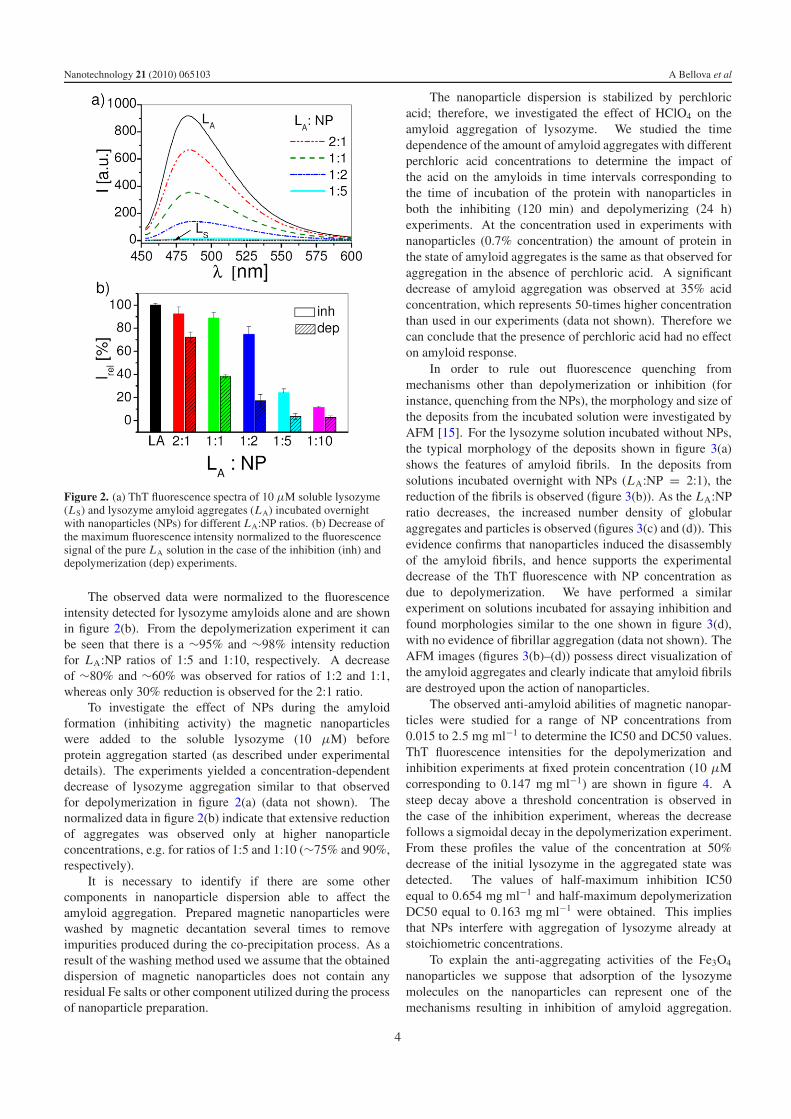
Figure 2. (a) ThT fluorescence spectra of 10 μM soluble lysozyme(LS) and lysozyme amyloid aggregates (LA) incubated overnightwith nanoparticles (NPs) for different LA:NP ratios. (b) Decrease ofthe maximum fluorescence intensity normalized to the fluorescencesignal of the pure LA solution in the case of the inhibition (inh) anddepolymerization (dep) experiments.
The observed data were normalized to the fluorescenceintensity detected for lysozyme amyloids alone and are shownin figure 2(b). From the depolymerization experiment it canbe seen that there is a ∼95% and ∼98% intensity reductionfor LA:NP ratios of 1:5 and 1:10, respectively. A decreaseof ∼80% and ∼60% was observed for ratios of 1:2 and 1:1,whereas only 30% reduction is observed for the 2:1 ratio.
To investigate the effect of NPs during the amyloidformation (inhibiting activity) the magnetic nanoparticleswere added to the soluble lysozyme (10 μM) beforeprotein aggregation started (as described under experimentaldetails). The experiments yielded a concentration-dependentdecrease of lysozyme aggregation similar to that observedfor depolymerization in figure 2(a) (data not shown). Thenormalized data in figure 2(b) indicate that extensive reductionof aggregates was observed only at higher nanoparticleconcentrations, e.g. for ratios of 1:5 and 1:10 (∼75% and 90%,respectively).
It is necessary to identify if there are some othercomponents in nanoparticle dispersion able to affect theamyloid aggregation. Prepared magnetic nanoparticles werewashed by magnetic decantation several times to removeimpurities produced during the co-precipitation process. As aresult of the washing method used we assume that the obtaineddispersion of magnetic nanoparticles does not contain anyresidual Fe salts or other component utilized during the processof nanoparticle preparation.
The nanoparticle dispersion is stabilized by perchloricacid; therefore, we investigated the effect of HClO4 on theamyloid aggregation of lysozyme. We studied the timedependence of the amount of amyloid aggregates with differentperchloric acid concentrations to determine the impact ofthe acid on the amyloids in time intervals corresponding tothe time of incubation of the protein with nanoparticles inboth the inhibiting (120 min) and depolymerizing (24 h)experiments. At the concentration used in experiments withnanoparticles (0.7% concentration) the amount of protein inthe state of amyloid aggregates is the same as that observed foraggregation in the absence of perchloric acid. A significantdecrease of amyloid aggregation was observed at 35% acidconcentration, which represents 50-times higher concentrationthan used in our experiments (data not shown). Therefore wecan conclude that the presence of perchloric acid had no effecton amyloid response.
In order to rule out fluorescence quenching frommechanisms other than depolymerization or inhibition (forinstance, quenching from the NPs), the morphology and size ofthe deposits from the incubated solution were investigated byAFM [15]. For the lysozyme solution incubated without NPs,the typical morphology of the deposits shown in figure 3(a)shows the features of amyloid fibrils. In the deposits fromsolutions incubated overnight with NPs (LA:NP = 2:1), thereduction of the fibrils is observed (figure 3(b)). As the LA:NPratio decreases, the increased number density of globularaggregates and particles is observed (figures 3(c) and (d)). Thisevidence confirms that nanoparticles induced the disassemblyof the amyloid fibrils, and hence supports the experimentaldecrease of the ThT fluorescence with NP concentration asdue to depolymerization. We have performed a similarexperiment on solutions incubated for assaying inhibition andfound morphologies similar to the one shown in figure 3(d),with no evidence of fibrillar aggregation (data not shown). TheAFM images (figures 3(b)–(d)) possess direct visualization ofthe amyloid aggregates and clearly indicate that amyloid fibrilsare destroyed upon the action of nanoparticles.
The observed anti-amyloid abilities of magnetic nanopar-ticles were studied for a range of NP concentrations from0.015 to 2.5 mg ml−1 to determine the IC50 and DC50 values.ThT fluorescence intensities for the depolymerization andinhibition experiments at fixed protein concentration (10 μMcorresponding to 0.147 mg ml−1) are shown in figure 4. Asteep decay above a threshold concentration is observed inthe case of the inhibition experiment, whereas the decreasefollows a sigmoidal decay in the depolymerization experiment.From these profiles the value of the concentration at 50%decrease of the initial lysozyme in the aggregated state wasdetected. The values of half-maximum inhibition IC50equal to 0.654 mg ml−1 and half-maximum depolymerizationDC50 equal to 0.163 mg ml−1 were obtained. This impliesthat NPs interfere with aggregation of lysozyme already atstoichiometric concentrations.
To explain the anti-aggregating activities of the Fe3O4
nanoparticles we suppose that adsorption of the lysozymemolecules on the nanoparticles can represent one of themechanisms resulting in inhibition of amyloid aggregation.
4

Nanotechnology 21 (2010) 065103 A Bellova et al
Figure 3. AFM images of (a) lysozyme amyloid fibrils LA (10 μg ml−1) and after incubation with Fe3O4 NPs with LA:NP ratio equal to2:1 (b), 1:1 (c) and 1:2 (d). Scale bar is 2 μm for all images.
Figure 4. Nanoparticle-induced inhibition (circles) anddepolymerization (triangles) of lysozyme aggregates at increasednanoparticle concentration detected by ThT assay. The curves wereobtained by fitting of the average values by nonlinear least-squaresmethod with the 3 parameter equation.
We assume that protein adsorption can lead to decreasingconcentration of the free lysozyme molecules in bulk anddiminish the nucleation process, which represents the initialstep in fibrillogenesis. Moreover, the nanoparticles can bindto active sites of protein and thus block formation of theamyloid aggregates. The ability of the nanoparticles to destroythe amyloid fibrils could by caused by interaction between
nanoparticles and hydrophobic residues of the fibrils and thusinterrupt the interface between two neighboring beta-sheets.
4. Conclusions
This study outlines the significant reduction of lysozymeamyloid aggregation by magnetic Fe3O4 nanoparticles. It wasfound that incubation of NPs with amyloid aggregates led toa decrease of the amount of amyloids by depolymerization ofamyloid structures. The NPs also inhibit lysozyme aggrega-tion; however this phenomenon requires higher nanoparticleconcentration. The ability to inhibit formation of amyloidsand to destroy amyloid aggregates exhibits concentration-dependent characteristics with IC50 = 0.654 mg ml−1 andDC50 = 0.163 mg ml−1. This implies that NPs decreasethe amount of lysozyme in an aggregate state already atstoichiometric concentration. More experiments are neededto explore the detailed molecular mechanisms of the anti-amyloidogenic ability of the NPs. However, our findingsindicate that NPs have a potential to be used for treatment ofamyloid diseases as the experimental data from various cell andanimal models used for the study of amyloid diseases suggestthat reduction of amyloid aggregation is beneficial [2, 3].Moreover, our experimental data provide evidence that in vitrointeraction of magnetic Fe3O4 nanoparticles with lysozymedo not promote amyloid aggregation. It is an importantobservation due to the fact that interaction of proteins with
5

Nanotechnology 21 (2010) 065103 A Bellova et al
nanoparticles possessing a strong ability to promote proteinamyloid aggregation can induce the onset or accelerate thedevelopment of amyloid disease. Our results, thus, suggest thelow potential biological risk in the case of applying this type ofnanoparticle in nanomedicine and nanodiagnostics.
We can finally conclude that this study, for the first time,proposes the potential use of magnetic Fe3O4 nanoparticles asthe therapeutic agent against amyloid diseases. Our resultsalso suggest their non-risk exploitation in nanomedicine andnanodiagnostics in view of their inhibitory effect on amyloidaggregation.
Acknowledgments
This work was supported by VEGA projects 7055, 0056, 0051and 0077, Project ESF 26220120021 and CEX NANOFLUIDSAS, Project EU-NMP-STRP 032652 BIODOT and APVVprojects-99-026505, -0347-06 and -0509-07.
References
[1] Sipe J D 2005 Amyloid Proteins (Weinheim: Wiley–VCH) andreferences inside
[2] Khlistunova I, Biernat J, Wang Y P, Pickhardt M,von Bergen M, Gazova Z, Mandelkow E M andMandelkow E 2006 J. Biol. Chem. 281 1205
[3] Roberson E D, Scearce-Levie K, Palop J J, Yan F, Cheng I H,Wu T, Gerstein H, Yu G Q and Mucke L 2007 Science316 750
[4] Knight J D and Miranker A D 2004 J. Mol. Biol. 341 1175[5] Relini A et al 2006 J. Biol. Chem. 281 16521[6] Powers E T and Kelly J W 2001 J. Am. Chem. Soc. 123 775[7] Lu J R, Perumal S, Powers E T, Kelly J W, Webster J R and
Penfold J 2003 J. Am. Chem. Soc. 125 3751[8] Linse S, Cabaleiro-Lago C, Xue W F, Lynch I, Lindman S,
Thulin E, Radford S E and Dawson K A 2007 Proc. NatlAcad. Sci. 104 8691
[9] Wu W H, Sun X, Yu Y P, Zhao L, Liu Q, Zhao Y F and Li Y M2008 Biochem. Biophys. Res. Commun. 373 315
[10] Rocha S, Thunemann A F, Pereira M do C, Coelho M,Mohwald H and Brezesinski G 2008 Biophys. Chem. 137 35
[11] Skaat H, Sorci M, Belfort G and Margel S 2008 J. Biomed.Mater. Res. A 91A 342
[12] Massart R 1981 IEEE Trans. Magn. 17 1247[13] Timko M et al 2004 Ind. J. Eng. Mater. Sci. 11 276[14] Cavallini M, Bystrenova E, Timko M, Koneracka M,
Zavisova V and Kopcansky P 2008 J. Phys.: Condens.Matter 20 204144
[15] Tessary I, Bisaglia M, Valle F, Samori B, Bergantino E,Mammi S and Bubacco L 2008 J. Biol. Chem. 283 16808
6