A Multiscale Approach to Characterize the Early Aggregation Steps of the Amyloid-Forming Peptide...
18
A Multiscale Approach to Characterize the Early Aggregation Steps of the Amyloid-Forming Peptide GNNQQNY from the Yeast Prion Sup-35 Jessica Nasica-Labouze 1 , Massimiliano Meli 2 , Philippe Derreumaux 3 , Giorgio Colombo 2 *, Normand Mousseau 1 * 1 De ´ partement de Physique and GEPROM, Universite ´ de Montre ´ al, Montre ´al, Que ´ bec, Canada, 2 Istituto di Chimica del Riconoscimento Molecolare, CNR, Milano, Italy, 3 Laboratoire de Biochimie The ´ orique, UPR9080 CNRS, Institut de Biologie Physico-Chimique, Universite ´ Paris 7, and Institut Universitaire de France, Paris, France Abstract The self-organization of peptides into amyloidogenic oligomers is one of the key events for a wide range of molecular and degenerative diseases. Atomic-resolution characterization of the mechanisms responsible for the aggregation process and the resulting structures is thus a necessary step to improve our understanding of the determinants of these pathologies. To address this issue, we combine the accelerated sampling properties of replica exchange molecular dynamics simulations based on the OPEP coarse-grained potential with the atomic resolution description of interactions provided by all-atom MD simulations, and investigate the oligomerization process of the GNNQQNY for three system sizes: 3-mers, 12-mers and 20- mers. Results for our integrated simulations show a rich variety of structural arrangements for aggregates of all sizes. Elongated fibril-like structures can form transiently in the 20-mer case, but they are not stable and easily interconvert in more globular and disordered forms. Our extensive characterization of the intermediate structures and their physico- chemical determinants points to a high degree of polymorphism for the GNNQQNY sequence that can be reflected at the macroscopic scale. Detailed mechanisms and structures that underlie amyloid aggregation are also provided. Citation: Nasica-Labouze J, Meli M, Derreumaux P, Colombo G, Mousseau N (2011) A Multiscale Approach to Characterize the Early Aggregation Steps of the Amyloid-Forming Peptide GNNQQNY from the Yeast Prion Sup-35. PLoS Comput Biol 7(5): e1002051. doi:10.1371/journal.pcbi.1002051 Editor: Vijay S. Pande, Stanford University, United States of America Received October 5, 2010; Accepted March 28, 2011; Published May 19, 2011 Copyright: ß 2011 Nasica-Labouze et al. This is an open-access article distributed under the terms of the Creative Commons Attribution License, which permits unrestricted use, distribution, and reproduction in any medium, provided the original author and source are credited. Funding: This work was partially supported by grants from the Natural Sciences and Engineering Research Council of Canada (http://www.nserc-crsng.gc.ca/), the Canada Research Chair Foundation (http://www.chairs-chaires.gc.ca/), the Fonds de la recherche en sante ´ du Que ´ bec (http://www.frsq.gouv.qc.ca/) and the Re ´ seau que ´be ´ cois de calcul de haute performance (http://rqchp.ca). The funders had no role in study design, data collection and analysis, decision to publish, or preparation of the manuscript. Competing Interests: The authors have declared that no competing interests exist. * E-mail: [email protected] (GC); [email protected] (NM) Introduction The aggregation of soluble peptides and proteins first into soluble oligomeric assemblies and then into insoluble amyloid fibrils is associated with the onset of misfolding diseases such as Alzheimer’s disease, Parkinson’s disease, type II diabetes and transmissible spongiform encephalopathies [1–5]. Though there is no sequence similarity, the final products of all amyloidogenic proteins display a similar cross-b structure [6,7] and the soluble oligomers of several proteins appear to share similar structural properties [8], suggesting common pathways for amyloid forma- tion [8–10]. Structural similarity does not, however, exclude diversity or polymorphism in the intermediates and products of amyloid assembly [11–24]. Many studies have shown that soluble oligomeric intermediates are more toxic than the full fibrils themselves [25,26]. These transient oligomers include low molecular weight aggregates (e.g. dimers [27] and tetramers [28]) and high molecular weight species (e.g., b-sheet rich annular protofibrils similar to pore-forming toxins [29–32]). While oligomers are considered as primary toxic species for most neurodegenerative diseases, there is recent experimental evidence that fragmentation or breakage of fibrils can contribute to the kinetics of aggregation and the amyloid cytotoxicity itself [33,34]. One important way for investigating amyloid fibril formation, polymorphism and cytotoxicity is offered by short protein fragments. Among them, GNNQQNY, from the N-terminal prion-determining domain of the yeast protein Sup35, is a paradigmatic example of a short sequence with the same properties as its corresponding full-length protein [35,36]. These properties include an amyloid fibril with a core cross-b spine, Congo-red binding and a nucleated-growth aggregation process [35]. In particular, X-ray diffraction of several micro-crystals provides a detailed atomic structure for different GNNQQNY fibrillar morphologies where the side-chains form self-comple- menting steric zippers [6,7,35–37]. As for all amyloid sequences, structural characterization of the intermediate GNNQQNY oligomers has been however precluded experimentally due to the high complexity of the aggregation process, and the short-lived and meta-stable character of the early aggregates. Computer simulations have proved useful complements to experiments for looking at the initial aggregation steps providing information, for example, about the presence of amorphous states in dynamic equilibrium with fibrillar and annular states [38–41] and the final steps of the polymerization-nucleation process [23,42]. They can provide atomic-resolution insights into several factors, ranging from the effect of sequence variations on aggregation tendencies to information on the stability of PLoS Computational Biology | www.ploscompbiol.org 1 May 2011 | Volume 7 | Issue 5 | e1002051
-
Upload
independent -
Category
Documents
-
view
3 -
download
0
Transcript of A Multiscale Approach to Characterize the Early Aggregation Steps of the Amyloid-Forming Peptide...
A Multiscale Approach to Characterize the EarlyAggregation Steps of the Amyloid-Forming PeptideGNNQQNY from the Yeast Prion Sup-35Jessica Nasica-Labouze1, Massimiliano Meli2, Philippe Derreumaux3, Giorgio Colombo2*, Normand
Mousseau1*
1 Departement de Physique and GEPROM, Universite de Montreal, Montreal, Quebec, Canada, 2 Istituto di Chimica del Riconoscimento Molecolare, CNR, Milano, Italy,
3 Laboratoire de Biochimie Theorique, UPR9080 CNRS, Institut de Biologie Physico-Chimique, Universite Paris 7, and Institut Universitaire de France, Paris, France
Abstract
The self-organization of peptides into amyloidogenic oligomers is one of the key events for a wide range of molecular anddegenerative diseases. Atomic-resolution characterization of the mechanisms responsible for the aggregation process andthe resulting structures is thus a necessary step to improve our understanding of the determinants of these pathologies. Toaddress this issue, we combine the accelerated sampling properties of replica exchange molecular dynamics simulationsbased on the OPEP coarse-grained potential with the atomic resolution description of interactions provided by all-atom MDsimulations, and investigate the oligomerization process of the GNNQQNY for three system sizes: 3-mers, 12-mers and 20-mers. Results for our integrated simulations show a rich variety of structural arrangements for aggregates of all sizes.Elongated fibril-like structures can form transiently in the 20-mer case, but they are not stable and easily interconvert inmore globular and disordered forms. Our extensive characterization of the intermediate structures and their physico-chemical determinants points to a high degree of polymorphism for the GNNQQNY sequence that can be reflected at themacroscopic scale. Detailed mechanisms and structures that underlie amyloid aggregation are also provided.
Citation: Nasica-Labouze J, Meli M, Derreumaux P, Colombo G, Mousseau N (2011) A Multiscale Approach to Characterize the Early Aggregation Steps of theAmyloid-Forming Peptide GNNQQNY from the Yeast Prion Sup-35. PLoS Comput Biol 7(5): e1002051. doi:10.1371/journal.pcbi.1002051
Editor: Vijay S. Pande, Stanford University, United States of America
Received October 5, 2010; Accepted March 28, 2011; Published May 19, 2011
Copyright: � 2011 Nasica-Labouze et al. This is an open-access article distributed under the terms of the Creative Commons Attribution License, which permitsunrestricted use, distribution, and reproduction in any medium, provided the original author and source are credited.
Funding: This work was partially supported by grants from the Natural Sciences and Engineering Research Council of Canada (http://www.nserc-crsng.gc.ca/),the Canada Research Chair Foundation (http://www.chairs-chaires.gc.ca/), the Fonds de la recherche en sante du Quebec (http://www.frsq.gouv.qc.ca/) and theReseau quebecois de calcul de haute performance (http://rqchp.ca). The funders had no role in study design, data collection and analysis, decision to publish, orpreparation of the manuscript.
Competing Interests: The authors have declared that no competing interests exist.
* E-mail: [email protected] (GC); [email protected] (NM)
Introduction
The aggregation of soluble peptides and proteins first into
soluble oligomeric assemblies and then into insoluble amyloid
fibrils is associated with the onset of misfolding diseases such as
Alzheimer’s disease, Parkinson’s disease, type II diabetes and
transmissible spongiform encephalopathies [1–5]. Though there is
no sequence similarity, the final products of all amyloidogenic
proteins display a similar cross-b structure [6,7] and the soluble
oligomers of several proteins appear to share similar structural
properties [8], suggesting common pathways for amyloid forma-
tion [8–10]. Structural similarity does not, however, exclude
diversity or polymorphism in the intermediates and products of
amyloid assembly [11–24].
Many studies have shown that soluble oligomeric intermediates
are more toxic than the full fibrils themselves [25,26]. These
transient oligomers include low molecular weight aggregates (e.g.
dimers [27] and tetramers [28]) and high molecular weight species
(e.g., b-sheet rich annular protofibrils similar to pore-forming
toxins [29–32]). While oligomers are considered as primary toxic
species for most neurodegenerative diseases, there is recent
experimental evidence that fragmentation or breakage of fibrils
can contribute to the kinetics of aggregation and the amyloid
cytotoxicity itself [33,34].
One important way for investigating amyloid fibril formation,
polymorphism and cytotoxicity is offered by short protein
fragments. Among them, GNNQQNY, from the N-terminal
prion-determining domain of the yeast protein Sup35, is a
paradigmatic example of a short sequence with the same
properties as its corresponding full-length protein [35,36]. These
properties include an amyloid fibril with a core cross-b spine,
Congo-red binding and a nucleated-growth aggregation process
[35]. In particular, X-ray diffraction of several micro-crystals
provides a detailed atomic structure for different GNNQQNY
fibrillar morphologies where the side-chains form self-comple-
menting steric zippers [6,7,35–37]. As for all amyloid sequences,
structural characterization of the intermediate GNNQQNY
oligomers has been however precluded experimentally due to
the high complexity of the aggregation process, and the short-lived
and meta-stable character of the early aggregates.
Computer simulations have proved useful complements to
experiments for looking at the initial aggregation steps providing
information, for example, about the presence of amorphous states
in dynamic equilibrium with fibrillar and annular states [38–41]
and the final steps of the polymerization-nucleation process
[23,42]. They can provide atomic-resolution insights into several
factors, ranging from the effect of sequence variations on
aggregation tendencies to information on the stability of
PLoS Computational Biology | www.ploscompbiol.org 1 May 2011 | Volume 7 | Issue 5 | e1002051

aggregates and the kinetics of aggregation. Due to lighter
computational costs, short peptides are more amenable to
simulations of the aggregation process than full-length proteins.
For example, a number of numerical works have characterized the
structures and free energy of small GNNQQNY aggregates
ranging from 2-mers to 8-mers starting from disordered states or
studied the stability of pre-formed GNNQQNY assemblies with
cross-b or annular morphologies [23,24,32,42–51].
In this paper, we push the boundaries of the GNNQQNY
oligomer size and investigate, through a multi-scale simulation
approach, the aggregation and polymorphism of three GNNQQNY
oligomer sizes: 3-mers, 12-mers and 20-mers. Our approach takes
advantage of the accelerated sampling properties of replica exchange
molecular dynamics (REMD) simulations [52] based on coarse-
grained models and of the accurate description of the physico-
chemical interactions between the peptides and the solvent by using
an all-atom model. More precisely, we first use REMD simulations
[52] with the coarse-grained potential OPEP [53,54], and then
analyze the stability and conformational properties of selected
aggregates by room temperature MD as well as REMD simulations
using the GROMOS force-field [55]. In total, we accumulated more
than 23.60 ms and 2.66 ms of coarse-grained and all-atom
simulations, respectively, allowing relevant statistical analysis. To
our knowledge, the present study reports the largest simulations of
spontaneous self-organization carried out at the atomic resolution on
an amyloid peptide without any pre-formed seed. Overall, the results
of our integrated simulations and analysis show the existence of a
high degree of polymorphism for the GNNQQNY sequence, even
for oligomeric assemblies containing as many as 20 monomers.
Materials and Methods
Simulations and analyses presented here couple a number of
approaches, which are described briefly in this section. The first set
of simulations uses the coarse-grained OPEP potential with
replica-exchange molecular dynamics (REMD). These are fol-
lowed by all-atom simulations using GROMACS with MD and
REMD. All simulations are labeled as follows: a number, which
indicates the number of monomers, two letters indicating the force
field (OP for OPEP and GR for GROMACS), a letter or number
indicating the simulation and a label for the specific conformation
studied (when appropriate) giving, for example: 01OP2-A1.
Replica-Exchange Molecular Dynamics (REMD)REMD is a thermodynamical sampling method that requires the
running of N MD trajectories (or replica) in parallel at N different
temperatures selected in order to optimize thermodynamical
sampling [52]. At regular time intervals, conformational exchanges
are attempted between adjacent simulation pairs according to the
Metropolis criterion with accept-reject probability:
p(i, j)~min 1:0, exp1
kBTi
{1
kBTj
� �Ei{Ej
� �� �
where, before the exchange, trajectory i at temperature Ti has an
energy Ei and trajectory j has an energy Ej at temperature Tj.
This broadly used method allows for conformations in a deep
local minimum to explore other regions of the energy landscape
by migrating to higher temperatures. While thermodynamical
properties converge faster than with single temperature standard
MD, dynamical information is lost due to temperature exchanges.
It is still possible, however, to derive thermodynamically putative
aggregation pathways by following the continuous trajectories
through temperature space.
The Optimal Potential For Efficient Peptide-StructurePrediction (OPEP) Force-Field
OPEP is a coarse-grained protein model that uses a detailed
representation of all backbone atoms (N, H, Ca, C and O) and
reduces each side-chain to one single bead with appropriate
geometrical parameters and van der Waals radius. The OPEP
energy function, which includes implicit effects of aqueous solution,
is expressed as a sum of local potentials (taking into account the
changes in bond lengths, bond angles, improper torsions of the side-
chains and backbone torsions), non-bonded potentials (taking into
account the hydrophobic and hydrophilic properties of each amino
acid) and hydrogen-bonding potentials (taking into account two-
and four- body interactions) [53]. OPEP has been extensively tested
on peptides using multiple approaches such as the activation-
relaxation technique [56], Monte Carlo [54], MD [39] and REMD
simulations [57], and greedy-based algorithms [58,59]. OPEP is
also appropriate for simulations of GNNQQNY. Preliminary test
simulations on this peptide’s dimer indicate that, at 300 K, the
GNNQQNY relative orientation is a 60 to 40 probability in favor of
the antiparallel dimer with a least two hydrogen bonds. This result is
in general agreement with what was found by Strodel et al. with
CHARMM19 and the implicit solvation potential EEFI [46] where
both orientations of the strands are visited with similar probabilities.
OPEP Simulation DetailsREMD were carried out using a 1.5 fs time-step, periodic
boundary conditions with box sizes depending on the systems and a
weak coupling to an external temperature bath [60,61,62]. Replica
exchanges were attempted every 5000 steps and configurations
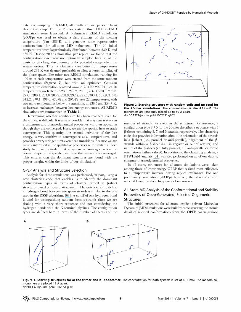
saved every 5000 steps. Initial structures for 3-mer and the 20-mer
simulations were constructed by placing random coil monomers
between 12 A to 50 A apart (Figure 1a and Figure 2). For the
12-mer, the initial chains occupied four rows, with each peptide
separated from the others by 15 A (Figure 1b). Because of the
Author Summary
The formation of amyloid fibrils is associated with manyneurodegenerative diseases such as Alzheimer’s, Creutz-feld-Jakob, Parkinson’s, the Prion disease and diabetesmellitus. In all cases, proteins misfold to form highlyordered insoluble aggregates called amyloid fibrils thatdeposit intra- and extracellularly and are resistant toproteases. All proteins are believed to have the instrinsiccapability of forming amyloid fibrils that share commonspecific structural properties that have been observed byX-ray crystallography and by NMR. However, little is knownabout the aggregation dynamics of amyloid assemblies,and their toxicity mechanism is therefore poorly under-stood. It is believed that small amyloid oligomers, formedon the aggregation pathway of full amyloid fibrils, are thetoxic species. A detailed atomic characterization of theoligomerization process is thus necessary to further ourunderstanding of the amyloid oligomer’s toxicity. Ourapproach here is to study the aggregation dynamics of a 7-residue amyloid peptide GNNQQNY through a combina-tion of numerical techniques. Our results suggest that thisamyloid sequence can form fibril-like structures and ispolymorphic, which agrees with recent experimentalobservations. The ability to fully characterize and describethe aggregation pathway of amyloid sequences numeri-cally is key to the development of future drugs to targetamyloid oligomers.
Study of GNNQQNY Peptide by Numerical Methods
PLoS Computational Biology | www.ploscompbiol.org 2 May 2011 | Volume 7 | Issue 5 | e1002051
extensive sampling of REMD, all results are independent from
this initial setup. For the 20-mer system, three OPEP-REMD
simulations were launched. A preliminary REMD simulation
(20OPp) was used to obtain a first estimate of the melting
temperature (Tm = 283 K) and generate some representative
conformations for all-atom MD refinement. The 20 initial
temperatures were logarithmically distributed between 230 K and
450 K. Despite 200-ns simulation per replica, we found that the
configuration space was not optimally sampled because of the
existence of a large discontinuity in the potential energy when the
system orders. Thus, a Gaussian distribution of temperatures
around 283 K was deemed preferable to allow a better sampling of
the phase space. The other two REMD simulations, running for
400 ns at each temperature, were started from the same random
configuration (Figure 2), but with an optimized Gaussian
temperature distribution centered around 283 K: 20OP1 uses 20
temperatures (in Kelvins: 223.8, 249.2, 260.1, 266.0, 270.3, 273.8,
277.1, 280.1, 283.0, 285.9, 288.9, 292.2, 295.7, 300.1, 305.9, 316.8,
342.2, 370.1, 398.0, 426.0) and 20OP2 uses 22 temperatures, with
two more temperatures below the transition, at 236.5 and 254.7 K,
to increase exchanges between low-energy structures. All REMD
simulations are summarized in Table 1.
Determining whether equilibrium has been reached, even for
the trimer, is difficult. It is always possible that a system is stuck in
a minimum and thermodynamical properties will then appear as
though they are converged. Here, we use the specific heat to track
convergence. This quantity, the second derivative of the free
energy, is very sensitive to convergence at all temperatures, and
provides a very stringent test even near transitions. Because we are
mostly interested in the qualitative properties of the systems under
study here, we consider that a system is converged when the
overall shape of the specific heat near the transition is converged.
This ensures that the dominant structures are found with the
proper weight, within the limits of our simulations.
OPEP Analysis and Structure SelectionAnalysis for these simulations was performed, in part, using a
new clustering code that enables us to identify the dominant
configuration types in terms of clusters formed in b-sheet
structures based on strand attachment. The criterion set to define
a hydrogen bond between two given strands is similar to the one
used in the DSSP algorithm. [63]. A cutoff of one hydrogen bond
is used for distinguishing random from b-strands since we are
dealing with a very short sequence and not considering the
hydrogen bonds with the N-terminal glycines. The configuration
types are defined here in terms of the number of sheets and the
number of strands per sheet in the structure. For instance, a
configuration type 8 7 5 for the 20-mer describes a structure with 3
b-sheets containing 8, 7 and 5 strands, respectively. The clustering
code also provides information about the orientation of the strands
in a b-sheet (i.e., parallel or anti-parallel), alignment of the b-
strands within a b-sheet (i.e., in register or out-of register) and
nature of the b-sheets (i.e. fully parallel, full anti-parallel or mixed
orientations within a sheet). In addition to the clustering analysis, a
PTWHAM analysis [64] was also performed on all of our data to
compute thermodynamical properties.
In all cases, structures for all-atom simulations were taken
among those of lower-energy OPEP that resisted most efficiently
to a temperature increase during replica exchanges. For one
preliminary simulation (20OPp) however, the structures were
selected based on their frequency of occurrence.
All-Atom MD Analysis of the Conformational and StabilityProperties of Opep-Generated, Selected OligomericStructures
The initial structures for all-atom, explicit solvent Molecular
Dynamics (MD) simulations were built by reconstructing the atomic
detail of selected conformations from the OPEP coarse-grained
Figure 1. Starting structures for a) the trimer and b) dodecamer. The concentration for both systems is set at 4.15 mM. The random coilmonomers are placed 15 A apart.doi:10.1371/journal.pcbi.1002051.g001
Figure 2. Starting structure with random coils and no seed forthe 20-mer simulations. The concentration is also 4.15 mM. Themonomers are randomly placed 12 to 50 A apart.doi:10.1371/journal.pcbi.1002051.g002
Study of GNNQQNY Peptide by Numerical Methods
PLoS Computational Biology | www.ploscompbiol.org 3 May 2011 | Volume 7 | Issue 5 | e1002051

runs. Reconstruction was carried out using the MAXSPROUT
server [65]. Refinement of side-chain rotameric states was
performed using the program IRECS [66,67], where the prediction
is guided by a combination of potential interaction and rotamer
scores calculated with probabilities from the backbone dependent
rotamer library. Resulting all-atom structures obtained with this
procedure were first minimized using the Macromodel package
(Schrodinger Incorporated, USA) for 5000 steps with Polak-Ribier
Conjugate Gradient method and an energy gradient criterion for
convergence set to 0.05 kJ/mol. This minimization protocol was
intended to initially remove unphysical contacts between atoms
resulting from the reconstruction procedure, and not to optimize
structures. At this stage, the Ca atoms were constrained to their
positions with the default force constant (25 kcal/mol A2).
The resulting minimized systems were then solvated in a cubic-
shaped box large enough to contain 1.0nm of solvent around each
initial aggregate. The simple point charge (SPC) water model was
used [68] to solvate each oligomer in the simulation box. Each
system was subsequently energy minimized with a steepest descent
method for 5000 steps. The minimization was considered to be
converged when the maximum force was smaller than
0.0001 kJ mol21 nm21. The initial step size for minimization
was 0.01 nm. The calculation of electrostatic forces was done with
the PME implementation of the Ewald summation method. The
LINCS [69] algorithm was used to constrain all bond lengths and
the SETTLE algorithm [70] for the water molecules. Simulations
were performed with a dielectric permittivity, = 1, and a time
step of 2 fs. Initial velocities were taken from a Maxwellian
distribution at the desired initial temperature of 300 K. The
density of the system was adjusted performing the first equilibra-
tion runs at NPT condition by weak coupling to a bath of constant
pressure (P0 = 1 bar, coupling time tP = 0.5 ps) [60] and the system
Table 1. Details of all simulations run for the trimer, dodecamer and 20-mer systems.
Length of OPEPsimulations(ns)(a)
OPEP - LABEL ofstructuresextracted(b)
Temperatures min-max(K) & numberof temperatures(c)
GROMACS - LABEL ofreconstructed OPEPextracted structures(b)
Length ofGROMACSsimulations(ns)(d)
Total numberof atoms(e)
Temperature(K)(f)
3-mer 50616 03OP1-A 222.5–525 16 03-GR1-A 100 6504 300
03OP1-B 222.5–525 16 03-GR1-B 100 5997 300
03OP1-C 222.5–525 16 03-GR1-C 100 5619 300
03OP1-D 222.5–525 16 03-GR1-D 100 5949 300
03OP1-E 222.5–525 16 03-GR1-E 100 6621 300
12-mer 125616 12OP1-A 222.5–525 16 12-GR1-A 100 56271 300
12OP1-B 222.5–525 16 12-GR1-B 100 40476 300
12OP1-C 222.5–525 16 12-GR1-C 100 15381 300
12OP1-D 222.5–525 16 12-GR1-D 100 16134 300
12OP1-E 222.5–525 16 12-GR1-E 100 17157 300
20-merOPp
200620 20OPp-A 234.6–447.6 20 20GRp-A1 100 25620 300
20OPp-A 234.6–447.6 20 20GRp-A2 100 25620 300
20OPp-B 234.6–447.6 20 20GRp-B1 100 27816 300
20OPp-B 234.6–447.6 20 20GRp-B2 100 27816 300
20OPp-B 234.6–447.6 20 20GRp-B3 (REMD) 10612 27816 see text
20OPp-C 234.6–447.6 20 20GRp-C1 100 49674 300
20OPp-C 234.6–447.6 20 20GRp-C2 100 49674 300
20OPp-D 234.6–447.6 20 20GRp-D1 100 23746 300
20OPp-D 234.6–447.6 20 20GRp-D2 100 23746 300
20-merOP2
400622 20OP2-A 223.8–425.9 22 20GR2-A 100 21586 300
20OP2-B 223.8–425.9 22 20GR2-B 100 24640 300
20OP2-C 223.8–425.9 22 20GR2-C 100 23554 300
20OP2-E 223.8–425.9 22 20GR2-E 100 26065 300
20OP2-N 223.8–425.9 22 20GR2-N1 100 32206 300
20OP2-N 223.8–425.9 22 20GR2-N2 (REMD) 10612 32206 see text
This table presents simulations done with OPEP (coarse-grained potential) and GROMACS (all-atom potential).(a)The total simulation time for OPEP REMD simulations in the format time_per_replica x number_of_replicas.(b)The label of the OPEP/GROMACS structures extracted. The label indicates the number of monomers, the potential used (OP for OPEP and GR for GROMACS), the
simulation index (1,2 or p (preliminary)) and the letter ID of the structure.(c)The range of temperatures (in K) used for OPEP REMD simulations.(d)The total simulation time for GROMACS simulations. MD simulations are indicated by only one number while, for REMD simulations, the total simulation time is given
in the format time_per_replica x number_of_replicas.(e)The total number of atoms in the system including protein and solvatation water atoms.(f)The temperature used in GROMACS simulations (in K).doi:10.1371/journal.pcbi.1002051.t001
Study of GNNQQNY Peptide by Numerical Methods
PLoS Computational Biology | www.ploscompbiol.org 4 May 2011 | Volume 7 | Issue 5 | e1002051

was weakly coupled to an external temperature bath [60] with a
coupling constant of 0.1 ps. The proteins and the rest of the system
were coupled separately to the temperature bath. Table 1
summarizes the simulation conditions and number of peptides
for each simulation. All simulations and analysis were carried out
using the GROMACS package (version 3.3) [71–73] and the
GROMOS96 43A1 force field [74–77].
For all MD simulations, aggregates were simulated at 300 K for
100 ns. REMD simulations were also used to investigate the
stability and the conformational preferences of two 20-mer
aggregates. The replica exchange simulations were carried out
using the Solute Tempering REMD [78] protocol using the
version implemented in GROMACS by de Groot and coworkers
[79]. Twelve temperatures between 308 K and 419 K were
selected according to [80] for an exchange probability of around
40%.
Results/Discussion
The aggregation process for the three types of GNNQQNY
oligomers – containing 3, 12 and 20 chains, respectively – was
studied by a multi-scale approach consisting in a preliminary,
thorough exploration of the phase space through REMD with the
OPEP coarse-grained potential, followed by the refinement of the
most representative aggregate structures obtained via all-atom MD
or REMD simulations in explicit solvent. The initial concentration
for the OPEP runs was around 4.15 mM. This concentration is
10 times higher than the concentration at which amyloid
GNNQQNY fibrils form in a few hours according to Nelson
et al. [9] allowing for the formation of ordered structures within
our simulation time frame. The diversity in the number of chains
allows us to examine possible intermediates and analyze molecular
mechanisms of polymorphism in amyloid aggregates.
For clarity, we first present and discuss results for the trimeric
and dodecameric systems as they will serve as basis for
understanding the results observed for the 20-mer presented in
the last part of this section.
Simulations of Trimeric SystemsCoarse-grained simulations. Coarse-Grained REMD
simulations were performed with 16 replicas for 50 ns at
temperatures discussed in the materials and methods section.
Although the system is not fully converged for the very low-
temperature replicas, the PTWHAM-generated specific heat
computed over two different time intervals shows that the
melting temperature, Tm, is well-established at ,294 K
(Figure 3). Below this temperature, a clustering analysis shows
that GNNQQNY monomers are assembled into ordered
structures with high b-sheet content, while above Tm, the system
visits mostly disordered structures with very low secondary
structure composition. The alignment of individual strands
within oligomers, the secondary structures and the configuration
types of the aggregates are summarized in Table 2.
Structurally, the trimer displays a strong tendency to form
ordered planar b-sheets below Tm (Figure 4, left part of thepanel). These appear rapidly, within 1 to 8 ns, in a mostly anti-
parallel organization. Following trajectories leading to ordered
structures, we see that the three-stranded b-sheet is always
preceded by the formation of a mostly anti-parallel dimer seed.
Averaging over all structures below Tm, we find that only a very
small proportion of structures just below Tm consist of a two-
stranded b-sheet interacting with one chain in coil conformation
(1.9%) or three random coil chains (1.1%). The peptides at a
temperature just below Tm prefer an anti-parallel b-strand order
(87% at 267 K) over a parallel arrangement (13% at 267 K), while
this proportion falls to 55–60% at the lowest temperatures. As seen
in Table 2, the three b-strands prefer to be perfectly aligned or in-
registered at the lowest temperatures and are typically shifted by
one residue, i.e. out-of-registered, at temperatures close to the
melting point. As the temperature increases, the population of two-
stranded and three-stranded b-sheets becomes very low, amount-
ing to 8% and 0% at 333 K and 352 K, respectively. Except for
the lowest temperatures, where mixed parallel/antiparallel sheets
are most common, there is a clear dominance of fully antiparallel
sheets for three-stranded structures while fully parallel sheets are
rare, even among the few three-stranded sheets found above Tm,
where they reach 21%, to 68% for fully anti-parallel.
All-atom MD simulations. Five representative OPEP-
generated structures, labeled 03OP1-A, 03OP1-B, 03OP1-C,
03OP1-D and 03OP1-E (Figure 4, left side of the panel),were then subjected to all-atom MD simulations as described in
materials and methods. These structures can be divided in two
sets: 03OP1-A, 03OP1-B, 03OP1-C are characterized by three-
stranded b-sheets with mixed parallel/anti-parallel b-strands,
while 03OP1-D and 03OP1-E display a fully anti-parallel three-
stranded b-sheet.
As seen in the final structures of the all-atom simulations
displayed in Figure 4 (right side of the panel), the five structures
show different evolutions after the 100 ns all-atom MD. The three
structures 03OP1-A, -B and -D tend towards configuration types
2-1, i.e. with one chain converted from b-strand to random coil
and the two other chains enhancing their b-sheet contents. This
inter-conversion is independent on the initial orientation of the
strands. In contrast, the other two structures 03OP1-C and –E
preserve their three-stranded b-sheet configurations and enhance
their b-sheet contents. Simulation 03GR1-C keeps its starting
mixed parallel/anti-parallel configuration of the strands; in the
simulation 03GR1-E, one of the peptide flips orientation leading
to a perfectly aligned mixed b-sheet from an initial fully anti-
parallel sheet.
Even though all-atom simulations cannot capture fully disor-
dered chains within 100 ns at 300 K, the coarse-grained and all-
atom simulations indicate that both parallel and anti-parallel
arrangements can be found in multiple meta-stable two-stranded
and three-stranded structures, with various registers of hydrogen
bonds contributing to the structural richness and conformational
variability of the trimeric aggregates.
Our trimeric results point to the existence of three minima
associated with parallel, antiparallel and mixed parallel/antipar-
allel b-sheet structures, and are consistent with previous
computational studies at the all-atom level on the GNNQQNY
trimer [44,45,51]. Our conformational distribution for the trimer
is not biased, therefore, from the use of the OPEP coarse-grained
potential. We emphasize that the population of the fully parallel
and antiparallel b-structures in small aggregates vary substantially
with the selected force field. Using CHARMM force field and the
EEF1 implicit water model, Wales et al. predicted equal
populations for both states from free energy calculations [46].
Lai et al. using multiple MD simulations with the Gromos force
field and the SPC explicit water models found many transitions
between both states [45], while Reddy et al. using the same
Gromos force field and the SPC explicit water model predicted a
much higher population for the parallel geometry [47].
Simulations of Dodecameric SystemsCoarse-grained simulations. OPEP-REMD was perfor-
med with the 16 replicas as in the case of the trimer, but each
for 125 ns. Within the first 25 ns, the system converges at low
Study of GNNQQNY Peptide by Numerical Methods
PLoS Computational Biology | www.ploscompbiol.org 5 May 2011 | Volume 7 | Issue 5 | e1002051
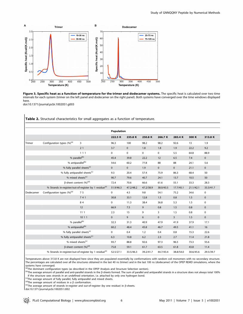
Figure 3. Specific heat as a function of temperature for the trimer and dodecamer systems. The specific heat is calculated over two timeintervals for each system (trimer on the left panel and dodecamer on the right panel). Both systems have converged over the time windows displayedhere.doi:10.1371/journal.pcbi.1002051.g003
Table 2. Structural characteristics for small aggregates as a function of temperature.
Population
222.5 K 235.8 K 250.8 K 266.7 K 283.4 K 300 K 313.8 K
Trimer Configuration types (%)(a) 3 96.3 100 98.2 98.2 92.6 13 1.9
2 1 3.7 0 1.8 1.8 1.9 22.2 9.2
1 1 1 0 0 0 0 5.5 64.8 88.9
% parallel(b) 45.4 39.8 22.2 12 6.5 7.4 0
% antiparallel(b) 54.6 60.2 77.8 88 88 24.1 5.6
% fully parallel sheets(c) 0 0 1.9 0 0 21.1 0
% fully antiparallel sheets(c) 9.3 20.4 57.4 75.9 86.3 68.4 50
% mixed sheets(c) 90.7 79.6 40.7 24.1 13.7 10.5 50
b-sheet content (%)(d) 72.4 70.6 66.6 61.6 55.1 33.3 28.3
% Strands in-register/out-of-register by 1 residue(e) 51.9/46.3 47.2/48.2 47.2/38.9 38.0/43.5 17.7/45.1 21.1/42.1 33.3/41.7
Dodecamer Configuration types (%)(a) 7 5 0 4.5 9.8 54.1 75.2 34.6 0
7 4 1 30.8 33.1 12.8 1.5 0.8 1.5 0
8 4 0 11.3 38.4 36.8 5.3 1.5 0
12 66.9 7.5 9 0.8 1.5 0.8 0
11 1 2.3 15 9 3 1.5 0.8 0
10 1 1 0 9 6 0 3 1.5 0
% parallel(b) 32.3 31.3 40.9 47.9 41.9 37.9 17.1
% antiparallel(b) 60.2 48.4 45.8 46.7 49.5 41.1 16
% fully parallel sheets(c) 0 0.4 1.2 0.4 0.8 15.3 22.6
% fully antiparallel sheets(c) 6.3 10.8 6.2 2.3 2.7 11.4 21.8
% mixed sheets(c) 93.7 88.8 92.6 97.3 96.5 73.3 55.6
b-sheet content (%)(d) 75.8 59.1 61.7 63.5 61.8 43.8 11.4
% Strands in-register/out-of-register by 1 residue(e) 52.5/27.1 33.5/46.3 39.2/41.7 44.7/43.4 38.8/54.0 30.6/45.6 29.5/39.7
Temperatures above 313.8 K are not displayed here since they are populated essentially by conformations with random coil monomers with no secondary structure.The percentages are calculated over all the structures obtained in the last 40 ns (trimer) and in the last 100 ns (dodecamer) of the OPEP REMD simulations, where thesystems have converged.(a)The dominant configuration types (as described in the OPEP Analysis and Structure Selection section).(b)The average amount of parallel and anti-parallel strands in the b-sheets formed. The sum of parallel and antiparallel strands in a structure does not always total 100%
if the structure sees strands in an undefined orientation, i.e. attached by only one hydrogen bond.(c)The average amount of fully parallel, fully antiparallel and mixed sheets.(d)The average amount of residues in a b conformation.(e)The average amount of strands in-register and out-of-register (by one residue) in b-sheets.doi:10.1371/journal.pcbi.1002051.t002
Study of GNNQQNY Peptide by Numerical Methods
PLoS Computational Biology | www.ploscompbiol.org 6 May 2011 | Volume 7 | Issue 5 | e1002051

temperature to b-sheet rich structures where the strands prefer an
antiparallel orientation, as for the trimer, but with a lower melting
temperature of 283 K (see Figure 3) even though the potential
energy per monomer in the ordered phase is much lower, rea-
ching 237.0 kcal/mol/monomer for the 12-mer compared to
218.4 kcal/mol/monomer for the trimer, indicating a clear bias
toward aggregation and resulting in a much more marked peak in
the specific heat.
Kinetically, the aggregation tendency for the dodecamer is to
first form one or two stable four-stranded b-sheets that show little
dissociation and that trigger the transient formation of one or two
longer b-sheets. The formation of a trimer that precedes the four-
stranded b-sheet shows, however, a higher dissociation/association
rate. Interestingly, the tendency of the GNNQQNY sequence to
form stable tetrameric aggregation nuclei had already been
noticed in a previous investigation on the system [44] and was
proposed by the Eisenberg group on the basis of entropic and
energetic arguments [7]. The final stable ordered structures are
shown in Figure 5 (left side of the panel).
As would be expected, a rich set of ordered configurations is
visited for the 12-mer (Table 2). Regrouping all structures below
melting, the dominant conformation, visited 63% of the time, is a
two b-sheet structure with a 7 or 8-strand sheet stabilized by a
smaller, 4–5 strand sheet positioned on top (Figure 5, struc-tures 12OP1-B to -E). Single sheets, with 11 or 12 strands also
appear with a frequency of 23.3% below melting (Figure 5,structure 12OP1-A). Surprisingly, strand orientation probabil-
ities vary significantly going from the 3-peptide to the 12-peptide
system. As for the 3-peptide system, the anti-parallel orientation is
favored below melting for the 12-peptide system especially at the
lowest two temperatures where the probability of forming anti-
parallel is between 60% and 45% compared to 30% for the
parallel. Then, as the temperature is increased, the amount of
parallel and anti-parallel orientation becomes almost the same,
suggesting that while anti-parallel orientation is energetically
Figure 4. Structures obtained for the trimeric simulations. Weshow, on the left-hand side panel, representative structures obtainedfrom the OPEP simulations and, on the right-hand side panel, therepresentative structures obtained after all-atom MD refinements.03OP1-A,-B,-C,-D and –E were extracted respectively at 222.5 K(probability of occurrence for this b-strand organization: 91%), 235.7 K(80%), 250.8 K (41%), 266.7 K (76%) and 283.4 K (86%). 03OP1-A to -C aremixed b-sheets while 03OP1-D and –E are fully antiparallel b-sheets. Theall-atom structures are represented in secondary structure cartoon andonly the tyrosines (most hydrophobic residues in the sequence) areshown in blue sticks (hydrogen atoms are omitted).doi:10.1371/journal.pcbi.1002051.g004
Figure 5. Structures obtained for the dodecameric simulations.We show, on the left-hand side panel, representative structuresobtained from the OPEP simulations and, on the right-hand side panel,representative structures obtained after all-atom MD refinements.12OP1-A,-B,-C,-D and –E were extracted respectively at 222.5 K,235.7 K, 250.8 K, 266.7 K and 283.4 K. 12OP1-A (top left structure) is along flat beta-sheet. 12OP1-B to -E (second left to bottom leftstructures) are made of 2 beta-sheets facing each other. Monomersforming b-sheets in the initial state are colored red or green. Thesecolors are kept in the final structure. The tyrosines are shown in bluesticks for the all-atom structures. During the all-atom MD simulation thestructures tend to be more globular but the strands see no exchangebetween the b-sheets, i.e. the red and green b-sheets do not dissociatefor the 12-mer system.doi:10.1371/journal.pcbi.1002051.g005
Study of GNNQQNY Peptide by Numerical Methods
PLoS Computational Biology | www.ploscompbiol.org 7 May 2011 | Volume 7 | Issue 5 | e1002051

Ta
ble
3.
Co
mp
aris
on
of
the
stru
ctu
ral
pro
pe
rtie
sb
etw
ee
nO
PEP
and
GR
OM
AC
Sst
ruct
ure
sfo
rse
lect
ed
stab
lest
ruct
ure
s.
b-S
he
et
Co
nte
ne
nt
(ba
sed
on
the
dss
pp
rog
ram
)(b)
%P
ara
lle
l-
An
tip
ara
lle
l(c)
%R
es
Ali
gn
(%1
00
Ali
gn
-%
Sh
ift
by
1R
es
-%
Sh
ift
by
2R
es…
)(d)
GR
OM
AC
S-
GR
OP
EP
-O
PG
RO
MA
CS
-G
RO
PE
P-
OP
GR
OM
AC
S-
GR
OP
EP
-O
P
Co
nfi
gu
rati
on
Ty
pe
(a)
Str
uct
ure
Fir
stC
lust
er
Fin
al
Str
uct
ure
CG
Min
Fir
stC
lust
er
Fin
al
Str
uct
ure
CG
Min
Fir
stC
lust
er
Fin
al
Str
uct
ure
CG
Min
Tri
me
rs3
03
_1
-A
38
%1
9%
71
%(1
00
%)
67
%5
0-
50
50
-0
50
-5
05
0-
50
50
-5
05
0-
50
50
-5
05
0-
50
30
3_
1-
B3
8%
24
%1
9%
(27
%)
67
%0
-5
05
0-
50
50
-5
05
0-
50
50
-5
05
0-
0-
50
50
-5
05
0-
50
30
3_
1-
C5
7%
62
%6
2%
(87
%)
67
%5
0-
50
50
-5
05
0-
50
50
-5
05
0-
50
50
-5
05
0-
50
50
-5
0
30
3_
1-
D4
8%
57
%5
2%
(73
%)
67
%0
-5
00
-5
00
-5
00
-1
00
50
-5
05
0-
50
50
-5
05
0-
50
30
3_
1-
E4
8%
52
%5
7%
(80
%)
53
%5
0-
50
0-
50
0-
50
0-
10
05
0-
0-
50
50
-0
-5
00
-5
0-
50
0-
50
-5
0
12
-me
rs1
21
2_
1-
A4
9%
35
%6
4%
(90
%)
60
%6
0-
20
40
-1
03
6-
64
36
-6
48
0-
20
50
-5
06
4-
18
-1
86
4-
18
-1
8
64
11
2_
1-
B1
4%
14
%4
5%
(63
%)
44
%1
3-
25
20
-4
02
2-
22
33
-3
33
5-
25
-3
70
-4
0-
60
33
-5
6-
11
33
-5
6-
11
84
12
_1
-C
35
%2
9%
49
%(6
8%
)4
2%
10
-2
04
0-
20
60
-4
04
0-
30
40
-4
0-
20
50
-2
0-
30
60
-2
0-
20
60
-2
0-
20
75
12
_1
-D
33
%2
4%
54
%(7
5%
)5
5%
11
-2
20
-3
04
0-
60
10
-6
04
5-
44
-1
15
0-
50
50
-5
05
0-
50
75
12
_1
-E
56
%5
1%
46
%5
1%
60
-4
05
0-
20
60
-4
06
0-
40
60
-3
0-
10
50
-5
06
0-
30
-1
05
0-
40
-10
20
-me
rs1
17
11
20
_p
-A
14
3%
38
%3
8%
(53
%)
37
%4
4-
65
6-
13
76
-2
44
4-
25
56
-3
1-
13
63
-1
9-
18
34
-3
3-
33
44
-3
7-
19
20
_p
-A
23
1%
34
%3
8%
(53
%)
37
%3
3-
05
0-
07
6-
24
44
-2
54
0-
40
-2
06
3-
31
-6
34
-3
3-
33
44
-3
7-
19
11
71
12
0_
p-
B1
52
%4
9%
36
%(5
0%
)6
6%
69
-1
95
9-
12
71
-24
75
-2
53
1-
44
-2
53
5-
47
-18
33
-3
9-
28
31
-4
4-
25
20
_p
-B
24
7%
38
%3
6%
(50
%)
66
%4
0-
13
38
-1
37
1-2
47
5-
25
34
-5
3-
13
31
-5
6-
13
33
-3
9-
28
31
-4
4-
25
11
71
12
0_
p-
C1
39
%3
5%
44
%(4
4%
)5
9%
44
-1
94
7-
20
72
-2
26
9-
25
31
-5
0-
19
54
-3
3-1
33
3-
39
-2
83
1-
44
-2
5
20
_p
-C
25
9%
36
%4
4%
(44
%)
59
%6
9-
25
69
-8
72
-2
26
9-
25
27
-4
7-
26
46
-5
43
3-
39
-2
83
1-
44
-2
5
12
71
20
_p
-D
14
4%
46
%4
4%
(62
%)
64
%2
4-
35
24
-3
53
5-
47
41
-47
59
-3
5-
66
5-
33
-65
9-
35
-6
59
-3
5-
6
20
_p
-D
22
7%
27
%4
4%
(62
%)
64
%2
9-
24
22
-2
23
5-
47
41
-4
76
3-
31
-6
61
-3
3-6
59
-3
5-
65
9-
35
-6
96
52
0_
2-
A4
4%
44
%3
4%
(47
%)
28
%6
9-
13
61
-1
17
2-
22
73
-1
33
1-
44
-25
39
-3
9-
22
56
-3
9-
52
0-
53
-2
7
87
52
0_
2-
B4
5%
39
%3
9%
(55
%)
71
%4
1-
41
17
-2
27
1-
18
41
-5
94
1-
18
-4
15
6-
28
-1
63
5-
53
-1
24
1-
24
-3
5
77
62
0_
2-
C3
6%
37
%5
2%
(73
%)
65
%4
4-
17
41
-3
57
6-
24
59
-4
15
0-
39
-1
13
5-
47
-1
83
0-
41
-2
94
1-
47
-1
2
88
42
0_
2-
E3
2%
33
%5
1%
(72
%)
63
%2
8-
11
42
-1
65
9-
41
50
-3
94
4-
39
-1
74
2-
37
-2
15
4-
23
-23
39
-3
3-
28
10
82
20
_2
-N
41
%3
9%
42
%(5
9%
)5
7%
39
-2
24
1-
24
31
-5
63
1-
56
44
-2
8-
28
29
-4
7-
24
44
-2
5-3
14
4-
25
-3
1
‘‘Fir
stC
lust
er’
’m
ean
sth
em
ost
rep
rese
nta
tive
stru
ctu
reo
fth
eG
RO
MA
CS
sim
ula
tio
ns.
‘‘Fin
alst
ruct
ure
’’is
the
fin
alco
nfo
rmat
ion
ob
tain
ed
atth
ee
nd
of
the
GR
OM
AC
Ssi
mu
lati
on
s.‘‘C
G’’
isth
est
ruct
ure
ext
ract
ed
atth
ee
nd
of
the
OP
EPsi
mu
lati
on
sb
efo
reth
ere
con
stru
ctio
no
fth
esi
de
chai
ns.
‘‘Min
’’in
dic
ate
sth
est
ruct
ure
resu
ltin
gfr
om
the
reco
nst
ruct
ion
of
the
sid
ech
ain
saf
ter
am
inim
izat
ion
ste
p.
(a) T
he
con
fig
ura
tio
nty
pe
(as
de
scri
be
din
the
OP
EPA
nal
ysis
and
Stru
ctu
reSe
lect
ion
sect
ion
).(b
) Th
eav
era
ge
amo
un
t(p
erc
en
tag
e)
of
resi
du
es
inab
con
form
atio
n.
For
OP
EP,
the
pe
rce
nta
ge
inb
rack
ets
has
be
en
calc
ula
ted
wit
ho
ut
taki
ng
the
Gly
cin
es
into
acco
un
t.(c
) Th
eav
era
ge
amo
un
t(p
erc
en
tag
e)
of
par
alle
lan
dan
ti-p
aral
lel
stra
nd
sin
ast
ruct
ure
.Th
esu
mo
fp
aral
lel
and
anti
par
alle
lst
ran
ds
ina
stru
ctu
red
oe
sn
ot
alw
ays
tota
l1
00
%if
the
stru
ctu
rese
es
stra
nd
sin
anu
nd
efi
ne
do
rie
nta
tio
n,
i.e.
atta
che
db
yo
nly
on
eh
ydro
ge
nb
on
d.
(d) T
he
ave
rag
eam
ou
nt
of
stra
nd
sin
-re
gis
ter
and
ou
t-o
f-re
gis
ter
(by
on
ere
sid
ue
).d
oi:1
0.1
37
1/j
ou
rnal
.pcb
i.10
02
05
1.t
00
3
Study of GNNQQNY Peptide by Numerical Methods
PLoS Computational Biology | www.ploscompbiol.org 8 May 2011 | Volume 7 | Issue 5 | e1002051
favored, it is rapidly overcome by the entropic gain of mixing
orientations. The alignment of the b-strands is a mix of perfectly
aligned strands and strands misaligned by one residue at all
temperatures below the melting point. Because sheets are longer
than for the trimer, the 12-mer comprises mostly b-sheets with
strands in mixed orientations at low temperatures below Tm with a
low probability of forming fully parallel or fully antiparallel sheets
(Table 2). Interestingly in the few and much smaller sheets
observed just above Tm, fully parallel and antiparallel b sheets
form with almost identical probability (data not shown), suggesting
that with slower growth, structures visited below Tm could be
more ordered.
All-atom MD simulations. The 5 most representative
structures obtained from OPEP REMD (labeled 12OP1-A to
12OP1-E) were further studied by all-atom MD. Representative
structures obtained from the latter simulations are shown in
Figure 5 right panel. The 12OP1-A OPEP structure is charac-
terized by the presence of a flat arrangement of b-sheets. It
undergoes significant rearrangements during the all-atom
evolution in explicit solvent (12GR1-A), as shown by the time
evolution of the radius of gyration (Figure S1), with the planar b-
sheet breaking into four fragments of two to four stranded b-sheets
that assemble on top of each other, with two central parallel b-
sheets covered on both sides by a perpendicular b-sheet. The
overall amount of b-sheet structure is conserved during the all-
atom simulation (Table 3).
Structure 12OP1-B is characterized by a mainly parallel twisted
b-sheet, with four strands packed on top. This structure is not
stable in the all-atom MD setting, simulation 12GR1-B, and
evolves towards a compact globular structure as shown by the
evolution of the radius of gyration in time (Figure S1).
Interestingly, the external side of the final aggregate is lined with
hydrophilic Asn and Gln side chains that provide favorable
contacts with the solvent. No specific order is observed for contacts
among these side chains, although some cases of interdigitation as
seen in the final steric zipper are noticed. The interior of the final
aggregate is lined with Tyr aromatic side chains.
Such a supramolecular organization of the peptides may be
representative of one of the soluble intermediates on the pathway
to fibril formation. Solubility is favored by the presence of
hydrophilic side chains on the external surface of the aggregate. At
the same time, the packing of the interior is not optimal, so that
the resulting structure may not be in the most favorable
arrangement to ensure lasting stability. Water can also access
the interior of the globular aggregate, disrupting inter-strand
hydrogen bonds, eventually favoring conformational changes.
Structures 12OP1-C and 12OP1-D are similar to 12OP1-B: the
main difference is that four strand pack with their long axis almost
perpendicular to the long axis of the extended b-sheet. The main
difference between 12OP1-C and 12OP1-D is that the planes
defined by the four strands have different inclinations with respect
to the plane of the long extended b-sheet. In the all-atom MD
setting — simulations 12GR1-C and 12GR1-D — these structures
evolve to less globular, but more compact final arrangements than
that observed above, with most of the Tyr side-chains in contact
with the solvent (Figure S1). The exterior of the aggregates is
lined with Asn, while the interior is more compact than for
12GR1-A and 12GR1-B and packed with the side-chains of Gln,
that form a network of van der Waals and hydrogen bonding
contacts.
Finally, structure 12OP1-E is characterized by two orthogonal
twisted b-sheets. The OPEP structure is very stable: it does not
undergo significant rearrangement during the all-atom MD,
contrary to the previous cases, and the b-sheet content remains
constant (Table 3). The oligomer is trapped in this conformation
by the extensive contacts packing determined by the Tyr side
chains in the two sheets. Moreover, the inter-sheet space is filled by
Asn and Gln side chains. However no specific packing into the
ordered steric zipper is evident.
Table 3 recapitulates the conformational heterogeneity and
plasticity of the 12-mer aggregates. As a general case, the presence
of explicit solvent tend to condense OPEP-generated structures, at
the expense of structured b-sheets and the associated parallel-
antiparallel structure, strand alignment and register. It must be
kept in mind, though, that MD simulations may be affected by
sampling limitations associated with the short runs and the
presence of solvent.
Overall, the combined results indicate that the configurational
richness increases from the trimer to the 12-mer and that the
critical nucleus has not yet been found. Though, the strands do not
see much exchange between sheets as seen in Figure 5. While
ordered 12-mers are energetically much more favorable than the
trimers, entropic factors may be considered prevalent, favoring a
wide variety of metastable structures. The presence of explicit
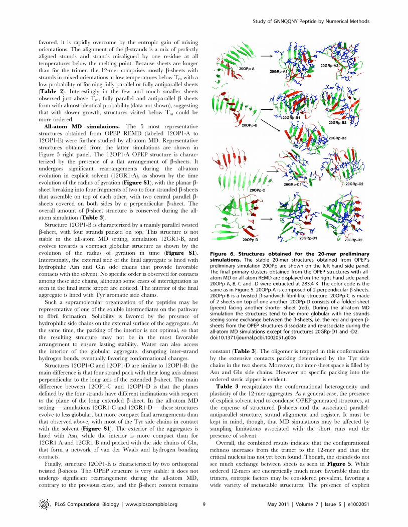
Figure 6. Structures obtained for the 20-mer preliminarysimulations. The stable 20-mer structures obtained from OPEP’spreliminary simulation 20OPp are shown on the left-hand side panel.The final primary clusters obtained from the OPEP structures with all-atom MD or all-atom REMD are displayed on the right-hand side panel.20OPp-A,-B,-C and -D were extracted at 283.4 K. The color code is thesame as in Figure 5. 20OPp-A is composed of 2 perpendicular b-sheets.20OPp-B is a twisted b-sandwich fibril-like structure. 20OPp-C is madeof 2 sheets on top of one another. 20OPp-D consists of a folded sheet(green) facing another shorter sheet (red). During the all-atom MDsimulation the structures tend to be more globular with the strandsseeing some exchange between the b-sheets, i.e. the red and green b-sheets from the OPEP structures dissociate and re-associate during theall-atom MD simulations except for structures 20GRp-D1 and -D2.doi:10.1371/journal.pcbi.1002051.g006
Study of GNNQQNY Peptide by Numerical Methods
PLoS Computational Biology | www.ploscompbiol.org 9 May 2011 | Volume 7 | Issue 5 | e1002051

solvent decreases significantly the stability of elongated b-sheets
either by increasing the effective hydrophobic interactions or
decreasing entropic gains, favoring rather more compact struc-
tures. Different molecular mechanisms may be responsible for the
stabilization of different conformations, endowed with different
solubility properties. Indeed, we have observed globular-like
structures with an external region decorated with hydrophilic
groups that may determine the oligomers to be soluble in aqueous
solution. In contrast, more ordered structures with higher b-sheet
content appear to expose more hydrophobic area to the contact
with the solvent. In turn, the latter may recruit more monomers or
preformed oligomers that can aggregate by the juxtaposition of
hydrophobic surfaces. The observations on the 12-mer systems
also underline the enormous structural diversity that characterizes
the aggregation of amyloidogenic peptides, which is reflected at
the macroscopic level in a high degree of polymorphism.
Simulations of 20-mer SystemsNext, we turned to the study of 20-mers in order to assess the
importance of the number of chains on the final supra-molecular
organization and determine whether new structural motifs can
emerge.
Coarse-grained simulations. Three REMD simulations
with OPEP were thus generated for the GNNQQNY 20-mer
systems: 20OPp, 20OP1 and 20OP2. A preliminary run 20OPp
was run to identify the four most common low-energy clusters,
from which we extract the central structure for each: 20OPp-A,
20OPp-B, 20OPp-C and 20OPp-D (Figure 6, left panel). These
were used as starting points for MD simulations with GROMACS.
The first three are two-sheet structures while the fourth is a three-
sheet configuration. What is particularly interesting here is that we
obtain a protofibril-like structure (20OPp-B) among the most
dominant clusters after only 200 ns starting from a random coil
configuration. Interestingly, the protofibril-like structure is possible
but not dominant in this preliminary simulation.
Following this preliminary run, we have performed two
additional simulations 400 ns-long 20OP1 and 20OP2 (Figure 7)
to attempt to better sample the phase space to determine the degree
of preference and the importance of the protofibril-like structure
among the morphologies accessible to that sequence for twenty
peptides. Even after 400 ns, however, neither simulation is fully
converged and the melting temperature is evaluated, from
specific heat, to be at 280 K or higher, with ordered structures
forming successfully below this temperature: the melting temp-
erature is likely to continue to increase with the simulation length
as the average nucleation time for the density used here appears
to be around 1 ms based on the fact that slightly more than half
the trajectories have not yet visited ordered structures during the
400 ns simulation. In spite of this limitation, we observe
significant exchange among the trajectories below melting,
suggesting that these achieve some degree of thermodynamic
equilibrium.
As for the 12-mer, aggregation is extremely favorable
energetically. The melting temperature for 20OP1 varies between
280.4 K and 289.2 K during the last 200 ns of simulation and the
energy of ordered structures at the lowest temperature, 223.8 K, is
on average 227.8 kcal/mol/monomer for 20OP1, as calculated
from the PTWHAM analysis. For 20OP2, the transition is
happening between 260.2 K and 290.5 K and the potential
energy of aggregated structures at the lowest temperature,
223.8 K, is on average 228.1 kcal/mol, which is comparable to
the energies of aggregated structures for 20OP1. Those energies
are about 10 Kcal/mol/monomer above the dodecamer struc-
tures’ energies at 222.5 K: clearly, the structures generated for the
20-mer are not as ordered as those found for the 12-mer due to the
much longer time needed to sample these energetically-favorable
conformations, but also because the entropic loss associated with
full-ordering is larger for the 20-mer. For both the 20OP1 and the
20OP2 simulation sets, random coil structures dominate at
simulations whose temperature is above 280 K.
Following specific trajectories, as they move through tempera-
tures, it is possible to identify sequences of steps leading to low-
energy ordered structures. In the more than 25 such events
observed in 20OP1 and 20OP2, the aggregation process is
systematically triggered by the formation of a few dimers, trimers
and/or tetramers seeds. The conformations obtained from both
20OP1 and 20OP2 are structurally similar in the sense that they
are almost always composed of three sheets composed of 5 to 9
strands each either facing each other in a triangle-like or organized
in a propeller-like or b-sandwich conformation (Figure 8).
Irrespective of the final shape, the system displays a strong
tendency to form b-sheets. The five final ordered structures
selected from 20OP2 and shown in Figure 8 are representative of
all three REMD simulation sets: below melting, the 20-chain
system mostly forms three b-sheets, but can also form two-sheet
structures. Looking at the statistics collected for 20OP1 and
20OP2 (Table 4), we observe that various three-b-sheet
Figure 7. Specific heat as a function of temperature for the two 20-mer simulations sets. The specific heat is calculated over two timeintervals for the systems 20OP1 (left panel) and 20OP2 (right panel) during the last 200 ns.doi:10.1371/journal.pcbi.1002051.g007
Study of GNNQQNY Peptide by Numerical Methods
PLoS Computational Biology | www.ploscompbiol.org 10 May 2011 | Volume 7 | Issue 5 | e1002051

configurations with juxtaposed b-sheets containing 8-7-5 mono-
mers or 9-6-5 monomers are frequent below Tm. Two-b-sheet
systems appear to be less frequent but are populated close to the
melting temperature as well. Although, sheet-lengths differ slightly
between simulations 20OP1 and 20OP2 for the dominant
structures, the overall results are consistent. We also observe a
high number of possible b-sandwich morphologies for the three-
sheet configuration among the final structures obtained where
some of them see their three sheets facing each other in a triangle
or twisted-around-each-other arrangements (Figure 8, structures
20OP2-A, -B, -C and -E). These topologies run from a rather well
defined 3-fold symmetry (20OP2-E) to more disordered confor-
mations with little symmetry (20OP2-A, -B and –C). The minimal
b-sheet unit contains four strands in 20OP2-E, five strands in
20OP2-A,-B and six strands in 20OP2-C. We note that the
structure 20OP2-E is reminiscent of the recently proposed
structure of Ab1–40 fibrils with a three-fold symmetry [14].
Interestingly, ordered two-sheet conformations with one sheet
slightly longer than the other, such as 11 9 and 12 8 (not shown,
but close to 20OP2-N, a 8 10 2 configuration in Figure 8),
represent a significant fraction of the accessible states either as a b-
sandwich –with an occasional insertion of a short b-sheet – or as
two perpendicular sheets.
In both REMDs, the b-sheets have a high probability of being
in a mixed anti-parallel/parallel orientation state due to their
length (over 70% below Tm) (Table 4). As for the dodecamer, as
T rises, sheets become shorter and we notice a rise of fully parallel
sheets at the five temperatures above Tm (data not shown).
Contrary to the 3-mer and 12-mer where the b-strands prefer an
anti-parallel orientation below 300 K, the parallel orientation of
the strands is preferred in the 20-mer at all temperatures. The
extended chains are also dominantly perfectly aligned at the lowest
temperatures and the structures see a mix of perfectly aligned
strands and strands misaligned by one residue close to the melting
point. The dominance of perfectly aligned strands and parallel
orientations of the chains is consistent with the experimental
observations that GNNQQNY fibrils display parallel b-strands
[9].
All-atom MD simulations of dominant clusters generated
by OPEP-REMD 20OPp. Consistent with the protocols
described above for the 3-mers and 12-mers, the dynamical
properties of 9 selected oligomeric conformations generated by the
OPEP simulations were refined by all-atom MD simulations in
explicit solvent (see Table 1).
The first set of all-atom MD simulations was run on the
structures selected from 20OPp calculations. OPEP runs identified
two main types of 3D organization for the 20mer: extended b-
sheet and globular-like structures. The former are characterized by
the presence of two parallel sheets, while the latter are
characterized by a circular organization of the strands, in a
mostly parallel arrangement. The major representatives of the
extended b-sheet like structures obtained from OPEP are labeled
20OPp-B and 20OPp-C; the globular structures are recapitulated
by 20OPp-A (which shows a compact part packed by a more
extended sheet) and 20OPp-D, see Figure 6. Two sets of 100 ns
MD simulations, starting with different initial velocities, were run
for each structure.
AGGREGATES WITH FACING B-SHEETS. The structure of 20OPp-B is
characterized by the presence of two perpendicular b-sheets. Each
b-sheet consists mainly of twisted parallel strands.
After the first MD run (20GRp-B1) the two sheets are oriented
anti-parallel to each other, forming a tight and elongated structure
(Figure 6). The Asn side chains from opposite sheets occupy the
inter-sheet space with some intertwining of the amide side chains.
Significant packing is also provided by the aromatic rings of
Tyrosine belonging to adjacent strands in the same sheet. The
100-ns generated aggregate also displays a significant degree of
twisting in the strands that make up the two facing antiparallel b-
sheets. This final structure is similar to that observed by others
[50]. It is important to underline that this twisted configuration
forms spontaneously during the MD simulation time starting from
a less compact structure.
In the second MD simulation (20GRp-B2), starting from the
same initial structure with a different set of velocities, the b-sheet
content decreases due a lower degree of packing of the Tyr side
chains and interdigitation of the Gln and Asn side-chains
(Figure 9). Packing interactions, however, still appear to be
important in stabilizing the compact structure.
During both 20GRp-B1 and 20GRp-B2 MD simulations, the
strands are dynamically interchanged between the two b-sheets.
Figure 8. Structures obtained for the 20-mer 20OP2 and 20GR2simulations. The final primary clusters obtained from the OPEPstructures with all-atom MD or all-atom REMD are displayed on theright-hand side panel. 20OP2-A,-B,-C,-E and -N were extractedrespectively at 260.1 K, 249.2 K, 254.7 K, 265.9 K and 292.2 K. Thedifferent sheets are distinguished by either a green, red or yellow colorand the tyrosines are shown in blue sticks for the all-atom structures.Structures 20OP2-A,-B,-C and -E are composed of 3 sheets twistedaround each other while structure 20OP2-N is a 2-sheet fibril-likeconformation. During the all-atom MD simulation the structures tend tobe more globular with the strands seeing some exchange between theb-sheets, i.e. the red, green and yellow b-sheets from the OPEPstructures dissociate and re-associate during the all-atom MD simula-tions except for the fibril-like structures 20GR2-N1 and -N2.doi:10.1371/journal.pcbi.1002051.g008
Study of GNNQQNY Peptide by Numerical Methods
PLoS Computational Biology | www.ploscompbiol.org 11 May 2011 | Volume 7 | Issue 5 | e1002051

Two elongated sheets may, however, evolve towards very
different supramolecular organizations. In 20OPp-C two elongat-
ed b-sheets, with mainly parallel b-strands, are in contact through
the terminal Tyr aromatic chains in an extended and non-compact
structure (Figure 6). In both all-atom MD simulations, 20GRp-
C1 and 20GRp-C2, the two b-sheets break up and reorganize in
more compact conformations. Though, for 20GRp-C2, the
strands do not interchange between sheets while 20GRp-C1 sees
some mixing between the strands of the 2 b-sheets. The compact
final structures still show some of the features we have described in
the previous case: anti-parallel orientations of facing sheets,
compaction of the side chains. One part of the long parallel b-
sheet, represented by a four-stranded unit, detaches from the
initial complex and packs onto the compact structure described
above, minimizing the exposed surface area. The resulting
structures correspond to a b-sandwich composed of three sheets.
Table 4. Structural characteristics for the 20-mer aggregates as a function of temperature below the melting point.
Population
223.8 K 249.3 K 260.1 K 265.9 K 270.3 K 273.8 K 277.1 K 280.1 K
20OP1 Configuration types (%)(a) 8 7 5 93.2 12.4 7.5 7.1 9.8 6.8 7.1 9.4
8 7 3 2 0.0 44.7 2.6 0.8 0.4 0.8 0.0 0.0
7 7 6 2.6 12.4 13.9 9.8 12.8 10.5 12.4 7.9
10 6 4 0.4 0.0 7.5 8.7 5.3 11.3 5.3 3.4
11 5 4 0.0 0.8 7.5 6.4 6.0 3.4 1.5 0.0
11 5 2 2 0.0 10.5 0.0 0.0 0.0 0.0 0.0 0.0
14 6 0.0 0.8 5.3 6.8 7.1 7.5 3.4 4.9
15 5 0.0 0.0 3.4 7.1 3.8 4.5 6.8 8.3
10 10 0.0 0.4 4.5 8.7 6.0 5.3 7.9 2.3
10 5 5 0.0 0.0 1.9 3.8 8.3 7.1 3.8 2.6
11 9 0.0 0.0 3.8 9.0 11.7 6.8 7.1 4.5
20 0.0 1.1 3.4 4.9 5.3 5.3 3.4 0.0
% parallel(b) 73.9 48.8 53.4 55.1 56.2 56.8 58.4 46.8
% antiparallel(b) 25.1 48.2 41.3 37.6 36.7 35.6 34.5 29.4
% fully parallel sheets(c) 26.2 22.4 19.7 15.1 11.8 14.5 11.1 16.1
% fully antiparallel sheets(c) 0.9 7.9 3.1 4.6 1.9 2.5 1.0 6.5
% mixed sheets(c) 72.9 69.7 77.2 80.3 86.3 83 87.9 77.4
b-sheet content (%)(d) 59.1 61.3 60.5 60.5 60.5 59.8 57.1 45.7
% Strands in-register/out-of-register by 1 residue(e) 32.1/38.0 43.1/31.2 42.6/33.8 41.2/36.9 40.1/38.4 38.8/38.6 39.7/38.6 38.7/39.8
20OP2 Configuration types (%)(a) 9 6 5 29.3 29.3 24.4 24.1 18.4 15.4 8.7 8.3
9 7 4 0.4 1.5 4.5 4.5 7.5 4.9 6.0 3.8
8 8 4 1.1 0.0 1.9 1.1 3.8 6.0 4.5 4.1
7 7 6 4.5 6.0 8.7 12.0 12.8 16.2 15.0 12.0
15 5 12.4 9.4 7.5 2.3 6.8 3.4 1.9 1.5
13 7 0.8 0.4 0.4 1.9 1.5 2.3 4.9 7.1
14 6 3.4 3.8 3.0 4.9 10.9 6.4 8.7 6.4
11 9 9.4 14.7 6.8 3.8 2.3 4.5 5.6 3.8
11 5 4 16.5 12.4 3.8 0.0 0.4 1.5 0.0 0.0
8 7 5 0.4 1.9 1.5 1.9 5.6 6.8 6.4 6.0
10 10 0.0 0.0 0.0 0.0 0.0 0.0 0.0 0.0
20 0.0 0.0 0.0 0.0 0.0 0.4 0.0 0.4
% parallel(b) 53.1 56.6 57.1 56.8 56.7 55.7 56.7 55.1
% antiparallel(b) 38.1 35.7 36.5 37.5 37.9 39.3 38.4 38.5
% fully parallel sheets(c) 7.4 16.9 15.0 14.2 14.1 14.1 18.7 17.1
% fully antiparallel sheets(c) 0.6 4.7 3.2 4.2 3.9 4.4 5.1 8.1
% mixed sheets(c) 92.0 78.5 81.8 81.6 82.0 81.5 76.2 74.8
b-sheet content (%)(d) 66.5 63.4 63.0 62.5 61.5 60.6 58.7 56.0
% Strands in-register/out-of-register by 1 residue(e) 51.0/36.3 50.0/34.0 48.1/35.1 44.6/37.0 42.4/37.2 42.3/37.2 39.9/39.1 39.7/39.4
Temperatures above 280.1 K are not displayed here since they are populated essentially by conformations with random coil monomers with no secondary structure.The percentages are calculated over all the structures obtained in the last 200 ns of both OPEP REMD simulations. For details on (a)–(e), see Table 2.doi:10.1371/journal.pcbi.1002051.t004
Study of GNNQQNY Peptide by Numerical Methods
PLoS Computational Biology | www.ploscompbiol.org 12 May 2011 | Volume 7 | Issue 5 | e1002051
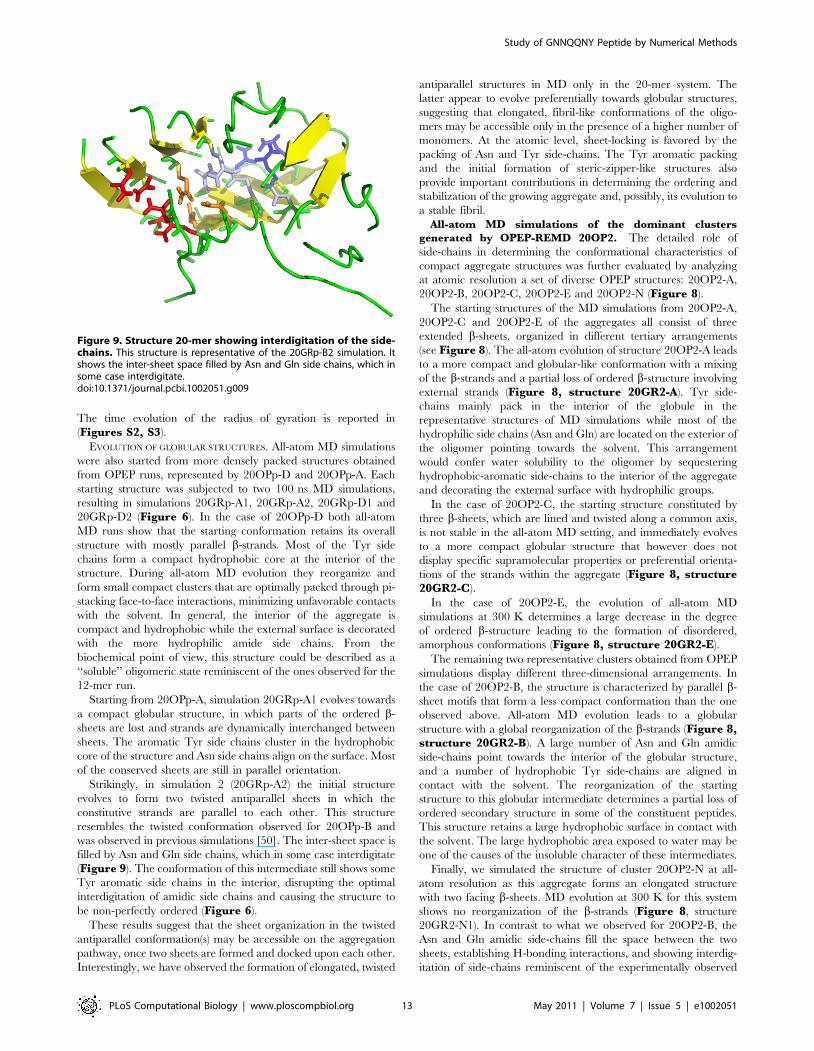
The time evolution of the radius of gyration is reported in
(Figures S2, S3).
EVOLUTION OF GLOBULAR STRUCTURES. All-atom MD simulations
were also started from more densely packed structures obtained
from OPEP runs, represented by 20OPp-D and 20OPp-A. Each
starting structure was subjected to two 100 ns MD simulations,
resulting in simulations 20GRp-A1, 20GRp-A2, 20GRp-D1 and
20GRp-D2 (Figure 6). In the case of 20OPp-D both all-atom
MD runs show that the starting conformation retains its overall
structure with mostly parallel b-strands. Most of the Tyr side
chains form a compact hydrophobic core at the interior of the
structure. During all-atom MD evolution they reorganize and
form small compact clusters that are optimally packed through pi-
stacking face-to-face interactions, minimizing unfavorable contacts
with the solvent. In general, the interior of the aggregate is
compact and hydrophobic while the external surface is decorated
with the more hydrophilic amide side chains. From the
biochemical point of view, this structure could be described as a
‘‘soluble’’ oligomeric state reminiscent of the ones observed for the
12-mer run.
Starting from 20OPp-A, simulation 20GRp-A1 evolves towards
a compact globular structure, in which parts of the ordered b-
sheets are lost and strands are dynamically interchanged between
sheets. The aromatic Tyr side chains cluster in the hydrophobic
core of the structure and Asn side chains align on the surface. Most
of the conserved sheets are still in parallel orientation.
Strikingly, in simulation 2 (20GRp-A2) the initial structure
evolves to form two twisted antiparallel sheets in which the
constitutive strands are parallel to each other. This structure
resembles the twisted conformation observed for 20OPp-B and
was observed in previous simulations [50]. The inter-sheet space is
filled by Asn and Gln side chains, which in some case interdigitate
(Figure 9). The conformation of this intermediate still shows some
Tyr aromatic side chains in the interior, disrupting the optimal
interdigitation of amidic side chains and causing the structure to
be non-perfectly ordered (Figure 6).
These results suggest that the sheet organization in the twisted
antiparallel conformation(s) may be accessible on the aggregation
pathway, once two sheets are formed and docked upon each other.
Interestingly, we have observed the formation of elongated, twisted
antiparallel structures in MD only in the 20-mer system. The
latter appear to evolve preferentially towards globular structures,
suggesting that elongated, fibril-like conformations of the oligo-
mers may be accessible only in the presence of a higher number of
monomers. At the atomic level, sheet-locking is favored by the
packing of Asn and Tyr side-chains. The Tyr aromatic packing
and the initial formation of steric-zipper-like structures also
provide important contributions in determining the ordering and
stabilization of the growing aggregate and, possibly, its evolution to
a stable fibril.
All-atom MD simulations of the dominant clusters
generated by OPEP-REMD 20OP2. The detailed role of
side-chains in determining the conformational characteristics of
compact aggregate structures was further evaluated by analyzing
at atomic resolution a set of diverse OPEP structures: 20OP2-A,
20OP2-B, 20OP2-C, 20OP2-E and 20OP2-N (Figure 8).
The starting structures of the MD simulations from 20OP2-A,
20OP2-C and 20OP2-E of the aggregates all consist of three
extended b-sheets, organized in different tertiary arrangements
(see Figure 8). The all-atom evolution of structure 20OP2-A leads
to a more compact and globular-like conformation with a mixing
of the b-strands and a partial loss of ordered b-structure involving
external strands (Figure 8, structure 20GR2-A). Tyr side-
chains mainly pack in the interior of the globule in the
representative structures of MD simulations while most of the
hydrophilic side chains (Asn and Gln) are located on the exterior of
the oligomer pointing towards the solvent. This arrangement
would confer water solubility to the oligomer by sequestering
hydrophobic-aromatic side-chains to the interior of the aggregate
and decorating the external surface with hydrophilic groups.
In the case of 20OP2-C, the starting structure constituted by
three b-sheets, which are lined and twisted along a common axis,
is not stable in the all-atom MD setting, and immediately evolves
to a more compact globular structure that however does not
display specific supramolecular properties or preferential orienta-
tions of the strands within the aggregate (Figure 8, structure20GR2-C).
In the case of 20OP2-E, the evolution of all-atom MD
simulations at 300 K determines a large decrease in the degree
of ordered b-structure leading to the formation of disordered,
amorphous conformations (Figure 8, structure 20GR2-E).
The remaining two representative clusters obtained from OPEP
simulations display different three-dimensional arrangements. In
the case of 20OP2-B, the structure is characterized by parallel b-
sheet motifs that form a less compact conformation than the one
observed above. All-atom MD evolution leads to a globular
structure with a global reorganization of the b-strands (Figure 8,structure 20GR2-B). A large number of Asn and Gln amidic
side-chains point towards the interior of the globular structure,
and a number of hydrophobic Tyr side-chains are aligned in
contact with the solvent. The reorganization of the starting
structure to this globular intermediate determines a partial loss of
ordered secondary structure in some of the constituent peptides.
This structure retains a large hydrophobic surface in contact with
the solvent. The large hydrophobic area exposed to water may be
one of the causes of the insoluble character of these intermediates.
Finally, we simulated the structure of cluster 20OP2-N at all-
atom resolution as this aggregate forms an elongated structure
with two facing b-sheets. MD evolution at 300 K for this system
shows no reorganization of the b-strands (Figure 8, structure
20GR2-N1). In contrast to what we observed for 20OP2-B, the
Asn and Gln amidic side-chains fill the space between the two
sheets, establishing H-bonding interactions, and showing interdig-
itation of side-chains reminiscent of the experimentally observed
Figure 9. Structure 20-mer showing interdigitation of the side-chains. This structure is representative of the 20GRp-B2 simulation. Itshows the inter-sheet space filled by Asn and Gln side chains, which insome case interdigitate.doi:10.1371/journal.pcbi.1002051.g009
Study of GNNQQNY Peptide by Numerical Methods
PLoS Computational Biology | www.ploscompbiol.org 13 May 2011 | Volume 7 | Issue 5 | e1002051
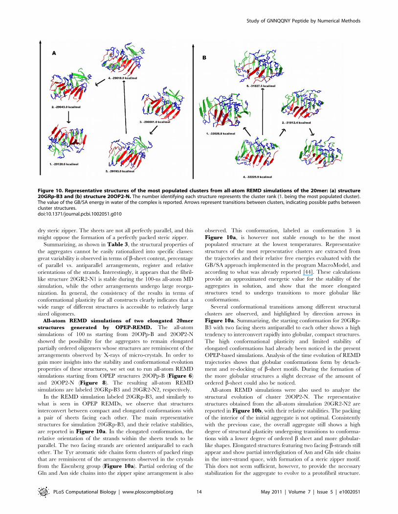
dry steric zipper. The sheets are not all perfectly parallel, and this
might oppose the formation of a perfectly packed steric zipper.
Summarizing, as shown in Table 3, the structural properties of
the aggregates cannot be easily rationalized into specific classes:
great variability is observed in terms of b-sheet content, percentage
of parallel vs. antiparallel arrangements, register and relative
orientations of the strands. Interestingly, it appears that the fibril-
like structure 20GR2-N1 is stable during the 100-ns all-atom MD
simulation, while the other arrangements undergo large reorga-
nization. In general, the consistency of the results in terms of
conformational plasticity for all constructs clearly indicates that a
wide range of different structures is accessible to relatively large
sized oligomers.
All-atom REMD simulations of two elongated 20mer
structures generated by OPEP-REMD. The all-atom
simulations of 100 ns starting from 20OPp-B and 20OP2-N
showed the possibility for the aggregates to remain elongated
partially ordered oligomers whose structures are reminiscent of the
arrangements observed by X-rays of micro-crystals. In order to
gain more insights into the stability and conformational evolution
properties of these structures, we set out to run all-atom REMD
simulations starting from OPEP structures 20OPp-B (Figure 6)
and 20OP2-N (Figure 8). The resulting all-atom REMD
simulations are labeled 20GRp-B3 and 20GR2-N2, respectively.
In the REMD simulation labeled 20GRp-B3, and similarly to
what is seen in OPEP REMDs, we observe that structures
interconvert between compact and elongated conformations with
a pair of sheets facing each other. The main representative
structures for simulation 20GRp-B3, and their relative stabilities,
are reported in Figure 10a. In the elongated conformation, the
relative orientation of the strands within the sheets tends to be
parallel. The two facing strands are oriented antiparallel to each
other. The Tyr aromatic side chains form clusters of packed rings
that are reminiscent of the arrangements observed in the crystals
from the Eisenberg group (Figure 10a). Partial ordering of the
Gln and Asn side chains into the zipper spine arrangement is also
observed. This conformation, labeled as conformation 3 in
Figure 10a, is however not stable enough to be the most
populated structure at the lowest temperatures. Representative
structures of the most representative clusters are extracted from
the trajectories and their relative free energies evaluated with the
GB/SA approach implemented in the program MacroModel, and
according to what was already reported [44]. These calculations
provide an approximated energetic value for the stability of the
aggregates in solution, and show that the more elongated
structures tend to undergo transitions to more globular like
conformations.
Several conformational transitions among different structural
clusters are observed, and highlighted by direction arrows in
Figure 10a. Summarizing, the starting conformation for 20GRp-
B3 with two facing sheets antiparallel to each other shows a high
tendency to interconvert rapidly into globular, compact structures.
The high conformational plasticity and limited stability of
elongated conformations had already been noticed in the present
OPEP-based simulations. Analysis of the time evolution of REMD
trajectories shows that globular conformations form by detach-
ment and re-docking of b-sheet motifs. During the formation of
the more globular structures a slight decrease of the amount of
ordered b-sheet could also be noticed.
All-atom REMD simulations were also used to analyze the
structural evolution of cluster 20OP2-N. The representative
structures obtained from the all-atom simulation 20GR2-N2 are
reported in Figure 10b, with their relative stabilities. The packing
of the interior of the initial aggregate is not optimal. Consistently
with the previous case, the overall aggregate still shows a high
degree of structural plasticity undergoing transitions to conforma-
tions with a lower degree of ordered b sheet and more globular-
like shapes. Elongated structures featuring two facing b-strands still
appear and show partial interdigitation of Asn and Gln side chains
in the inter-strand space, with formation of a steric zipper motif.
This does not seem sufficient, however, to provide the necessary
stabilization for the aggregate to evolve to a protofibril structure.
Figure 10. Representative structures of the most populated clusters from all-atom REMD simulations of the 20mer: (a) structure20GRp-B3 and (b) structure 20OP2-N. The number identifying each structure represents the cluster rank (1. being the most populated cluster).The value of the GB/SA energy in water of the complex is reported. Arrows represent transitions between clusters, indicating possible paths betweencluster structures.doi:10.1371/journal.pcbi.1002051.g010
Study of GNNQQNY Peptide by Numerical Methods
PLoS Computational Biology | www.ploscompbiol.org 14 May 2011 | Volume 7 | Issue 5 | e1002051

Once more, a number of transitions among different structural
families (Figure 10b) are observed.
Overall, we observe that the elongated structures reminiscent of
the one observed by the Eisenberg group in the fibril microcrystals
is accessible at room temperature but is not stable and can easily
interconvert into globular or more disordered conformations, even
in the presence of 20 monomers (see Table 3). According to our
observations, this happens both by detachment-reattachment of
strands or small oligomers from existing structures, or through
sliding/reptation moves without detaching from the aggregates
[81,82]. This behavior is widely observed across our 20-mer
simulations while the 12-mer system does not undergo much
reorganization during all-atom MD simulations. Interestingly,
when ordered supramolecular structure forms, with either an
elongated or a compact globular shape, there is no space for water
in the interior of the aggregate. Water exclusion from the core of
the aggregate may represent the first step leading to the formation
of a dry interface.
ConclusionsThe self-organization process of peptides and proteins into
oligomeric soluble and insoluble aggregates that eventually evolve
to fibrils is still difficult, if not impossible, to study at atomic
resolution using experimental approaches. In this paper, we have
carried out an extensive and comprehensive study of the formation
of oligomers of the model peptide GNNQQNY in different
conditions combining coarse-grained and all-atom simulation
approaches. Different numbers of peptides were used in several
simulations. In the smallest systems, composed of three strands, a
diverse set of structural motifs is accessible at room temperatures.
When bigger systems consisting of 12 chains are analyzed,
compact and globular structures begin to appear. Interestingly,
in some cases, globular oligomers expose hydrophilic side chains to
the contact with the water solvent, providing a viable model for
soluble intermediates that have been observed on pathway to the
formation of the final fibril. In parallel, at 300 K, globular
structures exposing a large amount of hydrophobic surface also
appear. These may represent possible nuclei for the growth of
bigger supramolecular structures.
In simulations conducted using 20 monomers, we have noticed
the appearance of elongated structures characterized by the
juxtaposition of two mainly parallel b-sheets with partial
interdigitation of amidic side chains reminiscent of the zipper-
spine observed in fibril microcrystals. It is important to notice,
however, that these structures are not stable in water solvent and
evolve towards more globular conformations. This observation
suggests that while ordered fibril-like structures are accessible on
the energy landscape, they need further stabilization by establish-
ing contacts with multiple copies of similar structures in order to
evolve to a fully fibrillar geometry. In this context, the formation of
this geometry would require the constructive interplay of many
factors and the entropic expense of such process would be clearly
very high, explaining the long lag phase times and very slow
kinetics of amyloid fibril formation. Moreover, the rich variety of
structures and conformational changes observed for the aggregates
may also reverberate into the fibril polymorphism observed at the
experimental scale.
In summary, our data and structural models represent valid
complements to experimental approaches in the attempt to shed
light on the supramolecular arrangements of amyloidogenic
oligomers, and lead the following conclusions.
First, the 20-mers of GNNQQNY are polymorphic and
endowed with a high degree of structural plasticity. Polymorphism
of the fibrillar products of amyloid aggregation has been observed
for many sequences by X-ray diffraction and solid-state NMR
experiments [6,14,21] and a recent computational study using a
simplified description, where the peptide has a single degree of
freedom with two minima [83]. Polymorphism of the low-n
oligomers with different registers of inter-peptide H-bonds and
orientations of the chains has also been reported using various
force fields and computational procedures [22,41,44,46,84].
Second, the 20-mers of GNNQQNY in explicit water are in
dynamic equilibrium, within at least 100 ns, between amorphous
structures (high probability) and configurations with three b-sheets
in various orientations (medium probability) and two b-sheets (low
probability). These two-b sheets, reminiscent of the cross-bstructures and the dry steric zipper observed experimentally for
mature fibrils, are not parallel, however, suggesting the existence
of a free energy barrier preventing the formation of a perfectly
packed steric zipper.
Third, there is a reorientation of the b-strands between the
GNNQQNY oligomers and fibrils. We find that an anti-parallel b-
strand alignment dominates over the parallel one in the 3 and 12
peptide systems. This contradiction with the fibrillar parallel b-
strand orientation [7] is however reconciled by the 20-peptide
systems, where a significant increase in the amount of parallel
strands and fraction of fully parallel b-sheets is observed.
Fourth, a common observation is that short amyloid peptide
fragments assume antiparallel b-strand geometries whereas longer
peptides, and proteins, often assume parallel geometries. Our
simulations along with other recent studies show this geometrical
property is more complex and depends strongly on the amino acid
composition. The dependence of b-strand orientation with
oligomer size occurs in the GNNQQNY (Sup35) peptide and
the VQIVYK (PH6) peptide, as reported by another computa-
tional study [85], because both short peptide fragments do not
contain any opposite charged amino acids at the extremities as
opposed to numerous fragments, such as the KLVFFAE (Ab(16–
22)) and the KFFE peptides. Table 2, for example, shows that the
free energy difference in favor of the antiparallel sheet decreases
from 0.70 kcal/mol for the trimer to only 0.05 kcal/mol for the
dodecamer at 300 K. This competition between the two b-strand
orientations during polymerization is also supported experimen-
tally by the co-existence of either antiparallel or parallel strands in
seeded hIAPP20–29 (SNNFGAILSS) fibrils [15], and the fact that
D23N-Ab40 forms fibrils with a majority having antiparallel b-
sheet structures and a minority having parallel b-sheet structures
[86]. We hypothesize that both short polar peptides and longer
peptides could display this b-strand orientation transition during
aggregation.
In addition, antiparallel b-sheets allow a higher potential
variability of the inter-chain H-bond geometry [87,88] accommo-
dating a higher number of possible strand conformations, which is
the favored situation in smaller aggregates. A parallel organization
could be more favorable for larger aggregates, which can for
instance create double-layered sheets that can pack with a parallel
arrangement of the strands as indicated by SS-NMR [89].
Moreover, in larger aggregates, the parallel arrangement might
be stabilized by the definition and stabilization of large numbers of
side-chain side-chain contacts. Therefore, our results are in
agreement with experiments but also suggest a more complex
relationship between the monomers during the assembly process,
something that cannot be measured experimentally.
Fifth, in terms of experimental relevance, it is important to note
that evidence exists showing that aggregation pathways can be
manipulated by the use of molecular chaperones. In the case of the
Sup35 prion protein, the chaperone Hsp104 catalyzes the
polymerization of seeds that are crucial for efficient amyloid
Study of GNNQQNY Peptide by Numerical Methods
PLoS Computational Biology | www.ploscompbiol.org 15 May 2011 | Volume 7 | Issue 5 | e1002051

formation [90]. Different chaperones such as Hsp70 and Hsp40,
on the contrary, prevent the assembly of aggregating species when
added during the polymerization reactions. In these cases, soluble
aggregates are formed, showing that chaperones can redirect
amyloidogenic polypeptides into non-amyloidogenic species.
Finally, the chaperonin TRiC has also been shown to be a
modulator of amyloid formation [91]. TRiC, in combination with
Hsp70 and 40, stimulates the reassembly of huntingtin oligomers
into soluble species, which are non-toxic. Clearly, the type of
binding and structural remodeling determined by different
chaperones is dictated by the details of the molecular recognition
between the oligomers and the interaction surfaces of the
chaperones. In this context, it is worth noting that generating
plausible models of possible oligomeric substrates for the
chaperones may be of great help in the design of optimized
systems aimed at modulating aggregate properties. Based on the
structural and surface properties of the oligomers, models of their
complexes with chaperones may be generated.
On this basis, the hydrophobic-hydrophilic profile of the
chaperone interaction surfaces could, for instance, be changed
by means of site-directed mutagenesis, affecting their activity and
ultimately the properties of the remodeled oligomers. This would
allow a rational manipulation of the amyloidogenic pathways,
helping to shed light on a very complex biological phenomenon.
A final consideration helpful to put our results in a biological
perspective is related to the importance of the knowledge of
oligomeric structures in the design of amyloidogenic inhibitors. In
this context, we are currently exploring the characterization of the
solvent accessible hydrophobic surface area of the 20-mers to
guide docking-experiments of small-molecule compounds (Congo
Red and EGCG in particular), in order to derive possible rules for
the rational selection of aggregation inhibitors. Preliminary data
and results show that this could be helpful in alleviating the
difficulties associated to drug-design when dealing with amyloid-
targets. Indeed, compared to classical drug-design efforts where
the target is an active site, with well-defined structure and cavities,
the variety of structures, mechanisms and conformational plasticity
of oligomers shown here confirm that rational design of
aggregation inhibitors is a daunting challenge. However, careful
characterization of oligomeric structures provides useful sugges-
tions for the design of possible inhibitors. Selective compounds or
peptidomimetics could be designed/selected to target the oligomer
conformations characterized by the presence of aromatic groups
on their external surface. These compounds would actually target
intermediates that are more prone to be insoluble or to favor the
addition of monomers through hydrophobic interactions. Inter-
estingly, most of the existing inhibitors of amyloidogenic pathways
are small molecules rich in aromatic functionalities, which can
target more than one single aggregating species, showing a general
mechanism of action [92].
Alternatively, one could design peptidomimetic-based or small
molecule chaperones that can stabilize soluble species, subtracting
them from the amyloidogenic pathway. This would lead to a
redirection of otherwise amyloidogenic peptides into non-amyloi-
dogenic species.
Supporting Information
Figure S1 Time evolution of the radius of gyration of the 12-mer
oligomers. From top to bottom: structures 12GR1-A, 12GR1-B,
12GR1-C, 12GR1-D and 12GR1-E. The structures shown are the
final structures of the all-atom MD simulations with GROMACS.
(TIF)
Figure S2 Time evolution of the radius of gyration of the 20-mer
oligomers for the preliminary simulation. Structures 20GRp-B1,
20GRp-B2, 20GRp-C1 and 20GRp-C2. The structures shown
are the final structures of the all-atom MD simulations with
GROMACS.
(TIF)
Figure S3 Time evolution of the radius of gyration of the 20-mer
oligomers for the preliminary simulation. Structures 20GRp-A1,
20GRp-A2, 20GRp-D1 and 20GRp-D2. The structures shown
are the final structures of the all-atom MD simulations with
GROMACS.
(TIF)
Author Contributions
Conceived and designed the experiments: JNL MM PD GC NM.
Performed the experiments: JNL MM. Analyzed the data: JNL MM PD
GC NM. Contributed reagents/materials/analysis tools: JNL MM PD GC
NM. Wrote the paper: JNL MM PD GC NM.
References
1. Chiti F, Dobson CM (2006) Protein misfolding, functional amyloid and humandisease. Annu Rev Biochem 75: 333–366.
2. Ross CA, Poirier MA (2004) Protein aggregation and neurodegenerative disease.
Nat Med 10: S10–S17.
3. Miranker AD (2004) Unzipping the mysteries of amyloid fibril formation. Proc
Natl Acad Sci U S A 101: 4335–4336.
4. Zerovnik E (2002) Amyloid-fibril formation. Proposed mechanisms and
relevance to conformational disease. Eur J Biochem 269: 3362–3371.
5. Dobson CM (1999) Protein misfolding, evolution and disease. Trends BiochemSci 24: 329–332.
6. Sawaya MR, Sambashivan S, Nelson R, Ivanova MI, Sievers SA, et al. (2007)Atomic structures of amyloid cross-b spines reveal varied steric zippers. Nature
447: 453–457.
7. Nelson R, Sawaya MR, Balbirnie M, Madsen AØ, Riekel C, et al. (2005)Structure of the cross-b spine of amyloid-like fibrils. Nature 435: 773–778.
8. Kayed R, Head E, Thompson JL, McIntire TM, Milton SC, et al. (2003)Common structure of soluble amyloid oligomers implies common mechanism of
pathogenesis. Science 300: 486–489.
9. Thirumalai D, Klimov DK, Dima RI (2003) Emerging ideas on the molecularbasis of protein and peptide aggregation. Curr Opin Struct Biol 13: 146–159.
10. Sunde M, Serpell LC, Bartlam M, Fraser PE, Pepys MB, et al. (1997) Commonstructure of amyloid fibrils by synchrotron X-ray diffraction. J Mol Biol 273:
729–739.
11. Fandrich M, Meinhardt J, Grigorieff N (2009) Structural polymorphism ofAlzheimer Ab and other amyloid fibrils. Prion 3: 89–93.
12. Celej MS, Caarls W, Demchenko AP, Jovin TM (2009) A triple emissionfluorescent probe reveals distinctive amyloid fibrillar polymorphism of wild-type
a-synuclein and its familial Parkinsons disease-mutants. Biochem 48:
7465–7472.
13. Meinhardt J, Sachse C, Hortschansky P, Grigorieff N, Fandrich M (2009) Ab(1–40) fibril polymorphism implies diverse interaction patterns in amyloid fibrils.
J Mol Biol 386: 869–877.
14. Paravastu AK, Leapman RD, Yau WM, Tycko R (2008) Molecular structural
basis for polymorphism in Alzheimer’s beta-amyloid fibrils. Proc Natl Acad
Sci U S A 105: 18349–54.
15. Madine J, Jack E, Stockley PG, Radford SE, Serpell LC, et al. (2008) Structuralinsights into the polymorphism of amyloid-like fibrils formed by region 20–29 of
amylin revealed by solid-state NMR and X-ray fiber diffraction. J Am Chem Soc
130: 14990–5001.
16. Verel R, Tomka IT, Bertozzi C, Cadalbert R, Kammerer RA, et al. (2008)Polymorphism in an amyloid-like fibril-forming model peptide. Angew Chem Int
Ed Engl 47: 5842–5.
17. Kodali R, Wetzel R (2007) Polymorphism in the intermediates and products of
amyloid assembly. Curr Opin Struct Biol 17: 48–57.
18. Petkova AT, Yau WM, Tycko R (2006) Experimental constraints on quaternary
structure in Alzheimer’s beta-amyloid fibrils. Biochem 45: 498–512.
19. Kreplak L, Aebi U (2006) From the polymorphism of amyloid fibrils to their
assembly mechanism and cytotoxicity. Adv Protein Chem 73: 217–33.
20. Anderson M, Bocharova OV, Makarava N, Breydo L, Salnikov VV, et al. (2006)
Polymorphism and ultrastructural organizations of prion protein amyloid fibrils:an insight from high-resolution atomic force microscopy. J Mol Biol 358: 580–96.
21. Petkova AT, Leapman RD, Guo WM, Mattson MP, Tycko R (2005) Self-
propagating, molecular-level polymorphism in Alzheimer’s beta-amyloid fibrils.
Science 307: 262–5.
Study of GNNQQNY Peptide by Numerical Methods
PLoS Computational Biology | www.ploscompbiol.org 16 May 2011 | Volume 7 | Issue 5 | e1002051
22. Mo Y, Lu Y, Wei G, Derreumaux P (2009) Structural diversity of the soluble
trimers of the human amylin(20–29) peptide revealed by molecular dynamics
simulations. J Chem Phys 130: 125101.
23. Park J, Kahng B, Hwang W (2009) Thermodynamic selection of steric zipper
patterns in the amyloid cross-beta spine. PLoS Comput Biol 5: e1000492.
24. Berryman JT, Radford SE, Harris SA (2009) Thermodynamic description of
polymorphism in Q- and N-rich peptide aggregates revealed by atomistic
simulation. Biophys J 97: 1–11.
25. Klein WL, Stine WB, Teplow DB (2004) Small assemblies of unmodifiedamyloid b-protein are the proximate neurotoxin in Alzheimer’s disease.
Neurobiol Aging 25: 569–580.
26. Bucciantini M, Giannoni E, Chiti F, Baroni F, Formigli L, et al. (2002) Inherent
toxicity of aggregates implies a common mechanism for protein misfolding
diseases. Nature 416: 507–511.
27. Shankar GM, Li S, Mehta TH, Garcia-Munoz A, Shepardson NE, et al. (2008)
Amyloid-b protein dimers isolated directly from Alzheimer’s brains impair
synaptic plasticity and memory. Nat Med 14: 837–842.
28. Ono K, Condron MM, Teplow DB (2009) Structure-neurotoxicity relationships
of amyloid beta-protein oligomers. Proc Natl Acad Sci U S A 106: 14745–50.
29. Lashuel HA, Hartley D, Petre BM, Walz T, Lansbury PT (2002) Amyloid pores
from pathogenic mutations. Nature 418: 291.
30. Goldberg MS, Lansbury PT (2000) Is there a cause-and-effect relationship
between alpha-synuclein fibrillization and Parkinson’s disease? Nature Cell Biol
2: E115–E119.
31. Lansbury PT (1999) Evolution of amyloid: what normal protein folding may tell
us about fibrillogenesis and disease. Proc Natl Acad Sci U S A 96: 3342–3344.
32. Zheng J, Jang H, Ma B, Nussinov R (2008) Annular structures as intermediates
in fibril formation of Alzheimer’s Ab-17–42. J Phys Chem B 112: 6856–6865.
33. Xue WF, Hellewell AL, Gosal WS, Homans SW, Hewitt EW, et al. (2009) Fibril
fragmentation enhances amyloid cytotoxicity. J Biol Chem 284: 34272–34282.
34. Knowles TP, Waudby CA, Devlin GL, Cohen SI, Aguzzi A, et al. (2009) An
analytical solution to the kinetics of breakable filament assembly. Science 326:
1533–1537.
35. Balbirnie M, Grothe R, Eisenberg DS (2001) An amyloid-forming peptide from
the yeast prion Sup35 reveals a dehydrated b-sheet structure for amyloid. Proc
Natl Acad Sci U S A 98: 2375–2380.
36. Diaz-Avalos R, Long C, Fontano E, Balbirnie M, Grothe R, et al. (2003) Cross-
beta order and diversity in nanocrystals of an amyloid-forming peptide. J Mol
Biol 330: 1165–1175.
37. Van der Wel PCA, Lewandowski JR, Griffin R G (2006) Solid-state NMR studyof amyloid nanocrystals and fibrils formed by the peptide GNNQQNY from
yeast prion protein Sup35p. J Am Chem Soc 129: 5117–5130.
38. Wei G, Song W, Derreumaux P, Mousseau N (2008) Self-assembly of amyloid-
forming peptides using molecular dynamics simulations and the OPEP coarse-
grained force field. Frontiers in Biosciences 13: 5681–5692.
39. Song W, Wei G, Mousseau N, Derreumaux P (2008) Self-assembly of the beta2-
microglobulin NHVTLSQ peptide using coarse-grained protein model reveals
b-barrel species. J Phys Chem B 112: 4410–4418.
40. Irback A, Mitternacht S (2007) Spontaneous b-barrel formation: an all-atom
Monte-Carlo study of Ab-16–22 oligomerization. Proteins 71: 207–214.
41. De Simone A, Derreumaux P (2010) Low molecular weight oligomers of
amyloid peptides display beta-barrel conformations: a replica exchange
molecular dynamics study in explicit solvent. J Chem Phys 132: 165103.
42. Periole X, Rampioni A, Vendruscolo M, Mark AE (2009) Factors that affect the
degree of twist in beta-sheet structures: a molecular dynamics simulation study of
a cross-beta filament of the GNNQQNY peptide. J Phys Chem B 113:
1728–1737.
43. Wang J, Tan C, Chen HF, Luo R (2008) All-atom simulations of amyloid fibrils
disaggregation. Biophys J 95: 5037–5047.
44. Meli M, Morra G, Colombo G (2008) Investigating the mechanism of peptide
aggregation: Insights from mixed Monte-Carlo molecular dynamics simulations.
Biophys J 94: 4414–4426.
45. Zhang Z, Chen H, Bai H, Lai L (2007) Molecular dynamics simulations on the
oligomer-formation process of the GNNQQNY peptide from yeast prion protein
Sup35. Biophys J 93: 1484–1492.
46. Strodel B, Whittleston CS, Wales DJ (2007) Thermodynamics and kinetics of
aggregation for the GNNQQNY peptide. J Am Chem Soc 129: 16005–16014.
47. Reddy AS, Chopra M, de Pablo JJ (2010) GNNQQNY—investigation of early
steps during amyloid formation. Biophys J 98: 1038–1045.
48. Vitagliano L, Esposito L, Pedone C, De Simone A (2008) Stability of single sheet
GNNQQNY aggregates analyzed by replica exchange molecular dynamics:
Antiparallel versus parallel association. Biochem Biophys Res Commun 377:
1036–1041.
49. Zheng J, Ma B, Tsai CJ, Nussinov R (2006) Structural stability and dynamics of
an amyloid-forming peptide GNNQQNY from the yeast prion Sup35. Biophys J
91: 824–833.
50. Esposito L, Pedone C, Vitagliano L (2006) Molecular dynamics analyses of cross-
b-spine steric zipper models: b-sheet twisting and aggregation. Proc Natl Acad
Sci U S A 103: 11533–11538.
51. Gsponer J, Haberthur U, Caflisch A (2003) The role of side-chain interactions in
the early steps of aggregation: Molecular dynamics simulations of an amyloid-
forming peptide from the yeast prion Sup35. Proc Natl Acad Sci U S A 100:
5154–5159.
52. Sugita Y, Okamoto Y (1999) Replica-exchange molecular dynamics method forprotein folding. Chem Phys Lett 314: 141–151.
53. Maupetit J, Tuffery P, Derreumaux P (2007) A coarse-grained protein force field
for folding and structure prediction. Proteins 69: 394–408.
54. Derreumaux P (1999) From polypeptide sequences to structures using Monte-
Carlo simulations and an optimized potential. J Chem Phys 111: 2301–2310.
55. Hess B, Kutzner C, Van Der Spoel D, Lindahl E (2008) GROMACS 4:
Algorithms for highly efficient load-balanced, scalable molecular simulation.J Chem Theory Comput 4: 435–447.
56. Chen W, Mousseau N, Derreumaux P (2006) The conformations of the amyloid-
beta(21–30) fragment can be described by three families in solution. J ChemPhys 125: 084911.
57. Melquiond A, Dong X, Mousseau N, Derreumaux P (2008) Role of the region23–28 in Abeta fibril formation: insights from simulations of the monomers and
dimmers of Alzheimer’s peptides Abeta40 and Abeta42. Curr Alzheimer Res 5:
244–250.
58. Maupetit J, Derreumaux P, Tuffery P (2009) PEP-FOLD: an online resource for
de novo peptide structure prediction. Nucleic Acids Res 37: W498–503.
59. Maupetit J, Derreumaux P, Tuffery P (2010) A fast method for large-scale de
novo peptide and miniprotein structure prediction. J Comput Chem 31:
726–738.
60. Berendsen HJC, Postma JPM, van Gunsteren WF, Dinola A, Haak JR (1984)
Molecular dynamics with coupling to an external bath. J Chem Phys 81:3684–3690.
61. Derreumaux P, Mousseau N (2007) Coarse-grained protein molecular dynamics
simulations. J Chem Phys 126: 025101.
62. Chebaro Y, Dong X, Laghaei R, Derreumaux P, Mousseau N (2009) Replica
exchange molecular dynamics simulations of coarse-grained proteins in implicitsolvent. J Phys Chem B 113: 267–274.
63. Carter P, Andersen CA, Rost B (2003) DSSPcont: Continuous secondary
structure assignments for proteins. Nucleic Acids Res 31: 3293–3295.
64. Yang L, Shao Q, Goa YQ (2009) Comparison between integrated and parallel
tempering methods in enhanced sampling simulations. J Chem Phys 130:124111.
65. Holm L, Sander C (1991) Database algorithm for generating protein backboneand side-chain co-ordinates from a C-alpha trace application to model building
and detection of co-ordinate errors. J Mol Biol 218: 183–194.
66. Hartmann C, Antes I, Lengauer T (2007) IRECS: A new algorithm for theselection of most probable ensembles of side-chain conformations in protein
models. Protein Sci 16: 294–1307.
67. Dunbrack RL, Jr., Cohen FE (1997) Bayesian statistical analysis of protein side-
chain rotamer preferences. Protein Sci 6: 1661–1681.
68. Berendsen HJC, Grigera JR, Straatsma TP (1987) The missing term in effectivepair potentials. J Phys Chem 91: 6269–6271.
69. Hess B, Bekker H, Berendsen HJC, Fraaije JGEM (1997) LINCS: A linearconstraint solver for molecular simulations. J Comp Chem 18: 1463–1472.
70. Miyamoto S, Kollman PA (1992) SETTLE: An analytical version of the SHAKE
and RATTLE algorithms for rigid water models. J Comp Chem 13: 952–962.
71. Berendsen HJC, van der Spoel D, van Drunen R (1995) GROMACS: A
message passing parallel molecular dynamics implementation. Comp PhysComm 91: 43–56.
72. Lindahl E, Hess B, van der Spoel D (2001) Gromacs 3.0: A package for
molecular simulation and trajectory analysis. J Mol Model 7: 306–317.
73. van der Spoel D, Lindahl E, Hess B, van Buuren AR, Apol E, et al. (2004)
Gromacs User Manual version 3.2. Nijenborgh 4, 9747 AG Groningen, TheNetherlands. 335 p. Available: www.gromacs.org.
74. van Gunsteren WF, Berendsen HJC (1987) GROMOS-87 manual. Nijenborgh
4, 9747 AG Groningen, The Netherlands, Biomos BV.
75. van Gunsteren WF, Daura WFX, Mark AE (1998) GROMOS Force Field.
Encyclopedia of Computational Chemistry 2: 1211–1216.
76. Scott WRP, Hunenberger PH, Tironi IG, Mark AE, Billeter SR, et al. (1999)
The GROMOS biomolecular simulation program package. J Phys Chem A 103:
3596–3607.
77. van Gunsteren WF, Billeter SR, Eising AA, Huenenberger PH, Krueger P, et al.
(1996) Biomolecular Simulation: The GROMOS96 Manual and User Guide.Switzerland, vdf Hochschulverlag, ETH Zurich.
78. Liu P, Friesner RA, Berne BJ (2005) Replica exchange with solute tempering: amethod for sampling biological systems in explicit water. Proc Natl Acad
Sci U S A 102: 13749–13754.
79. Kubitzki MB, de Groot BL (2007) Molecular dynamics simulations usingtemperature-enhanced essential dynamics replica exchange. Biophys J 92:
4262–4270.
80. van der Spoel D, Seibert MM (2006) Protein folding kinetics and thermody-
namics from atomistic simulations. Phys Rev E 96: 238102.
81. Wei G, Mousseau N, Derreumaux P (2007) Computational simulations of theearly steps of protein aggregation. Prion 1: 3–8.
82. Santini S, Wei G, Mousseau N, Derreumaux P (2004) Pathway complexity ofAlzheimer’s beta-amyloid Abeta16–22 peptide assembly. Structure 12:
1245–1255.
83. Pellarin R, Schuetz P, Guarnera E, Caflisch A (2010) Amyloid fibrilpolymorphism is under kinetic control. J Am Chem Soc 132: 14960–14970.
84. Santini S, Mousseau N, Derreumaux P (2004) In silico assembly of Alzheimer’sAbeta16–22 peptide into beta-sheets. J Am Chem Soc 126: 11509–11516.
Study of GNNQQNY Peptide by Numerical Methods
PLoS Computational Biology | www.ploscompbiol.org 17 May 2011 | Volume 7 | Issue 5 | e1002051
85. Li DW, Mohanty S, Irback A, Huo S (2008) Formation and growth of oligomers:
a Monte Carlo study of an amyloid tau fragment. PLoS Comput Biol 4:e1000238.
86. Tycko R, Sciarretta KL, Orgel JP, Meredith SC (2009) Evidence for novel beta-
sheet structures in Iowa mutant beta-amyloid fibrils. Biochemistry 48:6072–6084.
87. Salemme FR, Weatherford DW (1981) Conformational and geometricalproperties of beta-sheets in proteins. I. Parallel beta-sheets. J Mol Biol 146:
101–117.
88. Salemme FR, Weatherford DW (1981) Conformational and geometricalproperties of beta-sheets in proteins. II. Antiparallel and mixed beta-sheets.
J Mol Biol 146: 119–141.
89. Antzutkin ON, Balbach JJ, Leapman RD, Rizzo NW, Reed J, et al. (2000)
Multiple quantum solid-state NMW indicates a parallel, not antiparallel,organization of beta-sheets in Alzheimer’s beta-amyloid fibrils. Proc Natl Acad
Sci U S A 97: 13045–13050.
90. Shorter J, Lindquist S (2006) Destruction or potentiation of different prionscatalyzed by similar Hsp104 remodeling activities. Mol Cell 23: 425–438.
91. Behrends C, Langer CA, Boteva R, Bottcher UM, Stemp MJ, et al. (2006)Chaperonin TRiC promotes the assembly of polyQ expansion proteins into
nontoxic oligomers. Mol Cell 23: 887–97.
92. Ehrnhoefer DE, Bieschke J, Boeddrich A, Herbst M, Masino L, et al. (2008)EGCG redirects amyloidogenic polypeptides into unstructured, off-pathway
oligomers. Nat Struct Mol Biol 15: 558–566.
Study of GNNQQNY Peptide by Numerical Methods
PLoS Computational Biology | www.ploscompbiol.org 18 May 2011 | Volume 7 | Issue 5 | e1002051


![Ionic dimers in He droplets: Interaction potentials for Li[sub 2][sup +]–He,Na[sub 2][sup +]–He, and K[sub 2][sup +]–He and stability of the smaller clusters](https://static.fdokumen.com/doc/165x107/634156f0c38f655be209fcfa/ionic-dimers-in-he-droplets-interaction-potentials-for-lisub-2sup-henasub.jpg)

![The photophysics of singlet, triplet, and degradation trap states in 4,4-N,N[sup ʹ]-dicarbazolyl-1,1[sup ʹ]-biphenyl](https://static.fdokumen.com/doc/165x107/634397aac405478ed30633d9/the-photophysics-of-singlet-triplet-and-degradation-trap-states-in-44-nnsup.jpg)