Beta Amyloid Aggregation Inhibitors: Small Molecules as Candidate Drugs for Therapy of Alzheimers...
17
2990 Current Medicinal Chemistry, 2010, 17, 2990-3006 0929-8673/10 $55.00+.00 © 2010 Bentham Science Publishers Ltd. Beta Amyloid Aggregation Inhibitors: Small Molecules as Candidate Drugs for Therapy of Alzheimer’s Disease F. Re 1 , C. Airoldi 2 , C. Zona 2 , M. Masserini 1 , B. La Ferla 2 , N. Quattrocchi 1 and F. Nicotra* ,2 1 Department of Experimental Medicine, University of Milano Bicocca, via Cadore 48,20052 Monza, Italy 2 Department of Biotechnology and Biosciences, University of Milano Bicocca, piazza della Scienza 2, 20126, Milano, Italy Abstract: The progressive production and subsequent accumulation of -amyloid (A ), a proteolytic fragment of the membrane-associated amyloid precursor protein (APP), plays a central role in Alzheimer’s Disease (AD). A is released in a soluble form that may be responsible for cognitive dysfunction in the early stages of the disease, then progressively forms oligomeric, multimeric and fibrillar aggregates, triggering neurodegeneration. Eventually, the aggregation and ac- cumulation of A culminates with the formation of extracellular plaques, one of the morphological hallmarks of the dis- ease, detectable post-mortem in AD brains. In this review we report the known structural features of amyloid peptides and fibrils, and we give an overview of all small molecules that have been found to interact with A aggregation. Deeper knowledge of the mechanism leading to amyloid fibrils along with their molecular structure and the molecular interactions responsible for activity of small molecules could supply useful information for the design of new AD therapeutic agents. Keywords: -amyloid, Alzheimer’s disease, aggregation inhibitors. 1. INTRODUCTION Alzheimer’s disease (AD) is the most common cause of dementia among neurodegenerative diseases in the elderly population, affecting about 25 million people worldwide (2005). In Western countries it is the fourth common cause of death after heart disease, cancer and stroke. Its incidence and costs are predicted to raise due to increasing life expec- tancy and average age [1,2]. Hence it was estimated that the number of affected people will double every 20 years [3], emphasizing the role of AD as a major health problem that will gain even more ethical and economical relevance in near future. Clinical manifestations of this progressive and irre- versible dementia are memory loss and cognitive decline [4- 6]. At the microscopic level, the characteristic lesions in Alzheimer’s disease are extracellular senile or neuritic plaques, constituted by deposits of -amyloid (A ) peptide, and by intraneuronal fibrillary tangles in the medial temporal lobe structures and cortical areas of the brain, constituted by abnormally hyperphosphorylated protein tau, together with degeneration of the neurons and synapses. The mechanisms underlying AD are not yet completely clear but genetic, pathological and biochemical clues suggest that the progressive production and subsequent accumulation of the -amyloid (A ), a proteolytic fragment of the mem- brane-associated Amyloid Precursor Protein (APP), play a central role. A peptide, the major component of neuritic plaque, is formed in abnormal amounts by an aberrant cleav- age of Amyloid Precursor Protein (APP), a transmembrane glycoprotein (100–130 kDa) whose primary function is not known although it is implicated in neural plasticity and syn- apse formation of neurons. The major physiological route of APP processing in neurons is carried out by -secretase and -secretase to produce P3 protein. Instead, in AD an amyloi- dogenic pathway is boosted with the cleavage of APP by *Address correspondence to this author at the Department of Biotechnology and Biosciences, University of Milano Bicocca, piazza della Scienza 2, 20126, Milano, Italy; Tel: +39 02 6448 2152; Fax: +39 02 6448 2169; E-mail: [email protected] -secretase followed by -secretase, to produce A peptides with a number of amino acid residues ranging between 39 and 42. The predominant ones are A 1-40 (90%) and A 1- 42 (10%) as determined by biochemical studies on cell lysates and brains. In the brain, cells release this peptide in a soluble form that, under normal circumstances, is cleared. However, under abnormal conditions rendering more APP prone to -secretase cleavage, the A clearance mechanism in the process is overloaded. This eventually leads to accu- mulation of A that progressively forms oligomeric, mul- timeric and fibrillar aggregates, triggering neurodegenera- tion. The aggregation and accumulation of A culminates with the formation of extracellular plaques, one of the mor- phological hallmarks of the disease, detectable post-mortem in AD brains. 2. STRUCTURAL FEATURES The toxic effects manifested in AD are thought to arise as a result of conformational changes in A monomers from predominantly -helical to a mixture of folding intermedi- ates rich in -structures that lead to monomer aggregation and consequently fibril formation. In the last few years, it has been shown that soluble oligomers might be more impor- tant pathologically than fibrillar-amyloid deposits [7]. This amyloid forming tendency is highly modulated by the amino acid composition [8]. The sequence of A 1-42 is the following NH 2 -DEAFRHDSGYEVHHQKLVFFAEDVGSNKGAII GLMVGGVVIA-COOH The A 1-42 sequence is divided into two regions. Resi- dues 1-28 make up a relatively hydrophilic domain that in APP is extracellular. The C-terminal residue 28-42 repre- sents an hydrophobic domain associated with the cell mem- brane in APP. A 1-40 and A 1-42 peptides are mainly pre- sent as random coil in aqueous solution, but contain some secondary structure elements: a poly-proline II helix (PII) in the N-terminus, and two -strands in the central part and in the C-terminus [9]. The dynamics of monomeric A 1–40 in
Transcript of Beta Amyloid Aggregation Inhibitors: Small Molecules as Candidate Drugs for Therapy of Alzheimers...

2990 Current Medicinal Chemistry, 2010, 17, 2990-3006
0929-8673/10 $55.00+.00 © 2010 Bentham Science Publishers Ltd.
Beta Amyloid Aggregation Inhibitors: Small Molecules as Candidate
Drugs for Therapy of Alzheimer’s Disease
F. Re1, C. Airoldi2, C. Zona2, M. Masserini1, B. La Ferla2, N. Quattrocchi1 and F. Nicotra*,2
1Department of Experimental Medicine, University of Milano Bicocca, via Cadore 48,20052 Monza, Italy
2Department of Biotechnology and Biosciences, University of Milano Bicocca, piazza della Scienza 2, 20126, Milano,
Italy
Abstract: The progressive production and subsequent accumulation of -amyloid (A ), a proteolytic fragment of the membrane-associated amyloid precursor protein (APP), plays a central role in Alzheimer’s Disease (AD). A is released in a soluble form that may be responsible for cognitive dysfunction in the early stages of the disease, then progressively forms oligomeric, multimeric and fibrillar aggregates, triggering neurodegeneration. Eventually, the aggregation and ac-cumulation of A culminates with the formation of extracellular plaques, one of the morphological hallmarks of the dis-ease, detectable post-mortem in AD brains. In this review we report the known structural features of amyloid peptides and fibrils, and we give an overview of all small molecules that have been found to interact with A aggregation. Deeper knowledge of the mechanism leading to amyloid fibrils along with their molecular structure and the molecular interactions responsible for activity of small molecules could supply useful information for the design of new AD therapeutic agents.
Keywords: -amyloid, Alzheimer’s disease, aggregation inhibitors.
1. INTRODUCTION
Alzheimer’s disease (AD) is the most common cause of dementia among neurodegenerative diseases in the elderly population, affecting about 25 million people worldwide (2005). In Western countries it is the fourth common cause of death after heart disease, cancer and stroke. Its incidence and costs are predicted to raise due to increasing life expec-tancy and average age [1,2]. Hence it was estimated that the number of affected people will double every 20 years [3], emphasizing the role of AD as a major health problem that will gain even more ethical and economical relevance in near future. Clinical manifestations of this progressive and irre-versible dementia are memory loss and cognitive decline [4-6]. At the microscopic level, the characteristic lesions in Alzheimer’s disease are extracellular senile or neuritic plaques, constituted by deposits of -amyloid (A ) peptide, and by intraneuronal fibrillary tangles in the medial temporal lobe structures and cortical areas of the brain, constituted by abnormally hyperphosphorylated protein tau, together with degeneration of the neurons and synapses.
The mechanisms underlying AD are not yet completely clear but genetic, pathological and biochemical clues suggest that the progressive production and subsequent accumulation of the -amyloid (A ), a proteolytic fragment of the mem-brane-associated Amyloid Precursor Protein (APP), play a central role. A peptide, the major component of neuritic plaque, is formed in abnormal amounts by an aberrant cleav-age of Amyloid Precursor Protein (APP), a transmembrane glycoprotein (100–130 kDa) whose primary function is not known although it is implicated in neural plasticity and syn-apse formation of neurons. The major physiological route of APP processing in neurons is carried out by -secretase and -secretase to produce P3 protein. Instead, in AD an amyloi-
dogenic pathway is boosted with the cleavage of APP by
*Address correspondence to this author at the Department of Biotechnology and Biosciences, University of Milano Bicocca, piazza della Scienza 2, 20126, Milano, Italy; Tel: +39 02 6448 2152; Fax: +39 02 6448 2169; E-mail: [email protected]
-secretase followed by -secretase, to produce A peptides with a number of amino acid residues ranging between 39 and 42. The predominant ones are A 1-40 (90%) and A 1-42 (10%) as determined by biochemical studies on cell lysates and brains. In the brain, cells release this peptide in a soluble form that, under normal circumstances, is cleared. However, under abnormal conditions rendering more APP prone to -secretase cleavage, the A clearance mechanism in the process is overloaded. This eventually leads to accu-mulation of A that progressively forms oligomeric, mul-timeric and fibrillar aggregates, triggering neurodegenera-tion. The aggregation and accumulation of A culminates with the formation of extracellular plaques, one of the mor-phological hallmarks of the disease, detectable post-mortem in AD brains.
2. STRUCTURAL FEATURES
The toxic effects manifested in AD are thought to arise as a result of conformational changes in A monomers from predominantly -helical to a mixture of folding intermedi-ates rich in -structures that lead to monomer aggregation and consequently fibril formation. In the last few years, it has been shown that soluble oligomers might be more impor-tant pathologically than fibrillar-amyloid deposits [7]. This amyloid forming tendency is highly modulated by the amino acid composition [8].
The sequence of A 1-42 is the following
NH2-DEAFRHDSGYEVHHQKLVFFAEDVGSNKGAII GLMVGGVVIA-COOH
The A 1-42 sequence is divided into two regions. Resi-dues 1-28 make up a relatively hydrophilic domain that in APP is extracellular. The C-terminal residue 28-42 repre-sents an hydrophobic domain associated with the cell mem-brane in APP. A 1-40 and A 1-42 peptides are mainly pre-sent as random coil in aqueous solution, but contain some secondary structure elements: a poly-proline II helix (PII) in the N-terminus, and two -strands in the central part and in the C-terminus [9]. The dynamics of monomeric A 1–40 in
Beta Amyloid Aggregation Inhibitors Current Medicinal Chemistry, 2010 Vol. 17, No. 27 2991
aqueous solution was studied by Danielsson et al. [10] by heteronuclear nuclear magnetic resonance experiments.
The soluble monomeric peptide has a high tendency to form A oligomers, which eventually produce A fibrils [11]. At pH values comprised between 4 and 7, it rapidly precipitates giving rise to an oligomeric -sheet structure. Although A 1–40 and A 1–42 possess almost identical amino acid sequence, numerous in vitro and in vivo studies indicate that both peptides have different aggregation and deposition properties. In particular, A 1–42 aggregates much faster than A 1–40 in vitro and is more toxic than A 1–40. In 2008, Shen et al [12] performed parallel molecular dy-namics (MD) simulations on A 1–40 and A 1–42 to investi-gate their thermal unfolding processes with the aim to ex-plore the physical basis underlying the different dynamic behaviors of both A peptides.
NMR investigations on small A fragments [13] and A -(1–40)Met35ox suggest that in aqueous solution A peptides can be described as random coils, with only a small popula-tion of local nonrandom structures. Nevertheless, a critical role for in vivo conformational transitions between soluble helical and forms is suggested, although direct observation of these conformational transitions is difficult due to A pep-tides poor tendency to dissolve in water. For this reason, detailed structural studies are normally performed in mix-tures of water and organic fluorinated solvents, such as trifluoroethanol (TFE) or hexafluoroisopropanol (HFIP), and micellar solutions [14]. In these media, A peptides are not aggregated and have predominantly -helical conformations. Mixtures of water and TFE can also induce structure. This property was exploited by Tommaselli and co-workers [15] in order to characterize the conformational transitions of A peptides from helical to forms. With an integrated ap-proach of experimental (NMR and CD) and theoretical methods (MD, simulations, Fig. 1), they demonstrated that the 25-35 core sequence is a key motif in the -to- confor-mational transition. The conformation is stable in solutions containing 90–99% of water, and addition of appropriate amounts of HFIP turns the peptide conformation from to . Currently, attempts to identify peptide regions driving con-formational transitions induced by the surrounding medium, represent a possible approach to understand the molecular basis of AD. In addition, the structural characterization of a partially folded intermediate in the -to- transition and vice
versa could allow the rational design of molecules able to interfere with the aggregation process.
The structure of the oligomeric species is not known in detail until now. Recently, a specific A oligomer has been described, a dodecamer able to bind specifically to the den-dritic processes of the neuron and block the membrane po-tentiation [16,17]. From the structural point of view, the oli-gomer appears to be as a micelle of A -peptides with the hydrophobic C-terminus hidden in the micelle center and a critical 17.6 M micellar concentration [18]. The peptides in the oligomer are mainly unstructured [19]. In 2005, Laurents et al. proposed a different kind of A aggregated forms, the so-called -balls, detected at pH lower than 4 [20].
The product of small oligomers aggregation is the A protofibril. It has a rod-like structure with a length of 8-200 nm, characterized by a core with a high -sheet content, as suggested by its resistance to hydrogen exchange and its capacity to bind Congo Red and Thioflavin T. Protofibrils can elongate by association of smaller protofibrils with a speed depending on A concentration, pH, ionic strength and temperature. They can rapidly convert in A fibrils, when in presence of little preformed fibrils, even though they seem to be in a prevalent dynamic equilibrium with lower-order oli-gomers [21].
The final step in the A aggregation pathway is protofi-bril maturation into fibrils. This process may be driven through hydrophobic contact between 3 and 6 protofibrils that are supposed to form a fibril. A fibrils are composed of multiple protofibrils twisted around each other, resulting in filamentous structures with a diameter of 6-10 nm. A (1-40) fibrils are constituted by two or three protofibrils arranged to a left-handed superhelix forming an S-shaped fibril cross section. Amyloid fibrils have been defined as thermody-namically stable, structurally organized, highly insoluble, filamentous protein aggregates being composed of repeating units of -sheets aligned perpendicular to the fibre axis, with potentially both parallel and antiparallel organization. In the amyloid fibrils, this -sheet structure is limited in an ar-rangement in which the individual -strands run transversely to the main fibril axis. This structure is termed the cross- conformation.
The determination of the full molecular structures of amyloid fibrils requires specific experimental approaches [22,23], due to their noncrystalline, insoluble nature. While
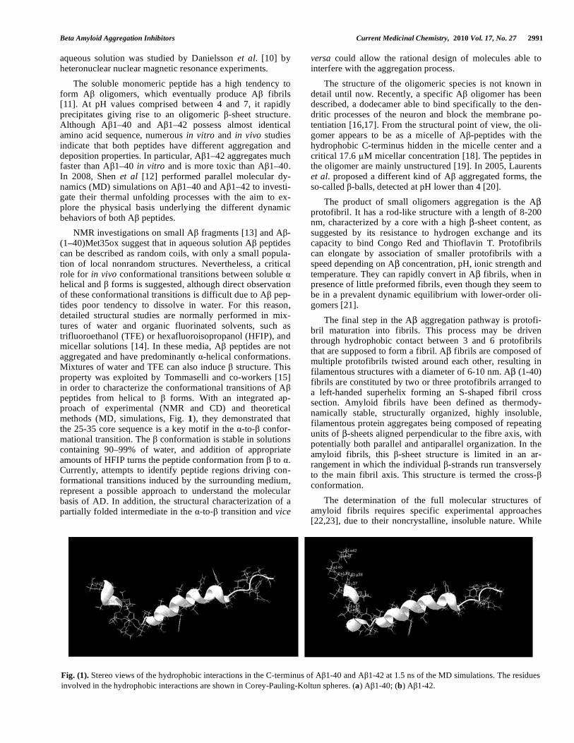
Fig. (1). Stereo views of the hydrophobic interactions in the C-terminus of A 1-40 and A 1-42 at 1.5 ns of the MD simulations. The residues involved in the hydrophobic interactions are shown in Corey-Pauling-Koltun spheres. (a) A 1-40; (b) A 1-42.
2992 Current Medicinal Chemistry, 2010 Vol. 17, No. 27 Re et al.
monomeric peptides present prevalently a random coil struc-ture, for amyloid fibrils the predominant secondary structural elements are -strands. The study of secondary structure in amyloid fibrils therefore consists on the identification of -strand and non- -strand segments (i.e., turns, loops, or bends). Different techniques, such as solid-state NMR [24], hydrogen/deuterium (H/D) exchange [25], proline-scanning mutagenesis [26], electron paramagnetic resonance (EPR) [27], infrared and Raman spectroscopies [28] have been ap-plied to the problem of secondary structure determination. Solid-state NMR and EPR [29] measurements have been employed also for the characterization of fibrils tertiary structure, that depends on the organization of -strand seg-ments into parallel or antiparallel -sheets. Quaternary struc-ture in amyloid fibrils is defined as the positions and orienta-tions of -sheets relative to one another. The most important techniques applied to these experimental studies are X-ray diffraction [30] and solid-state NMR [31]. The experimental determination of quaternary structure in amyloid fibrils has been relatively difficult. The last models for amyloid fibrils structures support the idea that its core contains two or more layers of -sheets. The quaternary structure is generated by a specific set of contacts among amino acid side chains that project from adjacent -sheets. In fibrils formed by relatively long peptides, the adjacent -sheets may be formed either by
-strands from the same or from different peptide molecules, indicating that the side chain contacts from which quaternary structure takes origin can be either intramolecular or inter-molecular.
3. INTERACTION WITH LIGANDS
The structural features of the interaction of ligands with A are mainly unknown, even if a great number of different molecules that interact with A peptides have been pro-posed. There are some sporadic exceptions among these compounds. Three significant cases are reported. The first example concerns the interaction of A with cyclodextrins, torus-shaped rings built up by different numbers of glucose residues. As firstly reported by Qin et al. [32], -cyclodextrin interacts with the A -peptide and the interac-tion is suggested to inhibit the formation of the soluble oli-gomers with an IC50 of 5mM, as determined with scintilla-tion proximity assay [33]. Camilleri et al. demonstrated by mass-spectrometry that the stoichiometry of the A -peptide and -cyclodextrin is 1:1 [34]. The main interaction with amino acid residues has been suggested to involve the aro-matic rings of phenylalanines and the hydrophobic cavity of cyclodextrin. In 2004, Danielsson and co-workers character-ized the interaction between A -peptide and -cyclodextrin by NMR [35]. The A -peptide has four putative aromatic binding sites, F4, Y10, F19 and F20. They demonstrated that if the phenylalanines at positions 19 and 20 in the fragment A 12-28 are replaced with glycines, thus removing all aro-matic side chains, no interaction occurs. This shows that the phenylalanines are necessary for -cyclodextrin-peptide in-teraction. For the central fragment A 12-28 where the phenylalanines are present, interaction occurs and Kd = 3.8 mM. Induced chemical shift changes suggested that the interaction mainly involves phenylalanine 19. This binding is not strong, but it is in agreement with the fibril reducing activity of -cyclodextrin determined earlier. In the case of the full-length peptide, the combined results from induced
peptide, the combined results from induced chemical shift changes and diffusion data were consistent with a two-site binding involving F19 and Y10 with a total apparent disso-ciation constant of Kd = 4 mM. The individual Kd for the two sites was determined to be Kd F = 4.7 mM and Kd Y = 6.6 mM, respectively, if the sites are assumed to be independent. The outermost N-terminal aromatic amino acid F4 did not seem to bind -cyclodextrin with any measurable affinity. This was tested by showing that the fragment A 1-9 does not bind -cyclodextrin. Other types of cyclodextrins were stud-ied and showed no binding. This may be due to the hydro-phobic pocket of -cyclodextrin that has optimal dimensions for interaction with the aromatic side chains. The - and -cyclodextrins have too small or too large cavities for interac-tion to take place.
The second example concerns the study of the interaction between gangliosides and A 1-40 peptide. This interaction was characterized in 2004 by Mandal et al. [36] in a mem-brane-mimicking environment, dissolving asialo-GM1 and ganglioside GT1b in a SDS (sodium dodecyl sulphate) solu-tion. As previously reported [37], A peptides associate with gangliosides in Alzheimer’s brains and form specific com-plexes with gangliosides in vitro. Different experimental evidences show that ganglioside binding induces A peptide oligomerization [38]. From the structural point of view, gan-gliosides are sialic acid–containing glycosphingolipids and consist of two main components: a hydrophobic ceramide unit, which anchors the gangliosides to the plasma mem-brane, and a hydrophilic oligosaccharide chain, to which one or more sialic acid units (N-acetylneuraminic acid) are at-tached. Gangliosides are abundant components of neuronal membranes and are involved in important neurobiological events such as neurodifferentiation, synaptogenesis, and syn-aptic transmission. Asialo-GM1 and GT1b ganglioside have four sugar moieties in common; however, GT1b has three additional sialic acids compared to asialoGM1 that does not contain sialic acid units. Asialo-GM1 and GT1b ganglioside binding to A 1-40 peptide were analyzed by heteronuclear NMR spectroscopy. Experimental data showed that, while GT1b is unable to bind A , asialo-GM1 addition induces appreciable chemical shift variations in A 1-40 amide pro-tons. In particular, asialo-GM1 docks between region I (E3–V24) and region II (G29–V40) of A . These evidences ex-plain why GM1 does not bind the shorter A 1-28 peptide [38a]. In fact, in the case of A 1-28, the absence of the main binding region G29–V40 would prevent GM1 binding. In addition, Matsuzaky and co-workers [39a] showed that gan-gliosides follow the order GT1b > GD1a > GD1b > GM1 to induce -sheet formation. These NMR data are in agreement with previous reports, suggesting that when the size of the ganglioside headgroup increases with the number of sialic acids (GT1b > GD1a > GD1b > GM1), it becomes difficult to accommodate the bulky saccharides between regions I and II of A and -sheet formation proceeds. The interaction of A peptide with the ganglioside GM1 is also currently under study by Bruix and co-workers [40]. It is worth noting that opposite results were obtained by Ariga et al. using a mem-brane-mimicking system composed of dipalmitoylphosphati-dylcholine, where A had lower affinity for asialoGM1 than for gangliosides with several sialic acids [39b].

Beta Amyloid Aggregation Inhibitors Current Medicinal Chemistry, 2010 Vol. 17, No. 27 2993
4. SMALL MOLECULE LIGANDS THAT INTERACT WITH A
Based on the conformation/oligomerization hypothesis, molecules able to stabilize the soluble A (sA ) conforma-tion, to destabilize the altered amyloidogenic conformer, and to prevent the required conformational transition could be effective inhibitors of amyloid plaque formation and very potent drug candidates for AD treatment.
4.1. Peptides
The intrinsic affinity of A for itself suggested that A –specific interactions could be the basis for the development of compounds binding to A and preventing its polymeriza-tion. It has been claimed that fibrillogenesis can be inhibited by short synthetic peptides partially homologous to A . Sev-eral elegant strategies have been used to design peptidic in-hibitors of A aggregation. Many of these approaches rely on using fragments of the parent A sequence as recognition elements in a dominant negative fashion. Typically, fibril-disrupting chemical elements are incorporated into A de-rived peptides in the form of N– or C–terminal modifica-tions, conformationally constrained amino acids, or modifi-cations to the peptide backbone. These concepts have also been applied to D–amino acid variants of the parent A se-quence. Given the hypothesis that aggregation intermediates are responsible for A toxicity, one approach to contrast this problem is to identify the regions of A that are at the basis of the polymerization into amyloid fibrils. Gazit et al. [41] analyzed a variety of short functional fragments from unre-lated amyloid–forming proteins and observed a remarkable occurrence of aromatic residues. They speculated that aro-matic residues raise the possibility that –stacking may play an important role in the molecular recognition and self–assembly process that lead to amyloid formation.
In A the aromatic residues are the two phenylalanine residues in position 19 and 20. In particular, the short frag-ment of A QKLVFF was shown to bind specifically to the full-length peptide [42]. Other studies have shown that not only QKLVFF, but also LVFFA and its derivatives [43] and LPFFD [44] are all potent inhibitors of amyloid formation. Another recent study demonstrated that the seven amino acid A fragment, KLVFFAE, forms well–ordered amyloid fibrils [24a]. These findings point to the pair of phenylalanine resi-dues as the major structural element that mediates binding of the QKLVFF peptide to the A polypeptide. As the forma-tion of amyloid fibrils is a process of molecular recognition and self–assembly, the high affinity and selectivity of the FF motif seems to provide the molecular recognition element needed for such process in the context of the full–length A . These candidate binding compounds could theoretically pre-vent aggregation, or even cause further association of toxic oligomers into larger nontoxic aggregates. Cairo and co–workers [45] proposed inhibitors designed as containing both the ‘‘recognition domain’’ (LVFF or KLVFF) and a ‘‘dis-rupting domain’’, a polypeptide chain with the ability to in-terfere with A aggregation. A series of variant of the KLVFF sequence were synthesized. Truncation of the C–terminal phenylalanine reduced the affinity by approximately 10–fold. The D–amino acid sequence KLVFF binds with similar affinity to the L–amino acid sequence, as reported by
Chalifour et al. [46]. Substitution of tyrosine at either the 19 or 20 position did not alter the affinity; however, replace-ment of both phenylalanine residues with tyrosine was det-rimental. Substitution with histidine in position 19, but not in position 20, led to a substantial loss of binding; nevertheless, a double histidine substitution partially restored binding. Substitution of phenylalanine with tryptophan residues gave mixed results. Murphy et al. focused their attention also on the “disrupting domain”; they demonstrated that KLVFF sequence, that also possess positively charged residues at the C-terminus, binds with higher affinities than does KLVFF alone. Among these peptides, KLVFFK4 (Kd = 37 μM) and KLVFFK6 (Kd = 40 μM) were the most potent in preventing A –associated toxicity. These results suggested that a pep-tide domain lacking direct homology with A can play a significant role in binding. The increased affinities of se-quences bearing lysine residues are not due to nonspecific Coulombic interactions of the peptides with the A –fibrils surfaces; in fact, there was a lack of affinity when three ly-sine residues were substituted with glutamic acid residues (four different isomers). These substitutions were useful to speculate that the position of the positively charged residues has a critical influence on A affinity. Placement of the posi-tively charged residues close to position 20 gives rise to the most potent ligand of the four isomers. Placing the lysines three residues apart from the region of residues 16–20 with intervening negatively charged residues reduces the affinity by 14–fold. Placement of positive charge at the N–terminus of the sequence of residues 16–20 results in an activity that is lower than that of compounds with a modification at C–terminus. Arginine–containing compounds were tested to determine the potency of compounds that incorporate posi-tively charged residues other than lysine. These compounds possess activity similar to that with lysine. Another example of peptide inhibitors containing the A amino acid sequence 16–20 was presented by Austen et al. [47]; they designed two peptides with RG–/–GR residues added at the N– and C–terminal ends of KLVFF to aid solubility, in particular the two molecules are RGKLVFFGR and RGKLVFFGR–NH2. On the basis of ThT results and TEM micrographs, both pep-tides inhibited the formation of A late aggregates, but the results for the inhibition of the oligomerization revealed that the second one had the best inhibitory effect, having the first one a slight blocking effect at higher concentrations. It is likely that the second one inhibits oligomeric formation by binding to the monomeric A molecule, thereby blocking the formation of early soluble aggregates, as shown by size ex-clusion chromatography and oligomeric specific ELISA. Interestingly, only RGKLVFF–NH2, which completely in-hibited the early aggregates of A , was able to protect cells from A toxicity. However, in the case of the first peptide, which showed only inhibition of the late aggregates of A , no neuroprotective effects were demonstrated. The results reported by Austen and co–workers provide clear evidence that the neuroprotective effects were due to the inhibition of formation of early aggregates rather than late aggregates of A . The above studies are quite significant as they suggest new approaches for the interference with A assembly and peptides that may be the lead compounds for the develop-ment of molecules preventing A aggregation in vivo. The use of multiple simultaneous interactions to enhance the af-finity and specificity of binding, known as multivalency, is
2994 Current Medicinal Chemistry, 2010 Vol. 17, No. 27 Re et al.
an important concept in biology. In the past, different types of scaffold molecules have been used to obtain multivalent structure, such as colloidal gold particles and poly(ethyleneglycol) (PEG). These approaches have some disadvantage, in particular the large size of the particles (PEG) and the lack of control over the number of peptides attached (colloidal gold). These disadvantages are overcome using dendrimers, multivalent macromolecules with regular, highly branched, structures and dimensions resembling those of small proteins. Starting from previous informations, Chafekar and co–workers [48] synthesized a first generation KLVFF dendrimer (K4 – four KLVFF on one scaffold) by using native chemical ligation and investigated the effect of dendritic KLVFF on the aggregation of A (1–42) as well as its effects on preformed aggregates. Their data show that the linkage of KLVFF to a dendrimeric scaffold potentiates its inhibitory effect on A aggregation and on the disassembly of pre–existing aggregates; in fact they used 20–fold lower concentration of K4 (per peptide subunit) to obtain a similar inhibition as KLVFF. Previously, a branched hexameric PEG linked to inverse FFVLK [49] sequence was shown to in-crease the potency of A inhibition, but also in this case the K4 conjugate from the study of Chafekar et al. gave stronger multivalent effect.
Looking at the A (1–42) sequence, Fülop et al. [50] pro-posed another inhibitor with a structure analogue to the se-quence 31–35, which plays important roles in A aggrega-tion [51]. Other types of peptides containing residues acting as –sheet breaker were proposed. In particular Soto and co–workers [44] proposed a –sheet breaker peptide iA 5 (LPFFD), designed from the central hydrophobic region of A . A proline residue was added into the sequence to disrupt
–sheet formation, as incorporation of this amino acid within a –pleated structure is highly unfavourable; an aspartic acid charged residue was added at the end of the peptide to in-crease solubility. It was demonstrated that iA 5 inhibits in a dose–dependent manner fibrillogenesis of either A (1–40) or A (1–42), disassembles preformed fibrils in vitro and pre-vents neuronal death induced by fibrils in cell culture. In addition, the peptide significantly reduced amyloid –protein deposition in vivo and completely blocked the formation of amyloid fibrils in a rat brain model of amyloidosis. Looking at these results, they hypothesized that –sheet breaker pep-tides probably inhibit amyloid formation by binding to monomeric/dimeric A peptides, thereby blocking the for-mation of the oligomeric –sheet–conformation precursor of the fibrils.
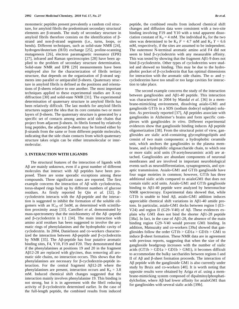
Etienne and co–workers [52] proposed peptides which contain , –disubstituited amino acid ( AA) in the hydro-phobic core of A (Fig. 2); in particular they utilized an al-
ternating AA/L–amino acid design to obtain a peptide that interacts with A by hydrogen bonding as well as by side–chain interactions. The AAs having side–chain larger than methyl stabilized extended peptide conforma-tions.
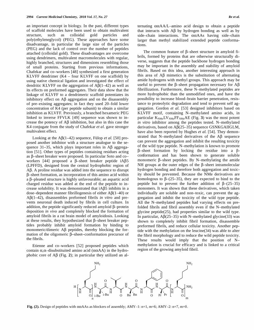
The common feature of –sheet structure in amyloid fi-brils, formed by proteins that are otherwise structurally di-verse, suggests that the peptide backbone hydrogen bonding may be important in the assembly and stability of amyloid fibrils. Based on this idea, another interesting approach in this area of A mimetics is the substitution of alternating amide hydrogens with methyl groups. This approach may be useful to prevent the –sheet propagation necessary for A fibrillization. Furthermore, these N–methylated peptides are more hydrophobic than the unmodified ones, and have the possibility to increase blood–brain barrier penetration, resis-tance to proteolytic degradation and tend to prevent self ag-gregation. Gordon et al. [53] designed inhibitors based on KLVFF motif, containing N–methylated amino acids, in particular KNMeLVNMeFFNMeAE (Fig. 3) was the most potent in vitro inhibitor among the peptides tested. N–methylated derivatives, based on A (25–35) sequence GSNKGAIIGLM, have also been reported by Hughes et al. [54]. They demon-strated that N–methylated derivatives of the A sequence can prevent the aggregation and inhibit the resulting toxicity of the wild type peptide. N–methylation is known to promote
–sheet formation by locking the residue into a -conformation and has been shown to generate soluble monomeric –sheet peptides. By N–methylating the amide NH groups at the outer edges of the –sheet intramolecular hydrogen bonding and therefore both aggregation and toxic-ity should be prevented. Because the NMe derivatives are homologous to –(25–35), they are expected to bind to the peptide but to prevent the further addition of –(25–35) monomers. It was shown that these derivatives, which taken individually are soluble and non–toxic, can prevent the ag-gregation and inhibit the toxicity of the wild type peptide. All the N–methylated peptides had varying effects on pre-folded fibrils and fibril assembly even if the N–methylated glycine peptide(25), had properties similar to the wild type. In particular, A (25–35) with N–methylated glycine(33) was shown to completely inhibit fibril formation, disassemble preformed fibrils, and reduce cellular toxicity. Another pep-tide with the methylation on the leucine(34) was able to alter the fibril morphology and to reduce the wild peptide toxicity. These results would imply that the position of N–methylation is crucial for efficacy and is linked to a critical region of the growing amyloid fibril.
NH
HN
NH
HN
NH
NH
NH2
O
O
O
O
O
O
O
NH2 NH2
HNH
i-Bu Bn Pri-Bu Bn Prn m
Fig. (2). Design of peptides with AAs as blockers of assembly; AMY–1: n=1, m=6; AMY–2: n=7, m=0.

Beta Amyloid Aggregation Inhibitors Current Medicinal Chemistry, 2010 Vol. 17, No. 27 2995
Gordon and co–workers [55] investigated also the effect of replacing the hydrogen bond donating amide NH back-bone with ester bond surrogates at alternating positions, to prevent the hydrogen bonding of the amyloidogenic peptide and to reduce the aggregation. In addition, the ester carbonyl is less basic than the amide carbonyl and, as a consequence, is a weaker hydrogen bond acceptor. At the same time, the ester bond shares many structural similarities with the amide bond, such as a predominance of the trans conformation and similar bond lengths and angles. Based on this information, they hypothesized that the substitutions would yield peptides with effective fibrillogenesis inhibitor activity and would permit a more direct assessment of the role of hydrogen bonds in stabilizing amyloid fibrils. The synthesized peptides presented two hydrogen bonding faces when arrayed in an extended –strand conformation; one face had normal hy-drogen bonding capabilities, but the other face was missing amide protons and its ability to hydrogen bond was severely limited.
Thus, the incorporation of ester bonds constitutes a more conservative substitution for peptide bonds than the incorpo-ration of N–methyl amino acids. The incorporation of two ester bonds at alternate residues of the A (16–20) peptide, similar to the incorporation of N–methyl amino acids, pre-vents the peptide from forming amyloid fibrils. The incorpo-ration of ester bonds also results in the formation of an effec-tive inhibitor of A (1–40) fibrillogenesis. They also ana-lyzed the different behavior of the A (16–20)ester peptide, the unmodified wild type A (16–20) and the inhibitor pep-tide A (16–20)N–methylated. All three peptides inhibited fibrillogenesis and disassembled preformed fibrils; the effi-cacy of A (16–20) ester is similar to that of A (16–20)N–methylated and both are better inhibitors than A (16–20). The similar inhibitory properties of A (16–20) ester com-pared to A (16–20)N–methylated also suggest that interfer-ing with hydrogen bonding is sufficient to prevent A (1–40) fibrillogenesis and that steric contributions from the N–methyl group are not required. What is also very interesting is the reported disassembly of pre–existing fibers, which may
suggest that many of these peptides can bind at the end of the fibrils and possibly influences the thermodynamic equilib-rium between oligomers and fibrils. Thus, it is clear that the considerable efforts in the rational design of A –targeting molecules based on the parent A sequence have been quite fruitful in providing a large class of compounds with differ-ent activity. These peptidomimetic molecules provide a class of compounds that may help to elucidate the mechanism of amyloid aggregation, perhaps by trapping intermediates, as well as providing inroads into the design of diagnostic and therapeutic reagents.
4.2. Non-Peptidic Small Molecules
Some non-peptidic small molecules are able not only to inhibit the formation and extension of –amyloid fibrils (fA ), but also to destabilize fA in vitro. These include naturally occurring or commercially available bioactive compounds, drugs, surfactants [56], Cu/Zn chelators [57], phenothiazines [58] and sulfonated dyes such as or Congo red (CR) (Fig. 4) and its derivatives [59]. Among them CR was the first small molecule reported to bind to amyloid in tissue sections, exhibiting the characteristic yellow–green birefringence under cross polarized light. Later thioflavin T (ThT) (Fig. 4) and thioflavin S (ThS) were also shown to stain amyloid deposits. These two dyes are the classical rea-gents to detect characteristic –sheet mediated fibrillization.
CR [60] and ThT [61] have also been shown to inhibit fi-brils formation at higher concentrations. Not surprisingly perhaps, many reported small molecule inhibitors are similar to CR and ThT, in that they are planar and aromatic. Accord-ing to Porat et al. [62], who reviewed the possible mode of interaction with A , small molecules with aromatic moieties can intercalate within grooves created by –sheets in both the soluble oligomeric forms as well as in the large fibrils. From this first study, several different types of molecules were found to be active in the inhibition of fA aggregation, with a wide range of activity, from less than 100 nM to more than 100 μM. In the table presented below, we resume the
H2N
MeN
NH
MeN
NH
MeN
NH
OH
NH2
O
O
O
O
O
O
O OH
O
Fig. (3). Design of peptides containing N–Methylated amino acids.
NN
NN
NH2
NaO3S
H2N
SO3Na
N
S
N
Cl
Congo Red Thioflavin T
Fig. (4). Structure of Congo red and Thioflavin T.

2996 Current Medicinal Chemistry, 2010 Vol. 17, No. 27 Re et al.
IC50 values of several compounds for the inhibition of A peptide aggregation. In particular compounds are classified for their inhibitory effect and can be roughly divided in two classes: bioactive molecules and drugs not related to Alz-heimer diseases. The compounds belonging to the first class include curcumin [63, 64], wine–related polyphenols [62] (myricetin, quercetin, morin, kaempferol, (+)–catechin and (–)–epicatechin, apomorphine [65], porphyrins [58, 66],
omega–3 fatty acid [67], tannic acid [62, 68], vitamin A and -carotene [69], rifampicin and rifamycin B [58], tetracy-
cline [68, 70], coenzyme Q10 [71]). Anti–inflammatory [72, 73] and anti–Parkinson agents [74, 75] belong to the second class, the latter being the most effective, with particular ref-erence to dopamine and L-DOPA. Analyzing the table pre-sented below, dopamine has the best performance. Dopamine and its precursor L–DOPA are used as drugs acting on the sympathetic nervous system. For example L–DOPA is used in the treatment of Parkinson’s disease and DOPA–responsive dystonia. Despite of its benefits in brain-related disease, until now, dopamine and its precursor have not been used for treatment of Alzheimer’s diseases even though in vitro studies were promising. The anti–amyloidogenic and fibril–destabilizing activity of the anti–Parkinsonians drugs, examined in the study of Ono et al. [74] (Table 1) was in the order dopamine > Selegiline ~ nordihydroguaiaretic acid (NDGA) > L–Dopa ~ Pergolide > Bromocriptine ~ Rifam-picin (RIF). In particular, dopamine exhibited potent anti–amyloidogenic and fibril–destabilizing effects and the effec-tive concentration (EC50) able to inhibit the formation or extension of fA s was in the order of 0.01–0.10 μM. This result was in contrast with previous works [76, 77] which identified in NDGA (Fig. 5), that has two ortho–dihydroxyphenyl rings symmetrically bound by a short car-bohydrate chain, the most effective molecule. The activity of NDGA could be ascribed to its compact and symmetric structure that might be suitable to specifically bind free A , thus inhibiting the polymerization of A into fA [76b]. Alternatively, this structure might be suitable for specific binding to fA and subsequent destabilization of the –sheet conformation of A rich in fA molecules.
Li et al. [78] reported that dopamine and L–DOPA were able to inhibit the formation of amyloid fibrils in a solution 1 mg/mL of A (1–40), in a range from 10 to 100 μM.
Other molecules that present a very high activity against amyloid fibrils and plaque formation are red wine and green tea related polyphenols [62]; these compounds were also
analyzed by Ono et al. [79], during their systematic work. The wine–related polyphenols inhibit fA formation from A (1–40) and A (1–42) as well as destabilized preformed fA (1–40) and fA (1–42) dose–dependently in vitro. Within this class of compounds tannic acid showed the best activity.
Tannic acid (TA), a commercial form of tannin, is a po-lymeric polyphenol with a weak acidity (pKa around 10) due to its phenol groups. Its structure is based mainly on glucose esters of gallic acid. It is a yellow to light brown amorphous powder which is highly soluble in water. Ono et al. exam-ined the effects of TA on the formation and extension of fA (1–40) and fA (1–42), as well as on the destabilization of fA s at pH 7.5 at 37 °C in vitro, using fluorescence spec-troscopy with ThT, Congo Red and electron microscopy [68]. They also compared the activity of TA with the anti–amyloidogenic and fibril destabilizing effects of Myricetin (Myr) (Fig. 6), RIF, Tetracycline (TC) and NDGA. TA ex-hibited potent anti–amyloidogenic and fibril–destabilizing effects and the EC50 for the formation or extension of fA s or for the destabilization of fA s were in the order of 0.012–0.065 μM for both A (1–40) and A (1–42). They suggested that the anti–amyloidogenic and fibril-destabilizing activity of NDGA and wine–related polyphenols may be classified in the following order: TA > NDGA = Myr = morin = quercetin > kaempferol > (+)–catechin = (–)–epicatechin > TC. Inter-estingly, TA, wine–related polyphenols, NDGA, RIF all pos-sess antioxidant activity. Tomiyama et al. [80]. suggested that RIF binds to A by hydrophobic interactions between its lipophilic ansa chain and the hydrophobic region of A , thus blocking the association between A molecules that lead to fA formation. The anti–amyloidogenic activity of TCs, and of small molecule anionic sulfonates or sulfates is also re-lated to the propensity to bind to A [77, 81].
Another important A -ligand is curcumin (diferulo-methane), a low molecular weight molecule with potent anti-oxidant and anti–inflammatory activities that has a favorable toxicity profile and is also under development as a potential cancer chemotherapeutic agent. Curcumin (Cur) (Fig. 6) is a major component of the yellow curry spice turmeric. This spice is used in the traditional diet and as an herbal medicine in India [82]. The frequency of Alzheimer’s disease in India is roughly one–quarter of that in the US (e.g., 0.7 vs. 3.1% between 70 and 79 year) [83]. Maybe not by chance Cur is a free radical scavenger [84], protecting the brain from lipid peroxidation [73a], and scavenges nitric oxide–based
HO
OH
OHHO
Nordihydroguaiaretic acid
O
O
NH
NN
N
O
O
O
H
HO
OH
O
O
OH
OH
OH
Rifampicin
Fig. (5). Structure of Nordihydroguaiaretic acid and Rifampicin.

Beta Amyloid Aggregation Inhibitors Current Medicinal Chemistry, 2010 Vol. 17, No. 27 2997
Table 1.
IC50 aggregation μM Compound
fA (1–40) fA (1–42) Ref.
Ability to pass
BBB (Ref.)
Acetopromazine maleate salt > 40 [58]
2–(4–Aminophenyl)–methylbenzothiazole 2.0 [58]
Amphotericin B 2.2 [58]
Apigenin > 40 [58]
Azure A 0.4 [58]
Azure B 0.3 [58]
Azure C 0.2 [58]
Basic blue 41 1.4 [58]
Bromocriptine 14 20 [74]
–Carotene 2.4 5.2 [69]
Catechin 2.9 5.3 [62, 78] [107-109]
Chlorpromazine hydrochloride > 40 [58]
Chlorazol black E 0.3 [58]
CoQ10 1.8 5.5 [71] [101]
Curcumin 0.19-081 0.63-1.1 [95a 99b] [112, 113]
Daunorubicin hydrochloride 1.4 [58]
Dihydrolipoic acid 3.9 20.8 [70a] [103]
2,2’-Dihydroxybenzophenone > 40 [58]
2–[4–(Dimethylamino)phenyl]–6–methylbenzothiazole 2.0 [58]
2, 4–Dinitrophenol 7 [79] [111]
3,3’–Dipropyl thiodicarbocyanine iodide 0.3 [58]
L–Dopa 5.0 1.7 [74] [100]
Dopamine 0.01 0.04 [74]
(–)–Epicatechin 2.8 5.6 [62, 78] [107-109]
(–)–Epicatechin 3–galiate 3.0 [62, 58] [110]
Exifone 0.7 [62, 58]
Ferric dehydroporphyrin IX 0.2 [58]
Ferulic acid 1.8 5.5 [70b]
Filipin III 14.6 [58]
Gossypetin 1.3 [62, 58]
Hematin (from bovine blood) 0.2 [58]
Hemin ~ 0.4 ~ 0.4 [66]
Hemin chloride 0.1 [58]
Hypericin 0.9 [62, 58]
Kaempferol 1.7 3.2 [62, 78] [107, 108]
Lacmoid 1.4 [58]
–Lipoic acid 3.2 20 [70a] [102]
Metylene Blue 2.3 12.5 [58, 80]
Methyl yellow 1.5 [58]
Morin 0.24 0.67 [62, 78] [107, 108]
Mycostatin 9.3 [58]

2998 Current Medicinal Chemistry, 2010 Vol. 17, No. 27 Re et al.
(Table 1). Contd…..
IC50 aggregation μM Compound
fA (1–40) fA (1–42) Ref.
Ability to pass
BBB (Ref.)
Myricetin 0.20-0.37 0.34-0.51 [68,78,81, 62, 71] [79, 107, 108]
NDGA 0.14-0.21 0.74-1.1 [62, 68, 63, 74, 69, 70a,
71]
3–Nitrophenol 80 [79] [111]
2,3,4,2’,4’–Pentahydroxybenzophenone 2.8 [62, 58]
Pergolide 0.89 2.0 [74] [74]
Perphenazine > 40 [58]
Phthalocyanine 3.2 [58]
Ponceau SS 1.2 [58]
Promazine hydrochloride > 40 [58]
Propionylpromazine hydrochloride > 40 [58]
Protoporphyrin IX ~ 65 ~ 65 [66]
Pseudohypericin 2.3 [58]
Purpurogallin 0.5 [62, 58]
Quercitin 0.24 0.72 [62, 78] [106-108]
Quinacrine 8.4 [58]
Quinacrine mustard 1.2 [58]
Retinal 0.18 0.89 [69]
Retinol 0.18 0.76 [69]
Retinoic acid 4.5 20 [69]
Rifampicin 4.9-9.7 9.1 [70a,b, 68, 58, 74] [104]
Rifamycin B 3.1 [58]
Rosmarinic Acid 0.29 1.1 [62, 63]
Selegiline 0.68 0.72 [74] [105]
Tannic acid 0.012 0.022 [62, 68]
Tetracycline 10 10 [70a,b, 68]
Thionin 0.3 [58]
2,3,4–Trihydroxybenzophenone 3.1 [62,58]
Zinc protoporphyrin IX ~ 5 ~ 5 [66]
OH
OOH
HO O
OH
OH
OH
Myricetin
O
HO
O
O
OH
O
Curcumin
Fig. (6). Structure of Myricetin and Curcumin.
radicals. Oral administration of Cur has been shown to be centrally neuroprotective. In 2001, Lim et al. [64b] sug-gested that Cur was able to block Alzheimer’s disease patho-
genesis at multiple sites of the inflammation cascade, but the direct effects of Cur on the formation and destabilization of fA remain unclear. Looking at these information, in 2005,

Beta Amyloid Aggregation Inhibitors Current Medicinal Chemistry, 2010 Vol. 17, No. 27 2999
Yang et al. [64a] worked on Cur as A inhibitor both in vitro and in vivo. They showed that under aggregating conditions in vitro, Cur inhibited aggregation (IC50 = 0.8 μM) and dis-aggregated fibrillar A 40 (EC50 = 1 μM). Later, other authors [62, 63, 70b] reported lower IC50. The effects of Cur did not depend on A sequence but on fibril–related confor-mation. Brain sections of Tg2576 mice with Alzheimer’s disease were incubated with Cur and revealed preferential labeling of amyloid plaques. In vivo studies showed that Cur injected peripherally into aged Tg mice crossed the blood–brain barrier and bound A plaques. When fed to aged Tg2576 mice with advanced amyloid accumulation, Cur la-beled plaques and reduced amyloid levels and plaque burden. Hence, Cur directly binds small –amyloid species thus blocking aggregation and fibril formation both in vitro and in vivo. These data suggested that low dose Cur effectively is able to disaggregate A as well as to prevent fibril and oli-gomer formation, supporting the rationale for Cur use in clinical trials preventing or treating AD. In 2007, Reinke et al. [85] studied the structure–activity relationships of amy-loid –aggregation inhibitors based on Cur. They studied the effect of three prominent features on inhibition of amyloid aggregation: the presence of two aromatic end groups, the substitution pattern of these aromatics, and the length and flexibility of the linker region. They found that modification of any one of the modules has profound effects on activity. To establish the contribution of each structural module to the activity, they assembled a collection of curcumin related compounds. Cur is composed of two relatively polar aro-matic groups connected by a rigid linker. Reinke et al., in their first experiments investigated whether both aromatic groups were required for activity using the ThT assay. They found that simple compounds with a single aromatic group, like vanillin, salicylic acid or ferulic acid, did not decrease aggregation of 25 μM A (1–42), even at high concentrations (500 μM). They also studied the relation between the sub-
stituent on the two phenyl group and the activity, observing that the aromatic end groups require one or more polar, hy-drogen bonding substitutions for optimal inhibition of A aggregation.
In Cur, CR, and Chrysamine G (CG) (Fig. 7) the dis-tances between the terminal aromatic regions are all strik-ingly similar, it was hypothesized that the length of the linker would be important for activity.
Reinkle et al. noticed that length and flexibility of the linker region are not independent variables and established that the optimal are rigid (less than two freely rotating car-bons) and restricted between 8 and 16 Å. Interestingly, many of the best amyloid ligands fall within the observed optimal range. For example, CR, CG, NDGA, Cur (Fig. 8) and ros-marinic acid all meet these requirements. In addition, the linker requirement seems to be one of the most significant features, as compounds that fulfill the other substructural requirements, such as the aromatic substitution, still fail to inhibit if they have a short linker. An interesting exception to this observation is resveratrol. Despite its short linker length, this compound is active, suggesting a possible alternative mechanism for inhibition or a possible different binding site on the amyloid peptide, such as the site occupied by ThT.
Overall, the anti–amyloidogenic molecules can be classi-fied into four group based on the activity. Dopamine and tannic acid were classified into the strongest anti–amyloidogenic group, compounds like NDGA, Cur, rosma-rinic acid or myricetin into the second group, L–DOPA or –carotene into the third group, RIF and TC into the fourth group. Poly(vinylsulfonic acid, sodium salt), 1,3–propanedisulfonic acid, disodium salt, –sheet breaker pep-tide and nicotine exhibited anti–amyloidogenic activity in a μM range.
NN
N
NHO
OH
COOH
HOOC
Fig. (7). Structure of Chrysamine G.
Fig. (8). Representative model showing how linker characteristic contribute to potency. Linker too long (b.), too short (c.) or too flexible (d.) can decrease the ability of the compound to interact with A .

3000 Current Medicinal Chemistry, 2010 Vol. 17, No. 27 Re et al.
5. CROSSING THE BLOOD BRAIN BARRIER (BBB)
An endothelial cell monolayer associated with pericytes and astrocytes, known as the blood brain barrier (BBB), separates the blood from the cerebral parenchyma; it is char-acterized by tight intracellular junctions and by the absence of fenestrations [86]. The BBB is the homeostatic defense mechanism against pathogens and toxins. Complex and highly regulated, the BBB screens the biochemical, physico-chemical and structural features of solutes at its periphery, thus affording barrier selectivity in the passage of desired molecules into the brain parenchyma. The BBB provides the brain with nutrients, prevents the introduction of harmful blood-borne substances, and restricts the movement of ions and fluid to ensure an optimal environment for brain func-tion. It protects the brain from substances which are neuro-toxic in physiological concentrations. As a consequence of its barrier properties, the BBB also prevents the movement of drugs from the blood into the brain, and therefore acts as an obstacle for the systemic delivery of neurotherapeutics. The BBB determines whether or not a given drug, unless lipid-soluble, small (< 600 Da), electrically neutral and weakly basic molecules, can reach the central nervous sys-tem (CNS), either by passive diffusion or through carrier or receptor systems [87] limiting the brain penetration. Fur-thermore, efflux transport systems may target the drugs that meet these criteria and export them from the brain. As a re-sult, the BBB excludes many small-molecule pharmaceuti-cals, and nearly all biopharmaceuticals such as genes and proteins fail to penetrate into the brain tissue to an apprecia-ble extent [88]. Thus, although the surface area of the human brain microvasculature available for drug transport (~ 20 m2) is more than adequate for treating the entire brain volume, the barrier properties of the BBB continue to restrict brain drug delivery via the bloodstream [89].
There is growing evidence for BBB damage in AD [90]. Most A plaques are located in the proximity, or are directly in contact with cerebral capillaries. Alterations of the expres-sion and distribution of the A transporters and tight junction proteins have been observed at the level of endothelial base-ment membrane of cerebral microvasculature, where the efflux of A from brain is regulated. These observation open the possibility of potential therapeutic alternatives of AD based on sealing a compromised BBB and reversing A deposition.
5.2. CEREBRAL CLEARANCE OF A ACROSS THE
BBB
The amyloid hypothesis states that a variety of neuro-toxic A species contribute to the pathogenesis of Alz-heimer's disease. Accordingly, a key determinant of disease onset and progression is the appropriate balance between A production and catabolism. It has been suggested that insuf-ficient clearance of A may account for elevated A levels in the brain and the accumulation of pathogenic amyloid depos-its in sporadic AD [89]. Ito et al. demonstrated the efflux clearance of [125I]A in rats to be 11.0 μL/(min g brain) [91]. Experimental evidence suggests that brain and blood A are in equilibrium, through the BBB, and that peripheral seques-tration of A may shift this equilibrium towards the blood, eventually drawing out its excess from the brain (“sink” ef-
fect) [92], reducing A -related pathology and dysfunction [93]. Clearance seems to be accomplished via two major pathways: proteolytic degradation and receptor-mediated transport from the brain.
Proteolytic Degradation
A number of proteases have been implicated in the prote-olytic clearance of A from the CNS, the two major en-dopeptidases involved in A degradation are zinc metalloen-dopeptidases, referred to as insulin degrading enzyme (IDE) or insulysin, neprilysin (NEP), endothelin converting en-zyme, and plasmin [94].
Receptor-Mediated Transport
Clearance is achieved by the brain-to-blood efflux trans-port system across the BBB. Increasing evidence suggests that the low-density lipoprotein receptor-related protein (LRP) and the receptor for advanced glycation end products (RAGE) are involved in receptor-mediated flux of A across the BBB [95]. While LRP appears to mediate the efflux of A from the brain to the periphery, RAGE has been strongly implicated in A influx back into the CNS. Therapeutic strategies therefore can be aimed at upregulation of LRP-1 or downregulation of RAGE.
ApoE, acting to traffic lipids throughout the brain, plays critical roles in regulating brain A peptide levels, as well as their deposition and clearance [96]. Recently, Jiang Q. et al demonstrated that lipidated forms of ApoE act to enhance the clearance of A peptides from the brain, facilitating the pro-teolytic degradation of A . They demonstrated that increase of lipidated forms of ApoE results in reduced A peptide and plaques levels in an animal model of AD [97].
The molecular pathways responsible for efflux and influx of A across the BBB suggest an array of potential therapeu-tic strategies that could be aimed at reducing the accumula-tion of cerebral A . The A clearance should preferably be performed before the proteins cluster into the toxic oli-gomers and can be achieved in many ways: 1) immunother-apy; 2) enzymatic degradation by metallo-endoproteases; 3) clearance stimulation by increasing transport over BBB by stimulating the LRP receptor or inhibiting RAGE (or P-gp). More recently, immunological approaches have been imple-mented to reduce A load in the CNS. Recent studies have demonstrated that beta amyloid burden in transgenic animal models can be reduced either by peripheral or cerebral infu-sion of exogenous anti-A antibodies (passive immuniza-tion) or by immunization with synthetic A peptide. Two basic mechanisms have been proposed to account for the findings of reduced amyloid load in transgenic animals with high anti-A antibody titers. First, anti-A antibodies may cross the BBB and bind to A in the brain, prompting diges-tion by activated microglia. Alternatively, antibodies or agents that have high affinity for A (ganglioside GM1 or gelsolin) may bind and sequester A in the periphery and either promote efflux from or inhibit influx back into brain [92, 98].
Another possibility to enhance the A clearance from the brain is the upregulation of A degradating enzymes, for example NEP activity can be upregulated by the administra-tion of somatostatin [98].

Beta Amyloid Aggregation Inhibitors Current Medicinal Chemistry, 2010 Vol. 17, No. 27 3001
6. CURRENT AND NOVEL THERAPEUTIC
APPROACHES FOR THE TREATMENT OF ALZHEI-
MER’S DISEASE
Goals of pharmacologic therapy for AD are to stabilize and modulate the expected decline of cognitive, functional and behavioral symptoms [114]. Available treatments are not intended to alter the progressive pathophysiology of the dis-ease, but rather are considered to be symptomatic therapies. Presently, cholinesterase inhibitors (ChEIs) are the first-line medications in the treatment of AD and are effective in de-laying cognitive impairment. Management of noncognitive psychiatric symptoms requires additional adjunctive medica-tions, such as antipsychotics, antidepressants, and anxiolytics [115]. Alternate treatments using nonsteroidal anti-inflam-matory drugs (NSAIDs), Gingko biloba, and hormone re-placement therapy have been used with varying success in the treatment of AD. Newer anti-amyloid medications in various stages of clinical trials show promise in modifying the disease process. Glutamate-mediated toxicity in the cere-bral cortex also has been hypothesized to contribute to the neurodegeneration of AD. In 2003, the FDA approved me-mantine, a low to moderate-affinity noncompetitive N-methyl-D-aspartate receptor antagonist, for the treatment of moderate to severe AD [116]. In patients with moderate to severe administration of a stable dose of memantine results in significant improvement in cognitive, functional, and global outcomes compared with placebo. Gingko extract has been used for thousands of years in traditional Chinese medicine for a variety of conditions. Recently, it has become more widely used in the United States to treat age-related physical and cognitive disorders [115]. The efficacy of gingko in the treatment of AD has been attributed to the fla-vonoid component for its antioxidative and antiperoxidative properties and the terpenoid component for its anti-inflammatory effects [117]. Estrogens are neuroprotective against oxidative stress, excitatory neurotoxicity, and ische-mia in the brain. Several small short-term randomized clini-cal trials and some epidemiologic studies have suggested efficacy of hormone therapy in the treatment of mild to mod-erate AD [118].
Currently there are four categories of anti-amyloid drugs in clinical trials with promise to modify the AD process: 1) immunotherapies; 2) secretase inhibitors; 3) selective A -lowering agents; and 4) anti-A aggregation agents. Several anti-aggregation agents, which are in clinical trials, are thought to prevent fibril formation and facilitate A clear-ance. Tramiprosate, a small glycosaminoglycan (GAG) mi-metic, binds to soluble A inhibiting fibril formation, result-ing in a decrease of soluble A levels in CSF and reducing amyloid plaque formation. In a 3-month phase II study in subjects with mild to moderate AD, tramiprosate decreased CSF A concentrations from baseline without any effect on cognitive function compared with placebo [119]. Colostrinin, a proline-rich peptide, inhibits A aggregation and has demonstrated moderate improvements in cognitive function for mild more than moderate AD. Unfortunately, these effects were not sustained over long term use [120].
6.1. Novel Therapeutical Promises Exploiting NPs
The currently available diagnostic tools do not allow to screen AD at an early stage, and the therapy is often com-promised by the difficulty to cross the BBB.
The idea to exploit a sort of “cargo” that allows drugs and diagnostic to cross the BBB is very attractive. To ac-complish this task, one must take advantage of one of the molecular transport systems permitting the uptake of nutri-ents into the brain. One of these transport systems, knows as receptor mediated transcytosis (RMT), employs the vesicular trafficking machinery of the endothelium to transport sub-strates in the brain. Once a chemical entity able to cross the BBB exploiting RMT is found, the drug can be covalently linked or encapsulated, generating a sort of cargo to cross BBB.
Recently, the emerging field of nanotechnology has gen-erated new promises to solve this problem. Nanoparticles (NPs) ranging in size from 10–1000 nm have several advan-tages: they present a good physical stability in biological fluids; they can be designed, using a wide range of materials, to control release of encapsulated drugs; NPs formulations can be suitable for either hydrophilic or lipophilic drugs. Moreover, NPs possess high drug-loading capacity; they may offer protection from degrading enzymes to sensitive molecules (e.g. peptides) during transportation, increasing their half-life in the blood stream [122]. Many A ligands described in this review cannot cross the BBB. Conjugation or incorporation into NPs might overcome this problem. The NPs surface can be modified with specific molecules to avoid recognition by the macrophages of the reticuloendo-thelial system (RES) and for prolonging the NPs blood circu-lation residence time [123]. Finally, the NPs surface can be modified with specific ligands in order to achieve site- spe-cific drug delivery and successful penetration of the BBB [124]. Many A ligands described in this review cannot cross the BBB. Conjugation or incorporation into NPs might overcome this problem.
Multiple presentation of a single ligand at the surface of NPs will exploit the cluster effect [125] that strongly en-hances the adhesion properties. Furthermore, NPs can be functionalized with molecules performing different roles, such as 1) interaction with BBB, 2) stimulation to cross BBB, 3) PET and MRI contrast agents. Such NPs combine the different properties required for diagnosis and therapy.
CONCLUSION
A wide number of small molecules have shown the ca-pacity to prevent A aggregation or even disaggregate A oligomers, fibrils and plaques. The structural features that favour this interaction are becoming more and more clear, We collected these interesting scientific results in this re-view, that will allow to generate therapeutics and diagnostics for Alzheimer’s disease provided that the big problem of transportation through the BBB is solved.

3002 Current Medicinal Chemistry, 2010 Vol. 17, No. 27 Re et al.
ACKNOWLEDGEMENTS
Funding support has been received from the European Community Seventh Framework Programme (FP7/2007e2013) under grant agreement no 212043.
ABBREVIATIONS
A = -amyloid
fA = A fibrils
sA = soluble A
AD = Alzheimer Disease
ADDLs = A -derived diffusible ligands
APP = amyloid precursor protein
BBB = brain-blood barrier
CG = Chrysamine G
ChEIs = cholinesterase inhibitors
CNS = central nervous system
COX = cytochrome-c-oxidase
CR = Congo Red
CSF = cerebrospinal fluid
Cur = Curcumin
GAG = glycosaminoglycan
GSH = Glutathione
HFIP = hexafluoroisopropanol
IDE = insulin degrading enzyme
LMW = low molecular weight
LRP = receptor-related protein
LTP = long-term potentiation
MAO-B = monoamine oxidase B
MPL = Mass-per-length
Myr = Myricetin
NEP = neprilysin
NGDA = nordiydroguaiaretic acid
NPs = nanoparticles
NSAIDs = nonsteroidal anti-inflammatory drugs
PET = Positron Emission Tomography
RAGE = receptor for advanced glycation end products
RIF = Rifampicin
ROS = reactive oxygen species
RMT = receptor-mediated transcytosis
SPECT = Single Photon Emission Computerized To-mography
TA = Tannic acid
TC = Tetracycline
TFE = trifluoroethanol
TfR = transferrin receptor
ThS = thioflavin S
ThT = thioflavin T
REFERENCES
[1] Brookmeyer, R; Gray, S.; Kawas C. Projections of alzheimer's disease in the united states and the public health impact of delaying disease onset. Am. J. Public Health, 1998, 88, 1337-1342.
[2] Brookmeyer, R.; Corrada, M.M.; Curriero, F.C; Kawas, C. Survival Following a Diagnosis of Alzheimer Disease. Arch. Neurol., 2002, 59, 1764-1767.
[3] Ferri, C.P.; Prince, M.; Brayne, C.; Brodaty, H.; Fratiglioni, L.; Ganguli, M.; Hall, K.; Hasegawa, K.; Hendrie, H.; Huang, Y.; Jorm, A.; Mathers, C.; Menezes, P. R.; Rimmer, E.; Scazufca, M.Global prevalence of dementia: a Delphi consensus study. Lancet,
2005, 366, 2112-2117. [4] Masters, C.L.; Cappai, R.; Barnham, K.J.; Villemagne, V.L. Mo-
lecular mechanisms for Alzheimer’s disease: implications for neu-roimaging and therapeutics. J. Neurochem., 2006, 97, 1700-1725.
[5] Selkoe, D.J. Alzheimer’s disease: genes, proteins, and therapy. Physiol. Rev., 2001, 81(2), 741-766.
[6] Sisodia, S.S. St George-Hyslop P.H. -secretase, notch, A and alzheimer’s disease: where do the presenilins fit in? Nat. Rev. Neu-
rosci., 2002, 3(4), 281-290. [7] a) Kayed, R.; Head, E.; Thompson, J.L.; McIntire, T.M.; Milton,
S.C.; Cotman, C.W.; Glabe, C.B. Common structure of soluble amyloid oligomers implies common mechanism of pathogenesis. Science, 2003, 300, 486-489; b) Cleary, J.P.; Walsh, D.M.; Hofmeister, J.J.; Shankar, G.M.; Kuskowski, M.A.; Selkoe, D.J.; Ashe, K.H. Natural oligomers of the amyloid ß-protein specifically disrupt cognitive function. Nat. Neuroscience, 2005, 8, 79-84; c) Demuro, A.; Mina, E.; Kayed, R.; Milton, S.C.; Parker, I.; Glabe, C.G. Calcium dysregulation and membrane disruption as a ubiqui-tous neurotoxic mechanism of soluble amyloid oligomers. J. Biol. Chem., 2005, 280, 17294-17300.
[8] Pawar, A.P.; Dubay, K.F.; Zurdo, J.; Chiti, F.; Vendruscolo, M.; Dobson, C.M. Prediction of "aggregation-prone" and "aggregation-susceptible" regions in proteins associated with neurodegenerative diseases. J. Mol. Biol., 2005, 350, 379-392.
[9] a) Danielsson, J.; Jarvet, J.; Damberg, P. and Graslund, A. The Alzheimer beta-peptide shows temperature-dependent transitions between left-handed 3-helix, betastrand and random coil secondary structures. FEBS J., 2005, 272, 3938-3949; b) Riek, R.; Guntert, P.; Dobeli, H.; Wipf, B.; Wuthrich, K. NMR studies in aqueous solu-tion fail to identify significant conformational differences between the monomeric forms of two Alzheimer peptides with widely dif-ferent plaque-competence, A (1–40)(ox) and A (1–42)(ox). Eur.
J. Biochem., 2001, 268, 5930-5936. [10] Danielsson, J.; Andersson, A.; Jarvet, J.; Graslund, A. 15N relaxa-
tion study of the amyloid beta-peptide: structural propensities and persistence length. Magn. Res. Chem., 2006, 44, S114-S121.
[11] Walsh, D.M.; Klyubin, I.; Fadeeva, J.V.; Cullen, W.K.; Anwyl, R.; Wolfe, M.S.; Rowan, M.J.; Selkoe, D.J. Amyloid-beta oligomers: their production, toxicity and therapeutic inhibition. Biochem. Soc. Trans., 2002, 30, 552-557.
[12] Shen, L.; Ji, H.-F.; Zhang, H.-Y. Why Is the C-terminus of A (1-42) more unfolded than that of A (1-40)? clues from hydrophobic interaction. J. Phys. Chem. B, 2008, 112, 3164-3167.
[13] a) Lee, J.P.; Stimson, E.R.; Ghilardi, J.R.; Mantyh, P.W.; Lu, Y.-A.; Felix, A.M.; Llanos, W.; Behbin, A.; Cummings, M.; Van Criekinge, M.; Timms, W.; Maggio, J.E. 1H NMR of A beta amy-loid peptide congeners in water solution. Conformational changes correlate with plaque competence. Biochemistry, 1995, 34, 5191-5200; b) Jarvet, J.; Damberg, P.; Bodell, K.; Eriksson, L.E.G.; Gruslund, A. Reversable random coil to beta-sheet transition and the early stage of aggregation of the Abeta(12-28) fragment from the Alzheimer peptide. J. Am. Chem. Soc., 2000, 122, 4261-4268; c) Zhang, S.; Iwata, K.; Lachenmann, M.J.; Peng, J.W.; Li, S.; Stimson, R.R.; Lu, Y.A.; Felix, A.M.; Maggio, J.E.; Lee, J.P. The

Beta Amyloid Aggregation Inhibitors Current Medicinal Chemistry, 2010 Vol. 17, No. 27 3003
Alzheimer's peptide a beta adopts a collapsed coil structure in wa-ter. J. Struct. Biol., 2000, 130, 130-141.
[14] Barrow, C.J.; Zagorski, M.G. Solution structures of beta peptide and its constituent fragments: relation to amyloid deposition. Sci-ence, 1991, 253, 179-182; b) Sticht, H.; Bayer, P.; Willbold, D.; Dames, S.; Hilbich, C.; Beyreuther, K.; Frank, R.W.; Roesch, P. Structure of Amyloid A4-(1–40)-Peptide of Alzheimer's Disease. Eur. J. Biochem., 1995, 233, 293-298; c) Kohno, T.; Kobayashi, K.; Maeda, T.; Sato, K.; Takashima, A. Three-dimensional struc-tures of the amyloid beta peptide (25-35) in membrane-mimicking environment. Biochemistry, 1996, 35, 16094-16104; d) D’Ursi, A.M.; Armenante, M.R.; Guerrini, R.; Salvadori, S.; Sorrentino, G.; Picone, D. Solution Structure of Amyloid -Peptide (25 35) in Dif-ferent Media. J. Med. Chem., 2004, 47, 4231-4238.
[15] Tomaselli, S.; Esposito, V.; Vangone, P.; van Nuland, N.A.J.; Bonvin, A.M.J.J.; Guerrini, R.; Tancredi, T.; Temussi, P.A.; Pi-cone, D. The -to- conformational transition of Alzheimer’s A -(1–42) peptide in aqueous media is reversible: a step by step con-formational analysis suggests the location of conformation seed-ing. ChemBioChem, 2006, 7, 257-267.
[16] Barghorn, S.; Nimmrich, V.; Striebinger, A.; Krantz, C.; Keller, P.; Janson, B.; Bahr, M.; Schmidt, M.; Bitner, R.S.; Harlan, J.; Barlow, E.; Ebert, U.; Hillen, H. Globular amyloid -peptide oligomer - a homogenous and stable neuropathological protein in Alzheimer's disease. J. Neurochem., 2005, 95(3), 834-847.
[17] Lesné, S.; Koh, M.T.; Kotilinek, L.; Kayed, R.; Glabe, C.G.; Yang, A.; Gallagher, M.; Ashe, K.H. A specific amyloid- protein assem-bly in the brain impairs memory. Nature, 2006, 440(7082), 352-357.
[18] a) Kayed, R.; Head, E.; Thompson, J.L.; McIntire, T.M.; Milton, S.C.; Cotman, C.W.; Glabe, C.G. Common structure of soluble amyloid oligomers implies common mechanism of pathogenesis. Science, 2003, 300(5618), 486-489; b) Sabaté, R.; Estelrich, J. Evi-dence of the existence of micelles in the fibrillogenesis of -amyloid peptide. J. Phys. Chem. B Condens Matter Mater Surf. In-
terfaces Biophys., 2005, 109(21), 11027-11032. [19] Chiti, F.; Dobson, C.M. Protein misfolding, functional amyloid,
and human disease. Annu Rev Biochem, 2006, 75, 333-366. [20] Laurents, D.V.; Gorman, P.M.; Guo, M.; Rico, M.; Chakrabartty,
A.; Bruix, M. Alzheimer's A 40 studied by NMR at low pH Re-veals that sodium 4,4-dimethyl-4-silapentane-1-sulfonate (DSS) binds and promotes -ball oligomerization. J. Biol. Chem., 2005, 280, 3675-3685.
[21] Blackley, H. K.; Sanders, G. H.; Davies, M. C.; Roberts, C. J.; Tendler, S. J.; Wilkinson, M. J. In-situ atomic force microscopy study of beta-amyloid fibrillization. J. Mol. Biol., 2000, 298, 833-840.
[22] Petkova, A.T.; Yau, W.-M.; Tycko, R. Experimental Constraints on Quaternary Structure in Alzheimer’s -Amyloid Fibrils. Biochemis-
try, 2006, 45, 498-512. [23] Tycko, R. Progress towards a molecular-level structural under-
standing of amyloid fibrils. Curr. Opin. Struct. Biol., 2004, 14, 96-103.
[24] a) Balbach, J.; Petkova, A.; Oyler, N.; Antzutkin, O.; Gordon, D.; Meredith, S.; Tycko, R. Amyloid fibril formation by A 16-22, a seven-residue fragment of the Alzheimer’s -amyloid peptide, and structural characterization by solid-state NMR. Biochemistry, 2000, 39, 13748-13759; b) Antzutkin, O.N.; Balbach, J.J.; Tycko, R. Site-specific identification of non- -strand conformations in Alz-heimer’s -amyloid fibrils by solid-state NMR. Biophys. J., 2003, 84, 3326-3335; c) Petkova, A.T.; Ishii, Y.; Balbach, J.J.; Antzutkin, O.N.; Leapman, R.D.; Delaglio, F.; Tycko, R. A structural model for Alzheimer’s -amyloid fibrils based on experimental con-straints from solid-state NMR. Proc. Natl. Acad. Sci. U.S.A., 2002, 99, 16742-16747; d) Petkova, A.T.; Leapman, R.D.; Guo, Z.H.; Yau, W.M.; Mattson, M.P.; Tycko, R. Self-propagating, molecular-level polymorphism in Alzheimer’s -amyloid fibrils. Science,
2005, 307, 262-265; e) Jaroniec, C.P.; MacPhee, C.E.; Astrof, N.S.; Dobson, C.M.; Griffin, R.G. Molecular conformation of a peptide fragment of transthyretin in an amyloid fibril. Proc. Natl. Acad. Sci. U.S.A., 2002, 99, 16748-16753; f) Jaroniec, C.P.; MacPhee, C.E.; Bajaj, V.S.; McMahon, M.T.; Dobson, C.M.; Griffin, R.G. High-resolution molecular structure of a peptide in an amyloid fi-bril determined by magic angle spinning NMR spectroscopy. Proc. Natl. Acad. Sci. U.S.A., 2004, 101, 711-716; g) Ritter, C.; Mad-delein, M.-L.; Siemer, A.B.; Lührs, T.; Ernst, M.; Meier, B.H.;
Saupe, S.J.; Riek, R. Correlation of structural elements and infec-tivity of the HET-s prion. Nature, 2005, 435, 844-848; h) Benzinger, T.L.S.; Gregory, D.M.; Burkoth, T.S.; Miller-Auer, H.; Lynn, D.G.; Botto, R.E.; Meredith, S.C. Two dimensional structure of -amyloid(10-35) fibrils. Biochemistry, 2000, 39, 3491-3499; i) Gregory, D.M.; Benzinger, T.L.S.; Burkoth, T. S.; Miller-Auer, H.; Lynn, D.G.; Meredith, S.C.; Botto, R.E. Dipolar recoupling NMR of biomolecular self-assemblies: determining inter- and intrastrand distances in fibrilized Alzheimer’s -amyloid peptide. Solid State
Nucl. Magn. Reson., 1998, 13, 149-166; l) Heller, J.; Kolbert, A.C.; Larsen, R.; Ernst, M.; Bekker, T.; Baldwin, M.; Prusiner, S.B.; Pines, A.; Wemmer, D.E. Solid-state NMR studies of the prion pro-tein H1 fragment. Protein Sci., 1996, 5, 1655-1661; m) Laws, D.D.; Bitter, H.-M.; Liu, K.; Ball, H.L.; Kaneko, K.; Wille, H.; Cohen, F.E.; Prusiner, S.B.; Pines, A.; Wemmer, D.E. Solid-state NMR studies of the secondary structure of a mutant prion protein frag-ment of 55 residues that induces neurodegeneration. Proc. Natl.
Acad. Sci. U.S.A., 2001, 98, 11686-11690. [25] a) Kheterpal, I.; Zhou, S.; Cook, K.D.; Wetzel, R. A amyloid
fibrils possess a core structure highly resistant to hydrogen ex-change. Proc. Natl. Acad. Sci. U.S.A., 2000, 97, 13597-13601; b) Hoshino, M.; Katou, H.; Hagihara, Y.; Hasegawa, K.; Naiki, H.; Goto, Y. Mapping the core of the 2-microglobulin amyloid fibril by H/D exchange. Nat. Struct. Biol., 2002, 9, 332-336; c) Ippel, J.H.; Olofsson, A.; Schleucher, J.; Lundgren, E.; Wijmenga, S.S. Probing solvent accessibility of amyloid fibrils by solution NMR spectroscopy. Proc. Natl. Acad. Sci. U.S.A., 2002, 99, 8648-8653; d) Kuwata, K.; Matumoto, T.; Cheng, H.; Nagayama, K.; James, T.L.; Roder, H. NMR-detected hydrogen exchange and molecular dynamics simulations provide structural insight into fibril forma-tion of prion protein fragment 106-126. Proc. Natl. Acad. Sci.
U.S.A., 2003, 100, 14790-14795; e) Yamaguchi, K.I.; Katou, H.; Hoshino, M.; Hasegawa, K.; Naiki, H.; Goto, Y. Core and hetero-geneity of 2-microglobulin amyloid fibrils as revealed by H/D exchange. J. Mol. Biol., 2004, 338, 559-571; f) Olofsson, A.; Ippel, J.H.; Wijmenga, S. S.; Lundgren, E.; Ohman, A. Probing solvent accessibility of transthyretin amyloid by solution NMR spectros-copy. J. Biol. Chem., 2004, 279, 5699-5707; g) Wang, S.S.S.; Tobler, S.A.; Good, T.A.; Fernandez, E.J. Hydrogen exchange-mass spectrometry analysis of -amyloid peptide structure. Bio-chemistry, 2003, 42, 9507-9514; h) Whittemore, N.A.; Mishra, R.; Kheterpal, I.; Williams, A.D.; Wetzel, R.; Serpersu, E.H. Hydro-gen-deuterium (H/D) exchange mapping of A (1-40) amyloid fibril secondary structure using nuclear magnetic resonance spectros-copy. Biochemistry, 2005, 44, 4434-4441.
[26] Williams, A.D.; Portelius, E.; Kheterpal, I.; Guo, J.T.; Cook, K.D.; Xu, Y.; Wetzel, R. Mapping A amyloid fibril secondary structure using scanning proline mutagenesis. J. Mol.Biol., 2004, 335, 833-842.
[27] a) Jayasinghe, S.A.; Langen, R. Identifying structural features of fibrillar islet amyloid polypeptide using site-directed spin labeling. J. Biol. Chem., 2004, 279, 48420-48425; b) Der-Sarkissian, A.; Jao, C.C.; Chen, J.; Langen, R. Structural organization of -synuclein fibrils studied by site directed spin labeling. J. Biol. Chem., 2003, 278, 37530-37535; c) Torok, M.; Milton, S.; Kayed, R.; Wu, P.; McIntire, T.; Glabe, C.G.; Langen, R. Structural and dynamic fea-tures of Alzheimer’s A peptide in amyloid fibrils studied by site-directed spin labeling. J. Biol. Chem., 2002, 277, 40810-40815.
[28] a) Fraser, P.E.; Nguyen, J.T.; Inouye, H.; Surewicz, W.K.; Selkoe, D.J.; Podlisny, M.B.; Kirschner, D.A. Fibril formation by primate, rodent, and Dutch-hemorrhagic analogs of Alzheimer amyloid -protein. Biochemistry, 1992, 31, 10716-10723; b) Dong, J.; At-wood, C.S.; Anderson, V.E.; Siedlak, S.L.; Smith, M.A.; Perry, G.; Carey, P.R. Metal binding and oxidation of amyloid-â within iso-lated senile plaque cores: Raman microscopic evidence. Biochemis-
try, 2003, 42, 2768-2773. [29] a) Margittai, M.; Langen, R. Template-assisted filament growth by
parallel stacking of tau. Proc. Natl. Acad. Sci. U.S.A., 2004, 101, 10278-10283; b) Serag, A.A.; Altenbach, C.; Gingery, M.; Hubbell, W.L.; Yeates, T.O. Arrangement of subunits and ordering of -strands in an amyloid sheet. Nat. Struct. Biol., 2002, 9, 734-739.
[30] a) Astbury, W.T.; Dickinson, S.; Bailey, K. The X-ray interpreta-tion of denaturation and the structure of the seed globulins. Bio-
chem. J., 1935, 29, 2351-2360; b) Eanes, E.D.; Glenner, G.G. X-ray diffraction studies on amyloid filaments. J. Histochem. Cyto-
chem., 1968, 16, 673-677; c) Sunde, M.; Blake, C.C.F. From the

3004 Current Medicinal Chemistry, 2010 Vol. 17, No. 27 Re et al.
globular to the fibrous state: protein structure and structural con-version in amyloid formation. Q. Rev. Biophys., 1998, 31, 1-39.
[31] Oyler, N.A.; Tycko, R. Absolute structural constraints on amyloid fibrils from solid state NMR spectroscopy of partially oriented samples. J. Am. Chem. Soc., 2004, 126, 4478-4479.
[32] Qin, X.R.; Abe, H.; Nakanishi, H. NMR and CD studies on the interaction of Alzheimer -amyloid peptide (12-28) with -cyclodextrin. Biochem. Biophys. Res. Commun., 2002, 297(4), 1011-1015.
[33] Yu, J.; Bakhos, L.; Chang, L.; Holterman, M.J.; Klein, W.L.; Ven-ton, D.L. Per-6-substituted -cyclodextrin libraries inhibit forma-tion of -amyloidpeptide (A )-derived, soluble oligomers. J. Mol. Neurosci., 2002, 19(1-2), 51-55.
[34] Camilleri, P.; Haskins, N.J.; Howlett, D.R. Cyclodextrin interacts with the Alzheimer amyloid -A4 peptide. FEBS Lett., 1994, 341(2-3), 256-258.
[35] Danielsson, J.; Jarvet, J.; Damberg, P.; Gräslund, A. Two-site bind-ing of -cyclodextrin to the Alzheimer A (1-40) peptide measured with combined PFGNMR diffusion and induced chemical shifts. Biochemistry, 2004, 43, 6261-6269.
[36] Mandal, P.K.; Pettegrew, J.W. Alzheimer’s disease: NMR studies of asialo (GM1) and trisialo (GT1b) ganglioside interactions with A (1-40) peptide in a membrane mimic environment. Neurochem.
Res., 2004, 29(2), 447-453. [37] a) Lambert, M.P.; Barlow, A.K.; Chromy, B.A.; Edwards, C.;
Freed, R.; Liosatos, M.; Morgan, T.E.; Rozovsky, I.; Trommer, B.; Viola, K.L.; Wals, P.; Zhang, C.; Finch, C.E.; Krafft, G.A.; Klein, W. L. Diffusible, nonfibrillar ligands derived from Abeta1-42 are potent central nervous system neurotoxins. Proc. Natl. Acad. Sci. USA, 1998, 95, 6448-6453; b) McLaurin, J.; Chakrabartty, A. Membrane disruption by Alzheimer beta-amyloid peptides medi-ated through specific binding to either phospholipids or gangli-osides: Implications for neurotoxicity. J. Biol. Chem., 1996, 271, 26482-26489.
[38] a) ChooSmith, L.P.; Surewicz, W.K. The interaction between Alz-heimer amyloid beta (1-40) peptide and gangliosides GM1-containing membranes. FEBS Lett., 1997, 402, 95-98; b) McLaurin, J.; Franklin, T.; Fraser, P.E.; Chakrabartty, A. Structural transitions associated with the interaction of Alzheimer beta-amyloid peptides with gangliosides. J. Biol. Chem., 1998, 273, 4506-4515; c) Mar-tins, I.C.; Kuperstein, I; Wilkinson, H.; Maes, E.; Mieke, M.; Jonckheere, W.; Van Gelder, P.; Hartmann, D.; D’Hooge, R.; De Strooper, B.; Schymkowitz, J.; Rousseau, F. Lipids revert inert A amyloid fibrils to neurotoxic protofibrils that affect learning in mice. EMBO J., 2008, 27, 224-233.
[39] a) Matsuzaki, K.; Horikiri, C. Interactions of amyloid beta-peptide (1-40) with ganglioside-containing membranes. Biochemistry, 1999, 38, 4137-4142; b) Ariga, T.; Kobayashi, A.; Kiso, M.; Ishida, H.; Miyatake, T. Characterization of high-affinity binding between gangliosides and amyloid beta-protein. Arch. Biochem. Biophys., 2001, 388, 225-230.
[40] Rodriguez, R.; Rico, M.; Santoro, J.; Bruix, M. J. Biol. Chem., submitted.
[41] Gazit, E. A possible role for -stacking in the self-assembly of amyloid fibrils. FASEB J., 2002, 16, 77-83.
[42] Tjernberg, L.O.; Näslund, J.; Lindqvist, F.; Johansson, J.; Karl-ström, A.R.; Thyberg, J.; Terenius, L.; Nordstedt, C. Arrest of -Amyloid Fibril Formation by a Pentapeptide Ligand. J. Biol. Chem., 1996, 271, 8545-8548.
[43] Findeis, M.A.; Musso, G.M.; Arico–Muendel, C.C.; Benjamin, H.W.; Hundal, A.M.; Lee, J–J.; Chin, J.; Kelley, M.; Wakefiel, J.; Hayward, N.J.; Molineaux, S.M. Modified-peptide inhibitors of amyloid beta-peptide polymerization. Biochemistry, 1999, 38, 6791-6800.
[44] Soto, C.; Sigurdsson, E.; Morelli, L.; Kumar, R.; Castano, E.; Fran-gione, B. Beta-sheet breaker peptides inhibit fibrillogenesis in a rat brain model of amyloidosis: implications for Alzheimer's therapy. Nat. Med., 1998, 4, 822-826.
[45] Cairo, C.C.; Strzelec, A.; Murphy, R.M.; Kiessling, L.L. Affinity-based inhibition of -amyloid toxicity. Biochem., 2002, 41, 8620-8629.
[46] Chalifour, R.J.; McLaughlin, R.W.; Lavoie, L.; Morisette, C.; Tremblay, N.; Boulè, M.; Sarazin, P.; Stéa, D.; Lacombe, D.; Tremblay, P.; Gervais, F. Stereoselective interactions of peptide in-hibitors with the -amyloid peptide. J. Biol. Chem., 2003, 278, 34874-34881.
[47] Austen, B.M.; Paleologu, K.E.; Ali, S.A.E.; Qureshi, M.M.; Allsop, D.; El–Agnaf, O.M.A. Designing peptide inhibitors for oligomeri-zation and toxicity of Alzheimer's -amyloid peptide. Biochemistry, 2008, 47, 1984-1992.
[48] Chafekar, S.M.; Malda, H.; Merkx, M.; Meijer, E.W.; Viertl, D.; Lashuel, H.A.; Baas, F.; Scheper, W. Branched KLVFF tetramers strongly potentiate inhibition of beta-amyloid aggregation. ChemBioChem, 2007, 8, 1857-1864.
[49] Zhang, G.; Leibowitz, M.J.; Sinko, P.J.; Stein, S. Multiple-peptide conjugates for binding -amyloid plaques of Alzheimer's disease. Bioconjug. Chem., 2003, 14, 86-92.
[50] Fülöp, L.; Zarándi, M.; Datki, Z.; Soós, K.; Penke, B. -Amyloid-derived pentapeptide RIIGLa inhibits A 1–42 aggregation and toxic-ity. Biochem. Biophys. Res. Commun., 2004, 324, 64-69.
[51] a) Bond, J.P.; Deverin, S.P.; Inouye, H.; El–Agnaf, O.M.A.; Teeter, M.M.; Kirschner, D. Assemblies of Alzheimer’s peptides A 25–35 and A 31–35: reverse-turn conformation and side-chain interac-tions revealed by X-ray diffraction. J. Struct. Biol., 2003, 141, 156-170; b) Liu, R.; McAllister, C.; Lyubchenko, Y.; Sierks, M.R. Residues 17-20 and 30-35 of beta-amyloid play critical roles in aggregation. J. Neurosci. Res., 2004, 75, 162-171.
[52] Etienne, M.; Aucoin, J.; Fu, Y.; McCarley, R.; Hammer R. Stoichiometric inhibition of amyloid beta-protein aggregation with peptides containing alternating alpha, alpha-disubstituted amino ac-ids. J. Am. Chem. Soc., 2006, 128, 3522-3523.
[53] Gordon, D.J.; Sciarretta, K.L.; Meredith, S.C. Inhibition of beta-amyloid(40) fibrillogenesis and disassembly of beta-amyloid(40) fibrils by short beta-amyloid congeners containing N-methyl amino acids at alternate residues. Biochemistry, 2001, 40, 8237-8245.
[54] Hughes, E.; Burke, R.M.; Doig, A.J. Inhibition of toxicity in the -amyloid peptide fragment -(25-35) using N-methylated deriva-tives. J. Biol. Chem., 2000, 275, 25109-25115.
[55] Gordon, D.J.; Meredith, S.C. Probing the role of backbone hydro-gen bonding in beta-amyloid fibrils with inhibitor peptides contain-ing ester bonds at alternate positions. Biochemistry, 2003, 42, 475-485.
[56] Sabate, R.; Estelrich, J. Stimulatory and inhibitory effects of alkyl bromide surfactants on -amyloid fibrillogenesis. Langmuir, 2005, 21, 6944-6949.
[57] Bush, A.I. Metal complexing agents as therapies for Alzheimer’s disease. Neurobiol. Aging, 2002, 23, 1031-1038.
[58] Taniguchi, S.; Suzuki, N.; Masuda, M.; Hisanaga, S.; Iwatsubo, T.; Goedert, M.; Hasegawa, M. Inhibition of heparin-induced Tau filament formation by phenothiazines, polyphenols, and porphy-rins. J. Biol. Chem., 2005, 280, 7614-7623.
[59] Pollack, S.J.; Sadler, I.I.J. Sulfonated dyes attenuate the toxic ef-fects of -amyloid in a structure-specific fashion. Neurosci. Lett., 1995, 197, 211-214.
[60] a) Lorenzo, A.; Yankner, B.A. -Amyloid neurotoxicity requires fibril formation and is inhibited by Congo red. Proc. Natl. Acad. Sci. USA, 1994, 91, 12243-12247; b) Necula, M.; Kayed, R.; Mil-ton, S.; Glabe, C.G. Small molecule inhibitors of aggregation indi-cate that amyloid oligomerization and fibrillization pathways are independent and distinct. J. Biol. Chem., 2007, 282, 10311-10324.
[61] Xie, Y.; Deng, S.; Chen, Z.; Yan, S.; Landry, D.W. Identification of small-molecule inhibitors of the A –ABAD interaction. Bioorg. Med. Chem. Lett., 2006, 16, 4657-4660.
[62] Porat, Y.; Abramowitz, A.; Gazit, E. Inhibition of amyloid fibril formation by polyphenols: structural similarity and aromatic inter-actions as a common inhibition mechanism. Chem. Biol. Drug Des., 2006, 67, 27-37.
[63] Ono, K.; Hasegawa, K.; Naiki, H.; Yamada, M. Curcumin has potent anti-amyloidogenic effects for Alzheimer’s -amyloid fibrils in vitro. J. Neurosci. Res., 2004, 75, 742-750.
[64] a) Yang, F.; Lim, G.P.; Begum, A.N.; Ubeda, O.J.; Simmons, M.R.; Ambegaokar, S.S.; Chen, P.; Kayed, R.; Glabe, C.G.; Frautschy, S.A.; Cole, G.M. Curcumin inhibits formation of amy-loid oligomers and fibrils, binds plaques, and reduces amyloid. In Vivo J. Biol. Chem., 2005, 280, 5892-5901. b) Lim, G.P; Chu, T.; Yang, F.; Beech, W.; Frautschy, S.A.; Cole, G.M. The curry spice curcumin reduces oxidative damage and amyloid pathology in an Alzheimer transgenic mouse. J. Neurosci., 2001, 21, 8370-8377.
[65] Lashuel, H.A.; Hartley, D.M.; Balakhaneh, D.; Aggarwal, A., Teichberg, S.; Callaway, D.J.E. New Class of Inhibitors of Amy-loid- Fibril Formation. J. Biol. Chem., 2002, 277, 42881-42890.

Beta Amyloid Aggregation Inhibitors Current Medicinal Chemistry, 2010 Vol. 17, No. 27 3005
[66] Howlett, D.; Cutler, P.; Heales, S.; Camilleri, P. Hemin and related porphyrins inhibit -amyloid aggregation. FEBS Lett., 1997, 417, 249-251.
[67] a) Howlett, D.; Cutler, P.; Heales, S.; Camilleri, P. Hemin and related porphyrins inhibit -amyloid aggregation. FEBS Lett., 1997, 417, 249-251; b) Lim, G.P.; Chu, T.; Morihara, T.; Yang, F.; Teter, B.; Ubeda, O.; Salem Jr, N.; Frautschy, S.A.; Cole, G.M. A diet en-riched with the omega-3 fatty acid docosahexaenoic acid reduces amyloid burden in an aged Alzheimer mouse model. J. Neurosci., 2005, 25, 3032-3040.
[68] Ono, K.; Hasegawa, K.; Naiki, H.; Yamada, M. Anti-amyloidogenic activity of tannic acid and its activity to destabilize Alzheimer’s -amyloid fibrils in vitro. Biochim. Biophys. Acta, 2004, 1690, 193-202.
[69] Ono, K.; Yoshiike, Y.; Takashima, A.; Hasegawa, K.; Naiki, H.; Yamada, M. Vitamin A exhibits potent antiamyloidogenic and fi-bril-destabilizing effects in vitro. Exp. Neurol., 2004, 189, 380-392.
[70] a) Ono, K.; Hirohata, M.; Yamada, M. -Lipoic acid exhibits anti-amyloidogenicity for -amyloid fibrils in vitro. Biochem. Biophys.
Res. Commun., 2006, 341, 1046-1052; b) Ono, K.; Hirohata, M.; Yamada, M. Ferulic acid destabilizes preformed -amyloid fibrils in vitro. Biochem. Biophys. Res. Commun., 2005, 336, 444-449.
[71] Ono, K.; Hasegawa, K.; Naiki, H.; Yamada, M. Preformed -amyloid fibrils are destabilized by coenzyme Q10 in vitro. Biochem. Biophys. Res. Commun., 2005, 330, 111-116.
[72] Hirohata, M.; Ono, K.; Naiki, H.; Yamada, M. Non-steroidal anti-inflammatory drugs have anti-amyloidogenic effects for Alz-heimer’s -amyloid fibrils in vitro. Neuropharmacology, 2005, 49, 1088-1099.
[73] a) Thomas, T.; Nadackal, G.; Thomas, K. Aspirin and non-steroidal anti-inflammatory drugs inhibit amyloid- aggregation. NeuroRe-
port, 2001, 12, 3263-3267; b) in’t Veld, B.A.; Ruitenberg, A.; Hofman, A.; er, L.J.; van Duijn, C.M.; Stijnen, T.; Reteler, M.M.B.; Stricker, B.H.C. Non steroidal anti-inflammatory drugs and the risk of Alzheimer’s disease. N. Engl. J. Med., 2001, 345, 1515-1519. c) Lim, G.P.; Yang, F.; Chu, T.; Chen, P.; Beech, W.; Teter, B.; Tran, T; Ubeda, O.; Hsiao Ashe, K.; Frautschy, S.A.; Cole, G.M. Ibuprofen suppresses plaque pathology and inflamma-tion in a mouse model for Alzheimer’s disease. J. Neurosci, 2000, 20, 5709-5714.
[74] Ono, K.; Hasegawa, K.; Naiki, H.; Yamada, M. Anti-Parkinsonian agents have anti-amyloidogenic activity for Alzheimer’s -amyloid fibrils in vitro. Neurochem. Int., 2006, 48, 275-285.
[75] Sano, M.; Ernesto, C.; Thomas, R.G.; Klauber, M.R.; Schafer, K; Grundman, M.; Woodbury, P.; Growdon, J.; Cotman, C.W.; Pfeif-fer, E.; Schneider, L.S.; Thal, L.J. A controlled trial of selegiline, alpha-tocopherol, or both as treatment for Alzheimer’s disease. N.
Engl. J. Med., 1997, 336, 1216-1222. [76] a) Ono, K.; Hasegawa, K.; Yamada, M.; Naiki, H. Nicotine breaks
down preformed Alzheimer’s -amyloid fibrils in vitro. Biol. Psy-chiatry, 2002, 52, 880-886; b) Ono, K.; Hasegawa, K.; Yoshiike, Y.; Takashima, A.; Yamada, M.; Naiki, H. Nordihydroguaiaretic acid potently breaks down pre-formed Alzheimer’s -amyloid fi-brils in vitro. J. Neurochem., 2002, 81, 434-440.
[77] Zeng, H.; Zhang, Y.; Peng, L.–J.; Shao, H.; Menon, N.K.; Yang, J.; Salomon, A.R.; Freidland, R.P.; Zagorski, M.G. Nicotine and Amyloid Formation. Biol. Psychiatry, 2001, 49, 248-257.
[78] Li, J.; Zhu, M.; Manning–Bog, A.B.; Di Monte, D.A.; Fink, A.L. Dopamine and L-dopa disaggregate amyloid fibrils: implications for Parkinson’s and Alzheimer’s disease. FASEB J., 2004, 18, 962-964.
[79] Ono, K.; Yoshiike, Y.; Takashima, A.; Hasegawa, K.; Naiki, H.; Yamada, M. Potent anti-amyloidogenic and fibril-destabilizing ef-fects of polyphenols in vitro: implications for the prevention and therapeutics of Alzheimer’s disease. J. Neurochem., 2003, 87, 172-181.
[80] a) Tomiyama, T.; Shoji, A.; Kataoka, K.–I.; Suwa, Y; Asano, S.; Kaneko, H.; Endo, N. Inhibition of amyloid protein aggregation and neurotoxicity by rifampicin. J. Biol. Chem., 1996, 271, 6839-6844; b) Tomiyama, T.; Asano, S.; Suwa, Y.; Morita, T.; Kataoka, K.; Mori, H.; Endo, N. Rifampicin prevents the aggregation and neurotoxicity of amyloid beta protein in vitro. Biochem. Biophys. Res. Commun., 1994, 204, 76-83.
[81] Kisilevsky, R.; Lemieux, L.J.; Fraser, P.E., Kong, X.; Hultin, P.G.; Szarek, W.A. Arresting amyloidosis in vivo using small-molecule anionic sulphonates or sulphates: implications for Alzheimer's dis-
ease. Nat. Med., 1995, 1, 143-148; b) Pappolla, M.; Bozner, P.; Soto, C.; Shao, H.; Robakis, N.K.; Zagorski, M.; Frangione, B.; Ghiso, J. Inhibition of Alzheimer -Fibrillogenesis by Melatonin. J.
Biol. Chem., 1998, 273, 7185-7188; c) Soto, C.; Kindy, M.S.; Baumann, M.; Frangione, B. Inhibition of Alzheimer’s amyloidosis by peptides that prevent -sheet conformation. Biochem. Biophys. Res. Commun., 1996, 226, 672-680; d) Forloni, G.; Colombo, L.; Girola, L.; Tagliavini, F.; Salmona, M. Anti-amyloidogenic activity of tetracyclines: studies in vitro. FEBS Let., 2001, 487, 404-407.
[82] Kelloff, G.J.; Crowell, J.A.; Hawk, E.T.; Steele, V.E.; Lubert, R.A.; Boone, C.W.; Covey, J.M.; Doody, L.A.; Omenn, G.S.; Greenwald, P.; Hong, W.K.; Parkinson, D.R.; Bagheri, D.; Baxter, G.T.; Blun-den, M.; Doeltz, M.K.; Eisenhauer, K.M.; Johnson, K.; Knapp, G.G.; Longfellow, D.G.; Malone, W.F.; Nayfield, S.G.; Seifried, H.E.; Swall, L.M.; Sigman, C.C. Strategy and planning for chemo-preventive drug development: clinical development plans II. J. Cell. Biochem. Suppl., 1996, 26, 54-62.
[83] Ganguli, M.; Chandra, V.; Kamboh, M.I.; Johnston, J.M.; Dodge, H.H.; Thelma, B.K.; Juyal, R.C.; Pandav, R.; Belle, S.H.; De-Kosky, S.T. Apolipoprotein E Polymorphism and Alzheimer Dis-ease. Lancet, 1993, 342, 697-699.
[84] Zhao, B.L.; Li, X.J.; He, R.G.; Cheng, S.J.; Xin, W.J. Scavenging effect of extracts of green tea and natural antioxidants on active oxygen radicals. Cell Biophys., 1989, 14, 175-185.
[85] Reinke, A.A.; Gestwicki, J.E. Structure–activity relationships of amyloid beta-aggregation inhibitors based on curcumin: influence of linker length and flexibility. Chem. Biol. Drug Des., 2007, 70, 206-215.
[86] a) Gururangan, S.; Friedman, H.S. Innovations in design and deliv-ery of chemotherapy for brain tumors. Neuroimag. Clin. N. Am., 2002, 12(4), 583-597; b) Brightman, M.W.; Reese, T.S. Junctions between intimately apposed cell membranes in the vertebrate brain. J. Cell Biol., 1969, 40(3), 648-677; c) Abraham, M.H.; Chadha, H.S.; Mitchell, R.C. Hydrogen bonding. 33. Factors that influence the distribution of solutes between blood and brain. J. Pharm. Sci., 1994, 83(9), 1257-1268.
[87] Jones, A.R.; Shusta, E.V. Blood-brain barrier transport of therapeu-tics via receptor-mediation. Pharm. Res., 2007, 24(9), 1759-1771.
[88] Pardridge, W.M. Brain Drug Targeting: The Future of Brain Drug Development, 2001, Cambridge University Press, Cambridge, UK.
[89] Hardy, J.; Selkoe, D.J. The amyloid hypothesis of Alzheimer’s disease: progress and problems on the road to therapeutics. Science, 2002, 297, 353-356.
[90] a) Bell, R. D.; Zlokovic, B. V. neurovascular mechanism and blood-brain barrier disorder in Alzheimer disease. Acta Neuropa-
thol., 2009, 118, 103-113; b) Donahue, J.E.; Johanson, C.E. Apol-ipoprotein, E. Amyloid-beta and blood-brain barrier permeability in Alzheimer disease. J. Neuropathol. Exp. Neurol., 2008, 67, 261-270.
[91] Ito, S.; Ohtsuki, S.; Terasaki, T. Functional characterization of the brain-to-blood efflux clearance of human amyloid- peptide (1-40) across the rat blood-brain barrier. Neurosci. Res., 2006, 56, 246-252.
[92] Matsuoka, Y.; Saito, M.; LaFrancois, J.; Saito, M.; Gaynor, K.; Olm, V.; Wang, L.; Casey, E.; Lu, Y.; Shiratori, C.; Lemere, C.; Duff, K. Novel therapeutic approach for the treatment of Alz-heimer's disease by peripheral administration of agents with an af-finity to beta-amyloid. J. Neurosci., 2003, 23(1), 29-33.
[93] Sagare, A.; Deane, R.; Bell, R.D.; Johnson, B.; Hamm, K.; Pendu, R.; Marky, A.; Lenting, P.J.; Wu, Z.; Zarcone, T.; Goate, A.; Mayo, K.; Perlmutter, D.; Coma, M.; Zhong, Z.; Zlokovic, B.V. Clearance of amyloid- by circulating lipoprotein receptors. Nat. Med., 2007, 13(9), 1029-1031.
[94] Selkoe, D.J. Clearing the brain's amyloid cobwebs. Neuron, 2001, 32,177-180.
[95] Zlokovic, B.V. Clearing amyloid through the blood-brain barrier. J. Neurochem., 2004, 89, 807-811.
[96] Holtzman, D.M. Role of apoE/A interactions in the pathogenesis of Alzheimer's disease and cerebral amyloid angiopathy. J. Mol.
Neurosci., 2001, 17, 147-155. [97] Jiang, Q.; Lee, C.Y.; Mandrekar, S.; Wilkinson, B.; Cramer, P.;
Zelcer, N.; Mann, K.; Lamb, B.; Willson, T.M.; Collins, J.L.; Richardson, J.C.; Smith, J.D.; Comery, T.A.; Riddell, D.; Holtzman, D.M.; Tontonoz, P.; Landreth, G.E. ApoE promotes the proteolytic degradation of A . Neuron, 2008, 58(5), 681-693.

3006 Current Medicinal Chemistry, 2010 Vol. 17, No. 27 Re et al.
[98] Solomon, B. Antibody-mediated immunotherapy for Alzheimer’s disease. Curr. Opin. Investig. Drugs, 2007, 8(7), 519-524.
[99] Saito, T.; Iwata, N.; Tsubuki, S.; Takaki, Y.; Takano, J.; Huang, S.M.; Suemoto, T.; Higuchi, M.; Saido, T.C. Somatostatin regu-lates brain amyloid beta peptide A 42 through modulation of pro-teolytic degradation. Nat. Med., 2005, 11, 434-439.
[100] Frii, M.L.; Paulson, O.B.; Hertz, M.M.; Bolwig, T.G. Blood-brain barrier permeability of L-dopa in man. Eur. J. Clin. Investig., 2008, 11(3), 231-234.
[101] a) Matthews, R.T.; Yang, L.; Browne, S.; Baik, M.; Beal, M.F.Coenzyme Q10 administration increases brain mitochondrial con-centrations and exerts neuroprotective effects. Proc. Natl. Acad. Sci. USA, 1998, 95(15), 8892-8897; b). Miles, M.W. The uptake and distribution of coenzyme Q10. A review. Mitochondrion, 2007, 7 Suppl, S72-S77.
[102] Farr, S.A.; Poon, H.F.; Dogrukol-Ak, D.; Drake, J.; Banks, W.A.; Eyerman, E.; Butterfield, D.A.; Morley, J.E. The antioxidants al-phalipoic acid and N-acetylcysteine reverse memory impairment and brain oxidative stress in aged SAMP8 mice. J.Neurochem., 2003, 84, 1173.
[103] Bano, M.; Bhatt D.K. Neuroprotective role of a novel combination of certain antioxidants on lindane (g-HCH) induced toxicity in cerebrum of mice. Res. J. Agric. Biol. Sci., 2007, 3(6), 664-669.
[104] Mindermann, T.; Zimmerli, W.; Gratzl, O. Rifampin concentrations in various compartments of the human brain: a novel method for determining drug levels in the cerebral extracellular space. Antimi-crob. Agents Chemother., 1998, 42, 2626-2629.
[105] Sam, E.; Sarre, S.; Michotte, Y.; Verbeke, N. Distribution of apo-morphine enantiomers in plasma, brain tissue and striatal extracel-lular fluid. Eur. J. Pharmacol., 1997, 329(1), 9-15.
[106] Youdim, K.A.; Qaiser, M.Z.; Begley, D.J.; Rice-Evans, C.A.; Ab-bott, N.J. Flavonoid permeability across an in situ model of blood-brain barrier. Free Radic. Biol. Med., 2004, 36(5), 592-604.
[107] a) Inanami, O.; Watanabe, Y.; Syuto, B.; Nakano, M.; Tsuji, M.; Kuwubara, M. Oral administration of (-) catechin protects against ischemia-reperfusion-induced neuronal death in the gerbil. Free Radic.Res., 1998, 29(4), 359-365; b) Shutenko, Z.; Henry, Y.; Pinard, E.; Seylaz, J.; Potier, P.; Berthet, F.; Girard, P.; Sercombe, R. Influence of the antioxidant quercetin in vivo on the level of ni-tric oxide determined by electron paramagnetic resonance in rat brain during global ischemia and reperfusion. Biochem. Pharma-
col., 1999, 57(2), 199-208; c) Bastianetto, S.; Zheng, W.H.; Quirion, R. Neuroprotective abilities of resveratrol and other red wine constituents against nitric oxide-related toxicity in cultured hippocampal neurons. Br. J. Pharmacol., 2000, 131, 711-720; d) Virgili, M.; Contestabile, A. Partial neuroprotection of in vivo exci-totoxic brain damage by chronic administration of the red wine an-tioxidant agent, trans-resveratrol in rats. Neurosci. Lett., 2000, 281(2-3), 123-126; e) Choi, Y.T.; Jung, C.H.; Lee, S.R.; Bae, J.H.; Baek, W.K.; Suh, M.H.; Park, J.; Park, C.W.; Suh S.I. The green tea polyphenol (-)-epigallocatechin gallate attenuates beta-amyloid-induced neurotoxicity in cultured hippocampal neurons. Life Sci., 2001, 70, 603-614; f) Levites, Y.; Weinreb, O.; Maor, G.; Youdim, M.B.; Mandel, S. Green tea polyphenol (-)-epigallocatechin-3-gallate prevents N-methyl-4-phenyl-1,2,3,6-tetrahydropyridine-induced dopaminergic neurodegeneration. J. Neurochem., 2001, 78, 1073-1082.
[108] Hirohata, M.; Hasegawa, K.; Tsutsumi–Yasuhara, S.; Ohhashi, Y.; Ookoshi, T.; Ono, K.; Yamada, M.; Naiki, H. The anti-amyloidogenic effect is exerted against Alzheimer’s -amyloid fi-brils in vitro by preferential and reversible binding of flavonoids to the amyloid fibril structure. Biochem., 2007, 46, 1888-1899.
[109] Abd El Mohsen, M.M.; Kuhnle, G.; Rechner, A.R.; Schroeter, H.; Rose, S.; Jenner, P.; Rice-Evans, C.A. Uptake and metabolism of epicatechin and its access to the brain after oral ingestion. Free
Radic. Res., 2002, 33(12), 1693-1702.
[110] Suganuma, M.; Okabe, S.; Oniyama, M.; Tada, Y.; Ito, H.; Fujiki, H. Wide distribution of [3H].(-)-epigallocatechin gallate, a cancer preventive tea polyphenol, in mouse tissue. Carcinogenesis, 1998, 19(10), 1771-1776.
[111] De Felice, F.G.; Houzel, J.–C.; Garcia–Abreu, J.; Louzada, Jr., P.R.F.; Afonso, R.C.; Meirelles, M.N.L.; Lent, R.; Moura Neto, V.; Ferreira, S.T. Inhibition of Alzheimer’s disease -amyloid aggrega-tion, neurotoxicity, and in vivo deposition by nitrophenols: implica-tions for Alzheimer’s therapy. FASEB J., 2001, 15, 1297-1299.
[112] a) Frautschy, S.A.; Hu, W.; Miller, S.A.; Kim, P.; Harris-White, M.E.; Cole, G.M. Phenolic anti-inflammatory antioxidant reversal of A -induced cognitive deficits and neuropathology. Neurobiol. Aging, 2001, 22, 993-1005; b) Garcia-Alloza, M.; Borrelli, L.A.; Rozkalne, A.; Hyman, B.T.; Bacskai, B.J. Curcumin labels amyloid pathology in vivo, disrupts existing plaques, and partially restores distorted neurites in an Alzheimer mouse model. J. Neurochem., 2007, 102, 1095-1104.
[113] Begum, A.N.; Jones, M.R.; Lim, G.P.; Morihara, T.; Kim, P.; Heath, D.D.; Rock, C.L.; Pruitt, M.A.; Yang, F.; Hudspeth, B.; Hu, S.; Faull, K.F.; Teter, B.; Cole, G.M.; Frautschy, S.A. Curcumin structure-function, bioavailability, and efficacy in models of neu-roinflammation and Alzheimer’s disease. J. Pharmacol. Exp. Ther., 2008, 326(1), 196-208.
[114] Farlow, M.R.; Cummings, J.L. Effective pharmacologic manage-ment of Alzheimer's disease. Am. J. Med., 2007, 120, 388-397.
[115] Doraiswamy, P.M. Non-cholinergic strategies for treating and preventing Alzheimer's disease. CNS Drugs, 2002, 16, 811-824.
[116] Tariot, P.N.; Farlow, M.R.; Grossberg, G.T.; Graham, S.M.; McDonald, S; Gergel, I. Memantine treatment in patients with moderate to severe Alzheimer disease already receiving donepezil: a randomized controlled trial. JAMA, 2004, 291, 317-324.
[117] Oken, B.S.; Storzbach, D.M.; Kaye, J.A. The efficacy of Ginkgo biloba on cognitive function in Alzheimer disease. Arch. Neurol., 1998, 55, 1409-1415.
[118] Henderson, V.W. Estrogen-containing hormone therapy and Alz-heimer's disease risk: understanding discrepant inferences from ob-servational and experimental research. Neuroscience, 2006, 138, 1031-1039.
[119] Aisen, P.S.; Gauthier, S.; Vellas, B.; Briand, R.; Saumier, D.; Laurin, J.; Garceau, D. Alzhemed: a potential treatment for Alz-heimer's disease. Curr. Alzheimer Res., 2007, 4, 473-478.
[120] Bilikiewicz, A.; Gaus, W. Colostrinin (a naturally occurring, proline-rich, polypeptide mixture) in the treatment of Alzheimer's disease. J. Alzheimer Dis., 2004, 6, 17-26.
[121] Mehnert, W.; Mäder, K. Solid lipid nanoparticles Production, char-acterization and applications. Adv. Drug Deliv. Rev., 2001, 47, 165-196.
[122] Roney, C.; Kulkarni, P.; Arora, V.; Antich, P.; Bonte, F.; Wu, A.; Mallikarjuana, N. N.; Manohar, S.; Liang, H.-F.; Kulkarni, A. R.; Sung, H.-W.; Sairam, M.; Aminabhavi, T. M. Targeted nanoparti-cles for drug delivery through the blood–brain barrier for Alz-heimer’s disease. J. Control. Rel., 2005, 108, 193-214; b) ner M. Preparation, characterisation and physico-chemical properties of solid lipid nanoparticles (SLN) and nanostructured lipid carrier (NLC): their benefits as colloidal drug carrier systems. Pharmazie, 2006, 61, 375-386.
[123] Moghimi, S.M.; Hunter, A.C.; Murray, J.C. Long-circulating and target-specific nanoparticles: theory to practice. Pharmacol. Rev., 2001, 53, 283-318.
[124] Kreuter J. Nanoparticulate systems for brain delivery of drugs. Adv. Drug Deliv. Rev., 2001, 47, 65-81.
[125] Rolland, A.; Turrin, C.O.; Caminade, A.M.; Majoral, J.P. Den-drimers and nanomedicine: multivalency in action. New J. Chem., 2009, 33, 1809-1824.
Received: March 29, 2010 Revised: June 28, 2010 Accepted: June 30, 2010