Interleukin-18, From Neuroinflammation to Alzheimers Disease
12
Current Pharmaceutical Design, 2010, 16, 4213-4224 4213 1381-6128/10 $55.00+.00 © 2010 Bentham Science Publishers Ltd. Interleukin-18, From Neuroinflammation to Alzheimer’s Disease Paola Bossù* 1 , Antonio Ciaramella 1 , Francesca Salani 1 , Diego Vanni 1 , Ilaria Palladino 1 , Carlo Caltagirone 1 and Giuseppe Scapigliati 2 1 Clinical and Behavioral Neurology, IRCCS Fondazione Santa Lucia, Rome and 2 La Tuscia University, Viterbo, Italy Abstract: A large body of evidence on brain development and ageing has revealed that inflammatory processes profoundly affect brain functions during life span of mammalians, including humans. Activation of innate immune mechanisms leading to pro-inflammatory cy- tokine up-regulation is involved in devastating and disabling human brain illnesses, as Alzheimer’s disease (AD), a progressive neurode- generative disease that causes dementia in the elderly. Emerging data indicate that the cytokine Interleukin (IL)-18, one of the key media- tor of inflammation and immune response, has relevance in the physiopathological processes of the brain, by ultimately influencing the integrity of neurons and putatively contributing to AD. In this review, the relationship between specific IL-18-mediated processes and AD neurodegeneration is summarized and clinical studies pointing to a role of the cytokine in the pathology are discussed. Altogether, the presented data indicate that a more complete knowledge of the molecular mechanisms underlying IL-18 implication in neuroinflam- matory and neurodegenerative pathways could contribute toward the development of new therapeutic strategies for AD. Keywords: Interleukin-18, Alzheimer's disease, neuroinflammation, cytokines. 1. INTRODUCTION The central nervous system (CNS) was considered in the past an immune-privileged site isolated from periphery, where antigens and pathogens were able to evade immune recognition and where inflammation occurred less commonly than in other organs of the body [1]. Accordingly, the brain was described as lacking of ade- quate lymphatic systems and the brain blood barrier (BBB) was viewed, at least under normal conditions, as an almost impenetrable boundary preventing cells or molecules of immune origin to entry. This peculiar lack of immune surveillance was seen as an evolu- tionary adaptation able to protect the highly vulnerable brain from the harmful effects of inflammation [2]. In the last years, an evolved idea of neuro-immune interaction has been developed that considers the brain no longer as isolated from the environment, but rather in strict contact with peripheral immune system, even in the absence of a BBB clear damage [3-5]. Microglia, which are the resident immune cells patrolling the cerebral microenvironment and responding to insults, are able to initiate an innate immune response [6,7]. Beside brain resident microglia and astrocytes, which can also take part, other blood-derived immunocompetent cells can play a crucial role in the central immune response. Leukocytes can enter the CNS and reach defined cerebral region, such as cerebrospinal fluid (CSF), meninges, and sometimes brain parenchyma. In fact, regardless to the presence of intact BBB, whose permeability to cells and macromolecules under healthy physiological circum- stances is tightly regulated, some cells infiltrating from the circula- tion into the brain parenchyma have been observed under normal conditions [8-11]. In addition, the BBB can acquire more perme- ability with ageing and in those brain illnesses where the BBB leak- age is not obvious, as neurodegenerative disorders including Alz- heimer’s Disease (AD), cellular elements of the immune system have been described to enter the brain and to actually play a role in modifying disease [12,13]. Accordingly, systemic immune cells including mediators of adaptive immunity, have gained an increas- ing importance in the limitation of neuroinflammatory processes and protection from neurodegenerative damage [14]. Furthermore, pleiotropic immune mediators, including molecules of the major histocompatibility complex (MHC), complement proteins and in *Address correspondence to this author at the IRCCS Fondazione Santa Lucia, Department of Clinical and Behavioral Neurology, Experimental Neuro-psychobiology Lab Via Ardeatina, 306, I-00179 Roma – Italy; Tel: +39 06 51501520; Fax: +39 06 51501552; E-mail: [email protected] particular cytokines, may have key actions in the context of neu- ronal system, further confirming the modern concept of dynamic neuro-immune crosstalk. Taken together, these observations con- verge in demonstrating that right because of their ability to influ- ence neuronal function and plasticity, the immune system compo- nents must undergo a strong control within the brain, in order to avoid deleterious effects in CNS development and function. This led to the new hypothesis that immune privilege exists in this site, not so much because the nervous and the immune systems are iso- lated one from another, but, on the contrary, because the two sys- tem networks are strictly connected and highly interacting [15]. Under this fascinating scenario of the immune-CNS relationship, where the brain is seen as capable of influencing immune processes and, conversely, changes in brain activity can occur following an immunological response, the specific purpose of the present review is to summarize the role of one pro-inflammatory cytokine, the in- terleukin (IL)-18, in Alzheimer’s disease. Albeit a large number of more exhaustive and authoritative appraisals already exist that have considered the impact of neuroinflammation in neurodegenerative diseases, a brief recapitulation on cytokine relevance in inflamma- tory brain processes precedes the more specific part of this report, mainly devoted to highlight the emerging evidence that points to an involvement of IL-18 in the pathogenic processes of AD and its potential implications in therapy. 2. INFLAMMATION AND CYTOKINES IN THE BRAIN Inflammatory responses generally exert a host-defensive role, representing the normal physiologic reaction of the organism to in- juries, which might further evolve as an adaptive response for re- storing homeostasis [16]. Likewise, inflammatory reactions take place in CNS in response to potentially pathogenic insults of either exogenous or endogenous nature. However, brain inflammatory mechanisms are characterized by peculiar features, mostly different from the rest of the body [17]. In fact, while swelling and exagger- ated inflammatory cell accumulation should be avoided in the cere- bral site because of the restraining presence of the skull and the high vulnerability of neurons, brain inflammation might account for the participation of immune mediators originating both locally and in the periphery. This process, initially addressed to counteract in- sults in CNS, is defined neuroinflammation and is characterized by the involvement of resident microglia, as first line of defense, and of leukocytes recruited from the periphery, at later stages. Upon activation, operated by invading viral or bacterial pathogens, trau- matic injuries, vascular disorders, autoimmune stimuli or even mis-
Transcript of Interleukin-18, From Neuroinflammation to Alzheimers Disease

Current Pharmaceutical Design, 2010, 16, 4213-4224 4213
1381-6128/10 $55.00+.00 © 2010 Bentham Science Publishers Ltd.
Interleukin-18, From Neuroinflammation to Alzheimer’s Disease
Paola Bossù*1, Antonio Ciaramella
1, Francesca Salani
1, Diego Vanni
1, Ilaria Palladino
1, Carlo Caltagirone
1
and Giuseppe Scapigliati2
1Clinical and Behavioral Neurology, IRCCS Fondazione Santa Lucia, Rome and
2La Tuscia University, Viterbo, Italy
Abstract: A large body of evidence on brain development and ageing has revealed that inflammatory processes profoundly affect brain
functions during life span of mammalians, including humans. Activation of innate immune mechanisms leading to pro-inflammatory cy-tokine up-regulation is involved in devastating and disabling human brain illnesses, as Alzheimer’s disease (AD), a progressive neurode-
generative disease that causes dementia in the elderly. Emerging data indicate that the cytokine Interleukin (IL)-18, one of the key media-tor of inflammation and immune response, has relevance in the physiopathological processes of the brain, by ultimately influencing the
integrity of neurons and putatively contributing to AD. In this review, the relationship between specific IL-18-mediated processes and AD neurodegeneration is summarized and clinical studies pointing to a role of the cytokine in the pathology are discussed. Altogether,
the presented data indicate that a more complete knowledge of the molecular mechanisms underlying IL-18 implication in neuroinflam-matory and neurodegenerative pathways could contribute toward the development of new therapeutic strategies for AD.
Keywords: Interleukin-18, Alzheimer's disease, neuroinflammation, cytokines.
1. INTRODUCTION
The central nervous system (CNS) was considered in the past an immune-privileged site isolated from periphery, where antigens and pathogens were able to evade immune recognition and where inflammation occurred less commonly than in other organs of the body [1]. Accordingly, the brain was described as lacking of ade-quate lymphatic systems and the brain blood barrier (BBB) was viewed, at least under normal conditions, as an almost impenetrable boundary preventing cells or molecules of immune origin to entry. This peculiar lack of immune surveillance was seen as an evolu-tionary adaptation able to protect the highly vulnerable brain from the harmful effects of inflammation [2]. In the last years, an evolved idea of neuro-immune interaction has been developed that considers the brain no longer as isolated from the environment, but rather in strict contact with peripheral immune system, even in the absence of a BBB clear damage [3-5]. Microglia, which are the resident immune cells patrolling the cerebral microenvironment and responding to insults, are able to initiate an innate immune response [6,7]. Beside brain resident microglia and astrocytes, which can also take part, other blood-derived immunocompetent cells can play a crucial role in the central immune response. Leukocytes can enter the CNS and reach defined cerebral region, such as cerebrospinal fluid (CSF), meninges, and sometimes brain parenchyma. In fact, regardless to the presence of intact BBB, whose permeability to cells and macromolecules under healthy physiological circum-stances is tightly regulated, some cells infiltrating from the circula-tion into the brain parenchyma have been observed under normal conditions [8-11]. In addition, the BBB can acquire more perme-ability with ageing and in those brain illnesses where the BBB leak-age is not obvious, as neurodegenerative disorders including Alz-heimer’s Disease (AD), cellular elements of the immune system have been described to enter the brain and to actually play a role in modifying disease [12,13]. Accordingly, systemic immune cells including mediators of adaptive immunity, have gained an increas-ing importance in the limitation of neuroinflammatory processes and protection from neurodegenerative damage [14]. Furthermore, pleiotropic immune mediators, including molecules of the major histocompatibility complex (MHC), complement proteins and in
*Address correspondence to this author at the IRCCS Fondazione Santa Lucia, Department of Clinical and Behavioral Neurology, Experimental
Neuro-psychobiology Lab Via Ardeatina, 306, I-00179 Roma – Italy; Tel: +39 06 51501520; Fax: +39 06 51501552;
E-mail: [email protected]
particular cytokines, may have key actions in the context of neu-ronal system, further confirming the modern concept of dynamic neuro-immune crosstalk. Taken together, these observations con-verge in demonstrating that right because of their ability to influ-ence neuronal function and plasticity, the immune system compo-nents must undergo a strong control within the brain, in order to avoid deleterious effects in CNS development and function. This led to the new hypothesis that immune privilege exists in this site, not so much because the nervous and the immune systems are iso-lated one from another, but, on the contrary, because the two sys-tem networks are strictly connected and highly interacting [15]. Under this fascinating scenario of the immune-CNS relationship, where the brain is seen as capable of influencing immune processes and, conversely,
changes in brain activity can occur following an
immunological response, the specific purpose of the present review
is to summarize the role of one pro-inflammatory cytokine, the in-terleukin (IL)-18, in Alzheimer’s disease. Albeit a large number of more exhaustive and authoritative appraisals already exist that have considered the impact of neuroinflammation in neurodegenerative diseases, a brief recapitulation on cytokine relevance in inflamma-tory brain processes precedes the more specific part of this report, mainly devoted to highlight the emerging evidence that points to an involvement of IL-18 in the pathogenic processes of AD and its potential implications in therapy.
2. INFLAMMATION AND CYTOKINES IN THE BRAIN
Inflammatory responses generally exert a host-defensive role, representing the normal physiologic reaction of the organism to in-juries, which might further evolve as an adaptive response for re-storing homeostasis [16]. Likewise, inflammatory reactions take place in CNS in response to potentially pathogenic insults of either exogenous or endogenous nature. However, brain inflammatory mechanisms are characterized by peculiar features, mostly different from the rest of the body [17]. In fact, while swelling and exagger-ated inflammatory cell accumulation should be avoided in the cere-bral site because of the restraining presence of the skull and the high vulnerability of neurons, brain inflammation might account for the participation of immune mediators originating both locally and in the periphery. This process, initially addressed to counteract in-sults in CNS, is defined neuroinflammation and is characterized by the involvement of resident microglia, as first line of defense, and of leukocytes recruited from the periphery, at later stages. Upon activation, operated by invading viral or bacterial pathogens, trau-matic injuries, vascular disorders, autoimmune stimuli or even mis-

4214 Current Pharmaceutical Design, 2010, Vol. 16, No. 38 Bossù et al.
folded proteins, all responsive cells concur to release regulatory substances (including cytokines) that exert both beneficial and harmful effects on the cellular environment. Albeit microglia acti-vation is prominent, the participation of leukocytes and regulatory substances from the vasculature are also pivotal events in the de-velopment of brain inflammatory lesions [18]. Moreover, the nature of immune cell infiltrate can vary a lot among different neuropa-thological situations and the several factors that regulate both the access of diverse inflammatory cell types and BBB permeability still need to be mostly clarified [19].
The term neuroinflammation has been commonly used only since 1995 onwards, but in the last fifteen years the number of arti-cles identified using this term as a search keyword have grown enormously, evidencing the increased interest in this very promis-ing research area [20]. Notably, a distinction in neuroinflammation could be made between its acute and chronic features. In fact, whilst acute neuroinflammatory responses are generally short-lived reactions with protective effects, chronic inflammatory processes in CNS are believed to lead to neuronal damage [21, 22]. Indeed, though conceived for being protective and involved to some extent in neurorepair and neuroprotection, the neuroinflammatory re-sponse can have detrimental consequences when, due to the persis-tence of triggering insults or to the failure in turning off the in-flammatory process, a sustained neuroimmune activation with the characteristics of a self-perpetuating chronic inflammation occurs and promotes brain tissue damage. This happens through the activa-tion of both resident and blood borne immune cells, and the produc-tion of soluble factors like chemokines, oxidative intermediates and pro-inflammatory cytokines. Furthermore, neuroinflammation can either partially disrupt the BBB or increase its permeability to com-ponents that wouldn’t normally pass it, including cytokines and other inflammatory cells, involved, to a different extent, in the sus-tainment and perpetuation of the inflammatory cascade, as well as its regulation [4, 23].
Notably, the inflammatory events occurring in the brain are mostly accompanied by immune peripheral changes, and peripheral inflammation has, in most of the cases, relevant consequences on central immune response [24]. Therefore, neuroinflammation, as conceptualized throughout this review, should be considered as a process that involves immune mediators of both central and periph-eral origins, which are not isolated one from another but rather function in a dynamic and interacting manner and which are differ-ently activated, depending on the diverse pathophysiological condi-tions [25]. In line with this last set of observations and based on the new concept of a dynamic neuro-immune communication, the neu-roinflammatory reactions have been largely associated to patho-genic alterations of the brain during development [26, 27] and in ageing [28, 13]. In old age, in particular, the increased triggering of microglia operated by toxic factors, would be controlled in a less efficient manner by the senescent immune system, increasing the risk for age-associated neurodegenerative diseases [29, 30]. Nota-bly, a low-grade, chronic up regulation of the inflammatory re-sponse is often present in elderly individuals and it is associated to several age-dependent diseases [31-34]. Consistently, a link be-tween inflammation and neuronal degeneration has been proposed in chronic neurodegenerative disorders, where inflammation is widely viewed as a risk factor and a contributor of the illness [35, 23, 36]. Several mediators, including cytokines, have a fundamental role in neuroinflammatory phenomena linked to neurodegeneration [37]. Cytokines are soluble proteins or, more generally, systemic chemical messengers with multiple actions, particularly able to regulate cell interactions during the immune response. They are promptly produced by immune cells in response to injury and in-flammation, but they can also mediate normal, ongoing signaling between cells of non immune tissues, including the nervous system. In fact, in CNS cytokines are the immune messengers to the brain, molecular mediators that signal the brain in response to a danger
stimulus through an elaborated coordination. They are locally pro-duced by microglia and astrocytes, but recent evidence indicates that also neurons can produce some of them [38], in particular in response to humoral and neural communication pathways triggered by cytokines produced in the periphery. Notably, cytokine receptors are present throughout CNS, therefore an increase in brain cytoki-nes could result in a complex change in neuroendocrine modula-tion. In addition, also peripherally produced cytokines themselves can enter the brain by means of different mechanisms, such as pas-sive diffusion at the circumventricular organs, active transport across BBB, or as released by endothelial cells of the BBB in re-sponse to peripheral stimuli [39].
By a functional point of view, cytokines are generally consid-ered to belong to two main subgroups with opposite pro-inflam-matory or anti-inflammatory effects, which can differentially affect the inflammatory pathways, by augmenting the immune response (the former) or dampening it (the latter). The balance between pro- and anti-inflammatory function of cytokines plays a central role in the outcome (healing or chronic stage) of inflammatory reactions. In particular, when pro-inflammatory cytokines are produced in high amounts, a tissue damage might occur, which in the specific case of CNS is represented by neuronal and synaptic damage. For this reason, physiological and molecular mechanisms have evolved to tightly control cytokine production and to prevent the injury dur-ing the host response [40]. However, it is of great importance to highlight that the distinction of cytokines with pro- or anti-inflammatory activity is only useful to describe the action of the molecules considered at a given time, and that the whole context, including the process timing, the presence of other modulating molecules and their concentration could itself influence the cyto-kine final effect on inflammation and neuronal function as well [41, 42]. In addition, cytokines can interact with each others, by stimu-lating or inhibiting each other’s production, and can also modulate the neuroendocrine system, thus influencing stress-mediated reac-tions and disease susceptibility [43]. In normal healthy conditions, brain cytokines might exert regulatory effects on neurogenesis, neu-rodifferentiation and neuroplasticity [15,44]. Consistently, the acti-vation of innate immune mechanisms leading to pro-inflammatory cytokine up-regulation appears to influence CNS development [45], as well as ageing and age-dependent diseases [31, 33]. Notably, several clinical and experimental evidences confirm a central role of pro-inflammatory cytokines in brain diseases, as brain infection, traumatic brain injury, stroke, demyelinating autoimmunity and, of particular interest for this review, in age-related neurodegenerative diseases. However, the exact role of neuroinflammation, especially in the latter type of brain diseases, could be multifaceted, having both promoting or limiting effects on the onset, progression and outcome of the illness, as extensively discussed somewhere else [42].
3. ALZHEIMER’S DISEASE AND NEUROINFLAMMATION
AD is an highly invalidating chronic neurodegenerative disor-der clinically manifested by progressive dementia. Its most diffuse form is a late-onset type of illness affecting subjects aged over 65 that is not directly linked to familiarly transmitted genetic factors, but it would rather be the consequence of a complex multifactorial pathogenic process, with still unclear etiology. However, age is the most relevant risk factor for AD. Although a preclinical form of AD, defined as Mild Cognitive Impairment (MCI), has been de-scribed in people with isolated memory deficit and increased risk to develop AD [46], diagnosis and treatment of affected subjects are up till now delayed and unable to impact the devastating strength of the illness.
Cerebral lesions characteristic of AD brain, mostly occurring in limbic and association cortices, are neurofibrillary tangles, consist-ing of aggregates of hyperphosphorylated tau protein found within neurons, and extracellular amyloid plaques mainly made of abnor-

Interleukin-18, From Neuroinflammation to Alzheimer’s Disease Current Pharmaceutical Design, 2010, Vol. 16, No. 38 4215
mally folded amyloid beta (A ) protein fragments that are produced by proteolytic processing from a larger protein called amyloid pre-cursor protein (APP). In some cases, A deposition also occurs in cerebral vessels and results in cerebral amyloid angiopathy. Signifi-cantly, in the close vicinity of amyloid plaques and A deposits, the presence of activated microglia that express a number of cytokines, represents another important distinctive neuropathological sign of AD [47]. Indeed, several pathological mechanisms crucially in-volved in AD pathogenesis have been so far disclosed, including the identification of a cascade of altered molecular events leading to A burden, which seems to be causally linked to pathogenic phe-nomena underlying synaptic and neuronal damage [48]. Within such scenario, where the primary insult might be due to the pres-ence of A , a perturbation of the immune mechanisms and several features of a chronic inflammatory response associated with neuro-toxicity have been largely described [32, 49]. Indeed, several lines of evidence indicate that inflammation is an early event in the pathogenesis of AD [50-53], and it has been also considered as a risk factor for cognitive decline in healthy elderly people [54]. Con-sistently, the age-related over-activation of the innate immunity has also been hypothesized to be a prodrome of AD [34]. In addition, some epidemiological and prospective population-based studies have suggested that the chronic use of non-steroidal anti-inflammatory drugs (NSAIDs) could be protective against AD [55, 56] by means of a number of molecular mechanisms that include the lowering of pathogenic A peptide production [57]. Unfortu-nately, clinical trials using NSAIDs failed in demonstrating the beneficial effect of these drugs in AD [58-60]. Even though these negative results could be attributed to several non optimized condi-tions, such as patient selection, modality of administration and even target of treatment [61], a definitive proof of NSAIDs efficacy in AD is still lacking. A deeper knowledge of neuroinflammatory mechanisms occurring during the disease development is warranted to further identify new efficacious anti-inflammatory therapies for this up to date incurable brain illness [62].
By a pathobiological point of view, the neuroinflammatory re-sponse that characterizes AD brain mainly consists of elements of the innate immune response, where the principal orchestrators ap-pears to be activated microglia, but astrocytes are also involved. More in particular, A -induced glial activation with the consequent expression increase of pro-inflammatory cytokines, like IL-1, IL-6 and tumor necrosis factor- (TNF ), which have been associated with neuron apoptosis and the formation of amyloid plaques and neurofibrillary tangles, as well as the resulting cytokine-mediated cognitive dysfunction, are generally accepted as important phenom-ena in AD pathophysiological processes [63-68]. However, it is worthy of note that, as before reported, cytokine actions are the fi-nal result of a complex network, which takes into account also the effects of other inflammatory and regulatory molecules that come from outside the brain [39] and which are able to influence the net effect of the inflammatory response on the neuropathological events. In addition, the contribute of pro-inflammatory cytokines to disease progression in chronic conditions might not always be det-rimental. In particular, the prototypical inflammatory cytokine IL-1, which has been considered one of the main players in both acute and chronic neurodegeneration [69, 70] may also have beneficial and protective consequences on AD plaque pathology, likely due to an ability to trigger adaptive innate immune processes [71]. In keeping with this last observation, recently emerging findings con-verge in indicating that although resident neuroinflammatory cells might exert a detrimental neurodegenerative role, differently, blood derived immune cells might control and limit the inflammatory damage in AD [14,18]. On the whole, the underlying cellular and molecular mechanisms of neuroinflammatory and neurodegenera-tive pathways, as well as the timing of their activation in relation to clinical changes, still need to be fully elucidated in AD. Thus, any attempt to decipher the role of each single component within the network of immune-inflammatory mediators crowding the brain
during neuropathological processes, would be a valuable result in order to accomplish advancements for therapeutic intervention in AD. In particular, while a huge body of evidences indicates that IL-1 is one of the most important pro-inflammatory cytokines involved in neuroinflammation and neurodegeneration, mounting evidence suggests that another molecule, related to IL-1 by means of com-mon similarities, namely IL-18, deserves further attention, as it might be a critical component of the brain's response to insults and a potential mediator of cerebral pathogenic processes.
4. IL-18 SYSTEM
IL-18, formerly defined as interferon inducing factor, is a cy-tokine belonging to the IL-1 family, and therefore also named IL-1F4, because of its properties shared with IL-1 as regards in par-ticular processing mechanisms, receptor complex structure and sig-nal transduction pathways. It possesses agonist, pleiotropic qualities and potent promoting activities in chronic inflammation and auto-immunity, as well as in the amplification of age-dependent inflam-matory processes [72-75]. As depicted in Fig. (1), IL-18 protein is the main constituent of a complex biological system, including the following molecular participants: the cytokine itself that acquires agonist activity after the enzymatic cleavage of its precursor form; the enzymes responsible for IL-18 maturation; the IL-18 specific receptor molecules, able to bind the cytokine and translate its signal within the target cells; and some IL-18 related molecules with modulator activity of the whole cytokine system. Likewise IL-1, also IL-18 is produced as a leaderless 24 kDa precursor protein, which is cleaved into its 18 kDa biologically active form by ex-tracellular and intracellular enzymes, among which the most spe-cific is the intracellular IL-1 converting enzyme (ICE), commonly known as caspase-1 [76, 77]. The activation of caspase-1 is, in turn, orchestrated by inflammasome, an intracellular multiprotein sys-tem, whose assembly is triggered by a defense reaction to danger signals [78]. Notably, transcription and processing of pro-IL-18 and ultimately its release as active cytokine is triggered by complex mo-lecular mechanisms that can be grouped into two main steps. Briefly, the first step consists of an increase in IL-18 mRNA ex-pression, which generally results by the engagement of Toll-like receptors (TLRs), extracellular sensors belonging to the family of pattern-recognition receptors (PRRs), molecules able to detect highly conserved components of foreign pathogens, referred to as pathogen-associated molecular patterns (PAMPs) [79, 80]. The sec-ond step leading to bioactive IL-18 production corresponds to the formation and activation of a caspase-1-activating multiprotein complex termed inflammasome, such as the NALP3 inflammasome [81]. The latter, also included in the PRRs, belongs to the family of nucleotide binding and oligomerization domain (NOD)-like recep-tor (NLR), intracellular sensors that are involved in recognizing the presence of pathogens (via PAMPs), as well as the presence of en-dogenous danger or stress signals (via danger-associated molecular patterns, DAMPs). As extensively and excellently reviewed in the last years, the PRRs, including TLRs and inflammasomes, are cru-cial components of the innate immune responses and inflammation [82, 83] and their activation ends up with the release of key media-tors of defense processes, like IL-18, which in fact is able to initiate and amplify a wide variety of effects associated with host responses to microbial invasion, injury and tissue damage.
IL-18 exerts its effects through the binding to a specific recep-tor complex (IL-18R), member of the IL-1 receptor/Toll-like recep-tor family. It is made of an heterodimeric complex consisting of two chains, both structurally characterized by three extracellular immunoglobulin domains and one intracellular portion containing the Toll/IL-1 receptor (TIR) domain. One of the two chains is a binding molecule, defined IL-18 receptor alpha chain (IL-18R ) and the other is a signaling accessory receptor beta chain (IL-18R ), which does not directly bind IL-18, but which is recruited into a signaling complex after the binding of IL-18 to the IL-18R . The presence of both chains is necessary for a functional receptor

4216 Current Pharmaceutical Design, 2010, Vol. 16, No. 38 Bossù et al.
[84-86]. Following the formation of the active receptor complex, IL-18 signals by using transduction pathways similar to those of IL-1 [87], therefore an interaction between the intracellular region of both receptor chains containing the Toll/IL-1R domain (TIR) with the adaptor molecule myeloid differentiation primary response pro-tein 88 (MyD88) occurs, which allows the activation of the IL-1R-associated kinase (IRAK)/tumor necrosis factor receptor-associated factor 6 (TRAF6) pathway. Again likewise IL-1, other than causing the translocation into the nucleus of the nuclear factor kappa beta (NF-kB), IL-18 can also activate c-Jun N-terminal kinase (JNK), mitogen-activated protein kinases as p38MAPK and phosphatidyli-nositol-3 kinase [88]. As regulatory component of the IL-18 system, an IL-18 binding protein (IL-18BP) that inhibits IL-18 binding to its receptor complex, is naturally produced and released into circu-lation, generally in a molar excess in comparison to the agonist cy-tokine. IL-18BP has also the ability to induce a negative feedback regulation of IL-18 production [89]. An additional modulator of the IL-18 bioactivity, less characterized than IL-18BP and to date de-scribed only in humans, is IL-1F7, another molecule belonging to the IL-1 family and processed by caspase-1 to generate the
mature
cytokine. It is able to weakly bind IL-18R without recruiting the IL-18R chain and to inhibit IL-18 activity by binding IL-18BP and thus enhancing its IL-18-neutralizing capacity [90, 91].
The main biological activity of IL-18 is to stimulate innate im-
munity, as well as T helper immune responses, serving as a link between innate and adaptive immune responses [92] through
mechanisms that include stimulation of adhesion molecules expres-sion, induction of chemokines and pro-inflammatory cytokine pro-duction, stimulation of natural killer (NK) cell activity, induction of Fas ligand (FasL) expression on T and NK cells, and stimulation of Th1, Th2 or Th17 responses, depending on its combination with other cytokines [88].
Owing to its important functions in immune regulation and in-flammatory response, IL-18 has been described to play an impor-tant role in several human autoimmune and inflammatory disease, such as systemic lupus erythematosus, macrophage activation syn-drome, rheumatoid arthritis, Crohn's disease, psoriasis and graft-versus-host disease. Additionally, a role for IL-18 has been deline-ated also in atherosclerosis, obesity and appetite control [93], sug-gesting that the pleiotropic activity of the IL-18 can influence sev-eral biological processes beyond inflammation.
5. IL-18 DEPENDENT MECHANISMS RELEVANT IN ALZ-HEIMER’S DISEASE
Compelling evidence indicates that IL-18, whose whole system (including IL-18, caspase-1, IL-18R, IL-18BP) is expressed in the brain of both rodents and humans, might be considered as a key player in some biological processes occurring within the CNS. The influence of IL-18 on neurodegeneration and neuroinflammation during brain infection, autoimmune demyelinating disease and brain injuries, in both immature and adult brain, has been excel-lently reviewed in an earlier report [94]. More recently, the evi-
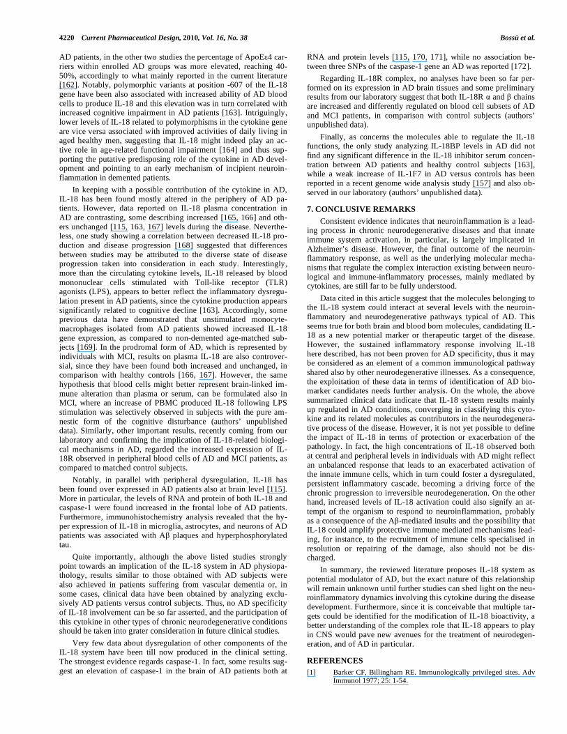
Fig. (1). The IL-18 system. The two signals necessary for production of bioactive IL-18 and the following interaction of the cytokine with the IL-18 receptor
chains leading to signal transduction are schematically depicted in the upper part of the cartoon (A). The regulatory components of the IL-18 system that can
inhibit IL-18 binding to its receptor complex and signal transduction, including IL-18BP, IL-1F7 (left side) and two isoforms of IL-18 receptor (right side), are
illustrated in the lower part of the figure (B).
IL-18Rα IL-18RβPro-IL-18
Caspase-1
IL-18
IL-18Rα IL-18Rβ
Truncated IL-18Rα
Soluble IL-18Rβ
IL-1F7
A
BIL-18BP
Inflammasome
IL-18 mRNA expression
Caspase-1 activation
IL-18BP/IL-18
IL-18Rα
IL-18RβIL-1F7/IL-18BP
TLRs

Interleukin-18, From Neuroinflammation to Alzheimer’s Disease Current Pharmaceutical Design, 2010, Vol. 16, No. 38 4217
dence about the ability of IL-18 to mediate the communication be-tween the nervous, the endocrine and the immune systems during stress has been reported [95]. Eventually, this latter group of authors has also comprehensively reviewed the role of the whole IL-18 system in CNS, under healthy and diseased conditions [96]. The specific intent of the present review is to focus on data pointing to the relevancy of IL-18-mediated biological mechanisms in AD physiopathology. Thus, cellular sources and triggering factors, as well as target cells, transduction pathways, biological functions and modulators of the IL-18 system, will be hereafter discussed under the light of their potential involvement in the progression of AD.
Although primary source of IL-18 are macrophages, its expres-sion is widespread and it has been detected in several different cell types, including monocytes, dendritic cells (DCs), Kupffer cells, keratinocytes, chondrocytes, osteoblasts and fibroblasts. Amongst brain cells, IL-18 can be mainly expressed by microglia, astrocytes, ependymal cells and neurons [96]. In addition, brain IL-18 expres-sion may be enhanced in vivo during neuroinflammatory events in response to the harmful effects of diverse exogenous or endogenous insulting stimuli, like brain infection, hypoxic-ischemic, hyperoxic and traumatic brain injury [94]. Regarding neurodegeneration, and AD in particular, it is likely that signals produced by stressed, dam-aged or otherwise malfunctioning brain cells, including also amy-loid misfolded proteins, could activate the innate immunity defense by eliciting the cytokine release, primarily by microglial cells, through the binding to PPRs, the receptors which mediate innate immunity. Accordingly, evidence emerging in the very last years suggests that components of two major families of PPRs, TLRs and NLRs, are indeed involved in AD neuroinflammation and neurode-generation.
As regards in particular the TLRs, although it is well estab-lished that they are essential for protective immunity against invad-ing pathogens, it is also true that inappropriate TLR responses can contribute to acute and chronic inflammation, and probably to neu-rodegeneration. Accordingly, one of the first studies pointing to a key role for innate immunity receptors in AD showed an interac-tion, leading to microglia activation and neuron toxicity, between aggregated A and the LPS receptor CD14, which is known to sig-naling by TLR4 [97]. In addition, microglia of AD mice have been observed to express increased levels of CD14 receptor molecule. In a further study, a CD14 up regulation was even observed within the brain of AD patients in microglia associated to A deposits, while evidence about a contribute of CD14 in phagocytosis of A fibrils was also provided [98]. Later on, cumulating research findings re-vealed, both in animals and humans, a general up regulation of TLR molecules in association with AD. More in particular, the involve-ment of CD14 and TLRs, (mainly TLR4 and TLR2) in microglia recognition of fibrillar A with subsequent cell activation, but also A clearance, was confirmed in a number of studies [99-105]. Fur-thermore, genetic studies revealed an association between CD14 and TLR4 polymorphisms and AD susceptibility [106, 107]. Thus, although TLR role in AD seems to be complex and the final out-come of the triggering of these receptors might be, dependently on several variables, both protective and harmful, the TLR-mediated innate immune response in the CNS appears somewhat relevant in AD [108]. It is known that TLR triggering, obtained for instance in myeloid cells by LPS treatment through CD14 binding, results in the activation of the transcription factor NF-kB, which in turn regu-lates the expression of a wide array of genes involved in inflamma-tory responses, including IL-18 [109-111]. Consistently, it has been reported that LPS can induce IL-18 expression in microglia [112]. A second important signal for IL-18 production within a neuroin-flammatory context is the activation of the inflammasome, a large complex containing molecules belonging to a cytosolic type of pat-tern recognition innate immune receptors. In particular, most in-flammasomes contain members of the NLR family that act as sen-sors for endogenous danger signals, which specifically trigger the
processing and release of the pro-inflammatory cytokines IL-1 and IL-18. In fact, this type of inflammasomes have the pivotal function to convert inactive procaspase-1 to active caspase-1, which is able to cleave the inactive IL-18 precursor to a secreted, active cytokine. In particular, amongst NLRs inflammasome components able to sense cellular damage, NALP3, whose activation can be initiated after A phagocytosis [113], might be a very important link be-tween AD and IL-18. In addition, other A -mediated mechanisms leading to the activation of caspase-1, such as the K
+ efflux-
dependent activation of NALP1, have been hypothesized to play a relevant role in the pathogenesis of AD [114]. Thus, taken all to-gether, data on A ability to trigger TLR and NLR by one hand, and evidences that TLR and NLR-mediated activation leads to IL-18 release during neuroinflammation on the other hand, suggest that microglia of AD patients can respond to A innate immunity recep-tor activation with an increased release of IL-18. In fact, the two events that are required for production of mature IL-18, i.e. the enhanced precursor synthesis, mainly regulated by TLR activation and transcriptionally by NF- B, and the precursor processing, which chiefly depends on inflammasome involvement and caspase-1 activation, both appear to occur in AD brain. As further confirma-tion of this observation, an increased expression of IL-18 protein and caspase-1 was specifically observed in the frontal lobe of AD brains. In this context, microglia, but also astrocytes and neurons stained with IL-18, were observed in the strict vicinity of amyloid deposits and neurofibrillary tangles [115].
Thus, although a direct proof of IL-18 production by human microglia following A -mediated TLR/NLR triggering is still miss-ing, it is highly conceivable that AD-specific pathogenic insults, such as A accumulation, can lead via PPRs activation to an in-creased release of IL-18 within the brain of AD subjects.
The effects of IL-18 are exerted following the binding of the processed, active cytokine to its specific receptor complex, which belongs to the IL-1/TLR family. As before reported, the activation of IL-18 mediated signaling pathways are triggered when both IL-18R and IL-18R chains of the receptor complex are co-expressed on the membrane of target cells. The IL-18R is present on both dif-ferent leukocyte populations and other cell types, including macro-phages, DCs, natural killer (NK), T and B lymphocytes, neutro-phils, basophils, mast cells, endothelial cells, smooth muscle cells, synovial fibroblasts, chondrocytes, and epithelial cells, underscor-ing the pleiotropic nature of the IL-18 system [88]. In the brain, a widespread constitutive expression of both and chains of the receptor complex was described, attributed to microglia, astrocytes and prevalently to neurons, throughout the brain. In addition, cul-tured microglia, astrocytes and neuronal cells can express the recep-tor and respond to IL-18 under different conditions [96, 116-118]. On the whole, cell types that express IL-18R in CNS can vary de-pending on environmental and pathogenic conditions. Intriguingly, in the CNS, a tight modulation of the IL-18 activity is required, as indicated by the existence of some peculiar isoforms of IL-18R with possible regulatory functions, further supporting the relevance of IL-18 system in brain physiopathology. More in particular, one study described the existence in mouse brain of a truncated decoy form of IL-18R chain, lacking the TIR domain [119], while other authors identified in brain rat a splice variant of IL-18R mRNA predicted to encode a soluble isoform of IL-1R , made by only one extracellular immunoglobulin domain, instead of three, and lacking the intracellular portion [117].
The signaling pathways mobilized by IL-18 binding to its re-ceptor complex includes the recruitment of MyD88 and the activa-tion of IRAK/ TRAF6 leading to translocation into the nucleus of NF-kB, as well as the activation of JNK/ p38MAPK pathway. Ac-cordingly, IL-18 dependent NF- B phosphorylation and JNK and p38MAPK activation were observed in brain cells under some con-ditions [96]. Interestingly, NF-kB signaling has been recently pro-posed as a molecular culprit of the innate immunity activation dur-

4218 Current Pharmaceutical Design, 2010, Vol. 16, No. 38 Bossù et al.
ing aging [114] and a therapeutic target in neurodegenerative dis-eases [120]. Notably, a link between AD and the over expression of IL-18 could be pointed out at signaling molecular level, since sev-eral components of the IL-18 signal transduction pathway result activated in AD. As an example, NF- B has been found activated in AD brain, in association with neurons and glial cells of A plaque surrounding areas. Furthermore, A stimulation was seen to induce an up regulation of NF- B activity in vitro. These observations have led to the suggestion that sustained NF- B activation, both consequence and cause of elevated cytokine levels occurring during chronic neuroinflammation, could result in increased A production and microglia activation, which in turn could lead to an amplifica-tion of AD pathogenic processes [121].
In a similar way, JNK and p38MAPK might be involved in A -induced responses and are activated in AD brain, particularly in the early stages of the disease [122, 123]. Hence, p38MAPK, which appears to be one of the potential molecular mediators of IL-18 sig-naling in the neurons [124], has been recently reviewed with regard to its role in AD pathophysiology, also beyond the neuroinflamma-tory context [125].
Similarly to what reported in the periphery, IL-18 appears to have multiple regulatory functions also in CNS, where it is sup-posed to affect neuroinflammatory, behavioral and stress-related mechanisms [96]. Some of these IL-18 mediated biological proc-esses might influence AD pathobiological pathways.
It is well known that IL-18 can amplify the innate immune re-sponse by inducing the release of other inflammatory cytokines [73, 74, 88, 126, 127], thus it is possible to predict that one important function played by IL-18 within the brain, would be addressed to microglial cells in particular and consist in amplifying the inflam-matory cascade occurring during neuroinflammatory phenomena. This has been confirmed by studies on IL-18 null mice, who have impaired microglia activation [128, 129]. In addition, some studies have demonstrated that IL-18 can stimulate the production of IL-1 in primary astrocytes and the release of various chemokines and pro-inflammatory cytokines, including IL-1 , IL-8, TNF- , IL-6 and IL-18 itself in microglial and astrocytic cell lines [118, 130, 131]. Differently, primary newborn microglia have been shown to respond to IL-18 by inhibiting the LPS induced release of IL-12 and lowering TNF- [116]. Regardless these apparent inconsistencies, probably related to the different function of the cytokine in the two different experimental systems, IL-18 has been identified as an im-portant contributor in several pathological conditions where neu-roinflammation occurs [94]. Within this scenario, where A or other AD-related stressor factors could induce the release of IL-18, likely via PRR triggering, it is conceivable that this cytokine would exacerbate the release by neighboring glial cells or peripheral im-mune cells, of diverse toxic inflammatory molecules, which may lead to a vicious cycle where inflammatory processes contribute to different aspects of AD neurodegeneration. In keeping with a po-tential contribute of peripheral immune cells in amplifying the AD inflammatory milieu, we recently demonstrated that myeloid-derived DCs obtained from AD patients show an over activated in-flammatory state and a reduced antigen presenting ability, as com-pared to cells of healthy control subjects [132]. Since in a previous in vitro study we have shown that A treatment is able to sustain inflammation by driving monocyte differentiation towards an IL-18 producing DC phenotype [133], it is probable that IL-18 would ex-acerbate a neuroinflammatory responses in a AD-specific and DC-dependent fashion also in vivo conditions. However, more studies are needed to clarify this issue and to enable the interpretation of the role, either protective or detrimental, of IL-18 within the physiopathological processes of AD. Regarding the peculiar ability of IL-18 to enhance both Th1- and Th2-driven immune responses, no studies focused to unravel this IL-18-mediated effect have been so far conducted in AD conditions, despite the existence of growing data indicating that T cell-mediated adaptive immunity may be in-
volved in this disease. Accordingly, T cells can infiltrate the AD brain [134], where they may be dysfunctional [135] and decreased in number in association with mild/moderate dementia [52], while in normal conditions, T cells can maintain neurogenesis, spatial learning and memory in adulthood [136]. These data might be taken as a suggestion of a possible contribution of IL-18 in triggering adaptive innate immune processes that can influence chronic neu-rodegeneration.
Other biological functions of IL-18 are the promotion of IFN- production other than from T cells also from natural killer (NK) cells, important cellular component of the innate immunity. In addi-tion, the cytokine is able to induce the enhancement of FasL ex-pression on T and NK cells, as well as the induction of apoptosis in Fas-expressing cells [88]. More recently, it has been indicated that IL-18 can cause fratricidal death in NK cells and this might occur via two mechanisms: by inducing the expression of FasL and by increasing the production of TNF- from NK cells. It is noteworthy that this IL-18-mediated NK cell killing has been hypothesized to be a negative-feedback mechanism to control and terminate a pro-inflammatory immune response [137]. Interestingly, some clinical studies have reported that NK from AD patients are over reactive and overproduce IFN- and TNF- [138]. Furthermore, increased apoptosis was found in AD-derived NK cells [139], suggesting that NK cell homeostasis may be indeed altered in AD. Finally, Fas-mediated apoptosis was implicated in AD [140, 141]. On the whole, this last set of data imply that careful consideration should be given to the role of IL-18 in AD, regarding in particular its ability to regu-late the functions of innate immune cells and their potential toxic effects on neurons.
In addition to the effects exerted on immune and glial cells, IL-18 can also directly target neuronal cells, as demonstrated by the broad distribution of IL-18R on brain cells. This peculiarity could underline the effects of IL-18 on neurodegeneration, ahead of its pro-inflammatory and immunomodulatory features. Currently available evidence shows that pro-inflammatory cytokines, includ-ing IL-18, may influence cognitive processes by affecting neuronal survival, synaptic plasticity, and neurogenesis. Indeed, data mainly coming from in vitro and animal studies provided clues for direct IL-18 regulation of neurotoxicity and synaptic function [94, 96]. In particular, it has been also proposed that age-related deficits in long term potentiation (LTP), likely dependent on microglia activation in older animals, may be due to increased hippocampal concentrations of IL-18 [142], confirming that inflammatory changes, apparently mediated by IL-18 may exert a negative impact on synaptic plastic-ity.
Neurogenesis regulation might be a further issue of IL-18 rele-vance in AD. In fact, AD pathogenic processes may implicate aber-rant neurogenic mechanisms and defects in brain repair functions [143, 144]. At the same time, it has been demonstrated that LPS-mediated activation strongly suppress neurogenesis in the adult ro-dent hippocampus [145, 146] and pro-inflammatory cytokines might mediate such effect. In particular, IL-18 has been suggested to either inhibit neuronal differentiation or induce neuronal cell death in cultured neural progenitors and also attenuate adult neuro-genesis [147, 148]. Notably, it is known that IL-18 is produced by ependymal cells, which are located in the brain zones, where adult neurogenesis occurs [96]. Altogether, these data further provide support for the putative involvement of IL-18 dysregulation in AD pathogenesis. Moreover, neuroimmune interaction in neurogenesis has been studied more in depth in the last years [149], suggesting that more attention should be given to decipher IL-18-dependent effects in neurorepairing processes during AD development.
As already extensively reviewed by other authors, a consistent body of literature reports on a crucial role for IL-18 in stress mechanisms, highlighting the possible neuroendocrine and neuro-immune regulatory function of the cytokine [95]. Since stress has a negative impact on cognitive function and increases the levels of

Interleukin-18, From Neuroinflammation to Alzheimer’s Disease Current Pharmaceutical Design, 2010, Vol. 16, No. 38 4219
inflammatory substances, also this set of data could be interpreted as supportive of an involvement of IL-18 in AD pathophysiological processes. Furthermore, the behavioral profile of AD patients re-sults profoundly altered, even independently from cognitive symp-toms [150] and IL-18 has been before associated with depressive and emotional disorders in different contexts, also by our group [96, 149, 151, 152]. However, this promising area of research needs to be better investigated and the understanding of underlying mecha-nisms of cytokine implication in behavioral and psychological symptoms of dementia (BPSD) in AD represents an intriguing chal-lenge for the development of future studies in the field of psycho-neuroimmunology. Accordingly, appetite disturbances are included among BPSD, and weight loss is a significant feature of AD, which may often precede the clinical diagnosis of dementia [153]. Inter-estingly, emerging evidence indicates that IL-18 is a homeostatic regulator of the energy balance, acting both centrally and peripher-ally as an inhibitor of appetite and feed efficiency [154, 155].
Finally, regarding the other members of the IL-18 system, such as IL-18BP and IL-1F7, very few studies regarding their role in CNS have been so far reported and only in one case they can be cited in favor of the hypothesis that IL-18 system is involved in AD. In fact, a weak association with AD can be made for IL-1F7, since it has been detected in the human brain under normal condi-tions [156] and its expression resulted slightly elevated in neocortex of AD patients [157].
6. CLINICAL EVIDENCE OF IL-18 DYSREGULATION IN AD
Among the numerous molecules which have been considered as potential mediators of neuroinflammation in AD, IL-18 is still poorly studied. However, there is an expanding evidence about a
potential involvement of this cytokine and its related molecules in AD patients, both at central and peripheral level. A summary of these observations, hereafter described in more details, is reported in Table 1.
Important data supporting the role of inflammation as driving force in AD comes from genetic studies. Accordingly, one recent meta analysis study conducted on genome wide analysis data indi-cates that genes involved in inflammation and immune response are indeed correlated with AD risk [158]. Although the general view suggests that also defined polymorphisms within some cytokine genes may predispose for AD risk, studies based on genomic analy-sis of single inflammatory cytokines in AD subjects are often con-tradictory, likely because of the following reasons: the small size of population analyzed, the ethnical differences and the relatively low impact of single gene variants linked to the multifaceted nature of the disease. To date, only three studies have been specifically ad-dressed to analyze IL-18 gene variants in AD patients. The first study, conducted in our laboratory within an Italian population, suggested a significant association between two functional single nucleotide polymorphisms (SNPs) of the cytokine gene promoter, i.e. -607 C/A (rs1946518) and -137 G/C (rs187238), and AD risk and outcome [159]. A second independent study further confirmed the association of the same polymorphisms of IL-18 gene with the risk to develop AD also in a Chinese population [160]. Finally, a third study, similarly to the first performed in Italians, provided negative results about association of the same polymorphisms and AD risk [161]. This inconsistency, apparently independent from population size and ethnicity, could be explained on the basis of some heterogeneity of AD patients enrolled, in fact while the study of Segat and co-authors indicated a presence of the 4 allele of the gene coding for apolipoprotein E (ApoE) in only in 27% of their
Table 1. Clinical Evidence of IL-18 System Dysregulation in Alzheimer’s Disease
Gene/Protein Type of Analysis Major Findings in AD Patients (vs. Control Subjects) References
Association between SNPs (rs1946518, rs187238) and disease risk/outcome [159,160] Genetic variability
Lack of association between SNPs (rs1946518, rs187238) and disease risk [161]
Brain RNA Increased in the frontal regions [115]
PBMC RNA Increased in macrophages [169]
Brain protein Increased in neurons and glial cells [115]
CSF protein Not detectable [167]
Increased [115]
Plasma/serum protein Increased [165,166,168]
Unchanged [115,163,167]
IL-18
PBMC protein Increased following LPS stimulation [163]
Genetic variability Lack of association between SNPs (rs501192, rs556205, rs530537) and disease risk [172]
Brain RNA Increased in cerebral cortex and frontal region [115,170,171]
Caspase-1
Brain protein Increased [115]
IL-18R PBMC protein Increased Authors’ unpublished
data
IL-18 BP Plasma/serum protein Unchanged [163]
Brain RNA Increased in neocortex [157] IL-1F7
PBMC protein Increased following LPS stimulation Authors’ unpublished
data
4220 Current Pharmaceutical Design, 2010, Vol. 16, No. 38 Bossù et al.
AD patients, in the other two studies the percentage of ApoE 4 car-riers within enrolled AD groups was more elevated, reaching 40-50%, accordingly to what mainly reported in the current literature [162]. Notably, polymorphic variants at position -607 of the IL-18 gene have been also associated with increased ability of AD blood cells to produce IL-18 and this elevation was in turn correlated with increased cognitive impairment in AD patients [163]. Intriguingly, lower levels of IL-18 related to polymorphisms in the cytokine gene are vice versa associated with improved activities of daily living in aged healthy men, suggesting that IL-18 might indeed play an ac-tive role in age-related functional impairment [164] and thus sup-porting the putative predisposing role of the cytokine in AD devel-opment and pointing to an early mechanism of incipient neuroin-flammation in demented patients.
In keeping with a possible contribution of the cytokine in AD, IL-18 has been found mostly altered in the periphery of AD pa-tients. However, data reported on IL-18 plasma concentration in AD are contrasting, some describing increased [165, 166] and oth-ers unchanged [115, 163, 167] levels during the disease. Neverthe-less, one study showing a correlation between decreased IL-18 pro-duction and disease progression [168] suggested that differences between studies may be attributed to the diverse state of disease progression taken into consideration in each study. Interestingly, more than the circulating cytokine levels, IL-18 released by blood mononuclear cells stimulated with Toll-like receptor (TLR) agonists (LPS), appears to better reflect the inflammatory dysregu-lation present in AD patients, since the cytokine production appears significantly related to cognitive decline [163]. Accordingly, some previous data have demonstrated that unstimulated monocyte-macrophages isolated from AD patients showed increased IL-18 gene expression, as compared to non-demented age-matched sub-jects [169]. In the prodromal form of AD, which is represented by individuals with MCI, results on plasma IL-18 are also controver-sial, since they have been found both increased and unchanged, in comparison with healthy controls [166, 167]. However, the same hypothesis that blood cells might better represent brain-linked im-mune alteration than plasma or serum, can be formulated also in MCI, where an increase of PBMC produced IL-18 following LPS stimulation was selectively observed in subjects with the pure am-nestic form of the cognitive disturbance (authors’ unpublished data). Similarly, other important results, recently coming from our laboratory and confirming the implication of IL-18-related biologi-cal mechanisms in AD, regarded the increased expression of IL-18R observed in peripheral blood cells of AD and MCI patients, as compared to matched control subjects.
Notably, in parallel with peripheral dysregulation, IL-18 has been found over expressed in AD patients also at brain level [115]. More in particular, the levels of RNA and protein of both IL-18 and caspase-1 were found increased in the frontal lobe of AD patients. Furthermore, immunohistochemistry analysis revealed that the hy-per expression of IL-18 in microglia, astrocytes, and neurons of AD patients was associated with A plaques and hyperphosphorylated tau.
Quite importantly, although the above listed studies strongly point towards an implication of the IL-18 system in AD physiopa-thology, results similar to those obtained with AD subjects were also achieved in patients suffering from vascular dementia or, in some cases, clinical data have been obtained by analyzing exclu-sively AD patients versus control subjects. Thus, no AD specificity of IL-18 involvement can be so far asserted, and the participation of this cytokine in other types of chronic neurodegenerative conditions should be taken into grater consideration in future clinical studies.
Very few data about dysregulation of other components of the IL-18 system have been till now produced in the clinical setting. The strongest evidence regards caspase-1. In fact, some results sug-gest an elevation of caspase-1 in the brain of AD patients both at
RNA and protein levels [115, 170, 171], while no association be-tween three SNPs of the caspase-1 gene an AD was reported [172].
Regarding IL-18R complex, no analyses have been so far per-formed on its expression in AD brain tissues and some preliminary results from our laboratory suggest that both IL-18R and chains are increased and differently regulated on blood cell subsets of AD and MCI patients, in comparison with control subjects (authors’ unpublished data).
Finally, as concerns the molecules able to regulate the IL-18 functions, the only study analyzing IL-18BP levels in AD did not find any significant difference in the IL-18 inhibitor serum concen-tration between AD patients and healthy control subjects [163], while a weak increase of IL-1F7 in AD versus controls has been reported in a recent genome wide analysis study [157] and also ob-served in our laboratory (authors’ unpublished data).
7. CONCLUSIVE REMARKS
Consistent evidence indicates that neuroinflammation is a lead-ing process in chronic neurodegenerative diseases and that innate immune system activation, in particular, is largely implicated in Alzheimer’s disease. However, the final outcome of the neuroin-flammatory response, as well as the underlying molecular mecha-nisms that regulate the complex interaction existing between neuro-logical and immune-inflammatory processes, mainly mediated by cytokines, are still far to be fully understood.
Data cited in this article suggest that the molecules belonging to the IL-18 system could interact at several levels with the neuroin-flammatory and neurodegenerative pathways typical of AD. This seems true for both brain and blood born molecules, candidating IL-18 as a new potential marker or therapeutic target of the disease. However, the sustained inflammatory response involving IL-18 here described, has not been proven for AD specificity, thus it may be considered as an element of a common immunological pathway shared also by other neurodegenerative illnesses. As a consequence, the exploitation of these data in terms of identification of AD bio-marker candidates needs further analysis. On the whole, the above summarized clinical data indicate that IL-18 system results mainly up regulated in AD conditions, converging in classifying this cyto-kine and its related molecules as contributors in the neurodegenera-tive process of the disease. However, it is not yet possible to define the impact of IL-18 in terms of protection or exacerbation of the pathology. In fact, the high concentrations of IL-18 observed both at central and peripheral levels in individuals with AD might reflect an unbalanced response that leads to an exacerbated activation of the innate immune cells, which in turn could foster a dysregulated, persistent inflammatory cascade, becoming a driving force of the chronic progression to irreversible neurodegeneration. On the other hand, increased levels of IL-18 activation could also signify an at-tempt of the organism to respond to neuroinflammation, probably as a consequence of the A -mediated insults and the possibility that IL-18 could amplify protective immune mediated mechanisms lead-ing, for instance, to the recruitment of immune cells specialised in resolution or repairing of the damage, also should not be dis-charged.
In summary, the reviewed literature proposes IL-18 system as potential modulator of AD, but the exact nature of this relationship will remain unknown until further studies can shed light on the neu-roinflammatory dynamics involving this cytokine during the disease development. Furthermore, since it is conceivable that multiple tar-gets could be identified for the modification of IL-18 bioactivity, a better understanding of the complex role that IL-18 appears to play in CNS would pave new avenues for the treatment of neurodegen-eration, and of AD in particular.
REFERENCES
[1] Barker CF, Billingham RE. Immunologically privileged sites. Adv Immunol 1977; 25: 1-54.

Interleukin-18, From Neuroinflammation to Alzheimer’s Disease Current Pharmaceutical Design, 2010, Vol. 16, No. 38 4221
[2] Perry VH, Andersson PB, Gordon S. Macrophages and inflamma-
tion in the central nervous system. Trends Neurosci 1993; 16: 268-73.
[3] Galea I, Bechmann I, Perry VH. What is immune privilege (not)? Trends Immunol 2007; 28: 12-8.
[4] Rivest S. Regulation of innate immune responses in the brain. Nat Rev Immunol 2009; 9: 429-39.
[5] McAllister AK, van de Water J. Breaking boundaries in neural-immune interactions. Neuron 2009; 64: 9-12.
[6] Nimmerjahn A, Kirchhoff F, Helmchen F. Resting microglial cells are highly dynamic surveillants of brain parenchyma in vivo. Sci-
ence 2005; 308: 1314-18. [7] Streit WJ, Conde JR, Fendrick SE, Flanary BE, Mariani CL. Role
of microglia in the central nervous system's immune response. Neu-rol Res 2005; 27: 685-91.
[8] Wekerle H, Linington C, Lassmann H, Meyermann R. Cellular immune reactivity within the CNS. Trends Neurosci 1986; 9: 271-
7. [9] Hickey WF. Leukocyte traffic in the central nervous system: the
participants and their roles. Semin Immunol 1999; 11: 125-37. [10] Ransohoff RM, Kivisäkk P, Kidd G. Three or more routes for leu-
kocyte migration into the central nervous system. Nat Rev Immu-nol 2003; 3; 569-81.
[11] Engelhardt B. Molecular mechanisms involved in T cell migration across the blood-brain barrier. J Neural Transm 2006; 113: 477-85.
[12] Simard AR, Soulet D, Gowing G, Julien JP, Rivest S. Bone mar-row-derived microglia play a critical role in restricting senile
plaque formation in Alzheimer’s disease. Neuron 2006; 49: 489-502.
[13] Lucin KM, Wyss-Coray T. Immune activation in brain aging and neurodegeneration: too much or too little? Neuron 2009; 64: 110-
22. [14] Schwartz M, Shechter R. Systemic inflammatory cells fight off
neurodegenerative disease. Nat Rev Neurol 2010; 6: 405-10. [15] Boulanger LM. Immune proteins in brain development and synap-
tic. Plasticity Neuron 2009; 64: 93-1. [16] Medzhitov R. Origin and physiological roles of inflammation.
Nature 2008; 454: 428-35. [17] Perry VH, Bell MD, Brown HC, Matyszak MK. Inflammation in
the nervous system. Curr Opin Neurobiol 1995; 5: 636-41. [18] Rezai-Zadeh K, Gate D, Town T. CNS infiltration of peripheral
immune cells: D-day for neurodegenerative disease? J Neuroim-mune Pharmacol 2009; 4: 462-75.
[19] Wilson EH, Weninger W, Hunter CH. Trafficking of immune cells in the central nervous system. J Clin Invest 2010; 120: 1368-80
[20] Streit WJ, Mrak RE, and Griffin WST Microglia and neuroinflam-mation: a pathological perspective. J Neuroinflamm 2004; 1: 14.
[21] Wyss-Coray T, Mucke L: Inflammation in neurodegenerative dis-ease--a double-edged sword. Neuron 2002; 35: 419-32.
[22] Lucas SM, Rothwell NJ, Gibson RM. The role of inflammation in CNS injury and disease. Br J Pharmacol 2006; 147 (Suppl 1):
S232-40. [23] Frank-Cannon TC, Alto LT, McAlpine FE, Tansey MG. Does neu-
roinflammation fan the flame in neurodegenerative diseases? Mol Neurodegener 2009; 4: 47.
[24] Perry VH. The influence of systemic inflammation on inflamma-tion in the brain: implications for chronic neurodegenerative dis-
ease. Brain Behav Immun 2004; 18: 407-13 [25] Steinman L. Nuanced roles of cytokines in three major human
brain disorders. J Clin Invest 2008; 118: 3557-63. [26] Hagberg H, Mallard C. Effect of inflammation on central nervous
system development and vulnerability. Curr Opin Neurol 2005; 18: 117-23
[27] Degos V, Favrais G, Kaindl AM, et al. Inflammation processes in perinatal brain damage. J Neural Transm 2010; 117(8): 1009-17.
[28] Dilger RN, Johnson RW. Aging, microglial cell priming, and the discordant central inflammatory response to signals from the pe-
ripheral immune system. J Leukoc Biol 2008; 84: 932-9. [29] Streit WJ. Microglial senescence: does the brain’s immune system
have an expiration date? Trends Neurosci 2006; 29: 506-10. [30] Ron-Harel N, Schwartz M. Immune senescence and brain aging:
can rejuvenation of immunity reverse memory loss? Trends Neuro-sci 2009; 32: 367-75.
[31] Franceschi C, Bonafe M, Valensin S, et al. Inflamm-aging. An evo-lutionary perspective on immunosenescence. Ann N Y Acad Sci
2000; 908: 208-18.
[32] Blasko I, Stampfer-Kountchev M, Robatscher P, Verhuis R,
Eikelenboom P, Grubeck-Loebenstein B. How chronic inflamma-tion can affect the brain and support the development of Alz-
heimer’s disease in old age: the role of microglia and astrocytes. Aging Cell 2004; 3: 169-76.
[33] Licastro F, Candore G, Lio D, et al. Innate immunity and inflam-mation in ageing: a key for understanding age-related diseases.
Immun Ageing 2005; 2: 8. [34] Giunta B, Fernandez F, Nikolic WV, et al. Inflammaging as a pro-
drome to Alzheimer’s disease. J Neuroinflamm 2008, 5: 51 [35] Zipp F, Aktas O. The brain as a target of inflammation: common
pathways link inflammatory and neurodegenerative diseases. Trends Neurosci 2006; 29: 518-27.
[36] Björkqvist M, Wild EJ, Tabrizi SJ. Harnessing immune alterations in neurodegenerative diseases. Neuron 2009; 64: 21-4.
[37] Eikelenboom P, Veerhuis R, Scheper W, Rozemuller AJ, van Gool WA, Hoozemans JJ. The significance of neuroinflammation in un-
derstanding Alzheimer’s disease. J Neural Transm 2006; 113: 1685-95.
[38] Besedovsky HO, del Rey A. Brain Cytokines as Integrators of the Immune-Neuroendocrine Network. In: Galoyan A, Besedovsky H,
Eds. Handbook of Neurochemistry and Molecular Neurobiology-Neuroimmunology. US: Springer 2008; pp. 4-17.
[39] Dunn AJ. Effects of the Immune System on Brain Neurochemistry In: Galoyan A., Besedovsky H, Eds. Handbook of Neurochemistry
and Molecular Neurobiology-Neuroimmunology. US: Springer 2008; pp. 38-59.
[40] Stoll G, Jander S, Schroeter M. Detrimental and beneficial effects of injury-induced inflammation and cytokine expression in the
nervous system. Adv Exp Med Biol 2002; 513: 87-113. [41] Allan, S.M. and Rothwell, N.J. Cytokines and acute neurodegen-
eration. Nat Rev Neurosci 2001; 2: 734-44. [42] Wyss-Coray T. Inflammation in Alzheimer disease: driving force,
bystander or beneficial response? Nat Med 2006; 12: 1005-15. [43] Marques-Deak A, Cizza G, Sternberg E. Brain-immune interactions
and disease susceptibility. Mol Psychiatry 2005; 10: 239-50. [44] Carpentier PA, Palmer TD. Immune influence on adult neural stem
cell regulation and function. Neuron 2009; 64: 79-92 [45] Deverman BE, Patterson PH. Cytokines and CNS development.
Neuron 2009; 64: 61-78 [46] Petersen RC, Smith GE, Waring SC, Ivnik RJ, Tangalos EG, Kok-
men E. Mild cognitive impairment: clinical characterization and outcome. Arch Neurol 1999; 56: 303-8.
[47] Mrak RE. Neuropathology and the neuroinflammation idea. J Alzh Dis 2009; 18: 473-81
[48] Selkoe DJ. Defining molecular targets to prevent Alzheimer dis-ease. Arch Neurol 2005; 62: 192-5.
[49] Akiyama H, Barger S, Barnum S, et al. Inflammation and Alz-heimer's disease. Neurobiol Aging 2000; 21: 383-421.
[50] Cagnin A, Brooks DJ, Kennedy AM, et al. In vivo-measurement of activated microglia in dementia. Lancet 2001; 358: 461-7.
[51] Craft JM, Watterson DM, Van Eldik LJ. Human amyloid beta-induced neuroinflammation is an early event in neurodegeneration.
Glia 2006; 53: 484-90. [52] Parachikova A, Agadjanyan MG, Cribbs DH, et al. Inflammatory
changes parallel the early stages of Alzheimer disease. Neurobiol Aging 2007; 28: 1821-33.
[53] Eikelenboom P, van Exel E, Hoozemans JJ, Veerhuis R, Rozemul-ler AJ, van Gool WA. Neuroinflammation - an early event in both
the history and pathogenesis of Alzheimer's disease. Neurodegener Dis 2010; 7: 38-41.
[54] Tan ZS, Beiser AS, Vasan RS, et al. Inflammatory markers and the risk of Alzheimer disease: the Framingham Study. Neurology
2007; 68: 1902-8. [55] McGeer PL, Schulzer M, McGeer EG. Arthritis and anti-
inflammatory agents as possible protective factors for Alzheimer’s disease: A review of 17 epidemiologic studies. Neurology 1996;
47: 425-32. [56] In’t Veld BA, Ruitenberg A, Hofman A, et al. Nonsteroidal anti-
inflammatory drugs and the risk of Alzheimer’s disease. N Engl J Med 2001; 345: 1515-21.
[57] Lleó A, Berezovska O, Herl L, et al. Nonsteroidal anti-inflammatory drugs lower Abeta42 and change presenilin 1 con-
formation. Nat Med 2004; 10: 1065-6.

4222 Current Pharmaceutical Design, 2010, Vol. 16, No. 38 Bossù et al.
[58] Aisen PS, Schafer A, Grundman M, et al. Effects of rofecoxib or
naproxen vs. placebo on Alzheimer disease progression: A random-ized controlled trial. JAMA 2003; 289: 2819-26.
[59] Reines SA, Block GA, Morris JC, et al. Rofecoxib: no effect on Alzheimer’s disease in a 1-year, randomized, blinded, controlled
study. Neurology 2004; 62: 66-71. [60] Wilcock GK, Black SE, Hendrix SB, et al. Efficacy and safety of
tarenflurbil in mild to moderate Alzheimer’s disease: A randomized phase II trial. Lancet Neurol 2008; 7: 483-93.
[61] McGeer PL, McGeer EG. NSAIDs and Alzheimer disease: Epide-miological, animal model and clinical studies. Neurobiol Aging
2007; 28: 639-47. [62] Hensley K. Neuroinflammation in Alzheimer's disease: mecha-
nisms, pathologic consequences, and potential for therapeutic ma-nipulation. J Alzheimers Dis 2010; 21: 1-14.
[63] Mrak RE, Griffin WS. Glia and their cytokines in progression of neurodegeneration. Neurobiol Aging 2005; 26: 349-54.
[64] Reichenberg A, Yirmiya R, Schuld A, et al. Cytokine-associated emotional and cognitive disturbances in humans. Arch Gen Psy-
chiatry 2001; 58: 445-52. [65] Weaver JD, Huang MH, Albert M, Harris T, Rowe JW, Seeman
TE. Interleukin-6 and risk of cognitive decline: MacArthur studies of successful aging. Neurology 2002; 59: 371-8.
[66] Yaffe K, Lindquist K, Penninx BW, et al. Inflammatory markers and cognition in well-functioning African-American and white eld-
ers. Neurology 2003; 61: 76-80. [67] Holmes C, El-Okl M, Williams A, Cunningham C, Wilcockson D,
Perry V. Systemic infection, interleukin 1b, and cognitive decline in Alzheimer’s disease. J Neurol Neurosurg Psychiatry 2003; 74:
788-9. [68] Glass CK, Saijo K, Winner B, Marchetto MC, Gage FH. Mecha-
nisms underlying inflammation in neurodegeneration. Cell 2010; 140: 918-34.
[69] Rothwell NJ, Luheshi GN. Interleukin 1 in the brain:biology, pa-thology and therapeutic target. Trends Neurosci 2000; 23: 618-25.
[70] Allan SM, Tyrrell PJ, Rothwell NJ. Interleukin-1 and neuronal in-jury. Nat Rev Immunol 2005; 5: 629-40.
[71] Shaftel SS, Griffin WST, O’Banion MK. The role of interleukin-1 in neuroinflammation and Alzheimer disease: an evolving perspec-
tive. J Neuroinflamm 2008; 5: 7. [72] Okamura H, Tsutsi H, Komatsu T, et al. Cloning of a new cytokine
that induces IFN-gamma production by T cells. Nature 1995; 378: 88-91.
[73] Gracie JA, Robertson SE, McInnes IB. Interleukin-18. J Leukoc Biol 2003; 73: 213-24.
[74] Dinarello CA. Interleukin 1 and interleukin 18 as mediators of in-flammation and the aging process. Am J Clin Nutr 2006; 83: 447S-
55. [75] Dinarello CA. Immunological and inflammatory functions of the
interleukin-1 family. Annu Rev Immunol 2009; 27: 519-50 [76] Ghayur T, Banerjee S, Hugunin M, et al. Caspase-1 processes IFN-
[gamma]inducing factor and regulates LPS-induced IFN-[gamma] production. Nature 1997; 386: 619-23.
[77] Gu Y, Kuida K, Tsutsui H, et al. Activation of interferon-gamma inducing factor mediated by interleukin-1beta converting enzyme.
Science 1997; 275: 206-9. [78] Martinon F, Mayor A, Tschopp J. The inflammasomes: guardians
of the body. Annu Rev Immunol 2009; 27: 229-65. [79] Janeway Jr. CA, Medzhitov R. Innate immune recognition. Ann
Rev Immunol 2002; 20: 197-216. [80] Akira S, Uematsu S, Takeuchi O. Pathogen recognition and innate
immunity. Cell 2006; 124: 783-801. [81] Petrilli V, Dostert C, Muruve DA, Tschopp J. The inflammasome:
a danger sensing complex triggering innate immunity. Curr Opin-ion Immunol 2007; 19: 615-22.
[82] Takeuchi O, Akira S. Pattern recognition receptors and inflamma-tion. Cell 2010; 140: 805-20
[83] Schroder K, Tschopp J. The inflammasomes. Cell 2010; 140: 821-32.
[84] Torigoe K, Ushio S, Okura T, et al. Purification and characteriza-tion of the human interleukin-18 receptor. J Biol Chem 1997; 272:
25737-42. [85] Sims JE. IL-1 and IL-18 receptors, and their extended family. Curr
Opin Immunol 2002; 14: 117-22. [86] Boraschi D, Tagliabue A. The interleukin-1 receptor family. Vitam
Horm 2006; 74: 229-54.
[87] Thomassen E, Bird TA, Renshaw BR, Kennedy MK, Sims JE.
Binding of interleukin-18 to the interleukin-1 receptor homologous receptor IL-1Rrp1 leads to activation of signaling pathways similar
to those used by interleukin-1. J Interferon Cytokine Res 1998; 18: 1077-88.
[88] Arend WP, Palmer G, Gabay C. IL-1, IL-18, and IL-33 families of cytokines. Immunol Rev 2008; 223: 20-38.
[89] Novick D, Schwartsburd B, Pinkus R, et al. A novel IL-18BP ELISA shows elevated serum IL-18BP in sepsis and extensive de-
crease of free IL-18. Cytokine 2001; 14: 334-42. [90] Kumar S, Hanning CR, Brigham-Burke MR, et al. Interleukin-
1F7B (IL-1H4/IL-1F7) is processed by caspase-1 and mature IL-1F7B binds to the IL-18 receptor but does not induce IFN-gamma
production. Cytokine 2002; 18: 61-71. [91] Bufler P, Azam T, Gamboni-Robertson F, et al. Complex of the IL-
1 homologue IL-1F7b and IL-18-binding protein reduces IL-18 ac-tivity. Proc Natl Acad Sci U S A 2002; 99: 13723-8.
[92] Okamura H, Tsutsui H, Kashiwamura S, Yoshimoto T, Nakanishi K. Interleukin18: a novel cytokine that augments both innate and
acquired immunity. Adv Immunol 1998; 70: 281-312. [93] Dinarello CA. Interleukin-18 and the pathogenesis of inflammatory
diseases. Semin Nephrol 2007; 27: 98-114. [94] Felderhoff-Mueser U, Schmidt OI, Oberholzer A, Bührer C, Stahel
PF. IL-18: a key player in neuroinflammation and neurodegenera-tion? Trends Neurosci 2005; 28: 487-93.
[95] Sugama S, Conti B. Interleukin-18 and stress. Brain Res Rev 2008; 58: 85-95.
[96] Alboni S, Cervia D, Sugama S, Conti B. Interleukin 18 in the CNS. J Neuroinflamm 2010; 7: 9.
[97] Fassbender K, Walter S, Kühl S, et al. The LPS receptor (CD14) links innate immunity with Alzheimer’s disease. FASEB J 2004;
18: 203-5. [98] Liu Y, Walter S, Stagi M, et al. LPS receptor (CD14): a receptor
for phagocytosis of Alzheimer’s amyloid peptide. Brain 2005; 128: 1778-89
[99] Landreth GE, Reed-Geaghan EG. Toll-like receptors in Alz-heimer’s disease. Curr Top Microbiol Immunol 2009; 336: 137-53.
[100] Reed-Geaghan EG, Savage JC, Hise AG, Landreth GE. CD14 and toll-like receptors 2 and 4 are required for fibrillar A{beta}-
stimulated microglial activation. J Neurosci 2009; 29: 11982-92. [101] Letiembre M, Liu Y, Walter S, et al. Screening of innate immune
receptors in neurodegenerative diseases: a similar pattern. Neuro-biol Aging 2009; 30: 759-68.
[102] Richard KL, Filali M, Prefontaine P, Rivest S. Toll-like receptor 2 acts as a natural innate immune receptor to clear amyloid beta 1-42
and delay the cognitive decline in a mouse model of Alzheimer’s disease. J Neurosci 2008; 28: 5784-93.
[103] Udan MLD, Ajit D, Crouse NR, Nichols MR. Toll-like receptors 2 and 4 mediate Aß(1-42) activation of the innate immune response
in a human monocytic cell line. J Neurochem 2008; 104: 524-33. [104] Tahara K, Kim H, Jin J, Maxwell J, Li L, Fukuchi K. Role of toll-
like receptor signaling in Abeta uptake and clearance. Brain 2006; 129: 3006-19
[105] Walter S, Letiembre M, Liu Y, et al. Role of the toll like receptor 4 in neuroinflammation in Alzheimer’s disease. Cell Physiol Bio-
chem 2007; 20: 947-56. [106] Minoretti P, Gazzaruso C, Vito C, et al. Effect of the functional
toll-like receptor 4 Asp299Gly polymorphism on susceptibility of late onset Alzheimer’s disease. Neurosci Lett 2006; 391: 147-9.
[107] Balistreri CR, Grimaldi MP, Chiappelli M, et al. Association be-tween the polymorphisms of TLR4 and CD14 genes and Alz-
heimer's disease. Curr Pharm Des 2008; 14: 2672-7. [108] Lehnardt S. Innate immunity and neuroinflammation in the CNS:
the role of microglia in Toll-like receptor-mediated neuronal injury. Glia 2010; 58: 253-63.
[109] Tone M, Thompson SA, Tone Y, Fairchild PJ, Waldmann H. Regu-lation of IL-18 (IFN -inducing factor) gene expression. J Immunol
1997; 159: 6156-63. [110] Manigold T, Böcker U, Traber P, et al. Lipopolysaccha-
ride/endotoxin induces IL-18 via CD14 in human peripheral blood mononuclear cells in vitro. Cytokine 2000; 12: 1788-92.
[111] Kawai T, Akira S. The role of pattern-recognition receptors in in-nate immunity: update on Toll-like receptors. Nature Immunol
2010; 11: 373-84.

Interleukin-18, From Neuroinflammation to Alzheimer’s Disease Current Pharmaceutical Design, 2010, Vol. 16, No. 38 4223
[112] Conti B, Park LC, Calingasan NY, et al. Cultures of astrocytes and
microglia express interleukin 18. Brain Res Mol Brain Res 1999; 67: 46-52.
[113] Halle A, Hornung V, Petzold G, et al. The NALP3 inflammasome is involved in the innate immune response to amyloid-[beta]. Nat
Immunol 2008; 9: 857-65. [114] Salminen A, Ojala J, Suuronen T, Kaarniranta K, Kauppinen A.
Amyloid-beta oligomers set fire to inflammasomes and induce Alzheimer's pathology. J Cell Mol Med 2008; 12: 2255-62.
[115] Ojala J, Alafuzoff I, Herukka SK, van Groen T, Tanila H, Pirttilä T. Expression of interleukin-18 is increased in the brains of Alz-
heimer's disease patients. Neurobiol Aging 2009; 30: 198-209. [116] Prinz M, Hanisch UK. Murine microglial cells produce and re-
spond to interleukin-18. J Neurochem 1999; 72: 2215-8. [117] Andre R, Wheeler RD, Collins PD, et al. Identification of a trun-
cated IL-18R beta mRNA: a putative regulator of IL-18 expressed in rat brain. J Neuroimmunol 2003; 145: 40-5.
[118] Das S, Mishra MK, Ghosh J, Basu A. Japanese Encephalitis Virus infection induces IL-18 and IL-1beta in microglia and astrocytes:
correlation with in vitro cytokine responsiveness of glial cells and subsequent neuronal death. J Neuroimmunol 2008; 195: 60-72.
[119] Alboni S, Cervia D, Ross B, et al. Mapping of the full length and the truncated interleukin-18 receptor alpha in the mouse brain. J
Neuroimmunol 2009; 214: 43-54. [120] Camandola S, Mattson MP. NF-kappa B as a therapeutic target in
neurodegenerative diseases. Expert Opin Ther Targets 2007; 11: 123-32.
[121] Granic I, Dolga AM, Nijholt IM, van Dijk G, Eisel UL. Inflamma-tion and NF-kappaB in Alzheimer's disease and diabetes. J Alz-
heimers Dis 2009; 16: 809-21. [122] Zhu X, Castellani RJ, Takeda A, et al. Differential activation of
neuronal ERK, JNK/SAPK and p38 in Alzheimer disease: the ‘two hit’ hypothesis. Mech Ageing Dev 2001; 123: 39-46.
[123] Sun A, Liu M, Nguyen XV, Bing G. p38MAP kinase is activated at early stages in Alzheimer’s disease brain. Exp Neurol 2003; 183:
394-405. [124] Curran BP, Murray HJ, O’Connor JJ. A role for c-Jun N-terminal
kinase in the inhibition of long-term potentiation by interleukin-1beta and longterm depression in the rat dentate gyrus in vitro.
Neuroscience 2003; 118: 347-57. [125] Munoz L, Ammit AJ. Targeting p38 MAPK pathway for the treat-
ment of Alzheimer's disease. Neuropharmacology 2010; 58: 561-68.
[126] Puren AJ, Razeghi P, Fantuzzi G, Dinarello CA. Interleukin-18 enhances lipopolysaccharide-induced interferon-gamma production
in human whole blood cultures. J Infect Dis 1998; 178: 1830-4. [127] Dinarello CA, Novick D, Puren AJ, et al. Overview of interleukin-
18: more than an interferon-gamma inducing factor. J Leukoc Biol 1998; 63: 658-64.
[128] Mori I, Hossain MJ, Takeda K, et al. Impaired microglial activation in the brain of IL-18-gene-disrupted mice after neurovirulent influ-
enza A virus infection. Virology 2001; 287: 163-70. [129] Sugama S, Wirz SA, Barr AM, Conti B, Bartfai T, Shibasaki T.
Interleukin-18 null mice show diminished microglial activation and reduced dopaminergic neuron loss following acute 1-methyl-4-
phenyl-1,2,3,6-tetrahydropyridine treatment. Neuroscience 2004; 128: 451-8.
[130] Culhane AC, Hall MD, Rothwell NJ, Luheshi GN. Cloning of rat brain interleukin-18 cDNA. Mol Psychiatry 1998; 3: 362-6.
[131] Wheeler RD, Brough D, Le Feuvre RA, et al. Interleukin-18 in-duces expression and release of cytokines from murine glial cells:
interactions with interleukin-1 beta. J Neurochem 2003; 85: 1412-20.
[132] Ciaramella A, Bizzoni F, Salani F, et al. Increased pro-inflammatory response by dendritic cells from patients with Alz-
heimer's disease. J Alzheimers Dis 2010; 19: 559-72. [133] Ciaramella A, Sanarico N, Bizzoni F, et al. Amyloid beta peptide
promotes differentiation of pro-inflammatory human myeloid den-dritic cells. Neurobiol Aging 2009; 30: 210-21.
[134] Itagaki S, McGeer PL, Akiyama H. Presence of T-cytotoxic sup-pressor and leucocyte common antigen positive cells in Alz-
heimer’s disease brain tissue. Neurosci Lett 1988; 91: 259-64. [135] Monsonego A, Imitola J, Zota V, Oida T, Weiner HL. Microglia
mediated nitric oxide cytotoxicity of T cells following amyloid beta peptide presentation to Th1 cells. J Immunol 2003; 171: 2216-24.
[136] Ziv Y, Ron N, Butovsky O, et al. Immune cells contribute to the
maintenance of neurogenesis and spatial learning abilities in adult-hood. Nat Neurosci 2006; 9: 268-75.
[137] Iannello A, Samarani S, Debbeche O, et al. Potential role of inter-leukin-18 in the immunopathogenesis of AIDS: involvement in
fratricidal killing of NK cells. J Virol 2009; 83: 5999-6010. [138] Solerte SB, Cravello L, Ferrari E, Fioravanti M. Overproduction of
IFN-gamma and TNF-alpha from natural killer (NK) cells is asso-ciated with abnormal NK reactivity and cognitive derangement in
Alzheimer's disease. Ann N Y Acad Sci 2000; 917: 331-40. [139] Schindowski K, Peters J, Gorriz C, et al. Apoptosis of CD4+ T and
natural killer cells in Alzheimer's disease. Pharmacopsychiatry 2006; 39: 220-8.
[140] Ethell DW, Buhler LA. Fas ligand-mediated apoptosis in degenera-tive disorders of the brain. J Clin Immunol 2003; 23: 439-46.
[141] Su JH, Anderson AJ, Cribbs DH, et al. Fas and Fas ligand are asso-ciated with neuritic degeneration in the AD brain and participate in
beta-amyloid induced neuronal death. Neurobiol Dis 2003; 12: 182-93.
[142] Griffin R, Nally R, Nolan Y, McCartney Y, Linden J, Lynch MA. The age-related attenuation in long-term potentiation is associated
with microglial activation. J Neurochem 2006; 99: 1263-72. [143] Armstrong RJ, Barker RA. Neurodegeneration: a failure of neu-
roregeneration? Lancet 2001; 358: 1174-6. [144] Jin K, Peel AL, Mao XO, et al. Increased hippocampal neurogene-
sis in Alzheimer's disease. Proc Natl Acad Sci U S A 2004; 101: 343-7.
[145] Ekdahl CT, Claasen JH, Bonde S, Kokaia Z, Lindvall O. Inflamma-tion is detrimental for neurogenesis in adult brain. Proc Natl Acad
Sci USA 2003; 100: 13632-37. [146] Monje ML, Toda H, Palmer TD. Inflammatory blockade restores
adult hippocampal neurogenesis. Science 2003; 302: 1760-65. [147] Liu YP, Lin HI, Tzeng SF. Tumor necrosis factor-alpha and inter-
leukin-18 modulate neuronal cell fate in embryonic neural progeni-tor culture. Brain Res 2005; 1054: 152-8.
[148] Zhu C, Huang Z, Gao J, et al. Irradiation to the immature brain attenuates neurogenesis and exacerbates subsequent hypoxic-
ischemic brain injury in the adult. J Neurochem 2009; 111: 1447-56.
[149] Molina-Holgado E, Molina-Holgado F. Mending the broken brain: neuroimmune interactions in neurogenesis. J Neurochem 2010;
114(5): 1277-90. [150] Spalletta G, Baldinetti F, Buccione I, et al. Cognition and behav-
iour are independent and heterogeneous dimensions in Alzheimer's disease. J Neurol 2004; 251: 688-95.
[151] Spalletta G, Bossu P, Ciaramella A, Bria P, Caltagirone C, Robin-son RG. The etiology of poststroke depression: a review of the lit-
erature and a new hypothesis involving inflammatory cytokines. Mol Psychiatry 2006; 11: 984-91.
[152] Bossù P, Salani F, Cacciari C, et al. Disease outcome, alexithymia and depression are differently associated with serum IL-18 levels in
acute stroke. Curr Neurovasc Res 2009; 6: 163-70. [153] Stewart R, Masaki K, Xue QL, et al. A 32-year prospective study
of change in body weight and incident dementia: the Honolulu-Asia Aging Study. Arch Neurol 2005; 62: 55-60.
[154] Netea MG, Joosten LA, Lewis E, et al. Deficiency of interleukin-18 in mice leads to hyperphagia, obesity and insulin resistance. Nat
Med 2006; 12: 650-6. [155] Zorrilla EP, Sanchez-Alavez M, Sugama S, et al. Interleukin-18
controls energy homeostasis by suppressing appetite and feed effi-ciency. Proc Natl Acad Sci USA 2007; 104: 11097-102.
[156] Pan G, Risser P, Mao W, et al. IL-1H, an interleukin 1-related pro-tein that binds IL-18 receptor/IL-1Rrp. Cytokine 2001; 13: 1-7.
[157] Tan MG, Chua WT, Esiri MM, Smith AD, Vinters HV, Lai MK. Genome wide profiling of altered gene expression in the neocortex
of Alzheimer's disease. J Neurosci Res 2010; 88: 1157-69. [158] Lambert JC, Grenier-Boley B, Chouraki V, et al. Implication of the
immune system in Alzheimer's disease: evidence from genome-wide pathway analysis. J Alzheimers Dis 2010; 20: 1107-18.
[159] Bossù P, Ciaramella A, Moro ML, et al. Interleukin 18 gene poly-morphisms predict risk and outcome of Alzheimer’s disease. J Neu-
rol Neurosurg Psychiatry 2007; 78: 807-11. [160] Yu JT, Tan L, Song JH, et al. Interleukin-18 promoter polymor-
phisms and risk of late onset Alzheimer’s disease. Brain Res 2009; 1253: 169-75.

4224 Current Pharmaceutical Design, 2010, Vol. 16, No. 38 Bossù et al.
[161] Segat L, Milanese M, Arosio B, Vergani C, Crovella S. Lack of
association between Interleukin-18 gene promoter polymorphisms and onset of Alzheimer’s disease. Neurobiol Aging 2010; 31: 162-
4. [162] Bu G. Apolipoprotein E and its receptors in Alzheimer's disease:
pathways, pathogenesis and therapy. Nat Rev Neurosci 2009; 10: 333-44.
[163] Bossù P, Ciaramella A, Salani F, et al. Interleukin-18 produced by peripheral blood cells is increased in Alzheimer's disease and corre-
lates with cognitive impairment. Brain Behav Immun 2008; 22: 487-92.
[164] Frayling TM, Rafiq S, Murray A, et al. An interleukin-18 polymor-phism is associated with reduced serum concentrations and better
physical functioning in older people. J Gerontol A Biol Sci Med Sci 2007; 62: 73-8.
[165] Malaguarnera L, Motta M, Di Rosa M, Anzaldi M, Malaguarnera M. Interleukin-18 and transforming growth factor-beta 1 plasma
levels in Alzheimer’s disease and vascular dementia. Neuropathol-ogy 2006; 26: 307-12.
[166] Ozturk C, Ozge A, Yalin OO, et al. The diagnostic role of serum inflammatory and soluble proteins on dementia subtypes: correla-
tion with cognitive and functional decline. Behav Neurol 2007; 18:
207-15. [167] Lindberg C, Chromek M, Ahrengart L, Brauner A, Schultzberg M,
Garlind A. Soluble interleukin-1 receptor type II, IL-18 and caspase-1 in mild cognitive impairment and severe Alzheimer’s
disease. Neurochem Int 2005; 46: 551-7. [168] Motta M, Imbesi R, Di Rosa M, Stivala F, Malaguarnera L. Altered
plasma cytokine levels in Alzheimer's disease: correlation with the disease progression. Immunol Lett 2007; 114: 46-51.
[169] Di Rosa M, Dell'Ombra N, Zambito AM, Malaguarnera M, Nicoletti F, Malaguarnera L. Chitotriosidase and inflammatory me-
diator levels in Alzheimer's disease and cerebrovascular dementia. Eur J Neurosci 2006; 23: 2648-56.
[170] Desjardins P, Ledoux S. Expression of ced-3 and ced-9 homologs in Alzheimer's disease cerebral cortex. Neurosci Lett 1998; 244:
69-72. [171] Pompl PN, Yemul S, Xiang Z, et al. Caspase gene expression in the
brain as a function of the clinical progression of Alzheimer disease. Arch Neurol 2003; 60: 369-76.
[172] Vázquez-Higuera JL, Rodríguez-Rodríguez E, Sánchez-Juan P, et al. Caspase-1 genetic variation is not associated with Alzheimer's
disease risk. BMC Med Genet 2010; 11: 32.
Received: November 8, 2010 Accepted: December 7, 2010