DRAFT ANGGARAN DASAR LEMBAGA SUMBERDAYA TERUMBU KARANG (LPS-TK) DESA MATTIRO DECENG
Upload
independentCategory
view
2download
0
Morphine Preconditioning Protects Against LPS-InducedNeuroinflammation and Memory Deficit
Farzaneh Rostami & Shahrbanoo Oryan &
Abolhassan Ahmadiani & Leila Dargahi
Received: 27 December 2011 /Accepted: 13 February 2012# Springer Science+Business Media, LLC 2012
Abstract Recent studies show that morphine possesses pro-tective preconditioning effects in different ischemia/reperfu-sionmodels. However, there is very little information about theantineuroinflammatory role of morphine and its protectiveeffect against memory deficit. In the present study, we evalu-ated the role of morphine preconditioning in a model of mildneuroinflammation induced by intraperitoneal lipopolysaccha-ride (LPS) injection (1 mg/kg). Rats were trained on passiveavoidance apparatus and challenged with LPS 20 h later. Fourhours after LPS, rats were subjected to passive avoidancetesting and then for the assessments of inflammatory andapoptotic cell death mediators in the hippocampus. LPSsignificantly increased the nuclear NF-κB and expressionof COX-2, IL-1β, and TNF-α, augmented the activity ofcaspase-3 and PARP cleavage, and in parallel shortened thelatencies to enter the dark compartment. Although morphineinjection in a noninflammatory context was able to induce aneuroinflammatory response and memory loss, morphine pre-conditioning at the dose of 4 mg/kg significantly preventedthe LPS-induced neuroinflammation and memory deficit.Morphine preconditioning was abolished by naloxone and,therefore, is dependent on opioid receptors. These resultssuggest that acute morphine injection, in spite of the inductionof a neuroinflammatory response and amnesia per se, exerts
an antineuroinflammatory role and protects from cell deathand memory deficit in an inflammatory context.
Keywords Preconditioning .Morphine . LPS . TLR-4 .
Naloxone
Introduction
From the past three decades, the immune-privileged assump-tion of the central nervous system (CNS) has been changed(Turrin and Rivest 2006). Recently, cumulative evidenceexpresses the role of neuroinflammation in the progressionof ischemia, brain trauma, stroke, and neurodegenerative dis-eases such as Alzheimer’s disease (Mucke and Eddleston1993; Kreutzberg 1996; Brown and Bal-Price 2003).
Many physiological and behavioral functions are modulatedby the opioidergic system, through three well-known classicopioid receptors, μ, δ, and κ (Kieffer 1995; Minami and Satoh1995). Recently, it has been shown that toll-like receptor 4(TLR-4) is also potentially involved in opioid functions andmay act as another opioid receptor (Hutchinson et al. 2008).Morphine, an opioid agent with agonistic effects on all opioidreceptors as well as TLR-4, has been well demonstrated tointerfere in the immune system functions (Hutchinson et al.2007, 2010a; Madera-Salcedo et al. 2011). Some studies showthat morphine can possess the peripheral anti-inflammatoryrole at a therapeutic dose (Askari et al. 2008) or potentiallyenhance the peripheral inflammation at ultralow concentration(Pourpak et al. 2004). Several studies have also revealed thesimilar immunomodulatory effect of morphine in the CNS. Forinstance, morphine exerts a neuroprotective effect againsthuman neuroblastoma oxidative stress and neuroinflammation(Rambhia et al. 2005), peroxynitrite-induced apoptosisin embryonic rat astrocyte (Kim et al. 2001),β-amyloid toxicity
F. Rostami :A. Ahmadiani : L. Dargahi (*)Neuroscience Research Center,Shahid Beheshti University of Medical Sciences,Tehran, Irane-mail: [email protected]
F. Rostami : S. OryanDepartment of Biology, School of Science,Tarbiat Moalem University,Tehran, Iran
J Mol NeurosciDOI 10.1007/s12031-012-9726-4
in HTP-11 neuroblastoma cell line (Pak et al. 2005), andlipopolysaccharide (LPS)- or 1-methyl-4-phenylpyridinium-induced dopaminergic neurotoxicity in rat primary mesen-cephalic neuron–glia cultures (Qian et al. 2007). Althoughglial activation and neuroinflammatory reaction is alsoreported to occur in response to opioids (Watkins et al.2009), the potential neuroprotective effect of opioidergiccompounds is associated with immune suppression (Kaoet al. 2008). Therefore, the modulation of opioidergic sys-tem has gained attention as a new neuroprotective strategyto protect brain against some detrimental insults accompa-nied with neuroinflammatory reactions, the phenomenonnamed preconditioning. Preconditioning is defined as aprotective event in which a prior stimulus or agent activatesendogenous protective mechanisms against a further delete-rious insult like stroke and ischemia (Barry and Zuo 2005).Divers stimuli, such as short episodes of ischemia and hypoxia(Gidday et al. 1994; Nandagopal et al. 2001) as well as lowdoses of endotoxin (Lin et al. 2009), have been shown toinduce a preconditioning effect in the brain. Morphine pre-conditioning and its protective effects are also reported insome in vitro and in vivo studies (Lim et al. 2004; Zhao etal. 2006). However, regarding the ability of morphine toactivate the TLR-4, it is not clear that morphine can synergis-tically enhance the neuroinflammatory response evoked byTLR-4 activation or can suppress it via a preconditioningeffect and exert an antineuroinflammatory and protectiverole.
The endotoxin LPS is the best-known target of innaterecognition which induces a robust inflammatory response byactivating the phagocytic cells (Wright 1999) and is used toinduce either peripheral or central inflammation in severalexperiments (Zujovic et al. 2001; Chakravarty and Herkenham2005; Cunningham et al. 2005). LPS induces the expression ofproinflammatory cytokines such as TNF-α, IL-1β, and IL-6by macrophages/microglia, lymphocytes, and endothelial cells(Lynn and Golenbock 1992; Ulevitch and Tobias 1995)through its binding to membrane CD14 receptors, whichtransfer LPS to TLR-4 via myeloid differentiation protein 2(Takeuchi and Akira 2001). TLR-4 has been known as asignal transducer receptor for LPS on immune cell surfaces(Hoshino et al. 1999; Qureshi et al. 1999). Interaction betweenLPS and TLR-4 also takes place in the circumventricularorgans, which allow intracellular signaling and then rapidtranscription of proinflammatory cytokines first within theseorgans and thereafter throughout the brain parenchyma duringsevere endotoxemia (Lacroix et al. 1998).
In the present study, using a combination of molecular andbehavioral experiments, we evaluated the role of morphinepretreatment on LPS-induced neuroinflammation and apopto-tic cell death in rat hippocampus. We also evaluated the effectof morphine preconditioning in LPS-induced memory impair-ment to show whether the potential anti-inflammatory and
protective role of morphine is as much as to improve thememory retention in an inflammatory context.
Materials and Methods
Animals
Adult male Wistar rats weighing 220–250 g were purchasedfrom the Neuroscience Research Center of Shahid BeheshtiUniversity of Medical Sciences, housed in groups of four understandard light and temperature regimes, and fed pelleted foodand water ad libitum. After at least 1 week of habituation tohandling, animals were admitted to the experimental proce-dures. All experiments were carried out in accordance with theNational Institutes of Health guidelines for the use of experi-mental animals and approved by the local ethical committee. Allefforts were made to minimize animal suffering and to reducethe number of animals used. Eight and four rats were used,respectively, for the behavioral and molecular experiments.
Intracerebroventricular Cannulation
Rats were anesthetized with chloral hydrate (400 mg/kg,intraperitoneal [i.p.]) (Liao et al. 2003) and positioned in astereotaxic apparatus (Stoelting Co., Wood Dale, IL, USA).A 23-gauge, stainless steel tubing guide cannula wasimplanted in the right lateral ventricle of the rat accordingto the coordinates of the atlas of Paxinos and Watson (2007),relative to the bregma: posterior −0.6 mm, lateral +1.6 mm,and ventral 3.7 mm from the surface of the skull. The guidecannula was anchored to the skull using mounting screws anddental caulk. The animals were allowed 3 days postoperativerecovery before being used for the experiments.
LPS Injection and Drug Administration
In this study, we had 14 experimental groups. In groups 1 and2, the rats received i.p. injection of vehicle (sterile phosphate-buffered saline [PBS], 1 mL/kg) or LPS (Sigma, St. Louis,MO, USA; Escherichia coli serotype 055:B5, 1 mg in 1 mLPBS, at the dose of 1 mg/kg) and 4 h later were subjected tomolecular assessment of neuroinflammation. In group 3, lpsRS(Invivogen, San Diego, CA, USA; TLR-4 antagonist, dis-solved in PBS) was injected at the dose of 40 μg/8 μL/rat,i.c.v, 35 min before LPS injection. In group 4, naloxone(Temad, Tehran, Iran; opioid receptor antagonist, dissolved inPBS) was injected at the dose of 4 mg/kg, i.p., 35 min beforeand 90 min after LPS injection. Groups 3 and 4 were also onlysubjected to molecular assessment of neuroinflammation.
In groups 5 and 6, the rats received i.p. injection of LPS orPBS, 20 h after passive avoidance training, and 4 h later were
J Mol Neurosci

subjected to memory retrieval testing and then molecularassessments.
In groups 7, 8, and 9, morphine sulfate (Temad, Tehran,Iran; dissolved in PBS) at the doses of 4, 7, and 10 mg/kg wasinjected i.p. 30 min before PBS injection. In groups 10, 11,and 12, morphine sulfate at the doses of 4, 7, and 10 mg/kgwas injected i.p. 30 min before LPS injection.
According to the significant anti-inflammatory and protec-tive role of morphine at the dose of 4 mg/kg, we designed thelast two groups: group 13 in which morphine pretreatmentwas combinedwith naloxone, 4 mg/kg, i.p., 35min before and85 min after LPS injection and group 14 in which morphinepretreatment was combined with lpsRS, 40 μg/8 μL/rat, i.c.v,35 min before LPS injection. The experimental timescale isshown schematically in Fig. 1.
Passive Avoidance Training and Testing
Passive (inhibitory) avoidance task was selected as the tool forthe assessment of memory retrieval in rats as described previ-ously (Pakpour et al. 2010). All behavioral training and testingwere performed between 10 am to 15 pm, during the lightperiod, and a habituation period of 1 h to the conditions of theexperimental room preceded all the behavioral procedures.
The test was performed in two consecutive days, an acqui-sition trial on the first day and a retention test 24 h later. On thetraining day, animals were placed into the light chamber of apassive avoidance apparatus (shuttle box), facing away fromthe door, which was in the closed position. After 10 s ofhabituation, the sliding door was opened, allowing the rat toenter the dark chamber. After entering the dark compartment,the rat was given a scrambled foot shock (1 mA for 1.5 s)through the stainless steel grid floor. The rats that reentered thedark chamber within a period of 120 s were omitted from thisstudy.
Twenty hours after the training session, rats were randomlyassigned to one of the ten abovementioned groups (groups 5–14)and were given injections of either BPS or LPS with or without
morphine and/or morphine plus naloxone or lpsRS pretreat-ments. In all these groups, 4 h after LPS or PBS injection,rats were placed in the light chamber for 10 s, after whichthe door was opened and they were given access to the darkcompartment of the apparatus within a period of 300 s.Memory retrieval was assessed by measuring the latency toenter the dark compartment.
Immunoblotting of Inflammatory and Apoptosis Markers
Four hours after LPS injection or immediately after behavioraltests, the rats were decapitated and the brains were immersed incold PBS. The hippocampi were then isolated from the brain,quickly immersed in liquid nitrogen, and stored at −70°C forlater use. We randomly used four rats per group when molec-ular experiments were performed.
Nuclear and Cytosolic Protein Extraction
Nuclear and cytosolic extracts were prepared as describedpreviously (Pradillo et al. 2005; Wang et al. 2006). In brief,hippocampus tissues were homogenized with 300 μL of coldbuffer A (10 mM HEPES [pH 7.9], 1 mM EDTA, 1 mMEGTA, 10mMKCl, 1 mMDTT, supplemented with completeprotease inhibitor tablet; Roche, Nutley, NJ, USA) using amicro-homogenizing system (Micro Smash MS-100) at 4°Cfor 45 s at 3,500 rpm. The cell suspension was incubated onice for 15 min and then Nonidet P-40 was added to reach a0.6% concentration. The tubes were gently vortexed for 15 sand then centrifuged at 4°C for 5 min at 8,000×g. The super-natant was collected and stored at −70°C for Western blotanalysis of cytosolic proteins. The pellets were resuspended in100 μL of cold buffer B (buffer A, supplemented with 20%glycerol and 0.4 M KCI) and gently shaken for 30 min at 4°C.Nuclear protein extracts were obtained by centrifugation at13,000×g for 5 min, and the supernatant was stored at −70°Cfor Western blot analysis of nuclear proteins.
Fig. 1 Schematic representation of the experimental timescale
J Mol Neurosci

Western Blot Analysis for COX-2, TNF-α, IL-1β,and Cleaved Caspase-3
The protein concentration of cytosolic extracts was determinedby the Bradford assay and equivalent amounts (40 μg protein)from each sample were subjected to 12% sodium dodecylsulfate polyacrylamide gel electrophoresis (SDS-PAGE) andtransferred to a polyvinylidene difluoride membrane. Afterincubation in blocking solution (2% nonfat dry milk in TBSTbuffer: 20 mM Tris–HCl, 150 mM NaCl, and 0.05% Tween20) for 75 min at room temperature, membranes were incubat-ed with the primary antibodies against TNF-α (Cell Signaling,Danvers, MA, USA; 1:1,000 dilution), COX-2 (ABR-AffinityBioReagents, Golden, CO, USA; 1:10,000 dilution), andcleaved caspase-3 (Cell Signaling, Danvers, MA, USA;1:1,000 dilution) at 4°C overnight. The next day, membraneswere washed three times in TBST and further incubated withappropriate horseradish peroxidase-conjugated secondaryantibodies (Cell Signaling, Danvers, MA, USA; 1:10,000dilutions) for 75 min at room temperature. After severalwashes, the blots were developed using the ECL AdvancedWestern Blotting Detection Kit (GE Healthcare, Amersham,Buckinghamshire, UK) following the manufacturer’s instruc-tions and exposed onto Kodak X-ray films. TNF-α incubatedmembranes were then incubated in stripping buffer (100 mM2-mercaptoethanol, 2% (w/v) SDS, 62.5 mM Tris–HClpH 6.7) for 5 min at 50°C and reprobed with primary antibodyagainst IL-1β (Santa Cruz Biotechnology, Inc., Santa Cruz,CA, USA; 1:250 dilution). To obtain loading controls, all theblots were incubated in stripping buffer and reprobed with theprimary antibody recognizing β-actin protein (Cell Signaling,Danvers, MA, USA; 1:1,000 dilution).The relative expressionof protein bands were quantified by scanning of the X-rayfilms and densitometric analysis with the ImageJ software.
Western Blot Analysis for NF-κB and PARP
The protein concentration of nuclear extracts was deter-mined by the Bradford assay and equivalent amounts(60 μg protein) from each sample were subjected to SDS-PAGE and then immunoblotting using primary antibodiesagainst NF-κB (Santa Cruz Biotechnology, Inc., Santa Cruz,CA, USA; 1/1,000 dilution) and PARP (Cell Signaling,Danvers, MA, USA; 1:1,000 dilution), as described above.
Statistical Analysis
Data from all the experiments are expressed as the mean±SEM. Statistical significance was assessed with one-wayanalysis of variance followed by Tukey’s post hoc multiplecomparison tests using the SPSS software. Significance wastaken at P<0.05.
Results
LPS-Induced Neuroinflammatory Response is Preventedby Naloxone and lpsRS
There are pieces of evidence showing that systemic LPSchallenge induces an acute neuroinflammatory reaction. Toaddress this issue, we first determined the hippocampal ex-pression of TNF-α and IL-1β, as proinflammatory cytokines,4 h after the i.p. injection of LPS. As it is shown in Fig. 2, LPSsignificantly increased the hippocampal expression levels ofTNF-α and IL-1β, compared with the vehicle-injected group(control). The main objectives of the present study were todetermine the potential anti-inflammatory and protective roleof morphine pretreatment and to evaluate whether this role ismediated through opioid receptors. To answer the latter issue,we intended to use naloxone for the pharmacologic inhibition
Fig. 2 Effects of naloxone and TLR-4 blockade on LPS-inducedneuroinflammatory response. Hippocampal expression of proinflamma-tory cytokines was assessed 4 h after the i.p. injection of LPS (1 mg/kg)by Western blotting. LPS significantly increased the expression levels ofTNF-α and IL-1β, compared with the PBS-injected group (control).Pretreatment with naloxone (4 mg/kg, i.p., 35 min before and 85 minafter LPS injection), as well as lpsRS (a TLR-4 antagonist; 40 μg/rat,intracerebroventricular (i.c.v.), 35 min before LPS injection) significantlyprotected against LPS-induced neuroinflammatory response. The quanti-tative data, presented as the mean±SEM, were obtained from densito-metric analysis of scanned X-ray films and normalized to correspondingimmunoblots of β-actin as the loading control. ***P<0.001 versus thecontrol group; ##P<0.01 and ###P<0.001 versus the LPS group
J Mol Neurosci

of opioid receptors. Considering the recent findings showingthat naloxone possesses TLR-4 antagonistic effects in additionto its pronounced antagonistic effects on opioid receptors, itwas necessary to evaluate firstly the effect of naloxone onLPS-induced neuroinflammation. Therefore, we designed apreliminary group receiving LPS with naloxone in the sameschedule which was assumed for the group receiving mor-phine plus naloxone treatment. As it is shown in Fig. 2,naloxone was able to significantly decrease the LPS-inducedinflammatory response. To subtract the antagonistic effect ofnaloxone on TLR-4 from opioid receptors, we designedanother group receiving LPS with lpsRS in the same schedulewhich was assumed for the group receiving morphine pluslpsRS. This TLR-4 antagonist did also prevent the LPS-induced neuroinflammatory response, and unexpectedly, therewas no significant difference between the anti-inflammatoryrole of lpsRS and naloxone.
These results were disappointing for the experimentsdesigned on the basis of the opioid receptor antagonist nalox-one. However, it was still interesting to know what willhappen if we administer morphine and naloxone in LPS-injected animals.
Morphine Pretreatment Prevents the LPS-InducedNeuroinflammatory Response
To determine whether morphine pretreatment can preventthe LPS-induced neuroinflammation, we first examined theeffect of three doses of morphine on the expression level ofinflammatory proteins in a noninflammatory context. Fourhours after PBS injection in morphine-pretreated groups,Western blot analysis revealed that morphine at the dose of4 mg/kg has no significant effect on TNF-α and IL-1βexpression compared with the control group (Fig. 3a). How-ever, morphine at the doses of 7 and 10 mg/kg significantlyincreased TNF-α expression and at the dose of 7 mg/kgsignificantly increased IL-1β expression (Fig. 3a). Althoughthe statistical data from the comparison between morphine-injected groups and LPS group are not shown in the figures, inmost cases, the inflammatory response triggered by a singlemorphine injection was not as much as that triggered by LPS.
Morphine pretreatment at all three doses significantly de-creased the expression level of TNF-α compared with thevehicle-treated LPS-injected group. This effect was only sig-nificant at the doses of 4 and 7 mg/kg morphine on theexpression of IL-1β (Fig. 3a). There was also a significantdifference between the anti-inflammatory effects of morphineat the doses of 4 and 10 mg/kg on both TNF-α and IL-1βexpression, and therefore, the 4-mg/kg dose of morphine wasselected for further mechanistic analysis.
To have more evidence showing the inflammatoryresponses triggered by a single morphine injection andthe anti-inflammatory role of morphine pretreatment in an
inflammatory context, we further analyzed the nuclear levelof NF-κB and also the cytosolic level of COX-2. As it isshown in Fig. 3b, c, LPS injection significantly increased thehippocampal nuclear NF-κB as well as COX-2 expression.Morphine pretreatment in PBS-injected rats at all three doseshad no significant effects on the nuclear level of NF-κB, but atthe doses of 7 and 10 mg/kg significantly increased the hip-pocampal levels of COX-2 (Fig. 3b, c). Morphine pretreat-ment at all three doses significantly reduced the nuclear levelof NF-κB in LPS-injected rats and also at the doses of 4 and7 mg/kg significantly decreased the hippocampal COX-2levels compared with the LPS group. In these two cases, theanti-inflammatory role of morphine at the dose of 4 mg/kgwas also more pronounced than at the dose of 10 mg/kg(Fig. 3b, c).
Morphine Pretreatment Prevents the LPS-InducedCaspase-3 Activation and PARP Cleavage
We assessed the occurrence of apoptotic cell death by mea-suring the activation of caspase-3 and also nuclear PARPcleavage in the rat hippocampus 4 h after LPS injection.LPS-injected rats showed significantly higher levels ofcleaved caspase-3 (Fig. 4a) and also nuclear PARP cleavagein comparison with the PBS-injected rats (Fig. 4b). Morphine-treated rats at the doses of 7 and 10 mg/kg also showedsignificantly higher levels of cleaved caspase-3 in their hip-pocampus cell lysate in comparison with the control group.However, morphine pretreatment at all three doses preventedthe activation of caspase-3 in LPS-injected rats, the effectwhich was also more pronounced at the dose of 4 mg/kgmorphine (Fig. 4a).
Morphine pretreatment at the dose of 4 mg/kg did alsosignificantly reduce the hippocampal nuclear PARP cleavagein LPS-injected rats, and this protective effect of morphinewas comparable to that observed in naloxone- or lpsRS-treated, LPS-injected groups. Naloxone and lpsRS, in parallelto their anti-inflammatory effects, significantly diminished thePARP cleavage in LPS-injected rats (Fig. 4b).
Anti-inflammatory and Protective Role of Morphineis Mediated through Opioid Receptors
The results mentioned above indicate that morphine cansignificantly reduce the LPS-induced neuroinflammatoryresponse and apoptotic cell death in the rat hippocampus,the effect which is also recapitulated by a TLR-4 antagonistand also naloxone, a mixed opioid and TLR-4 antagonist.Although we first intended to use naloxone as an opioidreceptor antagonist to investigate the role of opioid receptorsin the inflammatory/neurotoxic or antineuroinflammatory/protective effects of morphine in LPS-injected rats, afterreaching the above-described results, we got also interested
J Mol Neurosci

to know whether there is any additive or synergistic effectbetween the anti-inflammatory/protective role of naloxoneand morphine.
To answer this issue, we had two more groups of LPS-injected rats which received morphine at the dose of 4 mg/kg
plus naloxone or lpsRS. Surprisingly, naloxone, but notlpsRS, reversed the anti-inflammatory and protective role ofmorphine. As it is shown in Fig. 3a, b, naloxone antagonizedthe inhibitory effect of morphine 4 mg/kg and reversed theexpression level of TNF-α and COX-2 to some extent near to
Fig. 3 Effects of morphine pretreatment on LPS-induced neuroinflam-matory response. The hippocampal expression of TNF-α and IL-1β(a), COX-2 (b), and nuclear level of NF-κB (c) were determined byWestern blotting. All the panels show the representative immunoblotsof the control group, LPS (1 mg/kg, i.p.) group, groups receivingmorphine i.p. injections at the doses of 4, 7, and 10 mg/kg (M4, M7,and M10, respectively) 30 min before i.p. PBS injection, groupsreceiving morphine pretreatments at the doses of 4, 7, and 10 mg/kg(M4-LPS,M7-LPS, andM10-LPS, respectively) 30 min before i.p. LPSinjection, group receiving morphine at the dose of 4 mg/kg plus lpsRS
(40 μg/rat, i.c.v.) 30 and 35 min, respectively, before LPS injection(lpsRS-M4-LPS), and group receiving morphine at the dose of 4 mg/kg30 min before LPS plus naloxone (4 mg/kg, i.p.) 35 min before and85 min after LPS injection (N-M4-LPS). The quantitative data, pre-sented as the mean±SEM, were obtained from densitometric analysisof scanned X-ray films and normalized to corresponding immunoblotsof β-actin as the loading control. *P<0.05, **P<0.01, and ***P<0.001 versus the control group; #P<0.05, ##P<0.01, and ###P<0.001versus the LPS group; &P<0.05, &&P<0.01, and &&&P<0.001 versusthe M4-LPS group
J Mol Neurosci
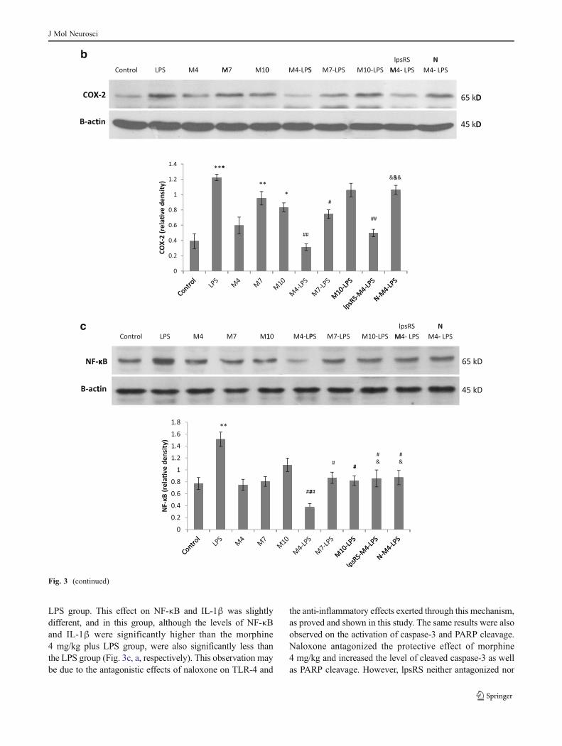
LPS group. This effect on NF-κB and IL-1β was slightlydifferent, and in this group, although the levels of NF-κBand IL-1β were significantly higher than the morphine4 mg/kg plus LPS group, were also significantly less thanthe LPS group (Fig. 3c, a, respectively). This observation maybe due to the antagonistic effects of naloxone on TLR-4 and
the anti-inflammatory effects exerted through this mechanism,as proved and shown in this study. The same results were alsoobserved on the activation of caspase-3 and PARP cleavage.Naloxone antagonized the protective effect of morphine4 mg/kg and increased the level of cleaved caspase-3 as wellas PARP cleavage. However, lpsRS neither antagonized nor
c
Fig. 3 (continued)
J Mol Neurosci

synergistically increased the anti-inflammatory and protectiveeffects of morphine (Fig. 4a, b).
Morphine Pretreatment Protects from LPS-Induced MemoryDeficit
In the present study, we also tried to investigate the effect ofmorphine on LPS-induced memory deficit. As it is shown inFig. 5, LPS significantly impaired the inhibitory avoidancememory retrieval and decreased the latency to enter the darkcompartment in comparison with the control group. Mor-phine injection in a noninflammatory context also impairedmemory retrieval (Fig. 5). Although all three doses of mor-phine significantly deceased the entry latency, this effectwas more pronounced at the doses of 4 and 7 mg/kg thanthe dose of 10 mg/kg. In spite of the potential amnesiceffect, morphine pretreatment at the doses of 4 and 10 mg/kg significantly prevented the LPS-induced memory deficit.In agreement with the above-described results, the protec-tive role of morphine on LPS-induced memory impairmentwas also reversed by naloxone (Fig. 5).
Discussion
In the current study, we showed the antineuroinflammatoryand memory recall-improving role of morphine precondi-tioning in LPS-induced neuroinflammation and also showedthat these effects are mediated through opioid receptors.
Acute peripheral infection has been demonstrated to induceneuroinflammatory responses in several studies (Chakravartyand Herkenham 2005; Cunningham et al. 2005). It is nowclear that, immediately after i.p. injection of LPS, it can berecognized by immune cells plasma membrane-boundedTLR-4 and TNF-α will be instantly secreted (Supajatura etal. 2001; Piliponsky et al. 2010). LPS/TLR-4 interaction hasbeen also proved in the CNS immediately following theexpression of proinflammatory cytokines during severe endo-toxemia (Laflamme and Rivest 2001). Our data showed asignificant increase in TNF-α and IL-1β levels in the rathippocampus 4 h after LPS administration and conceded theprevious studies and also our experiments to simulate neuro-inflammation. TNF-α and IL-1β are proinflammatory cyto-kines that are produced by microglia as well as in peripheraltissues (Merrill and Benveniste 1996; Bao et al. 2001).
Recently, it has been shown that naloxone, in addition to itsantagonistic effects on opioid receptors, possesses TLR-4antagonistic effects (Hutchinson et al. 2008) and has succes-sively calmed neuroinflammation in several studies (Liao etal. 2003; Lin et al. 2010). We assessed the role of naloxonepretreatment in LPS-induced neuroinflammation and, as wasexpected, it was able to suppress the neuroinflammatory
response as well as a TLR-4 antagonist. These data concededprevious documents about TLR-4 antagonistic and anti-inflammatory effects of naloxone and also raised the questionwhether the combination of naloxone with morphine willpotentiate or abolish the preconditioning effect of morphine.
Regarding the inflammatory versus anti-inflammatory andapoptotic versus protective effects, morphine has receivedintensive long-lasting studies. However, there is no data com-paring the in vivo effects of acute morphine injection innoninflammatory and inflammatory background. In this study,we showed that a single morphine injection at three differenttherapeutic doses exerts a proinflammatory response accom-panied with apoptotic cell death and passive avoidance mem-ory impairment. These results were expected because of thepotential agonistic effects of morphine on TLR-4 and activa-tion of microglial cells (Hutchinson et al. 2007). There are alsopieces of evidence showing the induction of apoptotic celldeath in the hippocampus and memory impairment in re-sponse to morphine (Spain and Newsom 1991; Zarrindast etal. 2006). It has been also demonstrated that chronic applica-tion of morphine triggers apoptosis in the rat spinal cord anddifferent brain areas (Boronat et al. 2001; Mao et al. 2002;Atici et al. 2004; Chen et al. 2008), in human and rodentperipheral immune system-related cells (Singhal et al. 1998;Yin et al. 1999), and in embryonic neuronal and glial cells (Huet al. 2002; Nasiraei-Moghadam et al. 2010). Our resultsproved that morphine induces a neuroinflammatory responseand apoptosis even in acute administration; however, thiseffect seems to be partly dose-dependent and to be enhancedas the morphine dose is increased. Furthermore, the results ofthis study showed that morphine administration before theinduction of a neuroinflammatory reaction exerts an oppositeprotective preconditioning effect. In general, preconditioning-induced protection describes a phenomenon in which a priorexposure to a stimulus or agent renders protection against asubsequent detrimental insult. For example, it has been shownthat mild activation of TLR-4 by LPS or TLR-9 by its ligand(CpG oligodeoxynucleotide) in different cerebral ischemiamodels reprograms the response to injury and improves theoutcome (Stevens et al. 2008; Vartanian et al. 2011). In theseexperiments, the preconditioning stimuli induce an inflamma-tory reaction, which occurs with morphine as well.
An essential feature of the early innate immune responseand activation of TLR-4 signaling by LPS is the secretion ofproinflammatory cytokines via the activation of signal trans-duction pathways, including NF-κB, which ultimately leads tothe transcription of chemokines, proinflammatory cytokines,and enzymes like IL-1β, TNF-α, and COX-2 (D’Acquisto etal. 1997; Liu and Malik 2006). Our results showed that NF-κBsignaling raises 4 h after LPS injection and is accompaniedwith the elevation of other inflammatory factors. The morphinepreconditioning effect prevents the activation of this
J Mol Neurosci

a
b
Fig. 4 Effects of morphine pretreatment on LPS-induced apoptoticcell death. The level of cleaved caspase-3 in the hippocampus celllysate and the level of PARP cleavage in the nuclear fraction weredetermined by Western blotting. a All the panels show the representa-tive immunoblots of the control group, LPS (1 mg/kg, i.p.) group,groups receiving morphine i.p. injections at the doses of 4, 7, and10 mg/kg (M4, M7, and M10, respectively) 30 min before i.p. PBSinjection, groups receiving morphine pretreatment at the doses of 4, 7,and 10 mg/kg (M4-LPS, M7-LPS, and M10-LPS, respectively) 30 minbefore i.p. LPS injection, group receiving morphine at the dose of4 mg/kg plus lpsRS (40 μg/rat, i.c.v.) 30 and 35 min, respectively,before LPS injection (lpsRS-M4-LPS), and group receiving morphineat the dose of 4 mg/kg 30 min before LPS plus naloxone (4 mg/kg, i.p.)35 min before and 85 min after LPS injection (N-M4-LPS). b The
panels show the representative immunoblots of the control group, LPS(1 mg/kg, i.p.) group, group receiving lpsRS (40 μg/rat; i.c.v.) 5 minbefore LPS (lpsRS-LPS), group receiving naloxone (4mg/kg; i.p.) 35minbefore and 120 min after LPS (N-LPS), group receiving morphine at thedose of 4 mg/kg 30 min before LPS (M4-LPS), and group receivingmorphine at the dose of 4 mg/kg 30 min before LPS plus naloxone(4 mg/kg, i.p.) 35 min before and 120 min after LPS injection (N-M4-LPS). The quantitative data, presented as the mean±SEM, were obtainedfrom densitometric analysis of scanned X-ray films and normalized tocorresponding immunoblots of β-actin as the loading control. *P<0.05,**P<0.01, and ***P<0.001 versus the control group; #P<0.05, ##P<0.01,and ###P<0.001 versus the LPS group; &P<0.05, &&P<0.01, and &&&P<0.001 versus the M4-LPS group
J Mol Neurosci

proinflammatory transcription factor in response to LPS chal-lenge and also reduces the expression of IL-1β, TNF-α, andCOX-2. Although there are some pieces of evidence showingthe induction of NF-κB signaling in response to chronicmorphine administration (Frässdorf et al. 2005; Roy et al.2006; El-Hage et al. 2008), we showed that a single morphineinjection causes no significant change in the basal level ofnuclear NF-κB in the noninflammatory context. In agreementwith other documents showing the induction of COX-2 inneuroinflammatory conditions (Giovannini et al. 2003; Sugi-moto and Iadecola 2003; Candelario-Jalil et al. 2005), ourresults showed that higher doses of morphine as well as LPSchallenge increases COX-2 expression in the rat hippocampusand that the morphine preconditioning effect prevents theinduction of COX-2 in response to LPS.
Caspase-3 is considered to be the main executioner caspaseinvolved in apoptotic cell death (Jänicke et al. 1998; Slee et al.2001) and the detection of an 89- or 24-kDa caspase cleavagefragment of PARP-1 is described as a hallmark of apoptosis(Lazebnik et al. 1994). Here, we showed that, 4 h after LPSchallenge, the level of cleaved caspase-3 as well as nuclearPARP cleavage is increased in the hippocampus and this is inagreement with other studies showing that LPS induces apo-ptosis (Comstock et al. 1998; Joshi et al. 2003). Although theapoptotic effects of morphine have been reported in a variety ofin vitro and in vivo models, there are some studies showing anantiapoptotic and protective effect of morphine preconditioningor postconditioning on ischemia (Okubo et al. 2004; Wang et
al. 2009). Here, we showed that, although an acute dose ofmorphine is able to induce apoptotic cell death in the rathippocampus, its preconditioning effect can protect the hippo-campus neuronal cells from the activation of caspase-3 andPARP cleavage. This is in contrast with a recent study showingthat morphine alone does not directly induce caspase activity,but enhances the LPS-induced caspase activity in spleen-,thymus-, and bone marrow-derived immune cells (Olin et al.2010).
There are pieces of evidence showing that learning andmemory processes can be affected by inflammatory reactionswithin the brain (Yirmiya and Goshen 2011). Some documentsalso mention a crucial role for NF-κB signaling in long-termmemory development (Albensi and Mattson 2000; Mattson etal. 2000; Kassed et al. 2002; Dash et al. 2005). There are alsosome studies revealing that pretraining and pretest administra-tion of morphine at the dose of 5 mg/kg, s.c., impairs thepassive avoidance memory retrieval (Zarrindast et al. 2006).Here, we showed that all three doses of morphine as well asLPS challenge impair the passive avoidance memory retentionand, interestingly, that morphine preconditioning can protectthe rats from LPS-induced memory deficit. Other investigatorshave also showed that morphine preconditioning or postcon-ditioning in ischemia/reperfusion models can improve bothshort-term and long-term memory performance (Pateliya etal. 2008; Rehni et al. 2008).
After confirming the anti-inflammatory and protective roleof morphine in LPS-induced neuroinflammation at both be-havioral and molecular levels, we used naloxone to evaluate ifthese effects are mediated through opioid receptors. Our resultsshowed that all of the protective preconditioning effects ofmorphine can be blocked with the opioid receptor antagonist.There are also some documents showing that the precondition-ing effect of morphine in ischemia/reperfusion models can beattenuated by naloxone and is dependent on the activation ofopioid receptors (McPherson and Yao 2001; Okubo et al.2004; Gwak et al. 2010). Regarding the fact that naloxone, inaddition to its antagonistic effects on opioid receptors, has alsothe same effects on TLR-4 (Hutchinson et al. 2007, 2010a, b),the conversion of the protective role of morphine may be dueto the blockage of TLR-4 by naloxone and exertion of theinnate proinflammatory and neurotoxic role of morphine. Toexclude this issue and be sure that the conversion of theprotective role of morphine by naloxone is only because ofopioid receptor blockage, we used a pure TLR-4 antagonist.Interestingly, the concurrent use of lpsRSwithmorphine neitherblocked the protective effect of morphine nor additively orsynergistically enhanced it. It, therefore, confirmed that thepreconditioning effect of morphine in LPS-induced neuroin-flammation is mediated through opioid receptors. In addition,the observation that the TLR-4 antagonist did not enhance theprotective effect of morphine can further emphasize that thepreconditioning effect of morphine is highly due to the
Fig. 5 Effects of morphine on passive avoidance memory retrieval innoninflammatory and inflammatory contexts. Twenty hours after passiveavoidance training, rats were assigned to ten groups and given injections ofeither BPS or LPS (1 mg/kg) with or without morphine and/or morphineplus naloxone or lpsRS pretreatments. The entry latency (mean±SEM)into the dark compartment was assessed 4 h later. Shorter entrance latencyindicates impaired memory retention. Morphine injection at the doses of 4,7, and 10 mg/kg as well as LPS impaired the passive avoidance memoryretrieval. Morphine pretreatment at the doses of 4 and 10 mg/kg protectedthe rats from LPS-induced memory deficit. Naloxone, but not lpsRS,halted the protective role of morphine 4 mg/kg in LPS-injected rats. *P<0.05 and ***P<0.001 versus the control group; ##P<0.01 and ###P<0.001versus the LPS group; &&P<0.01 versus the M4-LPS group
J Mol Neurosci

activation of opioid receptor signaling pathways, not because ofa simple competition between morphine and LPS on TLR-4.
Conclusion
It can be concluded that morphine has a preconditioning effecton LPS-induced neuroinflammation. Although morphine hasproinflammatory, neurotoxic, and amnesic effects in a nonin-flammatory context, it can prevent the induction of neuroin-flammatory response by LPS and also protect the hippocampalcells from apoptosis and also passive avoidance memory deficitin rats. The preconditioning protective role of morphine isdependent on opioid receptors. However, regarding the poten-tial effect of morphine on TLR-4, further studies are required todelineate whymorphine has no additive effects on LPS-inducedneuroinflammation and reversely can prevent it.
Acknowledgments The authors are grateful to the NeuroscienceResearch Center of Shahid Beheshti University of Medical Sciencesfor the financial support of this study.
References
Albensi BC, Mattson MP (2000) Evidence for the involvement of TNFand NF B in hippocampal synaptic plasticity. Synapse 35:151–159
Askari N, Mahboudi F, Haeri Rohani A et al (2008) Effects of singleadministration of morphine on G protein mRNA level in thepresence and absence of inflammation in the rat spinal cord.Scand J Immunol 67:47–52
Atici S, Cinel L, Cinel I et al (2004) Opioid neurotoxicity: comparisonof morphine and tramadol in an experimental rat model. Int JNeurosci 114:1001–1011
Bao L, Zhu Y, Elhassan AM et al (2001) Adjuvant-induced arthritis:IL-1 [beta], IL-6 and TNF-[alpha] are up-regulated in the spinalcord. Neuroreport 12:3905
Barry U, Zuo Z (2005) Opioids: old drugs for potential new applications.Curr Pharm Des 11:1343–1350
Boronat MA, García-Fuster MJ, García-Sevilla JA (2001) Chronicmorphine induces up–regulation of the pro–apoptotic Fas receptorand down–regulation of the anti-apoptotic Bcl-2 oncoprotein inrat brain. Br J Pharmacol 134:1263–1270
Brown GC, Bal-Price A (2003) Inflammatory neurodegeneration medi-ated by nitric oxide, glutamate, and mitochondria. Mol Neurobiol27:325–355
Candelario-Jalil E, Mhadu N, González-Falcón A et al (2005) Effectsof the cyclooxygenase-2 inhibitor nimesulide on cerebral infarctionand neurological deficits induced by permanent middle cerebralartery occlusion in the rat. J Neuroinflammation 2:3
Chakravarty S, Herkenham M (2005) Toll-like receptor 4 on nonhe-matopoietic cells sustains CNS inflammation during endotoxemia,independent of systemic cytokines. J Neurosci 25:1788
Chen Q, Cui J, Zhang Y, Yu LC (2008) Prolonged morphine applicationmodulates Bax and Hsp70 levels in primary rat neurons. NeurosciLett 441:311–314
Comstock KL, Krown KA, Page MT et al (1998) LPS-induced TNF-[alpha] release from and apoptosis in rat cardiomyocytes: obliga-tory role for CD14 in mediating the LPS response. J Mol CellCardiol 30:2761–2775
Cunningham C, Wilcockson DC, Campion S, Lunnon K, Perry VH(2005) Central and systemic endotoxin challenges exacerbate thelocal inflammatory response and increase neuronal death duringchronic neurodegeneration. J Neurosci 25:9275
D’Acquisto F, Iuvone T, Rombolą L, Sautebin L, Di Rosa M, CarnuccioR (1997) Involvement of NF-[kappa] B in the regulation ofcyclooxygenase-2 protein expression in LPS-stimulated J774 mac-rophages. FEBS Lett 418:175–178
Dash PK, Orsi SA, Moore AN (2005) Sequestration of serum responsefactor in the hippocampus impairs long term spatial memory. JNeurochem 93:269–278
El-Hage N, Bruce-Keller AJ, Yakovleva T et al (2008) Morphineexacerbates HIV-1 Tat-induced cytokine production in astrocytesthrough convergent effects on [Ca2+]i, NF-κB trafficking andtranscription. PLoS One 3:e4093
Frässdorf J, Weber NC, Obal D et al (2005) Morphine induces latecardioprotection in rat hearts in vivo: the involvement of opioidreceptors and nuclear transcription factor B. Anesth Analg101:934
Gidday JM, Fitzgibbons JC, Shah AR, Park T (1994) Neuroprotectionfrom ischemic brain injury by hypoxic preconditioning in theneonatal rat. Neurosci Lett 168:221–224
Giovannini M, Scali C, Prosperi C, Bellucci A, Pepeu G, Casamenti F(2003) Experimental brain inflammation and neurodegenerationas model of Alzheimer’s disease: protective effects of selectiveCOX-2 inhibitors. Int J Immunopathol Pharmacol 16:31
Gwak MS, Li L, Zuo Z (2010) Morphine preconditioning reduceslipopolysaccharide and interferon-[gamma]-induced mousemicroglial cell injury via [delta] 1 opioid receptor activation.Neuroscience 167:256–260
Hoshino K, Tsutsui H, Kawai T et al (1999) Cutting edge: generationof IL-18 receptor-deficient mice: evidence for IL-1 receptor-related protein as an essential IL-18 binding receptor. J Immunol162:5041
Hu S, Sheng WS, Lokensgard JR, Peterson PK (2002) Morphineinduces apoptosis of human microglia and neurons. Neurophar-macology 42:829–836
Hutchinson MR, Bland ST, Johnson KW, Rice KC, Maier SF, WatkinsLR (2007) Opioid-induced glial activation: mechanisms of activa-tion and implications for opioid analgesia, dependence, and reward.Sci World 98:111
Hutchinson M, Coats B, Lewis S et al (2008) Proinflammatory cyto-kines oppose opioid-induced acute and chronic analgesia. Brain,Behav, Immun 22:1178–1189
Hutchinson MR, Loram LC, Zhang Y et al (2010a) Evidence thattricyclic small molecules may possess toll-like receptor andmyeloid differentiation protein 2 activity. Neuroscience 168:551–563
Hutchinson MR, Zhang Y, Shridhar M et al (2010b) Evidence thatopioids may have toll-like receptor 4 and MD-2 effects. Brain,Behav, Immun 24:83–95
Jänicke RU, Sprengart ML, Wati MR, Porter AG (1998) Caspase-3 isrequired for DNA fragmentation and morphological changesassociated with apoptosis. J Biol Chem 273:9357
Joshi VD, Kalvakolanu DV, Cross AS (2003) Simultaneous activationof apoptosis and inflammation in pathogenesis of septic shock: ahypothesis1. FEBS Lett 555:180–184
Kao TK, Ou YC, Liao SL et al (2008) Opioids modulate post-ischemicprogression in a rat model of stroke. Neurochem Int 52:1256–1265
Kassed C, Willing A, Garbuzova-Davis S, Sanberg P, Pennypacker K(2002) Lack of NF-[kappa] B p50 exacerbates degeneration ofhippocampal neurons after chemical exposure and impairs learning.Exp Neurol 176:277–288
Kieffer BL (1995) Recent advances in molecular recognition and signaltransduction of active peptides: receptors for opioid peptides. CellMol Neurobiol 15:615–635
J Mol Neurosci

Kim MS, Cheong YP, So HS et al (2001) Protective effects of morphinein peroxynitrite-induced apoptosis of primary rat neonatal astro-cytes: potential involvement of G protein and phosphatidylinositol3-kinase (PI3 kinase). Biochem Pharmacol 61:779–786
Kreutzberg GW (1996) Microglia: a sensor for pathological events inthe CNS. Trends Neurosci 19:312–318
Lacroix S, Feinstein D, Rivest S (1998) The bacterial endotoxinlipopolysaccharide has the ability to target the brain in upregulatingits membrane CD14 receptor within specific cellular populations.Brain Pathol 8:625–640
Laflamme N, Rivest S (2001) Toll-like receptor 4: the missing link ofthe cerebral innate immune response triggered by circulatinggram-negative bacterial cell wall components. FASEB J 15:155
Lazebnik YA, Kaufmann SH, Desnoyers S, Poirier GG, Earnshaw WC(1994) Cleavage of poly(ADP-ribose) polymerase by a proteinasewith properties like ICE. Nature 371:346–347
Liao S, Chen W, Raung S, Chen C (2003) Neuroprotection of naloxoneagainst ischemic injury in rats: role of mu receptor antagonism.Neurosci Lett 345:169–172
Lim YJ, Zheng S, Zuo Z (2004) Morphine preconditions Purkinje cellsagainst cell death under in vitro simulated ischemia–reperfusionconditions. Anesthesiology 100:562
Lin HY, Huang CC, Chang KF (2009) Lipopolysaccharide precondition-ing reduces neuroinflammation against hypoxic ischemia and pro-vides long-term outcome of neuroprotection in neonatal rat. PediatrRes 66:254
Lin S, Tsai R, Tai Y et al (2010) Ultra-low dose naloxone upregulatesinterleukin-10 expression and suppresses neuroinflammation inmorphine-tolerant rat spinal cords. Behav Brain Res 207:30–36
Liu SF, Malik AB (2006) NF-κB activation as a pathological mechanismof septic shock and inflammation. Am J Physiol-Lung Cell MolPhysiol 290:L622–L645
Lynn W, Golenbock D (1992) Lipopolysaccharide antagonists. ImmunolToday 13:271–276
Madera-Salcedo IK, Cruz SL, Gonzalez-Espinosa C (2011) Morphinedecreases early peritoneal innate immunity responses in Swiss-Webster and C57BL6/J mice through the inhibition of mast cellTNF-[alpha] release. J Neuroimmunol 232:101–107
Mao J, Sung B, Ji RR, Lim G (2002) Neuronal apoptosis associatedwith morphine tolerance: evidence for an opioid-induced neuro-toxic mechanism. J Neurosci 22:7650
Mattson MP, Culmsee C, Yu ZF, Camandola S (2000) Roles of nuclearfactor B in neuronal survival and plasticity. J Neurochem 74:443–456
McPherson BC, Yao Z (2001) Morphine mimics preconditioning viafree radical signals and mitochondrial KATP channels in myocytes.Circulation 103:290–295
Merrill JE, Benveniste EN (1996) Cytokines in inflammatory brainlesions: helpful and harmful. Trends Neurosci 19:331–338
Minami M, Satoh M (1995) Molecular biology of the opioid receptors:structures, functions and distributions. Neurosci Res 23:121–145
Mucke L, Eddleston M (1993) Astrocytes in infectious and immune-mediated diseases of the central nervous system. FASEB J 7:1226
Nandagopal K, Dawson TM, Dawson VL (2001) Critical role for nitricoxide signaling in cardiac and neuronal ischemic preconditioningand tolerance. J Pharmacol Exp Ther 297:474
Nasiraei-Moghadam S, Kazeminezhad B, Dargahi L, Ahmadiani A(2010) Maternal oral consumption of morphine increases Bax/Bcl-2ratio and caspase 3 activity during early neural system developmentin rat embryos. J Mol Neurosci 41:156–164
Okubo S, Tanabe Y, Takeda K et al (2004) Ischemic preconditioningand morphine attenuate myocardial apoptosis and infarction afterischemia–reperfusion in rabbits: role of delta-opioid receptor. AmJ Physiol Heart Circ Physiol 287:H1786
Olin MR, Roy S, Molitor T (2010) In vivo morphine treatment syner-gistically increases LPS-induced caspase activity in immuneorgans. J Neuroimmune Pharm 5:546–552
Pak T, Cadet P, Mantione KJ, Stefano GB (2005) Morphine via nitricoxide modulates beta-amyloid metabolism: a novel protectivemechanism for Alzheimer’s disease. Med Sci Monit 11:BR357–BR366
Pakpour B, Ahmadi S, Nayer-Nouri T, Oryan S, Zarrindast MR (2010)Inhibitory avoidance memory deficit induced by scopolamine:interaction with glutamatergic system in the nucleus accumbens.Behav Pharmacol 21:719
Pateliya BB, Singh N, Jaggi AS (2008) Possible role of opioids and KATP channels in neuroprotective effect of postconditioning inmice. Biol Pharm Bull 31:1755–1760
Paxinos G, Watson C (2007) The rat brain in stereotaxic coordinate,sixth ed. Academic press, New York
Piliponsky AM, Chen CC, Grimbaldeston MA et al (2010) Mast cell-derived TNF can exacerbate mortality during severe bacterialinfections in C57BL/6-KitW-sh/W-sh mice. Am J Pathol 176:926
Pourpak Z, Ahmadiani A, Alebouyeh M (2004) Involvement of inter-leukin 1 in systemic morphine effects on paw oedema in a mousemodel of acute inflammation. Scand J Immunol 59:273–277
Pradillo JM, Romera C, Hurtado O et al (2005) TNFR1 upregulationmediates tolerance after brain ischemic preconditioning. J CerebBlood Flow Metab 25:193–203
Qian L, Tan K, Wei S et al (2007) Microglia-mediated neurotoxicity isinhibited by morphine through an opioid receptor-independentreduction of NADPH oxidase activity. J Immunol 179:1198
Qureshi S, Larivière L, Leveque G et al (1999) Endotoxin-tolerant micehavemutations in Toll-like receptor 4 (Tlr4). The J ExpMed 189:615
Rambhia S, Mantione KJ, Stefano GB, Cadet P (2005) Morphinemodulation of the ubiquitin–proteasome complex is neuroprotec-tive. Med Sci Monit: Int Med J Exp Clin Res 11:BR386
Rehni AK, Singh TG, Jaggi AS, Singh N (2008) Pharmacologicalpreconditioning of the brain: a possible interplay between opioidand calcitonin gene related peptide transduction systems. PharmacolRep 60:904–913
Roy S, Wang J, Kelschenbach J, Koodie L, Martin J (2006) Modulationof immune function by morphine: implications for susceptibility toinfection. J Neuroimmune Pharmacol 1:77–89
Singhal PC, Sharma P, Kapasi AA, ReddyK, Franki N, Gibbons N (1998)Morphine enhances macrophage apoptosis. J Immunol 160:1886
Slee EA, Adrain C, Martin SJ (2001) Executioner caspase-3, -6, and -7perform distinct, non-redundant roles during the demolition phaseof apoptosis. J Biol Chem 276:7320
Spain JW, Newsom GC (1991) Chronic opioids impair acquisition ofboth radial maze and Y-maze choice escape. Psychopharmacology105:101–106
Stevens SL, Ciesielski TM, Marsh BJ et al (2008) Toll-like receptor 9:a new target of ischemic preconditioning in the brain. J CerebBlood Flow Metab 28:1040–1047
Sugimoto K, Iadecola C (2003) Delayed effect of administration ofCOX-2 inhibitor in mice with acute cerebral ischemia. Brain Res960:273–276
Supajatura V, Ushio H, Nakao A, Okumura K, Ra C, Ogawa H (2001)Protective roles of mast cells against enterobacterial infection aremediated by Toll-like receptor 4. J Immunol 167:2250
Takeuchi O, Akira S (2001) Toll-like receptors; their physiological roleand signal transduction system. Int Immunopharmacol 1:625–635
Turrin NP, Rivest S (2006) Molecular and cellular immune mediatorsof neuroprotection. Mol Neurobiol 34:221–242
Ulevitch R, Tobias P (1995) Receptor-dependent mechanisms of cellstimulation by bacterial endotoxin. Annu Rev Immunol 13:437–457
Vartanian KB, Stevens SL, Marsh BJ, Williams-Karnesky R, LessovNS, Stenzel-Poore MP (2011) LPS preconditioning redirects TLRsignaling following stroke: TRIF-IRF3 plays a seminal role inmediating tolerance to ischemic injury. J Neuroinflammation 8:140
Wang Z, Kang J, Li Y, Yuan Z, Liu S, Sun L (2006) The effectsof dexamethasone on rat brain cortical nuclear factor kappa
J Mol Neurosci

B (NF-[kappa] B) in endotoxic shock. Toxicol Appl Pharmacol214:263–269
Wang Z, Zhao H, Peng J et al (2009) Anti-apoptotic effect of morphinepostconditioning on myocardial ischemia–reperfusion injury andits anti-oxidative stress mechanism. J Sun Yat-Sen Univ (MedSci) 5
Watkins LR, Hutchinson MR, Rice KC, Maier SF (2009) The “toll” ofopioid-induced glial activation: improving the clinical efficacy ofopioids by targeting glia. Trends Pharmacol Sci 30:581–591
Wright SD (1999) Toll, a new piece in the puzzle of innate immunity.The J Exp Med 189:605
Yin D, Mufson RA, Wang R, Shi Y (1999) Fas-mediated cell deathpromoted by opioids. Nature 397:218
Yirmiya R, Goshen I (2011) Immune modulation of learning, memory,neural plasticity and neurogenesis. Brain, Behav, Immun 25:181–213
Zarrindast MR, Fazli-Tabaei S, Ahmadi S, Yahyavi SH (2006) Effectof lithium onmorphine state-dependent memory of passive avoidancein mice. Physiol Behav 87:409–415
Zhao P, Huang Y, Zuo Z (2006) Opioid preconditioning induces opioidreceptor-dependent delayed neuroprotection against ischemia inrats. J Neuropathol Exp Neurol 65:945
Zujovic V, Schussler N, Jourdain D, Duverger D, Taupin V (2001) Invivo neutralization of endogenous brain fractalkine increases hip-pocampal TNF [alpha] and 8-isoprostane production induced byintracerebroventricular injection of LPS. J Neuroimmunol 115:135–143
J Mol Neurosci
Copyright © 2022 FDOKUMEN