Sfrp1 promotes neuroinflammation through the modulation of ...
109
Universidad Autónoma de Madrid Facultad de Ciencias Departamento de Biología Molecular Sfrp1 promotes neuroinflammation through the modulation of ADAM10 proteolytic activity PhD Thesis Javier Rueda Carrasco Madrid, 2017
-
Upload
khangminh22 -
Category
Documents
-
view
1 -
download
0
Transcript of Sfrp1 promotes neuroinflammation through the modulation of ...
Universidad Autónoma de Madrid
Facultad de Ciencias
Departamento de Biología Molecular
Sfrp1 promotes neuroinflammation through the modulation of ADAM10
proteolytic activity
PhD Thesis
Javier Rueda Carrasco
Madrid, 2017
Universidad Autónoma de Madrid
Facultad de Ciencias
Departamento de Biología Molecular
Sfrp1 promotes neuroinflammation through the modulation of ADAM10 proteolytic activity
Trabajo de Investigación que presenta
Javier Rueda Carrasco
Licenciado en Biotecnología
para optar al título de Doctor en Ciencias
por la Universidad Autónoma de Madrid.
Madrid, 2017
Fdo. Javier Rueda Carrasco
Codirigido por la Doctora
Paola Bovolenta Nicolao
Profesor de Investigación del CSIC
Fdo. Dra. Paola Bovolenta
Codirigido por la Doctora
Pilar Esteve Pastor
Científico titular del CSIC
Fdo. Dra. Pilar Esteve
Trabajo realizado en el Centro de Biología Molecular “Severo Ochoa”

INDEX
INDEX 5
INDEX ............................................................................................................................................................ 3
PRESENTACIÓN ............................................................................................................................................. 9
ABSTRACT ................................................................................................................................................... 13
ABBREVIATIONS .......................................................................................................................................... 17
INTRODUCTION .......................................................................................................................................... 23
1. Immune response in the Central Nervous System ......................................................................... 25
2. Cellular mediators of Neuroinflammation ..................................................................................... 27
Astrocytes .................................................................................................................................. 27
Microglia .................................................................................................................................... 29
3. Neuroimmune Regulators .............................................................................................................. 32
CX3CR1 ...................................................................................................................................... 33
CD200R ...................................................................................................................................... 33
TREM2 ....................................................................................................................................... 34
4. Models of Neuroinflammation ...................................................................................................... 35
Lipopolysaccharide induced Neuroinflammation ...................................................................... 35
Neuroinflammation associated with CNS autoimmunity .......................................................... 36
Neuroinflammation associated with Neurodegeneration ........................................................ 37
5. Secreted Frizzled-Related Protein 1 ............................................................................................... 39
Roles of Sfrp1 as regulator of cell-cell communication ............................................................. 40
Roles of Sfrp1 in pathogenesis .................................................................................................. 42
Implication of Sfrp1 in Alzheimer’s Disease .............................................................................. 43
OBJECTIVES ................................................................................................................................................. 47
MATERIALS AND METHODS........................................................................................................................ 51
1. Animals ........................................................................................................................................... 53
2. Intra-cerebral infusion ................................................................................................................... 53
6 Sfrp1 promotes neuroinflammation through the modulation of ADAM10 proteolytic activity
3. Experimental Autoimmune Encephalomyelitis .............................................................................. 54
4. Primary cultures ............................................................................................................................. 55
5. qRT-PCR .......................................................................................................................................... 56
6. Immunohistochemistry .................................................................................................................. 57
7. Western Blot and Immunoprecipitation ........................................................................................ 59
8. Analysis of ADAM10 activity .......................................................................................................... 60
9. ELISA ............................................................................................................................................... 61
RESULTS ...................................................................................................................................................... 65
1. Sfrp1 acts as a pro-inflammatory molecule in vitro ...................................................................... 67
Sfrp1 is expressed by glial cells .................................................................................................. 67
Pro-inflammatory stimuli induce Sfrp1 expression ................................................................... 68
Lack of Sfrp1 reduces cytokine production and secretion ........................................................ 69
Microglial activation is reduced in the absence of Sfrp1 ........................................................... 71
Sfrp1 addition is sufficient to activate microglial cells .............................................................. 74
2. Sfrp1 is necessary and sufficient to induce an inflammatory response in vivo ............................ 75
LPS induces Sfrp1 expression in vivo ......................................................................................... 76
Sfrp1 null mice present a poor response to LPS ........................................................................ 77
Sfrp1 null mice present normal glial development ................................................................... 80
Sfrp1 gene addition induces an inflammatory response ........................................................... 81
3. Sfrp1 exacerbated the symptomatology of EAE ............................................................................ 83
Sfrp1 expression is upregulated in the spinal cord of mice with EAE........................................ 84
Sfrp1 null mice develop a milder form of EAE ........................................................................... 85
4. Sfrp1 regulates microglial activation by modulating ADAM10 proteolytic activity ....................... 88
Sfrp1 regulates ADAM10 activity present in microglial cells ..................................................... 88
Sfrp1 potentiates ADAM10 shedding of TREM2 ....................................................................... 90
5. Sfrp1 neutralization as therapeutic target ..................................................................................... 91
Sfrp1 immunosuppression counteracts AD progression ........................................................... 91
INDEX 7
DISCUSSION ................................................................................................................................................ 95
1. Sfrp1 promotes a reactive state of microglial cells ........................................................................ 98
Sfrp1 contributes to exacerbate neuroinflammatory responses .............................................. 98
Sfrp1 may prime microglia cells predisposing them to activation .......................................... 100
2. Sfrp1 modulates microglial activity through ADAM10................................................................. 101
Sfrp1 modulates processing of ADAM10 immune-related substrates .................................... 101
Sfrp1 modulates ADAM10 activity in a substrate-specific manner ......................................... 103
3. Sfrp1 as therapeutic target to counteract neuroinflammation ................................................... 105
4. Future perspectives: Possible implication of Sfrp1 in age-related diseases ................................ 106
CONCLUSIONS .......................................................................................................................................... 111
CONCLUSIONES ........................................................................................................................................ 115
BIBLIOGRAPHY .......................................................................................................................................... 119

PRESENTACIÓN
PRESENTACIÓN 11
Una creciente evidencia indica la importancia de la respuesta inmune en la regulación
y el mantenimiento de la homeostasis del tejido neural. La pérdida de esta homeostasis
puede estar entre las posibles causas que contribuyen a la aparición o el desarrollo de
desórdenes neurológicos. Los procesos inflamatorios en el Sistema Nervioso Central
(SNC) están mediados por astrocitos y microglia, protegiéndolo frente a cualquier tipo
de daño en el intento de reparar el tejido. Sin embargo, cuando estos procesos
inflamatorios son exacerbados o dilatados en el tiempo inducen neurotoxicidad. La
neuroinflamación crónica ha sido implicada como uno de los detonantes de las diversas
enfermedades neurodegenerativas y autoinmunes que atacan al SNC.
Trabajo previo de nuestro laboratorio ha demostrado que Sfrp1 contribuye a la
progresión de la enfermedad de Alzheimer (EA), promoviendo la generación de péptidos
Aβ. Durante este estudio observamos que la inactivación genética de Sfrp1 va asociada
a unos niveles reducidos de inflamación. Esto unido a varios estudios que relacionan
altos niveles de expresión de Sfrp1 con enfermedades preifériacas asociadas a una
inflamación crónica, nos llevó a hipotetizar que Sfrp1 podría estar directamente
implicado en la regulación de la neuroinflamación.
Esta cuestión ha sido abordada a lo largo de esta tesis, aportando evidencias que la
sustentan. Hemos demostrado que la expresión de Sfrp1 se ve incrementada en
migroglia y astrocitos bajo diversas condiciones experimentales, usando modelos
animales de neuroinflamación, de EA o Encefalomielitis Autoinmune Experimental. Por
el contrario, la inactivación genética de Sfrp1 reduce severamente la activación glial,
provocando una mejora de los signos patológicos de los modelos. En cambio, la
sobreexpresión de Sfrp1 es suficiente para inducir una respuesta inflamatoria, mientras
que estudios preliminares indicamos que la neutralización de la actividad de Sfrp1
mejora la patología de la EA. En relación al posible mecanismo de acción, Sfrp1 parece
promover la neuroinflamación al regular el procesamiento de varios sustratos de
ADAM10 implicados en la activación de las células microgliales: TREM2, CD200 y CX3CL1.
Por tanto, proponemos una implicación directa de Sfrp1 en la modulación de la
actividad microglial durante la neuroinflamación, lo que sugiere que podría representar
una nueva diana terapéutica para atenuar los exacerbados procesos neuroinflamatorios
presentes en numerosas enfermedades neurodegenerativas.

ABSTRACT
ABSTRACT 15
Growing evidence suggests the importance of immune response regulation for the
maintenance of neural tissue homeostasis. Disruption of this homeostasis might be one
of the causes contributing to the onset and development of neurological disorders.
Inflammatory responses in the Central Nervous System (CNS) are mediated by
astrocytes and microglial cells, which help to protect from pathogen invasion and
respond to any kind of injury, in the attempt to repair the tissue. However, exacerbated
inflammatory responses lead to pathogenic neurotoxicity and chronic
neuroinflammation. The latter has been recognized as one of the drivers of diverse
neurodegenerative and autoimmune diseases of the CNS.
Previous work from our laboratory has demonstrated that Sfrp1 contributes to AD
progression by inducing Aβ peptide generation. In the course of this study, we also
observed that genetic inactivation of Sfrp1 was associated with particularly low levels of
neuroinflammation. Because an increased Sfrp1 expression has been reported in several
peripheral diseases associated with chronic inflammation, we hypothesised that Sfrp1
could directly contribute to the regulation of neuroinflammation.
In this thesis, we have addressed this issue, providing evidence that support this
hypothesis. Indeed, we show that Sfrp1 expression is upregulated in activated microglial
cells and reactive astrocytes under diverse experimental pro-inflammatory conditions,
including experimentally induced neuroinflammation, in mouse models for Alzheimer’s
Disease (AD) and in Experimental Autoimmune Encephalomyelitis. On the contrary,
genetic inactivation of Sfrp1 strongly reduces glial cells activation, ameliorating the
pathological traits of the diseases. Sfrp1 overexpression is sufficient to induce an
inflammatory response, activating glial cells and promoting the infiltration of immune
cells, whereas preliminary studies indicate that antibody-mediated neutralization of
Sfrp1 activity ameliorates AD pathological traits. From a mechanistic point of view, Sfrp1
seems to promote neuroinflammation by regulating ADAM10-mediated shedding of
TREM2, CD200 and CX3CL1, proteins implicated in the activation of microglial cells.
We thus propose that Sfrp1 is directly involved in modulating microglial activation
during brain inflammation. We also suggest that Sfrp1 may represent a new therapeutic
target to attenuate the exacerbated neuroinflammation present in numerous
neurodegenerative diseases.

ABBREVIATIONS
ABBREVIATIONS 19
AD Alzheimer's Disease
ADAM A Disintregrin And Metalloprotease ApoE Apolipoprotein E APP Amyloid Precursor Protein Arg1 Arginse 1 Aβ Amyloid Beta BACE β-site APP Cleaving Enzyme BBB Blood Brain Barrier BSA Bovine Serum Albumin Ca Calcium cc Corpus Callosum CD Cluster of Differentiation CNS Central Nervous System
CO2 Carbon Dioxide CR Complement Receptor CRD Cysteine Rich Domain
CSF Colony-Stimulating Factor DAMP Damage-Associated Molecular Pattern DAP12 DNAX Activation Protein 12 DMEM Dulbecco’s modified Eagle medium DNA DeoxyriboNucleic Acid E Embryonic day EAE Experimental Autoimmune Encephalomyelitis EDTA EthyleneDiamineTetraacetic Acid EGTA EthyleneGlycolTetraacetic Acid ELISA Enzyme-Linked ImmunoSorbent Assay
Fc Fragment crystallisable FCS Fetal Calf Serum Fz Frizzled
GAPDH GlycerAldehyde 3-Phosphate DeHydrogenase GFAP Glial Fibrillary Acid Protein GFP Green Fluorescent Protein GSK Glycogen Synthase Kinase H2O2 Hydrogen Peroxide HBSS Hank’s Balanced Salt Solution HCl HydroChloric acid HEK Human Embryonic Kidney cells Iba1 Ionized calcium Binding Adaptor 1
IFN Interferon IgG Immunoglobulin G IL Interleukin iNOS inducible Nitric Oxide Synthase IRES Internal Ribosome Entry Site ITAM Immune-receptor Tyrosine-based Activator Motive ITIM Immune-receptor Tyrosine-based Inhibition Motive L Ligand
20 Sfrp1 promotes neuroinflammation through the modulation of ADAM10 proteolytic activity
LPS Lipopolysaccharide
LV Lateral Ventricle Mac1 Macrophage-1 antigen (AKA CD11b) MBP Myelin Basic Protein Mg Magnesium MHC Major Histocompatibility Complex MOG Myelin Oligodendrocyte Glycoprotein mRNA messenger RNA MS Multiple Sclerosis NaCl Sodium Chloride NFκB Nuclear Factor Kappa-chain-enhancer of activated B cells NICD Notch IntraCellular Domain NSC Neural Stem Cell
NTR Netrin Related Domain P Postnatal day P- Phosphorylated PAMP Pathogen-Associated Molecular Pattern PBS Phosphate Buffer Saline PBSTw PBS Tween-20 PBT PBS Triton X-100 PCOLCE type-1 ProCOLlagen C-proteinase Enhancer protein PCR Polymerase Chain Reaction PFA ParaFormAldehyde PLP ProteoLipid Protein POD PerOxiDase PrP Prion Protein
PS Presinilin qPCR quantitative PCR qRT-PCR Real Time qPCR
R Receptor RANK Receptor Activator of NFκB RG Radial Glia rhSFRP1 recombinant human SFRP1 RNA RiboNucleic Acid SASP Senescence-Associated Secretory Phenotype Sfrp Secreted Frizzled Related Protein sTREM soluble TREM2 SVZ Sub-Ventricular Zone TBS Tris-Buffered Saline
TBST TBS Tween-20 TGFβ Transforming Growth Factor β TIMP Tissue Inhibitor of MetalloProteases TioS Thioflavin S TLR Toll-Like Receptor TNFα Tumour Necrosis Factor α TREM2 Triggering Receptor Expressed on Myeloid cells 2

INTRODUCTION
INTRODUCTION 25
The Central Nervous System (CNS) might represent the most complex entity in
existence. It derives from the embryonic neural plate, which subsequently folds to form
the neural tube. The neural tube is then patterned to form the eyes, brain and spinal
cord. Within the CNS, numerous cell types behave in concordance to maintain its
integrity and functions, intermingled in between a well-organised network of neurons,
glia and endothelial cells in contact to each other. In order to preserve neural
homeostasis, glial cells regulate diverse immune processes to secure proper functioning
of the CNS. When dysfunctional, detrimental immune processes favour the progression
of neurological disorders, interfering with motor control, perception, learning and
memory, which lead to the incapacitation of the individual. Furthermore, aging
represents a risk factor for many neurodegenerative events. In the developed world, the
increasing cohort of aged individuals make neuroinflammation an important therapeutic
target to make long lives worth living.
1. Immune response in the Central Nervous System
For a long time the CNS and the immune system have been thought to behave as two
independent and isolated systems (Medawar, 1948). But nowadays, this dogma appears
to be no longer valid as there is growing evidence that the two systems are
interconnected and synergize to regulate CNS homeostasis. Indeed, the activation and
infiltration of immune mediators into the CNS has been widely described under
homeostatic and pathological conditions and alterations of this crosstalk have been
correlated to the onset and progression of neurodegenerative diseases and aging (Lucin
& Wyss-Coray, 2009).
This dogma was based on the idea that the CNS was completely isolated from the
immune system and therefore this absolute isolation made the CNS considered as
immune privileged, suggesting that immune processes were of no importance in the
CNS. The Blood-Brain Barrier (BBB) was the only responsible for this isolation, shielding
the CNS from the entry of infectious agents. However, CNS immune privilege is now
known to be relative and the innate immune system has been shown to be active in the
CNS. Immune system functions are mediated by a complex crosstalk that involves every
cell type within the CNS (Lampron et al., 2013). More precisely, several observations
have described an active contribution of the BBB to the immune response. Importantly,
26 Sfrp1 promotes neuroinflammation through the modulation of ADAM10 proteolytic activity
an organised modulation of the permeability of the BBB allows peripheral immune cells
to cross the intact BBB (Carson et al., 2006), and afterwards the BBB modulates the
activity of infiltrated cells (Ifergan et al., 2008).
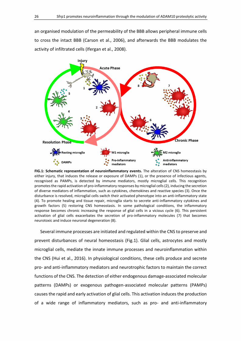
FIG.1: Schematic representation of neuroinflammatory events. The alteration of CNS homeostasis by either injury, that induces the release or exposure of DAMPs (1), or the presence of infectious agents, recognised as PAMPs, is detected by immune mediators, mostly microglial cells. This recognition promotes the rapid activation of pro-inflammatory responses by microglial cells (2), inducing the secretion of diverse mediators of inflammation, such as cytokines, chemokines and reactive species (3). Once the disturbance is resolved, microglial cells switch their activated phenotype into an anti-inflammatory state (4). To promote healing and tissue repair, microglia starts to secrete anti-inflammatory cytokines and growth factors (5) restoring CNS homeostasis. In some pathological conditions, the inflammatory response becomes chronic increasing the response of glial cells in a vicious cycle (6). This persistent activation of glial cells exacerbates the secretion of pro-inflammatory molecules (7) that becomes neurotoxic and induce neuronal degeneration (8).
Several immune processes are initiated and regulated within the CNS to preserve and
prevent disturbances of neural homeostasis (Fig.1). Glial cells, astrocytes and mostly
microglial cells, mediate the innate immune processes and neuroinflammation within
the CNS (Hui et al., 2016). In physiological conditions, these cells produce and secrete
pro- and anti-inflammatory mediators and neurotrophic factors to maintain the correct
functions of the CNS. The detection of either endogenous damage-associated molecular
patterns (DAMPs) or exogenous pathogen-associated molecular patterns (PAMPs)
causes the rapid and early activation of glial cells. This activation induces the production
of a wide range of inflammatory mediators, such as pro- and anti-inflammatory

INTRODUCTION 27
cytokines, chemokines and reactive species (Sochocka et al., 2016). An acute
inflammatory response is necessary for the clearance of debris and tissue repair of the
damaged area. After this period, a resolution phase begins, characterised by the
production of anti-inflammatory mediators (e.g., IL10 and TGFβ) and the depletion of
pro-inflammatory molecules (e.g., IL1β and TNFα) (Shichita et al., 2014). However, when
damaging agents or dysregulated activation of glial cells persist, the acute response
becomes a chronic inflammatory state, in which the magnified activation of glial cells
impairs the BBB and leads to tissue degeneration (Gualtierotti et al., 2017). It has been
proposed that sustained inflammatory events can contribute to aging. A systemic, low-
graded, chronic inflammation, named “Inflammaging”, is considered as a hallmark of
aging and a strong risk factor of many neurodegenerative disorders (Franceschi &
Campisi, 2014).
2. Cellular mediators of Neuroinflammation
As mentioned above, inflammatory processes within the CNS are mediated by
astrocytes and microglial cells. These cell types exert multiple functions in the CNS,
including protective and restorative responses to CNS infection or injury (Ransohoff &
Brown, 2012).
Astrocytes
Astrocytes are the most abundant cell type in the brain and have diverse roles
controlling numerous aspects of nervous system development, plasticity and disease.
Named because of their characteristic stellate shape, they were described for the first
time as neuroglia by Rudolf Virchow in 1856 (Parpura & Verkhratsky, 2012). However,
their morphology differs depending on the developmental stage, subtype and
localization (Tabata, 2015). The presence of genetically abnormal astrocytes or of
astrocytes that cannot perform their functions lead to neurodevelopmental (Sloan &
Barres, 2014) o neurodegenerative disorders (Belanger & Magistretti, 2009).
Astrocytes have a neural origin and, as neurons and oligodendrocytes, are born in a
temporally derived manner from subsequent divisions of neural stem cells (NSCs). The
first divisions of NSCs give rise to a restricted neurogenic wave, through the direct or
indirect asymmetric division of elongated NSCs called radial glial (RG) cells (Noctor et al.,
28 Sfrp1 promotes neuroinflammation through the modulation of ADAM10 proteolytic activity
2004). In general, at the end of neurogenesis, astrogenesis starts and has its peak around
postnatal day 7 (P7). During this period, RG divisions directly produce astrocytes, until
the last terminal division after which RG themselves differentiate into astrocytes (Pinto
& Gotz, 2007). Newborn astrocytes further divide symmetrically to expand different
populations of astrocytes with diverse specific positional and morphological identities
(Garcia-Marques & Lopez-Mascaraque, 2013) during the first three weeks of postnatal
development (Ge et al., 2012). Astrocytes in the adult subventricular and subgranular
zones of the brain, along the lateral ventricular walls and the dentate gyrus of the
hippocampus respectively, have been shown to generate new neurons and glial cells
and are therefore considered the NSCs of the adult brain (Kriegstein & Alvarez-Buylla,
2009).
The astrocytes are the only CNS cell type that contains and metabolizes glycogen,
thus representing the largest CNS energy storage, which is fundamental to support
neuronal function. Astrocytes also regulate postnatal angiogenesis and the formation
and permeability of the BBB by directly interacting with blood vessels through their
perivascular end-feet (Obermeier et al., 2013). Astrocytes influence the environmental
pH, ion homeostasis and regulate oxidative stress and blood flow (Takano et al., 2006).
Importantly, astrocytes modulate neuronal conductivity and synaptic plasticity thanks
to their close bidirectional interaction with neurons, forming the so-called tripartite
synapses (Araque et al., 1999) and to their recycling of neurotransmitters (Singh &
Abraham, 2017). Astrocytes also play critical roles in synapse formation, maturation and
elimination (Clarke & Barres, 2013; Chung et al., 2013).
Upon perturbation of CNS homeostasis, astrocytes respond quickly in a severity and
context-specific manner. They progressively modify their morphology, antigenicity and
functions. This reactive state has been reported to induce both potentially beneficial
and detrimental effects. The prototypical marker for their immunohistochemical
identification is the glial fibrillary acid protein (GFAP). Detectable in many astrocytes
throughout the CNS, GFAP expression increases as a sensitive and reliable marker that
labels reactive astrocytes in response to CNS injuries (Sofroniew & Vinters, 2010). Other
aspects of reactive astrogliosis are cell hypertrophy, proliferation and secretion of pro-
and anti-inflammatory mediators. Astrocyte can secrete a large number of cytokines and
INTRODUCTION 29
chemokines such as interleukin (IL)1β , IL6, IL8, IL10, IL17, IL27, TNFα, TGFβ, IFNγ, IFNβ,
CCL2, CCL3, CCL5, CXCL10, and CXCL12 (Qin & Benveniste, 2012). In response to severe
insults, astrocytes protect the CNS forming a so-called astrocytic scar. The formation of
this scar facilitates BBB repair, reduces edema after trauma, stroke or hydrocephalus,
stabilises extracellular matrix from excitotoxicity and oxidative stress, and limits the
spread of infiltrated cells or infectious agents from areas of damage or disease into the
healthy parenchyma (Sofroniew, 2009). In addition, astrocytes secrete ATP to induce a
rapid response of microglia to local injury (Davalos et al., 2005).
Microglia
Microglial cells were first described by Pío del Río-Hortega in 1919, among the cells
identified as the “third element” by Santiago Ramón y Cajal (Sierra et al., 2016).
Microglial cells are the CNS’s resident macrophages and the only cell type present in the
CNS parenchyma, with the exception of vascular cells, that do not have a neural origin
(Prinz & Priller, 2017). However, gene-expression profiles revealed that microglia differ
considerably from other tissue-resident macrophages (Gautier et al., 2012). Microglia
initiate, participate and regulate many important events of CNS development, its normal
homeostasis and its pathological conditions (Ransohoff & Cardona, 2010).
Although the myeloid origin of microglial cells has been widely accepted, the real
identity of microglial progenitors has been a matter of debate until recently (Tremblay
et al., 2015). Fate-map studies have demonstrated that microglia derive from yolk-sac
macrophages that colonise the neuroepithelium at E9.5 (Ginhoux et al., 2010). The first
gradual increase of microglial cells between E10 and E14 turns into a massive
proliferation around E17.5, accumulating in the choroid plexus primordium and
ventricles (Swinnen et al., 2013). After birth, the pool of microglial cells continues to rise
from 2% in newborn brain up to about 10% in the P14 brain (Alliot et al., 1999). During
this period, microglial cells scatter throughout the brain, undergoing morphological
(Reemst et al., 2016) changes from amoeboid proliferating and migrating microglia into
ramified parenchymal microglia. These modifications are associated with transcriptional
changes (Matcovitch-Natan et al., 2016). Under physiological conditions, microglia
locally self-renew slowly throughout life (Ajami et al., 2007), coupling proliferative and
apoptotic processes to maintain their number in the adult brain (Askew et al., 2017).
30 Sfrp1 promotes neuroinflammation through the modulation of ADAM10 proteolytic activity
During postnatal development, microglia play an important role in pruning of
synapses (Paolicelli & Ferretti, 2017) and phagocytosis of apoptotic newborn neurons
(Marin-Teva et al., 2011). This close interaction with neurons persist in the adult CNS,
where microglia remove apoptotic neurons (Neumann et al., 2009) and function as
dynamic regulators of synaptic plasticity (Wu et al., 2015), thus actively contributing to
learning and memory (Tremblay et al., 2010). In addition, microglia support neurons by
releasing neurotrophic factors (Heneka & O'Banion, 2007), and are required for
appropriate maturation of excitatory synapses (Salter & Beggs, 2014). All these
processes are supported by microglia remarkable ability of constantly surveying the
entire CNS (glia, blood vessels and neurons) with their highly motile processes typical of
the ramified microglia (Nimmerjahn et al., 2005), that paradoxically have been
commonly referred as resting microglia.
Microglial cells represent the first line of defence when CNS homeostasis is
challenged by a broad range of abnormal conditions, including injury, infection,
ischemia, toxic insults, trauma as well as different chemicals, cytokines, abnormally
folded or aggregated neurotoxic proteins (Luo & Chen, 2012). The microglial cell surface
is equipped with numerous transporters, channels and receptors for neurotransmitters,
neuro-hormones, neuromodulators, as well as wide range of receptors to detect PAMPs
or DAMPs within the CNS (Kettenmann et al., 2011). These include Toll-like receptors
(TLR), NOD-like receptors (NLR), and receptors for nucleic acids. In addition, they express
several families of receptors that enable phagocytosis of apoptotic cells, protein
aggregates and lipoprotein particles, such as ApoER. Microglia also capture and
endocyte immune complexes and complement-opsonized protein complexes through
Fc receptors and complement receptors. Microglia express chemokine receptors (e.g.,
CX3CR1) and immune receptors that regulate activation processes, such as members of
the immunoglobulin superfamily that deliver either activating (e.g., triggering receptor
expressed on myeloid cells 2, TREM2) or inhibitory signals (e.g., CD200R). Microglia
activity is also regulated by receptors of pro- and anti-inflammatory cytokines, produced
in the CNS by glial cells or that reach the CNS from the circulation (Colonna & Butovsky,
2017).
INTRODUCTION 31
Upon any type of disturbance of the CNS, resting microglia become activated,
proliferate, change cell morphology and migrate to the damaged area (Ransohoff &
Perry, 2009). Microglial activation is a complex process that may lead to a wide spectrum
of activations typically categorised as “classical M1” or “alternative M2” activation
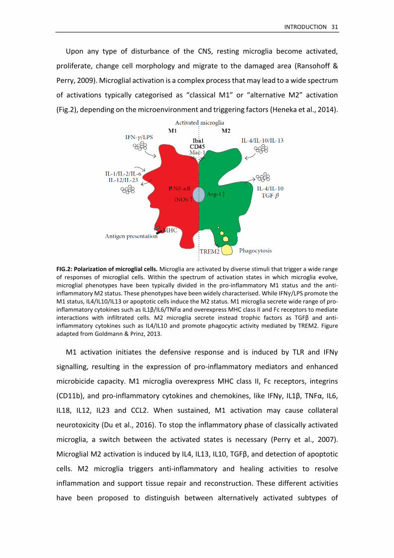
(Fig.2), depending on the microenvironment and triggering factors (Heneka et al., 2014).
FIG.2: Polarization of microglial cells. Microglia are activated by diverse stimuli that trigger a wide range of responses of microglial cells. Within the spectrum of activation states in which microglia evolve, microglial phenotypes have been typically divided in the pro-inflammatory M1 status and the anti-inflammatory M2 status. These phenotypes have been widely characterised. While IFNγ/LPS promote the M1 status, IL4/IL10/IL13 or apoptotic cells induce the M2 status. M1 microglia secrete wide range of pro-inflammatory cytokines such as IL1β/IL6/TNFα and overexpress MHC class II and Fc receptors to mediate interactions with infiltrated cells. M2 microglia secrete instead trophic factors as TGFβ and anti-inflammatory cytokines such as IL4/IL10 and promote phagocytic activity mediated by TREM2. Figure adapted from Goldmann & Prinz, 2013.
M1 activation initiates the defensive response and is induced by TLR and IFNγ
signalling, resulting in the expression of pro-inflammatory mediators and enhanced
microbicide capacity. M1 microglia overexpress MHC class II, Fc receptors, integrins
(CD11b), and pro-inflammatory cytokines and chemokines, like IFNγ, IL1β, TNFα, IL6,
IL18, IL12, IL23 and CCL2. When sustained, M1 activation may cause collateral
neurotoxicity (Du et al., 2016). To stop the inflammatory phase of classically activated
microglia, a switch between the activated states is necessary (Perry et al., 2007).
Microglial M2 activation is induced by IL4, IL13, IL10, TGFβ, and detection of apoptotic
cells. M2 microglia triggers anti-inflammatory and healing activities to resolve
inflammation and support tissue repair and reconstruction. These different activities
have been proposed to distinguish between alternatively activated subtypes of
32 Sfrp1 promotes neuroinflammation through the modulation of ADAM10 proteolytic activity
microglia (Colton, 2009). The M2 phenotype is characterised by enhanced phagocytic
activity, induced expression of arginase 1 and secretion of anti-inflammatory cytokines,
such as IL4, IL10, IL13, and TGFβ, and growth factors, such as insulin-like growth factor
(IGF1), fibroblast growth factor (FGF), colony-stimulating factor (CSF1), nerve growth
factor (NGF), brain-derived neurotrophic factor (BDNF), and glial derived neurotrophic
factor (GDNF) (Boche et al., 2013). The distinction of activated microglial states into
M1/M2 is a simplification since M1 and M2 represent the extremes of a range of
activated phenotypes.
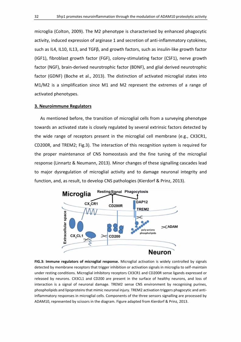
3. Neuroimmune Regulators
As mentioned before, the transition of microglial cells from a surveying phenotype
towards an activated state is closely regulated by several extrinsic factors detected by
the wide range of receptors present in the microglial cell membrane (e.g., CX3CR1,
CD200R, and TREM2; Fig.3). The interaction of this recognition system is required for
the proper maintenance of CNS homeostasis and the fine tuning of the microglial
response (Linnartz & Neumann, 2013). Minor changes of these signalling cascades lead
to major dysregulation of microglial activity and to damage neuronal integrity and
function, and, as result, to develop CNS pathologies (Kierdorf & Prinz, 2013).
FIG.3: Immune regulators of microglial response. Microglial activation is widely controlled by signals
detected by membrane receptors that trigger inhibition or activation signals in microglia to self-maintain
under resting conditions. Microglial inhibitory receptors CX3CR1 and CD200R sense ligands expressed or
released by neurons. CX3CL1 and CD200 are present in the surface of healthy neurons, and loss of
interaction is a signal of neuronal damage. TREM2 sense CNS environment by recognising purines,
phospholipids and lipoproteins that mimic neuronal injury. TREM2 activation triggers phagocytic and anti-
inflammatory responses in microglial cells. Components of the three sensors signalling are processed by
ADAM10, represented by scissors in the diagram. Figure adapted from Kierdorf & Prinz, 2013.
INTRODUCTION 33
CX3CR1
CX3CR1 is the receptor of the chemokine CX3CL1, also known as fractalkine or
neurotactin, and represents the only member of a monogamous ligand/receptor system
of the CX3C chemokine family (Ransohoff & El Khoury, 2015). The expression of CX3CR1
in the CNS is restricted to the microglial population (Jung et al., 2000), while the ligand
can be found in the neuronal membrane, as one of the two unique membrane-bound
chemokines, or in a secreted form (Hughes et al., 2002). The release of the chemokine
domain is mediated by ADAM10 (Hundhausen et al., 2007). CX3CR1/CX3CL1 signalling
plays a critical role regulating diverse microglial functions during physiological
conditions, modulating microglial surveillance and interaction with neurons implicated
in the maturation, activity and plasticity of neuronal connectivity (Paolicelli et al., 2014).
Under healthy conditions, high levels of secreted CX3CL1 have been found in the CNS
parenchyma, but if reduced promote microglial neurotoxicity (Cardona et al., 2006).
Furthermore, impaired signalling have been involved in the development of cognitive
impairment (Rogers et al., 2011) and chronic inflammatory diseases (Clark et al., 2011),
and the mediation of recruitment of immune cells in autoimmunity (Hertwig et al.,
2016).
CD200R
CD200R is another inhibitory receptor expressed in microglial cells (G. J. Wright et al.,
2000) and recently observed also in activated astrocytes (Hernangomez et al., 2016). Its
ligand, CD200 also known as OX2, is mainly expressed by neurons and, to a lower extent,
by astrocytes and oligodendrocytes (Koning et al., 2009). The interaction between
CD200 and CD200R keeps microglia in a resting state (Lyons et al., 2007), and most
importantly, induces the return of pro-inflammatory activated microglia to this state
(Walker & Lue, 2013). Both proteins are characterised by two immunoglobulins
domains, but they differ in that CD200 lacks the longer cytoplasmic tail of CD200R with
an immune-receptor tyrosine-based inhibition motif (ITIM) domain (Barclay et al., 2002).
Deficient interaction or expression of this immune-regulatory system has been reported
in AD (Walker et al., 2009) and demyelinating diseases (Hernangomez et al., 2012)
leading to chronic inflammation and contributing to aging (Hernangomez et al., 2014).

34 Sfrp1 promotes neuroinflammation through the modulation of ADAM10 proteolytic activity
The release of a functionally active soluble form of CD200 from the plasma-membrane
has been recently reported, as a consequence of an ADAM protease mediated shedding,
by ADAM10, 17 and 28. This soluble form seems to have detrimental
immunosuppressive effects in patients with leukaemia (Wong et al., 2016) but, at the
moment, there is no reported information about the impact of the CD200 soluble form
of in the brain.
TREM2
The Triggering Receptor Expressed on Myeloid cells 2 (TREM2) is a transmembrane
glycoprotein with an extracellular single immunoglobulin domain expressed only by
microglia in the CNS (Colonna & Wang, 2016). TREM2 binds poly-anions, such as
bacterial lipopolysaccharides (Y. Wang et al., 2015), phospholipids and lipoproteins such
as ApoE (Yeh et al., 2016). Ligand-TREM2 interaction transmits intracellular signalling
through the immune-receptor tyrosine-based activator motif (ITAM) of DAP12, with
which TREM2 is associated through their transmembrane regions, promoting
proliferation and survival (Otero et al., 2009), phagocytosis of apoptotic cells (Hsieh et
al., 2009), and attenuates microglial pro-inflammatory signalling (Turnbull et al., 2006).
Rare variants of TREM2 have been associated with an increased risk of developing
sporadic AD (Guerreiro et al., 2013; Jonsson et al., 2013). Even more rare mutations in
TREM2 or DAP12, that impair signalling, cause Nasu-Hakola disease, an inherited form
of dementia (Paloneva et al., 2002). Furthermore, deficiency of TREM2 in a mice model
of AD present less clustering of microglia surrounding the Aβ plaques (Jay et al., 2015),
which facilitates Aβ diffusion and consequently toxicity (Y. Wang et al., 2016). Recent
studies have also demonstrated that TREM2 is processed at the microglial cell surface
by ADAM10 and ADAM17 sheddases, generating a soluble form (Kleinberger et al.,
2014). This soluble TREM2 can be detected in the cerebrospinal fluid, acting as a marker
of microglial activation that correlates with neuronal injury markers (Suarez-Calvet et
al., 2016). Importantly, latest data indicate that soluble TREM2 may have its own
function triggering survival and pro-inflammatory responses, leading to microglial
activation (Zhong et al., 2017).
INTRODUCTION 35
4. Models of Neuroinflammation
The roman physician Aulus Celsus originally defined the term “inflammation” as a
process of “tumor, rubor, calor et dolor” (Celsus, 1478). This four cardinal sings imply
extravasation of the adaptive immune response mediators T and B-lymphocytes.
Notably, whereas this feature is seen in bacterial and viral infections and in autoimmune
diseases (e.g., Multiple Sclerosis, MS) of the CNS (Aguzzi et al., 2013), the term
“neuroinflammation” has gradually expanded to include conditions in which Celsus’ s
cardinal signs are not present. Neurodegenerative diseases, such as AD, are now
considered neuroinflammatory conditions, in which proliferation and activation of glial
cells are well-established hallmarks (Ransohoff, 2016).
Lipopolysaccharide induced Neuroinflammation
One of the typical approximations to study the immune system has been based on
the use of the endotoxin lipopolysaccharide (LPS). LPS, the best-characterised microbial
PAMP, is a major component of the outer membrane of Gram-negative bacteria and its
recognition induces a robust inflammatory response by phagocytic cells (S. D. Wright,
1999). Cell wall components of Gram-negative and Gram-positive bacteria stimulate
cytokine production, activating Toll-like receptors (TLRs) and pro-inflammatory
signalling (Nguyen et al., 2002). While TLR2 recognises Gram-positive bacteria, TLR4 is
critical for the recognition of LPS (S. J. Lee & Lee, 2002). Binding of LPS to TLR4 induces
the stimulation of nuclear factor-kappaB (NFκB) signalling pathway and cytokine
synthesis (Allan et al., 2005).
Systemic injection of LPS is recognised by TLR4-expressing microglia in the
circumventricular organs and choroid plexus, inducing the expression of TNFα that
spreads the pro-inflammatory signal through the brain parenchyma. Similarly, LPS
injected directly into the brain induces robust and transient microglial M1 activation and
expression of pro-inflammatory molecules, such as cytokines, chemokines and
complement system proteins, by a TLR4-dependent mechanism (Rivest, 2009). LPS
locally-injected stimulates microglial cells directly through the TLR4-NFκB pathway,
whereas activation of parenchymal microglia after a systemic LPS challenge depends on
TNFα-induced signalling (Nadeau & Rivest, 2000). Although TLR4 expression by
36 Sfrp1 promotes neuroinflammation through the modulation of ADAM10 proteolytic activity
astrocytes and neurons has been reported (Bowman et al., 2003), the response to LPS is
completely dependent on the presence of functional microglia (Holm et al., 2012).
Neuroinflammation associated with CNS autoimmunity
Experimental Autoimmune Encephalomyelitis (EAE) is the most commonly used
experimental model to study the prototypical inflammatory demyelinating disease of
the CNS, i.e., Multiple Sclerosis (MS). MS is a degenerative autoimmune disease with a
strong inflammatory component that attacks CNS myelin. From a clinical point to view,
MS exhibits a relapsing and remitting pattern of neurological deficits that can resolve
completely or leave residual disabilities, and occasionally derives in a continuous
progressive disease (Lassmann et al., 2012). The pathological features of MS are the
disruption of the BBB accompanied by an infiltration of peripheral macrophages and
lymphocytes, and an intrinsic gliosis, that ultimately results in glial scar formation
surrounding axonal degeneration and demyelination sites, which is known as sclerotic
plaque, the principal hallmark of MS (Pérez-Cerdá et al., 2016). It is controversial if
macrophage and lymphocyte infiltration (Barnett & Prineas, 2004), believed to be the
main trigger of CNS damage, is a cause or a consequence of the degeneration of neural
tissue (Stys et al., 2012). The induction of EAE is achieved by animal immunization and
therefore it is of no help to solve this controversy (Gran et al., 2008).
Most of the MS features, including paralysis, weight loss, demyelination,
inflammation and BBB disruption, are observed during EAE (Bennett et al., 2010). EAE is
typically induced by the sensitization of the innate immune system to different myelin
derived proteins such as myelin oligodendrocyte glycoprotein (MOG), myelin basic
protein (MBP), or proteolipid protein, injected together with an adjuvant, usually
complete Freund’s adjuvant, and pertussis toxin to relax the BBB (Gao & Tsirka, 2011).
The pathological lesions vary among strains and type of immunization, with monophasic
or sustained form of EAE. This depends on the presence or reduced resolution of
multifocal demyelination and infiltration of macrophages and CD4 positive T-cells
(Constantinescu et al., 2011). Macrophage infiltration strongly correlates with EAE
progression to severe disease, and disappearance of macrophage from the CNS lead to
symptom remission and possible recovery (Ajami et al., 2011).
INTRODUCTION 37
In response to passive immunization, microglia become activated and proliferate, this
neuroinflammatory process have been implicated in the onset and severity of EAE
clinical signs (Ding et al., 2014). During the onset and peak of EAE, microglia phagocytize
myelin, triggering the release of cytokines and chemokines that induce the disruption of
BBB integrity and the recruitment of macrophages and lymphocytes, that can be
activated by myelin presentation in MHC class II of microglial cells, acting as Antigen
Presenting Cells (Goldmann & Prinz, 2013). In addition, microglial cytokine release has
an important role in the regulation, proliferation and differentiation of the infiltrated T-
cells (Fletcher et al., 2010). Importantly, infiltrated macrophages and resident microglia
seems to have different roles in the course of EAE. Infiltrated macrophages are highly
inflammatory and demyelinating, whereas activated microglia appear to clear debris
(Yamasaki et al., 2014). Microglial overexpression of TREM2 increases the phagocytic
clearance of myelin debris, leading to improved tissue regeneration and to reduction of
the severity of the clinical symptoms (Takahashi et al., 2007).
Neuroinflammation associated with Neurodegeneration
Alzheimer’s disease is a neurodegenerative disease characterised by the
accumulation of amyloidβ (Aβ) containing plaques and Tau-positive neurofibrillary
tangles, accompanied by gliosis and neuronal loss (Alzheimer et al., 1995). Other
morphological features of AD include cerebrovascular amyloid angiopathy and major
synaptic alterations (Crews & Masliah, 2010). How AD develops is still unclear although
there are several well-established risk factors such as aging, diabetes, vascular
alterations, and a long etcetera. There are also several hypotheses on the possible
triggers. One of them, the amyloid hypothesis, links abnormal Aβ peptide aggregation
and accumulation with the other hallmarks of the disease. Toxic Aβ peptides are
generated by the action of two proteases: β-secretase (BACE1) and γ-secretase complex
(composed by PS1/2, Nicastrin and APH1), which sequentially cleave the amyloid
precursor protein (APP). Aβ deposition linearly and causally leads in a progressive
cascade to Tau pathology, neuroinflammation, synaptic dysfunction, neuronal loss and,
ultimately, dementia (Hardy & Selkoe, 2002). In the past, many clinical trials designed to
reduce Aβ levels show no efficacy. However, a recent clinical trial that selectively
targeted Aβ oligomers, the more toxic form of Aβ, showed some improvement,
38 Sfrp1 promotes neuroinflammation through the modulation of ADAM10 proteolytic activity
providing support to this hypothesis. Indeed, treatment with antibodies against
oligomeric Aβ reduced plaque burden and slowed the progression of cognitive decline
in AD patients (Sevigny et al., 2016).
The central role of Aβ accumulation in the pathogenesis of AD was revealed by the
discovery of mutations in the APP or Presinilin genes leading to altered APP processing
in familial forms of AD (Hardy & Allsop, 1991). These mutations have been subsequently
exploited to generate mouse models for AD (LaFerla & Green, 2012). One of the most
used is the APP;PS1 mouse line. These mice overexpress, under the control of the PrP
promoter, the human APP allele carrying the so-called Swedish mutation
(K594M/N595L) and a human PSEN1 allele containing a deletion of exon 9 (Jankowsky
et al., 2004).
The linearity of the amyloid hypothesis is nevertheless very controversial. The initial
causality has been modified (Selkoe & Hardy, 2016) to enhance the importance of Aβ
clearance impairment, that involve the expression of ApoE4 allele (Castellano et al.,
2011), and the misbalance of APP processing, leading to accumulation of longer Aβ
products (Aβ42/43) that are highly self-aggregating. The neuron-centric view has been
expanded towards the contribution of different cell-types, and their interactions, that
have been involved in the gradual evolution of the disease, trying to explain different
confusing steps of the cascade, such as the silent incubation period of Aβ accumulation
and the link between Aβ and neurotoxicity (De Strooper & Karran, 2016).
The role of neuroinflammation in the pathogenesis of AD has been intensively
investigated (Heneka et al., 2015). The prototypical hallmarks of AD, hyper-
phosphorylated Tau and oligomeric and fibrillary Aβ, are highly immunogenic and trigger
the activation of microglial cells and astrocytes, inducing the secretion of pro-
inflammatory cytokines (Grubman et al., 2016). In the brain of AD patients, reactive
astrocytes occupy peri-plaque positions, surrounding Aβ deposits in a manner
reminiscent of glial scarring (J. J. Rodriguez et al., 2009). Furthermore, there is a chronic
increase of the levels of pro-inflammatory cytokines (Brosseron et al., 2014).
Nevertheless, anti-inflammatory strategies seem to have unexpected negative effects
on Aβ processing and cognition (Chakrabarty et al., 2015). This demonstrates the
complexity of neuroinflammation associated to neurodegeneration since in some
INTRODUCTION 39
situations it can be beneficial and in other detrimental. Importantly, sporadic AD risk
genes are predominantly expressed by microglia. This indicate that genetically impaired
microglia may represent a risk for AD, further reinforcing the relevance of
neuroinflammation in AD (Skene & Grant, 2016). These genes are mostly involved in the
regulation of neuroinflammation and phagocytosis and include TREM2, CR1, CD33 and
ApoE4 (Villegas-Llerena et al., 2016). Moreover, it seems that the physiology of
microglial cells is impaired in the brain of AD patients likely because of chronic
stimulation or loss of function (Mosher & Wyss-Coray, 2014). This dysfunction
contributes to early synapse loss in AD, in part because complement-mediated synapse
pruning does not properly work (Hong et al., 2016).
Importantly, taking into account that aging is the greatest risk factor for AD, a recent
time-course study has demonstrated, comparing gene expression profiles in normal and
AD brains, that immune- and inflammation-associated genes were robustly upregulated
in aged brains compared to a modest response in AD patients (Cribbs et al., 2012). This
demonstrates the critical involvement of neuroinflammatory processes in AD
development and progression.
5. Secreted Frizzled-Related Protein 1
Secreted-Frizzled-Related-Proteins (Sfrps) compose a family of soluble factors with
five members in mammals (Sfrp1-5). These highly diffusible proteins have been widely
studied and characterised as modulators of Wnt signalling, an extensively used signalling
pathway that mediates cell-cell communication in development and adult tissue
homeostasis (Bovolenta et al., 2008). The different members of the family were
independently identified in two different contexts: related to early embryonic
development (Leyns et al., 1997) and in the modulation of apoptosis (Melkonyan et al.,
1997). This family received its name because the N-terminal region of the proteins
presents high sequence homology with the extracellular Wnt binding domain of the
Frizzled receptors (Hoang et al., 1996). The proteins are composed of two independently
folded domains. The domain similar to that of Frizzled receptor is called Cysteine Rich
Domain (CRD), owing to the ten conserved cysteine residues that conform a pattern of
disulphide bridges. The C-terminal domain contains a Netrin-related motif (NTR). In the
closely related Sfrp1/2/5, this domain is characterised by segments of positively charged
40 Sfrp1 promotes neuroinflammation through the modulation of ADAM10 proteolytic activity
residues and six cysteine residues that form three disulphide bridges (Chong et al.,
2002). This domain shares conformational similarities with a number of proteins,
including the axon-guidance protein netrin1, tissue inhibitors of metalloproteases
(TIMPs), type-1 procollagen C-proteinase enhancer proteins (PCOLCEs) and complement
proteins (Banyai & Patthy, 1999).
Roles of Sfrp1 as regulator of cell-cell communication
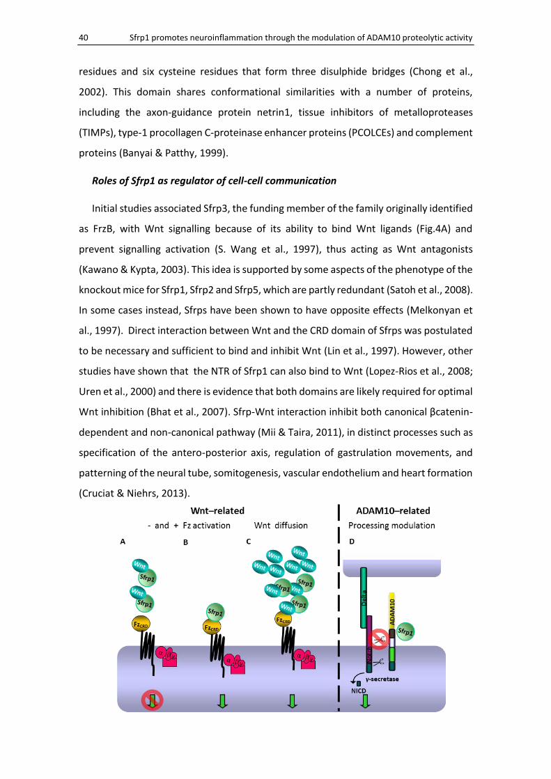
Initial studies associated Sfrp3, the funding member of the family originally identified
as FrzB, with Wnt signalling because of its ability to bind Wnt ligands (Fig.4A) and
prevent signalling activation (S. Wang et al., 1997), thus acting as Wnt antagonists
(Kawano & Kypta, 2003). This idea is supported by some aspects of the phenotype of the
knockout mice for Sfrp1, Sfrp2 and Sfrp5, which are partly redundant (Satoh et al., 2008).
In some cases instead, Sfrps have been shown to have opposite effects (Melkonyan et
al., 1997). Direct interaction between Wnt and the CRD domain of Sfrps was postulated
to be necessary and sufficient to bind and inhibit Wnt (Lin et al., 1997). However, other
studies have shown that the NTR of Sfrp1 can also bind to Wnt (Lopez-Rios et al., 2008;
Uren et al., 2000) and there is evidence that both domains are likely required for optimal
Wnt inhibition (Bhat et al., 2007). Sfrp-Wnt interaction inhibit both canonical βcatenin-
dependent and non-canonical pathway (Mii & Taira, 2011), in distinct processes such as
specification of the antero-posterior axis, regulation of gastrulation movements, and
patterning of the neural tube, somitogenesis, vascular endothelium and heart formation
(Cruciat & Niehrs, 2013).
INTRODUCTION 41
Beside inhibition of Wnt signalling, Sfrps have other Wnt-dependent and
independent roles, as shown by several studies from different laboratories, including
ours (Esteve & Bovolenta, 2010). Indeed, Sfrps can promote or suppress Wnt signalling
depending on their concentration and cellular context (Xavier et al., 2014). Direct
interaction between Sfrp1 and Frizzled receptors can induce the activation of the Wnt
non-canonical pathway (Fig.4B), providing axon-guidance information to retinal
ganglion cell axons (J. Rodriguez et al., 2005). Sfrp-Wnt interaction can also promote
Wnt diffusion (Fig.4C) in both Xenopus gastrulation (Mii & Taira, 2009) during optic cup
formation (Esteve et al., 2011b). Besides this Wnt signalling related functions, Sfrps can
regulate other pathways. Sfrp1 interaction with RANKL, a member of the TNF family,
prevents its binding to the activator of NFκB receptor (RANK), inhibiting osteoclast
formation (Hausler et al., 2004). In addition, the CRD of Sizzled, a Sfrp not expressed in
mammals, binds to BMP1/tolloid, impairing its metalloprotease activity required to
regulate a BMP signalling inhibitor, thereby regulating the pathway (H. X. Lee et al.,
2006). Our laboratory demonstrated that Sfrp1 interacts and negatively modulates the
activity of the A Disintegrin and Metalloprotease transmembrane protein ADAM10
(Fig.4D). This modulation interferes with ADAM10-mediated shedding of multiple
substrates including N-cadherin, L1 and Notch, during retinal and cortical neurogenesis,
adult brain homeostasis (Esteve et al., 2011a) and visual pathway establishment (Marcos
et al., 2015).
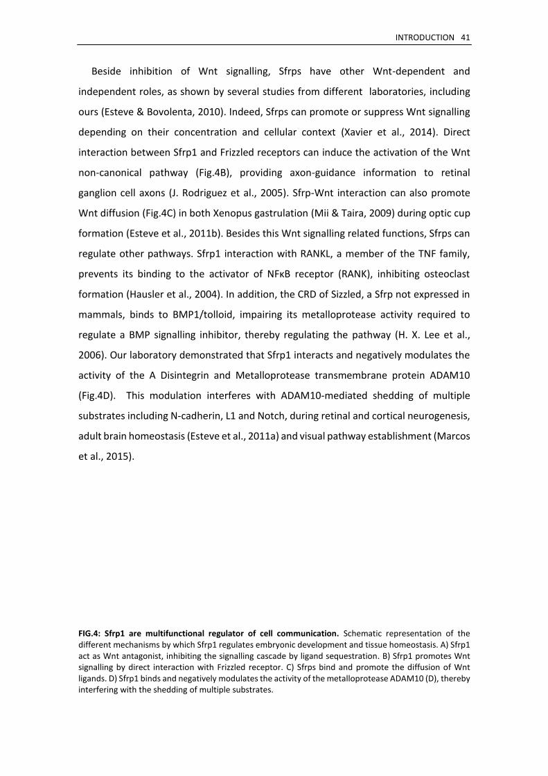
FIG.4: Sfrp1 are multifunctional regulator of cell communication. Schematic representation of the different mechanisms by which Sfrp1 regulates embryonic development and tissue homeostasis. A) Sfrp1 act as Wnt antagonist, inhibiting the signalling cascade by ligand sequestration. B) Sfrp1 promotes Wnt signalling by direct interaction with Frizzled receptor. C) Sfrps bind and promote the diffusion of Wnt ligands. D) Sfrp1 binds and negatively modulates the activity of the metalloprotease ADAM10 (D), thereby interfering with the shedding of multiple substrates.
42 Sfrp1 promotes neuroinflammation through the modulation of ADAM10 proteolytic activity
Roles of Sfrp1 in pathogenesis
Wnt signalling regulates diverse processes during embryonic development including
cell proliferation, patterning and fate determination (Clevers & Nusse, 2012). Mutations
or abnormal expression of components of the Wnt pathway have been associated with
tumorigenesis (Klaus & Birchmeier, 2008). In line with their function as inhibitors of Wnt
signalling, Sfrps seem to act as tumour suppressors. Loss or downregulation of Sfrps
expression, produced by allelic loss or promoter hyper-methylation, have been reported
in a variety of invasive carcinomas (Surana et al., 2014) and Sfrp1 promoter hyper-
methylation is an epigenetic marker for cancer detection and prognosis (Bovolenta et
al., 2008).
Sfrps have been also implicated in other pathologies (Esteve & Bovolenta, 2010). For
example, Sfrp1 expression is strongly increased in the retinas of patients with retinitis
pigmentosa, a degenerative disease characterised by the progressive loss of
photoreceptors (Hackam, 2005). Upregulation of Sfrp1 in the eye is associated with
elevated intraocular pressure, leading to glaucoma, perhaps inhibiting the Wnt pathway
(W. H. Wang et al., 2008). Outside the CNS, Sfrp1 overexpression contributes to the
pathogenesis of periodontitis (C. H. Li & Amar, 2007), rheumatoid arthritis (Walsh et al.,
2009) and lung emphysema (Foronjy et al., 2010), diseases that are all associated to
chronic inflammation.
With the exception of the retina, there is no description of CNS pathologies with a
direct relationship with Sfrp1 although the expression of diverse components of the Wnt
signalling pathway are disrupted in AD (Godoy et al., 2014), leading to an exacerbated
GSK3β activity that may represent a link between Aβ plaques and Tau neurofibrillary
tangles (Llorens-Martin et al., 2014). Furthermore, dysfunction of Wnt signalling has
been implicated in Aβ-mediated synaptic loss (Purro et al., 2014). The implication of Wnt
ligands in inflammatory processes is controversial. Wnt3a (Halleskog et al., 2011) and
Wnt5a (B. Li et al., 2011) might have pro-inflammatory properties but can also
counteract LPS-induced pro-inflammatory response (Halleskog & Schulte, 2013) and
induce an anti-inflammatory response (Di Liddo et al., 2015).
INTRODUCTION 43
Implication of Sfrp1 in Alzheimer’s Disease
ADAM10 is a constitutive transmembrane α-secretase highly active in the brain
(Jorissen et al., 2010). ADAM10 sheds a wide range of substrates (Weber & Saftig, 2012)
involved in the regulation of diverse processes in CNS development or degeneration,
such as neurogenesis, axon-guidance, synaptogenesis and neuroinflammation (Saftig &
Bovolenta, 2015). Among them, one of the most studied ADAM10 substrates is APP
(Huovila et al., 2005).
APP is expressed in many tissues but particularly concentrated at the neuronal
synapses. As mentioned above, APP is a protein widely accepted as a key factor in the
onset and development of AD (Hardy & Selkoe, 2002). APP is processed by two
alternative proteolytic pathways. In the so-called amyloidogenic pathway, APP is
sequentially cleaved by β and γ-secretases (BACE and PS1) to generate Aβ peptides that
aggregate and accumulate in Aβ plaques (Y. W. Zhang et al., 2011). APP can be also
processed through an alternative “non-amyloidogenic pathway” (Kuhn et al., 2010). In
this case, APP is first cleaved by the α-secretase ADAM10 within the Aβ peptide
sequence, precluding the formation of Aβ peptides (Lichtenthaler, 2011). This
alternative processing leads to the release of a soluble N-terminal extracellular domain
known as APPsα, which has been reported to have neurotrophic and neuroprotective
properties (Ring et al., 2007) and to promote adult neural stem cell proliferation (Caille
et al., 2004).
APP (Yasuoka et al., 2004), ADAM10 (Demars et al., 2011) and Sfrp1(Fig.5A) are co-
expressed in the sub-ventricular zone of the lateral ventricle of adult mouse brains.
Consistent with the finding that Sfrp1 acts as negative modulator of ADAM10, high levels
of soluble non-amyloidogenic APPsα are found in the ventricular walls of Sfrp1 null mice
(Esteve et al., 2011a), suggesting a possible involvement of Sfrp1 in the onset and
progression of AD.
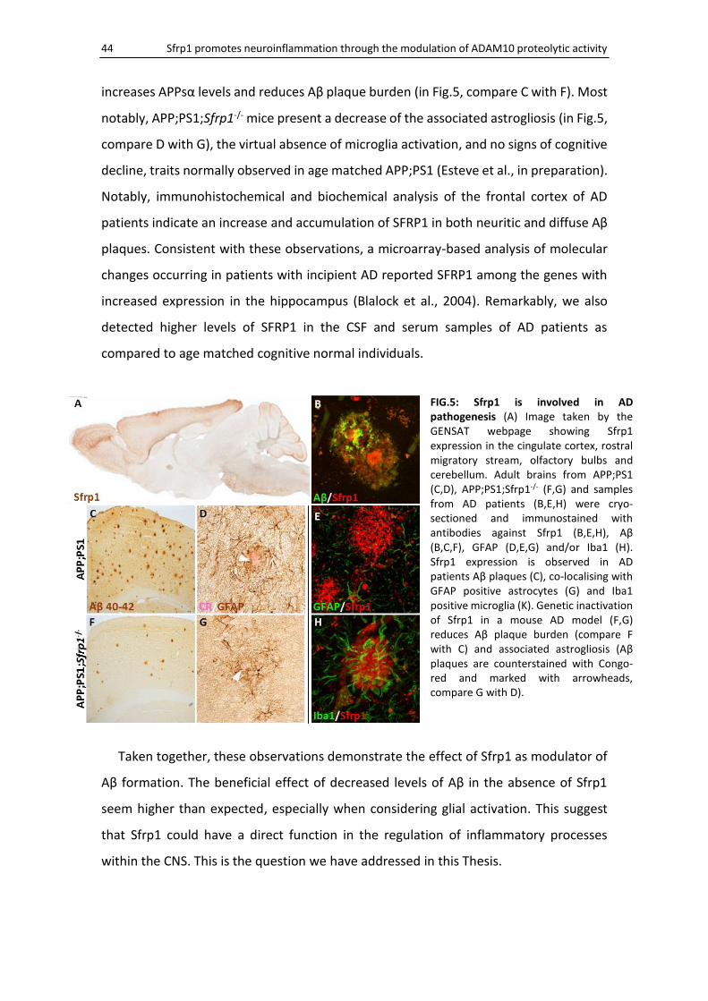
To test this possibility our laboratory has shown that Sfrp1 expression is increased in
a mouse model of AD, the APP;PS1, accumulating in the Aβ plaques (Fig.5B) and co-
localising with some surrounding reactive astrocytes (Fig.5E) and activated microglia
(Fig.5H). Furthermore, genetic inactivation of Sfrp1 in APP;PS1 mice (APP;PS1;Sfrp1-/-)
44 Sfrp1 promotes neuroinflammation through the modulation of ADAM10 proteolytic activity
increases APPsα levels and reduces Aβ plaque burden (in Fig.5, compare C with F). Most
notably, APP;PS1;Sfrp1-/- mice present a decrease of the associated astrogliosis (in Fig.5,
compare D with G), the virtual absence of microglia activation, and no signs of cognitive
decline, traits normally observed in age matched APP;PS1 (Esteve et al., in preparation).
Notably, immunohistochemical and biochemical analysis of the frontal cortex of AD
patients indicate an increase and accumulation of SFRP1 in both neuritic and diffuse Aβ
plaques. Consistent with these observations, a microarray-based analysis of molecular
changes occurring in patients with incipient AD reported SFRP1 among the genes with
increased expression in the hippocampus (Blalock et al., 2004). Remarkably, we also
detected higher levels of SFRP1 in the CSF and serum samples of AD patients as
compared to age matched cognitive normal individuals.
FIG.5: Sfrp1 is involved in AD pathogenesis (A) Image taken by the GENSAT webpage showing Sfrp1 expression in the cingulate cortex, rostral migratory stream, olfactory bulbs and cerebellum. Adult brains from APP;PS1 (C,D), APP;PS1;Sfrp1-/- (F,G) and samples from AD patients (B,E,H) were cryo-sectioned and immunostained with antibodies against Sfrp1 (B,E,H), Aβ (B,C,F), GFAP (D,E,G) and/or Iba1 (H). Sfrp1 expression is observed in AD patients Aβ plaques (C), co-localising with GFAP positive astrocytes (G) and Iba1 positive microglia (K). Genetic inactivation of Sfrp1 in a mouse AD model (F,G) reduces Aβ plaque burden (compare F with C) and associated astrogliosis (Aβ plaques are counterstained with Congo-red and marked with arrowheads, compare G with D).
Taken together, these observations demonstrate the effect of Sfrp1 as modulator of
Aβ formation. The beneficial effect of decreased levels of Aβ in the absence of Sfrp1
seem higher than expected, especially when considering glial activation. This suggest
that Sfrp1 could have a direct function in the regulation of inflammatory processes
within the CNS. This is the question we have addressed in this Thesis.

OBJECTIVES
OBJECTIVES 49
Sfrp1 protein is highly diffusible and acts as multifunctional extracellular regulator of
cell-to-cell communication. In addition, its expression increases in diverse diseases
associated with chronic inflammation. This points to a possible role of Sfrp1 in the
modulation of inflammatory processes. Previous work from our laboratory has also
demonstrated that Sfrp1 contributes to AD progression by modulating ADAM10-
mediated processing of APP. In the course of this study we noticed that
neuroinflammation was particularly low when Sfrp1 protein was absent from the brain
of mouse model of AD, pointing to a possible direct implication of Sfrp1 in the regulation
of brain inflammation.
We have thus undertaken this Thesis to determine whether Sfrp1 is involved in the
regulation of inflammatory processes in the CNS. The relevance of this general goal relies
on the growing evidence that inflammation in the CNS participates in the onset and
progression of neurological disorders.
The specific objectives for this work are the following:
- Analyse the changes of Sfrp1 expression under neuroinflammatory conditions.
- Test if Sfrp1 is necessary and sufficient for the development of an inflammatory
response within the CNS.
- Examine the possible mechanism of action through which Sfrp1 promotes
neuroinflammation.
- Study whether Sfrp1 could be considered as therapeutic target to attenuate
neuroinflammation typically observed in neurodegenerative diseases.

MATERIALS AND METHODS
MATERIALS AND METHODS 53
1. Animals
We used newborn and adult mice of both sexes. All mice were maintained under
specific pathogen–free conditions at the animal facilities of the Centro de Biología
Molecular Severo Ochoa, in accordance with current national and European guidelines
(Directive 2010/63/EU). All animal procedures were approved by the ethical committee
of the institute and of the Comunidad Autónoma de Madrid.
Sfrp1-/- mice were generated by inter-cross of the Sfrp1-/-;Sfrp2+/- mice in a
129/C57BL/6 background described in (Satoh et al., 2006), and back-crossed at least four
times with C57BL/6J to clean the background. Wild type animals were littermates
selected from heterozygous crosses. Sfrp1-/- brains present narrow ventricles and
shorter but thicker cortex (Esteve et al., Submitted). APP;PS1 transgenic mice where
generated as described (Jankowsky et al., 2004). Breeding pairs were kindly provided by
Dr. Torres-Aleman, Instituto Cajal, CSIC, Madrid, and only male mice were used.
2. Intra-cerebral infusion
For LPS infusion into the brain parenchyma, wild type (C57BL/6J) and Sfrp1-/- male
littermates of 10 weeks of age were used. Mice were anaesthetized with 4% inhaled
Isoflurane (Forane, AbbVie Farmacéutica) vaporised into a sealed anaesthetic induction
chamber (SurgiVet, Smiths Medical), placed into a stereotaxic apparatus (Stoelting) and
anaesthesia was maintained at 2.5% in 250ml/min oxygen flow. Under aseptic
conditions, a midline incision with scalpel was performded to reveal the skull and
bregma area was gently cleaned. Craniotomy was performed at precise point with a 23G
blunt needle, to allow injection into stereotaxic coordinates of 0.0mm
anterior/posterior, -1.0mm lateral, and -1.5mm dorsal/ventral from bregma, into the
top of corpus callosum (Lein et al., 2007). Vehicle (2.5µl sterile saline) or 5µg LPS
(Escherichia coli 0111:B4; Sigma Aldrich) in 2.5µl of sterile saline were delivered using a
10µl syringe with a fine 34G needle (Hamilton). Infusion was performed with a
Quintessential Stereotaxic Injector (Stoelting) at a rate of 0.5µl/min. The needle was
kept in this position for an additional 5min after injection and then retrieved slowly out
of the brain. Three days after infusion, when the inflammatory process reaches its peak
of activation (Rivest, 2009), mice were sacrificed and processed for either biochemical
or histological analysis.
54 Sfrp1 promotes neuroinflammation through the modulation of ADAM10 proteolytic activity
In the case of lentiviral infusion, the procedure was slightly different as lentiviral
particles were delivered intracerebroventricularly. First, lentiviral particles carrying GFP
or SFRP1-IRES-GFP into a pHRSIN vector (generated by O. Lancho in a collaboration
between our and M. Toribio’s laboratories) were obtained by transient transfection of
HEK-293T cells using Lipofectamine Reagent (Invitrogen). Three plasmids were
transfected: HIV-derived psPAX2 (gag/pol) and pMD2G (VSV envelope), and the
different lentiviral pHRSIN vectors. Culture supernatants were collected one and two
days after transfection and ultra-centrifuged. The pellets containing the lentiviral
particles were re-suspended in PBS (1x10⁸ TU/ml). Preparation of lentiviral particles was
performed by M.J. Martin-Bermejo. Small volumes of 2.5µl of GFP or SFRP1-IRES-GFP
lentiviral particles were delivered into the lateral ventricle in the following stereotaxic
coordinates of 0.5mm anterior/posterior, -1.0mm lateral, and -2.3mm dorsal/ventral
from bregma, following the same procedure described above. One or five months post-
injection, mice were sacrificed and processed for histological analysis.
3. Experimental Autoimmune Encephalomyelitis
Chronic EAE was inducted as described (Borroto et al., 2016). Briefly, female C57BL/6J
and Sfrp1-/- littermates of 8 to 10 weeks old mice were subcutaneously injected with
150mg of MOG35–55 (Espikem) emulsified in Freund’s complete adjuvant (Sigma Aldrich)
and supplemented with Mycobacterium tuberculosis (1mg/ml) (H37Ra strain from Difco)
into both femoral regions. The mice were immediately injected intraperitoneally with
200ng of pertussis toxin (Sigma Aldrich) and, again, 48 hours after immunization. The
animals were weighed and inspected for clinical signs of the disease on a daily basis by
an observer blind to the genotype. Clinical signs of EAE was assessed according to a
severity scale: 0) normal behaviour, no overt signs of disease; 1) weakness at the distal
portion of the tail; 1.5) complete flaccidity of the tail; 2) moderate hind limb weakness;
2.5) severe hind limb weakness; 3) ataxia; 3.5) partial hind limb paralysis; 4) complete
hind limb paralysis; 4.5) complete hind limb paralysis accompanied by muscle stiffness;
5) moribund state and hence sacrificed according to ethical procedures.
At day 16 after immunization, when the symptoms reached the peak of severity in
the wild type, a representative pool of mice were anesthetized and perfused
intracardially with 4% paraformaldehyde in 0.1M phosphate buffer (pH7.6). The spinal
MATERIALS AND METHODS 55
cords of the mice were dissected out and processed for histological analysis by
immunostaining. Statistical analysis was performed using Prism software (GraphPad) by
Mann–Whitney U nonparametric test.
4. Primary cultures
Glial primary cultures were established from cerebral cortices of newborns C57BL/6J
or Sfrp1-/- mice no older than three days. Cortices were dissected in Ca2+ and Mg2+ free
Hank’s Balanced Salt Solution (HBSS from Invitrogen). Tissue was finely chopped and
gently and mechanically dissociated with a Pasteur pipette, then incubated in HBSS for
10 minutes at 37ºC to allow auto-proteolysis in presence of 50μg/ml DNase1 (DN25 from
Sigma Aldrich). After that, cells were centrifuged for 8 minutes at 1000rpm. The pellet
was re-suspended in Dulbecco’s modified Eagle medium and F-12 nutrient mixture
(DMEM/F12 from Invitrogen) containing 10% Fetal Calf Serum (FCS from Invitrogen),
and gentamycin (Sigma Aldrich). Approximately cortices from 2 pups were platted in
75cm2 flask pre-treated with Poly-D-Lys (P7280 from Sigma Aldrich). Cells were cultured
at 37ºC in a humidified 5% CO2 incubator. The day after, the medium was replaced by
fresh one containing 10% conditioned medium from the L929 cell line containing m-CSF1
to improve microglial survival. The medium was not replaced for 2 weeks, when
confluence was normally achieved. For the analysis of mixed cultures, cell cultures were
detached as described (Saura et al., 2003). Briefly, cells were washed with warm
phosphate buffer saline (PBS), and then incubated with 0.25% trypsin (Invitrogen), 1mM
EDTA in PBS at 37ºC for 15min, until the majority of cells detached. After the addition of
DMEM/F12 supplemented with 10% FCS, cells were centrifuged for 5 minutes at
1000rpm, and re-suspended in DMEM/F12 10% FCS.
Purified microglial cultures were obtained by mechanical detachment of mixed glial
cultures. Flask containing mixed glial cultures were shaken and hand beaten for 15
minutes and then microglia was collected from the media. To maintain microglial
proliferation, the flask was filled with 50% of new growth medium and 50% of the
recovered medium. After 5 minutes centrifugation at 1000rpm cell pellet was re-
suspended in DMEM/F12 10% FCS.
56 Sfrp1 promotes neuroinflammation through the modulation of ADAM10 proteolytic activity
In both cases, cells were then seeded in different conditions depending on the
experiment. For immunostaining, cells were platted on Poly-D-Lysine coated glass
coverslips of 12mm diameter (Thermo scientific) at a density of 30.000 cells per cm2. For
protein or RNA quantification, cells were seeded directly on multi-well tissue culture
plates (Falcon) at a density of 105 cells per cm2. After 48 hours, cells were treated with
different agents. Different stimuli were diluted in DMEM/F12 0,5% FCS. Cells were
treated with Escherichia coli 0111:B4 lipopolysaccharide (LPS; L3024 from Sigma Aldrich)
at 1µg/ml, interleukin 6 (IL6, provided from Dr. Alarcón, eBiosciences) at 10ng/ml,
human recombinant SFRP1 protein (SRP3154 from Sigma Aldrich) at 0,5µg/ml, or Sfrp1
specific inhibitor (BML287 from Enzo Live Sciences) at 5µM. After treatment, cell media
were collected and directly frozen at -80ºC. Cells for imaging were fixed with warm 4%
paraformaldehyde (PFA30525 from Millipore) for 15 minutes at 37ºC and then washed
twice with PBS. Covers were maintained at 4ºC in PBS 0.025% sodium azide until
processed for immunocytochemistry. Cells for protein or RNA quantification were
directly processed and stored at -80ºC.
5. qRT-PCR
After treatments, cultured cells were collected in cold PBS and total RNA was
extracted with Trizol Reagent (Life technologies) following the protocol suggested by the
manufacturer. RNA was purified with RNeasy Lipid Mini Kit (Qiagen) following the
manufacturer’s protocol. The concentration of the samples was determined at 260nm
absorbance using a Nanodrop 1000 spectrophotometer (Thermo Scientific). Same
amounts of RNA were retro-transcribed using the First-Strand cDNA Synthesis Kit (GE
Healthcare) following the manufacturer’s guidelines to obtain a concentration of
2.5μg/μl supposing a 100% effectivity in reaction. Quantitative PCR was accomplished
following the protocol suggested by the manufacturer using Power SYBR Green Master
Mix (Applied Biosystems) in reactions of 10μl and 10ng total cDNA mold. The primers
were designed using Primer-Blast tool from NCBI. In all cases, the amplified region was
around 250 base pairs and covering an exon-exon junction (probes are listed in Table1).
GAPDH probe was used in parallel assay as housekeeping normalizer. The assay was
performed at least in triplicate in 384-well optical plates using an ABI Prism 7900
Sequence Detection System (Applied Biosystem). All data were captured using the
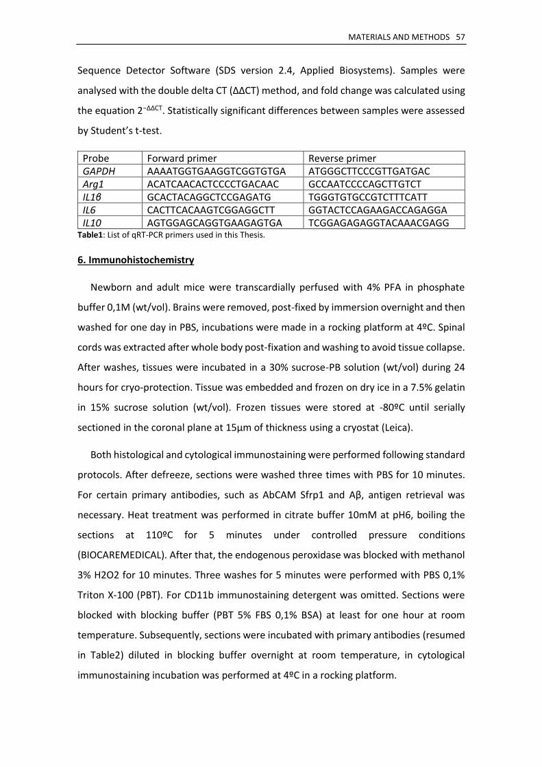
MATERIALS AND METHODS 57
Sequence Detector Software (SDS version 2.4, Applied Biosystems). Samples were
analysed with the double delta CT (ΔΔCT) method, and fold change was calculated using
the equation 2−ΔΔCT. Statistically significant differences between samples were assessed
by Student’s t-test.
Probe Forward primer Reverse primer GAPDH AAAATGGTGAAGGTCGGTGTGA ATGGGCTTCCCGTTGATGAC Arg1 ACATCAACACTCCCCTGACAAC GCCAATCCCCAGCTTGTCT IL1β GCACTACAGGCTCCGAGATG TGGGTGTGCCGTCTTTCATT IL6 CACTTCACAAGTCGGAGGCTT GGTACTCCAGAAGACCAGAGGA IL10 AGTGGAGCAGGTGAAGAGTGA TCGGAGAGAGGTACAAACGAGG
Table1: List of qRT-PCR primers used in this Thesis.
6. Immunohistochemistry
Newborn and adult mice were transcardially perfused with 4% PFA in phosphate
buffer 0,1M (wt/vol). Brains were removed, post-fixed by immersion overnight and then
washed for one day in PBS, incubations were made in a rocking platform at 4ºC. Spinal
cords was extracted after whole body post-fixation and washing to avoid tissue collapse.
After washes, tissues were incubated in a 30% sucrose-PB solution (wt/vol) during 24
hours for cryo-protection. Tissue was embedded and frozen on dry ice in a 7.5% gelatin
in 15% sucrose solution (wt/vol). Frozen tissues were stored at -80ºC until serially
sectioned in the coronal plane at 15µm of thickness using a cryostat (Leica).
Both histological and cytological immunostaining were performed following standard
protocols. After defreeze, sections were washed three times with PBS for 10 minutes.
For certain primary antibodies, such as AbCAM Sfrp1 and Aβ, antigen retrieval was
necessary. Heat treatment was performed in citrate buffer 10mM at pH6, boiling the
sections at 110ºC for 5 minutes under controlled pressure conditions
(BIOCAREMEDICAL). After that, the endogenous peroxidase was blocked with methanol
3% H2O2 for 10 minutes. Three washes for 5 minutes were performed with PBS 0,1%
Triton X-100 (PBT). For CD11b immunostaining detergent was omitted. Sections were
blocked with blocking buffer (PBT 5% FBS 0,1% BSA) at least for one hour at room
temperature. Subsequently, sections were incubated with primary antibodies (resumed
in Table2) diluted in blocking buffer overnight at room temperature, in cytological
immunostaining incubation was performed at 4ºC in a rocking platform.

58 Sfrp1 promotes neuroinflammation through the modulation of ADAM10 proteolytic activity
Antibody Anti- Source Host Dilution
Aβ (4G8) Covance Mouse monoclonal 1:500 Aβ (6E10) Covance Mouse monoclonal 1:500
β-galactosidase AbCAM Chicken polyclonal 1:2000
CD11b BD Rat monoclonal (biotin) 1:200
CD4 BD Rat monoclonal (biotin) 1:200 CD45 Serotec Rat monoclonal 1:500 GFAP Dako Rabbit polyclonal 1:1000 / 1:3000
GFAP (GA5) Millipore Mouse monoclonal 1:1000 / 1:2000 GFP AbCAM Chicken polyclonal 1:2000
Iba1 Wako Rabbit polyclonal 1:1000 / 1:3000 Iba1 Millipore Mouse monoclonal 1:1000 / 1:2000 MBP AbCAM Rabbit polyclonal 1:1000 NeuN Millipore Mouse monoclonal 1:1000 SFRP1 AbCAM Rabbit polyclonal 1:1000 / 1:3000 SFRP1 Sigma Rabbit polyclonal 1:1000 / 1:3000 SFRP1 Cell Signaling Rabbit polyclonal 1:1000 SFRP1 M. Dominguez* Mouse monoclonal 1:1000 / 1:2000
TauP (AT8) Thermo Mouse monoclonal 1:50 Table2: List of primary antibodies used for immunostaining in this Thesis. Source, type, and dilution are indicated. Dilution antibodies used in tissue/cells. *Antibody generated by our laboratory in collaboration with the laboratory of M. Dominguez (ISCIII).
After incubation with primary antibody, three washes for 10 minutes were performed
with PBT. Secondary antibodies were used at the indicated dilutions and incubated for
one hour at room temperature: donkey anti-rabbit Alexa488, donkey anti-rabbit
Alexa594, donkey anti-mouse Alexa488, donkey anti-mouse Alexa594, goat anti-chicken
Alexa488, goat anti-rat Alexa594, 1:1000/1:3000 (Molecular Probes, Invitrogen);
Streptavidin Alexa488, Streptavidin Alexa594, 1:500 (Molecular Probes, Invitrogen);
goat anti-mouse biotin, goat anti-rabbit biotin, 1:500 (Jackson Lab). In some cases, biotin
conjugated antibodies were used in combination with Streptavidin-POD (Jackson Lab).
Antibody staining was revealed with Diaminobenzidine. Sections were counterstained
with Hoechst (Sigma Aldrich). For fibrillary plaques detection sections were stained with
Thiofalvin S in some cases. All samples were mounted with Mowiol (Calbiochem).
The preparations were analysed with a microscope DMCTR5000 equipped with a
DFC350Fx monochrome camera or a DFC500 colour camera (Leica Microsystems).
Confocal imaging was realised with a confocal laser scanning microscope LSM710
coupled to a vertical microscope AxioImager.M2 (Zeiss). Fluorescence quantification
was assessed with ImageJ software (National Institute of Health). 3D reconstruction and
volumetric quantitation was performed using Imaris software (version 7.1.1, Bitplane).
MATERIALS AND METHODS 59
7. Western Blot and Immunoprecipitation
To obtain cellular protein fraction, tissue or cultured cells were equally treated with
RIPA buffer (NP-40 1%, Sodium deoxycholate 0.5%, SDS 0.1% in PBS 1X) complemented
with Proteases Inhibitor Complex (Roche) and PMSF. Tissue was homogenised and
disaggregated, previous to disrupt by sonication. Homogenate was stabilised on ice for
15 minutes and next centrifuged at 15000rpm for 30 minutes at 4ºC. Supernatants were
collected and protein concentration was quantified by BCA protein assay kit (Thermo
Scientific). Different samples were diluted at the same concentration and subsequently
diluted in Laemlli Buffer 5X (Glycerin 50%, SDS 10%, 0,025% Bromophenol blue, 10% β-
mercaptoethanol in 20mM TrisHCl pH6.8). Cell supernatants were directly diluted in
Laemlli Buffer. After re-suspension lysates were denaturalised at 100ºC for 5 minutes.
In the case of Immunoprecipitation, cells were collected in cold PBS and treated with
Brij96 lysis buffer (Brij96 0.33% v/v, NaCl 150mM in 20mM TrisHCl pH7.8)
complemented with Protease Inhibitor Complex (Roche) and PMSF 1mM. The cellular
homogenate was stabilised for 45 minutes on ice and next centrifuged at 15000rpm for
15 minutes. Supernatant aliquot was separated as Input and the rest was incubated for
4 hours with three subsequent preclearing protein G-agarose beads (Sigma Aldrich),
previously incubated overnight with an unspecific antibody of the same isotype as
desired (Mouse IgG1 Isotype Control, R&D Systems). After preclearing, the samples
were centrifuged at 3000rpm for 3 minutes at 4ºC, and the supernatant was next
incubated for 4 hours with protein G-agarose beads previously incubated overnight with
target antibody (2μg of antibody per 40μl beads). Washes of the beads were performed
before and after incubation with the lysate to avoid unspecificities. Beads were finally
re-suspended in Laemlli Buffer 2X and denaturalised at 100ºC for 5 minutes.
All samples were resolved in SDS-PAGE gels of acrylamide/bis-acrylamide 8-12% and
analysed by Western Blot. Proteins were transferred to nitrocellulose membranes by
dry iBlot2 (Invitrogen) and incubated with TBS (NaCl 150mM in TrisHCl 10mM pH8)
containing 0.1% Tween (TBST) and 10% non-fat milk for at least one hour. After blocking,
the membranes were incubated overnight at 4ºC with primary antibodies (described in
Table3) diluted in 5% Bovine Serum Albumin (Sigma Aldrich) in TBST. Afterwards, the
membranes were washed three times for 10 minutes with TBST, and incubated for one
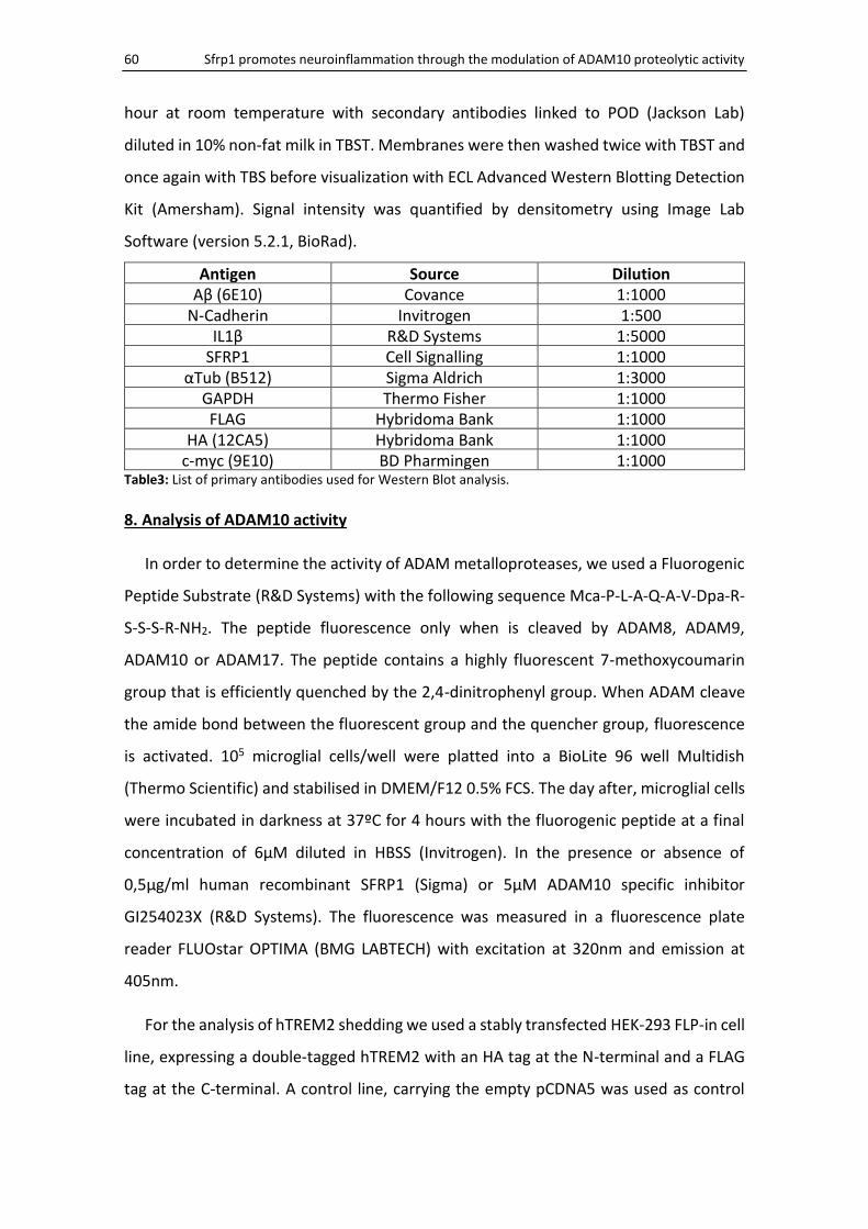
60 Sfrp1 promotes neuroinflammation through the modulation of ADAM10 proteolytic activity
hour at room temperature with secondary antibodies linked to POD (Jackson Lab)
diluted in 10% non-fat milk in TBST. Membranes were then washed twice with TBST and
once again with TBS before visualization with ECL Advanced Western Blotting Detection
Kit (Amersham). Signal intensity was quantified by densitometry using Image Lab
Software (version 5.2.1, BioRad).
Antigen Source Dilution Aβ (6E10) Covance 1:1000
N-Cadherin Invitrogen 1:500 IL1β R&D Systems 1:5000
SFRP1 Cell Signalling 1:1000 αTub (B512) Sigma Aldrich 1:3000
GAPDH Thermo Fisher 1:1000 FLAG Hybridoma Bank 1:1000
HA (12CA5) Hybridoma Bank 1:1000 c-myc (9E10) BD Pharmingen 1:1000
Table3: List of primary antibodies used for Western Blot analysis.
8. Analysis of ADAM10 activity
In order to determine the activity of ADAM metalloproteases, we used a Fluorogenic
Peptide Substrate (R&D Systems) with the following sequence Mca-P-L-A-Q-A-V-Dpa-R-
S-S-S-R-NH2. The peptide fluorescence only when is cleaved by ADAM8, ADAM9,
ADAM10 or ADAM17. The peptide contains a highly fluorescent 7-methoxycoumarin
group that is efficiently quenched by the 2,4-dinitrophenyl group. When ADAM cleave
the amide bond between the fluorescent group and the quencher group, fluorescence
is activated. 105 microglial cells/well were platted into a BioLite 96 well Multidish
(Thermo Scientific) and stabilised in DMEM/F12 0.5% FCS. The day after, microglial cells
were incubated in darkness at 37ºC for 4 hours with the fluorogenic peptide at a final
concentration of 6µM diluted in HBSS (Invitrogen). In the presence or absence of
0,5μg/ml human recombinant SFRP1 (Sigma) or 5μM ADAM10 specific inhibitor
GI254023X (R&D Systems). The fluorescence was measured in a fluorescence plate
reader FLUOstar OPTIMA (BMG LABTECH) with excitation at 320nm and emission at
405nm.
For the analysis of hTREM2 shedding we used a stably transfected HEK-293 FLP-in cell
line, expressing a double-tagged hTREM2 with an HA tag at the N-terminal and a FLAG
tag at the C-terminal. A control line, carrying the empty pCDNA5 was used as control
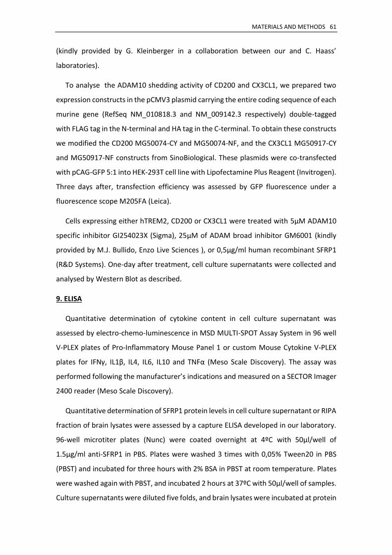
MATERIALS AND METHODS 61
(kindly provided by G. Kleinberger in a collaboration between our and C. Haass’
laboratories).
To analyse the ADAM10 shedding activity of CD200 and CX3CL1, we prepared two
expression constructs in the pCMV3 plasmid carrying the entire coding sequence of each
murine gene (RefSeq NM_010818.3 and NM_009142.3 respectively) double-tagged
with FLAG tag in the N-terminal and HA tag in the C-terminal. To obtain these constructs
we modified the CD200 MG50074-CY and MG50074-NF, and the CX3CL1 MG50917-CY
and MG50917-NF constructs from SinoBiological. These plasmids were co-transfected
with pCAG-GFP 5:1 into HEK-293T cell line with Lipofectamine Plus Reagent (Invitrogen).
Three days after, transfection efficiency was assessed by GFP fluorescence under a
fluorescence scope M205FA (Leica).
Cells expressing either hTREM2, CD200 or CX3CL1 were treated with 5μM ADAM10
specific inhibitor GI254023X (Sigma), 25μM of ADAM broad inhibitor GM6001 (kindly
provided by M.J. Bullido, Enzo Live Sciences ), or 0,5μg/ml human recombinant SFRP1
(R&D Systems). One-day after treatment, cell culture supernatants were collected and
analysed by Western Blot as described.
9. ELISA
Quantitative determination of cytokine content in cell culture supernatant was
assessed by electro-chemo-luminescence in MSD MULTI-SPOT Assay System in 96 well
V-PLEX plates of Pro-Inflammatory Mouse Panel 1 or custom Mouse Cytokine V-PLEX
plates for IFNγ, IL1β, IL4, IL6, IL10 and TNFα (Meso Scale Discovery). The assay was
performed following the manufacturer’s indications and measured on a SECTOR Imager
2400 reader (Meso Scale Discovery).
Quantitative determination of SFRP1 protein levels in cell culture supernatant or RIPA
fraction of brain lysates were assessed by a capture ELISA developed in our laboratory.
96-well microtiter plates (Nunc) were coated overnight at 4ºC with 50µl/well of
1.5µg/ml anti-SFRP1 in PBS. Plates were washed 3 times with 0,05% Tween20 in PBS
(PBST) and incubated for three hours with 2% BSA in PBST at room temperature. Plates
were washed again with PBST, and incubated 2 hours at 37ºC with 50µl/well of samples.
Culture supernatants were diluted five folds, and brain lysates were incubated at protein
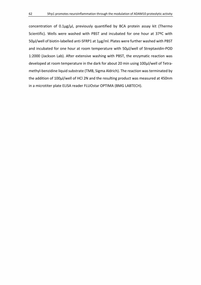
62 Sfrp1 promotes neuroinflammation through the modulation of ADAM10 proteolytic activity
concentration of 0.1µg/µl, previously quantified by BCA protein assay kit (Thermo
Scientific). Wells were washed with PBST and incubated for one hour at 37ºC with
50µl/well of biotin-labelled anti-SFRP1 at 1µg/ml. Plates were further washed with PBST
and incubated for one hour at room temperature with 50µl/well of Streptavidin-POD
1:2000 (Jackson Lab). After extensive washing with PBST, the enzymatic reaction was
developed at room temperature in the dark for about 20 min using 100µl/well of Tetra-
methyl-benzidine liquid substrate (TMB, Sigma Aldrich). The reaction was terminated by
the addition of 100µl/well of HCl 2N and the resulting product was measured at 450nm
in a microtiter plate ELISA reader FLUOstar OPTIMA (BMG LABTECH).

RESULTS
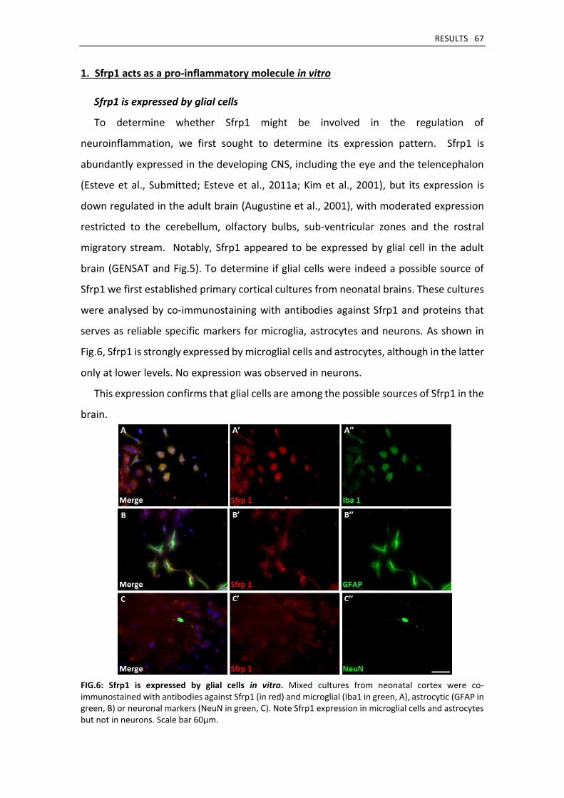
RESULTS 67
1. Sfrp1 acts as a pro-inflammatory molecule in vitro
Sfrp1 is expressed by glial cells
To determine whether Sfrp1 might be involved in the regulation of
neuroinflammation, we first sought to determine its expression pattern. Sfrp1 is
abundantly expressed in the developing CNS, including the eye and the telencephalon
(Esteve et al., Submitted; Esteve et al., 2011a; Kim et al., 2001), but its expression is
down regulated in the adult brain (Augustine et al., 2001), with moderated expression
restricted to the cerebellum, olfactory bulbs, sub-ventricular zones and the rostral
migratory stream. Notably, Sfrp1 appeared to be expressed by glial cell in the adult
brain (GENSAT and Fig.5). To determine if glial cells were indeed a possible source of
Sfrp1 we first established primary cortical cultures from neonatal brains. These cultures
were analysed by co-immunostaining with antibodies against Sfrp1 and proteins that
serves as reliable specific markers for microglia, astrocytes and neurons. As shown in
Fig.6, Sfrp1 is strongly expressed by microglial cells and astrocytes, although in the latter
only at lower levels. No expression was observed in neurons.
This expression confirms that glial cells are among the possible sources of Sfrp1 in the
brain.
FIG.6: Sfrp1 is expressed by glial cells in vitro. Mixed cultures from neonatal cortex were co-immunostained with antibodies against Sfrp1 (in red) and microglial (Iba1 in green, A), astrocytic (GFAP in green, B) or neuronal markers (NeuN in green, C). Note Sfrp1 expression in microglial cells and astrocytes but not in neurons. Scale bar 60μm.
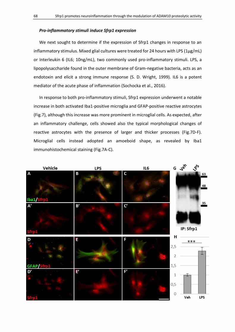
68 Sfrp1 promotes neuroinflammation through the modulation of ADAM10 proteolytic activity
Pro-inflammatory stimuli induce Sfrp1 expression
We next sought to determine if the expression of Sfrp1 changes in response to an
inflammatory stimulus. Mixed glial cultures were treated for 24 hours with LPS (1μg/mL)
or Interleukin 6 (IL6; 10ng/mL), two commonly used pro-inflammatory stimuli. LPS, a
lipopolysaccharide found in the outer membrane of Gram-negative bacteria, acts as an
endotoxin and elicit a strong immune response (S. D. Wright, 1999). IL6 is a potent
mediator of the acute phase of inflammation (Sochocka et al., 2016).
In response to both pro-inflammatory stimuli, Sfrp1 expression underwent a notable
increase in both activated Iba1-positive microglia and GFAP-positive reactive astrocytes
(Fig.7), although this increase was more prominent in microglial cells. As expected, after
an inflammatory challenge, cells showed also the typical morphological changes of
reactive astrocytes with the presence of larger and thicker processes (Fig.7D-F).
Microglial cells instead adopted an amoeboid shape, as revealed by Iba1
immunohistochemical staining (Fig.7A-C).
RESULTS 69
Sfrp1 increase was verified by immunoprecipitation and Western blot comparison of
the protein content in the culture media of LPS-treated and untreated cultures (Fig.7G),
consistent with the highly secreted nature of Sfrp1 protein. Similar results were
obtained using a homemade ELISA (Fig.7H), which revealed that Sfrp1 content in the
media of LPS-treated cells was more than double of that of control cultures.
Lack of Sfrp1 reduces cytokine production and secretion
CNS inflammation is inevitably linked to the activation of glial cells. When activated,
glial cells acquire a polarized phenotype characterized by a different morphology and
different functions required to respond to the insult the CNS has suffered. The main
outcome of this activation is the ability to secrete and respond to a large number of
cytokines (Shichita et al., 2014). The differential activation of glial cells triggers distinct
profiles of cytokine secretion to promote distinct outputs, conferring glial cells with a
pivotal role in modulating the type and extent of the neuroinflammatory response
(Heneka et al., 2014). Glial polarization has been widely studied in the case of microglial
cells and there are a number of well-described activation markers that are typically used
to characterize the so-called M1 or M2 state of microglial cells. The M1 phenotype is
usually defined by the expression of pro-inflammatory cytokines, whereas the M2 state
by the production of anti-inflammatory cytokines (Colton, 2009).
FIG.7: Sfrp1 expression increases in response to pro-inflammatory stimuli. New-born mixed glial cultures (A-F) treated with vehicle (A,D), 1μg/ml LPS (B,E) or 10ng/ml IL6 (C,F) for one day were co-immunostained with antibodies against Sfrp1 and a microglial (Iba1; A-C) or astrocytic markers (GFAP; D-F). Sfrp1 staining (red) increases upon LPS and IL6 treatment, compared to vehicle treated cultures. Note that pro-inflammatory stimuli induce morphological changes in activated microglia (amoeboid; B,C) and reactive astrocytes (larger processes; E,F). G) Immunoprecipitation and Western Blot analysis of the levels of Sfrp1 protein in vehicle or LPS treated cultures revealed that Sfrp1 is enriched in the medium of the cultures treated with LPS. H) ELISA quantification of Sfrp1 levels present in culture supernatants show a 2.25 fold increase of Sfrp1 in response to LPS. Error bars represent Standard Error. Statistical significance: ***P<0.001 by Student's t-test. Scale bar 60μm.
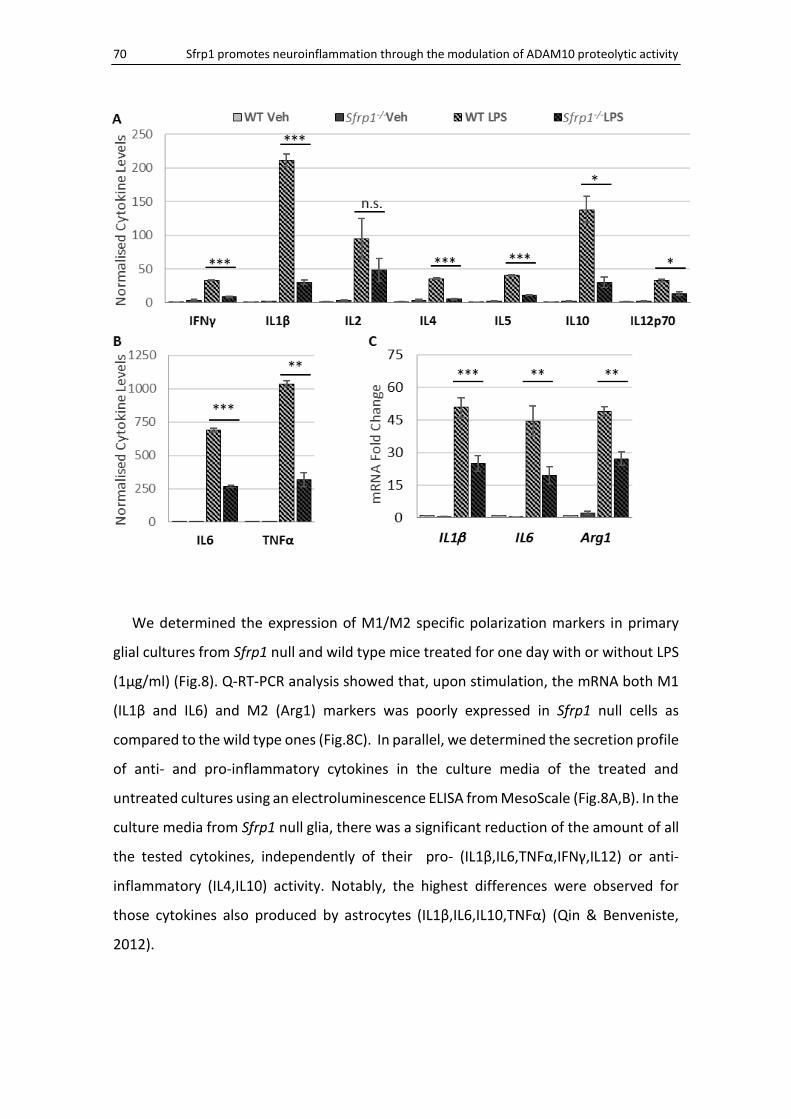
70 Sfrp1 promotes neuroinflammation through the modulation of ADAM10 proteolytic activity
We determined the expression of M1/M2 specific polarization markers in primary
glial cultures from Sfrp1 null and wild type mice treated for one day with or without LPS
(1µg/ml) (Fig.8). Q-RT-PCR analysis showed that, upon stimulation, the mRNA both M1
(IL1β and IL6) and M2 (Arg1) markers was poorly expressed in Sfrp1 null cells as
compared to the wild type ones (Fig.8C). In parallel, we determined the secretion profile
of anti- and pro-inflammatory cytokines in the culture media of the treated and
untreated cultures using an electroluminescence ELISA from MesoScale (Fig.8A,B). In the
culture media from Sfrp1 null glia, there was a significant reduction of the amount of all
the tested cytokines, independently of their pro- (IL1β,IL6,TNFα,IFNγ,IL12) or anti-
inflammatory (IL4,IL10) activity. Notably, the highest differences were observed for
those cytokines also produced by astrocytes (IL1β,IL6,IL10,TNFα) (Qin & Benveniste,
2012).
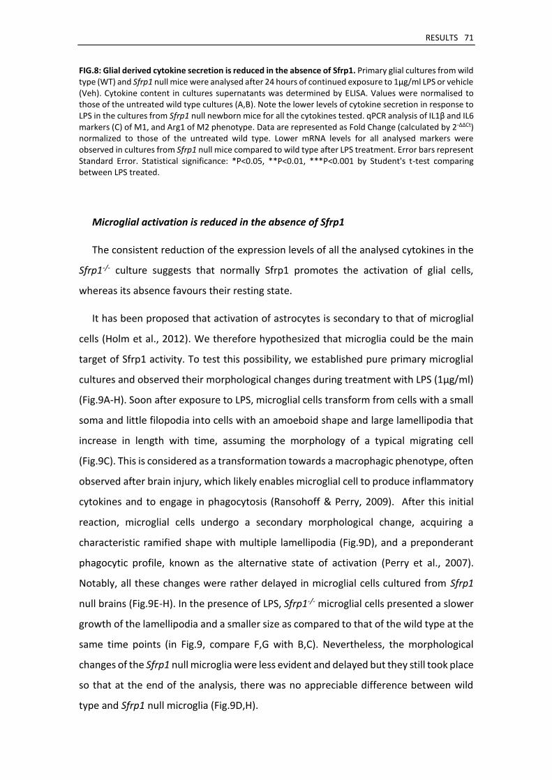
RESULTS 71
FIG.8: Glial derived cytokine secretion is reduced in the absence of Sfrp1. Primary glial cultures from wild type (WT) and Sfrp1 null mice were analysed after 24 hours of continued exposure to 1μg/ml LPS or vehicle (Veh). Cytokine content in cultures supernatants was determined by ELISA. Values were normalised to those of the untreated wild type cultures (A,B). Note the lower levels of cytokine secretion in response to LPS in the cultures from Sfrp1 null newborn mice for all the cytokines tested. qPCR analysis of IL1β and IL6 markers (C) of M1, and Arg1 of M2 phenotype. Data are represented as Fold Change (calculated by 2-ΔΔCt) normalized to those of the untreated wild type. Lower mRNA levels for all analysed markers were observed in cultures from Sfrp1 null mice compared to wild type after LPS treatment. Error bars represent Standard Error. Statistical significance: *P<0.05, **P<0.01, ***P<0.001 by Student's t-test comparing between LPS treated.
Microglial activation is reduced in the absence of Sfrp1
The consistent reduction of the expression levels of all the analysed cytokines in the
Sfrp1-/- culture suggests that normally Sfrp1 promotes the activation of glial cells,
whereas its absence favours their resting state.
It has been proposed that activation of astrocytes is secondary to that of microglial
cells (Holm et al., 2012). We therefore hypothesized that microglia could be the main
target of Sfrp1 activity. To test this possibility, we established pure primary microglial
cultures and observed their morphological changes during treatment with LPS (1µg/ml)
(Fig.9A-H). Soon after exposure to LPS, microglial cells transform from cells with a small
soma and little filopodia into cells with an amoeboid shape and large lamellipodia that
increase in length with time, assuming the morphology of a typical migrating cell
(Fig.9C). This is considered as a transformation towards a macrophagic phenotype, often
observed after brain injury, which likely enables microglial cell to produce inflammatory
cytokines and to engage in phagocytosis (Ransohoff & Perry, 2009). After this initial
reaction, microglial cells undergo a secondary morphological change, acquiring a
characteristic ramified shape with multiple lamellipodia (Fig.9D), and a preponderant
phagocytic profile, known as the alternative state of activation (Perry et al., 2007).
Notably, all these changes were rather delayed in microglial cells cultured from Sfrp1
null brains (Fig.9E-H). In the presence of LPS, Sfrp1-/- microglial cells presented a slower
growth of the lamellipodia and a smaller size as compared to that of the wild type at the
same time points (in Fig.9, compare F,G with B,C). Nevertheless, the morphological
changes of the Sfrp1 null microglia were less evident and delayed but they still took place
so that at the end of the analysis, there was no appreciable difference between wild
type and Sfrp1 null microglia (Fig.9D,H).
72 Sfrp1 promotes neuroinflammation through the modulation of ADAM10 proteolytic activity
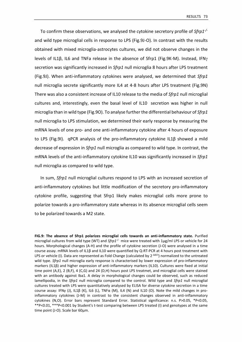
RESULTS 73
To confirm these observations, we analysed the cytokine secretory profile of Sfrp1-/-
and wild type microglial cells in response to LPS (Fig.9J-O). In contrast with the results
obtained with mixed microglia-astrocytes cultures, we did not observe changes in the
levels of IL1β, IL6 and TNFa release in the absence of Sfrp1 (Fig.9K-M). Instead, IFN
secretion was significantly increased in Sfrp1 null microglia 8 hours after LPS treatment
(Fig.9J). When anti-inflammatory cytokines were analysed, we determined that Sfrp1
null microglia secrete significantly more IL4 at 4-8 hours after LPS treatment (Fig.9N)
There was also a consistent increase of IL10 release to the media of Sfrp1 null microglial
cultures and, interestingly, even the basal level of IL10 secretion was higher in null
microglia than in wild type (Fig.9O). To analyse further the differential behaviour of Sfrp1
null microglia to LPS stimulation, we determined their early response by measuring the
mRNA levels of one pro- and one anti-inflammatory cytokine after 4 hours of exposure
to LPS (Fig.9I). qPCR analysis of the pro-inflammatory cytokine IL1β showed a mild
decrease of expression in Sfrp1 null microglia as compared to wild type. In contrast, the
mRNA levels of the anti-inflammatory cytokine IL10 was significantly increased in Sfrp1
null microglia as compared to wild type.
In sum, Sfrp1 null microglial cultures respond to LPS with an increased secretion of
anti-inflammatory cytokines but little modification of the secretory pro-inflammatory
cytokine profile, suggesting that Sfrp1 likely makes microglial cells more prone to
polarize towards a pro-inflammatory state whereas in its absence microglial cells seem
to be polarized towards a M2 state.
FIG.9: The absence of Sfrp1 polarizes microglial cells towards an anti-inflammatory state. Purified microglial cultures from wild type (WT) and Sfrp1-/- mice were treated with 1μg/ml LPS or vehicle for 24 hours. Morphological changes (A-H) and the profile of cytokine secretion (J-O) were analysed in a time course assay. mRNA levels of IL1β and IL10 were quantified by Q-RT-PCR at 4 hours post treatment with LPS or vehicle (I). Data are represented as Fold Change (calculated by 2-ΔΔCt) normalized to the untreated wild type. Sfrp1 null microglia early response is characterised by lower expression of pro-inflammatory markers (IL1β) and higher expression of anti-inflammatory markers (IL10). Cultures were fixed at initial time point (A,E), 2 (B,F), 4 (C,G) and 24 (D,H) hours post LPS treatmet, and microglial cells were stained with an antibody against Iba1. A delay in morphological changes could be observed, such as reduced lamellipodia, in the Sfrp1 null microglia compared to the control. Wild type and Sfrp1 null microglial cultures treated with LPS were quantitatively analysed by ELISA for diverse cytokine secretion in a time course assay: IFNγ (J), IL1β (K), IL6 (L), TNFα (M), IL4 (N) and IL10 (O). Note the mild changes in pro-inflammatory cytokines (J-M) in contrast to the consistent changes observed in anti-inflammatory cytokines (N,O). Error bars represent Standard Error. Statistical significance: n.s. P>0.05, *P<0.05, **P<0.01, ***P<0.001 by Student's t-test comparing between LPS treated (I) and genotypes at the same time point (J-O). Scale bar 60μm.
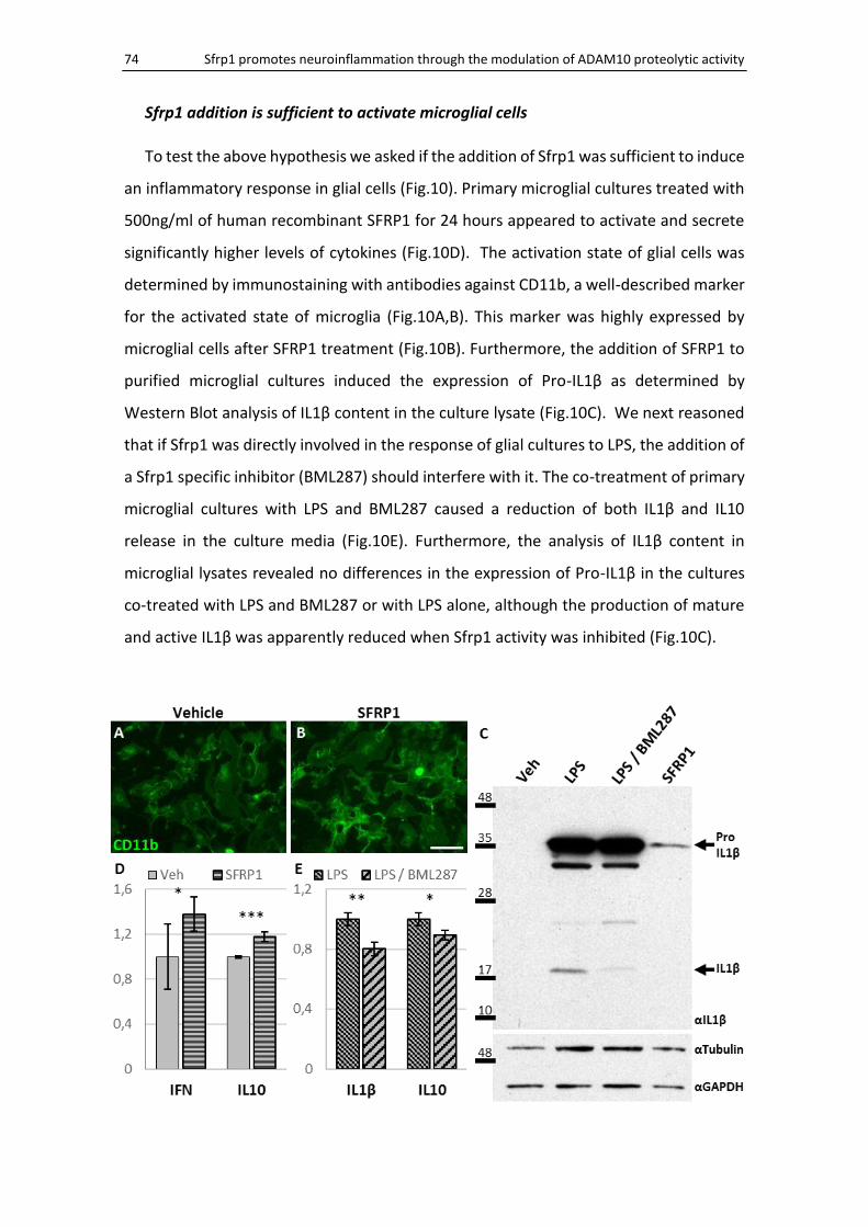
74 Sfrp1 promotes neuroinflammation through the modulation of ADAM10 proteolytic activity
Sfrp1 addition is sufficient to activate microglial cells
To test the above hypothesis we asked if the addition of Sfrp1 was sufficient to induce
an inflammatory response in glial cells (Fig.10). Primary microglial cultures treated with
500ng/ml of human recombinant SFRP1 for 24 hours appeared to activate and secrete
significantly higher levels of cytokines (Fig.10D). The activation state of glial cells was
determined by immunostaining with antibodies against CD11b, a well-described marker
for the activated state of microglia (Fig.10A,B). This marker was highly expressed by
microglial cells after SFRP1 treatment (Fig.10B). Furthermore, the addition of SFRP1 to
purified microglial cultures induced the expression of Pro-IL1β as determined by
Western Blot analysis of IL1β content in the culture lysate (Fig.10C). We next reasoned
that if Sfrp1 was directly involved in the response of glial cultures to LPS, the addition of
a Sfrp1 specific inhibitor (BML287) should interfere with it. The co-treatment of primary
microglial cultures with LPS and BML287 caused a reduction of both IL1β and IL10
release in the culture media (Fig.10E). Furthermore, the analysis of IL1β content in
microglial lysates revealed no differences in the expression of Pro-IL1β in the cultures
co-treated with LPS and BML287 or with LPS alone, although the production of mature
and active IL1β was apparently reduced when Sfrp1 activity was inhibited (Fig.10C).
RESULTS 75
2. Sfrp1 is necessary and sufficient to induce an inflammatory response in vivo
Altogether the results obtained with glial cell cultures provided evidence that Sfrp1
is sufficient to trigger microglial activation. However, under in vitro conditions,
microglial cells behaviour differ from brain microglia, because the environment
influences macrophage functions and gene expression (Gosselin et al., 2014). The next
obvious question was if this occured also in vivo, where microglial are embedded into a
different milieu, interacting with other cells types that also respond to insults.
To answer this question we took advantage again of LPS, as its infusion in the brain
represents a broadly applied tool to induce brain inflammatory processes (S. D. Wright,
1999). Specifically, we infused LPS (5μg in total) directly into the brain parenchyma by
intra-cerebral injection to groups of 4-6 animals, while controls were injected with
saline. Three days after surgery, when the inflammatory process reaches its peak of
activation (Rivest, 2009), the animals were sacrificed and the brains were analysed
either by histological or biochemical procedures. The correct localization of the injection
was verified during histological analysis, and badly injected animals were discarded.
FIG.10: SFRP1 treatment is sufficient to trigger an inflammatory response in cultured glial cells. A,B) Mixed glial cultures from wild type cortex treated with 500ng/ml human recombinant SFRP1 for 24 hours were stained with an antibody against CD11b (A,B). Note the higher levels of CD11b expression in the presence of SFRP1, indicating an activated state of microglial cells. C) Purified microglial cultures were treated with 500ng/ml SFRP1, 1μg/ml LPS, 5μM BML287, a specific Sfrp1 inhibitor, and/or vehicle for 24 hours. After treatment, cell lysates were assessed by Western Blot with an antibody against IL1β. Note that LPS induces a huge increment of Pro-IL1β (35KDa) expression and its cleavage into the active IL1β (17KDa) compared to vehicle treatment. Observe the reduced LPS mediated formation of mature IL1β in the presence of BML287. Conversely, SFRP1 addition induces the expression of Pro-IL1β. D,E) ELISA of cell supernatants to determine the content of IL1β and IL10 released by microglial cells. SFRP1 induces a significant increase in the secretion of both cytokines (D). BML287 prevents LPS-mediated release of both IL1β and IL10 (E). Error bars represent Standard Error. Statistical significance: *P<0.05, **P<0.01, ***P<0.001 by Student's t-test comparing between closed. Scale bar 60μm.
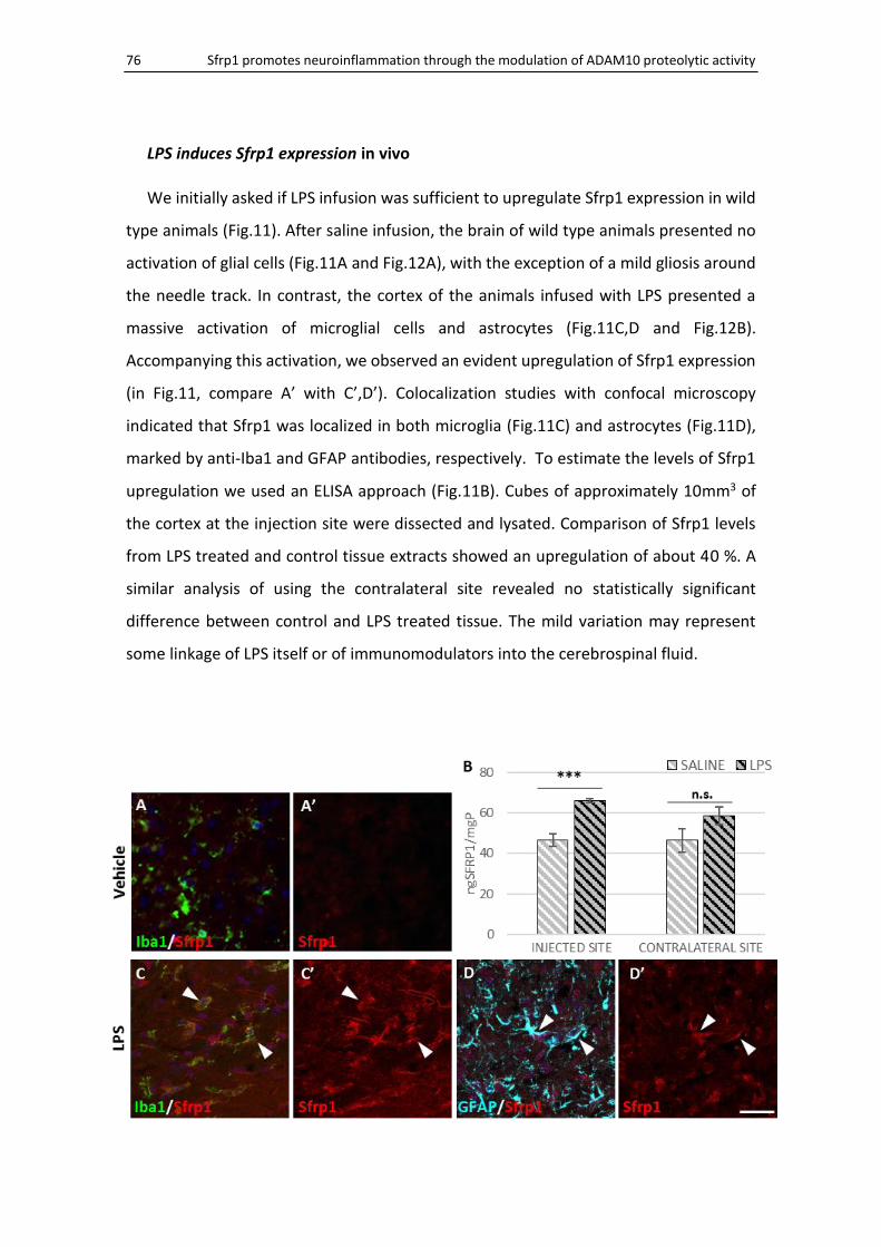
76 Sfrp1 promotes neuroinflammation through the modulation of ADAM10 proteolytic activity
LPS induces Sfrp1 expression in vivo
We initially asked if LPS infusion was sufficient to upregulate Sfrp1 expression in wild
type animals (Fig.11). After saline infusion, the brain of wild type animals presented no
activation of glial cells (Fig.11A and Fig.12A), with the exception of a mild gliosis around
the needle track. In contrast, the cortex of the animals infused with LPS presented a
massive activation of microglial cells and astrocytes (Fig.11C,D and Fig.12B).
Accompanying this activation, we observed an evident upregulation of Sfrp1 expression
(in Fig.11, compare A’ with C’,D’). Colocalization studies with confocal microscopy
indicated that Sfrp1 was localized in both microglia (Fig.11C) and astrocytes (Fig.11D),
marked by anti-Iba1 and GFAP antibodies, respectively. To estimate the levels of Sfrp1
upregulation we used an ELISA approach (Fig.11B). Cubes of approximately 10mm3 of
the cortex at the injection site were dissected and lysated. Comparison of Sfrp1 levels
from LPS treated and control tissue extracts showed an upregulation of about 40 %. A
similar analysis of using the contralateral site revealed no statistically significant
difference between control and LPS treated tissue. The mild variation may represent
some linkage of LPS itself or of immunomodulators into the cerebrospinal fluid.
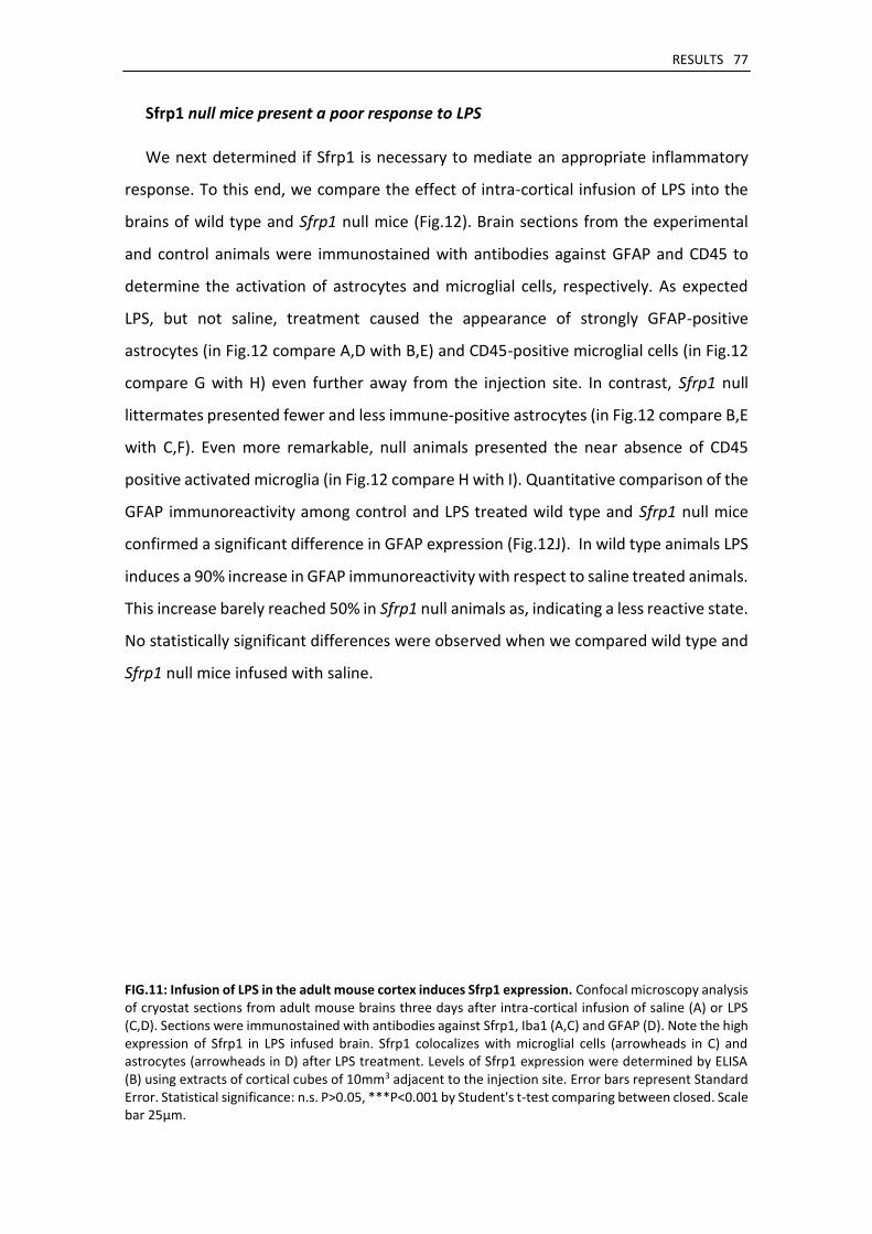
RESULTS 77
Sfrp1 null mice present a poor response to LPS
We next determined if Sfrp1 is necessary to mediate an appropriate inflammatory
response. To this end, we compare the effect of intra-cortical infusion of LPS into the
brains of wild type and Sfrp1 null mice (Fig.12). Brain sections from the experimental
and control animals were immunostained with antibodies against GFAP and CD45 to
determine the activation of astrocytes and microglial cells, respectively. As expected
LPS, but not saline, treatment caused the appearance of strongly GFAP-positive
astrocytes (in Fig.12 compare A,D with B,E) and CD45-positive microglial cells (in Fig.12
compare G with H) even further away from the injection site. In contrast, Sfrp1 null
littermates presented fewer and less immune-positive astrocytes (in Fig.12 compare B,E
with C,F). Even more remarkable, null animals presented the near absence of CD45
positive activated microglia (in Fig.12 compare H with I). Quantitative comparison of the
GFAP immunoreactivity among control and LPS treated wild type and Sfrp1 null mice
confirmed a significant difference in GFAP expression (Fig.12J). In wild type animals LPS
induces a 90% increase in GFAP immunoreactivity with respect to saline treated animals.
This increase barely reached 50% in Sfrp1 null animals as, indicating a less reactive state.
No statistically significant differences were observed when we compared wild type and
Sfrp1 null mice infused with saline.
FIG.11: Infusion of LPS in the adult mouse cortex induces Sfrp1 expression. Confocal microscopy analysis of cryostat sections from adult mouse brains three days after intra-cortical infusion of saline (A) or LPS (C,D). Sections were immunostained with antibodies against Sfrp1, Iba1 (A,C) and GFAP (D). Note the high expression of Sfrp1 in LPS infused brain. Sfrp1 colocalizes with microglial cells (arrowheads in C) and astrocytes (arrowheads in D) after LPS treatment. Levels of Sfrp1 expression were determined by ELISA (B) using extracts of cortical cubes of 10mm3 adjacent to the injection site. Error bars represent Standard Error. Statistical significance: n.s. P>0.05, ***P<0.001 by Student's t-test comparing between closed. Scale bar 25μm.
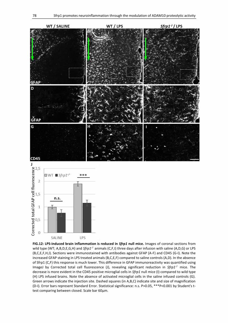
78 Sfrp1 promotes neuroinflammation through the modulation of ADAM10 proteolytic activity
FIG.12: LPS-induced brain inflammation is reduced in Sfrp1 null mice. Images of coronal sections from wild type (WT; A,B,D,E,G,H) and Sfrp1-/- animals (C,F,I) three days after infusion with saline (A,D,G) or LPS (B,C,E,F,H,I). Sections were immunostained with antibodies against GFAP (A-F) and CD45 (G-I). Note the increased GFAP staining in LPS treated animals (B,C,E,F) compared to saline controls (A,D). In the absence of Sfrp1 (C,F) this response is much lower. This difference in GFAP immunoreactivity was quantified using ImageJ by Corrected total cell fluorescence (J), revealing significant reduction in Sfrp1-/- mice. The decrease is more evident in the CD45 positive microglial cells in Sfrp1 null mice (I) compared to wild type (H) LPS infused brains. Note the absence of activated microglial cells in the saline infused controls (G). Green arrows indicate the injection site. Dashed squares (in A,B,C) indicate site and size of magnification (D-I). Error bars represent Standard Error. Statistical significance: n.s. P>0.05, ***P<0.001 by Student's t-test comparing between closed. Scale bar 60μm.
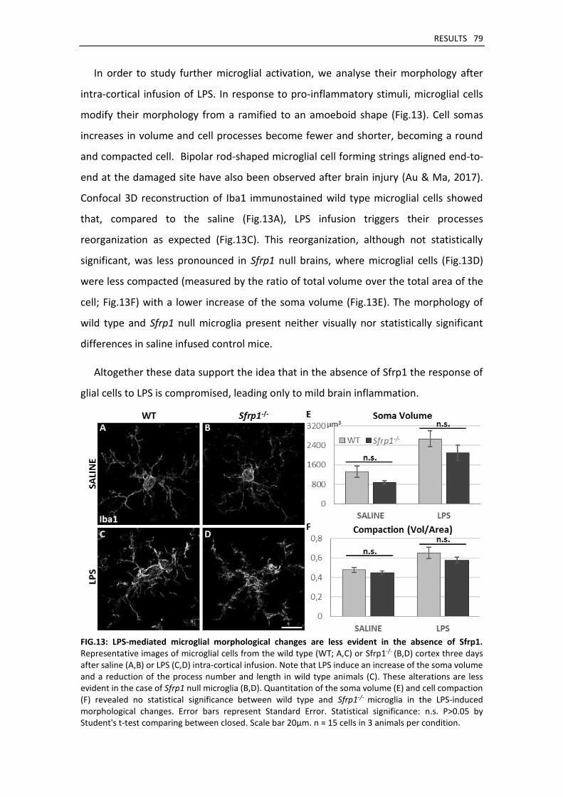
RESULTS 79
In order to study further microglial activation, we analyse their morphology after
intra-cortical infusion of LPS. In response to pro-inflammatory stimuli, microglial cells
modify their morphology from a ramified to an amoeboid shape (Fig.13). Cell somas
increases in volume and cell processes become fewer and shorter, becoming a round
and compacted cell. Bipolar rod-shaped microglial cell forming strings aligned end-to-
end at the damaged site have also been observed after brain injury (Au & Ma, 2017).
Confocal 3D reconstruction of Iba1 immunostained wild type microglial cells showed
that, compared to the saline (Fig.13A), LPS infusion triggers their processes
reorganization as expected (Fig.13C). This reorganization, although not statistically
significant, was less pronounced in Sfrp1 null brains, where microglial cells (Fig.13D)
were less compacted (measured by the ratio of total volume over the total area of the
cell; Fig.13F) with a lower increase of the soma volume (Fig.13E). The morphology of
wild type and Sfrp1 null microglia present neither visually nor statistically significant
differences in saline infused control mice.
Altogether these data support the idea that in the absence of Sfrp1 the response of
glial cells to LPS is compromised, leading only to mild brain inflammation.
FIG.13: LPS-mediated microglial morphological changes are less evident in the absence of Sfrp1. Representative images of microglial cells from the wild type (WT; A,C) or Sfrp1-/- (B,D) cortex three days after saline (A,B) or LPS (C,D) intra-cortical infusion. Note that LPS induce an increase of the soma volume and a reduction of the process number and length in wild type animals (C). These alterations are less evident in the case of Sfrp1 null microglia (B,D). Quantitation of the soma volume (E) and cell compaction (F) revealed no statistical significance between wild type and Sfrp1-/- microglia in the LPS-induced morphological changes. Error bars represent Standard Error. Statistical significance: n.s. P>0.05 by Student's t-test comparing between closed. Scale bar 20μm. n = 15 cells in 3 animals per condition.
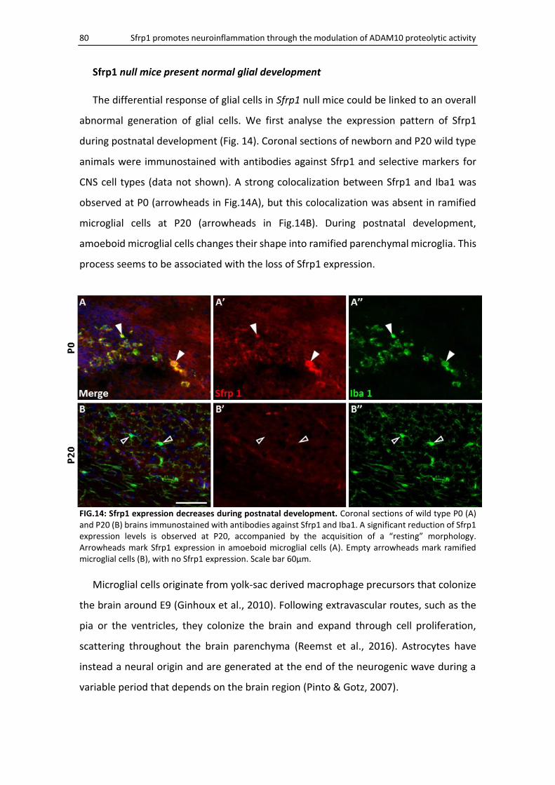
80 Sfrp1 promotes neuroinflammation through the modulation of ADAM10 proteolytic activity
Sfrp1 null mice present normal glial development
The differential response of glial cells in Sfrp1 null mice could be linked to an overall
abnormal generation of glial cells. We first analyse the expression pattern of Sfrp1
during postnatal development (Fig. 14). Coronal sections of newborn and P20 wild type
animals were immunostained with antibodies against Sfrp1 and selective markers for
CNS cell types (data not shown). A strong colocalization between Sfrp1 and Iba1 was
observed at P0 (arrowheads in Fig.14A), but this colocalization was absent in ramified
microglial cells at P20 (arrowheads in Fig.14B). During postnatal development,
amoeboid microglial cells changes their shape into ramified parenchymal microglia. This
process seems to be associated with the loss of Sfrp1 expression.
FIG.14: Sfrp1 expression decreases during postnatal development. Coronal sections of wild type P0 (A) and P20 (B) brains immunostained with antibodies against Sfrp1 and Iba1. A significant reduction of Sfrp1 expression levels is observed at P20, accompanied by the acquisition of a “resting” morphology. Arrowheads mark Sfrp1 expression in amoeboid microglial cells (A). Empty arrowheads mark ramified microglial cells (B), with no Sfrp1 expression. Scale bar 60μm.
Microglial cells originate from yolk-sac derived macrophage precursors that colonize
the brain around E9 (Ginhoux et al., 2010). Following extravascular routes, such as the
pia or the ventricles, they colonize the brain and expand through cell proliferation,
scattering throughout the brain parenchyma (Reemst et al., 2016). Astrocytes have
instead a neural origin and are generated at the end of the neurogenic wave during a
variable period that depends on the brain region (Pinto & Gotz, 2007).
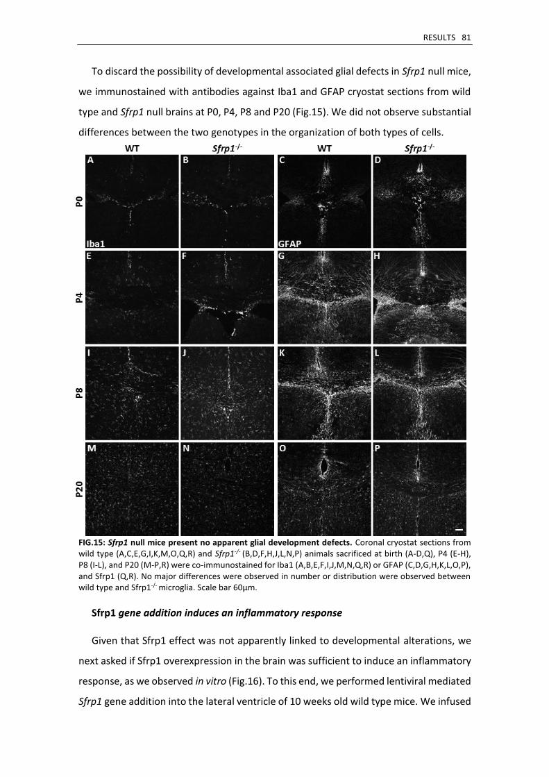
RESULTS 81
To discard the possibility of developmental associated glial defects in Sfrp1 null mice,
we immunostained with antibodies against Iba1 and GFAP cryostat sections from wild
type and Sfrp1 null brains at P0, P4, P8 and P20 (Fig.15). We did not observe substantial
differences between the two genotypes in the organization of both types of cells.
FIG.15: Sfrp1 null mice present no apparent glial development defects. Coronal cryostat sections from wild type (A,C,E,G,I,K,M,O,Q,R) and Sfrp1-/- (B,D,F,H,J,L,N,P) animals sacrificed at birth (A-D,Q), P4 (E-H), P8 (I-L), and P20 (M-P,R) were co-immunostained for Iba1 (A,B,E,F,I,J,M,N,Q,R) or GFAP (C,D,G,H,K,L,O,P), and Sfrp1 (Q,R). No major differences were observed in number or distribution were observed between wild type and Sfrp1-/- microglia. Scale bar 60μm.
Sfrp1 gene addition induces an inflammatory response
Given that Sfrp1 effect was not apparently linked to developmental alterations, we
next asked if Sfrp1 overexpression in the brain was sufficient to induce an inflammatory
response, as we observed in vitro (Fig.16). To this end, we performed lentiviral mediated
Sfrp1 gene addition into the lateral ventricle of 10 weeks old wild type mice. We infused
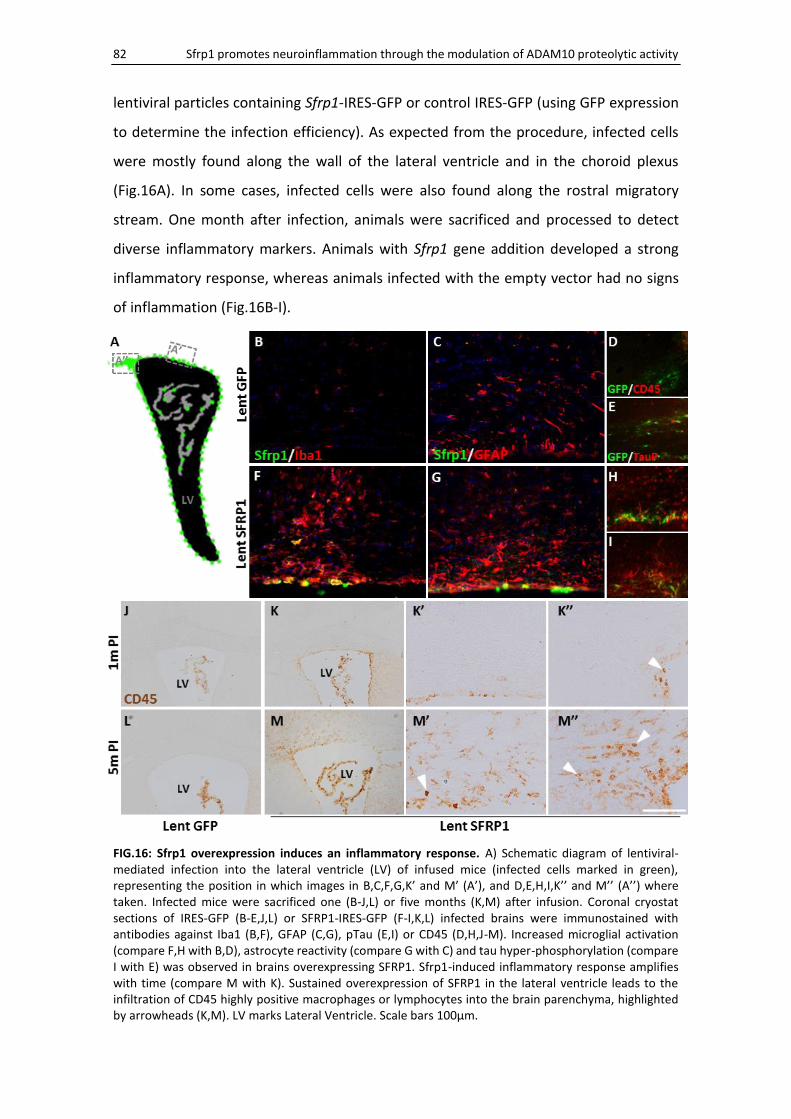
82 Sfrp1 promotes neuroinflammation through the modulation of ADAM10 proteolytic activity
lentiviral particles containing Sfrp1-IRES-GFP or control IRES-GFP (using GFP expression
to determine the infection efficiency). As expected from the procedure, infected cells
were mostly found along the wall of the lateral ventricle and in the choroid plexus
(Fig.16A). In some cases, infected cells were also found along the rostral migratory
stream. One month after infection, animals were sacrificed and processed to detect
diverse inflammatory markers. Animals with Sfrp1 gene addition developed a strong
inflammatory response, whereas animals infected with the empty vector had no signs
of inflammation (Fig.16B-I).
FIG.16: Sfrp1 overexpression induces an inflammatory response. A) Schematic diagram of lentiviral-mediated infection into the lateral ventricle (LV) of infused mice (infected cells marked in green), representing the position in which images in B,C,F,G,K’ and M’ (A’), and D,E,H,I,K’’ and M’’ (A’’) where taken. Infected mice were sacrificed one (B-J,L) or five months (K,M) after infusion. Coronal cryostat sections of IRES-GFP (B-E,J,L) or SFRP1-IRES-GFP (F-I,K,L) infected brains were immunostained with antibodies against Iba1 (B,F), GFAP (C,G), pTau (E,I) or CD45 (D,H,J-M). Increased microglial activation (compare F,H with B,D), astrocyte reactivity (compare G with C) and tau hyper-phosphorylation (compare I with E) was observed in brains overexpressing SFRP1. Sfrp1-induced inflammatory response amplifies with time (compare M with K). Sustained overexpression of SFRP1 in the lateral ventricle leads to the infiltration of CD45 highly positive macrophages or lymphocytes into the brain parenchyma, highlighted by arrowheads (K,M). LV marks Lateral Ventricle. Scale bars 100μm.
RESULTS 83
The cortex of Lent-Sfrp1 infected animals presented a large number of reactive
astrocytes, characterized by enlarged and long processes and a strong GFAP
immunoreactivity (Fig.16G). Similarly, a large number of Iba1-positive activated
microglial cells was detected in the surrounding of the site of infection (Fig.16F).
Microglia presented an amoeboid appearance with high expression of CD45 (Fig.16H), a
marker of microglial activation. Around the infected cells, there was also accumulation
of hyper-phosphorylated Tau (Fig.16I), an established marker for neurodegeneration
and inflammation (Grubman et al., 2016).
In order to determine whether or not this effect was transitory and the brain
eventually resolved the Sfrp1 induced neuroinflammation, we analyse the effect of
lentiviral infection after five months (Fig.16L,M). After such a period, inflammatory signs
were still persistent and neuroinflammation had actually amplified (in Fig.16, compare
M with K), in contrast to the animals infected with the empty vector (Fig.16L), which
were normal. Indeed, the cortex of Lent-Sfrp1 treated animals showed a large number
of CD45 positive cells, indicating the presence of activated microglial cells. Notably, in
the brain parenchyma there was also a number of highly positive CD45 and round-
shaped cells likely representing infiltrated macrophages or lymphocytes (arrowheads in
Fig.16M’ and M’’).
Together these results support the idea that Sfrp1 is necessary and sufficient to
trigger an inflammatory response in the brain.
3. Sfrp1 exacerbated the symptomatology of EAE
We reasoned that if Sfrp1 has a general pro-inflammatory activity in the brain, its
expression should be upregulated in other experimental paradigms with a clear
inflammatory component other than that induced by LPS.
Multiple Sclerosis is a degenerative autoimmune disease with a strong inflammatory
component. The principal model used to study this disease is the Experimental
Autoimmune Encephalomyelitis (EAE). EAE is generated by the passive immunization of
mice with Myelin Oligodendrocyte Glycoprotein (MOG) fragments, which triggers an
autoimmune response. We thus used this model to test further the implications of Sfrp1
in CNS inflammation.
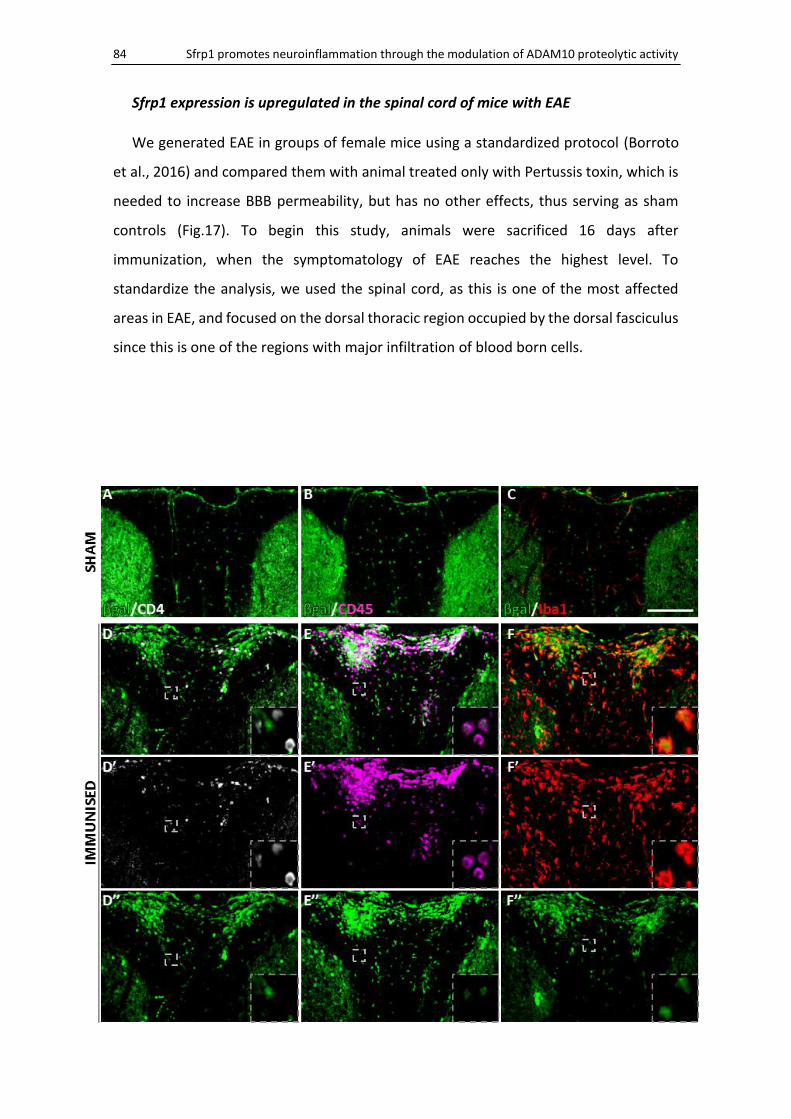
84 Sfrp1 promotes neuroinflammation through the modulation of ADAM10 proteolytic activity
Sfrp1 expression is upregulated in the spinal cord of mice with EAE
We generated EAE in groups of female mice using a standardized protocol (Borroto
et al., 2016) and compared them with animal treated only with Pertussis toxin, which is
needed to increase BBB permeability, but has no other effects, thus serving as sham
controls (Fig.17). To begin this study, animals were sacrificed 16 days after
immunization, when the symptomatology of EAE reaches the highest level. To
standardize the analysis, we used the spinal cord, as this is one of the most affected
areas in EAE, and focused on the dorsal thoracic region occupied by the dorsal fasciculus
since this is one of the regions with major infiltration of blood born cells.
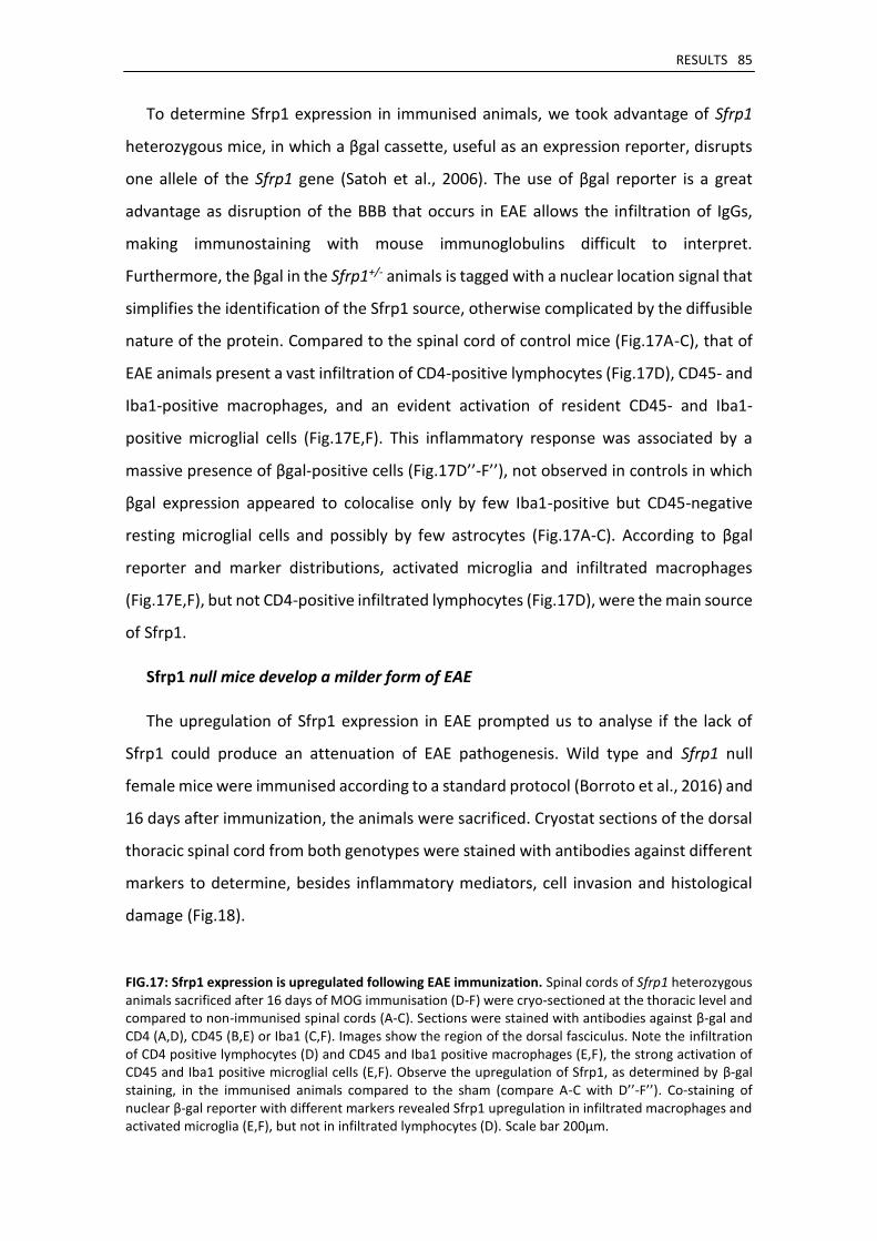
RESULTS 85
To determine Sfrp1 expression in immunised animals, we took advantage of Sfrp1
heterozygous mice, in which a βgal cassette, useful as an expression reporter, disrupts
one allele of the Sfrp1 gene (Satoh et al., 2006). The use of βgal reporter is a great
advantage as disruption of the BBB that occurs in EAE allows the infiltration of IgGs,
making immunostaining with mouse immunoglobulins difficult to interpret.
Furthermore, the βgal in the Sfrp1+/- animals is tagged with a nuclear location signal that
simplifies the identification of the Sfrp1 source, otherwise complicated by the diffusible
nature of the protein. Compared to the spinal cord of control mice (Fig.17A-C), that of
EAE animals present a vast infiltration of CD4-positive lymphocytes (Fig.17D), CD45- and
Iba1-positive macrophages, and an evident activation of resident CD45- and Iba1-
positive microglial cells (Fig.17E,F). This inflammatory response was associated by a
massive presence of βgal-positive cells (Fig.17D’’-F’’), not observed in controls in which
βgal expression appeared to colocalise only by few Iba1-positive but CD45-negative
resting microglial cells and possibly by few astrocytes (Fig.17A-C). According to βgal
reporter and marker distributions, activated microglia and infiltrated macrophages
(Fig.17E,F), but not CD4-positive infiltrated lymphocytes (Fig.17D), were the main source
of Sfrp1.
Sfrp1 null mice develop a milder form of EAE
The upregulation of Sfrp1 expression in EAE prompted us to analyse if the lack of
Sfrp1 could produce an attenuation of EAE pathogenesis. Wild type and Sfrp1 null
female mice were immunised according to a standard protocol (Borroto et al., 2016) and
16 days after immunization, the animals were sacrificed. Cryostat sections of the dorsal
thoracic spinal cord from both genotypes were stained with antibodies against different
markers to determine, besides inflammatory mediators, cell invasion and histological
damage (Fig.18).
FIG.17: Sfrp1 expression is upregulated following EAE immunization. Spinal cords of Sfrp1 heterozygous animals sacrificed after 16 days of MOG immunisation (D-F) were cryo-sectioned at the thoracic level and compared to non-immunised spinal cords (A-C). Sections were stained with antibodies against β-gal and CD4 (A,D), CD45 (B,E) or Iba1 (C,F). Images show the region of the dorsal fasciculus. Note the infiltration of CD4 positive lymphocytes (D) and CD45 and Iba1 positive macrophages (E,F), the strong activation of CD45 and Iba1 positive microglial cells (E,F). Observe the upregulation of Sfrp1, as determined by β-gal staining, in the immunised animals compared to the sham (compare A-C with D’’-F’’). Co-staining of nuclear β-gal reporter with different markers revealed Sfrp1 upregulation in infiltrated macrophages and activated microglia (E,F), but not in infiltrated lymphocytes (D). Scale bar 200μm.
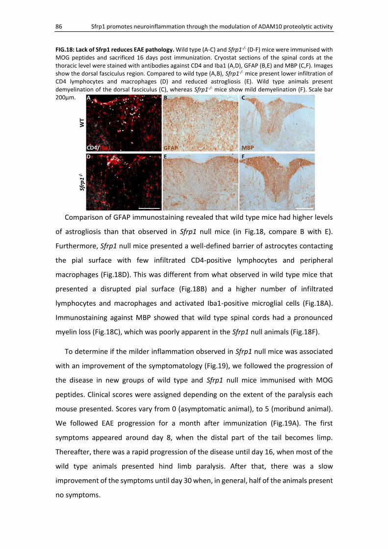
86 Sfrp1 promotes neuroinflammation through the modulation of ADAM10 proteolytic activity
FIG.18: Lack of Sfrp1 reduces EAE pathology. Wild type (A-C) and Sfrp1-/- (D-F) mice were immunised with MOG peptides and sacrificed 16 days post immunization. Cryostat sections of the spinal cords at the thoracic level were stained with antibodies against CD4 and Iba1 (A,D), GFAP (B,E) and MBP (C,F). Images show the dorsal fasciculus region. Compared to wild type (A,B), Sfrp1-/- mice present lower infiltration of CD4 lymphocytes and macrophages (D) and reduced astrogliosis (E). Wild type animals present demyelination of the dorsal fasciculus (C), whereas Sfrp1-/- mice show mild demyelination (F). Scale bar 200μm.
Comparison of GFAP immunostaining revealed that wild type mice had higher levels
of astrogliosis than that observed in Sfrp1 null mice (in Fig.18, compare B with E).
Furthermore, Sfrp1 null mice presented a well-defined barrier of astrocytes contacting
the pial surface with few infiltrated CD4-positive lymphocytes and peripheral
macrophages (Fig.18D). This was different from what observed in wild type mice that
presented a disrupted pial surface (Fig.18B) and a higher number of infiltrated
lymphocytes and macrophages and activated Iba1-positive microglial cells (Fig.18A).
Immunostaining against MBP showed that wild type spinal cords had a pronounced
myelin loss (Fig.18C), which was poorly apparent in the Sfrp1 null animals (Fig.18F).
To determine if the milder inflammation observed in Sfrp1 null mice was associated
with an improvement of the symptomatology (Fig.19), we followed the progression of
the disease in new groups of wild type and Sfrp1 null mice immunised with MOG
peptides. Clinical scores were assigned depending on the extent of the paralysis each
mouse presented. Scores vary from 0 (asymptomatic animal), to 5 (moribund animal).
We followed EAE progression for a month after immunization (Fig.19A). The first
symptoms appeared around day 8, when the distal part of the tail becomes limp.
Thereafter, there was a rapid progression of the disease until day 16, when most of the
wild type animals presented hind limb paralysis. After that, there was a slow
improvement of the symptoms until day 30 when, in general, half of the animals present
no symptoms.
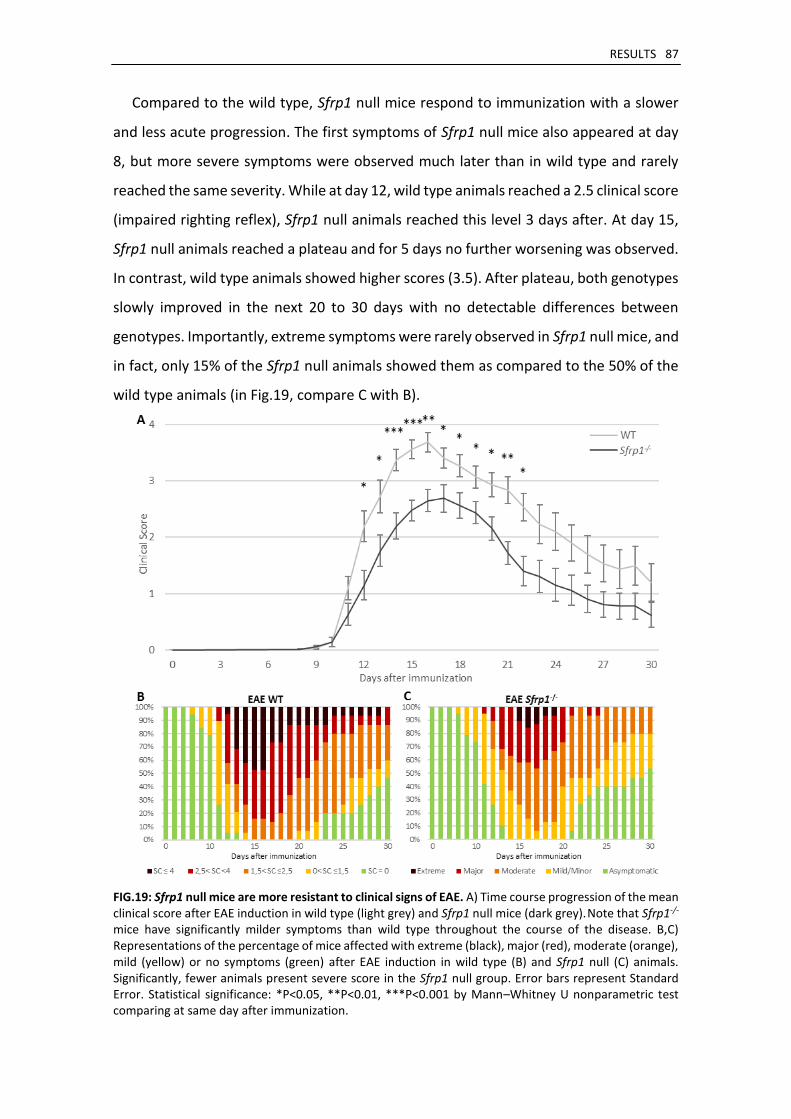
RESULTS 87
Compared to the wild type, Sfrp1 null mice respond to immunization with a slower
and less acute progression. The first symptoms of Sfrp1 null mice also appeared at day
8, but more severe symptoms were observed much later than in wild type and rarely
reached the same severity. While at day 12, wild type animals reached a 2.5 clinical score
(impaired righting reflex), Sfrp1 null animals reached this level 3 days after. At day 15,
Sfrp1 null animals reached a plateau and for 5 days no further worsening was observed.
In contrast, wild type animals showed higher scores (3.5). After plateau, both genotypes
slowly improved in the next 20 to 30 days with no detectable differences between
genotypes. Importantly, extreme symptoms were rarely observed in Sfrp1 null mice, and
in fact, only 15% of the Sfrp1 null animals showed them as compared to the 50% of the
wild type animals (in Fig.19, compare C with B).
FIG.19: Sfrp1 null mice are more resistant to clinical signs of EAE. A) Time course progression of the mean clinical score after EAE induction in wild type (light grey) and Sfrp1 null mice (dark grey). Note that Sfrp1-/- mice have significantly milder symptoms than wild type throughout the course of the disease. B,C) Representations of the percentage of mice affected with extreme (black), major (red), moderate (orange), mild (yellow) or no symptoms (green) after EAE induction in wild type (B) and Sfrp1 null (C) animals. Significantly, fewer animals present severe score in the Sfrp1 null group. Error bars represent Standard Error. Statistical significance: *P<0.05, **P<0.01, ***P<0.001 by Mann–Whitney U nonparametric test comparing at same day after immunization.
88 Sfrp1 promotes neuroinflammation through the modulation of ADAM10 proteolytic activity
Altogether, these data suggest that Sfrp1 contributes to exacerbate EAE pathology
and suggest that Sfrp1 neutralization could make animals more resistant to EAE
progression and severity.
4. Sfrp1 regulates microglial activation by modulating ADAM10 proteolytic activity
Altogether, the results of these studies indicate that Sfrp1 is required a prototypic
inflammatory response. Sfrp1 overexpression stimulate glial cells, particularly microglial
cells, likely priming their response to an insult. Given the already reported mechanisms
of action, Sfrp1 could exert this this function either by interaction with the Wnt signalling
pathway or by regulating ADAM10 processing activity. Alternatively, Sfrp1 could directly
interact with an unknown receptor.
Sfrp1 regulates ADAM10 activity present in microglial cells
According to the literature, ADAM10 sheds a wide range of substrates that mediate
inflammatory processes. We thus decided to test if Sfrp1 effects on microglial cells could
be explained through this mechanism. First, we analysed whether in microglial cells
cultures ADAM10 activity is modulated by Sfrp1, given the ubiquitous expression of
ADAM10 protein (Y. Zhang et al., 2014). To this end, we took advantage of a fluorogenic
peptide that contains a sequence recognised and cleaved by ADAM8, ADAM9, ADAM10
and ADAM17. Primary microglial cells from wild type and Sfrp1 null mice were incubated
with this peptide for four hours and the tissue culture medium was collected to
determine the generated fluorescent signal, indicative of the degree of enzymatic
activity present in the cells (Fig.20.A). Sfrp1 null microglia showed a 50% increase in
ADAM enzymatic activity with respect to that of wild type cells, and addition of a
selective ADAM10 inhibitor or of human recombinant SFRP1 protein decreased of about
a 30% ADAM activity (Fig.20B). This indicates that Sfrp1 modulates ADAM functions in
microglial cells.
Once verified that microglial ADAM10 activity is regulated by Sfrp1, we sought to
analyse if neuroinflammatory regulators processed by ADAM10 are differentially
proteolysed in the presence of Sfrp1. Among them, ADAM10 sheds at least three
transmembrane proteins that have been implicated in silencing and reshaping the
adverse innate immune response: TREM2 and the ligands of the ligand/receptor pairs
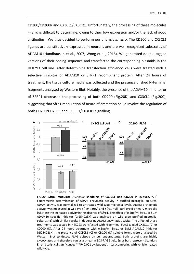
RESULTS 89
CD200/CD200R and CX3CL1/CX3CR1. Unfortunately, the processing of these molecules
in vivo is difficult to determine, owing to their low expression and/or the lack of good
antibodies. We thus decided to perform our analysis in vitro. The CD200 and CX3CL1
ligands are constitutively expressed in neurons and are well-recognised substrates of
ADAM10 (Hundhausen et al., 2007; Wong et al., 2016). We generated double-tagged
versions of their coding sequence and transfected the corresponding plasmids in the
HEK293 cell line. After determining transfection efficiency, cells were treated with a
selective inhibitor of ADAM10 or SFRP1 recombinant protein. After 24 hours of
treatment, the tissue culture media was collected and the presence of shed N-terminal
fragments analysed by Western Blot. Notably, the presence of the ADAM10 inhibitor or
of SFRP1 decreased the processing of both CD200 (Fig.20D) and CX3CL1 (Fig.20C),
suggesting that Sfrp1 modulation of neuroinflammation could involve the regulation of
both CD200/CD200R and CX3CL1/CX3CR1 signalling.
FIG.20: Sfrp1 modulate ADAM10 shedding of CX3CL1 and CD200 in culture. A,B) Fluorometric determination of ADAM enzymatic activity in purified microglial cultures. ADAM activity was normalized to untreated wild type microglia levels. ADAM proteolytic activity was measured in wild type (light grey) and Sfrp1 null (dark grey) primary microglia (A). Note the increased activity in the absence of Sfrp1. The effect of 0,5μg/ml Sfrp1 or 5μM ADAM10 specific inhibitor (GI254023X) was analysed on wild type purified microglial cultures (B) with similar results in decreasing ADAM enzymatic activity. The effect of these treatments was tested in HEK293 transfected with N-terminal FLAG tagged CX3CL1 (C) or CD200 (D). After 24 hours treatment with 0,5μg/ml Sfrp1 or 5μM ADAM10 inhibitor (GI254023X), the presence of CX3CL1 (C) or CD200 (D) soluble forms were analysed by Western Blot to detect FLAG epitope on cell supernatants. Both proteins are highly glycosylated and therefore run as a smear in SDS-PAGE gels. Error bars represent Standard Error. Statistical significance: ***P<0.001 by Student's t-test comparing with vehicle treated wild type.
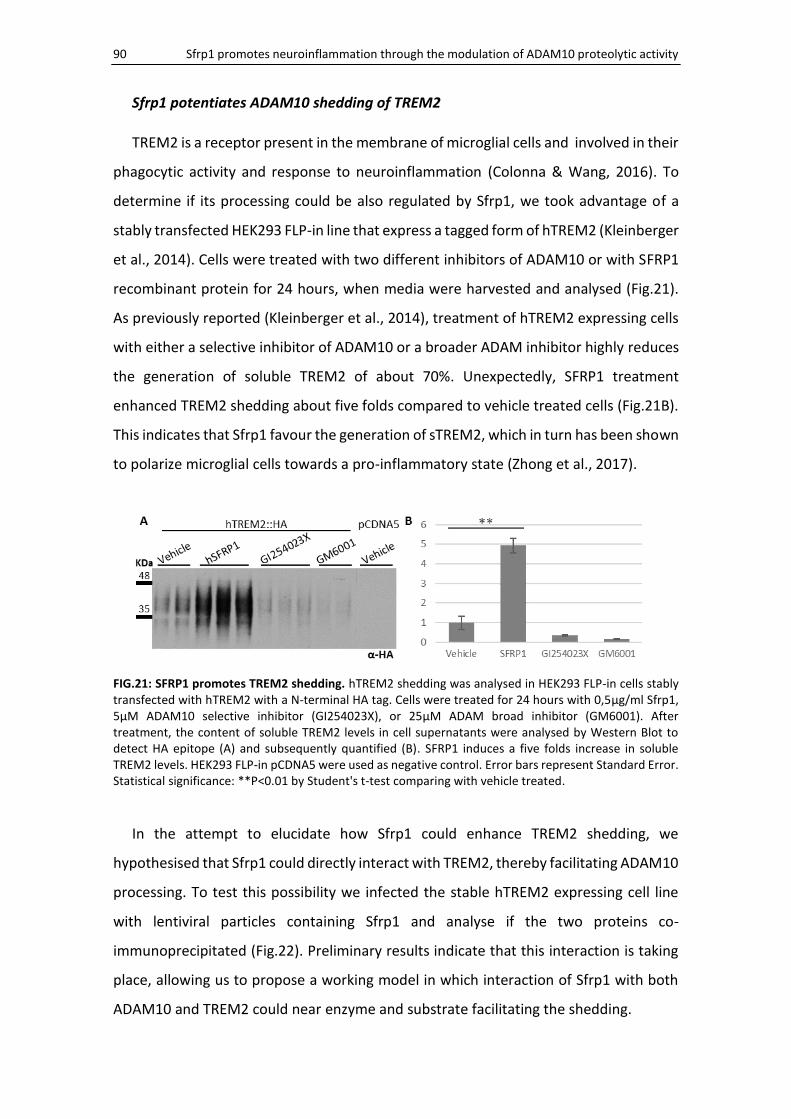
90 Sfrp1 promotes neuroinflammation through the modulation of ADAM10 proteolytic activity
Sfrp1 potentiates ADAM10 shedding of TREM2
TREM2 is a receptor present in the membrane of microglial cells and involved in their
phagocytic activity and response to neuroinflammation (Colonna & Wang, 2016). To
determine if its processing could be also regulated by Sfrp1, we took advantage of a
stably transfected HEK293 FLP-in line that express a tagged form of hTREM2 (Kleinberger
et al., 2014). Cells were treated with two different inhibitors of ADAM10 or with SFRP1
recombinant protein for 24 hours, when media were harvested and analysed (Fig.21).
As previously reported (Kleinberger et al., 2014), treatment of hTREM2 expressing cells
with either a selective inhibitor of ADAM10 or a broader ADAM inhibitor highly reduces
the generation of soluble TREM2 of about 70%. Unexpectedly, SFRP1 treatment
enhanced TREM2 shedding about five folds compared to vehicle treated cells (Fig.21B).
This indicates that Sfrp1 favour the generation of sTREM2, which in turn has been shown
to polarize microglial cells towards a pro-inflammatory state (Zhong et al., 2017).
FIG.21: SFRP1 promotes TREM2 shedding. hTREM2 shedding was analysed in HEK293 FLP-in cells stably transfected with hTREM2 with a N-terminal HA tag. Cells were treated for 24 hours with 0,5μg/ml Sfrp1, 5μM ADAM10 selective inhibitor (GI254023X), or 25μM ADAM broad inhibitor (GM6001). After treatment, the content of soluble TREM2 levels in cell supernatants were analysed by Western Blot to detect HA epitope (A) and subsequently quantified (B). SFRP1 induces a five folds increase in soluble TREM2 levels. HEK293 FLP-in pCDNA5 were used as negative control. Error bars represent Standard Error. Statistical significance: **P<0.01 by Student's t-test comparing with vehicle treated.
In the attempt to elucidate how Sfrp1 could enhance TREM2 shedding, we
hypothesised that Sfrp1 could directly interact with TREM2, thereby facilitating ADAM10
processing. To test this possibility we infected the stable hTREM2 expressing cell line
with lentiviral particles containing Sfrp1 and analyse if the two proteins co-
immunoprecipitated (Fig.22). Preliminary results indicate that this interaction is taking
place, allowing us to propose a working model in which interaction of Sfrp1 with both
ADAM10 and TREM2 could near enzyme and substrate facilitating the shedding.
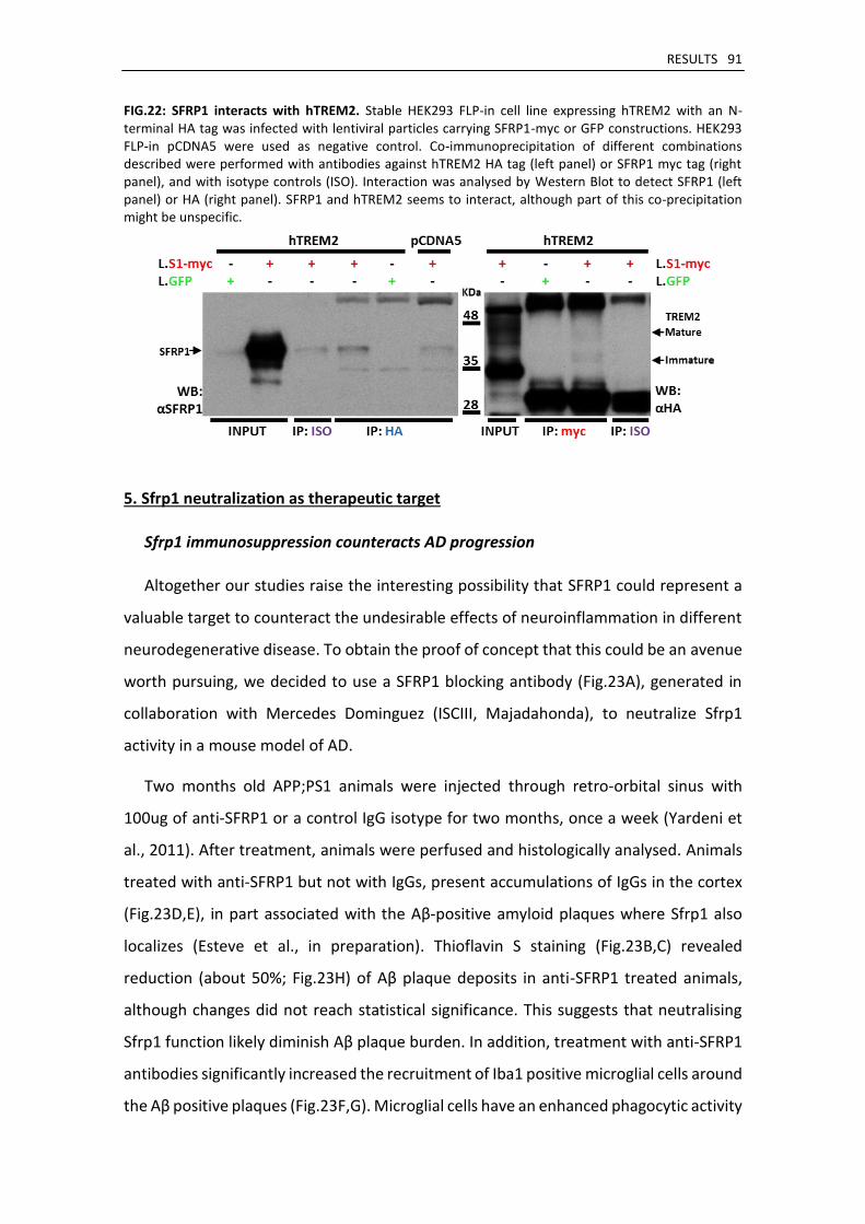
RESULTS 91
FIG.22: SFRP1 interacts with hTREM2. Stable HEK293 FLP-in cell line expressing hTREM2 with an N-terminal HA tag was infected with lentiviral particles carrying SFRP1-myc or GFP constructions. HEK293 FLP-in pCDNA5 were used as negative control. Co-immunoprecipitation of different combinations described were performed with antibodies against hTREM2 HA tag (left panel) or SFRP1 myc tag (right panel), and with isotype controls (ISO). Interaction was analysed by Western Blot to detect SFRP1 (left panel) or HA (right panel). SFRP1 and hTREM2 seems to interact, although part of this co-precipitation might be unspecific.
5. Sfrp1 neutralization as therapeutic target
Sfrp1 immunosuppression counteracts AD progression
Altogether our studies raise the interesting possibility that SFRP1 could represent a
valuable target to counteract the undesirable effects of neuroinflammation in different
neurodegenerative disease. To obtain the proof of concept that this could be an avenue
worth pursuing, we decided to use a SFRP1 blocking antibody (Fig.23A), generated in
collaboration with Mercedes Dominguez (ISCIII, Majadahonda), to neutralize Sfrp1
activity in a mouse model of AD.
Two months old APP;PS1 animals were injected through retro-orbital sinus with
100ug of anti-SFRP1 or a control IgG isotype for two months, once a week (Yardeni et
al., 2011). After treatment, animals were perfused and histologically analysed. Animals
treated with anti-SFRP1 but not with IgGs, present accumulations of IgGs in the cortex
(Fig.23D,E), in part associated with the Aβ-positive amyloid plaques where Sfrp1 also
localizes (Esteve et al., in preparation). Thioflavin S staining (Fig.23B,C) revealed
reduction (about 50%; Fig.23H) of Aβ plaque deposits in anti-SFRP1 treated animals,
although changes did not reach statistical significance. This suggests that neutralising
Sfrp1 function likely diminish Aβ plaque burden. In addition, treatment with anti-SFRP1
antibodies significantly increased the recruitment of Iba1 positive microglial cells around
the Aβ positive plaques (Fig.23F,G). Microglial cells have an enhanced phagocytic activity
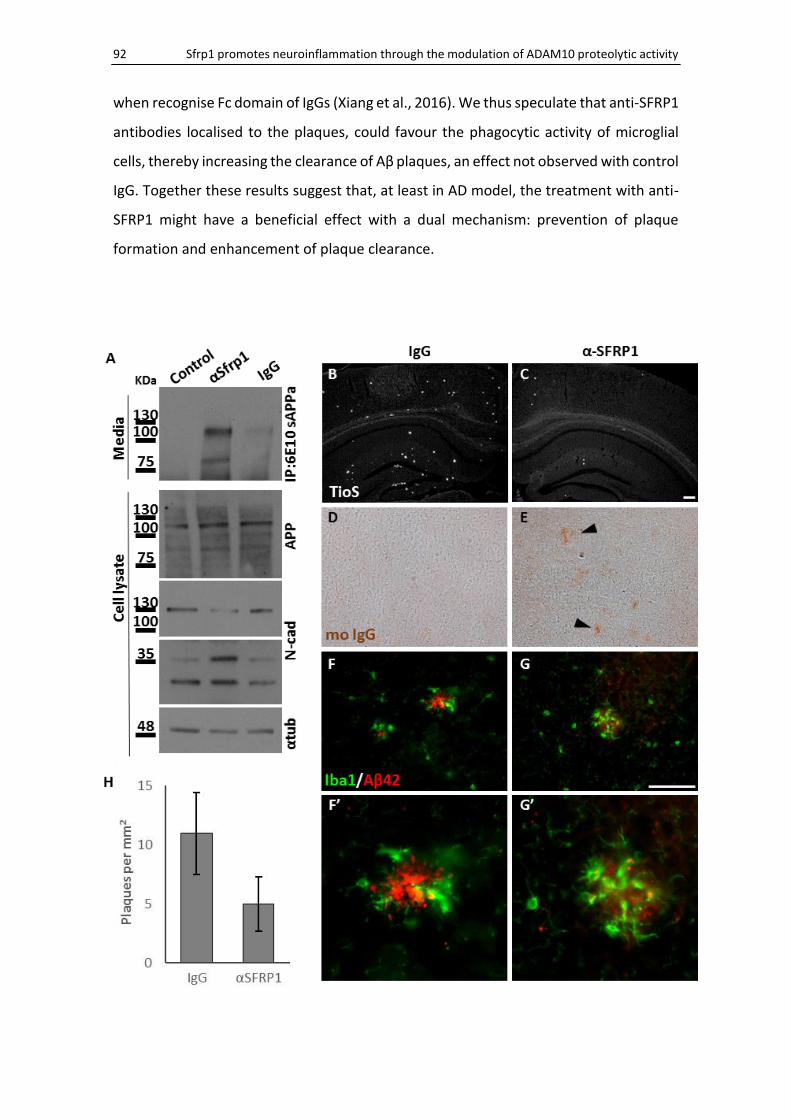
92 Sfrp1 promotes neuroinflammation through the modulation of ADAM10 proteolytic activity
when recognise Fc domain of IgGs (Xiang et al., 2016). We thus speculate that anti-SFRP1
antibodies localised to the plaques, could favour the phagocytic activity of microglial
cells, thereby increasing the clearance of Aβ plaques, an effect not observed with control
IgG. Together these results suggest that, at least in AD model, the treatment with anti-
SFRP1 might have a beneficial effect with a dual mechanism: prevention of plaque
formation and enhancement of plaque clearance.
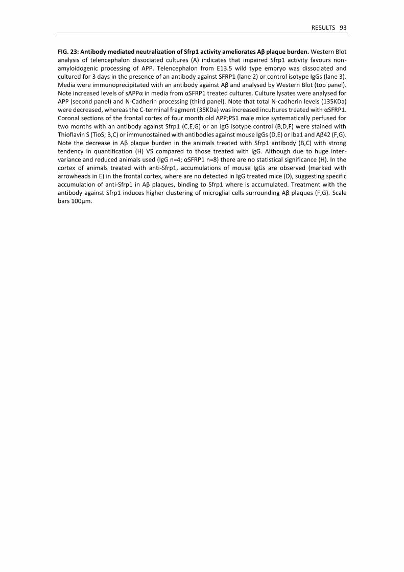
RESULTS 93
FIG. 23: Antibody mediated neutralization of Sfrp1 activity ameliorates Aβ plaque burden. Western Blot analysis of telencephalon dissociated cultures (A) indicates that impaired Sfrp1 activity favours non-amyloidogenic processing of APP. Telencephalon from E13.5 wild type embryo was dissociated and cultured for 3 days in the presence of an antibody against SFRP1 (lane 2) or control isotype IgGs (lane 3). Media were immunoprecipitated with an antibody against Aβ and analysed by Western Blot (top panel). Note increased levels of sAPPα in media from αSFRP1 treated cultures. Culture lysates were analysed for APP (second panel) and N-Cadherin processing (third panel). Note that total N-cadherin levels (135KDa) were decreased, whereas the C-terminal fragment (35KDa) was increased incultures treated with αSFRP1. Coronal sections of the frontal cortex of four month old APP;PS1 male mice systematically perfused for two months with an antibody against Sfrp1 (C,E,G) or an IgG isotype control (B,D,F) were stained with Thioflavin S (TioS; B,C) or immunostained with antibodies against mouse IgGs (D,E) or Iba1 and Aβ42 (F,G). Note the decrease in Aβ plaque burden in the animals treated with Sfrp1 antibody (B,C) with strong tendency in quantification (H) VS compared to those treated with IgG. Although due to huge inter-variance and reduced animals used (IgG n=4; αSFRP1 n=8) there are no statistical significance (H). In the cortex of animals treated with anti-Sfrp1, accumulations of mouse IgGs are observed (marked with arrowheads in E) in the frontal cortex, where are no detected in IgG treated mice (D), suggesting specific accumulation of anti-Sfrp1 in Aβ plaques, binding to Sfrp1 where is accumulated. Treatment with the antibody against Sfrp1 induces higher clustering of microglial cells surrounding Aβ plaques (F,G). Scale bars 100μm.

DISCUSSION
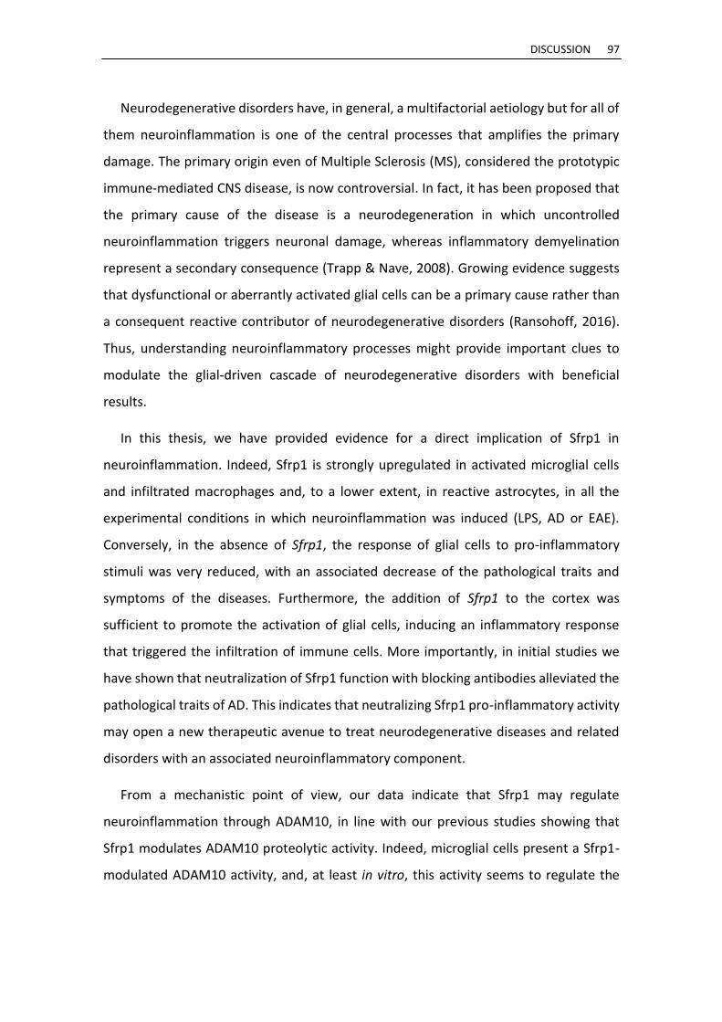
DISCUSSION 97
Neurodegenerative disorders have, in general, a multifactorial aetiology but for all of
them neuroinflammation is one of the central processes that amplifies the primary
damage. The primary origin even of Multiple Sclerosis (MS), considered the prototypic
immune-mediated CNS disease, is now controversial. In fact, it has been proposed that
the primary cause of the disease is a neurodegeneration in which uncontrolled
neuroinflammation triggers neuronal damage, whereas inflammatory demyelination
represent a secondary consequence (Trapp & Nave, 2008). Growing evidence suggests
that dysfunctional or aberrantly activated glial cells can be a primary cause rather than
a consequent reactive contributor of neurodegenerative disorders (Ransohoff, 2016).
Thus, understanding neuroinflammatory processes might provide important clues to
modulate the glial-driven cascade of neurodegenerative disorders with beneficial
results.
In this thesis, we have provided evidence for a direct implication of Sfrp1 in
neuroinflammation. Indeed, Sfrp1 is strongly upregulated in activated microglial cells
and infiltrated macrophages and, to a lower extent, in reactive astrocytes, in all the
experimental conditions in which neuroinflammation was induced (LPS, AD or EAE).
Conversely, in the absence of Sfrp1, the response of glial cells to pro-inflammatory
stimuli was very reduced, with an associated decrease of the pathological traits and
symptoms of the diseases. Furthermore, the addition of Sfrp1 to the cortex was
sufficient to promote the activation of glial cells, inducing an inflammatory response
that triggered the infiltration of immune cells. More importantly, in initial studies we
have shown that neutralization of Sfrp1 function with blocking antibodies alleviated the
pathological traits of AD. This indicates that neutralizing Sfrp1 pro-inflammatory activity
may open a new therapeutic avenue to treat neurodegenerative diseases and related
disorders with an associated neuroinflammatory component.
From a mechanistic point of view, our data indicate that Sfrp1 may regulate
neuroinflammation through ADAM10, in line with our previous studies showing that
Sfrp1 modulates ADAM10 proteolytic activity. Indeed, microglial cells present a Sfrp1-
modulated ADAM10 activity, and, at least in vitro, this activity seems to regulate the

98 Sfrp1 promotes neuroinflammation through the modulation of ADAM10 proteolytic activity
proteolysis of relevant immune modulators, such as microglial TREM2 and neuronal
CD200 and CX3CL1, in a substrate-dependent manner.
1. Sfrp1 promotes a reactive state of microglial cells
The observation that motivated our study was the strong decrease of gliosis
surrounding the deposits of Aβ in the brain of a mouse model of AD, when Sfrp1
expression was genetically inactivated. Aβ plaque burden is reduced in the absence of
Sfrp1 and this could explain the mild gliosis observed in the APP;PS1;Sfrp1-/- mice model.
However, gliosis was mild also surrounding the few Aβ plaques present in the
APP;PS1;Sfrp1-/-, pointing to the possibility that Sfrp1 could directly impinge upon
inflammatory events.
Sfrp1 contributes to exacerbate neuroinflammatory responses
It is important to remark that the absence of Sfrp1 does not completely impair the
inflammatory response. This is an important observation, because, as previously
mentioned, an acute inflammatory response is a necessary reaction to damage and has
beneficial functions because it promotes pathogen elimination, tissue repair and restore
CNS homeostasis (Sochocka et al., 2016). Both LPS-infused and MOG-immunised Sfrp1
null animals develop an inflammatory response, but this response was milder than that
of control animals and likely sufficient to neutralize the proinflammatory trigger and
thus prevent a chronic inflammatory state. This effect was particularly remarkable in
limiting the severity of the clinical symptoms of EAE, where we observed that only 15%
of the Sfrp1 null animals presented severe symptoms, in contrast to the 50% of the wild
types. Although, EAE progression is triggered by the infiltration of peripheral
macrophages and lymphocytes (Ajami et al., 2011), microglial activation is required for
the development and maintenance of inflammatory lesions associated to EAE (Ding et
al., 2014), likely favouring further infiltration of peripheral immune cells (Heppner et al.,
2005). Although, we believe that the EAE phenotype observed in Sfrp1-/- mice is likely
linked to a decreased activation of local microglila cells, we cannot discard that this
might be also associated to less reactive infiltrated macrophages, which also seem to
express Sfrp1 during EAE.
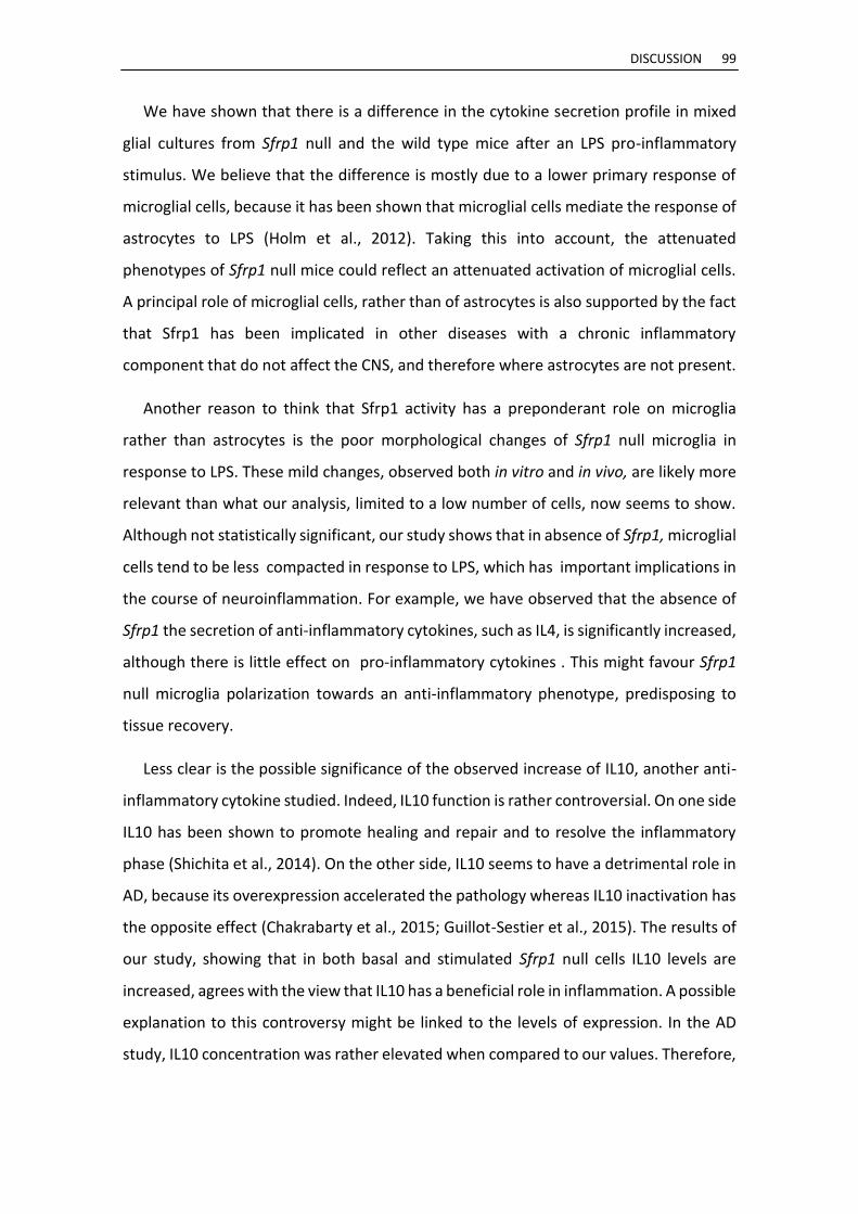
DISCUSSION 99
We have shown that there is a difference in the cytokine secretion profile in mixed
glial cultures from Sfrp1 null and the wild type mice after an LPS pro-inflammatory
stimulus. We believe that the difference is mostly due to a lower primary response of
microglial cells, because it has been shown that microglial cells mediate the response of
astrocytes to LPS (Holm et al., 2012). Taking this into account, the attenuated
phenotypes of Sfrp1 null mice could reflect an attenuated activation of microglial cells.
A principal role of microglial cells, rather than of astrocytes is also supported by the fact
that Sfrp1 has been implicated in other diseases with a chronic inflammatory
component that do not affect the CNS, and therefore where astrocytes are not present.
Another reason to think that Sfrp1 activity has a preponderant role on microglia
rather than astrocytes is the poor morphological changes of Sfrp1 null microglia in
response to LPS. These mild changes, observed both in vitro and in vivo, are likely more
relevant than what our analysis, limited to a low number of cells, now seems to show.
Although not statistically significant, our study shows that in absence of Sfrp1, microglial
cells tend to be less compacted in response to LPS, which has important implications in
the course of neuroinflammation. For example, we have observed that the absence of
Sfrp1 the secretion of anti-inflammatory cytokines, such as IL4, is significantly increased,
although there is little effect on pro-inflammatory cytokines . This might favour Sfrp1
null microglia polarization towards an anti-inflammatory phenotype, predisposing to
tissue recovery.
Less clear is the possible significance of the observed increase of IL10, another anti-
inflammatory cytokine studied. Indeed, IL10 function is rather controversial. On one side
IL10 has been shown to promote healing and repair and to resolve the inflammatory
phase (Shichita et al., 2014). On the other side, IL10 seems to have a detrimental role in
AD, because its overexpression accelerated the pathology whereas IL10 inactivation has
the opposite effect (Chakrabarty et al., 2015; Guillot-Sestier et al., 2015). The results of
our study, showing that in both basal and stimulated Sfrp1 null cells IL10 levels are
increased, agrees with the view that IL10 has a beneficial role in inflammation. A possible
explanation to this controversy might be linked to the levels of expression. In the AD
study, IL10 concentration was rather elevated when compared to our values. Therefore,
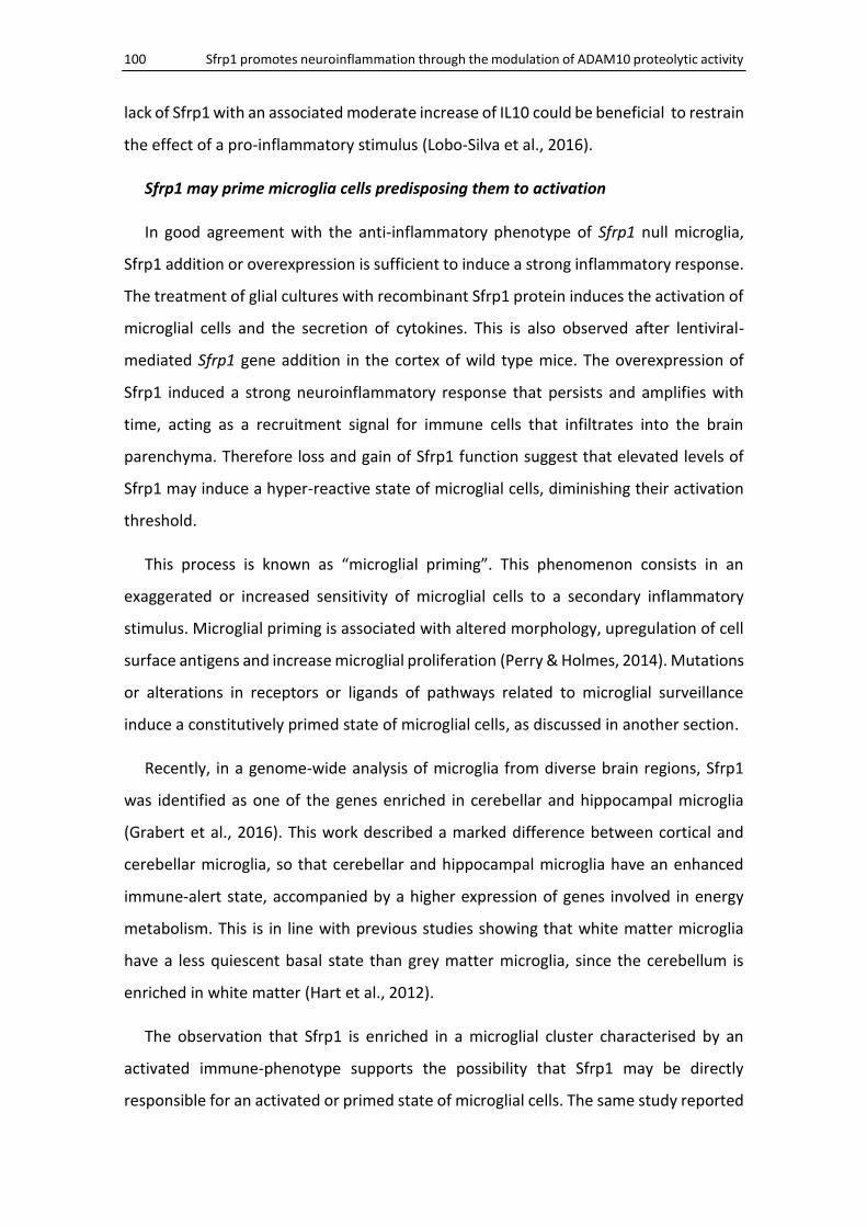
100 Sfrp1 promotes neuroinflammation through the modulation of ADAM10 proteolytic activity
lack of Sfrp1 with an associated moderate increase of IL10 could be beneficial to restrain
the effect of a pro-inflammatory stimulus (Lobo-Silva et al., 2016).
Sfrp1 may prime microglia cells predisposing them to activation
In good agreement with the anti-inflammatory phenotype of Sfrp1 null microglia,
Sfrp1 addition or overexpression is sufficient to induce a strong inflammatory response.
The treatment of glial cultures with recombinant Sfrp1 protein induces the activation of
microglial cells and the secretion of cytokines. This is also observed after lentiviral-
mediated Sfrp1 gene addition in the cortex of wild type mice. The overexpression of
Sfrp1 induced a strong neuroinflammatory response that persists and amplifies with
time, acting as a recruitment signal for immune cells that infiltrates into the brain
parenchyma. Therefore loss and gain of Sfrp1 function suggest that elevated levels of
Sfrp1 may induce a hyper-reactive state of microglial cells, diminishing their activation
threshold.
This process is known as “microglial priming”. This phenomenon consists in an
exaggerated or increased sensitivity of microglial cells to a secondary inflammatory
stimulus. Microglial priming is associated with altered morphology, upregulation of cell
surface antigens and increase microglial proliferation (Perry & Holmes, 2014). Mutations
or alterations in receptors or ligands of pathways related to microglial surveillance
induce a constitutively primed state of microglial cells, as discussed in another section.
Recently, in a genome-wide analysis of microglia from diverse brain regions, Sfrp1
was identified as one of the genes enriched in cerebellar and hippocampal microglia
(Grabert et al., 2016). This work described a marked difference between cortical and
cerebellar microglia, so that cerebellar and hippocampal microglia have an enhanced
immune-alert state, accompanied by a higher expression of genes involved in energy
metabolism. This is in line with previous studies showing that white matter microglia
have a less quiescent basal state than grey matter microglia, since the cerebellum is
enriched in white matter (Hart et al., 2012).
The observation that Sfrp1 is enriched in a microglial cluster characterised by an
activated immune-phenotype supports the possibility that Sfrp1 may be directly
responsible for an activated or primed state of microglial cells. The same study reported
DISCUSSION 101
that microglia aging is region-dependent, with cerebellar microglia being the more
prone to aging, because they are in immune-alert state with a higher expression of
immune amplifying genes (Grabert et al., 2016). This points to a possible implication of
Sfrp1 in making microglia more vulnerable to aging and age-related diseases owing to
alertness well above that of other regions.
2. Sfrp1 modulates microglial activity through ADAM10
Our data together with the literature mentioned above support the idea that Sfrp1
tunes microglial activation. High levels of Sfrp1 could lead to major dysregulation of
microglial functions and subsequently induce severe alterations of CNS homeostasis that
result in the development of CNS pathologies. Given the multifunctional activities of
Sfrp1 (Cruciat & Niehrs, 2013), there are several mechanisms by which Sfrp1 may
modulate microglial activity: the first could involve the Wnt signalling pathway; the
second the regulation of ADAM10 proteolytic activity or, as a third possibility, it might
involve the interaction with a previously undescribed receptor.
Sfrp1 can modulate the Wnt signalling pathway in both a positive and negative way
(Esteve & Bovolenta, 2010). Disruption of the Wnt signalling pathway has been
implicated in some of the alterations that characterize AD (Godoy et al., 2014) and
higher Sfrp1 levels could sequester Wnt ligands, reducing pathway activity (Kawano &
Kypta, 2003), thereby modulating microglial response (Halleskog & Schulte, 2013). We
cannot discard this possibility but we have evidence that in APP;PS1 mice that lack Sfrp1
Wnt/β-catenin signalling is not particularly affected, since qPCR analysis of the total
brain mRNA levels of Axin2, a read-out of Wnt pathway activity (Nusse & Clevers, 2017),
revealed no significant differences with those of the APP;PS1 mice alone.
Sfrp1 modulates processing of ADAM10 immune-related substrates
The other mechanism by which Sfrp1 could modify microglial activity implicates the
negative modulation of ADAM10 (Esteve et al., 2011a). ADAM10 is implicated in
neuroinflammatory and neurodegenerative events (Saftig & Bovolenta, 2015). Our
study shows that Sfrp1 can modulate ADAM10 activity in microglia (Fig.20). Among the
wide range of ADAM10 substrates, there are mediators of the microglial response, such
102 Sfrp1 promotes neuroinflammation through the modulation of ADAM10 proteolytic activity
as CX3CL1 (Hundhausen et al., 2007), CD200 (Wong et al., 2016) and TREM2 (Kleinberger
et al., 2014). All of them are required to maintain CNS homeostasis and microglia cells
in a quiescent state (Colonna & Butovsky, 2017). Mutations in the genes encoding these
regulators have been associated with susceptibility to develop neurodegenerative
diseases (Kierdorf & Prinz, 2013). It is therefore plausible that high levels of Sfrp1 could
disrupt their signalling, leading to an exacerbated response of microglial cells, as
observed upon Sfrp1 overexpression.
CX3CL1, CD200 and TREM2 have different characteristics. CX3CL1 and CD200 are
ligands, present in membrane-anchored and soluble forms, which are expressed mostly
by neurons (Hughes et al., 2002; Koning et al., 2009). Their different forms interact with
their respective receptors (CX3CR1 and CD200R) expressed in microglial cells (Jung et
al., 2000; G. J. Wright et al., 2000), maintaining microglial cells in a quiescent state. If
ligand-receptor interaction, and thus its signalling, is lost, neuronal damage appears
together with microglial neurotoxicity (Cardona et al., 2006; Lyons et al., 2007). We have
demonstrated that Sfrp1 impairs ADAM10-mediated shedding of both CX3CL1 and
CD200 in vitro. Therefore, Sfrp1-modulated shedding of these neuronal ligands could
limit their binding to the receptors on microglial cells, thereby allowing microglial
activation (since microglia are no longer kept in a quiescent state). This mechanism could
explain why Sfrp1 induced a primed or less quiescent microglia state.
The other ADAM10 substrate we have studied is TREM2. Differently from the other
two, this receptor is expressed by microglial cells (Colonna & Wang, 2016). Polyanions,
phospholipids and lipoproteins can bind and activate TREM2. Upon ligand binding,
TREM2 activates an intracellular signal that promotes phagocytosis (Hsieh et al., 2009),
survival (Otero et al., 2009) and attenuates microglial activation (Turnbull et al., 2006).
At the cell membrane, TREM2 activity is regulated by ADAM10 and ADAM17-mediated
proteolytic processing (Kleinberger et al., 2014). Aberrant TREM2 signalling has been
related to malfunction and senescence (Zheng et al., 2017) of microglial cells and
neurological disorders (Ulrich et al., 2017).
In contrast to what possibly expected, here we have shown that Sfrp1 enhances
TREM2 shedding, promoting the generation of soluble TREM2 (sTREM2). The functions
and interactions of TREM2 are still highly controversial, and even more debated is the

DISCUSSION 103
role of sTREM2. In a mouse model of AD, haploinsufficiency of TREM2, which
presumably lead to lower levels of active TREM2 at the plasma membrane, have been
shown to promote microglial dysfunction and to induce severe axonal dystrophy (Y.
Wang et al., 2015; Y. Wang et al., 2016). sTREM2 enhances microglial survival and is
sufficient to trigger pro-inflammatory responses (Zhong et al., 2017). Thus, Sfrp1-
promoted processing of TREM2 could have two detrimental effects. Both effects
correlate with those observed upon Sfrp1 overexpression, meaning an increase of
inflammation.
Notably, reduced function of TREM2 impairs microglial survival disrupting β-catenin
stabilization (Zheng et al., 2017). As stabilization of β-catenin allows its translocation to
the nucleus and thus its function as final effector of the Wnt/β-catenin pathway, the
shedding-mediated modulation of TREM2 activity may be an indirect mechanism with
which Sfrp1 could interfere with β-catenin induced gene transcription in microglial cells.
If this were the case, a possible explanation for the absence of changes in Axin2 mRNA
levels observed in AD mice might be the small proportion that microglial Axin2 might
represent over the total brain content. This establishes another level of Sfrp1-
dependent regulation to Wnt ligands activity that could explain their dual and context-
dependent effect on microglial response (Halleskog et al., 2011; Halleskog & Schulte,
2013; B. Li et al., 2011).
Sfrp1 modulates ADAM10 activity in a substrate-specific manner
The different effect of Sfrp1 on ADAM10-mediated shedding of different substrates
indicates that Sfrp1 function might depend on the nature of the substrate. We have
proposed that the Sfrp1 CRD could interact with the CRD present in ADAM10. This CRD
dimerization could bring ADAM10 catalytic domain close to the Sfrp1 NTR domain,
which is known to share similarities with TIMP inhibitors (Mott et al., 2000). Thereby,
Sfrp1 NTR would interfere with ADAM10 enzymatic activity by allosteric competition
with diverse substrates.
According to a database of predicted human protein-protein interactions (McDowall
et al., 2009), TREM2 has high probability of interacting with Netrin1. Netrin1 shares
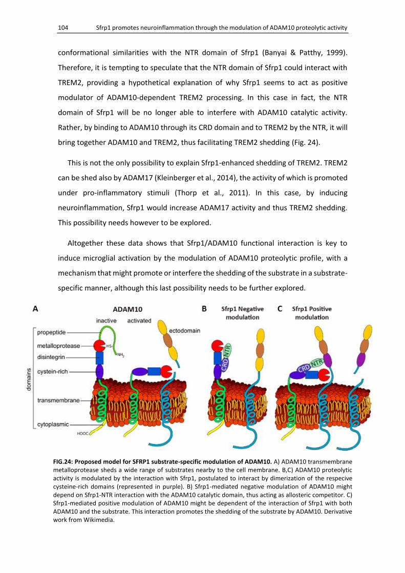
104 Sfrp1 promotes neuroinflammation through the modulation of ADAM10 proteolytic activity
conformational similarities with the NTR domain of Sfrp1 (Banyai & Patthy, 1999).
Therefore, it is tempting to speculate that the NTR domain of Sfrp1 could interact with
TREM2, providing a hypothetical explanation of why Sfrp1 seems to act as positive
modulator of ADAM10-dependent TREM2 processing. In this case in fact, the NTR
domain of Sfrp1 will be no longer able to interfere with ADAM10 catalytic activity.
Rather, by binding to ADAM10 through its CRD domain and to TREM2 by the NTR, it will
bring together ADAM10 and TREM2, thus facilitating TREM2 shedding (Fig. 24).
This is not the only possibility to explain Sfrp1-enhanced shedding of TREM2. TREM2
can be shed also by ADAM17 (Kleinberger et al., 2014), the activity of which is promoted
under pro-inflammatory stimuli (Thorp et al., 2011). In this case, by inducing
neuroinflammation, Sfrp1 would increase ADAM17 activity and thus TREM2 shedding.
This possibility needs however to be explored.
Altogether these data shows that Sfrp1/ADAM10 functional interaction is key to
induce microglial activation by the modulation of ADAM10 proteolytic profile, with a
mechanism that might promote or interfere the shedding of the substrate in a substrate-
specific manner, although this last possibility needs to be further explored.
FIG.24: Proposed model for SFRP1 substrate-specific modulation of ADAM10. A) ADAM10 transmembrane metalloprotease sheds a wide range of substrates nearby to the cell membrane. B,C) ADAM10 proteolytic activity is modulated by the interaction with Sfrp1, postulated to interact by dimerization of the respecive cysteine-rich domains (represented in purple). B) Sfrp1-mediated negative modulation of ADAM10 might depend on Sfrp1-NTR interaction with the ADAM10 catalytic domain, thus acting as allosteric competitor. C) Sfrp1-mediated positive modulation of ADAM10 might be dependent of the interaction of Sfrp1 with both ADAM10 and the substrate. This interaction promotes the shedding of the substrate by ADAM10. Derivative work from Wikimedia.
DISCUSSION 105
3. Sfrp1 as therapeutic target to counteract neuroinflammation
The results obtained in this study strongly support the idea that interfering with Sfrp1
activity may counteract the detrimental effect of neuroinflammation, suggesting that
Sfrp1 could be a promising therapeutic target for the treatment of neurological
disorders. Unspecific IgGs or Aβ-specific antibodies have been used in several clinical
trials to delay or reduce AD pathology (Lunemann et al., 2015). However, most of them
had no or limited success with the exception of that in which patients have been treated
with the specific antibody aducanumab against oligomeric Aβ (Sevigny et al., 2016). In
this case, patients presented a reduction of Aβ-deposition and a slower progression of
dementia. This encouraging study however can be applied only to AD patients. Our
approach targeting Sfrp1 to prevent exacerbated neuroinflammation could be applied
to a broader range of patients affected by diseases that involve chronic
neuroinflammation. This idea is supported by the results of our trial.
In a mouse model of AD, we systemically infused an antibody against SFRP1 to
neutralise Sfrp1 activity. After two months, treated animals present a reduced Aβ plaque
burden and accumulations of anti-SFRP1 in the cortex resembling Aβ deposits. Two
different and compatible effects of anti-SFRP1 may explain this reduction: reduced Aβ
deposition and enhanced of Aβ clearance. The first one would result from the
neutralisation of Sfrp1 activity as negative modulator of ADAM10 activity. This will
enhance ADAM10 processing of APP, thereby preventing Aβ formation. The second one
might be triggered by anti-SFRP1 accumulation in the Sfrp1 containing amyloid plaques.
“Opsonised-like” plaques would then trigger microglial TREM2-mediated phagocytic
activity (Xiang et al., 2016), thereby enhancing plaque clearance. As an additional
beneficial effect also TREM2-dependent, microglial clustering around the plaques
promoted by anti-SFRP1 antibodies might limit Aβ diffusion (Y. Wang et al., 2016) and
impair neuronal dystrophy (Yuan et al., 2016). TREM2 signalling has been also directly
related to mediate microglial Aβ uptake by interacting with Aβ-lipoproteins complexes
(Yeh et al., 2016). This suggest that Sfrp1 neutralization would limit the progression of
the disease reducing Aβ plaque burden and detrimental activation of glial cells, as well
as by promoting Aβ clearance and limiting Aβ diffusion by stabilizing TREM2 signalling.
106 Sfrp1 promotes neuroinflammation through the modulation of ADAM10 proteolytic activity
In conclusion, our data support the role of Sfrp1 as modulator of neuroinflammatory
responses in the CNS and shows that high Sfrp1 levels may lead to an exacerbated
microglial response with detrimental consequences for CNS homeostasis. The relevance
of Sfrp1 in microglial priming and senescence indicates that the role of this protein in
aging should be studied. Moreover, the possible Sfrp1 substrate-specific regulation of
ADAM10 must be confirmed and thereafter verified in vivo. Last, but not least, Sfrp1
neutralization might be a promising avenue for the treatment of the neuroinflammatory
processes involved in the onset and progression of diverse neurological disorders.
4. Future perspectives: Possible implication of Sfrp1 in age-related diseases
The data exposed in this thesis reveal a novel function of Sfrp1 as a pro-inflammatory
modulator of microglial activation. However, the relevance of increased levels of Sfrp1
on other brain cells types has not been assessed. Sfrp1 has been related to the
modulation of mesenchymal stem cells (MSC) function in vessel maturation (Dufourcq
et al., 2008). As MSC have been implicated in BBB stabilization and the regulation of its
permeability (Park et al., 2015), it might be worth studying the possible implication of
Sfrp1 in the modulation of BBB permeability. Sfrp1 direct action on neurons should be
also considered, since Wnt signalling (Budnik & Salinas, 2011) and ADAM10 (Prox et al.,
2013) have been implicated in synapse formation and remodelling, and their inhibition
induces synaptic degeneration and cognitive impairment (Marzo et al., 2016; Prox et al.,
2013). Sfrp1 impairs synaptic plasticity leading to an aged synaptic phenotype (Gogolla
et al., 2009). Both synaptic plasticity impairment (Cerpa et al., 2011) and endothelial-
derived BBB disruption (Winkler et al., 2015) are processes widely related to the onset
and progression of age- and neurodegenerative-related cognitive decline.
One of the principal theories of aging states the importance of inflammation in aging
events. The so-called “Inflammaging” seems to involve the upregulation of pro-
inflammatory mediators in the circulation. Inflammaging is described as a systemic, low-
graded, chronic inflammation in absence of infection (Franceschi et al., 2000) that is
present in aging individual and it is thought to facilitate neurodegenerative disorders
(Cribbs et al., 2012). Nonetheless, the specific aetiology of inflammaging remains
unknown, but cellular senescence has been described among its most likely sources
(Franceschi & Campisi, 2014).
DISCUSSION 107
Senescence is a response to damage and stress that supresses the proliferation of
tumorigenic cells (Campisi & d'Adda di Fagagna, 2007). Persistent senescent cells are
thought to drive aging and age-associated pathologies through an altered protein
secretion profile, which is called the senescence-associated secretory phenotype (SASP)
(Coppe et al., 2010). SASP factors include several pro-inflammatory cytokines and
chemokines, growth factors and regulators. Likewise, Sfrp1 has been described as a
SASP, necessary and sufficient to induce senescence phenotypes through inhibition of
Wnt signalling (Elzi et al., 2012). This fit well with the role of Sfrps as tumour suppressors
genes (Surana et al., 2014) and is supported by the strong evidence of the inverse
association between cancer and neurodegenerative diseases (Driver, 2014). Sfrp1
increased expression mediates the appearance of glaucoma (W. H. Wang et al., 2008)
inducing the senescence of trabecular meshwork cells that regulates the intraocular
pressure (Morgan et al., 2015). Glaucoma is considered an aging-associated disease, and
the strong association with AD have suggested glaucoma to represent a manifestation
of AD (Lai et al., 2017). In this direction, the appearance of senescent dystrophic
microglia has been related to represent a major contributor to the onset of sporadic AD
(Streit et al., 2009). All these data consistently supports the implication of Sfrp1 in the
induction inflammaging.
In this work, we have demonstrated that increased levels of Sfrp1 locally in the CNS
induce microglial activation but circulating Sfrp1 could also promote microglial priming.
Growing evidence associate peripheral diseases characterized by elevated systemic
inflammation, such as obesity, rheumatoid arthritis or diabetes type 2, with a higher risk
of developing cognitive decline (Marioni et al., 2010). It is possible that low-graded
chronic systemic inflammation triggers microglial priming, but both acute and chronic
systemic inflammation have been shown to increase the rate of cognitive decline in
patients with AD (Holmes et al., 2009). Related to that, unpublished data from our
laboratory indicate that Sfrp1 levels are increased in serum samples from elderly people.
In the same direction, Sfrp1 is one of the most significantly up-regulated (almost 200-
fold change) genes in monocytes of aged rats, together with a large number of
recognised markers of cytotoxicity, inflammation and migration (Martinez et al., 2015).
The increase of both, brain- and/or circulating-Sfrp1 levels, could represent an
108 Sfrp1 promotes neuroinflammation through the modulation of ADAM10 proteolytic activity
inflammatory stimulus that triggers microglial priming to exacerbate microglial
response, once known the CNS pro-inflammatory effect of Sfrp1 described in this work.
In addition and related to the increased levels of circulating Sfrp1 during aging, it has
been demonstrated the importance of circulating factors in the induction of age-related
cognitive impairment (Villeda et al., 2014). This enhances our data indicating Sfrp1 as
mediator of neuroinflammatory processes, that directly in the CNS or indirectly systemic
as an age-related SASP could promote dysfunction and/or priming of microglia,
triggering detrimental chronic inflammatory processes in the CNS promoting
neurodegenerative diseases.

CONCLUSIONS
CONCLUSIONS 113
- Sfrp1 is involved in neuroinflammation and its expression is upregulated under
pro-inflammatory conditions.
- Sfrp1 upregulation in response to a pro-inflammatory stimulus mostly depends
on microglial cells and to a lower extent on astrocytes.
- Overexpression of Sfrp1 is sufficient to induce an inflammatory response.
- Genetic inactivation of Sfrp1 reduces the response of glial cells to a pro-
inflammatory stimulus
- This milder response reduces the symptomatology of EAE and delays AD
progression.
- Sfrp1 seems to modulate ADAM10-mediated processing of different molecules
involved in the onset of microglial activation towards a pro-inflammatory state.
- Sfrp1-mediated modulation of ADAM10 proteolytic activity seems to be
substrate-dependent.
- Sfrp1 represents a potential therapeutic target to ameliorate exacerbated
neuroinflammatory processes characteristic of neurodegenerative diseases.

CONCLUSIONES
CONCLUSIONES 117
- La proteína Sfrp1 está implicada en procesos neuroinflamatorios y su expresión está
aumentada bajo condiciones pro-inflamatorias.
- El incremento de la expresión de Sfrp1, en respuesta a un estímulo pro-inflamatorio,
depende en su mayoría de células microgliales y en menor medida de astrocitos.
- La sobreexpresión de Sfrp1 es suficiente para inducir una respuesta inflamatoria.
- La inactivación genética de Sfrp1 disminuye la respuesta glial frente a un estímulo
pro-inflamatorio.
- Esta respuesta reducida va asociada a la disminución de la sintomatología asociada a
la EAE y retarda la progresión de la EA.
- Sfrp1 parece modular la actividad proteolítica de ADAM10, alterando el
procesamiento de diferentes sustratos implicados en la activación de la microglia
hacia un estado pro-inflamatorio.
- La modulación de ADAM10 mediada por Sfrp1 parece ser dependiente de sustrato.
- Sfrp1 representa una potencial diana terapéutica para atenuar los exacerbados
procesos neuroinflamatorios característicos de enfermedades neurodegenerativas.

BIBLIOGRAPHY
BIBLIOGRAPHY 121
Aguzzi, A., Barres, B. A., & Bennett, M. L. (2013). Microglia: scapegoat, saboteur, or something else? Science, 339(6116), 156-161.
Ajami, B., Bennett, J. L., Krieger, C., McNagny, K. M., & Rossi, F. M. (2011). Infiltrating monocytes trigger EAE progression, but do not contribute to the resident microglia pool. Nat Neurosci, 14(9), 1142-1149.
Ajami, B., Bennett, J. L., Krieger, C., Tetzlaff, W., & Rossi, F. M. (2007). Local self-renewal can sustain CNS microglia maintenance and function throughout adult life. Nat Neurosci, 10(12), 1538-1543.
Alzheimer, A., Stelzmann, R. A., Schnitzlein, H. N., & Murtagh, F. R. (1995). An English translation of Alzheimer's 1907 paper, "Uber eine eigenartige Erkankung der Hirnrinde". Clin Anat, 8(6), 429-431.
Allan, S. M., Tyrrell, P. J., & Rothwell, N. J. (2005). Interleukin-1 and neuronal injury. Nat Rev Immunol, 5(8), 629-640.
Alliot, F., Godin, I., & Pessac, B. (1999). Microglia derive from progenitors, originating from the yolk sac, and which proliferate in the brain. Brain Res Dev Brain Res, 117(2), 145-152.
Araque, A., Parpura, V., Sanzgiri, R. P., & Haydon, P. G. (1999). Tripartite synapses: glia, the unacknowledged partner. Trends Neurosci, 22(5), 208-215.
Askew, K., Li, K., Olmos-Alonso, A., Garcia-Moreno, F., Liang, Y., Richardson, P., Tipton, T., Chapman, M. A., Riecken, K., Beccari, S., Sierra, A., Molnar, Z., Cragg, M. S., Garaschuk, O., Perry, V. H., & Gomez-Nicola, D. (2017). Coupled Proliferation and Apoptosis Maintain the Rapid Turnover of Microglia in the Adult Brain. Cell Rep, 18(2), 391-405.
Au, N. P. B., & Ma, C. H. E. (2017). Recent Advances in the Study of Bipolar/Rod-Shaped Microglia and their Roles in Neurodegeneration. Front Aging Neurosci, 9, 128.
Augustine, C., Gunnersen, J., Spirkoska, V., & Tan, S. S. (2001). Place- and time-dependent expression of mouse sFRP-1 during development of the cerebral neocortex. Mech Dev, 109(2), 395-397.
Banyai, L., & Patthy, L. (1999). The NTR module: domains of netrins, secreted frizzled related proteins, and type I procollagen C-proteinase enhancer protein are homologous with tissue inhibitors of metalloproteases. Protein Sci, 8(8), 1636-1642.
Barclay, A. N., Wright, G. J., Brooke, G., & Brown, M. H. (2002). CD200 and membrane protein interactions in the control of myeloid cells. Trends Immunol, 23(6), 285-290.
Barnett, M. H., & Prineas, J. W. (2004). Relapsing and remitting multiple sclerosis: pathology of the newly forming lesion. Ann Neurol, 55(4), 458-468.
Belanger, M., & Magistretti, P. J. (2009). The role of astroglia in neuroprotection. Dialogues Clin Neurosci, 11(3), 281-295.
Bennett, J., Basivireddy, J., Kollar, A., Biron, K. E., Reickmann, P., Jefferies, W. A., & McQuaid, S. (2010). Blood-brain barrier disruption and enhanced vascular permeability in the multiple sclerosis model EAE. J Neuroimmunol, 229(1-2), 180-191.
Bhat, R. A., Stauffer, B., Komm, B. S., & Bodine, P. V. (2007). Structure-function analysis of secreted frizzled-related protein-1 for its Wnt antagonist function. J Cell Biochem, 102(6), 1519-1528.
Blalock, E. M., Geddes, J. W., Chen, K. C., Porter, N. M., Markesbery, W. R., & Landfield, P. W. (2004). Incipient Alzheimer's disease: microarray correlation analyses reveal major transcriptional and tumor suppressor responses. Proc Natl Acad Sci U S A, 101(7), 2173-2178.
122 Sfrp1 promotes neuroinflammation through the modulation of ADAM10 proteolytic activity
Boche, D., Perry, V. H., & Nicoll, J. A. (2013). Review: activation patterns of microglia and their identification in the human brain. Neuropathol Appl Neurobiol, 39(1), 3-18.
Borroto, A., Reyes-Garau, D., Jimenez, M. A., Carrasco, E., Moreno, B., Martinez-Pasamar, S., Cortes, J. R., Perona, A., Abia, D., Blanco, S., Fuentes, M., Arellano, I., Lobo, J., Heidarieh, H., Rueda, J., Esteve, P., Cibrian, D., Martinez-Riano, A., Mendoza, P., Prieto, C., Calleja, E., Oeste, C. L., Orfao, A., Fresno, M., Sanchez-Madrid, F., Alcami, A., Bovolenta, P., Martin, P., Villoslada, P., Morreale, A., Messeguer, A., & Alarcon, B. (2016). First-in-class inhibitor of the T cell receptor for the treatment of autoimmune diseases. Sci Transl Med, 8(370), 370ra184.
Bovolenta, P., Esteve, P., Ruiz, J. M., Cisneros, E., & Lopez-Rios, J. (2008). Beyond Wnt inhibition: new functions of secreted Frizzled-related proteins in development and disease. J Cell Sci, 121(Pt 6), 737-746.
Bowman, C. C., Rasley, A., Tranguch, S. L., & Marriott, I. (2003). Cultured astrocytes express toll-like receptors for bacterial products. Glia, 43(3), 281-291.
Brosseron, F., Krauthausen, M., Kummer, M., & Heneka, M. T. (2014). Body fluid cytokine levels in mild cognitive impairment and Alzheimer's disease: a comparative overview. Mol Neurobiol, 50(2), 534-544.
Budnik, V., & Salinas, P. C. (2011). Wnt signaling during synaptic development and plasticity. Curr Opin Neurobiol, 21(1), 151-159.
Caille, I., Allinquant, B., Dupont, E., Bouillot, C., Langer, A., Muller, U., & Prochiantz, A. (2004). Soluble form of amyloid precursor protein regulates proliferation of progenitors in the adult subventricular zone. Development, 131(9), 2173-2181.
Campisi, J., & d'Adda di Fagagna, F. (2007). Cellular senescence: when bad things happen to good cells. Nat Rev Mol Cell Biol, 8(9), 729-740.
Cardona, A. E., Pioro, E. P., Sasse, M. E., Kostenko, V., Cardona, S. M., Dijkstra, I. M., Huang, D., Kidd, G., Dombrowski, S., Dutta, R., Lee, J. C., Cook, D. N., Jung, S., Lira, S. A., Littman, D. R., & Ransohoff, R. M. (2006). Control of microglial neurotoxicity by the fractalkine receptor. Nat Neurosci, 9(7), 917-924.
Carson, M. J., Doose, J. M., Melchior, B., Schmid, C. D., & Ploix, C. C. (2006). CNS immune privilege: hiding in plain sight. Immunol Rev, 213, 48-65.
Castellano, J. M., Kim, J., Stewart, F. R., Jiang, H., DeMattos, R. B., Patterson, B. W., Fagan, A. M., Morris, J. C., Mawuenyega, K. G., Cruchaga, C., Goate, A. M., Bales, K. R., Paul, S. M., Bateman, R. J., & Holtzman, D. M. (2011). Human apoE isoforms differentially regulate brain amyloid-beta peptide clearance. Sci Transl Med, 3(89), 89ra57.
Celsus, A. C. (1478). De Medicina. Nicolaus Laurentius, Firenze.
Cerpa, W., Gambrill, A., Inestrosa, N. C., & Barria, A. (2011). Regulation of NMDA-receptor synaptic transmission by Wnt signaling. J Neurosci, 31(26), 9466-9471.
Clark, A. K., Staniland, A. A., & Malcangio, M. (2011). Fractalkine/CX3CR1 signalling in chronic pain and inflammation. Curr Pharm Biotechnol, 12(10), 1707-1714.
Clarke, L. E., & Barres, B. A. (2013). Emerging roles of astrocytes in neural circuit development. Nat Rev Neurosci, 14(5), 311-321.
Clevers, H., & Nusse, R. (2012). Wnt/beta-catenin signaling and disease. Cell, 149(6), 1192-1205.
BIBLIOGRAPHY 123
Colonna, M., & Butovsky, O. (2017). Microglia Function in the Central Nervous System During Health and Neurodegeneration. Annu Rev Immunol, 35, 441-468.
Colonna, M., & Wang, Y. (2016). TREM2 variants: new keys to decipher Alzheimer disease pathogenesis. Nat Rev Neurosci, 17(4), 201-207.
Colton, C. A. (2009). Heterogeneity of microglial activation in the innate immune response in the brain. J Neuroimmune Pharmacol, 4(4), 399-418.
Constantinescu, C. S., Farooqi, N., O'Brien, K., & Gran, B. (2011). Experimental autoimmune encephalomyelitis (EAE) as a model for multiple sclerosis (MS). Br J Pharmacol, 164(4), 1079-1106.
Coppe, J. P., Desprez, P. Y., Krtolica, A., & Campisi, J. (2010). The senescence-associated secretory phenotype: the dark side of tumor suppression. Annu Rev Pathol, 5, 99-118.
Crews, L., & Masliah, E. (2010). Molecular mechanisms of neurodegeneration in Alzheimer's disease. Hum Mol Genet, 19(R1), R12-20.
Cribbs, D. H., Berchtold, N. C., Perreau, V., Coleman, P. D., Rogers, J., Tenner, A. J., & Cotman, C. W. (2012). Extensive innate immune gene activation accompanies brain aging, increasing vulnerability to cognitive decline and neurodegeneration: a microarray study. J Neuroinflammation, 9, 179.
Cruciat, C. M., & Niehrs, C. (2013). Secreted and transmembrane wnt inhibitors and activators. Cold Spring Harb Perspect Biol, 5(3), a015081.
Chakrabarty, P., Li, A., Ceballos-Diaz, C., Eddy, J. A., Funk, C. C., Moore, B., DiNunno, N., Rosario, A. M., Cruz, P. E., Verbeeck, C., Sacino, A., Nix, S., Janus, C., Price, N. D., Das, P., & Golde, T. E. (2015). IL-10 alters immunoproteostasis in APP mice, increasing plaque burden and worsening cognitive behavior. Neuron, 85(3), 519-533.
Chong, J. M., Uren, A., Rubin, J. S., & Speicher, D. W. (2002). Disulfide bond assignments of secreted Frizzled-related protein-1 provide insights about Frizzled homology and netrin modules. J Biol Chem, 277(7), 5134-5144.
Chung, W. S., Clarke, L. E., Wang, G. X., Stafford, B. K., Sher, A., Chakraborty, C., Joung, J., Foo, L. C., Thompson, A., Chen, C., Smith, S. J., & Barres, B. A. (2013). Astrocytes mediate synapse elimination through MEGF10 and MERTK pathways. Nature, 504(7480), 394-400.
Davalos, D., Grutzendler, J., Yang, G., Kim, J. V., Zuo, Y., Jung, S., Littman, D. R., Dustin, M. L., & Gan, W. B. (2005). ATP mediates rapid microglial response to local brain injury in vivo. Nat Neurosci, 8(6), 752-758.
De Strooper, B., & Karran, E. (2016). The Cellular Phase of Alzheimer's Disease. Cell, 164(4), 603-615.
Demars, M. P., Bartholomew, A., Strakova, Z., & Lazarov, O. (2011). Soluble amyloid precursor protein: a novel proliferation factor of adult progenitor cells of ectodermal and mesodermal origin. Stem Cell Res Ther, 2(4), 36.
Di Liddo, R., Bertalot, T., Schuster, A., Schrenk, S., Tasso, A., Zanusso, I., Conconi, M. T., & Schafer, K. H. (2015). Anti-inflammatory activity of Wnt signaling in enteric nervous system: in vitro preliminary evidences in rat primary cultures. J Neuroinflammation, 12, 23.
Ding, Z., Mathur, V., Ho, P. P., James, M. L., Lucin, K. M., Hoehne, A., Alabsi, H., Gambhir, S. S., Steinman, L., Luo, J., & Wyss-Coray, T. (2014). Antiviral drug ganciclovir is a potent inhibitor of microglial proliferation and neuroinflammation. J Exp Med, 211(2), 189-198.
124 Sfrp1 promotes neuroinflammation through the modulation of ADAM10 proteolytic activity
Driver, J. A. (2014). Inverse association between cancer and neurodegenerative disease: review of the epidemiologic and biological evidence. Biogerontology, 15(6), 547-557.
Du, L., Zhang, Y., Chen, Y., Zhu, J., Yang, Y., & Zhang, H. L. (2016). Role of Microglia in Neurological Disorders and Their Potentials as a Therapeutic Target. Mol Neurobiol.
Dufourcq, P., Descamps, B., Tojais, N. F., Leroux, L., Oses, P., Daret, D., Moreau, C., Lamaziere, J. M., Couffinhal, T., & Duplaa, C. (2008). Secreted frizzled-related protein-1 enhances mesenchymal stem cell function in angiogenesis and contributes to neovessel maturation. Stem Cells, 26(11), 2991-3001.
Elzi, D. J., Song, M., Hakala, K., Weintraub, S. T., & Shiio, Y. (2012). Wnt antagonist SFRP1 functions as a secreted mediator of senescence. Mol Cell Biol, 32(21), 4388-4399.
Esteve, P., & Bovolenta, P. (2010). The Advantages and Disadvantages of Sfrp1 and Sfrp2 Expression in Pathological Events. The Tohoku Journal of Experimental Medicine, 221(1), 11-17.
Esteve, P., Crespo, I., Sandonis, A., & Bovolenta, P. (Submitted). Sfrp1 modulates cell-signalling events underlying telencephalic patterning, growth and differentiation.
Esteve, P., Rueda-Carrasco, J., Mateo, I., Martin-Bermejo, M., Sandonis, A., Crespo, I., Moreno, I., Aso, E., Garcia-Esparcia, P., Gomez-Tortosa, E., Rabano, A., Fortea, J., Alcolea, D., Lleo, A., Heneka, M. T., Ferrer, I., Dominguez, M., & Bovolenta, P. (in preparation). Elevated levels of Secreted-Frizzled-Related-Protein 1 contribute to Alzheimer’s disease pathogenesis.
Esteve, P., Sandonis, A., Cardozo, M., Malapeira, J., Ibanez, C., Crespo, I., Marcos, S., Gonzalez-Garcia, S., Toribio, M. L., Arribas, J., Shimono, A., Guerrero, I., & Bovolenta, P. (2011a). SFRPs act as negative modulators of ADAM10 to regulate retinal neurogenesis. Nat Neurosci, 14(5), 562-569.
Esteve, P., Sandonis, A., Ibanez, C., Shimono, A., Guerrero, I., & Bovolenta, P. (2011b). Secreted frizzled-related proteins are required for Wnt/beta-catenin signalling activation in the vertebrate optic cup. Development, 138(19), 4179-4184.
Fletcher, J. M., Lalor, S. J., Sweeney, C. M., Tubridy, N., & Mills, K. H. (2010). T cells in multiple sclerosis and experimental autoimmune encephalomyelitis. Clin Exp Immunol, 162(1), 1-11.
Foronjy, R., Imai, K., Shiomi, T., Mercer, B., Sklepkiewicz, P., Thankachen, J., Bodine, P., & D'Armiento, J. (2010). The divergent roles of secreted frizzled related protein-1 (SFRP1) in lung morphogenesis and emphysema. Am J Pathol, 177(2), 598-607.
Franceschi, C., Bonafe, M., Valensin, S., Olivieri, F., De Luca, M., Ottaviani, E., & De Benedictis, G. (2000). Inflamm-aging. An evolutionary perspective on immunosenescence. Ann N Y Acad Sci, 908, 244-254.
Franceschi, C., & Campisi, J. (2014). Chronic inflammation (inflammaging) and its potential contribution to age-associated diseases. J Gerontol A Biol Sci Med Sci, 69 Suppl 1, S4-9.
Gao, Z., & Tsirka, S. E. (2011). Animal Models of MS Reveal Multiple Roles of Microglia in Disease Pathogenesis. Neurol Res Int, 2011, 383087.
Garcia-Marques, J., & Lopez-Mascaraque, L. (2013). Clonal identity determines astrocyte cortical heterogeneity. Cereb Cortex, 23(6), 1463-1472.
Gautier, E. L., Shay, T., Miller, J., Greter, M., Jakubzick, C., Ivanov, S., Helft, J., Chow, A., Elpek, K. G., Gordonov, S., Mazloom, A. R., Ma'ayan, A., Chua, W. J., Hansen, T. H., Turley, S. J., Merad, M., Randolph, G. J., & Immunological Genome, C. (2012). Gene-expression profiles and transcriptional regulatory
BIBLIOGRAPHY 125
pathways that underlie the identity and diversity of mouse tissue macrophages. Nat Immunol, 13(11), 1118-1128.
Ge, W. P., Miyawaki, A., Gage, F. H., Jan, Y. N., & Jan, L. Y. (2012). Local generation of glia is a major astrocyte source in postnatal cortex. Nature, 484(7394), 376-380.
Ginhoux, F., Greter, M., Leboeuf, M., Nandi, S., See, P., Gokhan, S., Mehler, M. F., Conway, S. J., Ng, L. G., Stanley, E. R., Samokhvalov, I. M., & Merad, M. (2010). Fate mapping analysis reveals that adult microglia derive from primitive macrophages. Science, 330(6005), 841-845.
Godoy, J. A., Rios, J. A., Zolezzi, J. M., Braidy, N., & Inestrosa, N. C. (2014). Signaling pathway cross talk in Alzheimer's disease. Cell Commun Signal, 12, 23.
Gogolla, N., Galimberti, I., Deguchi, Y., & Caroni, P. (2009). Wnt signaling mediates experience-related regulation of synapse numbers and mossy fiber connectivities in the adult hippocampus. Neuron, 62(4), 510-525.
Goldmann, T., & Prinz, M. (2013). Role of microglia in CNS autoimmunity. Clin Dev Immunol, 2013, 208093.
Gosselin, D., Link, V. M., Romanoski, C. E., Fonseca, G. J., Eichenfield, D. Z., Spann, N. J., Stender, J. D., Chun, H. B., Garner, H., Geissmann, F., & Glass, C. K. (2014). Environment drives selection and function of enhancers controlling tissue-specific macrophage identities. Cell, 159(6), 1327-1340.
Grabert, K., Michoel, T., Karavolos, M. H., Clohisey, S., Baillie, J. K., Stevens, M. P., Freeman, T. C., Summers, K. M., & McColl, B. W. (2016). Microglial brain region-dependent diversity and selective regional sensitivities to aging. Nat Neurosci, 19(3), 504-516.
Gran, B., O'Brien, K., Fitzgerald, D., & Rostami, A. (2008). Experimental Autoimmune Encephalomyelitis. In A. Lajtha, A. Galoyan, & H. O. Besedovsky (Eds.), Handbook of Neurochemistry and Molecular Neurobiology: Neuroimmunology (pp. 355-377). Boston, MA: Springer US.
Grubman, A., Kanninen, K. M., & Malm, T. (2016). Multitasking Microglia and Alzheimer's Disease: Diversity, Tools and Therapeutic Targets. J Mol Neurosci, 60(3), 390-404.
Gualtierotti, R., Guarnaccia, L., Beretta, M., Navone, S. E., Campanella, R., Riboni, L., Rampini, P., & Marfia, G. (2017). Modulation of Neuroinflammation in the Central Nervous System: Role of Chemokines and Sphingolipids. Adv Ther, 34(2), 396-420.
Guerreiro, R., Wojtas, A., Bras, J., Carrasquillo, M., Rogaeva, E., Majounie, E., Cruchaga, C., Sassi, C., Kauwe, J. S., Younkin, S., Hazrati, L., Collinge, J., Pocock, J., Lashley, T., Williams, J., Lambert, J. C., Amouyel, P., Goate, A., Rademakers, R., Morgan, K., Powell, J., St George-Hyslop, P., Singleton, A., Hardy, J., & Alzheimer Genetic Analysis, G. (2013). TREM2 variants in Alzheimer's disease. N Engl J Med, 368(2), 117-127.
Guillot-Sestier, M. V., Doty, K. R., Gate, D., Rodriguez, J., Jr., Leung, B. P., Rezai-Zadeh, K., & Town, T. (2015). Il10 deficiency rebalances innate immunity to mitigate Alzheimer-like pathology. Neuron, 85(3), 534-548.
Hackam, A. S. (2005). The Wnt signaling pathway in retinal degenerations. IUBMB Life, 57(6), 381-388.
Halleskog, C., Mulder, J., Dahlstrom, J., Mackie, K., Hortobagyi, T., Tanila, H., Kumar Puli, L., Farber, K., Harkany, T., & Schulte, G. (2011). WNT signaling in activated microglia is proinflammatory. Glia, 59(1), 119-131.
126 Sfrp1 promotes neuroinflammation through the modulation of ADAM10 proteolytic activity
Halleskog, C., & Schulte, G. (2013). WNT-3A and WNT-5A counteract lipopolysaccharide-induced pro-inflammatory changes in mouse primary microglia. J Neurochem, 125(6), 803-808.
Hardy, J., & Allsop, D. (1991). Amyloid deposition as the central event in the aetiology of Alzheimer's disease. Trends Pharmacol Sci, 12(10), 383-388.
Hardy, J., & Selkoe, D. J. (2002). The amyloid hypothesis of Alzheimer's disease: progress and problems on the road to therapeutics. Science, 297(5580), 353-356.
Hart, A. D., Wyttenbach, A., Perry, V. H., & Teeling, J. L. (2012). Age related changes in microglial phenotype vary between CNS regions: grey versus white matter differences. Brain Behav Immun, 26(5), 754-765.
Hausler, K. D., Horwood, N. J., Chuman, Y., Fisher, J. L., Ellis, J., Martin, T. J., Rubin, J. S., & Gillespie, M. T. (2004). Secreted frizzled-related protein-1 inhibits RANKL-dependent osteoclast formation. J Bone Miner Res, 19(11), 1873-1881.
Heneka, M. T., Carson, M. J., Khoury, J. E., Landreth, G. E., Brosseron, F., Feinstein, D. L., Jacobs, A. H., Wyss-Coray, T., Vitorica, J., Ransohoff, R. M., Herrup, K., Frautschy, S. A., Finsen, B., Brown, G. C., Verkhratsky, A., Yamanaka, K., Koistinaho, J., Latz, E., Halle, A., Petzold, G. C., Town, T., Morgan, D., Shinohara, M. L., Perry, V. H., Holmes, C., Bazan, N. G., Brooks, D. J., Hunot, S., Joseph, B., Deigendesch, N., Garaschuk, O., Boddeke, E., Dinarello, C. A., Breitner, J. C., Cole, G. M., Golenbock, D. T., & Kummer, M. P. (2015). Neuroinflammation in Alzheimer's disease. The Lancet Neurology, 14(4), 388-405.
Heneka, M. T., Kummer, M. P., & Latz, E. (2014). Innate immune activation in neurodegenerative disease. Nat Rev Immunol, 14(7), 463-477.
Heneka, M. T., & O'Banion, M. K. (2007). Inflammatory processes in Alzheimer's disease. J Neuroimmunol, 184(1-2), 69-91.
Heppner, F. L., Greter, M., Marino, D., Falsig, J., Raivich, G., Hovelmeyer, N., Waisman, A., Rulicke, T., Prinz, M., Priller, J., Becher, B., & Aguzzi, A. (2005). Experimental autoimmune encephalomyelitis repressed by microglial paralysis. Nat Med, 11(2), 146-152.
Hernangomez, M., Carrillo-Salinas, F. J., Mecha, M., Correa, F., Mestre, L., Loria, F., Feliu, A., Docagne, F., & Guaza, C. (2014). Brain innate immunity in the regulation of neuroinflammation: therapeutic strategies by modulating CD200-CD200R interaction involve the cannabinoid system. Curr Pharm Des, 20(29), 4707-4722.
Hernangomez, M., Klusakova, I., Joukal, M., Hradilova-Svizenska, I., Guaza, C., & Dubovy, P. (2016). CD200R1 agonist attenuates glial activation, inflammatory reactions, and hypersensitivity immediately after its intrathecal application in a rat neuropathic pain model. J Neuroinflammation, 13, 43.
Hernangomez, M., Mestre, L., Correa, F. G., Loria, F., Mecha, M., Inigo, P. M., Docagne, F., Williams, R. O., Borrell, J., & Guaza, C. (2012). CD200-CD200R1 interaction contributes to neuroprotective effects of anandamide on experimentally induced inflammation. Glia, 60(9), 1437-1450.
Hertwig, L., Hamann, I., Romero-Suarez, S., Millward, J. M., Pietrek, R., Chanvillard, C., Stuis, H., Pollok, K., Ransohoff, R. M., Cardona, A. E., & Infante-Duarte, C. (2016). CX3CR1-dependent recruitment of mature NK cells into the central nervous system contributes to control autoimmune neuroinflammation. Eur J Immunol, 46(8), 1984-1996.
Hoang, B., Moos, M., Jr., Vukicevic, S., & Luyten, F. P. (1996). Primary structure and tissue distribution of FRZB, a novel protein related to Drosophila frizzled, suggest a role in skeletal morphogenesis. J Biol Chem, 271(42), 26131-26137.
BIBLIOGRAPHY 127
Holm, T. H., Draeby, D., & Owens, T. (2012). Microglia are required for astroglial Toll-like receptor 4 response and for optimal TLR2 and TLR3 response. Glia, 60(4), 630-638.
Holmes, C., Cunningham, C., Zotova, E., Woolford, J., Dean, C., Kerr, S., Culliford, D., & Perry, V. H. (2009). Systemic inflammation and disease progression in Alzheimer disease. Neurology, 73(10), 768-774.
Hong, S., Beja-Glasser, V. F., Nfonoyim, B. M., Frouin, A., Li, S., Ramakrishnan, S., Merry, K. M., Shi, Q., Rosenthal, A., Barres, B. A., Lemere, C. A., Selkoe, D. J., & Stevens, B. (2016). Complement and microglia mediate early synapse loss in Alzheimer mouse models. Science, 352(6286), 712-716.
Hsieh, C. L., Koike, M., Spusta, S. C., Niemi, E. C., Yenari, M., Nakamura, M. C., & Seaman, W. E. (2009). A role for TREM2 ligands in the phagocytosis of apoptotic neuronal cells by microglia. J Neurochem, 109(4), 1144-1156.
Hughes, P. M., Botham, M. S., Frentzel, S., Mir, A., & Perry, V. H. (2002). Expression of fractalkine (CX3CL1) and its receptor, CX3CR1, during acute and chronic inflammation in the rodent CNS. Glia, 37(4), 314-327.
Hui, C. W., Zhang, Y., & Herrup, K. (2016). Non-Neuronal Cells Are Required to Mediate the Effects of Neuroinflammation: Results from a Neuron-Enriched Culture System. PLoS One, 11(1), e0147134.
Hundhausen, C., Schulte, A., Schulz, B., Andrzejewski, M. G., Schwarz, N., von Hundelshausen, P., Winter, U., Paliga, K., Reiss, K., Saftig, P., Weber, C., & Ludwig, A. (2007). Regulated Shedding of Transmembrane Chemokines by the Disintegrin and Metalloproteinase 10 Facilitates Detachment of Adherent Leukocytes. The Journal of Immunology, 178(12), 8064-8072.
Huovila, A. P., Turner, A. J., Pelto-Huikko, M., Karkkainen, I., & Ortiz, R. M. (2005). Shedding light on ADAM metalloproteinases. Trends Biochem Sci, 30(7), 413-422.
Ifergan, I., Kebir, H., Bernard, M., Wosik, K., Dodelet-Devillers, A., Cayrol, R., Arbour, N., & Prat, A. (2008). The blood-brain barrier induces differentiation of migrating monocytes into Th17-polarizing dendritic cells. Brain, 131(Pt 3), 785-799.
Jankowsky, J. L., Fadale, D. J., Anderson, J., Xu, G. M., Gonzales, V., Jenkins, N. A., Copeland, N. G., Lee, M. K., Younkin, L. H., Wagner, S. L., Younkin, S. G., & Borchelt, D. R. (2004). Mutant presenilins specifically elevate the levels of the 42 residue beta-amyloid peptide in vivo: evidence for augmentation of a 42-specific gamma secretase. Hum Mol Genet, 13(2), 159-170.
Jay, T. R., Miller, C. M., Cheng, P. J., Graham, L. C., Bemiller, S., Broihier, M. L., Xu, G., Margevicius, D., Karlo, J. C., Sousa, G. L., Cotleur, A. C., Butovsky, O., Bekris, L., Staugaitis, S. M., Leverenz, J. B., Pimplikar, S. W., Landreth, G. E., Howell, G. R., Ransohoff, R. M., & Lamb, B. T. (2015). TREM2 deficiency eliminates TREM2+ inflammatory macrophages and ameliorates pathology in Alzheimer's disease mouse models. J Exp Med, 212(3), 287-295.
Jonsson, T., Stefansson, H., Steinberg, S., Jonsdottir, I., Jonsson, P. V., Snaedal, J., Bjornsson, S., Huttenlocher, J., Levey, A. I., Lah, J. J., Rujescu, D., Hampel, H., Giegling, I., Andreassen, O. A., Engedal, K., Ulstein, I., Djurovic, S., Ibrahim-Verbaas, C., Hofman, A., Ikram, M. A., van Duijn, C. M., Thorsteinsdottir, U., Kong, A., & Stefansson, K. (2013). Variant of TREM2 associated with the risk of Alzheimer's disease. N Engl J Med, 368(2), 107-116.
Jorissen, E., Prox, J., Bernreuther, C., Weber, S., Schwanbeck, R., Serneels, L., Snellinx, A., Craessaerts, K., Thathiah, A., Tesseur, I., Bartsch, U., Weskamp, G., Blobel, C. P., Glatzel, M., De Strooper, B., & Saftig, P. (2010). The disintegrin/metalloproteinase ADAM10 is essential for the establishment of the brain cortex. J Neurosci, 30(14), 4833-4844.
128 Sfrp1 promotes neuroinflammation through the modulation of ADAM10 proteolytic activity
Jung, S., Aliberti, J., Graemmel, P., Sunshine, M. J., Kreutzberg, G. W., Sher, A., & Littman, D. R. (2000). Analysis of fractalkine receptor CX(3)CR1 function by targeted deletion and green fluorescent protein reporter gene insertion. Mol Cell Biol, 20(11), 4106-4114.
Kawano, Y., & Kypta, R. (2003). Secreted antagonists of the Wnt signalling pathway. J Cell Sci, 116(Pt 13), 2627-2634.
Kettenmann, H., Hanisch, U. K., Noda, M., & Verkhratsky, A. (2011). Physiology of microglia. Physiol Rev, 91(2), 461-553.
Kierdorf, K., & Prinz, M. (2013). Factors regulating microglia activation. Front Cell Neurosci, 7, 44.
Kim, A. S., Lowenstein, D. H., & Pleasure, S. J. (2001). Wnt receptors and Wnt inhibitors are expressed in gradients in the developing telencephalon. Mech Dev, 103(1-2), 167-172.
Klaus, A., & Birchmeier, W. (2008). Wnt signalling and its impact on development and cancer. Nat Rev Cancer, 8(5), 387-398.
Kleinberger, G., Yamanishi, Y., Suarez-Calvet, M., Czirr, E., Lohmann, E., Cuyvers, E., Struyfs, H., Pettkus, N., Wenninger-Weinzierl, A., Mazaheri, F., Tahirovic, S., Lleo, A., Alcolea, D., Fortea, J., Willem, M., Lammich, S., Molinuevo, J. L., Sanchez-Valle, R., Antonell, A., Ramirez, A., Heneka, M. T., Sleegers, K., van der Zee, J., Martin, J. J., Engelborghs, S., Demirtas-Tatlidede, A., Zetterberg, H., Van Broeckhoven, C., Gurvit, H., Wyss-Coray, T., Hardy, J., Colonna, M., & Haass, C. (2014). TREM2 mutations implicated in neurodegeneration impair cell surface transport and phagocytosis. Sci Transl Med, 6(243), 243ra286.
Koning, N., Swaab, D. F., Hoek, R. M., & Huitinga, I. (2009). Distribution of the immune inhibitory molecules CD200 and CD200R in the normal central nervous system and multiple sclerosis lesions suggests neuron-glia and glia-glia interactions. J Neuropathol Exp Neurol, 68(2), 159-167.
Kriegstein, A., & Alvarez-Buylla, A. (2009). The glial nature of embryonic and adult neural stem cells. Annu Rev Neurosci, 32, 149-184.
Kuhn, P. H., Wang, H., Dislich, B., Colombo, A., Zeitschel, U., Ellwart, J. W., Kremmer, E., Rossner, S., & Lichtenthaler, S. F. (2010). ADAM10 is the physiologically relevant, constitutive alpha-secretase of the amyloid precursor protein in primary neurons. EMBO J, 29(17), 3020-3032.
LaFerla, F. M., & Green, K. N. (2012). Animal models of Alzheimer disease. Cold Spring Harb Perspect Med, 2(11).
Lai, S. W., Lin, C. L., & Liao, K. F. (2017). Glaucoma may be a non-memory manifestation of Alzheimer's disease in older people. Int Psychogeriatr, 1-7.
Lampron, A., Elali, A., & Rivest, S. (2013). Innate immunity in the CNS: redefining the relationship between the CNS and Its environment. Neuron, 78(2), 214-232.
Lassmann, H., van Horssen, J., & Mahad, D. (2012). Progressive multiple sclerosis: pathology and pathogenesis. Nat Rev Neurol, 8(11), 647-656.
Lee, H. X., Ambrosio, A. L., Reversade, B., & De Robertis, E. M. (2006). Embryonic dorsal-ventral signaling: secreted frizzled-related proteins as inhibitors of tolloid proteinases. Cell, 124(1), 147-159.
Lee, S. J., & Lee, S. (2002). Toll-like receptors and inflammation in the CNS. Curr Drug Targets Inflamm Allergy, 1(2), 181-191.
BIBLIOGRAPHY 129
Lein, E. S., Hawrylycz, M. J., Ao, N., Ayres, M., Bensinger, A., Bernard, A., Boe, A. F., Boguski, M. S., Brockway, K. S., Byrnes, E. J., Chen, L., Chen, L., Chen, T. M., Chin, M. C., Chong, J., Crook, B. E., Czaplinska, A., Dang, C. N., Datta, S., Dee, N. R., Desaki, A. L., Desta, T., Diep, E., Dolbeare, T. A., Donelan, M. J., Dong, H. W., Dougherty, J. G., Duncan, B. J., Ebbert, A. J., Eichele, G., Estin, L. K., Faber, C., Facer, B. A., Fields, R., Fischer, S. R., Fliss, T. P., Frensley, C., Gates, S. N., Glattfelder, K. J., Halverson, K. R., Hart, M. R., Hohmann, J. G., Howell, M. P., Jeung, D. P., Johnson, R. A., Karr, P. T., Kawal, R., Kidney, J. M., Knapik, R. H., Kuan, C. L., Lake, J. H., Laramee, A. R., Larsen, K. D., Lau, C., Lemon, T. A., Liang, A. J., Liu, Y., Luong, L. T., Michaels, J., Morgan, J. J., Morgan, R. J., Mortrud, M. T., Mosqueda, N. F., Ng, L. L., Ng, R., Orta, G. J., Overly, C. C., Pak, T. H., Parry, S. E., Pathak, S. D., Pearson, O. C., Puchalski, R. B., Riley, Z. L., Rockett, H. R., Rowland, S. A., Royall, J. J., Ruiz, M. J., Sarno, N. R., Schaffnit, K., Shapovalova, N. V., Sivisay, T., Slaughterbeck, C. R., Smith, S. C., Smith, K. A., Smith, B. I., Sodt, A. J., Stewart, N. N., Stumpf, K. R., Sunkin, S. M., Sutram, M., Tam, A., Teemer, C. D., Thaller, C., Thompson, C. L., Varnam, L. R., Visel, A., Whitlock, R. M., Wohnoutka, P. E., Wolkey, C. K., Wong, V. Y., Wood, M., Yaylaoglu, M. B., Young, R. C., Youngstrom, B. L., Yuan, X. F., Zhang, B., Zwingman, T. A., & Jones, A. R. (2007). Genome-wide atlas of gene expression in the adult mouse brain. Nature, 445(7124), 168-176.
Leyns, L., Bouwmeester, T., Kim, S. H., Piccolo, S., & De Robertis, E. M. (1997). Frzb-1 is a secreted antagonist of Wnt signaling expressed in the Spemann organizer. Cell, 88(6), 747-756.
Li, B., Zhong, L., Yang, X., Andersson, T., Huang, M., & Tang, S. J. (2011). WNT5A signaling contributes to Abeta-induced neuroinflammation and neurotoxicity. PLoS One, 6(8), e22920.
Li, C. H., & Amar, S. (2007). Inhibition of SFRP1 reduces severity of periodontitis. J Dent Res, 86(9), 873-877.
Lichtenthaler, S. F. (2011). alpha-secretase in Alzheimer's disease: molecular identity, regulation and therapeutic potential. J Neurochem, 116(1), 10-21.
Lin, K., Wang, S., Julius, M. A., Kitajewski, J., Moos, M., Jr., & Luyten, F. P. (1997). The cysteine-rich frizzled domain of Frzb-1 is required and sufficient for modulation of Wnt signaling. Proc Natl Acad Sci U S A, 94(21), 11196-11200.
Linnartz, B., & Neumann, H. (2013). Microglial activatory (immunoreceptor tyrosine-based activation motif)- and inhibitory (immunoreceptor tyrosine-based inhibition motif)-signaling receptors for recognition of the neuronal glycocalyx. Glia, 61(1), 37-46.
Lobo-Silva, D., Carriche, G. M., Castro, A. G., Roque, S., & Saraiva, M. (2016). Balancing the immune response in the brain: IL-10 and its regulation. J Neuroinflammation, 13(1), 297.
Lopez-Rios, J., Esteve, P., Ruiz, J. M., & Bovolenta, P. (2008). The Netrin-related domain of Sfrp1 interacts with Wnt ligands and antagonizes their activity in the anterior neural plate. Neural Dev, 3, 19.
Lucin, K. M., & Wyss-Coray, T. (2009). Immune activation in brain aging and neurodegeneration: too much or too little? Neuron, 64(1), 110-122.
Lunemann, J. D., Nimmerjahn, F., & Dalakas, M. C. (2015). Intravenous immunoglobulin in neurology--mode of action and clinical efficacy. Nat Rev Neurol, 11(2), 80-89.
Luo, X. G., & Chen, S. D. (2012). The changing phenotype of microglia from homeostasis to disease. Transl Neurodegener, 1(1), 9.
Lyons, A., Downer, E. J., Crotty, S., Nolan, Y. M., Mills, K. H., & Lynch, M. A. (2007). CD200 ligand receptor interaction modulates microglial activation in vivo and in vitro: a role for IL-4. J Neurosci, 27(31), 8309-8313.
130 Sfrp1 promotes neuroinflammation through the modulation of ADAM10 proteolytic activity
Llorens-Martin, M., Jurado, J., Hernandez, F., & Avila, J. (2014). GSK-3beta, a pivotal kinase in Alzheimer disease. Front Mol Neurosci, 7, 46.
Marcos, S., Nieto-Lopez, F., Sandonis, A., Cardozo, M. J., Di Marco, F., Esteve, P., & Bovolenta, P. (2015). Secreted frizzled related proteins modulate pathfinding and fasciculation of mouse retina ganglion cell axons by direct and indirect mechanisms. J Neurosci, 35(11), 4729-4740.
Marin-Teva, J. L., Cuadros, M. A., Martin-Oliva, D., & Navascues, J. (2011). Microglia and neuronal cell death. Neuron Glia Biol, 7(1), 25-40.
Marioni, R. E., Strachan, M. W., Reynolds, R. M., Lowe, G. D., Mitchell, R. J., Fowkes, F. G., Frier, B. M., Lee, A. J., Butcher, I., Rumley, A., Murray, G. D., Deary, I. J., & Price, J. F. (2010). Association between raised inflammatory markers and cognitive decline in elderly people with type 2 diabetes: the Edinburgh Type 2 Diabetes Study. Diabetes, 59(3), 710-713.
Martinez, L., Gomez, C., & Vazquez-Padron, R. I. (2015). Age-related changes in monocytes exacerbate neointimal hyperplasia after vascular injury. Oncotarget, 6(19), 17054-17064.
Marzo, A., Galli, S., Lopes, D., McLeod, F., Podpolny, M., Segovia-Roldan, M., Ciani, L., Purro, S., Cacucci, F., Gibb, A., & Salinas, P. C. (2016). Reversal of Synapse Degeneration by Restoring Wnt Signaling in the Adult Hippocampus. Curr Biol, 26(19), 2551-2561.
Matcovitch-Natan, O., Winter, D. R., Giladi, A., Vargas Aguilar, S., Spinrad, A., Sarrazin, S., Ben-Yehuda, H., David, E., Zelada Gonzalez, F., Perrin, P., Keren-Shaul, H., Gury, M., Lara-Astaiso, D., Thaiss, C. A., Cohen, M., Bahar Halpern, K., Baruch, K., Deczkowska, A., Lorenzo-Vivas, E., Itzkovitz, S., Elinav, E., Sieweke, M. H., Schwartz, M., & Amit, I. (2016). Microglia development follows a stepwise program to regulate brain homeostasis. Science, 353(6301), aad8670.
McDowall, M. D., Scott, M. S., & Barton, G. J. (2009). PIPs: human protein-protein interaction prediction database. Nucleic Acids Res, 37(Database issue), D651-656.
Medawar, P. B. (1948). Immunity to homologous grafted skin; the fate of skin homografts transplanted to the brain, to subcutaneous tissue, and to the anterior chamber of the eye. Br J Exp Pathol, 29(1), 58-69.
Melkonyan, H. S., Chang, W. C., Shapiro, J. P., Mahadevappa, M., Fitzpatrick, P. A., Kiefer, M. C., Tomei, L. D., & Umansky, S. R. (1997). SARPs: a family of secreted apoptosis-related proteins. Proc Natl Acad Sci U S A, 94(25), 13636-13641.
Mii, Y., & Taira, M. (2009). Secreted Frizzled-related proteins enhance the diffusion of Wnt ligands and expand their signalling range. Development, 136(24), 4083-4088.
Mii, Y., & Taira, M. (2011). Secreted Wnt "inhibitors" are not just inhibitors: regulation of extracellular Wnt by secreted Frizzled-related proteins. Dev Growth Differ, 53(8), 911-923.
Morgan, J. T., Raghunathan, V. K., Chang, Y. R., Murphy, C. J., & Russell, P. (2015). The intrinsic stiffness of human trabecular meshwork cells increases with senescence. Oncotarget, 6(17), 15362-15374.
Mosher, K. I., & Wyss-Coray, T. (2014). Microglial dysfunction in brain aging and Alzheimer's disease. Biochem Pharmacol, 88(4), 594-604.
Mott, J. D., Thomas, C. L., Rosenbach, M. T., Takahara, K., Greenspan, D. S., & Banda, M. J. (2000). Post-translational proteolytic processing of procollagen C-terminal proteinase enhancer releases a metalloproteinase inhibitor. J Biol Chem, 275(2), 1384-1390.

BIBLIOGRAPHY 131
Nadeau, S., & Rivest, S. (2000). Role of microglial-derived tumor necrosis factor in mediating CD14 transcription and nuclear factor kappa B activity in the brain during endotoxemia. J Neurosci, 20(9), 3456-3468.
Neumann, H., Kotter, M. R., & Franklin, R. J. (2009). Debris clearance by microglia: an essential link between degeneration and regeneration. Brain, 132(Pt 2), 288-295.
Nguyen, M. D., Julien, J. P., & Rivest, S. (2002). Innate immunity: the missing link in neuroprotection and neurodegeneration? Nat Rev Neurosci, 3(3), 216-227.
Nimmerjahn, A., Kirchhoff, F., & Helmchen, F. (2005). Resting microglial cells are highly dynamic surveillants of brain parenchyma in vivo. Science, 308(5726), 1314-1318.
Noctor, S. C., Martínez-Cerdeño, V., Ivic, L., & Kriegstein, A. R. (2004). Cortical neurons arise in symmetric and asymmetric division zones and migrate through specific phases. Nature Neuroscience, 7(2), 136-144.
Nusse, R., & Clevers, H. (2017). Wnt/beta-Catenin Signaling, Disease, and Emerging Therapeutic Modalities. Cell, 169(6), 985-999.
Obermeier, B., Daneman, R., & Ransohoff, R. M. (2013). Development, maintenance and disruption of the blood-brain barrier. Nat Med, 19(12), 1584-1596.
Otero, K., Turnbull, I. R., Poliani, P. L., Vermi, W., Cerutti, E., Aoshi, T., Tassi, I., Takai, T., Stanley, S. L., Miller, M., Shaw, A. S., & Colonna, M. (2009). Macrophage colony-stimulating factor induces the proliferation and survival of macrophages via a pathway involving DAP12 and beta-catenin. Nat Immunol, 10(7), 734-743.
Paloneva, J., Manninen, T., Christman, G., Hovanes, K., Mandelin, J., Adolfsson, R., Bianchin, M., Bird, T., Miranda, R., Salmaggi, A., Tranebjaerg, L., Konttinen, Y., & Peltonen, L. (2002). Mutations in two genes encoding different subunits of a receptor signaling complex result in an identical disease phenotype. Am J Hum Genet, 71(3), 656-662.
Paolicelli, R. C., Bisht, K., & Tremblay, M. E. (2014). Fractalkine regulation of microglial physiology and consequences on the brain and behavior. Front Cell Neurosci, 8, 129.
Paolicelli, R. C., & Ferretti, M. T. (2017). Function and Dysfunction of Microglia during Brain Development: Consequences for Synapses and Neural Circuits. Front Synaptic Neurosci, 9, 9.
Park, H. J., Shin, J. Y., Kim, H. N., Oh, S. H., Song, S. K., & Lee, P. H. (2015). Mesenchymal stem cells stabilize the blood-brain barrier through regulation of astrocytes. Stem Cell Res Ther, 6, 187.
Parpura, V., & Verkhratsky, A. (2012). Neuroglia at the crossroads of homoeostasis, metabolism and signalling: evolution of the concept. ASN Neuro, 4(4), 201-205.
Pérez-Cerdá, F., Sánchez-Gómez, M. V., & Matute, C. (2016). The link of inflammation and neurodegeneration in progressive multiple sclerosis. Multiple Sclerosis and Demyelinating Disorders, 1(1).
Perry, V. H., Cunningham, C., & Holmes, C. (2007). Systemic infections and inflammation affect chronic neurodegeneration. Nat Rev Immunol, 7(2), 161-167.
Perry, V. H., & Holmes, C. (2014). Microglial priming in neurodegenerative disease. Nat Rev Neurol, 10(4), 217-224.
Pinto, L., & Gotz, M. (2007). Radial glial cell heterogeneity--the source of diverse progeny in the CNS. Prog Neurobiol, 83(1), 2-23.
132 Sfrp1 promotes neuroinflammation through the modulation of ADAM10 proteolytic activity
Prinz, M., & Priller, J. (2017). The role of peripheral immune cells in the CNS in steady state and disease. Nat Neurosci, 20(2), 136-144.
Prox, J., Bernreuther, C., Altmeppen, H., Grendel, J., Glatzel, M., D'Hooge, R., Stroobants, S., Ahmed, T., Balschun, D., Willem, M., Lammich, S., Isbrandt, D., Schweizer, M., Horre, K., De Strooper, B., & Saftig, P. (2013). Postnatal disruption of the disintegrin/metalloproteinase ADAM10 in brain causes epileptic seizures, learning deficits, altered spine morphology, and defective synaptic functions. J Neurosci, 33(32), 12915-12928, 12928a.
Purro, S. A., Galli, S., & Salinas, P. C. (2014). Dysfunction of Wnt signaling and synaptic disassembly in neurodegenerative diseases. J Mol Cell Biol, 6(1), 75-80.
Qin, H., & Benveniste, E. N. (2012). ELISA methodology to quantify astrocyte production of cytokines/chemokines in vitro. Methods Mol Biol, 814, 235-249.
Ransohoff, R. M. (2016). How neuroinflammation contributes to neurodegeneration. Science, 353(6301), 777-783.
Ransohoff, R. M., & Brown, M. A. (2012). Innate immunity in the central nervous system. J Clin Invest, 122(4), 1164-1171.
Ransohoff, R. M., & Cardona, A. E. (2010). The myeloid cells of the central nervous system parenchyma. Nature, 468(7321), 253-262.
Ransohoff, R. M., & El Khoury, J. (2015). Microglia in Health and Disease. Cold Spring Harb Perspect Biol, 8(1), a020560.
Ransohoff, R. M., & Perry, V. H. (2009). Microglial physiology: unique stimuli, specialized responses. Annu Rev Immunol, 27, 119-145.
Reemst, K., Noctor, S. C., Lucassen, P. J., & Hol, E. M. (2016). The Indispensable Roles of Microglia and Astrocytes during Brain Development. Front Hum Neurosci, 10, 566.
Ring, S., Weyer, S. W., Kilian, S. B., Waldron, E., Pietrzik, C. U., Filippov, M. A., Herms, J., Buchholz, C., Eckman, C. B., Korte, M., Wolfer, D. P., & Muller, U. C. (2007). The secreted beta-amyloid precursor protein ectodomain APPs alpha is sufficient to rescue the anatomical, behavioral, and electrophysiological abnormalities of APP-deficient mice. J Neurosci, 27(29), 7817-7826.
Rivest, S. (2009). Regulation of innate immune responses in the brain. Nat Rev Immunol, 9(6), 429-439.
Rodriguez, J., Esteve, P., Weinl, C., Ruiz, J. M., Fermin, Y., Trousse, F., Dwivedy, A., Holt, C., & Bovolenta, P. (2005). SFRP1 regulates the growth of retinal ganglion cell axons through the Fz2 receptor. Nat Neurosci, 8(10), 1301-1309.
Rodriguez, J. J., Olabarria, M., Chvatal, A., & Verkhratsky, A. (2009). Astroglia in dementia and Alzheimer's disease. Cell Death Differ, 16(3), 378-385.
Rogers, J. T., Morganti, J. M., Bachstetter, A. D., Hudson, C. E., Peters, M. M., Grimmig, B. A., Weeber, E. J., Bickford, P. C., & Gemma, C. (2011). CX3CR1 deficiency leads to impairment of hippocampal cognitive function and synaptic plasticity. J Neurosci, 31(45), 16241-16250.
Saftig, P., & Bovolenta, P. (2015). Proteases at work: cues for understanding neural development and degeneration. Front Mol Neurosci, 8, 13.
BIBLIOGRAPHY 133
Salter, M. W., & Beggs, S. (2014). Sublime microglia: expanding roles for the guardians of the CNS. Cell, 158(1), 15-24.
Satoh, W., Gotoh, T., Tsunematsu, Y., Aizawa, S., & Shimono, A. (2006). Sfrp1 and Sfrp2 regulate anteroposterior axis elongation and somite segmentation during mouse embryogenesis. Development, 133(6), 989-999.
Satoh, W., Matsuyama, M., Takemura, H., Aizawa, S., & Shimono, A. (2008). Sfrp1, Sfrp2, and Sfrp5 regulate the Wnt/beta-catenin and the planar cell polarity pathways during early trunk formation in mouse. Genesis, 46(2), 92-103.
Saura, J., Tusell, J. M., & Serratosa, J. (2003). High-yield isolation of murine microglia by mild trypsinization. Glia, 44(3), 183-189.
Selkoe, D. J., & Hardy, J. (2016). The amyloid hypothesis of Alzheimer's disease at 25 years. EMBO Mol Med, 8(6), 595-608.
Sevigny, J., Chiao, P., Bussiere, T., Weinreb, P. H., Williams, L., Maier, M., Dunstan, R., Salloway, S., Chen, T., Ling, Y., O'Gorman, J., Qian, F., Arastu, M., Li, M., Chollate, S., Brennan, M. S., Quintero-Monzon, O., Scannevin, R. H., Arnold, H. M., Engber, T., Rhodes, K., Ferrero, J., Hang, Y., Mikulskis, A., Grimm, J., Hock, C., Nitsch, R. M., & Sandrock, A. (2016). The antibody aducanumab reduces Abeta plaques in Alzheimer's disease. Nature, 537(7618), 50-56.
Shichita, T., Ito, M., & Yoshimura, A. (2014). Post-ischemic inflammation regulates neural damage and protection. Front Cell Neurosci, 8, 319.
Sierra, A., de Castro, F., Del Rio-Hortega, J., Rafael Iglesias-Rozas, J., Garrosa, M., & Kettenmann, H. (2016). The "Big-Bang" for modern glial biology: Translation and comments on Pio del Rio-Hortega 1919 series of papers on microglia. Glia, 64(11), 1801-1840.
Singh, A., & Abraham, W. C. (2017). Astrocytes and synaptic plasticity in health and disease. Exp Brain Res, 235(6), 1645-1655.
Skene, N. G., & Grant, S. G. (2016). Identification of Vulnerable Cell Types in Major Brain Disorders Using Single Cell Transcriptomes and Expression Weighted Cell Type Enrichment. Front Neurosci, 10, 16.
Sloan, S. A., & Barres, B. A. (2014). Mechanisms of astrocyte development and their contributions to neurodevelopmental disorders. Curr Opin Neurobiol, 27, 75-81.
Sochocka, M., Diniz, B. S., & Leszek, J. (2016). Inflammatory Response in the CNS: Friend or Foe? Mol Neurobiol.
Sofroniew, M. V. (2009). Molecular dissection of reactive astrogliosis and glial scar formation. Trends Neurosci, 32(12), 638-647.
Sofroniew, M. V., & Vinters, H. V. (2010). Astrocytes: biology and pathology. Acta Neuropathol, 119(1), 7-35.
Streit, W. J., Braak, H., Xue, Q. S., & Bechmann, I. (2009). Dystrophic (senescent) rather than activated microglial cells are associated with tau pathology and likely precede neurodegeneration in Alzheimer's disease. Acta Neuropathol, 118(4), 475-485.
Stys, P. K., Zamponi, G. W., van Minnen, J., & Geurts, J. J. (2012). Will the real multiple sclerosis please stand up? Nat Rev Neurosci, 13(7), 507-514.

134 Sfrp1 promotes neuroinflammation through the modulation of ADAM10 proteolytic activity
Suarez-Calvet, M., Araque Caballero, M. A., Kleinberger, G., Bateman, R. J., Fagan, A. M., Morris, J. C., Levin, J., Danek, A., Ewers, M., Haass, C., & Dominantly Inherited Alzheimer, N. (2016). Early changes in CSF sTREM2 in dominantly inherited Alzheimer's disease occur after amyloid deposition and neuronal injury. Sci Transl Med, 8(369), 369ra178.
Surana, R., Sikka, S., Cai, W., Shin, E. M., Warrier, S. R., Tan, H. J., Arfuso, F., Fox, S. A., Dharmarajan, A. M., & Kumar, A. P. (2014). Secreted frizzled related proteins: Implications in cancers. Biochim Biophys Acta, 1845(1), 53-65.
Swinnen, N., Smolders, S., Avila, A., Notelaers, K., Paesen, R., Ameloot, M., Brone, B., Legendre, P., & Rigo, J. M. (2013). Complex invasion pattern of the cerebral cortex bymicroglial cells during development of the mouse embryo. Glia, 61(2), 150-163.
Tabata, H. (2015). Diverse subtypes of astrocytes and their development during corticogenesis. Front Neurosci, 9, 114.
Takahashi, K., Prinz, M., Stagi, M., Chechneva, O., & Neumann, H. (2007). TREM2-transduced myeloid precursors mediate nervous tissue debris clearance and facilitate recovery in an animal model of multiple sclerosis. PLoS Med, 4(4), e124.
Takano, T., Tian, G. F., Peng, W., Lou, N., Libionka, W., Han, X., & Nedergaard, M. (2006). Astrocyte-mediated control of cerebral blood flow. Nat Neurosci, 9(2), 260-267.
Thorp, E., Vaisar, T., Subramanian, M., Mautner, L., Blobel, C., & Tabas, I. (2011). Shedding of the Mer tyrosine kinase receptor is mediated by ADAM17 protein through a pathway involving reactive oxygen species, protein kinase Cdelta, and p38 mitogen-activated protein kinase (MAPK). J Biol Chem, 286(38), 33335-33344.
Trapp, B. D., & Nave, K. A. (2008). Multiple sclerosis: an immune or neurodegenerative disorder? Annu Rev Neurosci, 31, 247-269.
Tremblay, M. E., Lecours, C., Samson, L., Sanchez-Zafra, V., & Sierra, A. (2015). From the Cajal alumni Achucarro and Rio-Hortega to the rediscovery of never-resting microglia. Front Neuroanat, 9, 45.
Tremblay, M. E., Lowery, R. L., & Majewska, A. K. (2010). Microglial interactions with synapses are modulated by visual experience. PLoS Biol, 8(11), e1000527.
Turnbull, I. R., Gilfillan, S., Cella, M., Aoshi, T., Miller, M., Piccio, L., Hernandez, M., & Colonna, M. (2006). Cutting Edge: TREM-2 Attenuates Macrophage Activation. The Journal of Immunology, 177(6), 3520-3524.
Ulrich, J. D., Ulland, T. K., Colonna, M., & Holtzman, D. M. (2017). Elucidating the Role of TREM2 in Alzheimer's Disease. Neuron, 94(2), 237-248.
Uren, A., Reichsman, F., Anest, V., Taylor, W. G., Muraiso, K., Bottaro, D. P., Cumberledge, S., & Rubin, J. S. (2000). Secreted frizzled-related protein-1 binds directly to Wingless and is a biphasic modulator of Wnt signaling. J Biol Chem, 275(6), 4374-4382.
Villeda, S. A., Plambeck, K. E., Middeldorp, J., Castellano, J. M., Mosher, K. I., Luo, J., Smith, L. K., Bieri, G., Lin, K., Berdnik, D., Wabl, R., Udeochu, J., Wheatley, E. G., Zou, B., Simmons, D. A., Xie, X. S., Longo, F. M., & Wyss-Coray, T. (2014). Young blood reverses age-related impairments in cognitive function and synaptic plasticity in mice. Nat Med, 20(6), 659-663.
Villegas-Llerena, C., Phillips, A., Garcia-Reitboeck, P., Hardy, J., & Pocock, J. M. (2016). Microglial genes regulating neuroinflammation in the progression of Alzheimer's disease. Curr Opin Neurobiol, 36, 74-81.
BIBLIOGRAPHY 135
Walker, D. G., Dalsing-Hernandez, J. E., Campbell, N. A., & Lue, L. F. (2009). Decreased expression of CD200 and CD200 receptor in Alzheimer's disease: a potential mechanism leading to chronic inflammation. Exp Neurol, 215(1), 5-19.
Walker, D. G., & Lue, L. F. (2013). Understanding the neurobiology of CD200 and the CD200 receptor: a therapeutic target for controlling inflammation in human brains? Future Neurol, 8(3).
Walsh, N. C., Reinwald, S., Manning, C. A., Condon, K. W., Iwata, K., Burr, D. B., & Gravallese, E. M. (2009). Osteoblast function is compromised at sites of focal bone erosion in inflammatory arthritis. J Bone Miner Res, 24(9), 1572-1585.
Wang, S., Krinks, M., Lin, K., Luyten, F. P., & Moos, M., Jr. (1997). Frzb, a secreted protein expressed in the Spemann organizer, binds and inhibits Wnt-8. Cell, 88(6), 757-766.
Wang, W. H., McNatt, L. G., Pang, I. H., Millar, J. C., Hellberg, P. E., Hellberg, M. H., Steely, H. T., Rubin, J. S., Fingert, J. H., Sheffield, V. C., Stone, E. M., & Clark, A. F. (2008). Increased expression of the WNT antagonist sFRP-1 in glaucoma elevates intraocular pressure. J Clin Invest, 118(3), 1056-1064.
Wang, Y., Cella, M., Mallinson, K., Ulrich, J. D., Young, K. L., Robinette, M. L., Gilfillan, S., Krishnan, G. M., Sudhakar, S., Zinselmeyer, B. H., Holtzman, D. M., Cirrito, J. R., & Colonna, M. (2015). TREM2 lipid sensing sustains the microglial response in an Alzheimer's disease model. Cell, 160(6), 1061-1071.
Wang, Y., Ulland, T. K., Ulrich, J. D., Song, W., Tzaferis, J. A., Hole, J. T., Yuan, P., Mahan, T. E., Shi, Y., Gilfillan, S., Cella, M., Grutzendler, J., DeMattos, R. B., Cirrito, J. R., Holtzman, D. M., & Colonna, M. (2016). TREM2-mediated early microglial response limits diffusion and toxicity of amyloid plaques. J Exp Med, 213(5), 667-675.
Weber, S., & Saftig, P. (2012). Ectodomain shedding and ADAMs in development. Development, 139(20), 3693-3709.
Winkler, E. A., Nishida, Y., Sagare, A. P., Rege, S. V., Bell, R. D., Perlmutter, D., Sengillo, J. D., Hillman, S., Kong, P., Nelson, A. R., Sullivan, J. S., Zhao, Z., Meiselman, H. J., Wenby, R. B., Soto, J., Abel, E. D., Makshanoff, J., Zuniga, E., De Vivo, D. C., & Zlokovic, B. V. (2015). GLUT1 reductions exacerbate Alzheimer's disease vasculo-neuronal dysfunction and degeneration. Nat Neurosci, 18(4), 521-530.
Wong, K. K., Zhu, F., Khatri, I., Huo, Q., Spaner, D. E., & Gorczynski, R. M. (2016). Characterization of CD200 Ectodomain Shedding. PLoS One, 11(4), e0152073.
Wright, G. J., Puklavec, M. J., Willis, A. C., Hoek, R. M., Sedgwick, J. D., Brown, M. H., & Barclay, A. N. (2000). Lymphoid/neuronal cell surface OX2 glycoprotein recognizes a novel receptor on macrophages implicated in the control of their function. Immunity, 13(2), 233-242.
Wright, S. D. (1999). Toll, a new piece in the puzzle of innate immunity. J Exp Med, 189(4), 605-609.
Wu, Y., Dissing-Olesen, L., MacVicar, B. A., & Stevens, B. (2015). Microglia: Dynamic Mediators of Synapse Development and Plasticity. Trends Immunol, 36(10), 605-613.
Xavier, C. P., Melikova, M., Chuman, Y., Uren, A., Baljinnyam, B., & Rubin, J. S. (2014). Secreted Frizzled-related protein potentiation versus inhibition of Wnt3a/beta-catenin signaling. Cell Signal, 26(1), 94-101.
Xiang, X., Werner, G., Bohrmann, B., Liesz, A., Mazaheri, F., Capell, A., Feederle, R., Knuesel, I., Kleinberger, G., & Haass, C. (2016). TREM2 deficiency reduces the efficacy of immunotherapeutic amyloid clearance. EMBO Mol Med, 8(9), 992-1004.
136 Sfrp1 promotes neuroinflammation through the modulation of ADAM10 proteolytic activity
Yamasaki, R., Lu, H., Butovsky, O., Ohno, N., Rietsch, A. M., Cialic, R., Wu, P. M., Doykan, C. E., Lin, J., Cotleur, A. C., Kidd, G., Zorlu, M. M., Sun, N., Hu, W., Liu, L., Lee, J. C., Taylor, S. E., Uehlein, L., Dixon, D., Gu, J., Floruta, C. M., Zhu, M., Charo, I. F., Weiner, H. L., & Ransohoff, R. M. (2014). Differential roles of microglia and monocytes in the inflamed central nervous system. J Exp Med, 211(8), 1533-1549.
Yardeni, T., Eckhaus, M., Morris, H. D., Huizing, M., & Hoogstraten-Miller, S. (2011). Retro-orbital injections in mice. Lab Anim (NY), 40(5), 155-160.
Yasuoka, K., Hirata, K., Kuraoka, A., He, J. W., & Kawabuchi, M. (2004). Expression of amyloid precursor protein-like molecule in astroglial cells of the subventricular zone and rostral migratory stream of the adult rat forebrain. J Anat, 205(2), 135-146.
Yeh, F. L., Wang, Y., Tom, I., Gonzalez, L. C., & Sheng, M. (2016). TREM2 Binds to Apolipoproteins, Including APOE and CLU/APOJ, and Thereby Facilitates Uptake of Amyloid-Beta by Microglia. Neuron, 91(2), 328-340.
Yuan, P., Condello, C., Keene, C. D., Wang, Y., Bird, T. D., Paul, S. M., Luo, W., Colonna, M., Baddeley, D., & Grutzendler, J. (2016). TREM2 Haplodeficiency in Mice and Humans Impairs the Microglia Barrier Function Leading to Decreased Amyloid Compaction and Severe Axonal Dystrophy. Neuron, 90(4), 724-739.
Zhang, Y., Chen, K., Sloan, S. A., Bennett, M. L., Scholze, A. R., O'Keeffe, S., Phatnani, H. P., Guarnieri, P., Caneda, C., Ruderisch, N., Deng, S., Liddelow, S. A., Zhang, C., Daneman, R., Maniatis, T., Barres, B. A., & Wu, J. Q. (2014). An RNA-sequencing transcriptome and splicing database of glia, neurons, and vascular cells of the cerebral cortex. J Neurosci, 34(36), 11929-11947.
Zhang, Y. W., Thompson, R., Zhang, H., & Xu, H. (2011). APP processing in Alzheimer's disease. Mol Brain, 4, 3.
Zheng, H., Jia, L., Liu, C. C., Rong, Z., Zhong, L., Yang, L., Chen, X. F., Fryer, J. D., Wang, X., Zhang, Y. W., Xu, H., & Bu, G. (2017). TREM2 Promotes Microglial Survival by Activating Wnt/beta-Catenin Pathway. J Neurosci, 37(7), 1772-1784.
Zhong, L., Chen, X. F., Wang, T., Wang, Z., Liao, C., Wang, Z., Huang, R., Wang, D., Li, X., Wu, L., Jia, L., Zheng, H., Painter, M., Atagi, Y., Liu, C. C., Zhang, Y. W., Fryer, J. D., Xu, H., & Bu, G. (2017). Soluble TREM2 induces inflammatory responses and enhances microglial survival. J Exp Med, 214(3), 597-607.