Neuroinflammation and its resolution in Alzheimer's disease
100
From Division of Neurogeriatrics Center for Alzheimer Research Department of Neurobiology, Care Sciences and Society Karolinska Institutet, Stockholm, Sweden Neuroinflammation and its resolution in Alzheimer's disease Ying Wang Stockholm 2021
-
Upload
khangminh22 -
Category
Documents
-
view
0 -
download
0
Transcript of Neuroinflammation and its resolution in Alzheimer's disease
From Division of Neurogeriatrics Center for Alzheimer Research
Department of Neurobiology, Care Sciences and Society Karolinska Institutet, Stockholm, Sweden
Neuroinflammation and its resolution in Alzheimer's disease
Ying Wang
Stockholm 2021

All previously published papers were reproduced with permission from the publisher. Published by Karolinska Institutet. Printed by Universitetsservice US-AB, 2021 © Ying Wang, 2021 ISBN 978-91-8016-241-8
Neuroinflammation and its resolution in Alzheimer’s disease THESIS FOR DOCTORAL DEGREE (Ph.D.)
By
Ying Wang
The thesis will be defended in public at BioClinicum in J3:06 Ulf von Euler U410033300, Solna, 23rd June, 2021, at 15:00
Principal Supervisor: Dr. Erik Hjorth Karolinska Institutet Department of Neurobiology, Care Sciences and Society Division of Neurogeriatrics Co-supervisor(s): Professor Marianne Schultzberg Karolinska Institutet Department of Neurobiology, Care Sciences and Society Division of Neurogeriatrics Professor Maria Eriksdotter Karolinska Institutet Department of Neurobiology, Care Sciences and Society Division of Clinical Geriatrics
Opponent: Dr. Elizabeth Bradshaw Columbia University Medical Center Department of Neurology Examination Board: Professor Sven-Erik Dahlén Karolinska Institutet Institute of Environmental Medicine Docent Anna Erlandsson Uppsala University Department of Public Health and Caring Science Docent Camilla Nilsberth Linköping University Department of Biomedical and Clinical Sciences


To my family

ABSTRACT Alzheimer's disease (AD) is the most common dementia with high prevalence among an increasing aged population. Despite the existence of symptom-reliving drugs for AD, the clinical trials performed until now have failed to find drugs that cure or stop the progression of AD. New perspectives and strategies for treatments are therefore direly needed. Chronic inflammation as indicated by persistent activation of microglia and increased pro-inflammatory mediators is one of the major characteristics for AD, together with pathological accumulation of β-amyloid (Aβ), hyperphosphorylated tau proteins and neuronal loss. In normal physiological conditions, inflammation is ended by resolution, an active process associated with restoration and regeneration mediated by specialised pro-resolving lipid mediators (SPMs). Previous studies have shown that there are alterations in the resolution of inflammation in AD that can cause neurodegeneration by impairment in neuroprotective signalling, control of inflammation, and in the removal of the pathogenic Aβ peptide. The current studies focus on the impairment of pro-resolving mechanisms in the context of AD. The prospect of reducing harmful inflammation while at the same time increasing protective and pro-homeostatic activities present a promising strategy for treating AD.
In Paper I and II, we focused on answering the fundamental question, whether and how the neuroinflammation (Paper I) and its resolution (Paper II) are altered in AD patients. We aimed to identify dissimilar inflammation-related protein mediators (Paper I) and SPMs (Paper II) profiles in the cerebrospinal fluid (CSF) of patients diagnosed with subjective cognitive impairment (SCI), mild cognitive impairment (MCI) or AD. We found an inflammatory pattern in the CSF that could differentiate SCI and AD. Comorbidities have an influence on the inflammatory pattern. SPMs were decreased in the CSF of AD patients and were associated with AD pathologies and cognition, suggesting that SPMs have potential to be novel biomarkers for AD. In Paper III and IV, the aim of the studies was to explore the pro-resolving role of maresin 1 (MaR1) in the context of Aβ42-induced inflammation in human microglial cell models. In Paper III, AD-like neuroinflammation was induced exposure to Ab42 monomers in both human monocyte-derived microglia (MdM) and a differentiated human monocyte cell line (THP-1 cells). We showed that one of the SPMs MaR1 reduced Aβ42-induced elevation in pro-inflammatory activation and stimulated the Aβ42 uptake. In Paper IV, RNA-Sequencing (RNA-Seq) was used to study the effects of MaR1 on the transcriptome of Aβ42-treated MdM to obtain a broader view regarding the pro-resolving roles of MaR1. We found that Aβ42 up-regulated inflammatory pathways and that co-incubation with MaR1 down-regulated some of these pathways. Proteomics confirmed the finding.
In conclusion, the inflammation-related protein mediator profile and SPMs in CSF have a potential to contribute to the diagnosis of AD and are correlated to AD pathologies and cognition. SPM MaR1 attenuates AD-like neuroinflammation and supports the hypothesis that stimulating the resolution of inflammation could be a new therapeutic strategy in AD.
LIST OF SCIENTIFIC PAPERS
I. Ying Wang, Ceren Emre, Helena Gyllenhammar-Schill, Karin Fjellman, Helga Eyjolfsdottir, Maria Eriksdotter, Marianne Schultzberg, Erik Hjorth Cerebrospinal fluid inflammatory markers in Alzheimer’s disease: influence of comorbidities
Curr Alzheimer Res. 2021;doi:10.2174/1567205018666210330162207.
II. Khanh V. Do, Erik Hjorth, Ying Wang, Bokkyoo Jun, Marie-Audrey I. Kautzmann, Maria Eriksdotter, Marianne Schultzberg, Nicolas G. Bazan CSF profile of lipid mediators in Alzheimer’s disease
Manuscript
III. Ying Wang, Axel Leppert, Shuai Tan, Bram van der Gaag, Nailin Li, Marianne Schultzberg, Erik Hjorth
Maresin 1 attenuates pro-inflammatory activation induced by b-amyloid and stimulates its uptake. J Cell Mol Med. 2021;25(1):434-447. doi: 10.1111/jcmm.16098
IV. Ying Wang, Xiang Zhang, Henrik Biverstål, Nicolas G. Bazan, Shuai
Tan, Xiaofei Li, Nailin Li, Marianne Schultzberg, Erik Hjorth
Pro-resolving lipid mediator reduces Ab42-induced gene expression in monocyte-derived microglia Manuscript
CONTENTS
1 INTRODUCTION ................................................................................................... 1
1.1 An overview of Alzheimer's disease ...................................................................................................... 1 1.1.1 AD in a historical view..................................................................................................................... 1 1.1.2 Aspects of AD pathology ................................................................................................................. 2
Ab pathology ....................................................................................................................................... 2 Tau pathology ...................................................................................................................................... 4 Heterogeneity of AD and other pathologies .......................................................................................... 5
1.1.3 Clinical aspects of AD ..................................................................................................................... 5 Epidemiology ....................................................................................................................................... 5 Disease progression and diagnosis ....................................................................................................... 6 CSF biomarkers ................................................................................................................................... 7 Treatment ............................................................................................................................................ 8
1.2 Neuroinflammation ............................................................................................................................... 9 1.2.1 Neuroinflammation in AD ................................................................................................................ 9
Epidemiological evidence ..................................................................................................................... 9 Genetic evidence ................................................................................................................................ 10 Pathological and clinical evidence ..................................................................................................... 11
1.2.2 Microglia and astrocytes in neuroinflammation............................................................................... 12 Biology and heterogeneity of microglia .............................................................................................. 12 Microglia in AD ................................................................................................................................. 13 Cellular models to study microglia ..................................................................................................... 15 Astrocytes in AD ................................................................................................................................ 17
1.2.3 Molecular players in AD ................................................................................................................ 17 Cytokine and chemokine..................................................................................................................... 17 Lipid mediators .................................................................................................................................. 19 Other players ..................................................................................................................................... 20
1.3 Resolution of inflammation ................................................................................................................. 21 1.3.1 General aspects of resolution of inflammation ................................................................................ 21 1.3.2 Specialized pro-resolving mediators (SPMs) ................................................................................... 22
Overview ........................................................................................................................................... 22 SPMs as potential biomarkers and drugs ............................................................................................ 23 SPM maresin 1 (MaR1) ...................................................................................................................... 25
1.3.3 Resolution of inflammation in AD: a potential therapeutic target? ................................................... 27 Alteration of SPMs and their receptors in AD ..................................................................................... 27 SPMs as potential treatment in AD ..................................................................................................... 29
2 RESEARCH AIMS .............................................................................................. 31
3 MATERIALS AND METHODS ............................................................................ 35
3.1 Human CSF samples and clinical data ............................................................................................... 35 3.1.1 Human CSF samples ...................................................................................................................... 35 3.1.2 Clinical data and study cohorts ....................................................................................................... 35 3.1.3 Ethics information.......................................................................................................................... 36
3.2 Cell models .......................................................................................................................................... 36 3.2.1 THP-1 monocytic cell line.............................................................................................................. 36 3.2.2 Monocyte-derived microglia (MdM) .............................................................................................. 37 3.2.3 Ethics information.......................................................................................................................... 37
3.3 Techniques ........................................................................................................................................... 38 3.3.1 Immunoassays ............................................................................................................................... 38 3.3.2 Expression and purification of Ab42 monomers ............................................................................... 38 3.3.3 Bulk RNA-Sequencing (Seq) ......................................................................................................... 39

3.3.4 Liquid chromatography with tandem mass spectrometry (LC-MS/MS) ........................................... 40 3.3.5 Other commonly used techniques .................................................................................................. 40
3.4 Statistics .............................................................................................................................................. 42 3.4.1 Multivariate analysis (MVA) ......................................................................................................... 42 3.4.2 Univariate analysis ........................................................................................................................ 42 3.4.3 Analysis of RNA-Seq data ............................................................................................................. 42
4 RESULTS AND DISCUSSION ............................................................................ 45
4.1 Imbalance between neuroinflammation and its resolution in AD (Paper I and II) ........................... 45 4.1.1 Paper I: Cerebrospinal fluid inflammatory markers in Alzheimer’s disease: influence of comorbidities .............................................................................................................................................................. 45 4.1.2 Paper II: CSF profile of lipid mediators in Alzheimer’s disease ...................................................... 48
Altered levels of LMs in the CSF of patients with cognitive impairment .............................................. 48 LMs were correlated to cognition, AD biomarkers and inflammatory mediators ................................. 49
4.2 Therapeutic effects of MaR1 in the context of AD (Paper III and IV) .............................................. 50 4.2.1 Paper III: Maresin 1 attenuates pro-inflammatory activation induced by β-amyloid and stimulates its uptake .................................................................................................................................................... 50
MaR1 reduced Ab42-induced pro-inflammatory responses .................................................................. 50 MaR1 increased Ab42 uptake ............................................................................................................. 51 MaR1 reduced Ab42-induced cell death .............................................................................................. 52
4.2.2 Paper IV: Pro-resolving lipid mediator reduced Ab42-induced gene expression in monocyte-derived microglia ............................................................................................................................................... 52
Ab42-stimulated MdM – a good model to study AD-like inflammation ................................................. 52 MaR1 re-balanced the imbalanced immune network in AD................................................................. 53 MaR1 restored homeostasis at the protein level ................................................................................. 53
5 CONCLUSIONS .................................................................................................. 55
6 POINTS OF PERSPECTIVE ............................................................................... 57
7 ACKNOWLEDGEMENTS ................................................................................... 59
8 REFERENCES .................................................................................................... 63
LIST OF ABBREVIATIONS AA arachidonic acid Aβ amyloid β
AChE acetylcholine esterase AD Alzheimer's disease
ADRDA Alzheimer’s Disease and Related Disorders Association ALX/FPR2 lipoxin A4 receptor/formyl peptide receptor 2
APOE apolipoprotein E APP amyloid precursor protein
BBB blood brain barrier BLT1 leukotriene B4 receptor 1
CB1 cannabinoid receptor 1 CCL2 C-C motif chemokine ligand 2
ChemR23 chemerin receptor 23 CNS central nervous system
COX cyclooxygenase CSF cerebrospinal fluid
DA discriminant analysis DEGs differentially expressed genes
DHA docosahexaenoic acid DP prostaglandin D2 receptor
DPA docosapentaenoic acid DSM-IV Diagnostic and Statistical Manual, 4th edition
ELISA enzyme-linked immunosorbent assay EP prostaglandin E2 receptor
EPA eicosapentaenoic acid ESCs embryonic stem cells
FAD familial Alzheimer's disease FlSp Nephila clavipes flagelliform spidroin
FP prostaglandin F2a receptor FPKM fragment per kilobase million
GFAP glia fibrillary acidic protein GM-CSF granulocyte-macrophage colony-stimulating factor
GO Gene Ontology GPR G protein-coupled receptor
GSK3β glycogen synthase kinase 3 beta
GWAS large genome-wide association
HPGDS hematopoietic prostaglandin D synthase ICAM intercellular adhesion molecule
ICD-10 International Classification of Disease, 10th revision
IL interleukin IL-1ra interleukin-1 receptor antagonist
iNOS inducible nitric oxide synthase IP prostaglandin I2 receptor
iPSCs induced pluripotent stem cells JNK c-Jun N-terminal kinase
KEGG Kyoto encyclopedia of genes and genomes LC-MS/MS liquid chromatography with tandem mass spectrometry
LDH lactate dehydrogenase LGR6 leucine-rich repeat domain-containing G protein-coupled
receptor 6 LLOD lowest level of detection
LLOQ lowest level of quantitation
LOX lipoxygenase
LPS lipopolysaccharide LX lipoxin
MAPK mitogen-activated protein kinase MaR maresin
MCI mild cognitive impairment MCP-1 monocyte chemoattractant protein 1
MCTR maresin conjugates in tissue regeneration MdM monocyte-derived microglia
MMSE mini-mental state examination MSigDB molecular signature database
MVA multivariate analysis NF-κB nuclear factor κ-light-chain-enhancer of activated B cells
NfL neurofilament light NFTs neurofibrillary tangles
Ng neurogranin NGF nerve growth factor
NGI National Genomics Infrastructure NINCDS National Institute of Neurological and Communicative
Disorders and Stroke
NMDA N-methyl-D-aspartate
NO nitric oxide NSAIDs non-steroidal anti-inflammatory drugs
NT N-terminal domain OPLS orthogonal projections to latent structures
P-tau phosphorylated tau PCA principle component analysis
PD protectin PET positron emission tomography
PG prostaglandin PGES prostaglandin E synthases
PIB Pittsburgh compound B PlGF placental growth factor
PMA phorbol 12-myristate 13-acetate
PPAR-a peroxisome proliferator-activated receptor-a PSEN presenilin
PTX pentraxin ROR-α retinoic acid-related orphan receptor-α
Rv resolvin SAA serum amyloid A
SCI subjective cognitive impairment SPM specialized pro-resolving lipid mediator
STAT signal transducer and activator of transcription T-tau total tau
TEV tobacco etch virus TLR toll-like receptor
TNF-a tumour necrosis factor-a TP thromboxane receptor
TREM2 triggering receptor expressed on myeloid cells 2 TRPV1 transient receptor potential subtype V1
TX thromboxane VCAM vascular adhesion molecule
YKL-40 chitinase 3-like 1

1
1 INTRODUCTION 1.1 An overview of Alzheimer's disease Alzheimer's disease (AD) is the major cause of dementia and one of the major global public
health challenges of the 21st century considering its high prevalence, complicated
pathogenesis, its cruel disease course characterised by progressive deterioration and
disability, and the lack of disease-modifying drugs (see (1)). These features will be
discussed in this section to provide an overview.
1.1.1 AD in a historical view
The history of AD dates back to one century ago and below are some milestones in the
progress of understanding AD. AD was first described by a German psychiatrist and
pathologist Alois Alzheimer in 1906. He reported the case of a female patient who suffered
pronounced memory loss. At autopsy, Alois Alzheimer witnessed the pathologies of brain
shrinkage and abnormal deposits outside and inside neurons. Dr. Alzheimer also laid the
groundwork for understanding neurological diseases by establishing a relationship between
clinical symptoms and brain pathologies. "Alzheimer's Disease" was first named in 1910 by
Emil Kraepelin, a colleague of Alois Alzheimer.
In 1975, researchers developed the mini-mental state examination (MMSE) test, a
measurement scale for evaluating functional and cognitive impairment in the aged
population, paving the way for estimating the severity of cognitive impairment
quantitatively and recording the progression of the cognitive decline (2). In 1976, Katzman
K identified AD as one of the major causes of death, the most common cause of dementia
and a public health challenge in an editorial (3). In 1984, β-amyloid (Ab) protein, which is
the major component of plaques in AD brains, was identified by Glenner and Wong. They
purified Ab protein from cerebrovascular amyloidosis and completed the amino acid
sequence analysis (4). In 1986, Grundke-Iqbal I discovered that the microtubule-associated
protein tau was the key component of neurofibrillary tangles (NFTs) (5) and that tau was
abnormally hyperphosphorylated in the AD brain (6). In 1993, the first AD drug tacrine, an
acetylcholinesterase (AChE) inhibitor, was approved by the American Food and Drug
Administration, targeting cognitive and memory symptoms. Unfortunately, the effects of
tacrine are small for all outcomes (see (7)). In 2004, the use of an analogue of thioflavin T
for imaging amyloid in the brain was reported, i.e. the Pittsburgh Compound B (PIB) (8).
PIB enters the brain from the blood flow and binds to Ab deposits, where it could be
2
visualized using positron emission tomography (PET) (8). With the help of PIB-PET the
diagnosis of AD can be initiated at an early stage. The high cost and low availability of
PET limit the use of this method, and it is limited to the plaque pathology the link of which
to cognitive decline is not completely clear. However, a concept of molecular diagnosis of
AD was developed. In the past decades, many researchers have focussed on further
understanding the pathogenesis of AD, searching for new biomarkers to facilitate an early,
refined diagnosis and monitor the disease progress, and to develop new treatment strategies.
Many studies of good quality have been launched and the knowledge on AD keeps
accumulating. Scientists realize that the development of AD is insidious, complicated, and
heterogeneous. In addition to Ab and tau, there are many other factors and mechanisms that
are involved in the pathogenesis of AD, including inflammation (9, 10). There are many
scientific questions remaining to be answered, and more work is warranted to fill in the
blank(s) in the AD field.
1.1.2 Aspects of AD pathology
One century has passed since the first AD case was reported, and still the biology of AD
pathogenesis is not fully understood. The current view of AD pathogenesis hypothesizes
that Aβ aggregation, tau phosphorylation together with neuroinflammation continuously
cause neuronal loss, which results in clinically observable cognitive impairment when
reaching a critical level (see (10) and (11)).
Ab pathology
In the non-pathological condition, the concentration of Aβ in the brain is in balance by
homeostatic generation and clearance (proteostasis). The Aβ peptide, the length of which
varies from 36 to 43 amino acids, is produced from the transmembrane amyloid precursor
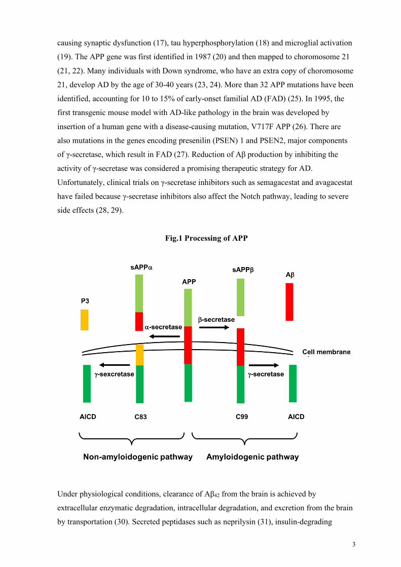
protein (APP) by sequential processing of γ-secretase and β-secretase enzymes (Fig. 1). The
Ab40 species is the most abundant form in the AD brain, but the Ab42 form is considered to
be the main pathological species (12). Aβ42 has been shown to be pro-inflammatory and
neurotoxic (13), and according to the amyloid cascade hypothesis it is believed to be the
main contributor to the development of AD (14). Mutation or overexpression of APP and γ-
secretase gene lead to the increased expression of Aβ peptide. If the Aβ42 concentration
rises above a critical threshold resulting from an imbalance between generation and
clearance, oligomers, fibrils, and senile plaques are formed, contributing to the
development of AD. Different aggregation states of Aβ42 have distinct properties regarding
neurotoxicity (15, 16). The oligomeric forms of Aβ42 are considered as the most toxic form,
3
causing synaptic dysfunction (17), tau hyperphosphorylation (18) and microglial activation
(19). The APP gene was first identified in 1987 (20) and then mapped to choromosome 21
(21, 22). Many individuals with Down syndrome, who have an extra copy of choromosome
21, develop AD by the age of 30-40 years (23, 24). More than 32 APP mutations have been
identified, accounting for 10 to 15% of early-onset familial AD (FAD) (25). In 1995, the
first transgenic mouse model with AD-like pathology in the brain was developed by
insertion of a human gene with a disease-causing mutation, V717F APP (26). There are
also mutations in the genes encoding presenilin (PSEN) 1 and PSEN2, major components
of γ-secretase, which result in FAD (27). Reduction of Aβ production by inhibiting the
activity of γ-secretase was considered a promising therapeutic strategy for AD.
Unfortunately, clinical trials on γ-secretase inhibitors such as semagacestat and avagacestat
have failed because γ-secretase inhibitors also affect the Notch pathway, leading to severe
side effects (28, 29).
Fig.1 Processing of APP
Under physiological conditions, clearance of Aβ42 from the brain is achieved by
extracellular enzymatic degradation, intracellular degradation, and excretion from the brain
by transportation (30). Secreted peptidases such as neprilysin (31), insulin-degrading
4
enzyme (32), matrix metalloproteinases (33), angiotensin-converting enzyme (34), etc, play
critical roles in the catabolism of Ab peptides. They have affinity for specific domains
within the amino acid sequence of Ab peptide and degrade the peptide to harmless forms
(31, 32, 35-38). Alternative pathways of degradation are autophagy (39), degradation by the
ubiquitin-proteasome system (40) and lysomal/endosomal degradation (41, 42). Myeloid
cells are major executors of uptake and phagocytosis of Ab (43, 44). In addition, Ab can be
cleared from the brain by being transported to the cerebrospinal fluid (CSF) (45) or to the
circulation by non-specific interstitial fluid flow (46). However, Aβ can also be reversely
transported from the circulation to the brain if the permeability of the blood brain barrier
(BBB) is compromised, or via the receptor for advanced glycation end products (47).
Keeping the balance between the efflux and influx of Aβ from and to the brain is crucial to
maintain a homeostatic microenvironment in the brain. Furthermore, promoting the
removal of Aβ from the brain is one of the major therapeutic strategies for AD.
Tau pathology
NFTs formed by abnormal phosphorylation of the tau protein is a classical
histopathological hallmark of AD. Tau is a microtubule-associated protein that is involved
in stabilizing microtubuli for efficient axonal transport. The tau protein has three domains:
N-terminal, mid-domain, and C-terminal domain that contains the microtubule-binding
repeats (48, 49). There are six isoforms of tau in the human brain and depending on the
number of microtubule-binding repeats, tau isoforms that are implicated in the pathogenesis
of AD fall into 3-repeat and 4-repeat groups (50, 51). In physiological conditions, the tau
protein folds over the microtubule-binding repeats and the ends approach each other (52).
However, in pathological conditions, the tau protein can adopt a conformation with exposed
residues that are prone to self-aggregation (53). Various post-translational modifications
can affect tau, such as hyperphosphorylation, truncation, acetylation, etc (54). In AD,
phosphorylation of the tau protein causes its detachment from the microtubuli and
subsequently results in their breakdown, axonal transport disturbance and synaptic
connectivity disruption (55-57). Detached hyperphosphorylated tau protein aggregates into
paired helical filaments as well as straight filaments which then form NFTs. NFT-
containing neurons may survive for decades (58), and tau species of small size can be
secreted e.g. via synaptic vesicles (59), exosomes (60) and translocation across the
membrane (61). On the other hand, tau species can be taken-up from the extracellular space
by endocytosis (62) and macropinocytosis (63). It is hypothesized that analogous with prion
disorders, toxic conformations of tau may act as a "seed", causing pathological
5
conformational changes of tau, and propagating through the neuronal network, from
subcortical areas to other areas (64-66). Myeloid cells are implicated in the spread of tau
after phagocytosis of extracellular tau (67). In the AD bran, NFTs first appear in the medial
temporal lobe, specifically in the entorhinal or transentorhinal cortex (Braak stage I and II),
then slowly progress to the limbic regions, particularly to the hippocampus region (Braak
stage III and IV), and then finally to the neocortex (Braak stage V and VI) (68). The pattern
of tau pathology development is closely associated with the clinical progression of AD,
from memory deficits to various cognitive impairments (69).
Tau pathology has been shown to interact with Aβ pathology (70, 71). There is evidence
that tau and Aβ can act in parallel pathways at an early stage, but tau phosphorylation can
also be a downstream event of Aβ pathology, and when tau and Aβ pathology overlaps,
their pathological effects can be enhanced (70, 71).
Heterogeneity of AD and other pathologies
Although senile plaques and NFTs are the most prominent and well-known pathologies in
the AD brain, it is the opinion of a growing number of researchers that Ab and tau cannot
fully explain the pathogenesis of AD. Patients with a diagnosis of AD may lack tau
pathology (72), while subjects with Aβ and tau pathologies may not develop dementia (14).
This highlights the heterogeneity and complexity of AD and encourages scientists to
expand their focus beyond Ab and tau. In recent years, various pathological processes in
addition to the ones related to Ab and tau have been observed in AD, including 1)
unresolved chronic inflammation as evidenced by persistent activation of microglia,
increased levels of pro-inflammatory mediators and decreased levels of specialized pro-
resolving lipid mediators (SPMs) (9), 2) mitochondrial dysfunction as evidenced by
mutations of mitochondrial DNA, impaired endoplasmic reticulum-mitochondria contacts,
oxidative stress and mitochondrial interactions with Ab (73-75), and 3) vascular alterations
as evidenced by disturbance of the BBB (76, 77), etc.
1.1.3 Clinical aspects of AD
Epidemiology
Epidemiological studies have shown that AD is the most prevalent dementia disorder,
afflicting an estimated 47 million people worldwide and accounting for 50-70% of all
dementia cases. The primary risk factor for AD is aging. Approximately 95% of all AD
cases are sporadic and diagnosed after the age of 65 years, while the other 5% are mainly
FAD with an early onset. Most of the genes with mutations that contribute to the

6
pathogenesis of FAD are involved in Aβ processing, such as APP, PSEN1 and PSEN2 (27),
while for sporadic AD, genes involved in lipid metabolism and innate immunity such as
apolipoprotein E (APOE) 4, triggering receptor expressed on myeloid cells 2 (TREM2) and
CD33, are prominent (27, 78, 79). Gender and lifestyle factors are also pronounced risk
factors. AD is more prevalent in females. Lifestyle-related risk factors, including diabetes,
high blood pressure, smoking, insufficient physical activity, have also been shown to
increase the risk for AD, and are potential primary prevention targets for AD (80). In
contrast, keeping oneself in good physical and mental condition has a preventive effect for
developing AD, and physical exercise and social activities are therefore highly
recommended (81-83).
Disease progression and diagnosis
As the development of AD is insidious and usually takes decades, the diagnosis includes
pre-clinical, mild cognitive impairment (MCI) and AD dementia stages. Sperling et al have
proposed three pre-clinical histopathological stages, during which molecular pathologies
gradually accumulate and finally result in cognitive impairment (84). In stage 1,
asymptomatic cerebral amyloidosis occurs, which is undetectable; in stage 2, abnormal tau
and Aβ levels are detectable in the CSF and brain, and evidence of synaptic dysfunction
and/or neurodegeneration appears; in stage 3, some patients report an experience of subtle
cognitive decline, although the objective clinical assessments do not indicate dementia (84).
The cognitive impairment that "the patient knows, but the doctor does not" is termed
subjective cognitive impairment (SCI), which means that the decreased cognition of
patients is still within the normal range on cognitive tests (85). Notably, SCI is not only an
early indicator for MCI and AD, but is also associated with other conditions, such as
depression, stroke, etc (85). Garcia et al reported that SCI patients with cardiovascular risk
factors, medial temporal lobe atrophy and central atrophy had an increased risk of
developing AD (86). Due to the scarcity of CSF samples from cognitively healthy control
subjects, patients diagnosed with SCI are commonly used as a reference group, or a
substitute for healthy controls in studies on AD focused on CSF factors. The collection of
CSF from healthy controls is a complicated enterprise that is beyond what many research
groups have access to. When progressing into MCI, patients are characterized by decreased
cognitive function in clinical assessment, but many remain to be functional members of
society (87). The diagnosis of MCI due to AD is based on the evaluation of AD biomarkers
(87). Positive biomarkers for both Aβ and neuronal injury suggest a high likelihood, while
7
negative biomarkers for both Aβ and neuronal injury indicate that the MCI is unlikely to be
caused by AD (87).
In the AD stage, the ability for patients to function at work or in regular household tasks
and social interactions is significantly impaired. The clinical manifestations vary between
individuals, depending on the involvement of brain functional regions. The most common
clinical symptom is the declining ability to remember new information, resulting from
pathological changes in the entorhinal cortex (Braak stage I and II) and hippocampus
(Braak stage III and IV) (88). Notably, some atypical clinical manifestations may develop
even earlier than memory loss, such as language, visual and executive problems (89, 90).
The involvement of other brain regions, such as basal forebrain (91) and locus coeruleus
(92), is found to begin earlier than in the entorhinal cortex and hippocampus. When
progressed into Braak stage V and VI, additional behavioural and cognitive symptoms
develop as more brain regions are affected. For example, the personality of the patient may
change if the prefrontal neocortex is involved. After the diagnosis of AD, the life span of
the patient is generally less than 10 years (93, 94). Traditionally, the diagnosis of AD is
based on a combination of medical history, clinical symptoms, and memory evaluation. The
most commonly used diagnostic criteria are the International Classification of Disease, 10th
revision (ICD-10), the Diagnostic and Statistical Manual, 4th edition (DSM-IV), and the
National Institute of Neurological and Communicative Disorders and Stroke and the
Alzheimer’s Disease and Related Disorders Association (NINCDS-ADRDA) workgroup in
1984 criteria (95). However, since these criteria require both impairment in memory and the
involvement of at least one non-memory brain region, the diagnosis of AD usually comes at
a stage that is beyond any realistically imaginable intervention. Attributing to the
development of new techniques to detect biomarkers, a molecule- and histometry-based
diagnosis has been proposed for AD (72, 96). The A, T, N System was established in 2018
to characterise AD (72, 96). "A" and "T" refers to Aβ and tau pathology, respectively, as
measured in CSF, or in the brain by amyloid PET; “N” refers to neurodegeneration, as
measured e.g. by hippocampal volume. A molecular profile of "A+T-N-, A+T+N-,
A+T+N+ and A+T-N+" indicates the diagnosis of AD (96). It may, however, be questioned
if a heterogenous and multifactorial disease such as AD can be defined by these factors
only.
CSF biomarkers
Early intervention for AD requires early diagnosis, and early diagnosis requires suitable
biomarkers. The CSF is a rich source of factors produced in the brain, and alterations in the

8
protein found in CSF conceivably reflect the disease progression in the brain. Therefore,
CSF is commonly regarded as a source of biomarkers for AD. In the following paragraphs,
recent data regarding AD biomarkers reflecting Ab pathology, tau pathology, neuroaxonal
degeneration, synaptic dysfunction and activation of glia will be discussed. Aβ and tau in
CSF are core biomarkers assisting the diagnosis of AD (97, 98). The CSF of AD patients is
characterized by decreased by approximately 50% of normal levels of Aβ42 (97). It is
hypothesized that the aggregation and depositing of Aβ in the AD brain result in decreased
CSF levels. When utilizing the ratio between Ab42 and Ab40 or between Ab42 and Ab38, the
diagnostic accuracy could be further increased (99). Total (t)- and phosphorylated (p)-tau
levels in the CSF are also cornerstone markers for biologically defining AD (96). In the
CSF, both t- and p-tau concentrations are significantly increased in AD (100, 101). A likely
explanation is that neurons secret tau protein as a response to Ab exposure (102). In recent
years, neurofilament light (NfL) has emerged as a general marker for neurodegeneration
(103), and increased levels of NfL were found in e.g. frontotemporal, HIV-associated and
vascular dementias (104). In AD, the CSF levels of NfL are elevated, and predict atrophy of
brain and worsening of cognition (105, 106). Synaptic loss is an early event in AD and is
correlated with cognitive decline (107). The dendritic protein neurogranin (Ng) is a CSF
biomarker for synaptic damage, shows elevated levels in AD and is correlated with t- and
p-tau levels and cognitive decline (108, 109). Since the levels of Ng are not dramatically
changed in the CSF of other neurodegenerative dementias, Ng has the potential to be an
AD-specific biomarker (110, 111). Neuroinflammation mediated by activated microglia and
astrocytes is another key pathological feature of AD (9). Biomarkers related to
inflammation will be discussed in the next section.
One of the drawbacks of using CSF biomarkers is the difficulty to evaluate brain region-
specific changes. As the involvement of brain regions is related to the disease progression
(e.g., Braak stages), the use of CSF biomarkers to monitor disease development may be
limited. Another disadvantage is that many of the CSF biomarkers are not specific. For
example, CSF levels of NfL are increased in several neurodegenerative diseases (103, 104).
Therefore, there is an urgent need to develop biomarker combinations to define the
pathological pattern in the CSF of AD, which may assist to increase the diagnostic
accuracy.
Treatment
Due to the lack of a disease-modifying treatment, AD has brought a large economic
burden to society, in addition to the suffering of relatives. It is estimated that the societal
9
cost per AD patient needing residential care is 72 500 Euro per year (112). Besides the
societal cost (41.7%), informal care costs (42.3%) and direct medical costs (16%) are also
heavy (1). The drugs available for the treatment of AD, i.e., various AChE inhibitors and
the N-methyl-D-aspartate (NMDA)-receptor antagonist Memantine™, can only slow the
progression of the disease, and are not very effective in doing so. Considering that no new
drug for AD has been approved for clinical use in the past 15 years, work on developing
new disease-modifying drugs for AD is urgently warranted.
Currently, researchers have put great efforts into developing drugs that reduce the Aβ42
burden in the AD brain (113). Clinical trials of β-secretase and γ-secretase inhibitors,
which reduce Aβ42 production, have failed due to severe side effects. Anti-Aβ monoclonal
antibodies, which increase removal of Aβ from the brain, have been tested extensively
during almost two decades. Of those, Aducanumab has been shown to improve cognition
of MCI and mild AD patients at the highest dose (10 mg/kg) in a phase 3 study (clinical
trial NCT02477800 and NCT02484547). There are other anti-Aβ monoclonal antibodies,
which have shown promising results in phase 1 or phase 2 studies and are now in phase 3
studies. For example, the antibody BAN2401, which binds to soluble and toxic Aβ
aggregates, has been shown to reduce Aβ burden in the brain and improve cognition in a
phase 2b study (clinical trial NCT01767311), and is now in phase 3 studies (clinical trial
NCT03887455). Clinical trials based on tau immunotherapy are also ongoing. Tau vaccine
AADvac1 (clinical trial NCT02579252) targeting truncated tau has shown some protective
effects in a phase 2 study. Since the pathogenesis of AD is complicated, novel therapeutic
strategies targeting pathological factors beyond Ab pathology and tau should be
considered, such as neuroinflammation.
1.2 Neuroinflammation 1.2.1 Neuroinflammation in AD
An increasing amount of epidemiological, genetic, pathological, and clinical evidence
shows that inflammation plays a major part in the pathogenesis of AD.
Epidemiological evidence
Historically, epidemiological studies have shown that anti-inflammatory therapies reduce
the risk of developing AD. In 1989, Jenkinson et al observed a low prevalence of AD in
rheumatoid arthritis patients treated with anti-inflammatory drugs (114). McGeer et al in
1990 further addressed the association between the use of anti-inflammatory drugs and the
development of AD in a study on a cohort of rheumatoid arthritis patients, showing that the
10
prevalence of AD among rheumatoid arthritis patients was lower, and that the anti-
inflammatory therapies for the rheumatoid arthritis patients might be the explanation (115).
Subsequent epidemiological studies on large cohorts have shown that non-steroidal anti-
inflammatory drugs (NSAIDs) decreased the relative risk for developing AD (116-121). In
an AD animal model, Bachstetter et al found that early treatment with anti-inflammatory
drugs attenuated AD pathology (122). Although these findings indicate that stopping
inflammation could be a therapeutic strategy for AD, clinical trials using NSAIDs to
prevent or cure AD in humans have largely failed. A large, randomized trial investigated if
the administration of anti-inflammatory drugs could prevent the development of AD
among individuals over 70 years with a familiar history of AD but was discontinued
because of an observed increased risk of developing cardiac disease (123). Another large,
randomized trial including more than 3 000 participants investigating if a low dose of
aspirin could improve the cognition of AD patients failed, which may due to that 30% of
the participants dropped at the follow-up cognitive tests (124). There are no current
treatment guidelines that recommend using NSAIDs to prevent or treat dementia. A
possible explanation for the failure of the clinical trials could be that inflammation plays a
dual role in the pathogenesis of AD: anti-inflammatory drugs may not only attenuate the
harmful processes of inflammation, but also block the protective ones, such as clearance of
Ab, etc. In this regard, stimulating the switch from harmful to beneficial processes, i.e.
promoting the resolution of inflammation, could be a more effective therapeutic strategy.
Genetic evidence
A number of large genome-wide association (GWAS) studies have identified a set of
inflammation-related susceptibility genes for AD (125-132), such as TREM2 (triggering
receptor) (78, 133), CD33 (surface receptor) (79, 128, 134), MS4A4AE/MS4A6A
(membrane-spanning proteins) (128), and CR1 (complement receptor 1) (135). TREM2 is a
receptor expressed on microglia and is responsible for their activation by forming a
complex with the transmembrane immune signalling adaptor (136). The R47H mutation of
TREM2 is a loss-of-function mutation impairing microglial phagocytosis and energy
metabolism (78, 133), carried by less than 0.5% of the population, and increasing the risk of
developing AD approximately three-fold (133). CD33 is a surface receptor containing a
tyrosine-based inhibitory motif, which plays an important role in the modulation of immune
cell response, such as the production of immune mediators, phagocytosis, etc (137). The
expression of CD33 on microglia is increased in AD and is associated with decreased
capability of microglia to take up Ab (134). The rs3865444C allele of CD33 is an AD-
11
associated single-nucleotide polymorphism with strong impact and is associated with
increased Ab pathology and microglial activation (79). Kramarz et al reported that adding
neuroinflammation-related genes to the Gene Ontology (GO) database can improve the
interpretation of AD-related transcriptome data (138). To translate the mutations of AD-
related genes to functional outcomes, more experimental studies are needed to increase the
understanding of AD pathogenesis and provide a basis for identifying novel therapeutic
targets.
Pathological and clinical evidence
The first piece of pathological evidence indicating the involvement of neuroinflammation in
AD is dated back to 1910. Oskar Fischer published a paper of nearly 100 pages describing
the pathological and clinical features of patients with plaques in the brain. He stated that the
deposition of plaques provoked inflammation resulting in neurodegeneration. However, he
did not provide solid histopathological evidence to support his statement. In the 1980’s,
studies were published describing activated microglia together with inflammatory
mediators, such as complement factors and immunoglobulins in the vicinity of Ab plaques
(139, 140). In 1996, a pathological study on post mortem brain tissue from AD patients
showed the occurrence of inflammation, whereas controls without dementia, but with a
high burden of AD pathology did not have inflammation in their brains (14). More recently,
increased activation of microglia in living AD patients was shown by PET studies (141,
142), and also in MCI patients (143, 144). Evidence of inflammation in AD has also been
provided by studies on CSF samples. Chitinase 3-like 1 (also known as YKL-40), a
glycoprotein enriched in astrocytes, shows promise as a candidate AD biomarker. In the
CSF of AD patients, YKL-40 was modestly increased and was correlated to tau levels and
cognition (145, 146). Soluble (s)TREM2, mainly produced by microglia, is another
candidate biomarker for AD. Increased levels of sTREM2 were observed prior to
symptomatic disease onset and were correlated to tau pathology (147). Furthermore,
alterations in various inflammatory mediators, including cytokines, chemokines, adhesion-
related molecules, have been observed in the CSF of AD patients. For example, Taipa et al
found that the levels of both pro-inflammatory mediators and anti-inflammatory cytokines
were higher in CSF from AD patients compared to non-demented controls (148). The levels
of intercellular adhesion molecule (ICAM)-1, vascular adhesion molecule 1 (VCAM-1)
were found to be higher in the CSF of AD patients (149, 150). Chemokine monocyte
chemoattractant protein (MCP)-1is another inflammatory factor shown to be increased in
the CSF of AD patients (151).
12
1.2.2 Microglia and astrocytes in neuroinflammation
Biology and heterogeneity of microglia
Studies on the cellular components of the central nervous system (CNS) date back to the
beginning of the 19th century. In 1856, the German pathologist Rudolf Virchow first
coined the term "glia", which means "glue" in Greek, to describe the non-neuronal tissues
in the CNS. In 1919, microglia were first visualized and described by the Spanish scientist
Pio del Rio Hortega, and were defined as a separate cell type (152). During ontogeny,
microglia are derived from the embryonic yolk sac precursors, enter the brain via the
lateral ventricles and leptomeninges by embryonic day 9.5 and then spread throughout the
cortical wall (153). Postnatally, microglia are more proliferative and active in performing
their functions than adult microglia (154). Their morphology is amoeboid and they are
actively involved in the establishment of neuronal networks by controlling the fate as well
as the number of neurons and their progenitor cells (154-156). Microglia phagocytose
apoptotic neurons and neuronal progenitor cells, remove dysfunctional or redundant
synapses, thereby remodelling the synaptic circuits (157-159). Microglia also support
other cellular components during CNS development, e.g. by contributing to
myelinogenesis through interaction with oligodendrocytes and their progenitors (160), and
to neovascularization by interacting with endothelial cells (161).
In adulthood, although less active, microglia perform similar roles as during development,
including synapse maintenance, trophic support, and phagocytic removal of cellular and
molecular debris (9, 162, 163). In the adult brain, microglia are considered the key effector
of immune activities. In the resting state, microglia continuously monitor a surrounding
microenvironment. Upon detecting a pathogenic object or condition, microglia transform
from ramified to ameboid morphology, and migrate toward the site of insult and contribute
to the initiation and progression of the inflammatory response (164-166).
Microglia exist in various phenotypic states when activated, indicative of the type of
activating insult and associated with different activities. Although today considered
somewhat controversial, there is a general division into two phenotypes: M1 and M2. The
M1 state is characterised by pro-inflammatory activities (such as secreting pro-
inflammatory cytokines), and if becoming chronic, by impaired phagocytic capacity (167-
169). In contrast, M2 microglia execute anti-inflammatory reactions, express anti-
inflammatory surface biomarkers and have stronger phagocytic capacity (170). Criticism
against the M1/M2 nomenclature can be raised due to the fact that there is always a
phenotype heterogeneity in the tissue, as well as in the cell culture dish, and that the
presence of a few cellular markers may not be the most appropriate basis for determining
13
the phenotypic state. In recent years, attributing to the development of the RNA-
Sequencing (RNA-Seq) technique, knowledge of the regional and population-based
heterogeneity of microglia in health and disease has advanced considerably. Using bulk
RNA-Seq, van der Poel et al discovered more than 400 differentially expressed genes
(DEGs) in human microglia from white matter and grey matter. Genes that were highly
expressed in grey matter were enriched in the "cytokine-mediated signalling" pathway,
while those highly expressed in white matter were related to the "chemotaxis" pathway
(171). Using snRNA-Seq, Emma et al identified 13 subclusters of microglia in the human
brain. Three of the clusters were enriched in homeostasis genes, three clusters were found
to have a high expression of phagocytic genes, another three clusters were enriched in both
homeostasis and neuron-related genes, two small clusters were related to inflammatory
responses, one cluster was associated with cellular stress, and one small cluster was
enriched in proliferation genes (172). When interpreting RNA-Seq data, one should be
aware that the natural specific signature of isolated microglia may be lost during sample
processing. Furthermore, the biological terms in the open access databases (e.g. GO) under
which genes are organized may not be specific or relevant for the disease of interest. For
example, Ab phagocytosis is distinct in many ways from the phagocytosis of other objects
(173). When analysing single-cell RNA-Seq data, the identification and annotation of
microglia clusters are flexible and subjective, which may lead to faulty conclusions if one
does not critically review how these clusters were identified and how their biological role
was derived. The results are also affected by the quality of RNA and the processing of the
RNA-Seq data. Therefore, results obtained from the RNA-Seq data need to be verified on a
protein and functional level.
Microglia in AD
In the pathological condition of AD, microglia appear to play a dual role during disease
progression. In vivo PET studies showed that in prodromal AD patients, microglial
activation was associated with a better prognosis, whereas increased microglial activity
later in the disease course was linked to a poor outcome (142), indicating that there is a
detrimental change in microglial activities during the pathogenesis. In the early stages of
AD, inflammatory activation of microglia can have beneficial effects. In experimental
settings, they contribute to the effective removal of Aβ and attempt to keep the brain in
homeostasis (174, 175). However, due to an increasing Aβ concentration and persistent
pro-inflammatory microenvironment, microglia appear to attain a more detrimental
phenotype that aggravates the disease (14, 176-178). In the late stage of AD, pronounced
14
pro-inflammatory activation is associated with inefficient clearance of Aβ, induction of tau
pathology and neuronal degeneration.
Clearance of Aβ can be achieved by microglial phagocytosis followed by intracellular
degradation (179), or by extracellular degradation by enzymes released from microglia or
other cells (180), as described in the previous sections. Aβ is recognized by microglial
receptors such as CD36, CD14, SCARA1 and toll-like receptors (TLRs), and is taken up
by microglia and enter the endolysosomal pathway (181-184). Mutations in the TREM2
and CD33 genes are correlated with impaired phagocytosis (78, 133, 185). The soluble
forms of Aβ, predominant in early stages of AD, can be degraded by enzymes, but fibrillar
Aβ in late stages are less prone to be degraded (179). When Aβ is recognized by microglia,
they become activated, leading to secretion of pro-inflammatory cytokines and chemokines
(183, 186), oxidative stress (187), and other neurotoxic activities (188). Since neurons have
receptors for pro-inflammatory cytokines (189), and the resulting activation of nuclear
factor (NF)-kB in neurons leads to activation of the APP gene, there is a vicious circle
maintained by exposure to undegraded and newly generated Aβ, leading to further
activation of microglia, and also reducing the phagocytic capacity of microglia to clear Aβ
from the brain. A vicious circle also exists between tau pathology and chronic
inflammation: the pro-inflammatory microenvironment shaped by microglia induces
phosphorylation of tau in neurons and p-tau-burdened neurons activate microglia (190-
192). Furthermore, activated microglia induce neurodegeneration by causing synaptic
dysfunction and by directly phagocytosing live neurons (193, 194). Pro-inflammatory
mediators released by microglia disrupt membrane conductance and potential, and thereby
the neuronal electrical signalling in the hippocampus (195-197), thus hypothetically
contributing to cognitive dysfunction. Taken together, microglia play a dual role in the
pathogenesis of AD, depending on their phenotype, the stage of disease, and the genetic
make-up of the affected individual.
Until very recently, bulk, and single-cell/nucleus RNA-Seq were predominantly used to
investigate the disease-specific transcriptome signature of microglia derived from
autopsies of brains from AD patients. Using bulk RNA-Seq, Srinivasan et al showed that
the damage-associated transcriptome profiles of microglia from human AD post mortem
brains were largely different form the profile seen in microglia from an AD-related mouse
model (198). A differential expression gene analysis between AD patients and age-
matched controls first demonstrated only 12 genes being significantly differently
expressed. Upon re-analysis after filtering out the outlier genes using DESeq2-provided
Cook's distance, the microglia from AD brains were found to exhibit a gene expression
15
profile indicating accelerated aging and upregulation of the APOE gene (198). Since the
results after filtering out outliers are quite different from those observed before, one should
be cautious when drawing conclusions. Also using bulk RNA-Seq, Alsema et al reported
that the gene expression profiles of microglia are not different in AD patients compared to
age-matched non-demented elderly (199). The results from these two studies indicate that
the gene expression profile of microglia in AD is not different, contradicting the plethora
of results obtained from studies on proteomics in microglia. This may be explained by the
limitations of the bulk RNA-Seq technique, which is based on transcriptomic information
obtained from a mixture of microglia with various phenotypes. Therefore, differences in
gene expression signature in disease-associated microglia may be hidden in the bulk of
expression. Supporting evidence for this hypothesis comes from single-cell/nucleus RNA-
Seq studies in which different microglial clusters are investigated separately. In 2019,
Mathys et al observed up-regulation of the APOE gene in microglia from AD patients
using single-nucleus RNA-Seq (200), in line with the findings from Srinivasan et al using
bulk RNA-Seq (198). In 2020, Olah et al discovered a cluster of microglia that was altered
in AD (201). This subset of microglia had a high expression of CD74, both in
transcriptome and protein level, and comprised 2-5% of the whole microglia population. In
the AD brain, the proportion of CD74 highly expressing microglia was reduced (201). In
2021, Gerrits et al identified an association between microglia clusters and the molecular
pathologies of AD. A population of microglia belonging to the phagocytic/activated cluster
was correlated with Ab load and located close to Ab plaques. Another population enriched
with the CX3R1, P2RY12, GRID2, ADGRB3 and DPP10 genes was associated with p-tau
load (172). When interpreting these results, it is important to note that the microglia were
obtained from autopsy samples, and the results may therefore reflect transcriptomic
information of very late-stage AD, while in MCI or prodromal AD, the transcriptome of
microglia may be different.
Cellular models to study microglia
The crucial roles that microglia play in both health and disease emphasize the need for valid
and effective in vitro methods and models to investigate mechanisms and responses in
microglia, and their regulation. The existing microglia in vitro models include cell lines,
primary microglia, and stem cell/monocyte-derived microglia. Microglial cell lines are
available from human (e.g. CHME-3 (202), CHME-5 (203) and HMO6 (204)), mouse (e.g.
BV2 (205)), rat (e.g. HAPI (206)) and macaque origins (207). These cell lines are
commonly produced from primary embryonic microglia that are transformed with
16
oncogenes to create an immortalized cell line. The advantages of using cell lines include
high accessibility, and that they are easily propagated and maintained. The disadvantages
are susceptibility to dedifferentiation, alteration of phenotype due to transformation and
genetic mutations during culture, altogether resulting in the genetic and functional
differences of cell lines compared to primary microglia.
Methodologies to isolate and culture primary microglia from human (206), non-human
primates (208) and rodents (209) are available. Most of these methods start with the
dissociation of the tissue, followed by cell-sorting using antibody-conjugated magnetic
beads (210), or by flow-cytometry (206). Although human primary microglia obtained from
neurosurgery is an ideal model, the practical use is limited by the scarcity of this resource
and that it is an invasive and complicated isolation procedure. Rodent primary microglia
represent a commonly used model with the advantages of 1) allowing studies on specific
pathogenic genes (i.e. transgenic and knock-in mice); 2) the post-mortem delay, which
affects the quality of microglia, can be strictly controlled; 3) the genetic background of the
obtained microglia is homogenous, therefore the results can be repeated in other research
groups, although the homogenous genetic background can also be argued to be a
disadvantage when translating to humans. The disadvantages of using rodent-derived
primary microglia are that the genetic background of rodents is very different from humans
and, as already mentioned, homogeneous (211-213), and that they in the majority of cases
are derived from pre-natal brain tissue.
Microglia can also be differentiated from monocytes and stem cells including embryonic
stem cells (ESCs) and induced pluripotent stem cells (iPSCs). ESCs are obtained from the
blastocyst, whereas iPSCs are mostly generated from fibroblasts of adults (214). The
advantages of using stem cell-derived microglia are that they are more similar to human
primary microglia compared to cell lines and mouse primary microglia, that they bring the
genetic information of the donors, and that they can be differentiated to other CNS cells in
parallel (211). However, the differentiation procedure is complicated and time-consuming.
It usually takes 4 to 8 weeks to complete the microglia differentiation protocol (211).
Compared to stem cell-derived microglia, monocyte-derived microglia (MdM) are easier to
obtain and differentiate. In addition, MdM are more similar to human primary microglia
than stem cell-derived microglia (unpublished data in Paper IV). In 2012, Etemad et al
were the first to produce microglial-like cells from human peripheral blood monocytes by
culturing the monocytes with a combination of immune-related mediators (granulocyte-
macrophage colony-stimulating factor (GM-CSF), M-CSF, MCP-1) and nerve growth
factor (NGF) b (215). In 2014, Ohgidani et al showed that the MdM carried genetic
17
information of the donors and could reflect pathological changes of a disease in the brain
(216). Nasu-Hakola disease is a rare autosomal recessive disorder caused by the mutation
of the microglia-expressed gene TREM2 or the DNAX-activation protein 12 (216). The
MdM generated from patients diagnosed with Nasu-Hakola disease exhibited delayed but
more marked pro-inflammatory responses (216). In 2017, Ryan et al proved the similarity
of MdM to human primary microglia in a transcriptomic level and utilized MdM to
investigate the effects of a gene variant related to AD (213).
Astrocytes in AD
Another important cellular component of the inflammatory response in the brain is the
astrocyte. It may be argued that astrocytes receive an unfairly low amount of attention than
microglia when in fact, astrocytes are the most abundant glial cell type in the CNS, and the
importance of astrocyte in the pathogenesis of AD should not be underestimated. In health,
astrocytes contribute to the formation and function of synapses (217, 218), modulation of
neuronal plasticity and excitability (219), extracellular potassium buffering (220), and
formation of the BBB and neurovascular unit (221), etc. In AD, reactive astrocytes are
characterised by elevated expression of glia fibrillary acidic protein (GFAP) and are often
found to accumulate around the Ab plaques, both in human AD and in animal models (222,
223). As previously mentioned, YKL-40 (a glycoprotein enriched in astrocytes (224)) is
increased in the CSF of AD patients. Experimental evidence suggests that astrocytes play a
dual role in the development of AD. On one hand, when exposed to Ab, astrocytes
exacerbate neuroinflammation by secreting pro-inflammatory mediators, such as cytokines
and nitric oxide (NO) (225). On the other hand, astrocytes contribute to the clearance of Ab
by expression of Ab-degrading enzymes (226), mediating ApoE lipidation to assist
microglia-mediated Ab removal (227) and by transporting soluble Ab out of the CNS via
the water channel protein aquaporin 4 (228).
1.2.3 Molecular players in AD
Cytokine and chemokine
Cytokines and chemokines are the key inflammatory protein mediators in AD (9). MCI
patients with increased pro-inflammatory cytokines and decreased anti-inflammatory
cytokines in the CSF have a higher risk to develop AD (229). Similarly, pro-inflammatory
signalling is found to be up-regulated while the anti-inflammatory signalling is down-
regulated in AD. The major sources for cytokines and chemokines are microglia and
astrocytes (230, 231). When exposed to Ab, the release of cytokines and chemokines by
18
microglia is increased. In an AD mice model, the cytokine levels in the brain were found to
be correlated to Ab load (232). Interestingly, the pro-inflammatory microenvironment
shaped by the inflammatory mediators in turn affects the phenotypes and functions of
microglia and astrocytes in the CNS (section 1.2.2).
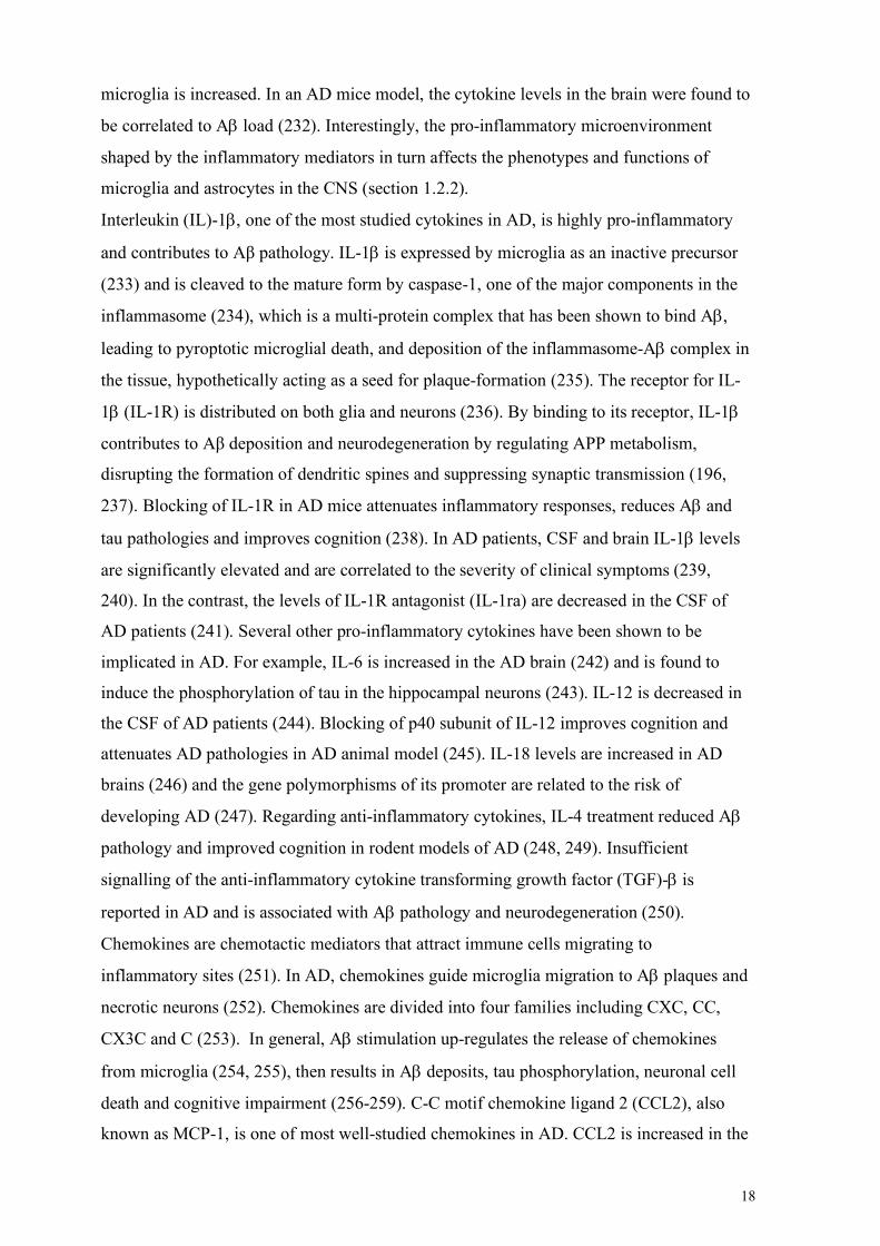
Interleukin (IL)-1b, one of the most studied cytokines in AD, is highly pro-inflammatory
and contributes to Aβ pathology. IL-1b is expressed by microglia as an inactive precursor
(233) and is cleaved to the mature form by caspase-1, one of the major components in the
inflammasome (234), which is a multi-protein complex that has been shown to bind Ab,
leading to pyroptotic microglial death, and deposition of the inflammasome-Ab complex in
the tissue, hypothetically acting as a seed for plaque-formation (235). The receptor for IL-
1b (IL-1R) is distributed on both glia and neurons (236). By binding to its receptor, IL-1b
contributes to Aβ deposition and neurodegeneration by regulating APP metabolism,
disrupting the formation of dendritic spines and suppressing synaptic transmission (196,
237). Blocking of IL-1R in AD mice attenuates inflammatory responses, reduces Ab and
tau pathologies and improves cognition (238). In AD patients, CSF and brain IL-1b levels
are significantly elevated and are correlated to the severity of clinical symptoms (239,
240). In the contrast, the levels of IL-1R antagonist (IL-1ra) are decreased in the CSF of
AD patients (241). Several other pro-inflammatory cytokines have been shown to be
implicated in AD. For example, IL-6 is increased in the AD brain (242) and is found to
induce the phosphorylation of tau in the hippocampal neurons (243). IL-12 is decreased in
the CSF of AD patients (244). Blocking of p40 subunit of IL-12 improves cognition and
attenuates AD pathologies in AD animal model (245). IL-18 levels are increased in AD
brains (246) and the gene polymorphisms of its promoter are related to the risk of
developing AD (247). Regarding anti-inflammatory cytokines, IL-4 treatment reduced Ab
pathology and improved cognition in rodent models of AD (248, 249). Insufficient
signalling of the anti-inflammatory cytokine transforming growth factor (TGF)-b is
reported in AD and is associated with Ab pathology and neurodegeneration (250).
Chemokines are chemotactic mediators that attract immune cells migrating to
inflammatory sites (251). In AD, chemokines guide microglia migration to Ab plaques and
necrotic neurons (252). Chemokines are divided into four families including CXC, CC,
CX3C and C (253). In general, Ab stimulation up-regulates the release of chemokines
from microglia (254, 255), then results in Ab deposits, tau phosphorylation, neuronal cell
death and cognitive impairment (256-259). C-C motif chemokine ligand 2 (CCL2), also
known as MCP-1, is one of most well-studied chemokines in AD. CCL2 is increased in the
19
AD brain, and is localized in Ab plaques, microvessels, neurons, microglia, and astrocytes
(242, 260-262). CCL2 has also been found to be increased in the CSF and plasma of AD
patients (151, 263), and is correlated to cognitive impairment (264, 265). Gene
polymorphisms of CCL2 are associated with the risk of developing AD (266). In response
to Ab stimulation, microglia and astrocytes increase the secretion of CCL2 (267, 268); in
response to CCL2, microglia and astrocytes increase the production of cytokines and the
formation of Ab oligomer (269, 270). These evidences indicate a harmful role of CCL2 in
AD. Surprisingly, a deficiency of CCR2, the receptor for CCL2, accelerates the disease
progression in AD mice model (271). This may be due to the failure of immune cell
recruitment, which is likely to be mediated by CCR2 (271). Other chemokines, such as
CXCL8, CXCL10, CX3CL1, CCL5, CXCL12, etc, are involved in the pathogenesis of AD
as well (272).
Lipid mediators
Lipid mediators (LMs) including prostaglandins (PGs), leukotrienes (LTs), as well as the
SPMs play important roles in neuroinflammation in the context of AD. SPMs will be
discussed in detail in the next section. PGs constitute a family of small inflammatory LMs
generated by a series of enzymatic reactions that start from arachidonic acid (AA) released
from membrane phospholipids. AA is metabolized by lipoxygenase (LOX) and
cyclooxygenase (COX), to form PGH2 with subsequent enzymatic processing through
specific pathways to yield more PGs including PGD2, PGE2, PGF2a, PGI2, as well as the
thromboxane (TX) TXA2, the receptors for which are prostaglandin D2 receptor (DP),
prostaglandin E2 receptor (EP), prostaglandin F2a receptor (FP), prostaglandin I2 receptor
(IP) and thromboxane receptor (TP), respectively. PGD2, the most abundant PG in the
brain, is synthesized by hematopoietic prostaglandin D synthase (HPGDS) (273). PGD2 has
two receptors with opposite functions where DP1 is neuroprotective and DP2 neurotoxic
(274). In AD, HPGDS and PGD2 levels are increased in microglia and astrocytes
surrounding the Ab plaques, as observed both in human brain and in a mouse AD model
(275). PGE2 is synthesized by prostaglandin E synthases (PGESs) and binds to four
receptors (EP1-4). PGES1 and PGES2 were increased in the middle frontal gyrus of AD
brain (276, 277).
The PGE2 receptors EP1, 2 and 3 have been demonstrated in microglia and neurons, while
EP4 is restricted to neurons (274). EP1 signalling is associated with increased Ab
pathology and Ab-induced neurotoxicity (278, 279). EP1-knockout AD mice were shown
20
to have reduced Ab plaques (278). Neurons from the EP1-knockout mice or neurons
incubated with an EP1 antagonist were more resistant to Ab-induced neuronal toxicity
(278, 279). EP2 signalling was shown to be associated with increased pro-inflammatory
reactions, decreased Ab phagocytosis by microglia and increased Ab-induced
neurotoxicity. Deletion of EP2 reduced Ab burden and oxidative damage in AD mice
model (280). A microglia-specific knock-out of EP2 exhibit decreased toxic inflammation,
increased Ab removal and microglia chemotaxis, elevated cytoprotective signalling, and
reduced synaptic injury and cognitive impairment (281, 282). In a mouse AD model,
deletion of EP3 was found to decrease inflammatory reactions and the production of Ab,
while increasing the levels of presynaptic proteins (283). The levels of EP4 have been
shown to be reduced in the brains of AD and MCI patients (284). Treatment of microglia in
vitro with an EP4 agonist decreased inflammation and increased Ab uptake (284). Deletion
of EP4 in an AD mouse model increased Ab pathology and pro-inflammatory cytokine
secretion (284). PGF2a and TXA2 have not been extensively studied in the context of AD.
A study on AD brains demonstrated increased levels of PGF2a and 8-iso-PGF2a in the
hippocampus (285). Some studies suggest that activation of TP is associated with increased
APP and Ab production (286, 287). Even though additional studies are needed to clarify
how LM signalling is implicated in AD, LMs and their receptors have a potential as
biomarkers as well as treatment targets for AD.
Other players
Complement factors, mainly derived from microglia, are increased in the AD brain, and
also play an important role in the development of AD (14). Aβ activates the complement
system, which in turn contributes to Aβ depositing (288). In animal models of AD, both
Aβ and tau pathologies were reduced following inhibition of the pro-inflammatory
complement factor C5a receptor (289). In addition, inducible nitric oxide synthase (iNOS)
in microglia produces the free radical NO when stimulated with cytokines (290). iNOS
levels are increased in the AD brain, and NO has been shown to cause mitochondrial
dysfunction, synaptic and axonal damage, and neuronal cell death (291-293). Furthermore,
the structure of Aβ can be modified by NO through peroxynitrite formation. Nitrated Aβ
tends to be more prone to aggregate and causes more severe neuronal injury compared to
normal Aβ (294).
21
1.3 Resolution of inflammation 1.3.1 General aspects of resolution of inflammation
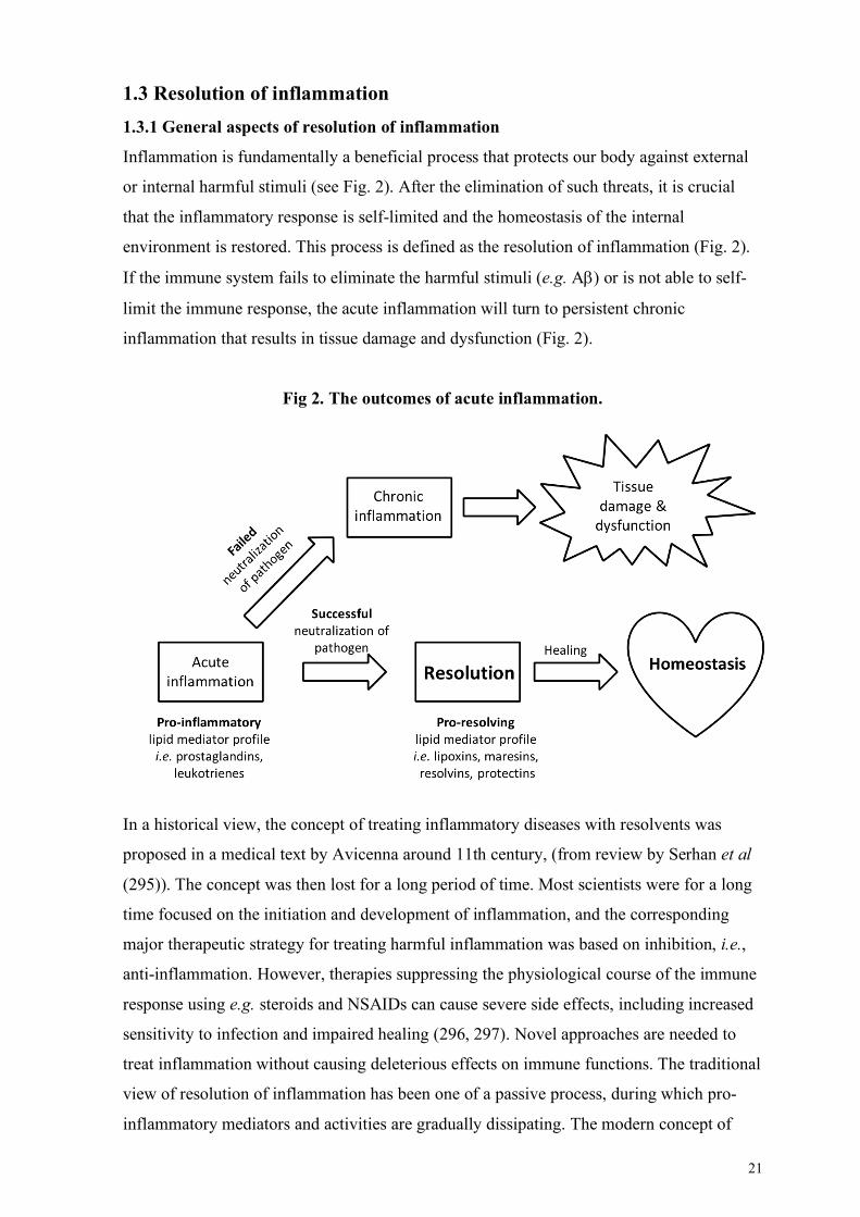
Inflammation is fundamentally a beneficial process that protects our body against external
or internal harmful stimuli (see Fig. 2). After the elimination of such threats, it is crucial
that the inflammatory response is self-limited and the homeostasis of the internal
environment is restored. This process is defined as the resolution of inflammation (Fig. 2).
If the immune system fails to eliminate the harmful stimuli (e.g. Ab) or is not able to self-
limit the immune response, the acute inflammation will turn to persistent chronic
inflammation that results in tissue damage and dysfunction (Fig. 2).
Fig 2. The outcomes of acute inflammation.
In a historical view, the concept of treating inflammatory diseases with resolvents was
proposed in a medical text by Avicenna around 11th century, (from review by Serhan et al
(295)). The concept was then lost for a long period of time. Most scientists were for a long
time focused on the initiation and development of inflammation, and the corresponding
major therapeutic strategy for treating harmful inflammation was based on inhibition, i.e.,
anti-inflammation. However, therapies suppressing the physiological course of the immune
response using e.g. steroids and NSAIDs can cause severe side effects, including increased
sensitivity to infection and impaired healing (296, 297). Novel approaches are needed to
treat inflammation without causing deleterious effects on immune functions. The traditional
view of resolution of inflammation has been one of a passive process, during which pro-
inflammatory mediators and activities are gradually dissipating. The modern concept of
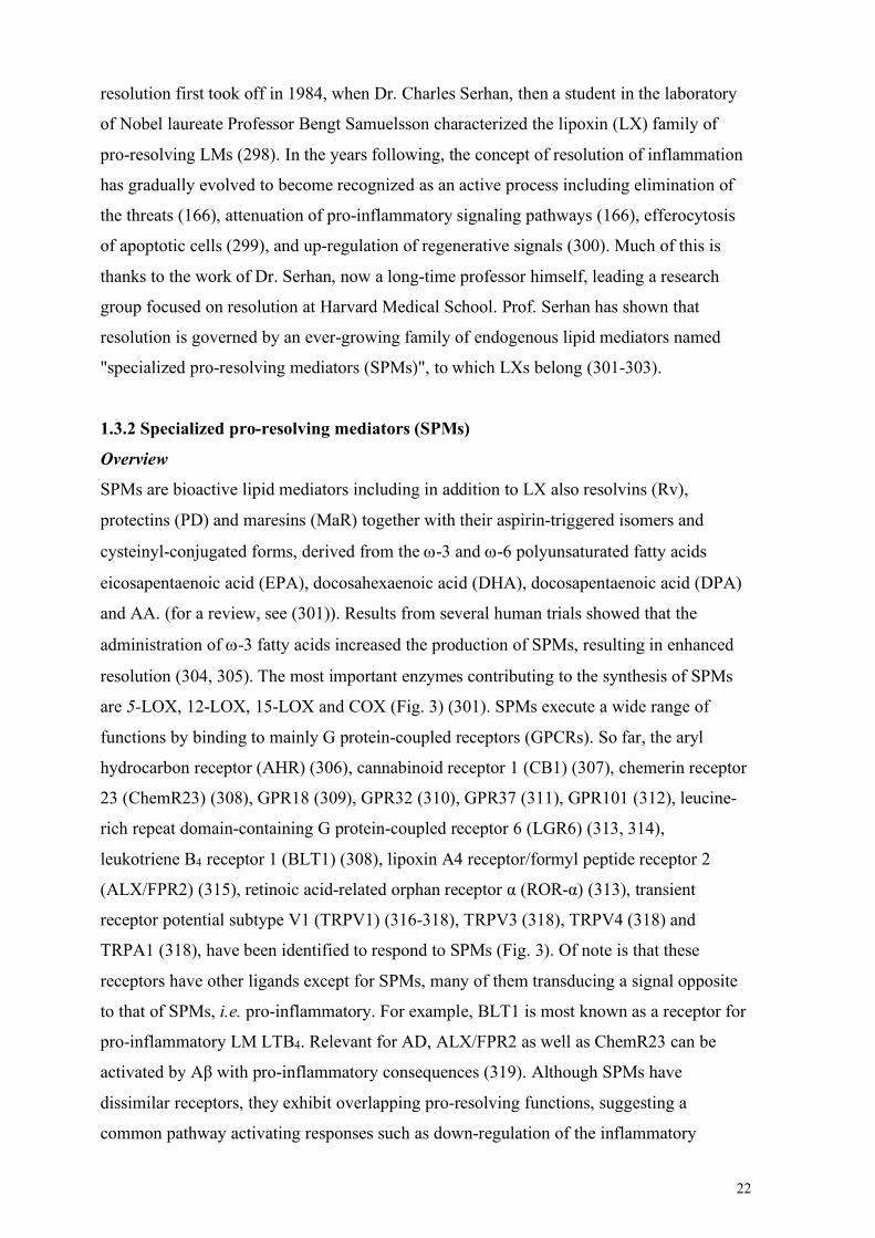
22
resolution first took off in 1984, when Dr. Charles Serhan, then a student in the laboratory
of Nobel laureate Professor Bengt Samuelsson characterized the lipoxin (LX) family of
pro-resolving LMs (298). In the years following, the concept of resolution of inflammation
has gradually evolved to become recognized as an active process including elimination of
the threats (166), attenuation of pro-inflammatory signaling pathways (166), efferocytosis
of apoptotic cells (299), and up-regulation of regenerative signals (300). Much of this is
thanks to the work of Dr. Serhan, now a long-time professor himself, leading a research
group focused on resolution at Harvard Medical School. Prof. Serhan has shown that
resolution is governed by an ever-growing family of endogenous lipid mediators named
"specialized pro-resolving mediators (SPMs)", to which LXs belong (301-303).
1.3.2 Specialized pro-resolving mediators (SPMs)
Overview
SPMs are bioactive lipid mediators including in addition to LX also resolvins (Rv),
protectins (PD) and maresins (MaR) together with their aspirin-triggered isomers and
cysteinyl-conjugated forms, derived from the w-3 and w-6 polyunsaturated fatty acids
eicosapentaenoic acid (EPA), docosahexaenoic acid (DHA), docosapentaenoic acid (DPA)
and AA. (for a review, see (301)). Results from several human trials showed that the
administration of w-3 fatty acids increased the production of SPMs, resulting in enhanced
resolution (304, 305). The most important enzymes contributing to the synthesis of SPMs
are 5-LOX, 12-LOX, 15-LOX and COX (Fig. 3) (301). SPMs execute a wide range of
functions by binding to mainly G protein-coupled receptors (GPCRs). So far, the aryl
hydrocarbon receptor (AHR) (306), cannabinoid receptor 1 (CB1) (307), chemerin receptor
23 (ChemR23) (308), GPR18 (309), GPR32 (310), GPR37 (311), GPR101 (312), leucine-
rich repeat domain-containing G protein-coupled receptor 6 (LGR6) (313, 314),
leukotriene B4 receptor 1 (BLT1) (308), lipoxin A4 receptor/formyl peptide receptor 2
(ALX/FPR2) (315), retinoic acid-related orphan receptor α (ROR-α) (313), transient
receptor potential subtype V1 (TRPV1) (316-318), TRPV3 (318), TRPV4 (318) and
TRPA1 (318), have been identified to respond to SPMs (Fig. 3). Of note is that these
receptors have other ligands except for SPMs, many of them transducing a signal opposite
to that of SPMs, i.e. pro-inflammatory. For example, BLT1 is most known as a receptor for
pro-inflammatory LM LTB4. Relevant for AD, ALX/FPR2 as well as ChemR23 can be
activated by Aβ with pro-inflammatory consequences (319). Although SPMs have
dissimilar receptors, they exhibit overlapping pro-resolving functions, suggesting a
common pathway activating responses such as down-regulation of the inflammatory
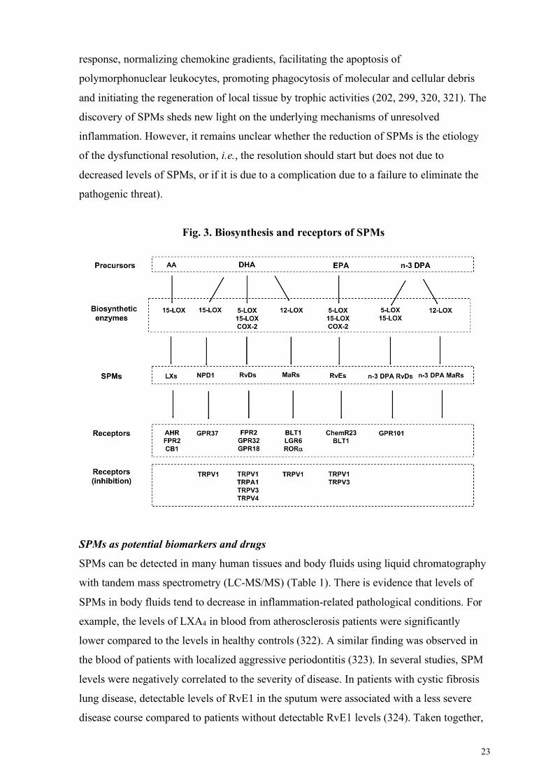
23
response, normalizing chemokine gradients, facilitating the apoptosis of
polymorphonuclear leukocytes, promoting phagocytosis of molecular and cellular debris
and initiating the regeneration of local tissue by trophic activities (202, 299, 320, 321). The
discovery of SPMs sheds new light on the underlying mechanisms of unresolved
inflammation. However, it remains unclear whether the reduction of SPMs is the etiology
of the dysfunctional resolution, i.e., the resolution should start but does not due to
decreased levels of SPMs, or if it is due to a complication due to a failure to eliminate the
pathogenic threat).
Fig. 3. Biosynthesis and receptors of SPMs
SPMs as potential biomarkers and drugs
SPMs can be detected in many human tissues and body fluids using liquid chromatography
with tandem mass spectrometry (LC-MS/MS) (Table 1). There is evidence that levels of
SPMs in body fluids tend to decrease in inflammation-related pathological conditions. For
example, the levels of LXA4 in blood from atherosclerosis patients were significantly
lower compared to the levels in healthy controls (322). A similar finding was observed in
the blood of patients with localized aggressive periodontitis (323). In several studies, SPM
levels were negatively correlated to the severity of disease. In patients with cystic fibrosis
lung disease, detectable levels of RvE1 in the sputum were associated with a less severe
disease course compared to patients without detectable RvE1 levels (324). Taken together,
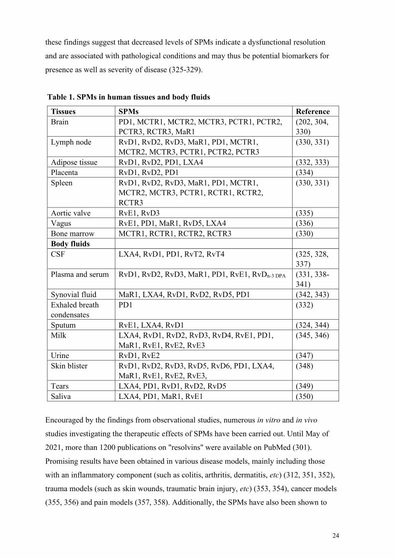
24
these findings suggest that decreased levels of SPMs indicate a dysfunctional resolution
and are associated with pathological conditions and may thus be potential biomarkers for
presence as well as severity of disease (325-329).
Table 1. SPMs in human tissues and body fluids
Tissues SPMs Reference Brain PD1, MCTR1, MCTR2, MCTR3, PCTR1, PCTR2,
PCTR3, RCTR3, MaR1 (202, 304, 330)
Lymph node RvD1, RvD2, RvD3, MaR1, PD1, MCTR1, MCTR2, MCTR3, PCTR1, PCTR2, PCTR3
(330, 331)
Adipose tissue RvD1, RvD2, PD1, LXA4 (332, 333) Placenta RvD1, RvD2, PD1 (334) Spleen RvD1, RvD2, RvD3, MaR1, PD1, MCTR1,
MCTR2, MCTR3, PCTR1, RCTR1, RCTR2, RCTR3
(330, 331)
Aortic valve RvE1, RvD3 (335) Vagus RvE1, PD1, MaR1, RvD5, LXA4 (336) Bone marrow MCTR1, RCTR1, RCTR2, RCTR3 (330) Body fluids CSF LXA4, RvD1, PD1, RvT2, RvT4 (325, 328,
337) Plasma and serum RvD1, RvD2, RvD3, MaR1, PD1, RvE1, RvDn-3 DPA (331, 338-
341) Synovial fluid MaR1, LXA4, RvD1, RvD2, RvD5, PD1 (342, 343) Exhaled breath condensates
PD1 (332)
Sputum RvE1, LXA4, RvD1 (324, 344) Milk LXA4, RvD1, RvD2, RvD3, RvD4, RvE1, PD1,
MaR1, RvE1, RvE2, RvE3 (345, 346)
Urine RvD1, RvE2 (347) Skin blister RvD1, RvD2, RvD3, RvD5, RvD6, PD1, LXA4,
MaR1, RvE1, RvE2, RvE3, (348)
Tears LXA4, PD1, RvD1, RvD2, RvD5 (349) Saliva LXA4, PD1, MaR1, RvE1 (350)
Encouraged by the findings from observational studies, numerous in vitro and in vivo
studies investigating the therapeutic effects of SPMs have been carried out. Until May of
2021, more than 1200 publications on "resolvins" were available on PubMed (301).
Promising results have been obtained in various disease models, mainly including those
with an inflammatory component (such as colitis, arthritis, dermatitis, etc) (312, 351, 352),
trauma models (such as skin wounds, traumatic brain injury, etc) (353, 354), cancer models
(355, 356) and pain models (357, 358). Additionally, the SPMs have also been shown to
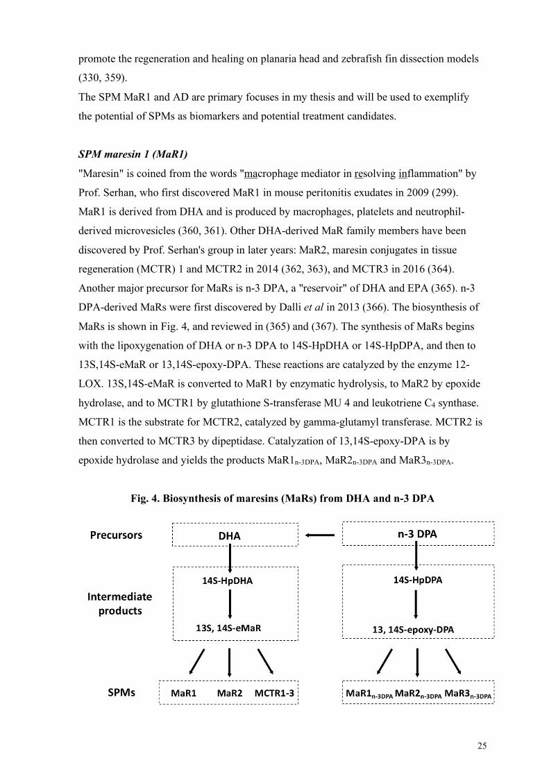
25
promote the regeneration and healing on planaria head and zebrafish fin dissection models
(330, 359).
The SPM MaR1 and AD are primary focuses in my thesis and will be used to exemplify
the potential of SPMs as biomarkers and potential treatment candidates.
SPM maresin 1 (MaR1)
"Maresin" is coined from the words "macrophage mediator in resolving inflammation" by
Prof. Serhan, who first discovered MaR1 in mouse peritonitis exudates in 2009 (299).
MaR1 is derived from DHA and is produced by macrophages, platelets and neutrophil-
derived microvesicles (360, 361). Other DHA-derived MaR family members have been
discovered by Prof. Serhan's group in later years: MaR2, maresin conjugates in tissue
regeneration (MCTR) 1 and MCTR2 in 2014 (362, 363), and MCTR3 in 2016 (364).
Another major precursor for MaRs is n-3 DPA, a "reservoir" of DHA and EPA (365). n-3
DPA-derived MaRs were first discovered by Dalli et al in 2013 (366). The biosynthesis of
MaRs is shown in Fig. 4, and reviewed in (365) and (367). The synthesis of MaRs begins
with the lipoxygenation of DHA or n-3 DPA to 14S-HpDHA or 14S-HpDPA, and then to
13S,14S-eMaR or 13,14S-epoxy-DPA. These reactions are catalyzed by the enzyme 12-
LOX. 13S,14S-eMaR is converted to MaR1 by enzymatic hydrolysis, to MaR2 by epoxide
hydrolase, and to MCTR1 by glutathione S-transferase MU 4 and leukotriene C4 synthase.
MCTR1 is the substrate for MCTR2, catalyzed by gamma-glutamyl transferase. MCTR2 is
then converted to MCTR3 by dipeptidase. Catalyzation of 13,14S-epoxy-DPA is by
epoxide hydrolase and yields the products MaR1n-3DPA, MaR2n-3DPA and MaR3n-3DPA.
Fig. 4. Biosynthesis of maresins (MaRs) from DHA and n-3 DPA
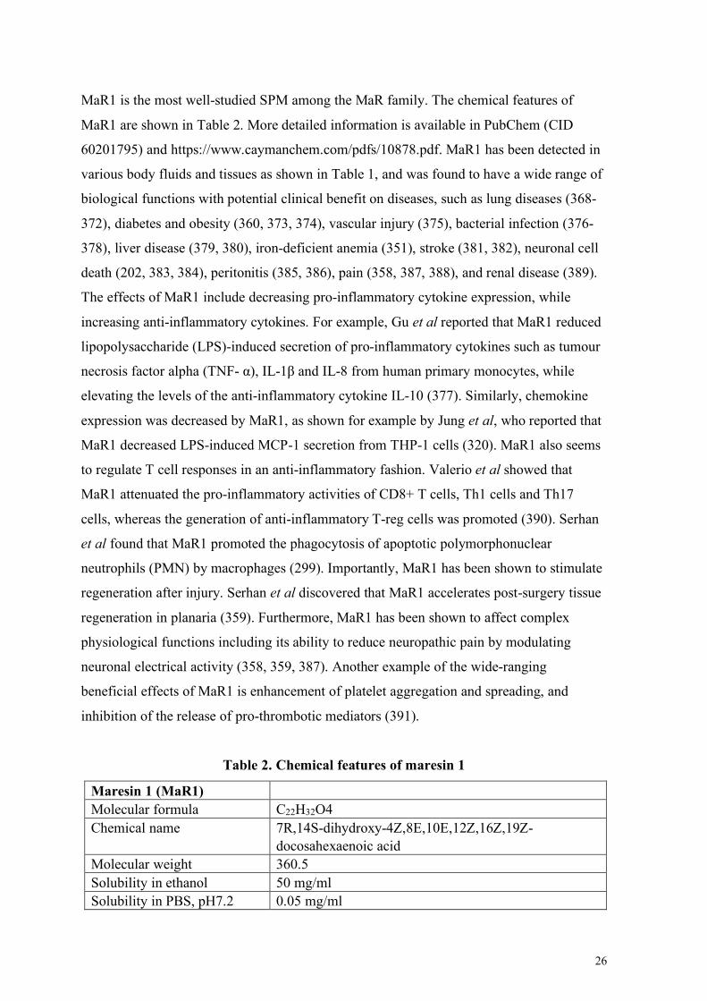
26
MaR1 is the most well-studied SPM among the MaR family. The chemical features of
MaR1 are shown in Table 2. More detailed information is available in PubChem (CID
60201795) and https://www.caymanchem.com/pdfs/10878.pdf. MaR1 has been detected in
various body fluids and tissues as shown in Table 1, and was found to have a wide range of
biological functions with potential clinical benefit on diseases, such as lung diseases (368-
372), diabetes and obesity (360, 373, 374), vascular injury (375), bacterial infection (376-
378), liver disease (379, 380), iron-deficient anemia (351), stroke (381, 382), neuronal cell
death (202, 383, 384), peritonitis (385, 386), pain (358, 387, 388), and renal disease (389).
The effects of MaR1 include decreasing pro-inflammatory cytokine expression, while
increasing anti-inflammatory cytokines. For example, Gu et al reported that MaR1 reduced
lipopolysaccharide (LPS)-induced secretion of pro-inflammatory cytokines such as tumour
necrosis factor alpha (TNF- α), IL-1β and IL-8 from human primary monocytes, while
elevating the levels of the anti-inflammatory cytokine IL-10 (377). Similarly, chemokine
expression was decreased by MaR1, as shown for example by Jung et al, who reported that
MaR1 decreased LPS-induced MCP-1 secretion from THP-1 cells (320). MaR1 also seems
to regulate T cell responses in an anti-inflammatory fashion. Valerio et al showed that
MaR1 attenuated the pro-inflammatory activities of CD8+ T cells, Th1 cells and Th17
cells, whereas the generation of anti-inflammatory T-reg cells was promoted (390). Serhan
et al found that MaR1 promoted the phagocytosis of apoptotic polymorphonuclear
neutrophils (PMN) by macrophages (299). Importantly, MaR1 has been shown to stimulate
regeneration after injury. Serhan et al discovered that MaR1 accelerates post-surgery tissue
regeneration in planaria (359). Furthermore, MaR1 has been shown to affect complex
physiological functions including its ability to reduce neuropathic pain by modulating
neuronal electrical activity (358, 359, 387). Another example of the wide-ranging
beneficial effects of MaR1 is enhancement of platelet aggregation and spreading, and
inhibition of the release of pro-thrombotic mediators (391).
Table 2. Chemical features of maresin 1
Maresin 1 (MaR1) Molecular formula C22H32O4 Chemical name 7R,14S-dihydroxy-4Z,8E,10E,12Z,16Z,19Z-
docosahexaenoic acid Molecular weight 360.5 Solubility in ethanol 50 mg/ml Solubility in PBS, pH7.2 0.05 mg/ml
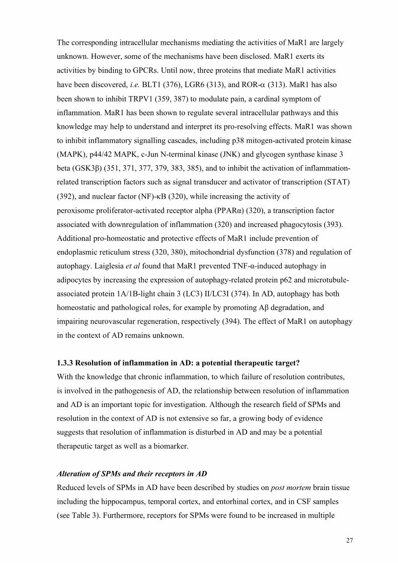
27
The corresponding intracellular mechanisms mediating the activities of MaR1 are largely
unknown. However, some of the mechanisms have been disclosed. MaR1 exerts its
activities by binding to GPCRs. Until now, three proteins that mediate MaR1 activities
have been discovered, i.e. BLT1 (376), LGR6 (313), and ROR-a (313). MaR1 has also
been shown to inhibit TRPV1 (359, 387) to modulate pain, a cardinal symptom of
inflammation. MaR1 has been shown to regulate several intracellular pathways and this
knowledge may help to understand and interpret its pro-resolving effects. MaR1 was shown
to inhibit inflammatory signalling cascades, including p38 mitogen-activated protein kinase
(MAPK), p44/42 MAPK, c-Jun N-terminal kinase (JNK) and glycogen synthase kinase 3
beta (GSK3β) (351, 371, 377, 379, 383, 385), and to inhibit the activation of inflammation-
related transcription factors such as signal transducer and activator of transcription (STAT)
(392), and nuclear factor (NF)-kB (320), while increasing the activity of
peroxisome proliferator-activated receptor alpha (PPARα) (320), a transcription factor
associated with downregulation of inflammation (320) and increased phagocytosis (393).
Additional pro-homeostatic and protective effects of MaR1 include prevention of
endoplasmic reticulum stress (320, 380), mitochondrial dysfunction (378) and regulation of
autophagy. Laiglesia et al found that MaR1 prevented TNF-α-induced autophagy in
adipocytes by increasing the expression of autophagy-related protein p62 and microtubule-
associated protein 1A/1B-light chain 3 (LC3) II/LC3I (374). In AD, autophagy has both
homeostatic and pathological roles, for example by promoting Aβ degradation, and
impairing neurovascular regeneration, respectively (394). The effect of MaR1 on autophagy
in the context of AD remains unknown.
1.3.3 Resolution of inflammation in AD: a potential therapeutic target?
With the knowledge that chronic inflammation, to which failure of resolution contributes,
is involved in the pathogenesis of AD, the relationship between resolution of inflammation
and AD is an important topic for investigation. Although the research field of SPMs and
resolution in the context of AD is not extensive so far, a growing body of evidence
suggests that resolution of inflammation is disturbed in AD and may be a potential
therapeutic target as well as a biomarker.
Alteration of SPMs and their receptors in AD
Reduced levels of SPMs in AD have been described by studies on post mortem brain tissue
including the hippocampus, temporal cortex, and entorhinal cortex, and in CSF samples
(see Table 3). Furthermore, receptors for SPMs were found to be increased in multiple
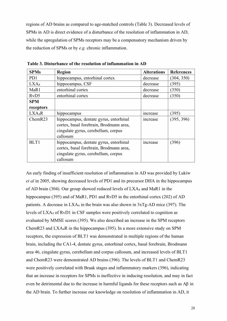
28
regions of AD brains as compared to age-matched controls (Table 3). Decreased levels of
SPMs in AD is direct evidence of a disturbance of the resolution of inflammation in AD,
while the upregulation of SPMs receptors may be a compensatory mechanism driven by
the reduction of SPMs or by e.g. chronic inflammation.
Table 3. Disturbance of the resolution of inflammation in AD
SPMs Region Alterations References PD1 hippocampus, entorhinal cortex decrease (304, 350) LXA4 hippocampus, CSF decrease (395) MaR1 entorhinal cortex decrease (350) RvD5 entorhinal cortex decrease (350) SPM receptors
LXA4R hippocampus increase (395) ChemR23 hippocampus, dentate gyrus, entorhinal
cortex, basal forebrain, Brodmann area, cingulate gyrus, cerebellum, corpus callosum
increase (395, 396)
BLT1 hippocampus, dentate gyrus, entorhinal cortex, basal forebrain, Brodmann area, cingulate gyrus, cerebellum, corpus callosum
increase (396)
An early finding of insufficient resolution of inflammation in AD was provided by Lukiw
et al in 2005, showing decreased levels of PD1 and its precursor DHA in the hippocampus
of AD brain (304). Our group showed reduced levels of LXA4 and MaR1 in the
hippocampus (395) and of MaR1, PD1 and RvD5 in the entorhinal cortex (202) of AD
patients. A decrease in LXA4 in the brain was also shown in 3xTg-AD mice (397). The
levels of LXA4 of RvD1 in CSF samples were positively correlated to cognition as
evaluated by MMSE scores (395). We also described an increase in the SPM receptors
ChemR23 and LXA4R in the hippocampus (395). In a more extensive study on SPM
receptors, the expression of BLT1 was demonstrated in multiple regions of the human
brain, including the CA1-4, dentate gyrus, entorhinal cortex, basal forebrain, Brodmann
area 46, cingulate gyrus, cerebellum and corpus callosum, and increased levels of BLT1
and ChemR23 were demonstrated AD brains (396). The levels of BLT1 and ChemR23
were positively correlated with Braak stages and inflammatory markers (396), indicating
that an increase in receptors for SPMs is ineffective in inducing resolution, and may in fact
even be detrimental due to the increase in harmful ligands for these receptors such as Ab in
the AD brain. To further increase our knowledge on resolution of inflammation in AD, it
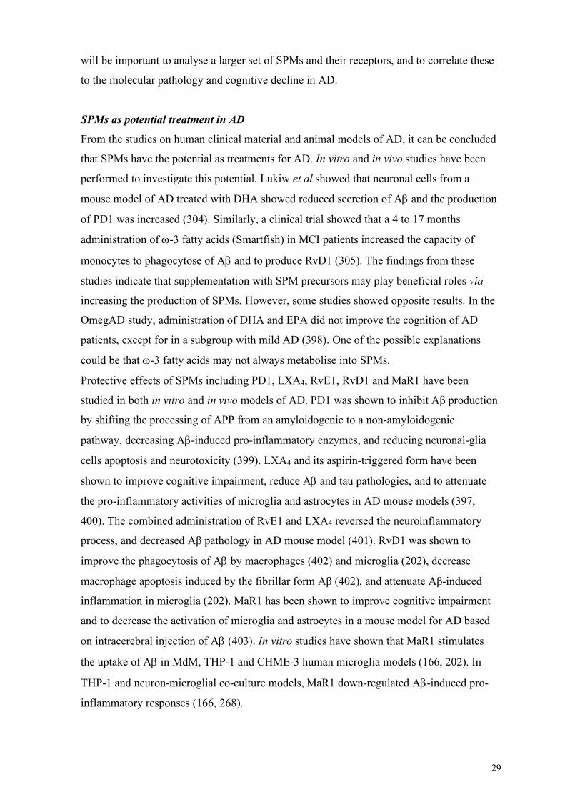
29
will be important to analyse a larger set of SPMs and their receptors, and to correlate these
to the molecular pathology and cognitive decline in AD.
SPMs as potential treatment in AD
From the studies on human clinical material and animal models of AD, it can be concluded
that SPMs have the potential as treatments for AD. In vitro and in vivo studies have been
performed to investigate this potential. Lukiw et al showed that neuronal cells from a
mouse model of AD treated with DHA showed reduced secretion of Ab and the production
of PD1 was increased (304). Similarly, a clinical trial showed that a 4 to 17 months
administration of w-3 fatty acids (Smartfish) in MCI patients increased the capacity of
monocytes to phagocytose of Ab and to produce RvD1 (305). The findings from these
studies indicate that supplementation with SPM precursors may play beneficial roles via
increasing the production of SPMs. However, some studies showed opposite results. In the
OmegAD study, administration of DHA and EPA did not improve the cognition of AD
patients, except for in a subgroup with mild AD (398). One of the possible explanations
could be that w-3 fatty acids may not always metabolise into SPMs.
Protective effects of SPMs including PD1, LXA4, RvE1, RvD1 and MaR1 have been
studied in both in vitro and in vivo models of AD. PD1 was shown to inhibit Aβ production
by shifting the processing of APP from an amyloidogenic to a non-amyloidogenic
pathway, decreasing Ab-induced pro-inflammatory enzymes, and reducing neuronal-glia
cells apoptosis and neurotoxicity (399). LXA4 and its aspirin-triggered form have been
shown to improve cognitive impairment, reduce Ab and tau pathologies, and to attenuate
the pro-inflammatory activities of microglia and astrocytes in AD mouse models (397,
400). The combined administration of RvE1 and LXA4 reversed the neuroinflammatory
process, and decreased Aβ pathology in AD mouse model (401). RvD1 was shown to
improve the phagocytosis of Ab by macrophages (402) and microglia (202), decrease
macrophage apoptosis induced by the fibrillar form Aβ (402), and attenuate Aβ-induced
inflammation in microglia (202). MaR1 has been shown to improve cognitive impairment
and to decrease the activation of microglia and astrocytes in a mouse model for AD based
on intracerebral injection of Ab (403). In vitro studies have shown that MaR1 stimulates
the uptake of Ab in MdM, THP-1 and CHME-3 human microglia models (166, 202). In
THP-1 and neuron-microglial co-culture models, MaR1 down-regulated Ab-induced pro-
inflammatory responses (166, 268).

30
It can be hypothesized with confidence that SPMs have a potential being drugs for AD in
humans. The existing experimental evidence supports this hypothesis, and since SPMs are
endogenously produced in the human body (and brain), treatment with SPMs or analogues
thereof may be associated with few or mild side effects. Protective effects of SPMs have
been observed in mouse models of AD (401), brain injury (404) and Down syndrome
(405). In these studies, intraperitoneal, subcutaneous, or oral administration has been used
to supply SPMs, indicating their effective transfer across the BBB. Altogether, stimulating
resolution in the AD brain is a novel promising treatment target for this terrible disease.
31
2 RESEARCH AIMS The aim of this project was to study neuroinflammation and its resolution in Alzheimer’s
disease (AD) by analysis of cerebrospinal fluid (CSF) (Paper I and II) and investigating
the potential of the specialized pro-resolving lipid mediator (SPM) maresin 1 (MaR1) to
resolve Ab-induced AD-like inflammation in microglia (Paper III and IV). An overview of
the project and sub-studies are shown in Fig. 5 (for Paper I and II) and Fig. 6 (for Paper
III and IV).
Specific aims for each paper:
Paper I: To identify the profile of inflammatory protein mediators in the CSF of patients
diagnosed with subjective cognitive impairment (SCI) or AD, taking into account the
presence of comorbidities.
Paper II: To analyse pro-inflammatory and pro-resolving lipid mediators (LMs) in CSF to
investigate differences between patients diagnosed with SCI, mild cognitive impairment
(MCI) and AD, and correlations to cognition and AD biomarkers.
Paper III: To explore the effects of MaR1 on β-amyloid (Aβ)-induced inflammation and
Aβ uptake in a human macrophage model, as well as a a human microglial cell model -
monocyte-derived microglia (MdM).
Paper IV: To investigate if MaR1 normalizes Aβ-induced alterations in transcriptome and
protein levels in MdM.
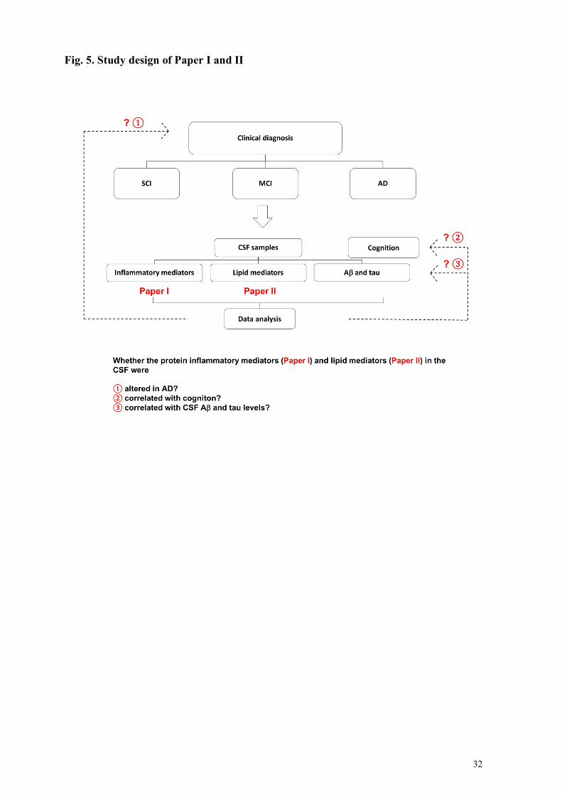
32
Fig. 5. Study design of Paper I and II
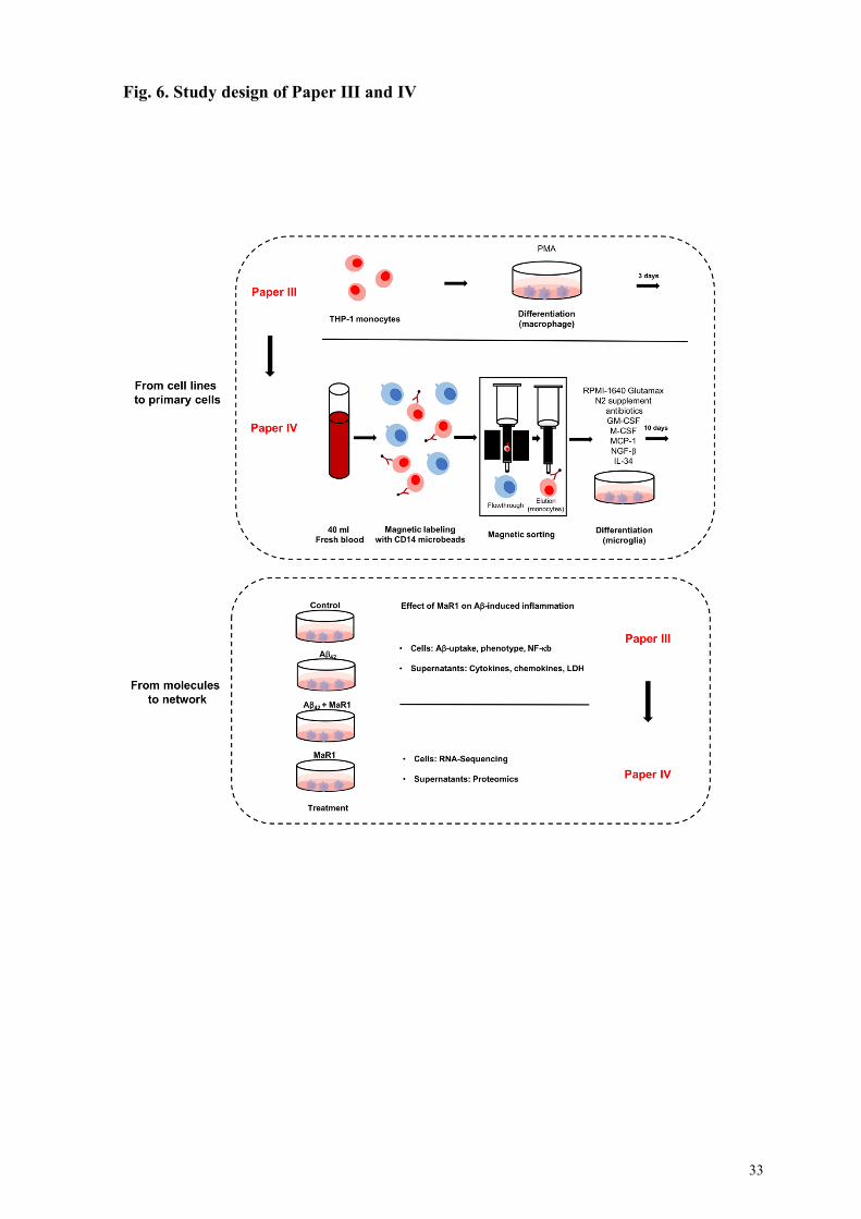
33
Fig. 6. Study design of Paper III and IV


35
3 MATERIALS AND METHODS 3.1 Human CSF samples and clinical data 3.1.1 Human CSF samples
CSF is secreted by the choroid plexus in the lateral and fourth ventricles and serves as a
"sink" that receives the metabolite generated by brain function. The connections between
CSF and the fluid compartments inside the brain makes it possible in some cases to view
pathological changes in brain in the CSF, and it is thus a major source of biomarkers for
neurodegenerative disorders. Compared to blood, another major source of biomarkers, CSF
reflects the conditions in brain much better. Although the CSFs are obtained from an
invasive procedure in clinic, lumbar puncture can be considered as a relatively harmless
standard procedure. Another advantage of using CSF samples as study material is that since
it is collected from the living person, longitudinal studies are possible, allowing the
scientists to investigate dynamic changes of biomarkers. CSF samples can be used for
biomarker studies for multiple purposes, such as assist diagnosis, predict prognosis, and
evaluate disease severity. Research focused on discovering new biomarkers in CSF (as well
as other tissues), is based on hypothesis-based approaches, where a factor or factors are
targeted, and explorative approaches using -omics technologies to look for new biomarkers
by measuring a large set of factors.
In Paper I and II, CSF samples were obtained from the Gedoc biobank (part of Stockholms
Medicinska Biobank, SMB) at the Memory Clinic at Karolinska University Hospital.
Patients visiting the Memory clinic at Karolinska University Hospital (as well as many
other hospitals) are routinely subjected to lumbar puncture and sampling of CSF for
diagnostic purposes. After informed consent, patients donate a volume of CSF to the
biobank of the Memory clinic (Gedoc), making it available for researchers together with
data on age, gender and a limited number of clinical parameters. Factors of inflammation
(Paper I) and homeostasis (Paper II) were analysed in the CSF samples from the same
persons.
3.1.2 Clinical data and study cohorts
In Paper I and II, the clinical data on age, gender, diagnosis, cognition, and AD
biomarkers were retrieved from the Gedoc database. The diagnosis of i) AD was based on
the ICD-10 criteria (406), ii) MCI was based on the Winblad criteria, and iii) SCI was
established when results from the clinical assessments did not indicate cognitive
impairment despite subjective complaints of memory problems by the patients (85, 407).
Cognitive impairment was evaluated by mini-mental score examination (MMSE) test (2),
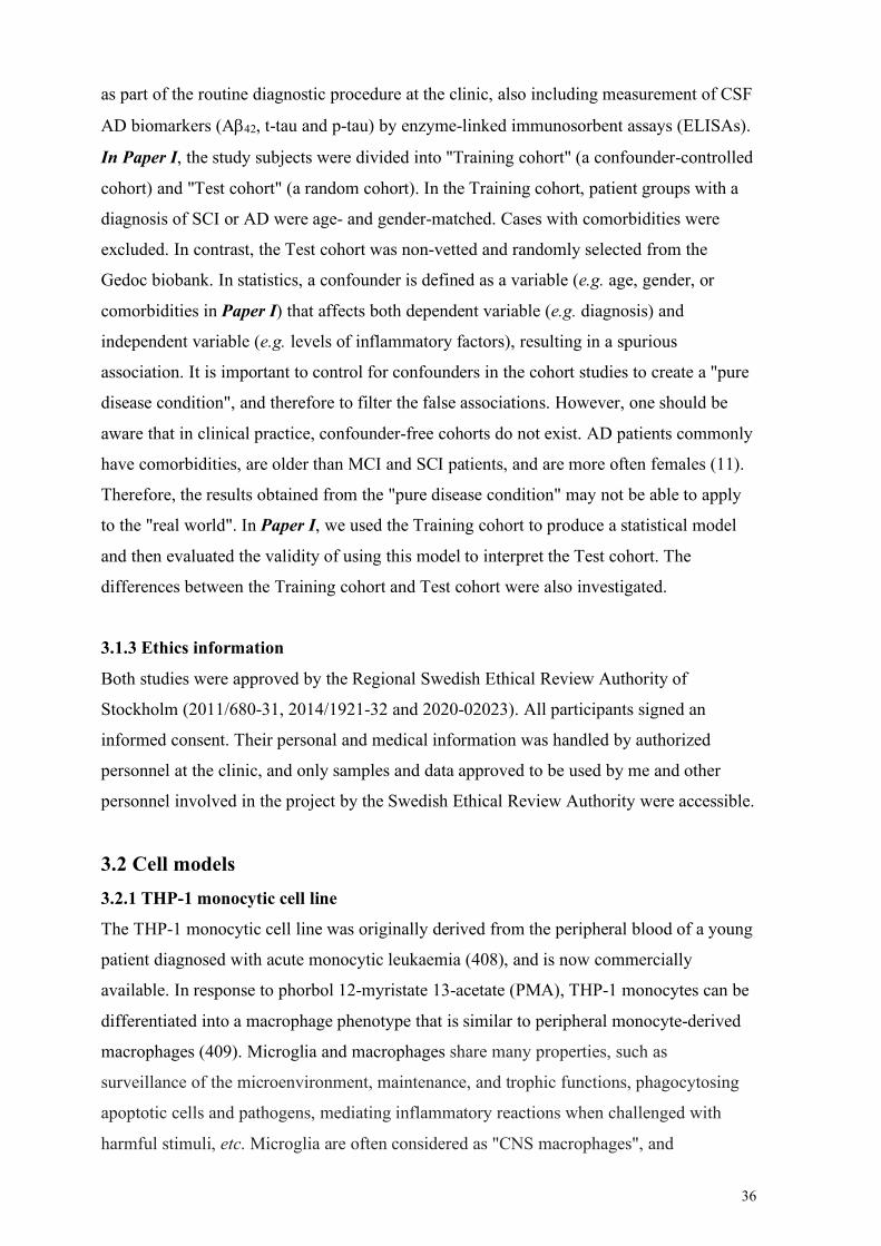
36
as part of the routine diagnostic procedure at the clinic, also including measurement of CSF
AD biomarkers (Ab42, t-tau and p-tau) by enzyme-linked immunosorbent assays (ELISAs).
In Paper I, the study subjects were divided into "Training cohort" (a confounder-controlled
cohort) and "Test cohort" (a random cohort). In the Training cohort, patient groups with a
diagnosis of SCI or AD were age- and gender-matched. Cases with comorbidities were
excluded. In contrast, the Test cohort was non-vetted and randomly selected from the
Gedoc biobank. In statistics, a confounder is defined as a variable (e.g. age, gender, or
comorbidities in Paper I) that affects both dependent variable (e.g. diagnosis) and
independent variable (e.g. levels of inflammatory factors), resulting in a spurious
association. It is important to control for confounders in the cohort studies to create a "pure
disease condition", and therefore to filter the false associations. However, one should be
aware that in clinical practice, confounder-free cohorts do not exist. AD patients commonly
have comorbidities, are older than MCI and SCI patients, and are more often females (11).
Therefore, the results obtained from the "pure disease condition" may not be able to apply
to the "real world". In Paper I, we used the Training cohort to produce a statistical model
and then evaluated the validity of using this model to interpret the Test cohort. The
differences between the Training cohort and Test cohort were also investigated.
3.1.3 Ethics information
Both studies were approved by the Regional Swedish Ethical Review Authority of
Stockholm (2011/680-31, 2014/1921-32 and 2020-02023). All participants signed an
informed consent. Their personal and medical information was handled by authorized
personnel at the clinic, and only samples and data approved to be used by me and other
personnel involved in the project by the Swedish Ethical Review Authority were accessible.
3.2 Cell models 3.2.1 THP-1 monocytic cell line
The THP-1 monocytic cell line was originally derived from the peripheral blood of a young
patient diagnosed with acute monocytic leukaemia (408), and is now commercially
available. In response to phorbol 12-myristate 13-acetate (PMA), THP-1 monocytes can be
differentiated into a macrophage phenotype that is similar to peripheral monocyte-derived
macrophages (409). Microglia and macrophages share many properties, such as
surveillance of the microenvironment, maintenance, and trophic functions, phagocytosing
apoptotic cells and pathogens, mediating inflammatory reactions when challenged with
harmful stimuli, etc. Microglia are often considered as "CNS macrophages", and

37
macrophages in different forms are commonly used as the substitute of primary microglia
(178, 410-413). As microglia and THP-1 cells respond to Ab42 stimulation in a similar
pattern, Ab42-treated THP-1 cells have been used as an in vitro model to investigate AD-
relevant inflammation (178, 410-413).
One of the advantages of cell lines is that genetic manipulations are easy to perform, such
as gene knock-down and knock-in. In Paper III, THP-1 cells and THP1-LuciaTM NF-kB
cells were differentiated by 72 h exposure to PMA and used as an in vitro model of
neuroinflammation in AD by treatment with Ab42 for 24 h. THP1-LuciaTM NF-kB cells
have been designed for monitoring the activation of NF-kB and contain an NF-kB-
inducible Luc reporter. As a result, the activation of NF-kB can be monitored by measuring
the activity of secreted luciferase. Advantages and disadvantages of using cell lines as in
vitro models are discussed in section 1.2.2.
3.2.2 Monocyte-derived microglia (MdM)
MdM is a novel microglial model, which shares many similarities with human primary
microglia, and is more similar to primary microglia compared to mouse primary microglia
and human microglial cell lines (213), thus being the most relevant human microglial
model available, except for primary microglia from the adult human brain. In Paper III and
IV, peripheral blood monocytes were either obtained commercially or isolated from fresh
blood samples from healthy volunteers using CD14-based magnetic sorting. The monocytes
were differentiated to microglia by incubating with a cocktail of cytokines (GM-CSF, M-
CSF, MCP-1, IL-34) and a trophic factor (NGF-b) for 10 days (213). One disadvantage of
the MdM model is that sufficient numbers of cells may not be obtained for methods such as
Western Blot, requiring large amounts of material. However, this problem could be
lessened by using a substitute cellular model for such methods, for example THP-1 (Paper
III). More information regarding the usage of MdM are in section 1.2.2.
3.2.3 Ethics information
All blood handling and experimental procedures were performed in compliance with the
protocols approved by the Regional Ethical Review Authority in Stockholm (2019-04340,
2019-0484 and 2020-05146).
38
3.3 Techniques 3.3.1 Immunoassays
Immunoassays including traditional sandwich ELISAs and a multiplex electrochemical
immunoassay developed by Meso Scale Discovery (Stockholm, Sweden) were used to
determine the concentrations of the inflammatory mediators in CSF samples and
supernatants from cell cultures.
The word "sandwich" is an excellent metaphor to describe the principle of ELISA: capture
antibodies and detection antibodies are the two pieces of bread and the target proteins are
the fillings in between. Sandwich ELISAs were used to analyse the proteins YKL-40, IL-
6Ra and IL-1ra in CSF samples (Paper I and II), TNF-a, IL-6 and IL-1b (Paper III) and
IL-1ra (Paper IV) in cell culture supernatants. A major drawback of the technique is that
only one molecule can be analysed in each well, and therefore the consumption of samples
is considerable. To decrease consumption, multiplexed assays have been developed, and an
assay based on electrochemiluminescence developed by Meso Scale Discovery was
employed in all four papers in this thesis. This technique allows the quantification of
multiple analytes in one well, requiring only a small amount of sample. The advantages of
the electrochemiluminescence also include higher sensitivity and a wider dynamic range,
compared to ELISA. In brief, high-binding carbon electrodes bind different capture
antibodies to different spots in the wells of the microplate, allowing the attachment of
multiple standards and analytes in the samples. Detection antibodies conjugated with
electrochemiluminescent labels are then added. To produce a detectable signal, electricity is
applied to electrodes underneath the wells, inducing light emission from the
electrochemiluminescent labels. The light intensity is proportional to the number of
analyte-antibody complexes in each spot, and thus to the levels of the analytes in the
sample. The concentrations of 37 inflammatory mediators including chemokines, cytokines
and vascular injury-related molecules were determined in CSF samples (Paper I and II)
and MdM supernatants (Paper IV) using a human neuroinflammatory panel. A total of 10
chemokines were measured in the THP-1 cell supernatants (Paper III).
3.3.2 Expression and purification of Ab42 monomers
The monomeric form of Ab42 was used to induce AD-like neuroinflammation in microglial
models in Paper III and IV. The Aβ42 monomers were produced in collaboration with Prof.
Jan Johansson (Department of Biosciences and Nutrition, Karolinska Institutet). The
production and purification procedures were performed as previously described (414). In
brief, a fusion protein, composed of the Aβ42 amino sequence and an N-terminal domain
39
(NT) of Nephila clavipes flagelliform spidroin (FlSp), with a tobacco etch virus (TEV)
protease recognition site in between, was expressed in BL21*(DE3) pLysS E coli cells. The
fusion protein is referred to as NT*FlSp-Aβ42 in the following text. After lysis of the
bacteria, NT*FlSp-Aβ42 was separated from local bacterial proteins using an immobilized
metal ion affinity chromatography column and was then cleaved by TEV protease.
Subsequently, the Aβ42 monomers were isolated from a mixture of Aβ42, NT*FlSp and TEV
using a Superdex30 PG column (26/600). As the Aβ42 monomers were produced in
bacteria, lipopolysaccharide (LPS) contamination was analysed by ELISA. LPS was not
detectable in any of the batches used in the studies.
3.3.3 Bulk RNA-Sequencing (Seq)
RNA-Seq, also known as whole transcriptome shotgun sequencing, is a methodology to
disclose the presence and amount of RNA in a biological sample and has gained increasing
technical refinement and popularity in the last two decades. Compared to traditional
sequencing methods, such as Sanger sequencing and microarray-based sequencing, RNA-
Seq offers a broader coverage and higher resolution of the dynamic nature of the
transcriptome. In addition to quantifying gene transcription, RNA-Seq technique also
allows the discovery of novel transcripts, the analysis of different types of RNA including
non-coding RNA, the investigation on functional pathways, etc. There are two types of
RNA-Seq technique that have different scopes, i.e. bulk RNA-Seq and single-cell or single-
nucleus RNA-Seq. The data obtained from bulk RNA-Seq reflect the average gene
expression from one biological sample that is produced from thousands of cells. Therefore,
bulk RNA-Seq is normally used to investigate the differential expression of genes across
samples and conditions. Single-cell/-nucleus RNA-Seq provides an opportunity to explore
the gene expression profile at the single cell level. It can show the heterogeneity of cells in
one condition, as well as differences in cellular constituents across conditions. The usage of
the RNA-Seq technique to study microglia from AD patients was reviewed in section 1.2.2,
with a discussion on advantages and limitations.
In Paper IV, RNA-Seq was used to screen the effects of MaR1 on Aβ42-induced
inflammation on MdM. The basic steps for performing bulk RNA-Seq are to 1) extract total
RNA, 2) check the quality of RNA, 3) isolate a specific set of RNA (mRNA, rRNA, long
ncRNA, small ncRNA), 4) make DNA constructs through cDNA synthesis, 5) perform
library preparation that allows DNA to adhere to the flow-wells, 6) PCR amplification, and
7) next-generation sequencing. Total RNA was extracted from the MdM using QIAGEN
RNeasy Mini kit following the supplier's instructions. The integrity of the RNA was
40
determined using Agilent RNA 600 Nano kit and the Agilent 2000 Bioanalyzer system.
With the support from the National Genomics Infrastructure (NGI) at Science for Life
Laboratory (Stockholm, Sweden), mRNA in the samples was isolated using a poly-A tail-
based selection protocol and was reversely transcribed to cDNA. The libraries were then
loaded into a S4 flow cell for sequencing.
3.3.4 Liquid chromatography with tandem mass spectrometry (LC-MS/MS)
The LC-MS/MS method was used in Paper II to determine the levels of LMs in CSF
samples in collaboration with Professor Nicolas G. Bazan (Neuroscience Center of
Excellence, School of Medicine, Louisiana State University Health New Orleans). LC
enables lipid separation from a biological sample, producing a refined material to analyse
with MS, which is used to identify the mass, chemical composition, and structure of an
unknown molecule or to determine the concentration of a pre-known molecule, based on
analysis of the ratio between mass and charge of the particles. LC-MS/MS is the most
commonly used method to determine the levels of SPMs in biological samples. In Paper II,
a total of 21 LMs including SPMs, pro-inflammatory LMs, their precursors and the
intermediate products in the metabolic pathways were analysed.
An alternative method to analyse LMs is ELISA. Compared to ELISA, the LC-MS/MS
approach allows considerably more specificity, and is in some cases more sensitive, this
being highly dependent on the analyte and the instrumentation available. Furthermore, LC-
MS/MS is by its nature multiplexed and produces more data than a regular ELISA allows.
The range of ELISAs available for measuring SPMs, and pro-inflammatory LMs in general,
is limited to a few LMs, while LC-MS/MS can measure any molecules with known
characteristics regarding the parameters measured in the instrument. A radioactively
labelled standard of the analyte increases the specificity and sensitivity of the measurement.
However, LC-MS/MS is much more costly and demands a highly trained and experienced
operator to produce optimal results. ELISA on the other hand is cheaper and technically
uncomplicated. When there is a large batch of samples, one may consider using ELISA to
measure the SPMs in all samples, then using LC-MS/MS to verify some of the results.
3.3.5 Other commonly used techniques
In Paper III and IV, immunocytochemistry was used to detect microglial markers and SPM
receptors in the cells. Immunocytochemistry is a classical technique to identify the presence
and location of a molecular target by using a (primary) antibody that binds to a specific
epitope on the antigenic target that is then visualized in a microscope by a fluorescent or
41
colorimetric system conjugated to a secondary antibody with affinity for the species in
which the primary antibody was made. In Paper III, Western blot was used to analyse the
phosphorylation of kinases in THP-1 cells. Western blot is a semi-quantitative method to
evaluate the levels of denatured proteins that are separated by electrophoresis according to
molecular weight. In Paper III, flow-cytometry was used to assess phagocytosis of Aβ42
using HilyteFluor 488-conjugated Aβ42, and to evaluate the expression of cell surface
markers using antibodies. Flow cytometry is a technique used to detect and measure
physical and biological characteristics of a population of cells or particles. Commonly, flow
cytometry is used to detect the presence of specific molecules in cells by
immunocytochemical staining with an antibody conjugated with a fluorophore.
Immunocytochemistry, Western blot, and flow cytometry are commonly used techniques,
which are all based on the principle that an antibody specifically binds to an antigen.
Therefore, the key issue for these methods is to confirm that the antibody-target protein is
specific. To achieve this, the reaction surface should always be blocked with inert protein
such as bovine serum albumin or serum, before adding the primary antibodies to the
sample. It is also necessary to confirm the specificity of the primary antibody, which can be
accomplished by using a blocking peptide (or the entire protein, or other molecule, that is
targeted), especially for antibodies that have not been carefully characterized previously. If
the primary antibody is specific, pre-incubation of the antibody with sufficient blocking
peptide (or entire protein) will decrease its binding to the target protein in the biological
sample. Another commonly used method to assess specificity is to test the antibody on cells
in which expression of the target protein has been deleted (e.g. by a knock-out procedure).
For immunocytochemistry, a negative control should be included to evaluate the unspecific
binding between the secondary antibody and the material analysed, so that a signal coming
from the binding of the secondary antibody to the primary can be controlled for. To
evaluate unspecific binding in flow cytometry, an isotype control antibody is used which
maintains similar biological properties as the primary antibody but does not bind to the
specific epitope. The signal from the isotype control antibody is considered as background.
In Paper III, the lactate dehydrogenase (LDH) assay was used to evaluate Aβ42-induced
cell death. LDH is a cytosolic enzyme catalysing the metabolism of NADH to NAD. When
the membrane permeability increases due to cell death, LDH leaks out to cell supernatants.
LDH assay is based on its capacity to oxidize lactate into pyruvate, which reacts with
tetrazolium salt to form formazan that appears orange color. The advantages of the method
are that it is quick, simple, and reliable. However, as serum has inherent LDH activity, the
background of the assay from the normal cell culture condition is high.
42
3.4 Statistics 3.4.1 Multivariate analysis (MVA)
The MVA methods principal component analysis (PCA) and orthogonal projections to
latent structures (OPLS) were used in Paper I and IV. The advantage of MVA is that
multiple variables could be analysed together and provide a pattern of the data, such as
class separation and clusters. In Paper I, multiple inflammatory mediators were analysed
together to identify an inflammatory pattern in the CSF. A pattern is conceivably more
powerful than individual factors to assist diagnosis. MVA relies on patterns of covariance
between multiple factors that distinguishes e.g. AD and SCI, rather than the differences
between AD and SCI analysed by univariate analysis of individual factors one at a time,
therefore type I and type II errors is largely avoided in MVA when compared to univariate
analysis. MVA can be used to add credibility to univariate comparisons, as well as in its
own right to provide predictions and classifications.
PCA and OPLS are two widely used MVA methods and one of the major differences is that
PCA is an unsupervised model while OPLS is supervised. PCA examines the interrelations
among multiple variables and then puts similar samples together and dissimilar samples
apart, to provide an overview of the data. PCA is a non-parametric analysis that is
independent of the distribution of variables. OPLS is a prediction and regression model
investigating the association between descriptor matrix X and response matrix Y. If Y
matrix is composed of discrete variables (for instance, diagnosis, treatment), the model is
called OPLS- discriminant analysis (DA). The predictive quality of the model, the variables
which contribute to the class discrimination and how strong their impact is, are also
outcomes by OPLS.
3.4.2 Univariate analysis
Univariate analysis was used through Paper I to IV. The data were normalized to the
average of that individual experiment. Kruskal–Wallis ANOVA test was used to analyse
group differences, with the built-in post hoc test, or manually with Mann-Whitney U-test
with Bonferroni correction for multiple comparisons. Correlations were analysed by the
Spearman Rank-Order test. A P value of <0.05 was considered statistically significant.
3.4.3 Analysis of RNA-Seq data
The RNA-Seq data analysis could roughly divided into four phases: 1) quality control and
alignment; 2) quantification of transcript abundance; 3) data filtering and normalization; 4)
43
differential expression analysis and functional pathway analysis. For each step, numerous
computational approaches are available to choose from. Different combinations of analysis
methods could have substantial effects on the conclusions drawn from the data. To increase
the likelihood to produce reasonable results, the selection of the analytical tools should be
based on the biological question. The results obtained from the RNA-Seq should be verified
with analyses at the protein level so that functional inferences can be made. In Paper IV,
the first two analysis steps were completed by NGI. Genes with an average expression of
less than 1 fragment per kilobase million (FPKM) were filtered from the subsequent
analysis. To increases the quality of the differential expression and functional pathway
analysis, we selected two different databases, which are Kyoto encyclopedia of genes and
genomes (KEGG) and the molecular signature database (MSigDB). The two databases
were employed in parallel, and obtained similar results, indicating a high reliability of the
obtained results. Based on our scientific questions, we also manually selected genes for
analysis. For example, a set of genes that were related to the clearance of Aβ42 were picked
out after literature review and their expression upon treatment with Aβ42 or MaR1, or their
combination was investigated. The key findings from RNA-Seq analysis were verified at
the protein level


45
4 RESULTS AND DISCUSSION 4.1 Imbalance between neuroinflammation and its resolution in AD
(Paper I and II) A disturbance in the resolution of inflammation in AD is evidenced by i) decreased SPMs
in post mortem brains of AD patients, and ii) attenuated neuroinflammation by SPMs in in
vitro and in vivo AD models. However, potential alterations in the levels of SPMs in the
CSF of AD patients have rarely been studied. In fact, the presence of SPMs in CSF has not
been studied extensively. Previously, our research group have used EIAs to analyse the
SPMs LXA4 and RvD1 in the CSF of AD patients and reported a significant reduction in
LXA4 (395). Encouraged by these findings, we have conducted a more extensive study on a
larger cohort using LC-MS/MS (Fig. 5). A total of 135 patients diagnosed with SCI (n =
52), MCI (n = 43) or AD (n = 40) were recruited to the study. In Paper I, we characterized
neuroinflammation in these patients by analysis of the CSF levels of forty-three protein
inflammatory mediators including cytokines, chemokines and vascular damage-related
factors using immunoassays. In Paper II, a total of 21 LMs including pro-inflammatory
LMs, SPMs and their precursors were assessed in the CSF of SCI, MCI, and AD patients
by LC-MS/MS.
4.1.1 Paper I: Cerebrospinal fluid inflammatory markers in Alzheimer’s disease:
influence of comorbidities
Analysis of SCI and AD cases using a 37-plex human neuroinflammatory panel together
with ELISAs, we found that nineteen out of 43 protein inflammatory factors passed the
quality criteria and were included in further analysis. The study was based on analysis of
two cohorts, a so-called Training cohort, a gender- and age-matched cohort where patients
with comorbidities were excluded, and a Test cohort, based on randomly selected cases.
Results from univariate analysis of the Training cohort showed that the levels of IL-6Ra
and IL-10 were lower in AD cases, while YKL-40 and IL-1ra levels were higher. When
performing the same analysis in the Test cohort we found a profile of differences. In
contrast to the results from the Training cohort, the concentrations of IL-6Ra were higher
in the AD cases than in SCI cases. Also, IL-15, placental growth factor (PlGF) and serum
amyloid A (SAA) were significantly higher in AD cases in the Test cohort, while IL-10, IL-
12 and CXCL10 levels were higher in SCI. When performing a comparison between the
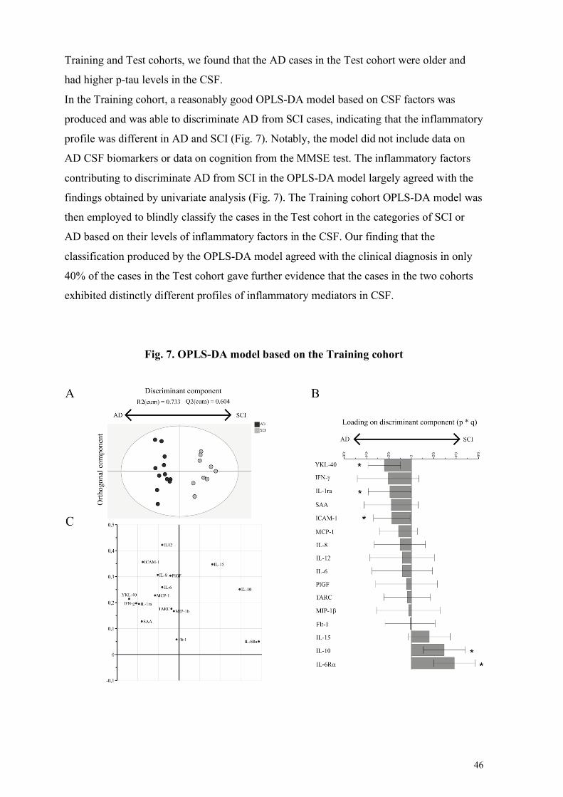
46
Training and Test cohorts, we found that the AD cases in the Test cohort were older and
had higher p-tau levels in the CSF.
In the Training cohort, a reasonably good OPLS-DA model based on CSF factors was
produced and was able to discriminate AD from SCI cases, indicating that the inflammatory
profile was different in AD and SCI (Fig. 7). Notably, the model did not include data on
AD CSF biomarkers or data on cognition from the MMSE test. The inflammatory factors
contributing to discriminate AD from SCI in the OPLS-DA model largely agreed with the
findings obtained by univariate analysis (Fig. 7). The Training cohort OPLS-DA model was
then employed to blindly classify the cases in the Test cohort in the categories of SCI or
AD based on their levels of inflammatory factors in the CSF. Our finding that the
classification produced by the OPLS-DA model agreed with the clinical diagnosis in only
40% of the cases in the Test cohort gave further evidence that the cases in the two cohorts
exhibited distinctly different profiles of inflammatory mediators in CSF.
Fig. 7. OPLS-DA model based on the Training cohort
47
Our study highlighted the field of biomarkers in AD from two important perspectives. (1)
Analysis of a pattern of molecules is more powerful than analysis of individual molecules
in differentiating AD from other conditions. The majority of studies on the inflammatory
mediators in AD are focused on individual factors (240, 415). The redundancy, high
variability, and the low specificity of inflammatory mediators to a specific pathology limit
their capacity to be used as diagnostic biomarkers. An emerging view is that combining
several biomarkers, thus providing the basis to define a pattern, is a more efficient strategy
(416). MVA models identify patterns by analysing multiple single variables in (n)-
dimensional space onto which the variables are projected in such a way that their
covariances can be explained. Clustering of individual observations in this space indicates a
similar pattern of variables, and if the observations in this cluster belong to e.g. cases with a
diagnosis of AD, a pathological pattern is shown. An MVA discriminant model produced
by cases with a known classification (Training cohort) could be used to predict and classify
unknown cases (Test cohort).
(2) The presence of comorbidities may act as a confounder when e.g. deciding on the
course of treatment and care for a patient, or when enrolling participants for clinical studies.
Scientists usually prefer to conduct their exploratory studies in a "pure" disease condition
and want to exclude the effects of confounders on their results. In our study, after to our
best capacity removing confounders such as gender, age, and comorbidities we obtained a
cohort with a different inflammatory pattern in their CSF compared to a randomly selected
cohort. As AD is prevalent in the aged population, AD patients are commonly afflicted by
other diseases in addition to AD, which may bias the results of studies on inflammatory
factors. In the future, it is worthy to study whether and how different kinds of comorbidities
such as other dementias, tumours, stroke, psychiatric disorders, affect the inflammatory
pattern in the CSF. One should also be aware that the reality of clinic may be better
represented by the Test cohort, since there are no exclusion criteria applied in the real
world. In our study, the OPLS-DA model produced using the Training cohort only partly
agreed with the clinical diagnosis when applied to the Test cohort.
As AD has a complicated and heterogeneous etiology, biomarkers, in addition to Ab and
tau would be useful for an early and more refined diagnosis of AD. An example of such a
refinement could be the identification of patients with comorbidities which can be more, or
less responsive to different treatment and care strategies. Inflammation, a sensitive indicator
of harmful conditions in the body, presents numerous markers that are can be investigated
for this purpose, a prospect that was the focus of the present study. Further studies are
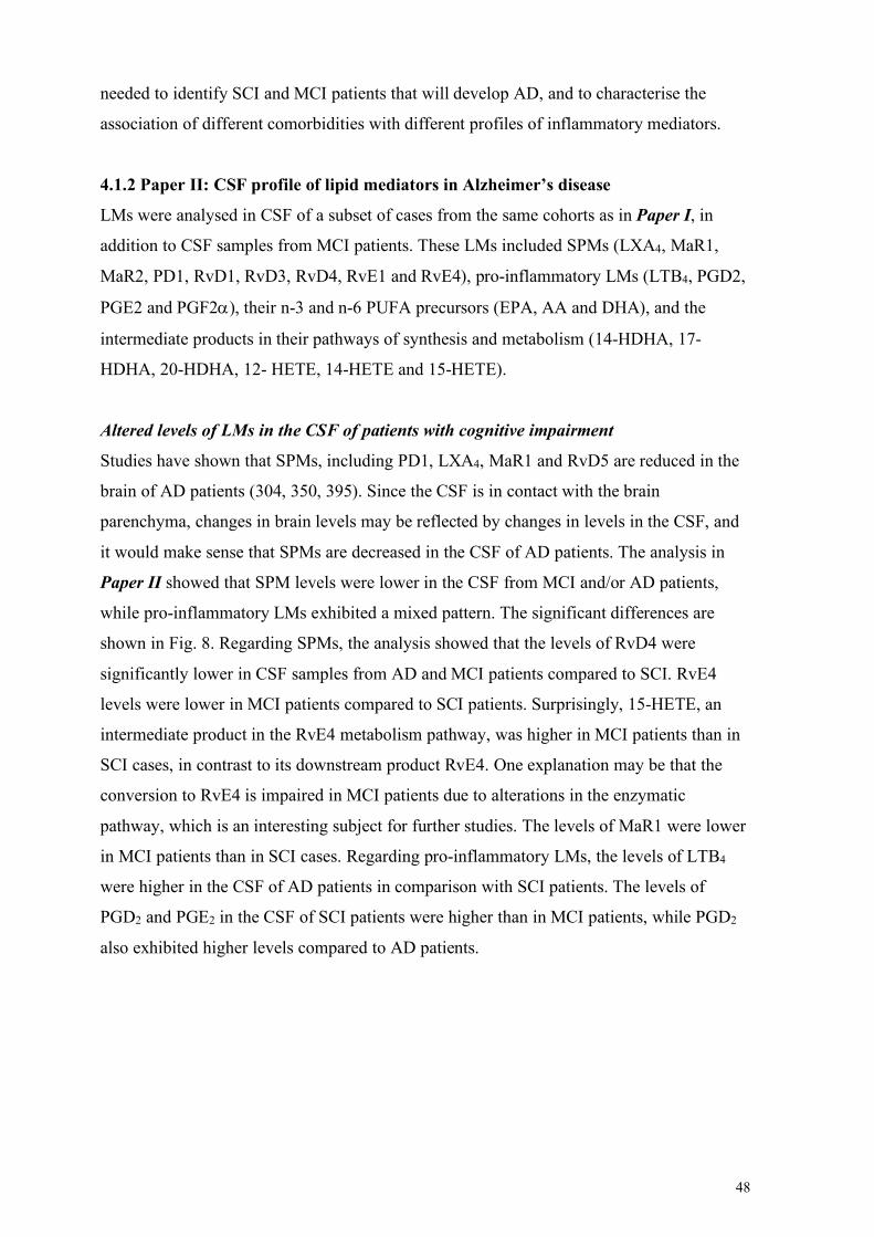
48
needed to identify SCI and MCI patients that will develop AD, and to characterise the
association of different comorbidities with different profiles of inflammatory mediators.
4.1.2 Paper II: CSF profile of lipid mediators in Alzheimer’s disease
LMs were analysed in CSF of a subset of cases from the same cohorts as in Paper I, in
addition to CSF samples from MCI patients. These LMs included SPMs (LXA4, MaR1,
MaR2, PD1, RvD1, RvD3, RvD4, RvE1 and RvE4), pro-inflammatory LMs (LTB4, PGD2,
PGE2 and PGF2a), their n-3 and n-6 PUFA precursors (EPA, AA and DHA), and the
intermediate products in their pathways of synthesis and metabolism (14-HDHA, 17-
HDHA, 20-HDHA, 12- HETE, 14-HETE and 15-HETE).
Altered levels of LMs in the CSF of patients with cognitive impairment
Studies have shown that SPMs, including PD1, LXA4, MaR1 and RvD5 are reduced in the
brain of AD patients (304, 350, 395). Since the CSF is in contact with the brain
parenchyma, changes in brain levels may be reflected by changes in levels in the CSF, and
it would make sense that SPMs are decreased in the CSF of AD patients. The analysis in
Paper II showed that SPM levels were lower in the CSF from MCI and/or AD patients,
while pro-inflammatory LMs exhibited a mixed pattern. The significant differences are
shown in Fig. 8. Regarding SPMs, the analysis showed that the levels of RvD4 were
significantly lower in CSF samples from AD and MCI patients compared to SCI. RvE4
levels were lower in MCI patients compared to SCI patients. Surprisingly, 15-HETE, an
intermediate product in the RvE4 metabolism pathway, was higher in MCI patients than in
SCI cases, in contrast to its downstream product RvE4. One explanation may be that the
conversion to RvE4 is impaired in MCI patients due to alterations in the enzymatic
pathway, which is an interesting subject for further studies. The levels of MaR1 were lower
in MCI patients than in SCI cases. Regarding pro-inflammatory LMs, the levels of LTB4
were higher in the CSF of AD patients in comparison with SCI patients. The levels of
PGD2 and PGE2 in the CSF of SCI patients were higher than in MCI patients, while PGD2
also exhibited higher levels compared to AD patients.
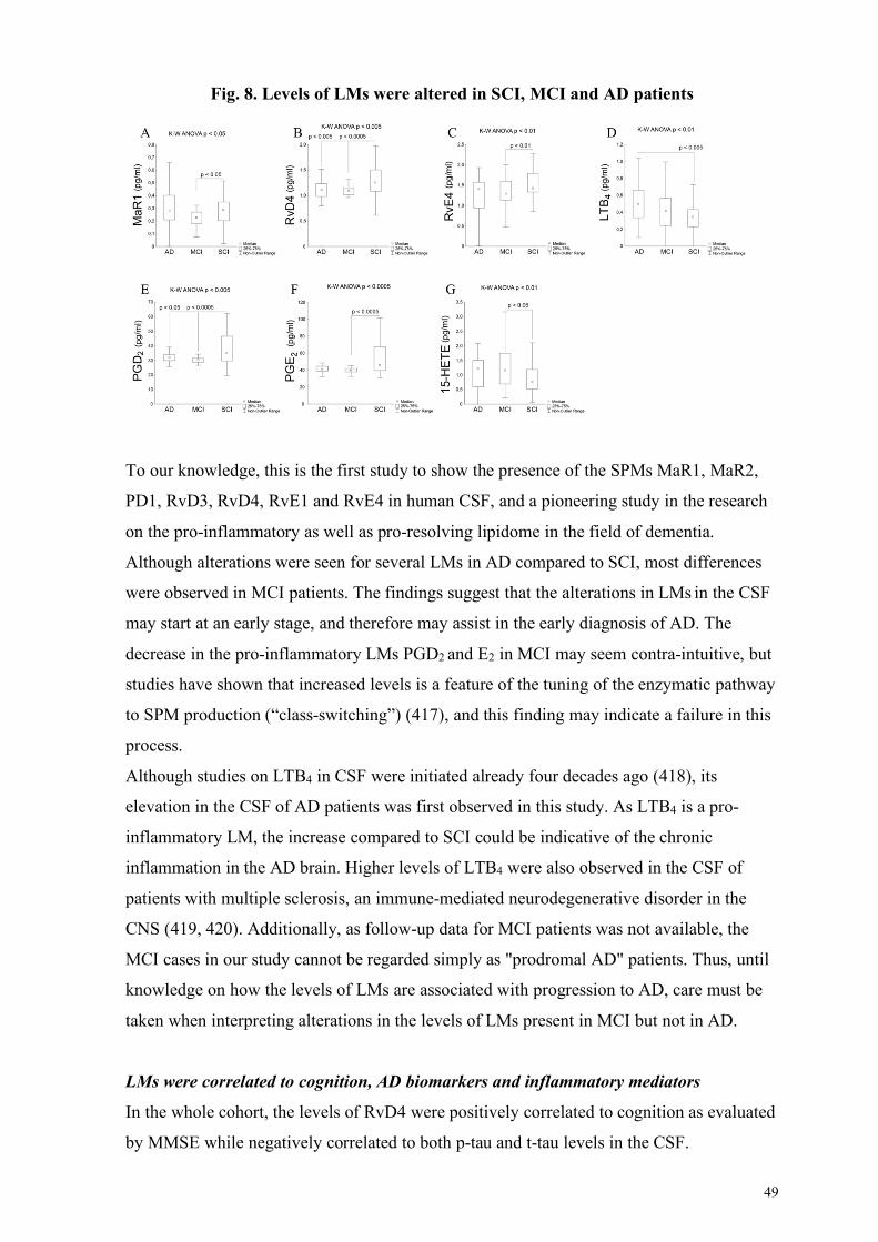
49
Fig. 8. Levels of LMs were altered in SCI, MCI and AD patients
To our knowledge, this is the first study to show the presence of the SPMs MaR1, MaR2,
PD1, RvD3, RvD4, RvE1 and RvE4 in human CSF, and a pioneering study in the research
on the pro-inflammatory as well as pro-resolving lipidome in the field of dementia.
Although alterations were seen for several LMs in AD compared to SCI, most differences
were observed in MCI patients. The findings suggest that the alterations in LMs in the CSF
may start at an early stage, and therefore may assist in the early diagnosis of AD. The
decrease in the pro-inflammatory LMs PGD2 and E2 in MCI may seem contra-intuitive, but
studies have shown that increased levels is a feature of the tuning of the enzymatic pathway
to SPM production (“class-switching”) (417), and this finding may indicate a failure in this
process.
Although studies on LTB4 in CSF were initiated already four decades ago (418), its
elevation in the CSF of AD patients was first observed in this study. As LTB4 is a pro-
inflammatory LM, the increase compared to SCI could be indicative of the chronic
inflammation in the AD brain. Higher levels of LTB4 were also observed in the CSF of
patients with multiple sclerosis, an immune-mediated neurodegenerative disorder in the
CNS (419, 420). Additionally, as follow-up data for MCI patients was not available, the
MCI cases in our study cannot be regarded simply as "prodromal AD" patients. Thus, until
knowledge on how the levels of LMs are associated with progression to AD, care must be
taken when interpreting alterations in the levels of LMs present in MCI but not in AD.
LMs were correlated to cognition, AD biomarkers and inflammatory mediators
In the whole cohort, the levels of RvD4 were positively correlated to cognition as evaluated
by MMSE while negatively correlated to both p-tau and t-tau levels in the CSF.
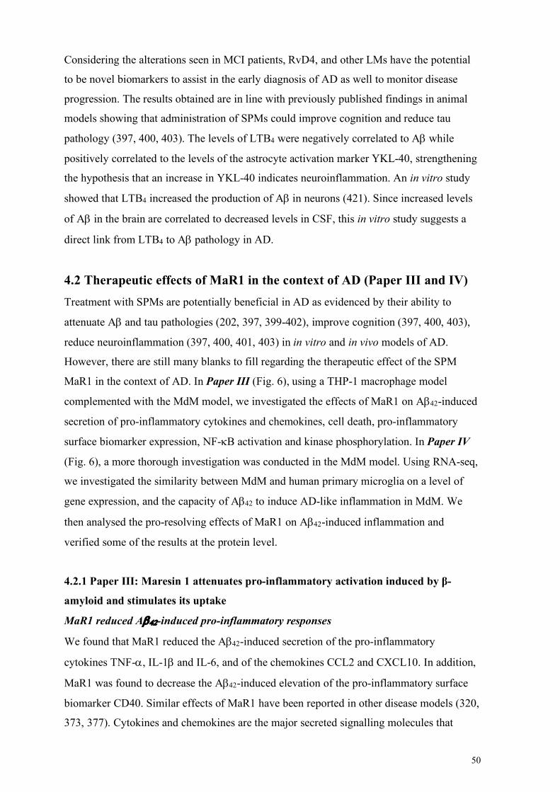
50
Considering the alterations seen in MCI patients, RvD4, and other LMs have the potential
to be novel biomarkers to assist in the early diagnosis of AD as well to monitor disease
progression. The results obtained are in line with previously published findings in animal
models showing that administration of SPMs could improve cognition and reduce tau
pathology (397, 400, 403). The levels of LTB4 were negatively correlated to Ab while
positively correlated to the levels of the astrocyte activation marker YKL-40, strengthening
the hypothesis that an increase in YKL-40 indicates neuroinflammation. An in vitro study
showed that LTB4 increased the production of Ab in neurons (421). Since increased levels
of Ab in the brain are correlated to decreased levels in CSF, this in vitro study suggests a
direct link from LTB4 to Ab pathology in AD.
4.2 Therapeutic effects of MaR1 in the context of AD (Paper III and IV) Treatment with SPMs are potentially beneficial in AD as evidenced by their ability to
attenuate Ab and tau pathologies (202, 397, 399-402), improve cognition (397, 400, 403),
reduce neuroinflammation (397, 400, 401, 403) in in vitro and in vivo models of AD.
However, there are still many blanks to fill regarding the therapeutic effect of the SPM
MaR1 in the context of AD. In Paper III (Fig. 6), using a THP-1 macrophage model
complemented with the MdM model, we investigated the effects of MaR1 on Ab42-induced
secretion of pro-inflammatory cytokines and chemokines, cell death, pro-inflammatory
surface biomarker expression, NF-kB activation and kinase phosphorylation. In Paper IV
(Fig. 6), a more thorough investigation was conducted in the MdM model. Using RNA-seq,
we investigated the similarity between MdM and human primary microglia on a level of
gene expression, and the capacity of Ab42 to induce AD-like inflammation in MdM. We
then analysed the pro-resolving effects of MaR1 on Ab42-induced inflammation and
verified some of the results at the protein level.
4.2.1 Paper III: Maresin 1 attenuates pro-inflammatory activation induced by β-
amyloid and stimulates its uptake
MaR1 reduced Ab42-induced pro-inflammatory responses
We found that MaR1 reduced the Ab42-induced secretion of the pro-inflammatory
cytokines TNF-a, IL-1b and IL-6, and of the chemokines CCL2 and CXCL10. In addition,
MaR1 was found to decrease the Ab42-induced elevation of the pro-inflammatory surface
biomarker CD40. Similar effects of MaR1 have been reported in other disease models (320,
373, 377). Cytokines and chemokines are the major secreted signalling molecules that
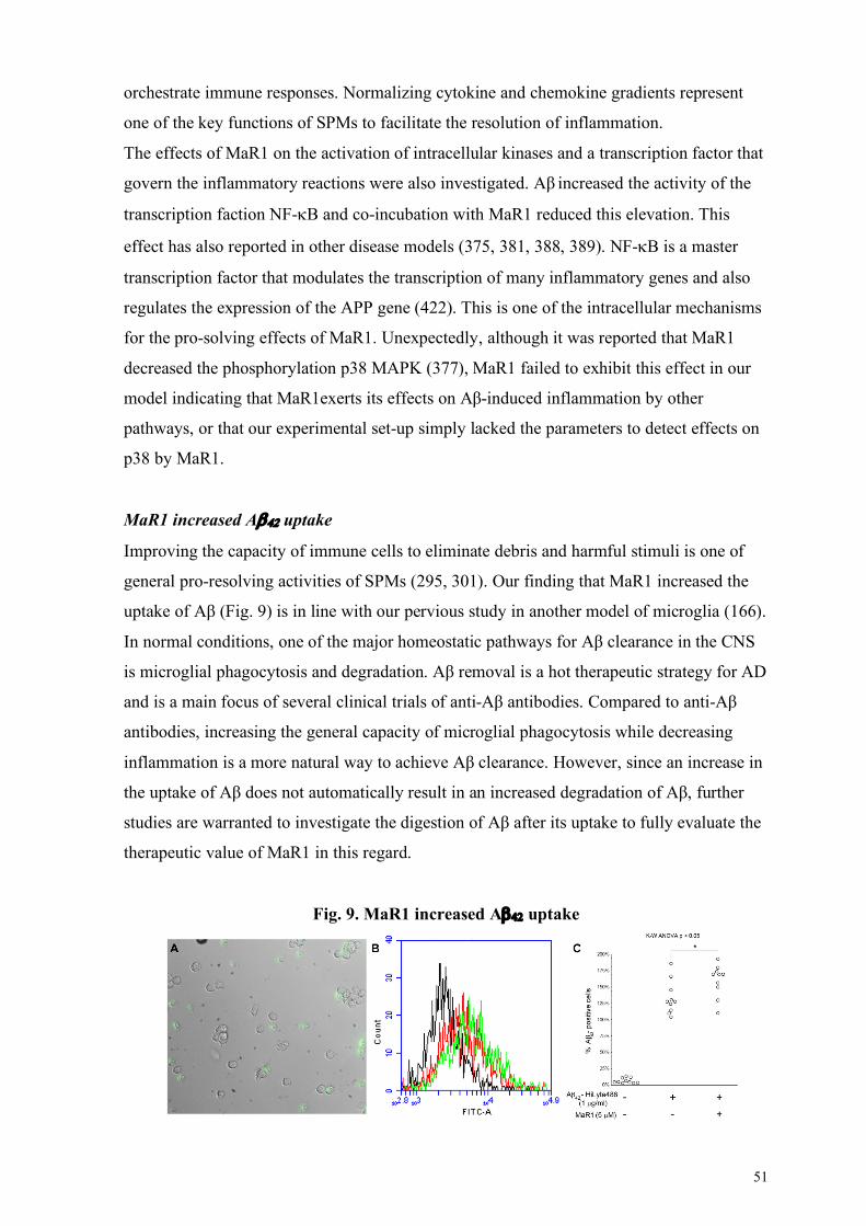
51
orchestrate immune responses. Normalizing cytokine and chemokine gradients represent
one of the key functions of SPMs to facilitate the resolution of inflammation.
The effects of MaR1 on the activation of intracellular kinases and a transcription factor that
govern the inflammatory reactions were also investigated. Aβ increased the activity of the
transcription faction NF-kB and co-incubation with MaR1 reduced this elevation. This
effect has also reported in other disease models (375, 381, 388, 389). NF-kB is a master
transcription factor that modulates the transcription of many inflammatory genes and also
regulates the expression of the APP gene (422). This is one of the intracellular mechanisms
for the pro-solving effects of MaR1. Unexpectedly, although it was reported that MaR1
decreased the phosphorylation p38 MAPK (377), MaR1 failed to exhibit this effect in our
model indicating that MaR1exerts its effects on Aβ-induced inflammation by other
pathways, or that our experimental set-up simply lacked the parameters to detect effects on
p38 by MaR1.
MaR1 increased Ab42 uptake
Improving the capacity of immune cells to eliminate debris and harmful stimuli is one of
general pro-resolving activities of SPMs (295, 301). Our finding that MaR1 increased the
uptake of Aβ (Fig. 9) is in line with our pervious study in another model of microglia (166).
In normal conditions, one of the major homeostatic pathways for Aβ clearance in the CNS
is microglial phagocytosis and degradation. Aβ removal is a hot therapeutic strategy for AD
and is a main focus of several clinical trials of anti-Aβ antibodies. Compared to anti-Aβ
antibodies, increasing the general capacity of microglial phagocytosis while decreasing
inflammation is a more natural way to achieve Aβ clearance. However, since an increase in
the uptake of Aβ does not automatically result in an increased degradation of Aβ, further
studies are warranted to investigate the digestion of Aβ after its uptake to fully evaluate the
therapeutic value of MaR1 in this regard.
Fig. 9. MaR1 increased Ab42 uptake

52
MaR1 reduced Ab42-induced cell death
Aβ significantly increased cell death of the differentiated THP1 cells, and co-incubation
with MaR1 decreased this cytotoxic effect. The ability of MaR1 to improve cell survival
has been reported in various models (202, 384).
4.2.2 Paper IV: Pro-resolving lipid mediator reduced Ab42-induced gene expression in
monocyte-derived microglia
Ab42-stimulated MdM – a good model to study AD-like inflammation
To better understand the neuroinflammation in AD and to discover new therapeutic targets
regarding inflammation and its resolution, the knowledge of microglia in the context of AD
must be increased. To achieve this, a relevant microglial model that exhibits AD-like
pathology is needed. In Paper IV, we used RNA-seq to show that MdM were largely
similar to human primary microglia on the level of gene expression and showed that Ab42-
stimulated MdM is a good microglial model to study AD-like inflammation.
PCA was performed to elucidate similarities and differences regarding the gene expression
in MdM, iPSC-derived microglia and human primary microglia isolated from surgery. The
PCA plot showed that these different cells were closely clustered together, suggesting
similarities in their transcriptomes. Microglial genes and resolution-related genes were
transcribed in MdM, with confirmation of gene expression data in the protein level for a set
of microglia-associated proteins including HLA-DR, P2RY12, Iba-1, MCSFR and TREM-
2, and the MaR1 receptor BLT1. These data suggest that MdM are similar to primary
human microglia.
A series of disease ontology analyses was performed on Ab42-stimulated MdM. We found
that 1) AD-risk genes, obtained from GWAS studies by literature review, were transcribed
in the Ab42-stimulated MdM; 2) 17 of the top 20 DEGs in comparison with control
conditions were associated with AD to some extent. They included AD risk genes, genes
with an expression shown to be altered in AD, and genes associated with AD pathology and
with cognitive impairment in AD patients; 3) the DEGs were enriched in the "Alzheimer
Disease" term when performing pathway analysis using the KEGG). Furthermore, 16 of the
17 secreted protein inflammatory mediators analysed in the culture medium that were up-
regulated by Ab42 were reported to be associated with AD, and Ab42 was taken up by the
MdM (shown in both Paper III and IV). Taken together, the results support that MdM
incubated with Ab42 exhibit and AD-like inflammation.
53
MaR1 re-balanced the imbalanced immune network in AD
The ability of MaR1 to affect Ab42-induced gene expression in MdM was investigated by
pathway analysis of the RNA-Seq data (control vs. Ab42 DEGs and Ab42 vs. Ab42 + MaR1
DEGs) using KEGG and MSigDB. All of the top 10 KEGG pathways comparing control
and Ab42 were related to inflammation (immune pathways) and were upregulated by Ab42.
Eight of these were significantly downregulated by co-treatment with MaR1. These
pathways which included chemokine signalling, cytokine-cytokine receptor interaction,
viral protein interaction with cytokine and cytokine receptor, NF-kB signalling, TNF-
signalling, NOD-like receptor signalling, Toll-like receptor signalling, and the C-type lectin
receptor signalling pathway, formed a closely interacting network. Similar results were
obtained upon analysis using MSigDB, which is a more extensive database compared to
KEGG. To analyse effects on transcription factors, a prediction based on MSigDB (C3
subdatabase) was performed. Gene expression of the transcription factors NF-kB
(particularly subunit p65) was increased by Ab42 and decreased by co-incubation with
MaR1.
These findings advance the investigation on the effects of MaR1 from those based on
differences in individual molecules to the level of networks of pathways of inflammation
and other areas of physiology. The advantages of using the RNA-Seq technique in this
study are that :1) it allows screening of the effects of MaR1 on the transcription of more
than 60,000 genes, providing a much more encompassing view compared to previous
studies; 2) the focus of study is switched from individual immune molecules to functional
pathways, which is more relevant since the inflammatory response is orchestrated by
numerous pathways of the immune system. From a network perspective, we concluded that
the balance of the immune network in microglia was disturbed by Ab42, and that MaR1 was
influential in restoring this balance.
MaR1 restored homeostasis at the protein level
To verify the RNA-Seq results at a protein level, a total of 38 secreted inflammatory
mediators were analysed in the culture supernatants. The mRNA levels and the protein
concentrations were found to be in line with each other. The PCA plot shows a marked
separation between cells incubated with Ab42 and control cells, while the cells co-incubated
with Ab42 and MaR1 showed an intermediate position, indicating that MaR1 at least partly
reduced the Ab42-induced inflammatory secretory pattern. Using univariate analysis, MaR1
was found to decrease the Ab42-induced secretion of 17 inflammatory factors. Twelve of
54
these factors have not been reported previously affected by MaR1 in the context of AD.
Seven factors, including CCL11, CCL13, CCL22, CCL26, IL-13, vascular endothelial
growth factor receptor (VEGFR) 1 and pentraxin 3 (PTX3), were never reported to be
affected by MaR1 in any disease model. Notably, this is the first study to show that Ab42
induced both PTX3 mRNA and protein. PTX3 is a pattern recognition factor that belongs to
the “long” pentraxins, which are not yet well investigated in AD (see (423)), while other
more well-known PTXs such as C-reactive protein, and amyloid P were increased in human
post mortem AD brains (424), where their expression is associated with the pathology of
plaques and tangles, PTX1 was increased in both brain and plasma of AD mice (425),
PTX2 predicted brain atrophy and cognition impairment in AD (426). To summarize Paper
IV, MdM represent a relevant and useful in vitro microglial model to study AD-like
inflammation. Furthermore, MaR1 had an ameliorating effect on Aβ42-induced changes on
several genes and proteins of importance in AD, highlighting its potential as treatment for
AD.

55
5 CONCLUSIONS The major aim of this thesis was to investigate if pro-inflammatory and pro-resolving
mediators are altered in the context of AD (Paper I and II), and whether the SPM MaR1
can be used as a potential drug to attenuate AD-like inflammation (Paper III and IV). We
used CSF from patients diagnosed with SCI, MCI and AD (Paper I and II), and microglial
in vitro models (Paper III and IV) to answer the scientific questions. The key findings of
the constituent papers are summarized as follows:
• Paper I and II
o The alterations of inflammatory mediators (including protein mediators and LMs) in
the CSF indicated a shift of inflammatory profile from pro-resolving to pro-
inflammatory as part of AD pathology.
o The SPM RvD4 could serve as a novel biomarker for AD, as the levels of RvD4
were decreased in the CSF from AD patients and were correlated to cognition and
tau pathology.
o Confounders including comorbidities affected results and conclusions in biomarker
studies. It is important to study biomarkers in both a confounder-controlled cohort
and a random-selected cohort.
o Pattern-based diagnosis could be achieved by using an MVA model.
• Paper III and IV:
o MaR1 promoted the resolution of inflammation in the context of AD, as evidenced
by attenuating Ab42-induced inflammatory reactions, stimulating Ab42 uptake and
decreasing Ab42-induced cell death, therefore MaR1 has a potential to serve as a
drug for AD.
o We reported that PTX3 was upregulated by Ab42, and that MaR1 prevented the
effect of Ab42. In addition, we discovered that MaR1 promoted resolution by
reducing the expression of CCL11, CCL13, CCL22, CCL26, IL-13 and VEGFR1.
o To understand the neuroinflammation in AD, a relevant and practically useful
microglial model is needed. MdM are largely similar to primary human microglia,
and Ab42 stimulation could induce AD-like inflammation in MdM. Therefore Ab42-
stimulated MdM could be a novel in vitro model to investigate neuroinflammation
in the context of AD.

57
6 POINTS OF PERSPECTIVE The field of resolution is young and keeps growing rapidly. The basis for understanding the
resolution of inflammation is characterisation of the SPMs with regard to e.g. structure,
synthesis, receptors and functions. It is also important to identify the pro-resolving effects
of SPMs and the underlying mechanisms in disease models. Results from studies in animal
models are promising. The translation from SPMs to clinical drugs should be considered.
• Basic information for SPMs: SPMs exhibit their effects by binding to receptors.
However, receptors for SPMs remain largely unknow, indicating the need to
discover more SPM receptors, which will aid in understanding the pro-resolving
signalling, thus helping receptors for SPMs to become therapeutic targets.
• Pharmacokinetics for SPMs: When it comes to treatment of CNS diseases, the
capability of drugs to pass the BBB must be considered. For SPMs, however, there
is evidence indicating that they can enter the brain from the periphery. The
underlying mechanisms for the distribution and transportation of SPMs in tissues
and organs also need further investigation.
• SPMs in AD: New SPMs keep being discovered, and, so far, studies on resolution
in AD have only concerned some of the SPMs. Ideally, all SPMs should be
investigated if they could be biomarkers or drugs for AD.
• Clinical translation: I look forward to clinical trials using SPMs being started in
the near future.

59
7 ACKNOWLEDGEMENTS The PhD studies will be a precious experience in my life. I would like to thank my family
who supported my decision to perform my PhD studies in Sweden, and who are always
with me no matter what happens.
When I recall my scientific journey, many thanks to my supervisors, mentors, colleagues
who company me grew. I realized that my scientific career started as early as when I was a
second-year medical student in the university. In 2012, I met my enlightener for scientific
journey, Youqiong, Professor at Jilin University, giving lectures in our anatomy class.
Under his guidance, I measured distances on MRI images to identify the safety range for
brain surgery and published my first paper. I learnt how to extract a scientific question from
the clinical practice and design the scientific research, and then use the results to guide
clinical practice. I want to thank Youqiong for culturing my interests for scientific research.
In 2013, I met Hongliang, who was a clinical doctor completing his PhD study at NVS
department at Karolinska Institutet (KI) and later became the external mentor for my PhD
studies. Under his guidance, I learnt to analyse clinical data (e.g. investigated the
association between levels of fasting plasma glucose and disease severity of Guillain-Barré
syndrome), and write literature reviews (e.g. summarized the complications for Guillain-
Barré syndrome). I would like to thank Hongliang for supervising me doing clinical studies
and writing reviews with great patience and give me many good suggestions for career
development. In 2014, I got a position in Dr. Hatch's lab in University of Manitoba,
Canada, as an exchange student for 9 months. I worked with his master student Hieu
studying about BBB. I have learnt many techniques during this period, which paved the
way for my PHD study. I want to thank Dr. Hatch for offering this wonderful opportunity
to me and thank Songyan who recommended me to Dr. Hatch's lab and helped me a lot to
fit the daily life in Canada. In 2015, I met Jiachun and Jie. Jiachun was my clinical
supervisor who let me know how to be a clinical doctor. I'd like to thank Jiachun for
helping me develop clinical skills and thank him for supporting my decision to quit the
study in Jilin University and start the PhD studies in KI. Jie was a professor worked in both
Jilin University and KI. I worked a short while with her in 2016 and performed a few
experiments on animals. I want to thank Jie for involving me in her project, and particularly
thank her for recommending me to KI. Additionally, I want to thank Yanmei, who gave
lectures in our philosophy classes when I was a college student and later became my friend.
When I was in the university, we always said that medicine is philosophy. Now I want to
60
say that science is also philosophy, philosophy is everywhere. I'd like to thank Yanmei for
discussing philosophy issues with me since 2010.
The 11th of November in 2016, my PhD journey began. I now started my scientific career
in an official way!
To Dr. Erik Hjorth, my main supervisor. Thanks for selecting me to be your first PhD
student. I am your first PhD student, and you are my first and last major supervisor for
PhD. These four and half years have witnessed us growing up together. Though at the
beginning of my PhD studies, both of us were not skilled at acting as PhD student or
supervisor, we grew together as time goes. We kept communicating with and learning from
each other. In the end, both of us significantly improved ourselves. You are a simple and
nice person. I appreciate your supervision and the changes you made for me.
To professor Marianne Schultzberg, my co-supervisor. Thanks for having me in your lab.
You are an experienced scientist and supervisor, I am very impressed that you could always
find a wise way to solve tough problems, no matter if the problems are about science or
beyond science. I am also impressed that you are strict with every detail and want to have
everything to be perfect. By working with you, I have learnt how to deal with problems
independently.
To professor Maria Eriksdotter, my co-supervisor. Thanks for helping us with the CSF
studies and giving good suggestions for my PhD project.
Thanks to Porfessor Charles Serhan who is the king of the resolution field. I have
followed your wonderful research during my entire PhD studies. I am really interested in
the resolution. Thanks for your great contributions to the field.
Thanks to China Scholarship Council for donating around 600,000 SEK stipend to my
PhD studies.
Thanks to Karolinska Institutet to be a fantastic academic institution that is open to
students from all over the world. Thanks for organizing Nobel lectures every year.
Thanks to Ceren. After spending the whole PhD life in the same group with you, I must
conclude that you are such a wonderful colleague! I appreciate all the support I got from
you during these four years. I will always be prepared to help you if you need it in the
future. Finally, good luck for your thesis defence.
Thanks to all former and present group members as well as internship students: Lotta
Granholm, Mingqin, Xiuzhe, Sara, Helena, Veronica, Bram, Zaki, Eric Hamlett,
Meghan, Lea, Miranda, Felicia, Lisanne and Silvia.

61
Thanks to all collaborators and their PIs from other groups. Thanks to Jan for Ab
purification collaboration, Nailin for blood collection collaboration, Maria A for
mitochondria study collaboration, Xiang for RNA-Seq data analysis collaboration,
Professor Nicolas Bazan for SPM detection collaboration. Thanks to Axel for teaching me
how to purify Ab. It was a new and complicated technique for me at the beginning. Thanks
for always helping me to solve the problems very patiently. Thanks to Henrik and Rakesh
for showing me the new method to purify Ab and giving me a batch of Ab. Thanks to Gefei
for helping with trouble shooting. Thanks to Luana for introducing the mitochondrial field
to me. Thanks to Xiaofei for answering questions regarding RNA-Seq. Thanks to Mattias
in NGI for introducing the RNA-Seq technique and service to me. Thanks to Rui for
answering questions regarding RNA-Seq data analysis in the drop-in sections. Thanks to all
the co-authors in my papers: Khanh, Bokkyoo, Helena G, Karin, Helga and Shuai.
Thanks to Raul for helping me in a very hard time when I first came. Thanks to Eva,
Maria Roos and Johanna for handling general issues for everybody in the department.
Thanks to all PIs and their group members in the Division for creating a nice atmosphere
and being there for knowledge and advice.
Thanks to my host Elasa for allowing me to change my rental contract several times when
the date moving from Huddinge to Solna was changed several times. Although I felt a little
bit weird, I was moved when you prayed devoutly for me. Your cute dog Lily also brought
happiness to me.
Thanks to Yang Yu, Yang Gao, Sailan and Junyu for celebrating many Chinese or
Swedish festivals together with Yang Yu's mahjong, board games and cute cat Nature.
Nature is always the star. I hope that all of you could have a Nature in the future after
playing with Yang's Nature. Thanks to Chenhong, Lisha, Mingchao, Fengna, Richeng,
Jingdian, Chao and Yiqun for celebrating Chinese New Year together. Thanks to Hui,
Zhulin, Bo, Xinyue and Jinming for happy times together.
Thanks to Covid-19 for letting me stay at home, therefore I saved a lot of money because I
could not travel. (I'm Joking...I hate it! Traveling all over the world is one of my dreams.)
Thanks to Liyan, my travel partner, who went to many countries together with me
including Iceland, Portugal, Spain, Italy, Latvia and Denmark, many cities and places
within Sweden including Helsingborg, Ven, Malmö, Lund, Ystad, Kosta, Kalmar, Öland,
Visby, Jönköping, Linköping, Norrköping, Nyköping, Karlstad, Sigtuna, Uppsala, Nacka,
Öregrund and Kiruna, and also many places in and around Stockholm, including nearly all
the famous landmarks, popular Chinese restaurants and our favourite Swedish bar Akkurat.
I really enjoyed the time spending together with you. Thanks to my other friends, most of

62
whom have been mentioned before for traveling with me to Finland, France, Switzerland,
Göteborg, Eslöv, Kristianstad and Umeå. Thanks to Zilong for driving me, Liyan and Tian
to many places, and having many good talks in the car. Thanks to Tian for being in charge
of playing music in the car, bringing up topics to discuss and cooking delicious meals for
us. Thanks to Xintong for inviting me to your lovely home in Eslöv. I also enjoyed the time
eating delicious rice noodles with you in Lund, and the time playing with your cute cat
Facai.
63
8 REFERENCES 1. Winblad B, Amouyel P, Andrieu S, Ballard C, Brayne C, Brodaty H, et al. Defeating Alzheimer's disease and other dementias: a priority for European science and society. Lancet Neurol. 2016;15(5):455-532. 2. Folstein M, Folstein S, McHugh P. “Mini-mental state”. A practical method for grading the cognitive state of patients for the clinician. J Psychiatr Res. 1975;12(3):189-98. 3. Katzman R. The prevalence and malignancy of Alzheimer Disease. Arch Neurol. 1976;33(4). 4. Glenner G, Wong C. Alzheimer's disease: initial report of the purification and characterization of a novel cerebrovascular amyloid protein. Biochem Biophys Res Commun. 1984;120(3):885-90. 5. Grundke-Iqbal I, Iqbal K, Quinlan M, Tung Y, Zaidi M, Wisniewski H. Microtubule-associated protein tau. A component of Alzheimer paired helical filaments. J Biol Chem. 1986;261(13):6084-9. 6. Grundke-Iqbal I, Iqbal K, Tung Y, Quinlan M, Wisniewski H, Binder L. Abnormal phosphorylation of the microtubule-associated protein tau (tau) in Alzheimer cytoskeletal pathology. Proc Natl Acad Sci U S A. 1986;83(13):4913-7. 7. Qizilbash N, Birks J, López Arrieta J, Lewington S, Szeto S. Tacrine for Alzheimer's disease. Cochrane Database Syst Rev. 1999(2):CD000202. 8. Klunk WE, Engler H, Nordberg A, Wang Y, Blomqvist G, Holt DP, et al. Imaging brain amyloid in Alzheimer's disease with Pittsburgh Compound-B. Ann Neurol. 2004;55(3):306-19. 9. Heneka MT, Carson MJ, Khoury JE, Landreth GE, Brosseron F, Feinstein DL, et al. Neuroinflammation in Alzheimer's disease. Lancet Neurol. 2015;14(4):388-405. 10. Heneka MT, Golenbock DT, Latz E. Innate immunity in Alzheimer's disease. Nat Immunol. 2015;16(3):229-36. 11. Scheltens P, Blennow K, Breteler MM, de Strooper B, Frisoni GB, Salloway S, et al. Alzheimer's disease. Lancet. 2016;388(10043):505-17. 12. Selkoe DJ. Alzheimer's disease results from the cerebral accumulation and cytotoxicity of amyloid-β protein. J Alzheimers Dis. 2001;3(1):75-80. 13. Roher A, Chaney M, Kuo Y, Webster S, Stine W, Haverkamp L, et al. Morphology and toxicity of Abeta-(1-42) dimer derived from neuritic and vascular amyloid deposits of Alzheimer's disease. J Biol Chem. 1996;271(34):20631-5. 14. Lue L, Brachova L, Civin W, Rogers J. Inflammation, Aβ deposition, and neurofibrillary tangle formation as correlates of Alzheimer's disease neurodegeneration. J Neuropathol Exp Neurol. 1996;55(10):1083-8. 15. Lesne SE, Sherman MA, Grant M, Kuskowski M, Schneider JA, Bennett DA, et al. Brain amyloid-β oligomers in ageing and Alzheimer's disease. Brain. 2013;136(Pt 5):1383-98. 16. Kayed R, Lasagna-Reeves CA. Molecular mechanisms of amyloid oligomers toxicity. J Alzheimers Dis. 2013;33 Suppl 1:S67-78. 17. Wirths O, Multhaup G, Czech C, Blanchard V, Moussaoui S, Tremp G, et al. Intraneuronal Aβ accumulation precedes plaque formation in β-amyloid precursor protein and presenilin-1 double-transgenic mice. Neurosci Lett. 2001;306(1-2):116-20. 18. Shankar GM, Li S, Mehta TH, Garcia-Munoz A, Shepardson NE, Smith I, et al. Amyloid-β protein dimers isolated directly from Alzheimer's brains impair synaptic plasticity and memory. Nat Med. 2008;14(8):837-42. 19. Sondag CM, Dhawan G, Combs CK. β amyloid oligomers and fibrils stimulate differential activation of primary microglia. J Neuroinflammation. 2009;6:1.
64
20. Kang J, Lemaire H-G, Unterbeck A, Salbaum JM, Masters CL, Grzeschik K-H, et al. The precursor of Alzheimer's disease amyloid A4 protein resembles a cell-surface receptor. Nature. 1987;325(6106):733-6. 21. Goldgaber D, Lerman MI, McBride OW, Saffiotti U, Gajdusek DC. Characterization and chromosomal localization of a cDNA encoding brain amyloid of Alzheimer's disease. Science. 1987;235(4791):877-80. 22. Tanzi R, Gusella J, Watkins P, Bruns G, St George-Hyslop P, Van Keuren M, et al. Amyloid beta protein gene: cDNA, mRNA distribution, and genetic linkage near the Alzheimer locus. Science. 1987;235(4791):880-4. 23. Burger P, Vogel F. The development of the pathologic changes of Alzheimer's disease and senile dementia in patients. Am J Pathol. 1973;73(2):457–76. 24. Oyama F, Cairns NJ, Shimada H, Oyama R, Titani K, Ihara Y. Down's syndrome: up-regulation of beta-amyloid protein precursor and tau mRNAs and their defective coordination. J Neurochem. 1994;62(3):1062-6. 25. Bekris LM, Yu C-E, Bird TD, Tsuang DW. Genetics of Alzheimer Disease. J Geriatr Psychiatry Neurol. 2010;23(4):213-27. 26. Games D, Adams D, Alessandrini R, Barbour R, Borthelette P, Blackwell C, et al. Alzheimer-type neuropathology in transgenic mice overexpressing V717F β-amyloid precursor protein. Nature. 1995;373:523-7. 27. Karch CM, Goate AM. Alzheimer's disease risk genes and mechanisms of disease pathogenesis. Biol Psychiatry. 2015;77(1):43-51. 28. Coric V, van Dyck CH, Salloway S, Andreasen N, Brody M, Richter RW, et al. Safety and tolerability of the gamma-secretase inhibitor avagacestat in a phase 2 study of mild to moderate Alzheimer disease. Arch Neurol. 2012;69(11):1430-40. 29. Doody RS, Raman R, Farlow M, Iwatsubo T, Vellas B, Joffe S, et al. A phase 3 trial of semagacestat for treatment of Alzheimer's disease. N Engl J Med. 2013;369(4):341-50. 30. Zuroff L, Daley D, Black KL, Koronyo-Hamaoui M. Clearance of cerebral Aβ in Alzheimer's disease: reassessing the role of microglia and monocytes. Cell Mol Life Sci. 2017;74(12):2167-201. 31. Kanemitsu H, Tomiyama T, Mori H. Human neprilysin is capable of degrading amyloid β peptide not only in the monomeric form but also the pathological oligomeric form. Neurosci Lett. 2003;350(2):113-6. 32. Mukherjee A, Song E, Kihiko-Ehmann M, Goodman JP, Jr., Pyrek JS, Estus S, et al. Insulysin hydrolyzes amyloid-β peptides to products that are neither neurotoxic nor deposit on amyloid plaques. J Neurosci. 2000;20(23):8745-9. 33. Zhao L, Lin S, Bales KR, Gelfanova V, Koger D, Delong C, et al. Macrophage-mediated degradation of beta-amyloid via an apolipoprotein E isoform-dependent mechanism. J Neurosci. 2009;29(11):3603-12. 34. Zou K, Maeda T, Watanabe A, Liu J, Liu S, Oba R, et al. Aβ42-to-Aβ40- and angiotensin-converting activities in different domains of angiotensin-converting enzyme. J Biol Chem. 2009;284(46):31914-20. 35. Hersh LB, Rodgers DW. Neprilysin and amyloid β peptide degradation. Curr Alzheimer Res. 2008;5(2):225-31. 36. Saido T, Leissring MA. Proteolytic degradation of amyloid-β protein. Cold Spring Harb Perspect Med. 2012;2(6):a006379. 37. Qiu WQ, Folstein MF. Insulin, insulin-degrading enzyme and amyloid-β peptide in Alzheimer's disease: review and hypothesis. Neurobiol Aging. 2006;27(2):190-8. 38. Yan P, Hu X, Song H, Yin K, Bateman RJ, Cirrito JR, et al. Matrix metalloproteinase-9 degrades amyloid-β fibrils in vitro and compact plaques in situ. J Biol Chem. 2006;281(34):24566-74.
65
39. Zheng L, Roberg K, Jerhammar F, Marcusson J, Terman A. Autophagy of amyloid beta-protein in differentiated neuroblastoma cells exposed to oxidative stress. Neurosci Lett. 2006;394(3):184-9. 40. Keller JN, Hanni KB, Markesbery WR. Impaired proteasome function in Alzheimer's disease. J Neurochem. 2000;75(1):436-9. 41. Gowrishankar S, Yuan P, Wu Y, Schrag M, Paradise S, Grutzendler J, et al. Massive accumulation of luminal protease-deficient axonal lysosomes at Alzheimer's disease amyloid plaques. Proc Natl Acad Sci U S A. 2015;112(28):E3699-708. 42. Yang AJ, Chandswangbhuvana D, Margol L, Glabe CG. Loss of endosomal/lysosomal membrane impermeability is an early event in amyloid Aβ1-42 pathogenesis. J Neurosci Res. 1998;52(6):691-8. 43. Paresce DM, Ghosh RN, Maxfield FR. Microglial cells internalize aggregates of the Alzheimer's disease amyloid β-protein via a scavenger receptor. Neuron. 1996;17(3):553-65. 44. Chung H, Brazil MI, Soe TT, Maxfield FR. Uptake, degradation, and release of fibrillar and soluble forms of Alzheimer's amyloid β-peptide by microglial cells. J Biol Chem. 1999;274(45):32301-8. 45. Ghersi-Egea JF, Gorevic PD, Ghiso J, Frangione B, Patlak CS, Fenstermacher JD. Fate of Cerebrospinal Fluid-Borne Amyloid β-Peptide: Rapid Clearance into Blood and Appreciable Accumulation by Cerebral Arteries. J Neurochem. 2002;67(2):880-3. 46. Zlokovic BV. Clearing amyloid through the blood-brain barrier. J Neurochem. 2004;89(4):807-11. 47. Deane R, Du Yan S, Submamaryan RK, LaRue B, Jovanovic S, Hogg E, et al. RAGE mediates amyloid-β peptide transport across the blood-brain barrier and accumulation in brain. Nat Med. 2003;9(7):907-13. 48. Himmler A, Drechsel D, Kirschner MW, Martin DW, Jr. Tau consists of a set of proteins with repeated C-terminal microtubule-binding domains and variable N-terminal domains. Mol Cell Biol. 1989;9(4):1381-8. 49. Lee G, Cowan N, Kirschner M. The primary structure and heterogeneity of tau protein from mouse brain. Science. 1988;239(4837):285-8. 50. Goedert M, Spillantini MG, Jakes R, Rutherford D, Crowther RA. Multiple isoforms of human microtubule-associated protein tau: sequences and localization in neurofibrillary tangles of Alzheimer's disease. Neuron. 1989;3(4):519-26. 51. Lewis J, Dickson DW. Propagation of tau pathology: hypotheses, discoveries, and yet unresolved questions from experimental and human brain studies. Acta Neuropathol. 2016;131(1):27-48. 52. Jeganathan S, von Bergen M, Brutlach H, Steinhoff HJ, Mandelkow E. Global hairpin folding of tau in solution. Biochemistry. 2006;45(7):2283-93. 53. Mirbaha H, Chen D, Morazova OA, Ruff KM, Sharma AM, Liu X, et al. Inert and seed-competent tau monomers suggest structural origins of aggregation. Elife. 2018;7. 54. Guo T, Noble W, Hanger DP. Roles of tau protein in health and disease. Acta Neuropathol. 2017;133(5):665-704. 55. Mudher A, Shepherd D, Newman TA, Mildren P, Jukes JP, Squire A, et al. GSK-3β inhibition reverses axonal transport defects and behavioural phenotypes in Drosophila. Mol Psychiatry. 2004;9(5):522-30. 56. Cowan Catherine M, Chee F, Shepherd D, Mudher A. Disruption of neuronal function by soluble hyperphosphorylated tau in a Drosophila model of tauopathy. Biochem Soc Trans. 2010;38(2):564-70. 57. Chee FC, Mudher A, Cuttle MF, Newman TA, MacKay D, Lovestone S, et al. Over-expression of tau results in defective synaptic transmission in Drosophila neuromuscular junctions. Neurobiol Dis. 2005;20(3):918-28.

66
58. Morsch R, Simon W, Coleman PD. Neurons may live for decades with neurofibrillary tangles. J Neuropathol Exp Neurol. 1999;58(2):188-97. 59. Pooler AM, Phillips EC, Lau DH, Noble W, Hanger DP. Physiological release of endogenous tau is stimulated by neuronal activity. EMBO Rep. 2013;14(4):389-94. 60. Saman S, Kim W, Raya M, Visnick Y, Miro S, Saman S, et al. Exosome-associated tau is secreted in tauopathy models and is selectively phosphorylated in cerebrospinal fluid in early Alzheimer disease. J Biol Chem. 2012;287(6):3842-9. 61. Katsinelos T, Zeitler M, Dimou E, Karakatsani A, Muller HM, Nachman E, et al. Unconventional secretion mediates the trans-cellular spreading of tau. Cell Rep. 2018;23(7):2039-55. 62. Wu JW, Herman M, Liu L, Simoes S, Acker CM, Figueroa H, et al. Small misfolded tau species are internalized via bulk endocytosis and anterogradely and retrogradely transported in neurons. J Biol Chem. 2013;288(3):1856-70. 63. Holmes BB, DeVos SL, Kfoury N, Li M, Jacks R, Yanamandra K, et al. Heparan sulfate proteoglycans mediate internalization and propagation of specific proteopathic seeds. Proc Natl Acad Sci U S A. 2013;110(33):E3138-47. 64. Jucker M, Walker LC. Self-propagation of pathogenic protein aggregates in neurodegenerative diseases. Nature. 2013;501(7465):45-51. 65. Braak H, Del Tredici K. Potential Pathways of Abnormal Tau and α-Synuclein Dissemination in Sporadic Alzheimer's and Parkinson's Diseases. Cold Spring Harb Perspect Biol. 2016;8(11). 66. Iqbal K, Braak E, Braak H, Zaidi T, Grundke-Iqbal I. A silver impregnation method for labeling both Alzheimer paired helical filaments and their polypeptides separated by sodium dodecyl sulfate-polyacrylamide gel electrophoresis. Neurobiol Aging. 1991;12(4):357-61. 67. Asai H, Ikezu S, Tsunoda S, Medalla M, Luebke J, Haydar T, et al. Depletion of microglia and inhibition of exosome synthesis halt tau propagation. Nat Neurosci. 2015;18(11):1584-93. 68. Vogels T, Leuzy A, Cicognola C, Ashton NJ, Smolek T, Novak M, et al. Propagation of Tau Pathology: Integrating Insights From Postmortem and In Vivo Studies. Biol Psychiatry. 2020;87(9):808-18. 69. Qian J, Hyman BT, Betensky RA. Neurofibrillary tangle stage and the rate of progression of Alzheimer symptoms: modeling using an autopsy cohort and application to clinical trial design. JAMA Neurol. 2017;74(5):540-8. 70. Small SA, Duff K. Linking Aβ and Tau in Late-Onset Alzheimer's Disease: A Dual Pathway Hypothesis. Neuron. 2008;60(4):534-42. 71. Lloret A, Fuchsberger T, Giraldo E, Vina J. Molecular mechanisms linking amyloid beta toxicity and Tau hyperphosphorylation in Alzheimers disease. Free Radic Biol Med. 2015;83:186-91. 72. Knopman D, Haeberlein S, Carrillo M, Hendrix J, Kerchner G, Margolin R, et al. The national institute on aging and the Alzheimer's association research framework for Alzheimer's disease: perspectives from the research roundtable. Alzheimers Dement. 2018;14(4):563-75. 73. Wilson EL, Metzakopian E. ER-mitochondria contact sites in neurodegeneration: genetic screening approaches to investigate novel disease mechanisms. Cell Death Differ. 2020. 74. Kobro-Flatmoen A, Lagartos-Donate MJ, Aman Y, Edison P, Witter MP, Fang EF. Re-emphasizing early Alzheimer's disease pathology starting in select entorhinal neurons, with a special focus on mitophagy. Ageing Res Rev. 2021;67:101307. 75. Fang EF, Hou Y, Palikaras K, Adriaanse BA, Kerr JS, Yang B, et al. Mitophagy inhibits amyloid-beta and tau pathology and reverses cognitive deficits in models of Alzheimer's disease. Nat Neurosci. 2019;22(3):401-12.
67
76. Janota C, Lemere CA, Brito MA. Dissecting the contribution of vascular alterations and aging to Alzheimer's disease. Mol Neurobiol. 2016;53(6):3793-811. 77. Sweeney MD, Sagare AP, Zlokovic BV. Blood-brain barrier breakdown in Alzheimer disease and other neurodegenerative disorders. Nat Rev Neurol. 2018;14(3):133-50. 78. Jonsson T, Stefansson H, Steinberg S, Jonsdottir I, Jonsson PV, Snaedal J, et al. Variant of TREM2 associated with the risk of Alzheimer's disease. N Engl J Med. 2013;368(2):107-16. 79. Bradshaw EM, Chibnik LB, Keenan BT, Ottoboni L, Raj T, Tang A, et al. CD33 Alzheimer's disease locus: altered monocyte function and amyloid biology. Nat Neurosci. 2013;16(7):848-50. 80. Sindi S, Ngandu T, Hovatta I, Kåreholt I, Antikainen R, Hänninen T, et al. Baseline Telomere Length and Effects of a Multidomain Lifestyle Intervention on Cognition: The FINGER Randomized Controlled Trial. J Alzheimers Dis. 2017;59(4):1459-70. 81. De la Rosa A, Olaso-Gonzalez G, Arc-Chagnaud C, Millan F, Salvador-Pascual A, Garcia-Lucerga C, et al. Physical exercise in the prevention and treatment of Alzheimer's disease. J Sport Health Sci. 2020;9(5):394-404. 82. Fratiglioni L, Paillard-Borg S, Winblad B. An active and socially integrated lifestyle in late life might protect against dementia. Lancet Neurol. 2004;3(6):343-53. 83. Wilson RS, Segawa E, Boyle PA, Bennett DA. Influence of late-life cognitive activity on cognitive health. Neurology. 2012;78(15):1123-9. 84. Sperling RA, Aisen PS, Beckett LA, Bennett DA, Craft S, Fagan AM, et al. Toward defining the preclinical stages of Alzheimer's disease: recommendations from the National Institute on Aging-Alzheimer's Association workgroups on diagnostic guidelines for Alzheimer's disease. Alzheimers Dement. 2011;7(3):280-92. 85. Reisberg B, Prichep L, Mosconi L, John ER, Glodzik-Sobanska L, Boksay I, et al. The pre-mild cognitive impairment, subjective cognitive impairment stage of Alzheimer's disease. Alzheimers Dement. 2008;4(1 Suppl 1):S98-S108. 86. Garcia-Ptacek S, Cavallin L, Kareholt I, Kramberger MG, Winblad B, Jelic V, et al. Subjective cognitive impairment subjects in our clinical practice. Dement Geriatr Cogn Dis Extra. 2014;4(3):419-30. 87. Albert M, DeKosky S, Dickson D, Dubois B, Feldman H, Fox N, et al. The diagnosis of mild cognitive impairment due to Alzheimer's disease: recommendations from the National Institute on Aging-Alzheimer's Association workgroups on diagnostic guidelines for Alzheimer's disease. Alzheimers Dement. 2011;7(3):270-9. 88. Olajide OJ, Suvanto ME, Chapman CA. Molecular mechanisms of neurodegeneration in the entorhinal cortex that underlie its selective vulnerability during the pathogenesis of Alzheimer's disease. Biol Open. 2021;10(1). 89. Barnes J, Dickerson BC, Frost C, Jiskoot LC, Wolk D, van der Flier WM. Alzheimer's disease first symptoms are age dependent: Evidence from the NACC dataset. Alzheimers Dement. 2015;11(11):1349-57. 90. Murray ME, Graff-Radford NR, Ross OA, Petersen RC, Duara R, Dickson DW. Neuropathologically defined subtypes of Alzheimer's disease with distinct clinical characteristics: a retrospective study. Lancet Neurol. 2011;10(9):785-96. 91. Schmitz TW, Nathan Spreng R, Alzheimer's Disease Neuroimaging I. Basal forebrain degeneration precedes and predicts the cortical spread of Alzheimer's pathology. Nat Commun. 2016;7:13249. 92. Olivieri P, Lagarde J, Lehericy S, Valabregue R, Michel A, Mace P, et al. Early alteration of the locus coeruleus in phenotypic variants of Alzheimer's disease. Ann Clin Transl Neurol. 2019;6(7):1345-51. 93. Helzner EP, Scarmeas N, Cosentino S, Tang MX, Schupf N, Stern Y. Survival in Alzheimer disease: a multiethnic, population-based study of incident cases. Neurology. 2008;71(19):1489-95.
68
94. Xie J, Brayne C, Matthews FE, Medical Research Council Cognitive F, Ageing Study c. Survival times in people with dementia: analysis from population based cohort study with 14 year follow-up. BMJ. 2008;336(7638):258-62. 95. McKhann G, Drachman D, Folstein M, Katzman R, Price D, Stadlan EM. Clinical diagnosis of Alzheimer's disease: report of the NINCDS-ADRDA Work Group under the auspices of Department of Health and Human Services Task Force on Alzheimer's Disease. Neurology. 1984;34(7):939-44. 96. Jack C, Bennett D, Blennow K, Carrillo M, Dunn B, Haeberlein S, et al. NIA-AA research framework: toward a biological definition of Alzheimer's disease. Alzheimers Dement. 2018;14(4):535-62. 97. Olsson B, Lautner R, Andreasson U, Öhrfelt A, Portelius E, Bjerke M, et al. CSF and blood biomarkers for the diagnosis of Alzheimer's disease: a systematic review and meta-analysis. Lancet Neurol. 2016;15(7):673-84. 98. Mattsson N, Insel PS, Landau S, Jagust W, Donohue M, Shaw LM, et al. Diagnostic accuracy of CSF Ab42 and florbetapir PET for Alzheimer's disease. Ann Clin Transl Neurol. 2014;1(8):534-43. 99. Janelidze S, Zetterberg H, Mattsson N, Palmqvist S, Vanderstichele H, Lindberg O, et al. CSF Aβ42/Aβ40 and Aβ42/Aβ38 ratios: better diagnostic markers of Alzheimer disease. Ann Clin Transl Neurol. 2016;3(3):154-65. 100. Andreasen N, Vanmechelen E, Vanderstichele H, Davidsson P, Blennow K. Cerebrospinal fluid levels of total-tau, phospho-tau and Aβ 42 predicts development of Alzheimer's disease in patients with mild cognitive impairment. Acta Neurol Scand Suppl. 2003;179:47-51. 101. Andreasen N, Minthon L, Davidsson P, Vanmechelen E, Vanderstichele H, Winblad B, et al. Evaluation of CSF-tau and CSF-Aβ42 as diagnostic markers for Alzheimer disease in clinical practice. Arch Neurol. 2001;58(3):373-9. 102. Sato C, Barthelemy NR, Mawuenyega KG, Patterson BW, Gordon BA, Jockel-Balsarotti J, et al. Tau kinetics in neurons and the human central nervous system. Neuron. 2018;97(6):1284-98 e7. 103. Khalil M, Teunissen CE, Otto M, Piehl F, Sormani MP, Gattringer T, et al. Neurofilaments as biomarkers in neurological disorders. Nat Rev Neurol. 2018;14(10):577-89. 104. Bridel C, van Wieringen WN, Zetterberg H, Tijms BM, Teunissen CE, and the NFLG, et al. Diagnostic value of cerebrospinal fluid neurofilament light protein in neurology: a systematic review and meta-analysis. JAMA Neurol. 2019. 105. Dhiman K, Gupta VB, Villemagne VL, Eratne D, Graham PL, Fowler C, et al. Cerebrospinal fluid neurofilament light concentration predicts brain atrophy and cognition in Alzheimer's disease. Alzheimers Dement (Amst). 2020;12(1):e12005. 106. Mattsson N, Insel PS, Palmqvist S, Portelius E, Zetterberg H, Weiner M, et al. Cerebrospinal fluid tau, neurogranin, and neurofilament light in Alzheimer's disease. EMBO Mol Med. 2016;8(10):1184-96. 107. Terry RD, Masliah E, Salmon DP, Butters N, DeTeresa R, Hill R, et al. Physical basis of cognitive alterations in Alzheimer's disease: synapse loss is the major correlate of cognitive impairment. Ann Neurol. 1991;30(4):572-80. 108. Kvartsberg H, Duits FH, Ingelsson M, Andreasen N, Ohrfelt A, Andersson K, et al. Cerebrospinal fluid levels of the synaptic protein neurogranin correlates with cognitive decline in prodromal Alzheimer's disease. Alzheimers Dement. 2015;11(10):1180-90. 109. Casaletto KB, Elahi FM, Bettcher BM, Neuhaus J, Bendlin BB, Asthana S, et al. Neurogranin, a synaptic protein, is associated with memory independent of Alzheimer biomarkers. Neurology. 2017;89(17):1782-8.
69
110. Wellington H, Paterson RW, Portelius E, Tornqvist U, Magdalinou N, Fox NC, et al. Increased CSF neurogranin concentration is specific to Alzheimer disease. Neurology. 2016;86(9):829-35. 111. Portelius E, Olsson B, Hoglund K, Cullen NC, Kvartsberg H, Andreasson U, et al. Cerebrospinal fluid neurogranin concentration in neurodegeneration: relation to clinical phenotypes and neuropathology. Acta Neuropathol. 2018;136(3):363-76. 112. Gustavsson A, Brinck P, Bergvall N, Kolasa K, Wimo A, Winblad B, et al. Predictors of costs of care in Alzheimer's disease: a multinational sample of 1222 patients. Alzheimers Dement. 2011;7(3):318-27. 113. Cummings J, Morstorf T, Lee G. Alzheimer's drug-development pipeline: 2016. Alzheimers Dement (N Y). 2016;2(4):222-32. 114. Jenkinson ML, Bliss MR, Brain AT, Scott DL. Rheumatold arthritis and senile dementia of the Alzhelmer's type. Br J Rheumatol. 1989;28(1):86-7. 115. McGeer P, McGeer E, Rogers J, Sibley J. Anti-inflammatory drugs and Alzheimer disease. Lancet. 1990;335(8696). 116. Weggen S, Eriksen J, Das P, Sagi S, Wang R, Pietrzik CF, KA. Smith, TE. Murphy, MP. Bulter, T. , et al. A subset of NSAIDs lower amyloidogenic Aβ42 independently of cyclooxygenase activity. Nature. 2001;414(6860):212-6. 117. Zhou Y, Su Y, Li B, Liu F, Ryder JW, Wu X, et al. Nonsteroidal anti-inflammatory drugs can lower amyloidogenic Aβ42 by inhibiting Rho. Science. 2003;302(5648):1215-7. 118. in 't Veld BA, Ruitenberg A, Hofman A, Launer LJ, van Duijn CM, Stijnen T, et al. Nonsteroidal antiinflammatory drugs and the risk of Alzheimer's disease. N Engl J Med. 2001;345(21):1515-21. 119. Stewart WF, Kawas C, Corrada M, Metter EJ. Risk of Alzheimer's disease and duration of NSAID use. Neurology. 1997;48(3):626-32. 120. Zandi PP, Anthony JC, Hayden KM, Mehta K, Mayer L, Breitner JC, et al. Reduced incidence of AD with NSAID but not H2 receptor antagonists: the Cache County Study. Neurology. 2002;59(6):880-6. 121. Yip AG, Green RC, Huyck M, Cupples LA, Farrer LA, Group MS. Nonsteroidal anti-inflammatory drug use and Alzheimer's disease risk: the MIRAGE Study. BMC Geriatr. 2005;5:2. 122. Bachstetter AD, Norris CM, Sompol P, Wilcock DM, Goulding D, Neltner JH, et al. Early stage drug treatment that normalizes proinflammatory cytokine production attenuates synaptic dysfunction in a mouse model that exhibits age-dependent progression of Alzheimer's disease-related pathology. J Neurosci. 2012;32(30):10201-10. 123. Group AR, Lyketsos CG, Breitner JC, Green RC, Martin BK, Meinert C, et al. Naproxen and celecoxib do not prevent AD in early results from a randomized controlled trial. Neurology. 2007;68(21):1800-8. 124. Price JF, Stewart MC, Deary IJ, Murray GD, Sandercock P, Butcher I, et al. Low dose aspirin and cognitive function in middle aged to elderly adults: randomised controlled trial. BMJ. 2008;337:a1198. 125. Lambert JC, Ibrahim-Verbaas CA, Harold D, Naj AC, Sims R, Bellenguez C, et al. Meta-analysis of 74,046 individuals identifies 11 new susceptibility loci for Alzheimer's disease. Nat Genet. 2013;45(12):1452-8. 126. Jones L, Holmans PA, Hamshere ML, Harold D, Moskvina V, Ivanov D, et al. Genetic evidence implicates the immune system and cholesterol metabolism in the aetiology of Alzheimer's disease. PLoS One. 2010;5(11):e13950. 127. Toral-Rios D, Franco-Bocanegra D, Rosas-Carrasco O, Mena-Barranco F, Carvajal-Garcia R, Meraz-Rios MA, et al. Evaluation of inflammation-related genes polymorphisms in Mexican with Alzheimer's disease: a pilot study. Front Cell Neurosci. 2015;9:148.

70
128. Hollingworth P, Harold D, Sims R, Gerrish A, Lambert JC, Carrasquillo MM, et al. Common variants at ABCA7, MS4A6A/MS4A4E, EPHA1, CD33 and CD2AP are associated with Alzheimer's disease. Nat Genet. 2011;43(5):429-35. 129. Huang KL, Marcora E, Pimenova AA, Di Narzo AF, Kapoor M, Jin SC, et al. A common haplotype lowers PU.1 expression in myeloid cells and delays onset of Alzheimer's disease. Nat Neurosci. 2017;20(8):1052-61. 130. Naj AC, Jun G, Beecham GW, Wang LS, Vardarajan BN, Buros J, et al. Common variants at MS4A4/MS4A6E, CD2AP, CD33 and EPHA1 are associated with late-onset Alzheimer's disease. Nat Genet. 2011;43(5):436-41. 131. Ramanan VK, Risacher SL, Nho K, Kim S, Shen L, McDonald BC, et al. GWAS of longitudinal amyloid accumulation on 18F-florbetapir PET in Alzheimer's disease implicates microglial activation gene IL1RAP. Brain. 2015;138(Pt 10):3076-88. 132. Sims R, van der Lee SJ, Naj AC, Bellenguez C, Badarinarayan N, Jakobsdottir J, et al. Rare coding variants in PLCG2, ABI3, and TREM2 implicate microglial-mediated innate immunity in Alzheimer's disease. Nat Genet. 2017;49(9):1373-84. 133. Guerreiro R, Wojtas A, Bras J, Carrasquillo M, Rogaeva E, Majounie E, et al. TREM2 variants in Alzheimer's disease. N Engl J Med. 2013;368(2):117-27. 134. Griciuc A, Serrano-Pozo A, Parrado AR, Lesinski AN, Asselin CN, Mullin K, et al. Alzheimer's disease risk gene CD33 inhibits microglial uptake of amyloid β. Neuron. 2013;78(4):631-43. 135. Lambert JC, Heath S, Even G, Campion D, Sleegers K, Hiltunen M, et al. Genome-wide association study identifies variants at CLU and CR1 associated with Alzheimer's disease. Nat Genet. 2009;41(10):1094-9. 136. Konishi H, Kiyama H. Microglial TREM2/DAP12 signaling: a double-edged sword in neural diseases. Front Cell Neurosci. 2018;12:206. 137. Zhao L. CD33 in Alzheimer's disease - biology, pathogenesis, and therapeutics: a mini-review. Gerontology. 2019;65(4):323-31. 138. Kramarz B, Huntley RP, Rodriguez-Lopez M, Roncaglia P, Saverimuttu SCC, Parkinson H, et al. Gene ontology curation of neuroinflammation biology improves the interpretation of Alzheimer's disease gene expression data. J Alzheimers Dis. 2020;75(4):1417-35. 139. Eikelenboom P, Stam FC. Immunoglobulins and complement factors in senile plaques. An immunoperoxidase study. Acta Neuropathol. 1982;57(2-3):239-42. 140. McGeer PL, Itagaki S, Tago H, McGeer EG. Reactive microglia in patients with senile dementia of the Alzheimer type are positive for the histocompatibility glycoprotein HLA-DR. Neurosci Lett. 1987;79(1-2):195-200. 141. Cagnin A, Brooks DJ, Kennedy AM, Gunn RN, Myers R, Turkheimer FE, et al. In-vivo measurement of activated microglia in dementia. Lancet. 2001;358(9280):461-7. 142. Hamelin L, Lagarde J, Dorothée G, Potier M, Corlier F, Kuhnast B, et al. Distinct dynamic profiles of microglial activation are associated with progression of Alzheimer's disease. Brain. 2018;141(6):1855-70. 143. Yasuno F, Kosaka J, Ota M, Higuchi M, Ito H, Fujimura Y, et al. Increased binding of peripheral benzodiazepine receptor in mild cognitive impairment-dementia converters measured by positron emission tomography with [(1)(1)C]DAA1106. Psychiatry Res. 2012;203(1):67-74. 144. Yasuno F, Ota M, Kosaka J, Ito H, Higuchi M, Doronbekov TK, et al. Increased binding of peripheral benzodiazepine receptor in Alzheimer's disease measured by positron emission tomography with [11C]DAA1106. Biol Psychiatry. 2008;64(10):835-41. 145. Craig-Schapiro R, Perrin RJ, Roe CM, Xiong C, Carter D, Cairns NJ, et al. YKL-40: a novel prognostic fluid biomarker for preclinical Alzheimer's disease. Biol Psychiatry. 2010;68(10):903-12.

71
146. Wennström M, Surova Y, Hall S, Nilsson C, Minthon L, Hansson O, et al. The inflammatory marker YKL-40 is elevated in cerebrospinal fluid from patients with Alzheimer's but not Parkinson's disease or dementia with Lewy bodies. PLoS One. 2015;10(8):e0135458. 147. Suarez-Calvet M, Kleinberger G, Araque Caballero MA, Brendel M, Rominger A, Alcolea D, et al. sTREM2 cerebrospinal fluid levels are a potential biomarker for microglia activity in early-stage Alzheimer's disease and associate with neuronal injury markers. EMBO Mol Med. 2016;8(5):466-76. 148. Taipa R, das Neves S, Sousa A, Fernandes J, Pinto C, Correia A, et al. Proinflammatory and anti-inflammatory cytokines in the CSF of patients with Alzheimer's disease and their correlation with cognitive decline. Neurobiol Aging. 2019;76:125-32. 149. Janelidze S, Mattsson N, Stomrud E, Lindberg O, Palmqvist S, Zetterberg H, et al. CSF biomarkers of neuroinflammation and cerebrovascular dysfunction in early Alzheimer disease. Neurology. 2018;91(9):e867-e77. 150. Popp J, Oikonomidi A, Tautvydaitė D, Dayon L, Bacher M, Migliavacca E, et al. Markers of neuroinflammation associated with Alzheimer’s disease pathology in older adults. Brain Behav Immun. 2017;62:203-11. 151. Correa JD, Starling D, Teixeira AL, Caramelli P, Silva TA. Chemokines in CSF of Alzheimer's disease patients. Arq Neuropsiquiatr. 2011;69(3):455-9. 152. Rı ́o-Hortega Pd. El tercer elemento de los centros nerviosos I. La mi- croglı ́a en estado normal. Bol de la Soc Esp de Biol. 1919;IX:68–82. 153. Tay TL, Savage JC, Hui CW, Bisht K, Tremblay ME. Microglia across the lifespan: from origin to function in brain development, plasticity and cognition. J Physiol. 2017;595(6):1929-45. 154. Matcovitch-Natan O, Winter DR, Giladi A, Vargas Aguilar S, Spinrad A, Sarrazin S, et al. Microglia development follows a stepwise program to regulate brain homeostasis. Science. 2016;353(6301):aad8670. 155. Frost JL, Schafer DP. Microglia: architects of the developing nervous system. Trends Cell Biol. 2016;26(8):587-97. 156. Ashwell K. Microglia and cell death in the developing mouse cerebellum. Brain Res Dev Brain Res. 1990;55(2):219-30. 157. Peri F, Nusslein-Volhard C. Live imaging of neuronal degradation by microglia reveals a role for v0-ATPase a1 in phagosomal fusion in vivo. Cell. 2008;133(5):916-27. 158. Marin-Teva JL, Dusart I, Colin C, Gervais A, van Rooijen N, Mallat M. Microglia promote the death of developing Purkinje cells. Neuron. 2004;41(4):535-47. 159. Tremblay ME, Lowery RL, Majewska AK. Microglial interactions with synapses are modulated by visual experience. PLoS Biol. 2010;8(11):e1000527. 160. Wlodarczyk A, Holtman IR, Krueger M, Yogev N, Bruttger J, Khorooshi R, et al. A novel microglial subset plays a key role in myelinogenesis in developing brain. EMBO J. 2017;36(22):3292-308. 161. Fantin A, Vieira JM, Gestri G, Denti L, Schwarz Q, Prykhozhij S, et al. Tissue macrophages act as cellular chaperones for vascular anastomosis downstream of VEGF-mediated endothelial tip cell induction. Blood. 2010;116(5):829-40. 162. Ji K, Akgul G, Wollmuth L, Tsirka S. Microglia actively regulate the number of functional synapses. PLoS One. 2013;8(2):e56293. 163. Paolicelli R, Bolasco G, Pagani F, Maggi L, Scianni M, Panzanelli P, et al. Synaptic pruning by microglia is necessary for normal brain development. Science. 2011;333(6048):1456-8. 164. Stence N, Waite M, Dailey ME. Dynamics of microglial activation: a confocal time-lapse analysis in hippocampal slices. Glia. 2001;33(3):256-66. 165. Nimmerjahn A, Kirchhoff F, Helmchen F. Resting microglial cells are highly dynamic surveillants of brain parenchyma in vivo. Science. 2005;308(5726):1314-8.

72
166. Wang Y, Leppert A, Tan S, van der Gaag B, Li N, Schultzberg M, et al. Maresin 1 attenuates pro-inflammatory activation induced by beta-amyloid and stimulates its uptake. J Cell Mol Med. 2020;25(1):434-47. 167. Paresce D, Chung H, Maxfield F. Slow degradation of aggregates of the Alzheimer's disease amyloid beta-protein by microglial cells. J Biol Chem. 1997;272(46):29390-7. 168. Hickman SE, Allison EK, El Khoury J. Microglial dysfunction and defective beta-amyloid clearance pathways in aging Alzheimer's disease mice. J Neurosci. 2008;28(33):8354-60. 169. Mawuenyega KG, Sigurdson W, Ovod V, Munsell L, Kasten T, Morris JC, et al. Decreased clearance of CNS β-amyloid in Alzheimer's disease. Science. 2010;330(6012):1774. 170. Ponomarev ED, Maresz K, Tan Y, Dittel BN. CNS-Derived Interleukin-4 Is Essential for the Regulation of Autoimmune Inflammation and Induces a State of Alternative Activation in Microglial Cells. J Neurosci. 2007;27(40):10714-21. 171. van der Poel M, Ulas T, Mizee MR, Hsiao CC, Miedema SSM, Adelia, et al. Transcriptional profiling of human microglia reveals grey-white matter heterogeneity and multiple sclerosis-associated changes. Nat Commun. 2019;10(1):1139. 172. Gerrits E, Brouwer N, Kooistra SM, Woodbury ME, Vermeiren Y, Lambourne M, et al. Distinct amyloid-β and tau-associated microglia profiles in Alzheimer's disease. Acta Neuropathol. 2021;141(5):681-96. 173. Koenigsknecht J, Landreth G. Microglial phagocytosis of fibrillar beta-amyloid through a beta1 integrin-dependent mechanism. J Neurosci. 2004;24(44):9838-46. 174. Shigematsu K, McGeer PL, Walker DG, Ishii T, McGeer EG. Reactive microglia/macrophages phagocytose amyloid precursor protein produced by neurons following neural damage. J Neurosci Res. 1992;31(3):443-53. 175. Frautschy S, Cole G, Baird A. Phagocytosis and deposition of vascular beta-amyloid in rat brains injected with Alzheimer beta-amyloid. Am J Pathol. 1992;140(6):1389-99. 176. Vodovotz Y, Lucia M, Flanders K, Chesler L, Xie Q, Smith T, et al. Inducible nitric oxide synthase in tangle-bearing neurons of patients with Alzheimer's disease. J Exp Med. 1996;84(4):1425-33. 177. Lue L, Rydel R, Brigham E, Yang L, Hampel H, Murphy GM J, et al. Inflammatory repertoire of Alzheimer's disease and nondemented elderly microglia in vitro. Glia. 2001;35(1):72-9. 178. McDonald D, Brunden K, Landreth G. Amyloid fibrils activate tyrosine kinase-dependent signaling and superoxide production in microglia. J Neurosci. 1997;17(7):2284-94. 179. Sole-Domenech S, Cruz DL, Capetillo-Zarate E, Maxfield FR. The endocytic pathway in microglia during health, aging and Alzheimer's disease. Ageing Res Rev. 2016;32:89-103. 180. Kurochkin I, Goto S. Alzheimer's β-amyloid peptide specifically interacts with and is degraded by insulin degrading enzyme. FEBS Lett. 1994;345(1):33-7. 181. Bamberger M, Harris M, McDonald D, Husemann J, Landreth G. A cell surface receptor complex for fibrillar β-amyloid mediates microglial activation. J Neurosci. 2003;23(7):2665-74. 182. Paresce DM, Ghosh RN, Maxfield FR. Microglial cells internalize aggregates of the Alzheimer's disease amyloid β-protein via a scavenger receptor. Neuron. 1996;17(3):553-65. 183. Stewart CR, Stuart LM, Wilkinson K, van Gils JM, Deng J, Halle A, et al. CD36 ligands promote sterile inflammation through assembly of a Toll-like receptor 4 and 6 heterodimer. Nat Immunol. 2010;11(2):155-61.
73
184. Liu Y, Walter S, Stagi M, Cherny D, Letiembre M, Schulz-Schaeffer W, et al. LPS receptor (CD14): a receptor for phagocytosis of Alzheimer's amyloid peptide. Brain. 2005;128(8):1778-89. 185. Bradshaw EM, Chibnik LB, Keenan BT, Ottoboni L, Raj T, Tang A, et al. CD33 Alzheimer's disease locus: altered monocyte function and amyloid biology. Nat Neurosci. 2013;16(7):848-50. 186. El Khoury JB, Moore KJ, Means TK, Leung J, Terada K, Toft M, et al. CD36 mediates the innate host response to β-amyloid. J Exp Med. 2003;197(12):1657-66. 187. Harris ME, Hensley K, Butterfield DA, Leedle RA, Carney JM. Direct evidence of oxidative injury produced by the Alzheimer's β-Amyloid peptide (1–40) in cultured hippocampal neurons. Exp Neurol. 1995;131(2):193-202. 188. May PC, Gitter BD, Waters DC, Simmons LK, Becker GW, Small JS, et al. β-amyloid peptide in vitro toxicity: Lot-to-lot variability. Neurobiol Aging. 1992;13(5):605-7. 189. Ban E, Milon G, Prudhomme N, Fillion G, Haour F. Receptors for interleukin-1 (α and β) in mouse brain: Mapping and neuronal localization in hippocampus. Neuroscience. 1991;43(1):21-30. 190. Maphis N, Xu G, Kokiko-Cochran ON, Cardona AE, Ransohoff RM, Lamb BT, et al. Loss of tau rescues inflammation-mediated neurodegeneration. Front Neurosci. 2015;9:196. 191. Li Y, Liu L, Barger S, Griffin W. Interleukin-1 mediates pathological effects of microglia on tau phosphorylation and on synaptophysin synthesis in cortical neurons through a p38-MAPK Pathway. J Neurosci. 2003;23(5):1605–11. 192. Lee YJ, Kim JE, Kwak MH, Go JUN, Yang SY, Kwon HS, et al. Selenium treatment significantly inhibits tumor necrosis factor-α-induced cell death and tau hyperphosphorylation in neuroblastoma cells. Mol Med Rep. 2014;10(4):1869-74. 193. Brown GC, Neher JJ. Microglial phagocytosis of live neurons. Nat Rev Neurosci. 2014;15(4):209-16. 194. Rajendran L, Paolicelli RC. Microglia-mediated synapse loss in Alzheimer's disease. J Neurosci. 2018;38(12):2911-9. 195. Cumiskey D, Pickering M, O'Connor JJ. Interleukin-18 mediated inhibition of LTP in the rat dentate gyrus is attenuated in the presence of mGluR antagonists. Neurosci Lett. 2007;412(3):206-10. 196. Lynch MA. Neuroinflammatory changes negatively impact on LTP: A focus on IL-1β. Brain Research. 2015;1621:197-204. 197. Yirmiya R, Goshen I. Immune modulation of learning, memory, neural plasticity and neurogenesis. Brain Behav Immun. 2011;25(2):181-213. 198. Srinivasan K, Friedman BA, Etxeberria A, Huntley MA, van der Brug MP, Foreman O, et al. Alzheimer's patient microglia exhibit enhanced aging and unique transcriptional activation. Cell Rep. 2020;31(13):107843. 199. Alsema AM, Jiang Q, Kracht L, Gerrits E, Dubbelaar ML, Miedema A, et al. Profiling microglia from Alzheimer's disease donors and non-demented elderly in acute human postmortem cortical tissue. Front Mol Neurosci. 2020;13:134. 200. Mathys H, Davila-Velderrain J, Peng Z, Gao F, Mohammadi S, Young JZ, et al. Single-cell transcriptomic analysis of Alzheimer's disease. Nature. 2019;570(7761):332-7. 201. Olah M, Menon V, Habib N, Taga MF, Ma Y, Yung CJ, et al. Single cell RNA sequencing of human microglia uncovers a subset associated with Alzheimer's disease. Nat Commun. 2020;11(1):6129. 202. Zhu M, Wang X, Hjorth E, Colas RA, Schroeder L, Granholm AC, et al. Pro-resolving lipid mediators improve neuronal survival and increase Aβ42 phagocytosis. Mol Neurobiol. 2016;53(4):2733-49. 203. Janabi N, Peudenier S, Heron B, Ng KH, Tardieu M. Establishment of human microglial cell lines after transfection of primary cultures of embryonic microglial cells with the SV40 large T antigen. Neurosci Lett. 1995;195(2):105-8.
74
204. Nagai A, Nakagawa E, Hatori K, Choi HB, McLarnon JG, Lee MA, et al. Generation and characterization of immortalized human microglial cell lines: expression of cytokines and chemokines. Neurobiol Dis. 2001;8(6):1057-68. 205. Blasi E, Barluzzi R, Bocchini V, Mazzolla R, Bistoni F. Immortalization of murine microglial cells by a v-raf/v-myc carrying retrovirus. J Neuroimmunol. 1990;27(2-3):229-37. 206. Cheepsunthorn P, Radov L, Menzies S, Reid J, Connor JR. Characterization of a novel brain-derived microglial cell line isolated from neonatal rat brain. Glia. 2001;35(1):53-62. 207. Garcia-Mesa Y, Jay TR, Checkley MA, Luttge B, Dobrowolski C, Valadkhan S, et al. Immortalization of primary microglia: a new platform to study HIV regulation in the central nervous system. J Neurovirol. 2017;23(1):47-66. 208. Zuiderwijk-Sick EA, van der Putten C, Bsibsi M, Deuzing IP, de Boer W, Persoon-Deen C, et al. Differentiation of primary adult microglia alters their response to TLR8-mediated activation but not their capacity as APC. Glia. 2007;55(15):1589-600. 209. Ni M, Aschner M. Neonatal rat primary microglia: isolation, culturing, and selected applications. Curr Protoc Toxicol. 2010;Chapter 12:Unit 12 7. 210. Nikodemova M, Watters JJ. Efficient isolation of live microglia with preserved phenotypes from adult mouse brain. J Neuroinflammation. 2012;9:147. 211. Timmerman R, Burm SM, Bajramovic JJ. An overview of in vitro methods to study microglia. Front Cell Neurosci. 2018;12:242. 212. Du Y, Deng W, Wang Z, Ning M, Zhang W, Zhou Y, et al. Differential subnetwork of chemokines/cytokines in human, mouse, and rat brain cells after oxygen-glucose deprivation. J Cereb Blood Flow Metab. 2017;37(4):1425-34. 213. Ryan K, White C, Patel K, Xu J, Olah M, Replogle J, et al. A human microglia-like cellular model for assessing the effects of neurodegenerative disease gene variants. Sci Transl Med. 2017;9(421). 214. Muffat J, Li Y, Yuan B, Mitalipova M, Omer A, Corcoran S, et al. Efficient derivation of microglia-like cells from human pluripotent stem cells. Nat Med. 2016;22(11):1358-67. 215. Etemad S, Zamin RM, Ruitenberg MJ, Filgueira L. A novel in vitro human microglia model: characterization of human monocyte-derived microglia. J Neurosci Methods. 2012;209(1):79-89. 216. Ohgidani M, Kato TA, Setoyama D, Sagata N, Hashimoto R, Shigenobu K, et al. Direct induction of ramified microglia-like cells from human monocytes: Dynamic microglial dysfunction in Nasu-Hakola disease. Sci Rep. 2014;4(1):4957. 217. Christopherson KS, Ullian EM, Stokes CC, Mullowney CE, Hell JW, Agah A, et al. Thrombospondins are astrocyte-secreted proteins that promote CNS synaptogenesis. Cell. 2005;120(3):421-33. 218. Allen NJ, Bennett ML, Foo LC, Wang GX, Chakraborty C, Smith SJ, et al. Astrocyte glypicans 4 and 6 promote formation of excitatory synapses via GluA1 AMPA receptors. Nature. 2012;486(7403):410-4. 219. Perez-Catalan NA, Doe CQ, Ackerman SD. The role of astrocyte-mediated plasticity in neural circuit development and function. Neural Dev. 2021;16(1):1. 220. Hertz L. Possible role of neuroglia: a potassium-mediated neuronal--neuroglial--neuronal impulse transmission system. Nature. 1965;206(989):1091-4. 221. Abbott NJ, Patabendige AA, Dolman DE, Yusof SR, Begley DJ. Structure and function of the blood-brain barrier. Neurobiol Dis. 2010;37(1):13-25. 222. Thangavel R, Stolmeier D, Yang X, Anantharam P, Zaheer A. Expression of glia maturation factor in neuropathological lesions of Alzheimer's disease. Neuropathol Appl Neurobiol. 2012;38(6):572-81. 223. Olabarria M, Noristani HN, Verkhratsky A, Rodriguez JJ. Concomitant astroglial atrophy and astrogliosis in a triple transgenic animal model of Alzheimer's disease. Glia. 2010;58(7):831-8.
75
224. Querol-Vilaseca M, Colom-Cadena M, Pegueroles J, San Martin-Paniello C, Clarimon J, Belbin O, et al. YKL-40 (Chitinase 3-like I) is expressed in a subset of astrocytes in Alzheimer's disease and other tauopathies. J Neuroinflammation. 2017;14(1):118. 225. DeWitt DA, Perry G, Cohen M, Doller C, Silver J. Astrocytes regulate microglial phagocytosis of senile plaque cores of Alzheimer's disease. Exp Neurol. 1998;149(2):329-40. 226. Pihlaja R, Koistinaho J, Kauppinen R, Sandholm J, Tanila H, Koistinaho M. Multiple cellular and molecular mechanisms are involved in human Abeta clearance by transplanted adult astrocytes. Glia. 2011;59(11):1643-57. 227. Koistinaho M, Lin S, Wu X, Esterman M, Koger D, Hanson J, et al. Apolipoprotein E promotes astrocyte colocalization and degradation of deposited amyloid-beta peptides. Nat Med. 2004;10(7):719-26. 228. Iliff JJ, Wang M, Liao Y, Plogg BA, Peng W, Gundersen GA, et al. A paravascular pathway facilitates CSF flow through the brain parenchyma and the clearance of interstitial solutes, including amyloid beta. Sci Transl Med. 2012;4(147):147ra11. 229. Tarkowski E, Andreasen N, Tarkowski A, Blennow K. Intrathecal inflammation precedes development of Alzheimer's disease. J Neurol Neurosurg Psychiatry. 2003;74(9):1200-5. 230. Liu C, Cui G, Zhu M, Kang X, Guo H. Neuroinflammation in Alzheimer's disease: chemokines produced by astrocytes and chemokine receptors. Int J Clin Exp Pathol. 2014;7(12):8342-55. 231. Lue LF, Rydel R, Brigham EF, Yang LB, Hampel H, Murphy GM, Jr., et al. Inflammatory repertoire of Alzheimer's disease and nondemented elderly microglia in vitro. Glia. 2001;35(1):72-9. 232. Patel NS, Paris D, Mathura V, Quadros AN, Crawford FC, Mullan MJ. Inflammatory cytokine levels correlate with amyloid load in transgenic mouse models of Alzheimer's disease. J Neuroinflammation. 2005;2(1):9. 233. Akama KT, Van Eldik LJ. β-amyloid stimulation of inducible nitric-oxide synthase in astrocytes is interleukin-1beta- and tumor necrosis factor-alpha (TNFalpha)-dependent, and involves a TNFalpha receptor-associated factor- and NFkappaB-inducing kinase-dependent signaling mechanism. J Biol Chem. 2000;275(11):7918-24. 234. Black R, Kronheim S, Merriam J, March C, Hopp T. A pre-aspartate-specific protease from human leukocytes that cleaves pro-interleukin-1 beta. J Biol Chem. 1989;264(10):5323-6. 235. Heneka MT, McManus RM, Latz E. Inflammasome signalling in brain function and neurodegenerative disease. Nat Rev Neurosci. 2018;19(10):610-21. 236. Ban EM. Interleukin-1 receptors in the brain: characterization by quantitative in situ autoradiography. Immunomethods. 1994;5(1):31-40. 237. Mrak RE, Sheng JG, Griffin WST. Glial cytokines in Alzheimer's disease: Review and pathogenic implications. Hum Pathol. 1995;26(8):816-23. 238. Kitazawa M, Cheng D, Tsukamoto MR, Koike MA, Wes PD, Vasilevko V, et al. Blocking IL-1 signaling rescues cognition, attenuates tau pathology, and restores neuronal beta-catenin pathway function in an Alzheimer's disease model. J Immunol. 2011;187(12):6539-49. 239. Griffin W, Stanley L, Ling C, White L, MacLeod V, Perrot L, et al. Brain interleukin 1 and S-100 immunoreactivity are elevated in Down syndrome and Alzheimer disease. Proc Natl Acad Sci U S A. 1989;86(19):7611-5. 240. Cacabelos R, Barquero M, García P, Alvarez X, Varela dSE. Cerebrospinal fluid interleukin-1β (IL-1β) in Alzheimer's disease and neurological disorders. Methods Find Exp Clin Pharmacol. 1991;13(7):455-8.
76
241. Tarkowski E, Liljeroth AM, Nilsson A, Minthon L, Blennow K. Decreased levels of intrathecal interleukin 1 receptor antagonist in Alzheimer's disease. Dement Geriatr Cogn Disord. 2001;12(5):314-7. 242. Sokolova A, Hill MD, Rahimi F, Warden LA, Halliday GM, Shepherd CE. Monocyte chemoattractant protein-1 plays a dominant role in the chronic inflammation observed in Alzheimer's disease. Brain Pathol. 2009;19(3):392-8. 243. Quintanilla RA, Orellana DI, Gonzalez-Billault C, Maccioni RB. Interleukin-6 induces Alzheimer-type phosphorylation of tau protein by deregulating the cdk5/p35 pathway. Exp Cell Res. 2004;295(1):245-57. 244. Johansson P, Almqvist EG, Wallin A, Johansson JO, Andreasson U, Blennow K, et al. Reduced cerebrospinal fluid concentration of interleukin-12/23 subunit p40 in patients with cognitive impairment. PLoS One. 2017;12(5):e0176760. 245. Vom Berg J, Prokop S, Miller KR, Obst J, Kalin RE, Lopategui-Cabezas I, et al. Inhibition of IL-12/IL-23 signaling reduces Alzheimer's disease-like pathology and cognitive decline. Nat Med. 2012;18(12):1812-9. 246. Ojala J, Alafuzoff I, Herukka SK, van Groen T, Tanila H, Pirttila T. Expression of interleukin-18 is increased in the brains of Alzheimer's disease patients. Neurobiol Aging. 2009;30(2):198-209. 247. Yu JT, Tan L, Song JH, Sun YP, Chen W, Miao D, et al. Interleukin-18 promoter polymorphisms and risk of late onset Alzheimer's disease. Brain Res. 2009;1253:169-75. 248. Shimizu E, Kawahara K, Kajizono M, Sawada M, Nakayama H. IL-4-induced selective clearance of oligomeric beta-amyloid peptide(1-42) by rat primary type 2 microglia. J Immunol. 2008;181(9):6503-13. 249. Kawahara K, Suenobu M, Yoshida A, Koga K, Hyodo A, Ohtsuka H, et al. Intracerebral microinjection of interleukin-4/interleukin-13 reduces beta-amyloid accumulation in the ipsilateral side and improves cognitive deficits in young amyloid precursor protein 23 mice. Neuroscience. 2012;207:243-60. 250. Tesseur I, Zou K, Esposito L, Bard F, Berber E, Can JV, et al. Deficiency in neuronal TGF-beta signaling promotes neurodegeneration and Alzheimer's pathology. J Clin Invest. 2006;116(11):3060-9. 251. Luster AD. Chemokines--chemotactic cytokines that mediate inflammation. N Engl J Med. 1998;338(7):436-45. 252. Savarin-Vuaillat C, Ransohoff RM. Chemokines and chemokine receptors in neurological disease: Raise, retain, or reduce? Neurotherapeutics. 2007;4(4):590-601. 253. Bachelerie F, Ben-Baruch A, Burkhardt AM, Combadiere C, Farber JM, Graham GJ, et al. International Union of Basic and Clinical Pharmacology. [corrected]. LXXXIX. Update on the extended family of chemokine receptors and introducing a new nomenclature for atypical chemokine receptors. Pharmacol Rev. 2014;66(1):1-79. 254. Smits H, Rijsmus A, van Loon J, Wat J, Verhoef J, Boven L, et al. Amyloid-β-induced chemokine production in primary human macrophages and astrocytes. J Neuroimmunol. 2002;127(1-2):160-8. 255. Lue L, Walker D, Rogers J. Modeling microglial activation in Alzheimer's disease with human postmortem microglial cultures. Neurobiol Aging. 2001;22(6):945-56. 256. Fuhrmann M, Bittner T, Jung CK, Burgold S, Page RM, Mitteregger G, et al. Microglial Cx3cr1 knockout prevents neuron loss in a mouse model of Alzheimer's disease. Nat Neurosci. 2010;13(4):411-3. 257. Lee S, Varvel NH, Konerth ME, Xu G, Cardona AE, Ransohoff RM, et al. CX3CR1 deficiency alters microglial activation and reduces beta-amyloid deposition in two Alzheimer's disease mouse models. Am J Pathol. 2010;177(5):2549-62. 258. Cho S-H, Sun B, Zhou Y, Kauppinen TM, Halabisky B, Wes P, et al. CX3CR1protein signaling modulates microglial activation and protects against plaque-independent
77
cognitive deficits in a mouse model of Alzheimer disease. J Biol Chem. 2011;286(37):32713-22. 259. Vaz M, Domingues C, Trindade D, Barra C, Oliveira JM, Rosa IM, et al. IL-8 and MCP-1 impact on tau phosphorylation and phosphatase activity. Curr Alzheimer Res. 2020;17(11):985-1000. 260. Vukic V, Callaghan D, Walker D, Lue LF, Liu QY, Couraud PO, et al. Expression of inflammatory genes induced by beta-amyloid peptides in human brain endothelial cells and in Alzheimer's brain is mediated by the JNK-AP1 signaling pathway. Neurobiol Dis. 2009;34(1):95-106. 261. Grammas P, Ovase R. Inflammatory factors are elevated in brain microvessels in Alzheimer's disease. Neurobiol Aging. 2001;22(6):837-42. 262. Ishizuka K, Kimura T, Igata-yi R, Katsuragi S, Takamatsu J, Miyakawa T. Identification of monocyte chemoattractant protein-1 in senile plaques and reactive microglia of Alzheimer's disease. Psychiatry Clin Neurosci. 1997;51(3):135-8. 263. Zhang R, Miller RG, Madison C, Jin X, Honrada R, Harris W, et al. Systemic immune system alterations in early stages of Alzheimer's disease. J Neuroimmunol. 2013;256(1-2):38-42. 264. Galimberti D, Fenoglio C, Lovati C, Venturelli E, Guidi I, Corra B, et al. Serum MCP-1 levels are increased in mild cognitive impairment and mild Alzheimer's disease. Neurobiol Aging. 2006;27(12):1763-8. 265. Kimura A, Yoshikura N, Hayashi Y, Inuzuka T. Cerebrospinal fluid C-C motif chemokine ligand 2 correlates with brain atrophy and cognitive impairment in Alzheimer's disease. J Alzheimers Dis. 2018;61(2):581-8. 266. Xu L, Dong Q, Xu L, Zou W, Li H. The MCP-1 A-2518G polymorphism increases the risk of Alzheimer's disease: A case-control study. Neurosci Lett. 2021;749:135710. 267. Smits HA, Rijsmus A, van Loon JH, Wat JW, Verhoef J, Boven LA, et al. Amyloid-β-induced chemokine production in primary human macrophages and astrocytes. J Neuroimmunol. 2002;127(1-2):160-8. 268. Yin P, Wang S, Wei Y, Wang X, Zhang J, Yin X, et al. Maresin1 decreased microglial chemotaxis and ameliorated inflammation induced by amyloid-β42 in neuron-microglia co-culture models. J Alzheimers Dis. 2020;73(2):503-15. 269. Kiyota T, Yamamoto M, Xiong H, Lambert MP, Klein WL, Gendelman HE, et al. CCL2 accelerates microglia-mediated Abeta oligomer formation and progression of neurocognitive dysfunction. PLoS One. 2009;4(7):e6197. 270. Semple BD, Frugier T, Morganti-Kossmann MC. CCL2 modulates cytokine production in cultured mouse astrocytes. J Neuroinflammation. 2010;7:67. 271. El Khoury J, Toft M, Hickman SE, Means TK, Terada K, Geula C, et al. Ccr2 deficiency impairs microglial accumulation and accelerates progression of Alzheimer-like disease. Nat Med. 2007;13(4):432-8. 272. Zuena AR, Casolini P, Lattanzi R, Maftei D. Chemokines in Alzheimer's disease: new insights into prokineticins, chemokine-like proteins. Front Pharmacol. 2019;10:622. 273. Saito S, Tsuda H, Michimata T. Prostaglandin D2 and reproduction. Am J Reprod Immunol. 2002;47(5):295-302. 274. Biringer RG. The role of eicosanoids in Alzheimer's disease. Int J Environ Res Public Health. 2019;16(14). 275. Mohri I, Kadoyama K, Kanekiyo T, Sato Y, Kagitani-Shimono K, Saito Y, et al. Hematopoietic prostaglandin D synthase and DP1 receptor are selectively upregulated in microglia and astrocytes within senile plaques from human patients and in a mouse model of Alzheimer disease. J Neuropathol Exp Neurol. 2007;66(6):469-80. 276. Chaudhry UA, Zhuang H, Crain BJ, Dore S. Elevated microsomal prostaglandin-E synthase-1 in Alzheimer's disease. Alzheimers Dement. 2008;4(1):6-13.
78
277. Chaudhry U, Zhuang H, Dore S. Microsomal prostaglandin E synthase-2: cellular distribution and expression in Alzheimer's disease. Exp Neurol. 2010;223(2):359-65. 278. Zhen G, Kim YT, Li RC, Yocum J, Kapoor N, Langer J, et al. PGE2 EP1 receptor exacerbated neurotoxicity in a mouse model of cerebral ischemia and Alzheimer's disease. Neurobiol Aging. 2012;33(9):2215-9. 279. Li X, Rose SE, Montine KS, Keene CD, Montine TJ. Antagonism of neuronal prostaglandin E(2) receptor subtype 1 mitigates amyloid beta neurotoxicity in vitro. J Neuroimmune Pharmacol. 2013;8(1):87-93. 280. Liang X, Wang Q, Hand T, Wu L, Breyer RM, Montine TJ, et al. Deletion of the prostaglandin E2 EP2 receptor reduces oxidative damage and amyloid burden in a model of Alzheimer's disease. J Neurosci. 2005;25(44):10180-7. 281. Shie FS, Montine KS, Breyer RM, Montine TJ. Microglial EP2 as a new target to increase amyloid beta phagocytosis and decrease amyloid beta-induced damage to neurons. Brain Pathol. 2005;15(2):134-8. 282. Johansson JU, Woodling NS, Wang Q, Panchal M, Liang X, Trueba-Saiz A, et al. Prostaglandin signaling suppresses beneficial microglial function in Alzheimer's disease models. J Clin Invest. 2015;125(1):350-64. 283. Shi J, Wang Q, Johansson JU, Liang X, Woodling NS, Priyam P, et al. Inflammatory prostaglandin E2 signaling in a mouse model of Alzheimer disease. Ann Neurol. 2012;72(5):788-98. 284. Woodling NS, Wang Q, Priyam PG, Larkin P, Shi J, Johansson JU, et al. Suppression of Alzheimer-associated inflammation by microglial prostaglandin-E2 EP4 receptor signaling. J Neurosci. 2014;34(17):5882-94. 285. Casadesus G, Smith MA, Basu S, Hua J, Capobianco DE, Siedlak SL, et al. Increased isoprostane and prostaglandin are prominent in neurons in Alzheimer disease. Mol Neurodegener. 2007;2:2. 286. Herbst-Robinson KJ, Liu L, James M, Yao Y, Xie SX, Brunden KR. Inflammatory eicosanoids increase amyloid precursor protein expression via activation of multiple neuronal receptors. Sci Rep. 2015;5:18286. 287. Soper JH, Sugiyama S, Herbst-Robinson K, James MJ, Wang X, Trojanowski JQ, et al. Brain-penetrant tetrahydronaphthalene thromboxane A2-prostanoid (TP) receptor antagonists as prototype therapeutics for Alzheimer's disease. ACS Chem Neurosci. 2012;3(11):928-40. 288. Strohmeyer R, Ramirez M, Cole GJ, Mueller K, Rogers J. Association of factor H of the alternative pathway of complement with agrin and complement receptor 3 in the Alzheimer's disease brain. J Neuroimmunol. 2002;131(1-2):135-46. 289. Fonseca MI, Ager RR, Chu S-H, Yazan O, Sanderson SD, LaFerla FM, et al. Treatment with a C5aR antagonist decreases pathology and enhances behavioral performance in murine models of Alzheimer’s disease. J Immunol. 2009;183(2):1375-83. 290. Green SJ, Scheller LF, Marletta MA, Seguin MC, Klotz FW, Slayter M, et al. Nitric oxide: cytokine-regulation of nitric oxide in host resistance to intracellular pathogens. Immunol Lett. 1994;43(1-2):87-94. 291. Vodovotz Y, Lucia MS, Flanders KC, Chesler L, Xie QW, Smith TW, et al. Inducible nitric oxide synthase in tangle-bearing neurons of patients with Alzheimer's disease. J Exp Med. 1996;184(4):1425-33. 292. Ghasemi M, Mayasi Y, Hannoun A, Eslami SM, Carandang R. Nitric oxide and mitochondrial function in neurological diseases. Neuroscience. 2018;376:48-71. 293. Law A, Gauthier S, R. Q. Say NO to Alzheimer's disease: the putative links between nitric oxide and dementia of the Alzheimer's type. Brain Res Brain Res Rev. 2001;35(1):73-96.
79
294. Youm YH, Grant RW, McCabe LR, Albarado DC, Nguyen KY, Ravussin A, et al. Canonical Nlrp3 inflammasome links systemic low-grade inflammation to functional decline in aging. Cell Metab. 2013;18(4):519-32. 295. Serhan CN, Chiang N, Dalli J, Levy BD. Lipid mediators in the resolution of inflammation. Cold Spring Harb Perspect Biol. 2014;7(2):a016311. 296. Wang AS, Armstrong EJ, Armstrong AW. Corticosteroids and wound healing: clinical considerations in the perioperative period. Am J Surg. 2013;206(3):410-7. 297. Major EO, Yousry TA, Clifford DB. Pathogenesis of progressive multifocal leukoencephalopathy and risks associated with treatments for multiple sclerosis: a decade of lessons learned. Lancet Neurol. 2018;17(5):467-80. 298. Serhan CN, Hamberg M, Samuelsson B. Trihydroxytetraenes: a novel series of compounds formed from arachidonic acid in human leukocytes. Biochem Biophys Res Commun. 1984;118(3):943-9. 299. Serhan CN, Yang R, Martinod K, Kasuga K, Pillai PS, Porter TF, et al. Maresins: novel macrophage mediators with potent antiinflammatory and proresolving actions. J Exp Med. 2009;206(1):15-23. 300. Chiang N, de la Rosa X, Libreros S, Pan H, Dreyfuss JM, Serhan CN. Cysteinyl-specialized proresolving mediators link resolution of infectious inflammation and tissue regeneration via TRAF3 activation. Proc Natl Acad Sci U S A. 2021;118(10). 301. Chiang N, Serhan CN. Specialized pro-resolving mediator network: an update on production and actions. Essays Biochem. 2020;64(3):443-62. 302. Serhan CN, Hong S, Gronert K, Colgan SP, Devchand PR, Mirick G, et al. Resolvins: a family of bioactive products of omega-3 fatty acid transformation circuits initiated by aspirin treatment that counter proinflammation signals. J Exp Med. 2002;196(8):1025-37. 303. Serhan CN, Clish CB, Brannon J, Colgan SP, Chiang N, Gronert K. Novel functional sets of lipid-derived mediators with antiinflammatory actions generated from omega-3 fatty acids via cyclooxygenase 2-nonsteroidal antiinflammatory drugs and transcellular processing. J Exp Med. 2000;192(8):1197-204. 304. Lukiw W, Cui J, Marcheselli V, Bodker M, Botkjaer A, Gotlinger K, et al. A role for docosahexaenoic acid-derived neuroprotectin D1 in neural cell survival and Alzheimer disease. J Clin Invest. 2005;115(10):2774-83. 305. Fiala M, Halder RC, Sagong B, Ross O, Sayre J, Porter V, et al. omega-3 supplementation increases amyloid-β phagocytosis and resolvin D1 in patients with minor cognitive impairment. FASEB J. 2015;29(7):2681-9. 306. Machado FS, Johndrow JE, Esper L, Dias A, Bafica A, Serhan CN, et al. Anti-inflammatory actions of lipoxin A4 and aspirin-triggered lipoxin are SOCS-2 dependent. Nat Med. 2006;12(3):330-4. 307. Pamplona FA, Ferreira J, Menezes de Lima O, Jr., Duarte FS, Bento AF, Forner S, et al. Anti-inflammatory lipoxin A4 is an endogenous allosteric enhancer of CB1 cannabinoid receptor. Proc Natl Acad Sci U S A. 2012;109(51):21134-9. 308. Yang M, Lippestad M, Hodges RR, Fjaervoll HK, Fjaervoll KA, Bair JA, et al. RvE1 uses the LTB4 receptor BLT1 to increase [Ca(2+)]i and stimulate mucin secretion in cultured rat and human conjunctival goblet cells. Ocul Surf. 2020;18(3):470-82. 309. Chiang N, Dalli J, Colas RA, Serhan CN. Identification of resolvin D2 receptor mediating resolution of infections and organ protection. J Exp Med. 2015;212(8):1203-17. 310. Chiang N, Barnaeva E, Hu X, Marugan J, Southall N, Ferrer M, et al. Identification of chemotype agonists for human resolvin D1 receptor DRV1 with pro-resolving functions. Cell Chem Biol. 2019;26(2):244-54 e4. 311. Bang S, Xie YK, Zhang ZJ, Wang Z, Xu ZZ, Ji RR. GPR37 regulates macrophage phagocytosis and resolution of inflammatory pain. J Clin Invest. 2018;128(8):3568-82.
80
312. Flak MB, Koenis DS, Sobrino A, Smith J, Pistorius K, Palmas F, et al. GPR101 mediates the pro-resolving actions of RvD5n-3 DPA in arthritis and infections. J Clin Invest. 2020;130(1):359-73. 313. Im DS. Maresin-1 resolution with RORα and LGR6. Prog Lipid Res. 2020;78:101034. 314. Chiang N, Libreros S, Norris PC, de la Rosa X, Serhan CN. Maresin 1 activates LGR6 receptor promoting phagocyte immunoresolvent functions. J Clin Invest. 2019;129(12):5294-311. 315. Maderna P, Cottell DC, Toivonen T, Dufton N, Dalli J, Perretti M, et al. FPR2/ALX receptor expression and internalization are critical for lipoxin A4 and annexin-derived peptide-stimulated phagocytosis. FASEB J. 2010;24(11):4240-9. 316. Serhan CN, Dalli J, Karamnov S, Choi A, Park CK, Xu ZZ, et al. Macrophage proresolving mediator maresin 1 stimulates tissue regeneration and controls pain. FASEB J. 2012;26(4):1755-65. 317. Park CK, Lu N, Xu ZZ, Liu T, Serhan CN, Ji RR. Resolving TRPV1- and TNF-α-mediated spinal cord synaptic plasticity and inflammatory pain with neuroprotectin D1. J Neurosci. 2011;31(42):15072-85. 318. Bang S, Yoo S, Yang TJ, Cho H, Kim YG, Hwang SW. Resolvin D1 attenuates activation of sensory transient receptor potential channels leading to multiple anti-nociception. Br J Pharmacol. 2010;161(3):707-20. 319. Tiffany HL, Lavigne MC, Cui YH, Wang JM, Leto TL, Gao JL, et al. Amyloid-beta induces chemotaxis and oxidant stress by acting at formylpeptide receptor 2, a G protein-coupled receptor expressed in phagocytes and brain. J Biol Chem. 2001;276(26):23645-52. 320. Jung T, Park H, Choi G, Kim D, Ahn S, Kim D, et al. Maresin 1 attenuates pro-inflammatory reactions and ER stress in HUVECs via PPARα-mediated pathway. Mol Cell Biochem. 2018;488(1-2):335-47. 321. Godson C, Mitchell S, Harvey K, Petasis NA, Hogg N, Brady HR. Cutting edge: lipoxins rapidly stimulate nonphlogistic phagocytosis of apoptotic neutrophils by monocyte-derived macrophages. J Immunol. 2000;164(4):1663-7. 322. Ho KJ, Spite M, Owens CD, Lancero H, Kroemer AH, Pande R, et al. Aspirin-triggered lipoxin and resolvin E1 modulate vascular smooth muscle phenotype and correlate with peripheral atherosclerosis. Am J Pathol. 2010;177(4):2116-23. 323. Fredman G, Oh SF, Ayilavarapu S, Hasturk H, Serhan CN, Van Dyke TE. Impaired phagocytosis in localized aggressive periodontitis: rescue by Resolvin E1. PLoS One. 2011;6(9):e24422. 324. Yang J, Eiserich JP, Cross CE, Morrissey BM, Hammock BD. Metabolomic profiling of regulatory lipid mediators in sputum from adult cystic fibrosis patients. Free Radic Biol Med. 2012;53(1):160-71. 325. Pruss H, Rosche B, Sullivan AB, Brommer B, Wengert O, Gronert K, et al. Proresolution lipid mediators in multiple sclerosis - differential, disease severity-dependent synthesis - a clinical pilot trial. PLoS One. 2013;8(2):e55859. 326. Eickmeier O, Fussbroich D, Mueller K, Serve F, Smaczny C, Zielen S, et al. Pro-resolving lipid mediator Resolvin D1 serves as a marker of lung disease in cystic fibrosis. PLoS One. 2017;12(2):e0171249. 327. Fosshaug LE, Colas RA, Anstensrud AK, Gregersen I, Nymo S, Sagen EL, et al. Early increase of specialized pro-resolving lipid mediators in patients with ST-elevation myocardial infarction. EBioMedicine. 2019;46:264-73. 328. Kooij G, Troletti CD, Leuti A, Norris PC, Riley I, Albanese M, et al. Specialized pro-resolving lipid mediators are differentially altered in peripheral blood of patients with multiple sclerosis and attenuate monocyte and blood-brain barrier dysfunction. Haematologica. 2020;105(8):2056-70.
81
329. Gomez EA, Colas RA, Souza PR, Hands R, Lewis MJ, Bessant C, et al. Blood pro-resolving mediators are linked with synovial pathology and are predictive of DMARD responsiveness in rheumatoid arthritis. Nat Commun. 2020;11(1):5420. 330. de la Rosa X, Norris PC, Chiang N, Rodriguez AR, Spur BW, Serhan CN. Identification and complete stereochemical assignments of the new resolvin conjugates in tissue regeneration in human tissues that stimulate proresolving phagocyte functions and tissue regeneration. Am J Pathol. 2018;188(4):950-66. 331. Colas RA, Shinohara M, Dalli J, Chiang N, Serhan CN. Identification and signature profiles for pro-resolving and inflammatory lipid mediators in human tissue. Am J Physiol Cell Physiol. 2014;307(1):C39-54. 332. Claria J, Nguyen BT, Madenci AL, Ozaki CK, Serhan CN. Diversity of lipid mediators in human adipose tissue depots. Am J Physiol Cell Physiol. 2013;304(12):C1141-9. 333. Clària J, Dalli J, Yacoubian S, Gao F, Serhan CN. Resolvin D1 and Resolvin D2 Govern Local Inflammatory Tone in Obese Fat. J Immunol. 2012;189(5):2597-605. 334. Keelan JA, Mas E, D'Vaz N, Dunstan JA, Li S, Barden AE, et al. Effects of maternal n-3 fatty acid supplementation on placental cytokines, pro-resolving lipid mediators and their precursors. Reproduction. 2015;149(2):171-8. 335. Artiach G, Carracedo M, Plunde O, Wheelock CE, Thul S, Sjovall P, et al. Omega-3 polyunsaturated fatty acids decrease aortic valve disease through the resolvin E1 and chemR23 Axis. Circulation. 2020;142(8):776-89. 336. Serhan CN, de la Rosa X, Jouvene CC. Cutting edge: human vagus produces specialized proresolving mediators of inflammation with electrical stimulation reducing proinflammatory eicosanoids. J Immunol. 2018;201(11):3161-5. 337. Colas RA, Nhat LTH, Thuong NTT, Gomez EA, Ly L, Thanh HH, et al. Proresolving mediator profiles in cerebrospinal fluid are linked with disease severity and outcome in adults with tuberculous meningitis. FASEB J. 2019;33(11):13028-39. 338. Mas E, Croft KD, Zahra P, Barden A, Mori TA. Resolvins D1, D2, and other mediators of self-limited resolution of inflammation in human blood following n-3 fatty acid supplementation. Clin Chem. 2012;58(10):1476-84. 339. Colas RA, Souza PR, Walker ME, Burton M, Zaslona Z, Curtis AM, et al. Impaired production and diurnal regulation of vascular RvDn-3 DPA increase systemic inflammation and cardiovascular disease. Circ Res. 2018;122(6):855-63. 340. Norris PC, Skulas-Ray AC, Riley I, Richter CK, Kris-Etherton PM, Jensen GL, et al. Identification of specialized pro-resolving mediator clusters from healthy adults after intravenous low-dose endotoxin and omega-3 supplementation: a methodological validation. Sci Rep. 2018;8(1):18050. 341. Perucci L, Santos TA, Santos P, Teixeira LC, Alpoim P, Gomes K, et al. Preeclampsia is associated with reduced resolvin D1 and maresin 1 to leukotriene B4 ratios in the plasma. Am J Reprod Immunol. 2019. 342. Giera M, Ioan-Facsinay A, Toes R, Gao F, Dalli J, Deelder AM, et al. Lipid and lipid mediator profiling of human synovial fluid in rheumatoid arthritis patients by means of LC-MS/MS. Biochim Biophys Acta. 2012;1821(11):1415-24. 343. Sano Y, Toyoshima S, Miki Y, Taketomi Y, Ito M, Lee H, et al. Activation of inflammation and resolution pathways of lipid mediators in synovial fluid from patients with severe rheumatoid arthritis compared with severe osteoarthritis. Asia Pac Allergy. 2020;10(2):e21. 344. Isopi E, Mattoscio D, Codagnone M, Mari VC, Lamolinara A, Patruno S, et al. Resolvin D1 reduces lung infection and inflammation activating resolution in cystic fibrosis. Front Immunol. 2020;11:581. 345. Weiss GA, Troxler H, Klinke G, Rogler D, Braegger C, Hersberger M. High levels of anti-inflammatory and pro-resolving lipid mediators lipoxins and resolvins and declining
82
docosahexaenoic acid levels in human milk during the first month of lactation. Lipids Health Dis. 2013;12:89. 346. Arnardottir H, Orr SK, Dalli J, Serhan CN. Human milk proresolving mediators stimulate resolution of acute inflammation. Mucosal Immunol. 2016;9(3):757-66. 347. Sasaki A, Fukuda H, Shiida N, Tanaka N, Furugen A, Ogura J, et al. Determination of omega-6 and omega-3 PUFA metabolites in human urine samples using UPLC/MS/MS. Anal Bioanal Chem. 2015;407(6):1625-39. 348. Motwani MP, Colas RA, George MJ, Flint JD, Dalli J, Richard-Loendt A, et al. Pro-resolving mediators promote resolution in a human skin model of UV-killed Escherichia coli-driven acute inflammation. JCI Insight. 2018;3(6). 349. English JT, Norris PC, Hodges RR, Dartt DA, Serhan CN. Identification and profiling of specialized pro-resolving mediators in human tears by lipid mediator metabolomics. Prostaglandins Leukot Essent Fatty Acids. 2017;117:17-27. 350. Tobon-Arroyave SI, Isaza-Guzman DM, Gomez-Ortega J, Florez-Alzate AA. Salivary levels of specialized pro-resolving lipid mediators as indicators of periodontal health/disease status. J Clin Periodontol. 2019;46(10):978-90. 351. Wang H, Shi P, Huang C, Liu Q. Maresin 1 ameliorates iron-deficient anemia in IL-10-/- mice with spontaneous colitis by the inhibition of hepcidin expression though the IL-6/STAT3 pathway. Am J Transl Res. 2016;8(6):2758-66. 352. Sawada Y, Honda T, Nakamizo S, Otsuka A, Ogawa N, Kobayashi Y, et al. Resolvin E1 attenuates murine psoriatic dermatitis. Sci Rep. 2018;8(1):11873. 353. Menon R, Krzyszczyk P, Berthiaume F. Pro-Resolution Potency of Resolvins D1, D2 and E1 on Neutrophil Migration and in Dermal Wound Healing. Nano Life. 2017;7(1). 354. Berg RWV, Davidsson J, Lidin E, Angeria M, Risling M, Gunther M. Brain tissue saving effects by single-dose intralesional administration of Neuroprotectin D1 on experimental focal penetrating brain injury in rats. J Clin Neurosci. 2019;64:227-33. 355. Sulciner ML, Serhan CN, Gilligan MM, Mudge DK, Chang J, Gartung A, et al. Resolvins suppress tumor growth and enhance cancer therapy. J Exp Med. 2018;215(1):115-40. 356. Gilligan MM, Gartung A, Sulciner ML, Norris PC, Sukhatme VP, Bielenberg DR, et al. Aspirin-triggered proresolving mediators stimulate resolution in cancer. Proc Natl Acad Sci U S A. 2019;116(13):6292-7. 357. Luo X, Gu Y, Tao X, Serhan CN, Ji RR. Resolvin D5 inhibits neuropathic and inflammatory pain in male but not female mice: distinct actions of D-series resolvins in chemotherapy-induced peripheral neuropathy. Front Pharmacol. 2019;10:745. 358. Fattori V, Pinho-Ribeiro FA, Staurengo-Ferrari L, Borghi SM, Rossaneis AC, Casagrande R, et al. The specialised pro-resolving lipid mediator maresin 1 reduces inflammatory pain with a long-lasting analgesic effect. Br J Pharmacol. 2019;176(11):1728-44. 359. Serhan CN, Dalli J, Karamnov S, Choi A, Park C-K, Xu Z-Z, et al. Macrophage proresolving mediator maresin 1 stimulates tissue regeneration and controls pain. FASEB J. 2012;26(4):1755-65. 360. Hong S, Lu Y, Tian H, Alapure BV, Wang Q, Bunnell BA, et al. Maresin-like lipid mediators are produced by leukocytes and platelets and rescue reparative function of diabetes-impaired macrophages. Chem Biol. 2014;21(10):1318-29. 361. Dalli J, Serhan CN. Specific lipid mediator signatures of human phagocytes: microparticles stimulate macrophage efferocytosis and pro-resolving mediators. Blood. 2012;120(15):e60-e72. 362. Deng B, Wang CW, Arnardottir HH, Li Y, Cheng CY, Dalli J, et al. Maresin biosynthesis and identification of maresin 2, a new anti-inflammatory and pro-resolving mediator from human macrophages. PLoS One. 2014;9(7):e102362.
83
363. Dalli J, Chiang N, Serhan CN. Identification of 14-series sulfido-conjugated mediators that promote resolution of infection and organ protection. Proc Natl Acad Sci U S A. 2014;111(44):E4753-61. 364. Dalli J, Sanger JM, Rodriguez AR, Chiang N, Spur BW, Serhan CN. Identification and actions of a novel third maresin conjugate in tissue regeneration: MCTR3. PLoS One. 2016;11(2):e0149319. 365. Drouin G, Rioux V, Legrand P. The n-3 docosapentaenoic acid (DPA): A new player in the n-3 long chain polyunsaturated fatty acid family. Biochimie. 2019;159:36-48. 366. Dalli J, Colas RA, Serhan CN. Novel n-3 immunoresolvents: structures and actions. Sci Rep. 2013;3:1940. 367. Dalli J, Serhan CN. Pro-resolving mediators in regulating and conferring macrophage function. Front Immunol. 2017;8:1400. 368. Sun Q, Wu Y, Zhao F, Wang J. Maresin 1 ameliorates lung ischemia/reperfusion injury by suppressing oxidative stress via activation of the Nrf-2-mediated HO-1 signaling Pathway. Oxid Med Cell Longev. 2017;2017:9634803. 369. Sun Q, Wu Y, Zhao F, Wang J. Maresin 1 inhibits transforming growth factor-beta1-induced proliferation, migration and differentiation in human lung fibroblasts. Mol Med Rep. 2017;16(2):1523-9. 370. Nordgren TM, Heires AJ, Wyatt TA, Poole J, LeVan TD, Cerutis DRR, Debra J Maresin-1 reduces the pro-inflammatory response of bronchial epithelial cells to organic dust. Respir Res. 2013;14:51. 371. Gong J, Wu Z-y, Qi H, Chen L, Li H-b, Li B, et al. Maresin 1 mitigates LPS-induced acute lung injury in mice. Br J Pharmacol. 2014;171(14):3539-50. 372. Gong J, Liu H, Wu J, Qi H, Wu ZY, Shu HQ, et al. Maresin 1 Prevents Lipopolysaccharide-Induced Neutrophil Survival and Accelerates Resolution of Acute Lung Injury. Shock. 2015;44(4):371-80. 373. Martinez-Fernandez L, Gonzalez-Muniesa P, Laiglesia LM, Sainz N, Prieto-Hontoria PL, Escote X, et al. Maresin 1 improves insulin sensitivity and attenuates adipose tissue inflammation in ob/ob and diet-induced obese mice. FASEB J. 2017;31(5):2135-45. 374. Laiglesia LM, Lorente-Cebrián S, López-Yoldi M, Lanas R, Sáinz N, Martínez JA, et al. Maresin 1 inhibits TNF-alpha-induced lipolysis and autophagy in 3T3-L1 adipocytes. J Cel Physiol. 2018;233(3):2238-46. 375. Sampson AP, Chatterjee A, Sharma A, Chen M, Toy R, Mottola G, et al. The pro-resolving lipid mediator maresin 1 (MaR1) attenuates inflammatory signaling pathways in vascular smooth muscle and endothelial cells. PLoS One. 2014;9(11). 376. Colas RA, Dalli J, Chiang N, Vlasakov I, Sanger JM, Riley IR, et al. Identification and actions of the maresin 1 metabolome in infectious inflammation. J Immunol. 2016;197(11):4444-52. 377. Gu Z, Lamont GJ, Lamont RJ, Uriarte SM, Wang H, Scott DA. Resolvin D1, resolvin D2 and maresin 1 activate the GSK3beta anti-inflammatory axis in TLR4-engaged human monocytes. Innate Immun. 2016;22(3):186-95. 378. Gu J, Luo L, Wang Q, Yan S, Lin J, Li D, et al. Maresin 1 attenuates mitochondrial dysfunction through the ALX/cAMP/ROS pathway in the cecal ligation and puncture mouse model and sepsis patients. Lab Invest. 2018;98(6):715-33. 379. Li R, Wang Y, Zhao E, Wu K, Li W, Shi L, et al. Maresin 1, a proresolving lipid mediator, mitigates carbon tetrachloride-induced liver injury in mice. Oxid Med Cell Longev. 2016;2016:9203716. 380. Rius B, Duran-Güell M, Flores-Costa R, López-Vicario C, Lopategi A, Alcaraz-Quiles J, et al. The specialized proresolving lipid mediator maresin 1 protects hepatocytes from lipotoxic and hypoxia-induced endoplasmic reticulum stress. FASEB J. 2017;31(12):5384-98.
84
381. Xian W, Wu Y, Xiong W, Li L, Li T, Pan S, et al. The pro-resolving lipid mediator Maresin 1 protects against cerebral ischemia/reperfusion injury by attenuating the pro-inflammatory response. Biochem Biophys Res Commun. 2016;472(1):175-81. 382. Xian W, Li T, Li L, Hu L, Cao J. Maresin 1 attenuates the inflammatory response and mitochondrial damage in mice with cerebral ischemia/reperfusion in a SIRT1-dependent manner. Brain Res. 2019;1711:83-90. 383. Francos-Quijorna I, Santos-Nogueira E, Gronert K, Sullivan AB, Kopp MA, Brommer B, et al. Maresin 1 promotes inflammatory resolution, neuroprotection, and functional neurological recovery after spinal cord injury. J Neurosci. 2017;37(48):11731-43. 384. Ohuchi K, Ono Y, Joho M, Tsuruma K, Ogami S, Yamane S, et al. A docosahexaenoic acid-derived pro-resolving agent, Maresin 1, protects motor neuron cells death. Neurochem Res. 2018;43(7):1413-23. 385. Du L, Li Y, Liu W. Maresin 1 regulates autophagy and inflammation in human periodontal ligament cells through glycogen synthase kinase-3beta/beta-catenin pathway under inflammatory conditions. Arch Oral Biol. 2018;87:242-7. 386. Wang CW, Colas RA, Dalli J, Arnardottir HH, Nguyen D, Hasturk H, et al. Maresin 1 biosynthesis and proresolving anti-infective functions with human-localized aggressive periodontitis leukocytes. Infect Immun. 2015;84(3):658-65. 387. Park C-K. Maresin 1 Inhibits TRPV1 in temporomandibular joint-related trigeminal nociceptive neurons and TMJ inflammation-induced synaptic plasticity in the trigeminal nucleus. Mediators Inflamm. 2015;2015:1-9. 388. Gao J, Tang C, Tai LW, Ouyang Y, Li N, Hu Z, et al. Pro-resolving mediator maresin 1 ameliorates pain hypersensitivity in a rat spinal nerve ligation model of neuropathic pain. J Pain Res. 2018;11:1511-9. 389. Qiu Y, Wu Y, Zhao H, Sun H, Gao S. Maresin 1 mitigates renal ischemia/reperfusion injury in mice via inhibition of the TLR4/MAPK/NF-kappaB pathways and activation of the Nrf2 pathway. Drug Des Devel Ther. 2019;13:739-45. 390. Chiurchiu V, Leuti A, Dalli J, Jacobsson A, Battistini L, Maccarrone M, et al. Proresolving lipid mediators resolvin D1, resolvin D2, and maresin 1 are critical in modulating T cell responses. Sci Transl Med. 2016;8(353):353ra111. 391. Lannan KL, Spinelli SL, Blumberg N, Phipps RP. Maresin 1 induces a novel pro-resolving phenotype in human platelets. J Thromb Haemost. 2017;15(4):802-13. 392. Dakin SG, Colas RA, Newton J, Gwilym S, Jones N, Reid HAB, et al. 15-Epi-LXA4 and MaR1 counter inflammation in stromal cells from patients with Achilles tendinopathy and rupture. FASEB J. 2019;33(7):8043-54. 393. Lecca D, Janda E, Mulas G, Diana A, Martino C, Angius F, et al. Boosting phagocytosis and anti-inflammatory phenotype in microglia mediates neuroprotection by PPARγ agonist MDG548 in Parkinson's disease models. Br J Pharmacol. 2018;175(16):3298-314. 394. Li Q, Liu Y, Sun M. Autophagy and Alzheimer’s Disease. Cell Mol Neurobiol. 2016;37(3):377-88. 395. Wang X, Zhu M, Hjorth E, Cortes-Toro V, Eyjolfsdottir H, Graff C, et al. Resolution of inflammation is altered in Alzheimer's disease. Alzheimers Dement. 2015;11(1):40-50 e1-2. 396. Emre C, Hjorth E, Bharani K, Carroll S, Granholm AC, Schultzberg M. Receptors for pro-resolving mediators are increased in Alzheimer's disease brain. Brain Pathol. 2020;30(3):614-40. 397. Dunn HC, Ager RR, Baglietto-Vargas D, Cheng D, Kitazawa M, Cribbs DH, et al. Restoration of lipoxin A4 signaling reduces Alzheimer's disease-like pathology in the 3xTg-AD mouse model. J Alzheimers Dis. 2015;43(3):893-903. 398. Freund-Levi Y, Eriksdotter-Jonhagen M, Cederholm T, Basun H, Faxen-Irving G, Garlind A, et al. Omega-3 fatty acid treatment in 174 patients with mild to moderate
85
Alzheimer disease: OmegAD study: a randomized double-blind trial. Arch Neurol. 2006;63(10):1402-8. 399. Zhao Y, Calon F, Julien C, Winkler JW, Petasis NA, Lukiw WJ, et al. Docosahexaenoic acid-derived neuroprotectin D1 induces neuronal survival via secretase- and PPARγ-mediated mechanisms in Alzheimer's disease models. PLoS One. 2011;6(1):e15816. 400. Medeiros R, Kitazawa M, Passos GF, Baglietto-Vargas D, Cheng D, Cribbs DH, et al. Aspirin-triggered lipoxin A4 stimulates alternative activation of microglia and reduces Alzheimer disease-like pathology in mice. Am J Pathol. 2013;182(5):1780-9. 401. Kantarci A, Aytan N, Palaska I, Stephens D, Crabtree L, Benincasa C, et al. Combined administration of resolvin E1 and lipoxin A4 resolves inflammation in a murine model of Alzheimer's disease. Exp Neurol. 2018;300:111-20. 402. Mizwicki MT, Liu G, Fiala M, Magpantay L, Sayre J, Siani A, et al. 1α,25-dihydroxyvitamin D3 and resolvin D1 retune the balance between amyloid-β phagocytosis and inflammation in Alzheimer's disease patients. J Alzheimers Dis. 2013;34(1):155-70. 403. Yin P, Wang X, Wang S, Wei Y, Feng J, Zhu M. Maresin 1 improves cognitive decline and ameliorates inflammation in a mouse model of Alzheimer's disease. Front Cell Neurosci. 2019;13:466. 404. Harrison JL, Rowe RK, Ellis TW, Yee NS, O'Hara BF, Adelson PD, et al. Resolvins AT-D1 and E1 differentially impact functional outcome, post-traumatic sleep, and microglial activation following diffuse brain injury in the mouse. Brain Behav Immun. 2015;47:131-40. 405. Hamlett ED, Hjorth E, Ledreux A, Gilmore A, Schultzberg M, Granholm AC. RvE1 treatment prevents memory loss and neuroinflammation in the Ts65Dn mouse model of Down syndrome. Glia. 2020;68(7):1347-60. 406. Naik M, Nygaard HA. Diagnosing dementia -- ICD-10 not so bad after all: a comparison between dementia criteria according to DSM-IV and ICD-10. Int J Geriatr Psychiatry. 2008;23(3):279-82. 407. Reisberg B, Gauthier S. Current evidence for subjective cognitive impairment (SCI) as the pre-mild cognitive impairment (MCI) stage of subsequently manifest Alzheimer's disease. Int Psychogeriatr. 2008;20(1):1-16. 408. Tsuchiya S, Yamabe M, Yamaguchi Y, Kobayashi Y, Konno T, Tada K. Establishment and characterization of a human acute monocytic leukemia cell line (THP-1). Int J Cancer. 1980;26(2):171-6. 409. Schwende H, Fitzke E, Ambs P, Dieter P. Differences in the state of differentiation of THP-1 cells induced by phorbol ester and 1,25-dihydroxyvitamin D3. J Leukoc Biol. 1996;59(4):555-61. 410. McDonald D, Bamberger M, Combs C, Landreth G. β-Amyloid fibrils activate parallel mitogen-activated protein kinase pathways in microglia and THP1 monocytes. J Neurosci. 1998;18:4451-60. 411. Yates S, Burgess L, Kocsis-Angle J, Antal J, Dority M, Embury P, et al. Amyloid β and amylin fibrils induce increases in proinflammatory cytokine and chemokine production by THP-1 cells and murine microglia. J Neurochem. 2000;74:1017-25. 412. Chong Y, Sung J, Shin S, Chung J, Suh Y. Effects of the β-amyloid and carboxyl-terminal fragment of Alzheimer's amyloid precursor protein on the production of the tumor necrosis factor-α and matrix metalloproteinase-9 by human monocytic THP-1. J Biol Chem. 2001;276(26):23511-7. 413. Chen J, Li Q, Jiang G, Liu J, Cheng Q. IL-18 induced IL-23/IL-17 expression impairs Aβ clearance in cultured THP-1 and BV2 cells. Cytokine. 2019;119:113-8. 414. Abelein A, Chen G, Kitoka K, Aleksis R, Oleskovs F, Sarr M, et al. High-yield production of amyloid-β peptide enabled by a customized spider silk domain. Sci Rep. 2020;10:235.
86
415. Chen X, Hu Y, Cao Z, Liu Q, Cheng Y. Cerebrospinal fluid inflammatory cytokine aberrations in Alzheimer's disease, Parkinson's disease and amyotrophic lateral sclerosis: a systematic review and meta-analysis. Front Immunol. 2018;9:2122. 416. Llano D, Devanarayan V, Simon A, (ADNI) AsDNI. Evaluation of plasma proteomic data for Alzheimer disease state classification and for the prediction of progression from mild cognitive impairment to Alzheimer disease. Alzheimer Dis Assoc Disord. 2013;27(3):233-43. 417. Levy BD, Clish CB, Schmidt B, Gronert K, Serhan CN. Lipid mediator class switching during acute inflammation: signals in resolution. Nat Immunol. 2001;2(7):612-9. 418. Westcott JY, Murphy RC, Stenmark K. Eicosanoids in human ventricular cerebrospinal fluid following severe brain injury. Prostaglandins. 1987;34(6):877-87. 419. Neu I, Mallinger J, Wildfeuer A, Mehlber L. Leukotrienes in the cerebrospinal fluid of multiple sclerosis patients. Acta Neurol Scand. 1992;86(6):586-7. 420. Neu IS, Metzger G, Zschocke J, Zelezny R, Mayatepek E. Leukotrienes in patients with clinically active multiple sclerosis. Acta Neurol Scand. 2002;105(1):63-6. 421. Joshi YB, Di Meco A, Pratico D. Modulation of amyloid-beta production by leukotriene B4 via the gamma-secretase pathway. J Alzheimers Dis. 2014;38(3):503-6. 422. Chen CH, Zhou W, Liu S, Deng Y, Cai F, Tone M, et al. Increased NF-kappaB signalling up-regulates BACE1 expression and its therapeutic potential in Alzheimer's disease. Int J Neuropsychopharmacol. 2012;15(1):77-90. 423. Osera C, Pascale A, Amadio M, Venturini L, Govoni S, Ricevuti G. Pentraxins and Alzheimer's disease: at the interface between biomarkers and pharmacological targets. Ageing Res Rev. 2012;11(2):189-98. 424. Yasojima K, Schwab C, McGeer EG, McGeer PL. Human neurons generate C-reactive protein and amyloid P: upregulation in Alzheimer's disease. Brain Res. 2000;887(1):80-9. 425. Ma QL, Teng E, Zuo X, Jones M, Teter B, Zhao EY, et al. Neuronal pentraxin 1: A synaptic-derived plasma biomarker in Alzheimer's disease. Neurobiol Dis. 2018;114:120-8. 426. Swanson A, Willette AA, Alzheimer's Disease Neuroimaging I. Neuronal Pentraxin 2 predicts medial temporal atrophy and memory decline across the Alzheimer's disease spectrum. Brain Behav Immun. 2016;58:201-8.