![Turning Back [updated 6.5.2015]](https://static.fdokumen.com/doc/165x107/6335f35102a8c1a4ec01fd86/turning-back-updated-652015.jpg)
The Interleukin-1 Family: Back to the Future
29
THE INTERLEUKIN-1 FAMILY: BACK TO THE FUTURE Cecilia Garlanda 1 , Charles A. Dinarello 2,3 , and Alberto Mantovani 1,4 1 Humanitas Clinical and Research Center, Via Manzoni 56, 20089 Rozzano, Italy 2 Division of Infectious Diseases, University of Colorado School of Medicine, Aurora, Colorado, USA 3 Department of Medicine, Radboud University Medical Center, Nijmegen, The Netherlands 4 BIOMETRA Department, Università degli Studi di Milano Abstract Interleukin-1 (IL-1) is a central mediator of innate immunity and inflammation. The IL-1 family includes 7 ligands with agonist activity (IL-1α and β, IL-18, IL-33, IL-36α, β, γ), three receptor antagonists (IL-1Ra, IL-36Ra, IL-38) and an anti-inflammatory cytokine (IL-37). Members of the IL-1 Receptor (IL-1R) family include 6 receptor chains forming 4 signaling receptor complexes, two decoy receptors (IL-1R2, IL-18BP) and two negative regulators (TIR8 or SIGIRR, IL-1RAcPb). A tight regulation via receptor antagonists, decoy receptors and signaling inhibitors ensures a balance between amplification of innate immunity and uncontrolled inflammation. All cells of the innate immune system express and/or are affected by IL-1 family members. Moreover, IL-1 family members play a key role in the differentiation and function of polarized innate and adaptive lymphoid cells. Here we will review the key properties of IL-1 family members, with emphasis on pathways of negative regulation and orchestration of innate and adaptive immunity. INTRODUCTION Interleukin-1 (IL-1) was the first interleukin to be identified (for recent review, (Dinarello, 2009, 2010; Dinarello et al., 2012; Gabay et al., 2010; Sims and Smith, 2010) and has served as a groundbreaking molecule with implications extending far beyond its extended family. The original description of a cytokine acting at vanishingly low concentrations on cells and organs as diverse as the hypothalamus and T lymphocytes was without precedent in biology (for review (Dinarello, 2009): pleiotropism turned out to be a common property among cytokines. Early on it was realized that IL-1 was responsible for resistance against microbes (van der Meer et al., 1988) a discovery upstream of the identification of the TIR domain (originally standing for Toll-IL-1 resistance; shared by the IL-1 receptor (IL-1R) and Toll- like receptors (TLR)), and of the inflammasome. Along the same line the function of MyD88 as a key adaptor was first discovered for IL-1R and then extended to TLR (Muzio et al., 1998; Muzio et al., 1997; Sims and Smith, 2010). The IL-1R2 receptor was identified as a decoy for IL-1 (Colotta et al., 1993), a paradigm shift since the original definition of “receptor” by Langley in the XIX century (Langley, 1906). Decoy receptors have since emerged as a general strategy conserved in evolution to limit the action of cytokines, © 2013 Elsevier Inc. All rights reserved. Correspondence to: Alberto Mantovani, MD, Humanitas Clinical and Research Center, Via Manzoni 56, 20089 Rozzano, Italy, [email protected]. Publisher's Disclaimer: This is a PDF file of an unedited manuscript that has been accepted for publication. As a service to our customers we are providing this early version of the manuscript. The manuscript will undergo copyediting, typesetting, and review of the resulting proof before it is published in its final citable form. Please note that during the production process errors may be discovered which could affect the content, and all legal disclaimers that apply to the journal pertain. NIH Public Access Author Manuscript Immunity. Author manuscript; available in PMC 2014 December 12. Published in final edited form as: Immunity. 2013 December 12; 39(6): 1003–1018. doi:10.1016/j.immuni.2013.11.010. NIH-PA Author Manuscript NIH-PA Author Manuscript NIH-PA Author Manuscript
-
Upload
independent -
Category
Documents
-
view
1 -
download
0
Transcript of The Interleukin-1 Family: Back to the Future

THE INTERLEUKIN-1 FAMILY: BACK TO THE FUTURE
Cecilia Garlanda1, Charles A. Dinarello2,3, and Alberto Mantovani1,4
1Humanitas Clinical and Research Center, Via Manzoni 56, 20089 Rozzano, Italy 2Division ofInfectious Diseases, University of Colorado School of Medicine, Aurora, Colorado, USA3Department of Medicine, Radboud University Medical Center, Nijmegen, The Netherlands4BIOMETRA Department, Università degli Studi di Milano
AbstractInterleukin-1 (IL-1) is a central mediator of innate immunity and inflammation. The IL-1 familyincludes 7 ligands with agonist activity (IL-1α and β, IL-18, IL-33, IL-36α, β, γ), three receptorantagonists (IL-1Ra, IL-36Ra, IL-38) and an anti-inflammatory cytokine (IL-37). Members of theIL-1 Receptor (IL-1R) family include 6 receptor chains forming 4 signaling receptor complexes,two decoy receptors (IL-1R2, IL-18BP) and two negative regulators (TIR8 or SIGIRR,IL-1RAcPb). A tight regulation via receptor antagonists, decoy receptors and signaling inhibitorsensures a balance between amplification of innate immunity and uncontrolled inflammation. Allcells of the innate immune system express and/or are affected by IL-1 family members. Moreover,IL-1 family members play a key role in the differentiation and function of polarized innate andadaptive lymphoid cells. Here we will review the key properties of IL-1 family members, withemphasis on pathways of negative regulation and orchestration of innate and adaptive immunity.
INTRODUCTIONInterleukin-1 (IL-1) was the first interleukin to be identified (for recent review, (Dinarello,2009, 2010; Dinarello et al., 2012; Gabay et al., 2010; Sims and Smith, 2010) and has servedas a groundbreaking molecule with implications extending far beyond its extended family.The original description of a cytokine acting at vanishingly low concentrations on cells andorgans as diverse as the hypothalamus and T lymphocytes was without precedent in biology(for review (Dinarello, 2009): pleiotropism turned out to be a common property amongcytokines. Early on it was realized that IL-1 was responsible for resistance against microbes(van der Meer et al., 1988) a discovery upstream of the identification of the TIR domain(originally standing for Toll-IL-1 resistance; shared by the IL-1 receptor (IL-1R) and Toll-like receptors (TLR)), and of the inflammasome. Along the same line the function ofMyD88 as a key adaptor was first discovered for IL-1R and then extended to TLR (Muzio etal., 1998; Muzio et al., 1997; Sims and Smith, 2010). The IL-1R2 receptor was identified asa decoy for IL-1 (Colotta et al., 1993), a paradigm shift since the original definition of“receptor” by Langley in the XIX century (Langley, 1906). Decoy receptors have sinceemerged as a general strategy conserved in evolution to limit the action of cytokines,
© 2013 Elsevier Inc. All rights reserved.
Correspondence to: Alberto Mantovani, MD, Humanitas Clinical and Research Center, Via Manzoni 56, 20089 Rozzano, Italy,[email protected].
Publisher's Disclaimer: This is a PDF file of an unedited manuscript that has been accepted for publication. As a service to ourcustomers we are providing this early version of the manuscript. The manuscript will undergo copyediting, typesetting, and review ofthe resulting proof before it is published in its final citable form. Please note that during the production process errors may bediscovered which could affect the content, and all legal disclaimers that apply to the journal pertain.
NIH Public AccessAuthor ManuscriptImmunity. Author manuscript; available in PMC 2014 December 12.
Published in final edited form as:Immunity. 2013 December 12; 39(6): 1003–1018. doi:10.1016/j.immuni.2013.11.010.
NIH
-PA Author Manuscript
NIH
-PA Author Manuscript
NIH
-PA Author Manuscript

chemokines and growth factors. Thus, IL-1 has served as a forerunner for intercellularmolecules and paradigms, which have had a broad impact in immunology and medicine atlarge.
The IL-1 and IL-1R families have grown impressively in size, complexity, and division oflabour (Dinarello et al., 2010; Dinarello, 2009, 2010; Dinarello et al., 2012; Gabay et al.,2010). The discovery of innate lymphoid cells and the dissection of pathways of T celldifferentiation have revealed essential functions for IL-1, IL-18 and IL-33 and have openedvistas on their functions (O’Shea and Paul, 2010; Spits et al., 2013). No less important havebeen the clinical implications of IL-1 research. Autoinflammatory diseases are uniquely IL-1mediated disorders and anti-IL-1 therapies have had a tremendous impact on inflammatorydiseases (Dinarello, 2010; Dinarello et al., 2012). Here we will review the commoncharacteristics of IL-1 family members and specific receptors. Emphasis will be on theimmunobiology of IL-1, its relatives and their receptors with a focus on selected cytokines(e.g. IL-33, IL-18, IL-36), pathways of negative regulation, orchestration of innate andadaptive lymphoid cells, and clinical implications.
AN OVERVIEW OF IL-1 AND IL-1R FAMILY MEMBERSAs shown in Fig.1A and B and Table 1, IL-1 family ligands include 7 molecules withagonist activity (IL-1α, IL-1β, IL-18, IL-33, IL-36α, β, and γ), three receptor antagonists(IL-1Ra, IL-36Ra and IL-38), and an anti-inflammatory cytokine (IL-37). The IL-1R familymembers include 11 molecules. A simplified nomenclature for IL-1R members is proposedhere: IL-1R1 (IL-1RI); IL-1R2 (IL-1RII), IL-1R3 (IL-1RAcP), IL-1R4 (ST2), IL-1R5(IL-18Rα), IL-1R6 (IL-1Rrp2, IL-36R), IL-1R7 (IL-18Rβ), IL-1R8 (TIR8, also known asSIGIRR), IL-1R9 (TIGIRR-2), IL-1R10 (TIGIRR-1).
The receptor chains are generally characterized by an extracellular portion consisting ofthree Ig-like domains. Notable exceptions are the IL-18 binding protein (IL-18BP) andTIR8, which have a single Ig domain. The intracellular portions are characterized by a TIRdomain essential for signaling via the MyD88 adaptor. The canonical TIR domain present insignaling receptors of the IL-1 family is shared by TLR. As discussed below, at least IL-1αand possibly IL-33 are preformed and released upon tissue damage, acting as bona fidealarmins. Thus, the TIR domain is a key transducer in sensing of microbes, tissue damage,driving amplification of innate immunity and inflammation. IL-1R signaling will not bereviewed here and the reader is referred to (Sims and Smith, 2010). Four signaling receptorcomplexes are formed: the IL-1 receptor (IL-1R1 and IL-1RAcP); the IL-33 receptor (ST2and IL-1RAcP); the IL-18 receptor (IL-18Rα and IL-18Rβ); the IL-36 receptor (IL-1Rrp2and IL-1RAcP). Two members of the family are decoy receptors (IL-R2 and IL-18BP).TIR8 has no ligand and acts as a negative regulator.
IL-1IL-1 affects virtually all cells and organs and is a major pathogenic mediator ofautoinflammatory, autoimmune, infectious and degenerative diseases (Dinarello, 2009,2010; Dinarello et al., 2012; Gabay et al., 2010; Sims and Smith, 2010). Here we willsummarize functions related to immunity and then focus on selected aspects (e.g. why twoIL-1s; cancer; metabolism). Fig. 2A highlights actions of IL-1 which are directly related toimmunity. The effects of IL-1 on the central nervous system include fever (IL-1 is theclassic endogenous pyrogen (Dinarello, 2009, 2010; Sims and Smith, 2010)) and activationof the hypothalamus-pituitary-adrenal (HPA) axis. At elevated temperature, leukocytemigration is increased. Cortisol downstream of the HPA axis has a regulatory function oninnate immunity and inflammation. The general significance of the acute phase response,triggered via IL-6 in the liver, is to amplify innate resistance mediated by the humoral arm
Garlanda et al. Page 2
Immunity. Author manuscript; available in PMC 2014 December 12.
NIH
-PA Author Manuscript
NIH
-PA Author Manuscript
NIH
-PA Author Manuscript
of innate immunity (e.g. C-reactive protein (CRP); mannose binding lectin; complementcomponents) and to regulate tissue damage (e.g. α1-antitrypsin). Induction of adhesionmolecules in endothelial cells and chemokines results in amplification of leukocyterecruitment and innate resistance to infection. IL-1 markedly prolongs the lifespan andstimulates the effector function of neutrophils and macrophages (Mantovani et al., 2011). Inaddition, IL-1 orchestrates the differentiation and function of innate and adaptive lymphoidcells (see below).
IL-1α and IL-1β, are encoded by distinct genes, bind to the same receptor (IL-1R1) and havesimilar biological properties. However, distinctions do exist and impact on immunity,inflammation and cancer. The IL-1α precursor is constitutively present in epithelial layers ofthe entire gastrointestinal tract, lung, liver, kidney, endothelial cells, and astrocytes. Uponcell death by necrosis, as occurs in ischemic diseases such as myocardial infarction, stroke,acute renal failure and tumor necrosis, the IL-1α precursor is released. Unlike the IL-1βprecursor, the IL-1α precursor is fully active and functions as an “alarmin” by rapidlyinitiating a cascade of inflammatory cytokines and chemokines, which accounts for sterileinflammation (Chen et al., 2007; Rider et al., 2011). Thus, IL-1α mediates the early phasesof sterile inflammation. In addition to the IL-1α precursor released from necrotic cells, thereis a membrane form of IL-1α present on activated monocytes. However, circulating IL-1α israrely detected even in persons with severe infections but is contained in apoptotic bodiesreleased from endothelial cells (Berda-Haddad et al., 2011).
In contrast, IL-1β is produced by hematopoietic cells such as blood monocytes, tissuemacrophages, skin dendritic cells and brain microglia in response TLR, activatedcomplement components, other cytokines (such as TNF-α) and IL-1 itself (Dinarello, 2011).Unlike the IL-1α precursor, the IL-1β precursor is not active but is cleaved by caspase-1,releasing the active cytokine into the extracellular space. Although caspase-1 is abundant inhematopoietic cells, the proenzyme (procaspase-1) first requires cleavage by theinflammasome. The key component of this macromolecular complex is the protein-nucleotide-binding domain and leucine-rich repeat pyrin containing protein-3 (NLRP3)(Hoffman et al., 2001), also called cryopyrin. Single amino acid gain of function mutationsin cryopyrin result in high amounts of actively secreted IL-1β. Elevated secretion of IL-1β islinked to inflammation in patients with these mutations, termed autoinflammatory diseases.However, not all IL-1β-mediated inflammation is due to NLRP3 or caspase-1 activity. Micedeficient in caspase-1 develop the same IL-1β-mediated disease as do wild-type mice(Dinarello, 2009, 2011). Extracellular cleavage of the inactive IL-1β precursor by neutrophilenzymes such a proteinase-3 and elastase generate active IL-1β because the cleavage site isclose to that of caspase-1 (Dinarello, 2011). In addition, caspase-11 provides a non-canonical inflammasome component important for the response to cholera toxin B orselected microbes (Kayagaki et al., 2011).
Another distinction between IL-1α and IL-1β can be found in carcinogenesis; mice deficientin IL-1β develop fewer tumors compared IL-1α-deficient or wild-type mice (Krelin et al.,2007). IL-1β induces tumor angiogenesis and metastatic spread of tumors (Carmi et al.,2013). IL-1 is indeed an important component of the inflammatory microenvironment oftumors (Mantovani et al., 2008). Early observations on augmentation of metastasis by IL-1were subsequently extended to primary carcinogenesis in different organs (Apte et al., 2006;Salcedo et al., 2013). IL-1α is a component of the intrinsic pathway linking genetic eventscausing cancer (Ras mutation) and the orchestration of cancer-related inflammation (Salcedoet al., 2013). Moreover, IL-1 plays a critical role in inflammatory conditions which increasecancer incidence (extrinsic pathway) as revealed by carcinogenesis in the pancreas, skin andliver (Salcedo et al., 2013). Given the diversity of cancer-related inflammation in differentorgans, exploitation of anti-IL-1 strategies in human cancer will require careful dissection of
Garlanda et al. Page 3
Immunity. Author manuscript; available in PMC 2014 December 12.
NIH
-PA Author Manuscript
NIH
-PA Author Manuscript
NIH
-PA Author Manuscript
its role in different human neoplasms. Neutralizing antibodies to IL-1α have entered clinicaltrial in cachectic patients with terminal colon cancer with encouraging results (Hong et al.,2011).
Differences between IL-1α and IL-1β are for the most part differences due to cell sources ofeach cytokine and release mechanisms but not to differences in downstream eventsfollowing receptor engagement. Having said that, IL-1α localizes to the nucleus andfunctions as a component of transcription whereas IL-1β has never been observed in thenucleus. The propiece of IL-1α contains a string of basic amino acids termed the nuclearlocalization sequence (NLS). The NLS accounts for the binding of the entire IL-1αprecursor to DNA. The IL-1α propiece itself may act as an oncoprotein and expression ofthe propiece induces neoplastic changes in cells. The IL-1α precursor shuttles between thecytosol and nucleus with amazing rapidity (Cohen et al., 2010). Upon a signal to initiateapoptosis, cytosolic IL-1α moves to the nucleus and remains tightly bound to chromatin. Incontrast, with a signal to undergo necrosis, for example due to hypoxia, IL-1α leaves thenucleus and resides in the cytosolic compartment (Cohen et al., 2010). With necrotic celldeath, the IL-1α precursor is released (Rider et al., 2011) initiating neutrophilicinflammation (Rider et al., 2011), whereas in cells dying of apoptosis, chromatin-boundIL-1α is unavailable for initiating inflammation.
IL-1 production can be a consequence of alterations in the homeostatic metabolic state asobserved in obesity (dysbiosis) (Tack et al., 2012) (Fig. 2A). Recent results have highlighteddifferential induction and role of IL-1α and IL-1β in fat induced vascular responses andatherosclerosis (Freigang et al., 2013). Atheroma formation has long been associated withIL-1β. In an unexpected twist, fatty acids were found to elicit release of IL-1α but not ofIL-1β. This selective induction was secondary to mitochondrial uncoupling which blockedcholesterol crystal-elicited IL-1β production. Thus metabolic stress triggers a selectiveIL-1α-sustained pathway of vascular inflammation and pathology.
IL-1 is likely to orient macrophages to aerobic glycolysis as lipopolysaccharide (LPS) does(Rodriguez-Prados et al., 2010) and in turn it is downstream of metabolic responses elicitedby microbial sensing. Accumulation of succinate in classically activated M1 macrophages(Tannahill et al., 2013) stabilizes hypoxia-inducible factor α (HIF-1α) which in turn inducesIL-1β. Thus, a metabolic signal represented by succinate serves as a selective trigger ofHIF-1α-dependent IL-1β production and as an amplifier of innate immunity andinflammation.
IL-33IL-33 is a cytokine mainly involved in type 2 immunity and inflammation. Its main effectson innate and adaptive cells, including innate lymphoid cell-2 (ILC2), T helper-2 (Th2) cellsand alternatively activated M2 polarized macrophages, are consistent with this generalfunction (Fig.2B).
IL-33 is a 30 kDa protein, composed of an N-terminal domain containing a chromatinbinding motif and a 18 kDa C-terminal region rich in β-sheets, expressed in a constitutive orinducible manner by several stromal, parenchymal and hematopoietic cell types. IL-33 isinactivated by caspase-1 (Cayrol and Girard, 2009) and the 30KDa protein is one of itsbioactive forms. IL-33 is released as a bioactive molecule mainly upon necrotic cell death orsecreted by unconventional mechanisms. As for other IL-1 family members, calpain and theneutrophil serine proteases cathepsin G and elastase cleave human IL-33 and generate morepotent mature forms (Lefrancais et al., 2012).
Garlanda et al. Page 4
Immunity. Author manuscript; available in PMC 2014 December 12.
NIH
-PA Author Manuscript
NIH
-PA Author Manuscript
NIH
-PA Author Manuscript
Fig. 2B presents in a schematic way the spectrum of action of IL-33. Mature IL-33 signalsthrough the ST2 receptor, which associates with IL-1AcP to induce MyD88-dependentsignalling (Fig. 1A). The crystal structure of IL-33 in complex with the ectodomain of ST2has recently been solved (Liu et al., 2013). ST2 is expressed on various innate and adaptiveimmune cell types, and drives the production of type 2 cytokines, which are responsible ofprotective type 2 inflammatory responses in infection and tissue repair, as well as ofdetrimental allergic responses. Genetic variation in the coding region at the IL1RL1, thegene coding for ST2, results in different concentrations of soluble ST2 (Ho et al., 2013).
Negative regulation of IL-33 activity is provided by soluble ST2 and soluble IL-1RAcPwhich together bind IL-33 and prevent its activity. In addition, TIR8 has been reported tonegatively regulate IL-33-dependent signalling and IL-33-driven allergic responses (Buleket al., 2009). Finally, negative regulation of IL-33-dependent lung inflammation is exertedthrough post-translational regulation of IL-33R expression by polyubiquitinilation anddegradation (Zhao et al., 2012).
IL-33-activated DC favour polarization of Th2 cells (Besnard et al., 2011). By inducing IL-5production in several cell types, ILCs in particular, and by promoting eosinophil maturation,IL-33 causes eosinophilia in vivo (Bouffi et al., 2013; Ikutani et al., 2012; Pecaric-Petkovicet al., 2009).
IL-33 amplifies innate immunity and inflammatory responses, not necessarily dependent onthe development of adaptive Th2 cell responses (Oboki et al., 2010). In macrophages, IL-33favours LPS-dependent cytokine and chemokine production, and M2 macrophagepolarization (Espinassous et al., 2009; Kurowska-Stolarska et al., 2009). Macrophage-derived IL-33 has been shown to be a regulator of trophoblast proliferation and placentalgrowth during early pregnancy (Fock et al., 2013). In a murine model of sepsis, IL-33 hasbeen found to enhance neutrophil recruitment to the site of infection and bacterial clearanceby preventing TLR-mediated downregulation of the CXCR2 chemokine receptor (Alves-Filho et al., 2010).
IL-33 is a driver of type 2 inflammatory conditions such as asthma, fibrosis and response toparasites (Liew et al., 2010; McHedlidze et al., 2013; Sica et al., 2013). IL-33 plays a keyrole in models of allergic lung inflammation, influenza virus or A. fumigatus induced airwayhyperreactivity (Albacker et al., 2013; Tjota et al., 2013). Sources of IL-33 include epithelialcells, endothelial cells and fibroblasts. IL-33 targets a wide range of immunocompetent cells(Fig. 2B) and drives Th2 cell polarization, eosinophil recruitment, goblet cell hyperplasiaand mucus secretion.
In humans, genome wide association studies (GWAS) have identified single nucleotidepolymorphisms (SNPs) in the genes encoding ST2 and IL-33 associated with asthma(Moffatt et al., 2010). In chronic obstructive pulmonary disease (COPD), IL-13-dependentlung disease has been shown to depend on long-term epithelial progenitor cells programmedfor excess IL-33 production (Byers et al., 2013).
IL-33 plays protective role against parasites ranging from nematode expulsion, to control ofToxoplasma gondii encephalitis (Moro et al., 2010; Neill et al., 2010). IL-13 produced byILCs is responsible of this protective response (Moro et al., 2010; Neill et al., 2010).Interestingly, IL-1β suppresses IL-25 and IL-33 production maintaining chronicity ofhelminthic infection (Zaiss et al., 2013).
IL-33 has also been shown to be involved in a variety of pathological conditions unrelated totype 2 polarization including arthritis, sepsis, fungal and viral infections. In cardiovasculardisease models, IL-33 plays a protective role, by reducing atherosclerotic plaque formation
Garlanda et al. Page 5
Immunity. Author manuscript; available in PMC 2014 December 12.
NIH
-PA Author Manuscript
NIH
-PA Author Manuscript
NIH
-PA Author Manuscript

(Miller et al., 2008), or by reducing cardiac hypertrophy secondary to pressure overload(Sanada et al., 2007). IL-33 is also involved in inducing ILC2-dependent increases ineosinophils and M2 polarized macrophages of visceral adipose tissue, which are implicatedin metabolic homeostasis (Molofsky et al., 2013). A reflection of the role of IL-33 in diversepathologies is the finding of the predictive value of increased ST2 serum concentrations inGraft versus Host Disease after allogeneic stem cell transplantation (Vander Lugt et al.,2013).
Thus, IL-33 is a key player in pathological conditions related to type 2 responses, as well asin unrelated conditions, by affecting innate and adaptive lymphoid cells (ILC2 and Th2cells) and by regulating the function of innate effectors including macrophages and mastcells.
IL-18First described in 1989 as “interferon-γ (IFN-γ)-inducing factor”, IL-18 is closely related toIL-1β in that both are first synthesized as inactive precursors, both require caspase-1 forcleavage, both can be processed extracellularly by proteinase-3 and both have decoyreceptors. Similar to IL-1α, the IL-18 precursor is found in nearly all the same mesenchymalcells in health in humans and mice (Puren et al., 1999) and IL-18 is associated with themonocyte membrane, as is IL-1α (Bellora et al., 2012). The tertiary structure of the IL-18precursor is closely related to that of the IL-37 precursor and the intron-exon borders of theIL-18 and IL-37 genes suggest a close relationship. Indeed, IL-37 binds to the IL-18receptor.
As discussed below, IL-18 is an important component of polarized Th1 cell and NK cellresponses and of the interplay between macrophages and NK cells (Fig. 3A). IL-18 has beenimplicated in several autoimmune diseases, myocardial pathology, emphysema, metabolicsyndrome, psoriasis, inflammatory bowel disease, macrophage activation syndrome, sepsisand acute kidney injury. In some models of disease as in age-related macular degeneration,IL-18 is protective (Doyle et al., 2012). IL-18 and IL-18R-deficient aged mice,spontaneously develop a metabolic syndrome similar to that of humans with diabetes,insulin resistance and atherosclerosis (Netea et al., 2006).
Induction of IFN-γ by IL-18 requires caspase-1 processing (Siegmund et al., 2001b).Interestingly, Fas sustained liver injury is IL-18 dependent but caspase-1 independent,implying an alternative processing pathway (Brydges et al., 2009). IL-18 can contribute tosome of the manifestations of inflammasome- mediated caspase-1 activation inautoinflammatory diseases (Towne et al., 2011).
Any phenotypic characteristic of caspase-1-deficient mice should be studied to assesswhether it is due to reduced IL-1β or IL-18 activity. The caspase-1-deficient mouse isresistant to dextran sulfate sodium (DSS)-induced colitis (Siegmund et al., 2001b) but theIL-1β-deficient mouse is susceptible in the same disease model (Bersudsky et al., 2013).Since neutralizing antibodies to IL-18 are protective in the DSS colitis model, caspase-1deficiency appears to prevent processing of IL-18 (Siegmund et al., 2001a) rather thanIL-1β. On the other hand, there are examples where caspase-1 processing of IL-18 is notrequired. For example, Fas ligand stimulation results in release of biologically active IL-18in caspase 1-deficient murine macrophages.
Mice harboring the mutations prevalent in cryopyrin associated periodic syndrome (CAPS)spontaneously develop inflammatory characteristics such as neutrophilia and elevatedcirculating concentrations of IL-1β and IL-18 and die in the neonatal period. Genetic
Garlanda et al. Page 6
Immunity. Author manuscript; available in PMC 2014 December 12.
NIH
-PA Author Manuscript
NIH
-PA Author Manuscript
NIH
-PA Author Manuscript

analysis suggests that IL-18 and IL-1β have complementary roles in the pathogenesis ofcutaneous and systemic manifestations of CAPS (Brydges et al., 2013).
In heart ischemia, the inflammatory cascade begins with activation of caspase-1, whichleads to increased release of IL-1β; IL-1β then increases the processing and release of activeIL-18 from the stored pools of the inactive IL-18 precursor (Pomerantz et al., 2001) and tocounter the inflammatory cascade, IL-18BP reduces the activity of IL-18. Damage to theheart is likely due to Fas signaling. Fas induces inflammatory cytokine production, includingIL-18. In addition to inducing IL-18, Fas signaling activates caspase-8 in macrophages anddendritic cells, which results in processing and release of mature IL-1β and IL-18 (Bossalleret al., 2012). Thus, IL-18 is a key component of polarized type 1 innate and adaptiveresponses. However, its role extends beyond type 1 immunity to include autoinflammationand tissue damage.
IL-36IL-36 family members IL-36α (IL-1F6), IL-36β (IL-1F8) and IL-36γ (IL-1F9) bind toIL-1Rrp2 and use IL-1RAcP as a coreceptor (Fig. 1A) (Dinarello, 2011; Sims and Smith,2010). IL-36Ra (IL-1F5), which shares more than 50% homology with IL-1Ra, is a receptorantagonist. IL-36α, IL-36β, IL-36γ, and IL-36Ra do not contain caspase cleavage sites andN-terminal truncation at the level of the A-X-Asp motif conserved in all IL-1 familymembers dramatically increases their pro-inflammatory activity (Towne et al., 2011).
IL-36 is produced by innate immune cells and lymphocytes inducing the production of pro-inflammatory cytokines, chemokines, and costimulatory molecules, thus promoting Th1 andTh17 cell polarization (Vigne et al., 2011; Vigne et al., 2012) (Fig. 3B). There is evidencethat IL-36 is involved in innate immunity and inflammation in the skin and lung whereepithelial cells can be a source of this cytokine. Here IL-36 is involved in pathologicalconditions including psoriasis and A. fumigatus infection (Blumberg et al., 2007; Gresnigt etal., 2013; Tortola et al., 2012). In general, IL-36 mirrors IL-1 and may serve as an amplifierof innate immunity and a mediator of inflammation in selected tissues, such as skin andlung.
IL-37IL-37 (IL-1F7) has 5 splice variants and isoform IL-37b is the most complete. An instabilitysequence limits the half-life of IL-37 mRNA. Although IL-1β or TLRs increase IL-37, theanti-inflammatory cytokine transforming growth factor-β (TGF-β) was the most effectivestimulus (Nold et al., 2010). Gene databases do not contain a murine homologue for IL-37,but human IL-37 is functional in mouse cells (Bufler et al., 2002; Sharma et al., 2008).Silencing of IL-37 in human blood monocytes with siRNA results in a 2-3 fold increase inLPS and IL-1β-induced cytokines (Nold et al., 2010), suggesting that endogenous IL-37serves as a natural brake of inflammation.
Similar to IL-1α and IL-33, IL-37 is found in the nucleus where the cytokine functions intranscription suppressing gene expression (Sharma et al., 2008). The nuclear translocationand anti-inflammatory properties of IL-37 appear to be linked to binding to the Smad3(Grimsby et al., 2004) transcription factor for the anti-inflammatory and immunosuppressiveproperties of TGF-β. Recombinant IL-37 binds the IL-18Rα chain (Kumar et al., 2002; Noldet al., 2013). Despite binding to the IL-18Rα chain, IL-37 does not act as a classical receptorantagonist (Bufler et al., 2002; Nold-Petry et al., 2009). Available evidence suggests thatIL-37 engages the IL-18Rα to deliver an inhibitory signal by using TIR8 (Nold et al., 2013).
Garlanda et al. Page 7
Immunity. Author manuscript; available in PMC 2014 December 12.
NIH
-PA Author Manuscript
NIH
-PA Author Manuscript
NIH
-PA Author Manuscript
To define the in vivo function of IL-37, transgenic mice expressing the full-length IL-37bisoform (IL-37Tg) have been generated (Nold et al., 2010). IL-37Tg mice are protectedagainst LPS challenge with less hypothermia, acidosis, hyperkalemia, hepatitis, dehydrationand inflammatory cytokines (Nold et al., 2010).
IL-37Tg mice are protected against inflammatory conditions involving the gut (DSS-induced colitis) (McNamee et al., 2011), the heart (ischemic heart injury) (Yousif et al.,2011) and the liver (ischemia and reperfusion) (Sakai et al., 2012). Protection is associatedwith decreased concentrations of inflammatory cytokines and chemokines, and increasedIL-10 (colitis). In addition, there is evidence that IL-37 expressing DC have defectivecapacity to activate effective T cell responses and induce T regulatory cells (Luo et al.,2013). IL-37 thus emerges as an inhibitor of adaptive immunity.
IL-38IL-38 (IL-1F10) was originally identified in silico. The definition of its precise functionrequires continued investigation. The IL-38 gene is located in the IL-1 family cluster onchromosome 2 next to the genes encoding the IL-1 receptor antagonist (IL-1Ra) andIL-36Ra. IL-38 shares 41% homology with IL-1Ra, a 43% homology with IL-36Ra and hasa three-dimensional structure similar to IL-1Ra. By immunohistochemistry, the IL-38protein is expressed in the basal epithelia of skin and in proliferating B cells of the tonsil.
The primary translated product, the IL-38 precursor of 152 amino acids, lacks a signalpeptide. The natural N-terminus is unknown and there is no caspase-1 consensus cleavagesite for IL-38. The putative antagonistic role of IL-38 is based on its amino acid homologyto the naturally occurring IL-1Ra as well as the observation that IL-38 binds to theextracellular domain of recombinant IL-1R1. However, in that study, the binding affinity ofrecombinant IL-38 was lower than those of IL-1Ra and IL-1β. IL-38 binds to theextracellular domain of recombinant IL-1Rrp2 (IL-36R) expressed as an IgG1 fusion protein(van de Veerdonk et al., 2012). IL-38 does not bind to IL-1R1, IL-1R3, or to IL-18Rα butonly to the IL-36R. In human peripheral blood mononuclear cells (PBMC) stimulated withIL-36γ in the presence of IL-38, the production IL-8 is reduced by 42%. By comparison, theeffect of IL-36Ra on IL-36-driven IL-8 is decreased by 75% (van de Veerdonk et al., 2012).It has been concluded that the ability of IL-38 to act as a receptor antagonist for the IL-36Rhas a biological basis but this property of IL-38 is weak, therefore, IL-38 appears to act as apartial receptor antagonist of the IL-36R. In the case of the IL-36Ra, its antagonist propertyis determined by generating different N-termini and testing for biological activity (seeabove). Thus it remains unclear if an alternate N-terminus may reveal a more robust functionfor recombinant IL-38.
Genetic association studies indicate that IL-38 may be involved in human inflammatorydiseases. In a GWAS of 66,185 subjects with elevated C-reactive protein, loci implicated inthe metabolic syndrome include NLRP3, IL-6R and IL-38 (Dehghan et al., 2011). Allelecombinations that include IL-38 polymorphisms are associated with psoriatic arthritis andankylosing spondylitis (Maksymowych et al., 2006; Rahman et al., 2006), suggesting thatIL-38 plays an important role in the pathogenesis of these inflammatory diseases. Becauseof the role that Th17 cells play in the pathogenesis of autoimmune diseases, it is possiblethat IL-38 is involved in the regulation of IL-17 production. Indeed, IL-38 inhibits Candidaalbicans-induced IL-17 as well as IL-22 production from human memory T-cells. In thepresence of IL-38, the production of IL-17A is reduced by 37 %, and of IL-22 by 39%. Inthe presence of IL-1Ra, the reduction of IL-17A and IL-22 is 82 % and 71 %, respectively(van de Veerdonk et al., 2012). Therefore, if IL-38 is blocking the IL-1 receptor, the effect isweak compared to IL-1Ra. Thus, available information suggests that IL-38 is a negative
Garlanda et al. Page 8
Immunity. Author manuscript; available in PMC 2014 December 12.
NIH
-PA Author Manuscript
NIH
-PA Author Manuscript
NIH
-PA Author Manuscript

regulator structurally and functionally related to receptor antagonists, involved in humaninflammation and autoimmunity. Further work is required to validate this view and todissect the mode of action of this cytokine.
NEGATIVE REGULATORSThe IL-1 system is tightly regulated at multiple levels by diverse mechanisms includingreceptor antagonists, decoy receptors, dominant negative receptor complexes, and negativeregulators (Fig. 4). In addition, soluble forms of signalling receptors or accessory proteins(e.g. ST2 and IL-1RAcP) may act as decoys or negative regulators by trapping the ligands.The existence of a wide range of negative regulators emphasizes the need for tight control ofthe IL-1 system, which mediates potentially devastating local and systemic inflammatoryreactions. Here we will focus on mechanisms of negative regulation intrinsic to the IL-1family, including decoy receptors, receptor antagonists and TIR8.
The IL-1 receptor family includes two decoy receptors, IL-1R2 and IL-18BP (Fig. 1 and 4).IL-1R2 has a short cytoplasmic tail and no TIR domain, and does not signal. It is released ina soluble form (Kuhn et al., 2007; Lorenzen et al., 2012) as a consequence of proteolyticprocessing or alternative splicing. The overall structure of the IL-1β-IL-1R2-IL-1RAcPcomplex is similar to that of the signalling ligand-receptor complex (Wang et al., 2010).
IL-1R2 negatively regulates IL-1 activity by different mechanisms (Fig. 4). By binding withhigh affinity, IL-1R2 acts as a molecular trap for IL-1 (Colotta et al., 1993; Re et al., 1996).Interestingly, IL-1R2 binds IL-1Ra at least 100 times less efficiently than the agonists.Moreover, IL-1R2 forms a complex with IL-1 and the IL-1RAcP, exerting a dominant-negative effect. Finally, soluble IL-1R2 and soluble IL-1RAcP bind pro-IL-1β with highaffinity and block its processing by caspase-1 (Smith et al., 2003). IL-1R2 is present in thecytoplasm and interacts with pro-IL-1α preventing cleavage and activation by differentenzymes (calpain, granzyme B, chymase, and elastase) (Zheng et al., 2013). Caspase-1cleaves IL-1R2 causing dissociation from IL-1α, calpain processing, and completerestoration of IL-1α activity after necrosis or during regulated secretion. Since IL-1R2 isexpressed by a limited set of cells, this would represent a further mechanism of negativecontrol of IL-1α by IL-1R2 during necrosis, restricted to specific cell types (Zheng et al.,2013).
In contrast to IL-1R1, which is expressed by a large variety of cell types, IL-1R2 isexpressed by a more limited set of cell types, including monocytes, polarized M2macrophages, microglial cells, neutrophils, B cells, and T regulatory (Treg) cells (Colotta etal., 1993; Martin et al., 2013; Mercer et al., 2010; Re et al., 1996).
Anti-inflammatory signals enhance IL-1R2 expression, including glucocorticoid hormones(GCs), prostaglandins, Th2 cell-associated cytokines (IL-4 and IL-13), IL-27 (Colotta et al.,1993), suggesting that induction of IL-1R2 contributes to the anti-inflammatory effect ofthese mediators. During pregnancy, chorionic gonadotropins down-regulate the synthesisand release of IL-1R2 by endometrial epithelial cells thus favouring embryonic IL-1βactivities essential for attachment of the blastocyst. Increased blood concentrations ofIL-1R2 have been detected in a wide range of human disorders and may reflect theactivation of endogenous pathways of negative regulation of inflammation.
IL-18BP is comprised of only one Ig-like domain and is structurally and functionally similarto IL-1R2 (Novick et al., 1999). By preventing the binding of the agonist to IL-18R, andneutralizing IL-18 activity, IL-18BP dampens IFN-γ production and consequently limitsTh1 cell responses (Kim et al., 2000; Novick et al., 1999) (Fig. 4). The gene is induced byIFN-γ, thus indicating that IL-18BP is part of a negative feedback loop controlling IFN-γ-
Garlanda et al. Page 9
Immunity. Author manuscript; available in PMC 2014 December 12.
NIH
-PA Author Manuscript
NIH
-PA Author Manuscript
NIH
-PA Author Manuscript

dependent inflammation, and by IL-27. In homeostatic conditions, IL-18BP is present in thecirculation at concentrations of 20-fold molar excess to IL-18, thus representing a defaultmechanism limiting IL-18 activity. In several IFN-γ-mediated autoimmune conditions, theconcentrations of free IL-18 compared to IL-18 bound to IL-18BP is important indetermining the severity of the disease (Novick et al., 2010). IL-18BP also binds IL-37complex and it may act as a dominant negative.
The IL-1 ligand family includes two receptor antagonists, IL-1Ra and IL-36Ra (Dinarello,2010). IL-1Ra binds IL-1R1 with an affinity higher than that of IL-1 but fails to recruit theIL-1RAcP. In addition to secreted soluble IL-1Ra, there are the two intracellular isoformswhich are considered a reservoir of IL-1Ra, to be released upon cell death, limiting the pro-inflammatory action of tissue damage.
Deficiency of IL-1Ra in mice results in spontaneous and lethal arteritis, destructive arthritisand psoriatic-like skin lesions, as well as increased susceptibility to carcinogenesis (Horai etal., 2000; Nicklin et al., 2000). Children born with a genetic deficiency of IL-1Ra orfunctional inactive IL-1Ra suffer from severe systemic and local inflammation, includingpustular skin eruptions, vasculitis, osteolytic lesions and sterile osteomyelitis (Aksentijevichet al., 2009; Reddy et al., 2009).
IL-36Ra acts as a specific receptor antagonist for IL-1Rrp2 and prevents the activity ofIL-36. IL-36Ra negatively regulates the IL-36-elicited pathway comprising IL-23, IL-17 andIL-22 and development of psoriasiform dermatitis (Blumberg et al., 2007; Tortola et al.,2012). Mutations of the IL-36Ra gene are associated to a rare life-threatening form ofpsoriasis (Marrakchi et al., 2011).
TIR8 (also known as SIGIRR) is an atypical receptor characterized by a single Ig domainand a TIR domain with two substitutions (Ser447 and Tyr536 replaced by Cys222 andLeu305) which early on suggested non-conventional signalling (Fig. 1A and 4). TIR8 isexpressed in several tissues, particularly in the kidney, digestive tract, liver, lung andlymphoid organs (Garlanda et al., 2009).
TIR8 inhibits NF-kB and JNK activation following stimulation of IL-1R family members orTLRs (IL-1RI, IL-18R, ST2, TLR1, TLR2, TLR3, TLR4, TLR7 and TLR9) (Bulek et al.,2009; Garlanda et al., 2004; Lech et al., 2008; Wald et al., 2003), by interfering with therecruitment of TIR-containing adaptor molecules (Wald et al., 2003). TIR8 can also regulatemTOR kinase activity in Th17 lymphocytes (Gulen et al., 2010) and in intestinal epithelialcells (Xiao et al., 2010).
Studies with deficient mice have shown that TIR8 plays non-redundant roles in regulatingpotentially detrimental inflammatory responses associated to infections, such astuberculosis, candidiasis, aspergillosis, P. aeruginosa infection (Bozza et al., 2008; Chen etal., 2011; Garlanda et al., 2007a; Huang et al., 2006). TIR8-deficiency causes more severegut inflammation during DSS colitis (Garlanda et al., 2004; Xiao et al., 2007) and increasedsusceptibility to intestinal carcinogenesis (Garlanda et al., 2007b; Xiao et al., 2007; Xiao etal., 2010). In a murine model of Chronic Lymphocytic Leukemia (CLL), TIR8-deficiencyleads to a more severe and earlier appearance of monoclonal B-cell expansions and toshortened life span, in agreement with TIR8 downmodulation in human malignant B cells(Bertilaccio et al., 2011).
In kidney ischemia or transplantation, TIR8-deficiency causes increased renal injury orsevere graft rejection, associated with excessive inflammation and amplified adaptiveimmune responses against donor antigens (Lech et al., 2009; Noris et al., 2009). TIR8
Garlanda et al. Page 10
Immunity. Author manuscript; available in PMC 2014 December 12.
NIH
-PA Author Manuscript
NIH
-PA Author Manuscript
NIH
-PA Author Manuscript
modulated LPS-induced microglia activation and neuroinflammation, cognitive and synapticfunctions in hippocampal tissue in response to IL-1α and high mobility group box 1.
TIR8-deficiency is also associated with increased susceptibility to develop allergy andautoimmunity in various models including systemic lupus erythematosus, lupus nephritis,(Lech et al., 2008; Lech et al., 2010), arthritis (Drexler et al., 2010), experimentalautoimmune encephalomyelitis, and psoriasis (Russell et al., 2013). In the latter cases, TIR8dampens IL-1-dependent differentiation of Th17 and Tγδ17 cells (Gulen et al., 2010; Russellet al., 2013).
IL-1RAcPb is an alternative form of IL-1RAcP restricted to the central nervous system(CNS), generated by alternative splicing, in which the prototypical IL-1RAcP C-terminalexon 12 is skipped and an alternative exon 12b is used (Smith et al., 2009). The exon 12bencodes a sequence of approximately 140 additional amino acids in the C-terminal of theTIR domain causing a changed configuration in the DD loop and aD helix regions of theIL-1RAcPb TIR domain, and affecting the interaction with MyD88 and IRAK4. IL-1RAcPbcould also be recruited to other IL-1RAcP-utilizing receptors, such as ST2 and IL-1Rrp2,which are expressed in the CNS. IL-1RAcPb-deficiency is associated with neuronal loss,suggesting that it may dampen the neurotoxic effects of IL-1 by modulating the intracellularsignalling and gene expression response to LPS-induced IL-1, or possibly to other cytokinesacting through IL-1RAcP (Smith et al., 2009).
ORCHESTRATION OF INNATE AND ADAPTIVE IMMUNOCOMPETENTCELLS
IL-1 family members affect virtually all cells of the innate immune system, includingmacrophages, neutrophils, eosinophils, basophils and mast cells (Fig. 2A). This aspect haspreviously been reviewed (Dinarello, 2009; Dinarello et al., 2012; Gabay et al., 2010; Simsand Smith, 2010) and specific topics (e.g. M2 macrophage polarization for IL-33) arediscussed above. Here we will focus on interaction with lymphoid cells.
One of the early descriptions of, and bioassay for, IL-1 was based on costimulation of Tlymphocyte proliferation, the so-called lymphocyte activation factor (LAF) activity andassay (Dinarello, 2009). The perception that IL-1 affects lymphoid cells has waned for along time to be revisited with the realization that polarized T cell and ILC differentiation andfunction involves members of the IL-1 family (Fig. 5) (O’Shea and Paul, 2010; Spits et al.,2013).
ILC are a complex and heterogeneous group of lymphoid cells involved in innate immunityand tissue remodeling (Spits et al., 2013). ILCs have been recently divided in differentfunctional subsets: group 1 ILCs includes NK and other IFN-γ-producing ILCs; group 2ILCs includes cells that produce type 2 cytokines (IL-4, IL-5, IL-9 and IL-13); group 3 ILCsproduce IL-17 and/or IL-22. In analogy with T cell polarization, ILCs depend on γ chaincytokines and IL-1 family members for their specific development and activity. IL-18,together with IL-15 is a major stimulus for IFN-γ production by group 1 ILCs; IL-33, actingin synergy with IL-25, is a potent inducer of the expansion of type 2 ILCs and of theproduction of extremely high amounts of Th2 cell associated cytokines, in particular IL-5and IL-13; IL-1β and IL-23 activate group 3 ILCs to produce IL-17 and IL-22 (Fig. 5).
NK cells are prototypic ILC1 cells. IL-18 is a key mediator in the interaction between NKcells and DC or macrophages (e.g. (Bellora et al., 2012; Mailliard et al., 2005). Macrophage-derived IL-18 promotes NK cell activation and, via DC, activation of adaptive responses. Insecondary lymphoid tissues, DC-derived IL-1β preferentially promotes ILC3 phenotype and
Garlanda et al. Page 11
Immunity. Author manuscript; available in PMC 2014 December 12.
NIH
-PA Author Manuscript
NIH
-PA Author Manuscript
NIH
-PA Author Manuscript
expansion from immature NK cells, at the expense of development and maturation ofconventional IFN-γ-producing NK cells, which express lower amounts of IL-1RI (Hughes etal., 2010).
In an interesting unexpected twist, IL-33 has been recently involved in the response to beevenom phospholipase A2 (PLA2) (Palm et al., 2013). PLA2, a conserved component ofmany venoms, triggers release of IL-33 and activation of ILC2s. The adaptive Th2 responseagainst PLA2 is also IL-33 and ST2 dependent. Thus IL-33 is a key element of the innateresponse to a conserved component of venoms.
IL-1 family members are key components of T cell polarization and function (O’Shea andPaul, 2010). IL-18 was originally identified based on its capacity to costimulate IFN-γproduction. It is therefore not surprising that IL-18 has been associated with Th1 celldifferentiation (Santarlasci et al., 2013). IL-18 is dispensable for recruitment of naïve T cellsto become Th1 cells. The IL-18R is induced by IL-12 in Th1 cells and this cytokineamplifies their expansion and IFN-γ production.
It has long been held that IL-1R1 is not expressed in Th1 cells. Recent results with humanlymphocytes dispel this oversimplification. It has found that a subset of Th1 clones, inparticular those characterized by expression of CD161, expresses IL-1R1 (Cosmi et al.,2008; Maggi et al., 2012). These “non-classical” Th1 cells derive from Th17 cells. Thus,expression of IL-1R is a feature of “non-classical” Th1 cells ontogenetically related to Th17cells. The actual role of IL-1 in the function of this Th1 subset remains to be elucidated.
The IL-1 family member associated with Th2 cell differentiation and function is IL-33 (seeabove). Unlike Th1 cells, Th2 cells express the IL-33 receptor chain ST2 (Guo et al., 2009).IL-33 is dispensable for Th2 cell differentiation but it amplifies cytokine production (inparticular IL-5 and IL-13) by these cells.
Analysis of the differentiation of human naïve T cells to Th17 cells originally revealed a keyrole of IL-1 (Acosta-Rodriguez et al., 2007; Annunziato et al., 2007). Accordingly, IL-1 isrequired for development of Th17 cell-sustained autoimmunity (e.g. (Chung et al., 2009;Sutton et al., 2006; Veldhoen et al., 2006).
IL-1 has more subtle actions in Th17 cells than promotion of differentiation. Candidaalbicans and Staphylococcus aureus specific human Th17 cells produce IL-17, but differ interms of IL-10 and IFN-γ. IL-1 is essential to generate C. albicans-induced cells capable ofproducing both IL-17 and IFN-γ. IL-1 also inhibits IL-10 production, which otherwisedampens IFN-γ secretion (Zielinski et al., 2012). Thus IL-1 plays a role in fine tuning thecytokine repertoire of Th17 cells specific for different pathogens.
Tγδ cells are the major source of IL-17 early after infection (Chien et al., 2013). IL-1 is notrequired for thymic differentiation of Tγδ17. However, Tγδ17 express the IL-1R and IL-1 inconcert with IL-23 drives their activation and IL-1 production, thus setting in motion anamplification circuit (Duan et al., 2010; Zeng et al., 2012).
Methicillin-resistant Staphilococcus aureus represents a serious clinical problem. In a mousemodel of skin infection, resistance to S. aureus depends on IL-1, which promotes IL-17production by Tγδ cells (Myles et al., 2013). Keratinocytes and possibly myeloid cells arethe major source of IL-1β. The same cells produce IL-19, IL-20 and IL-24, three members ofthe IL-10 family. These cytokines act via the IL-20 receptor and elicit degradation of thetranscription factor cEBP, thus resulting in feedback inhibition of IL-1.
Garlanda et al. Page 12
Immunity. Author manuscript; available in PMC 2014 December 12.
NIH
-PA Author Manuscript
NIH
-PA Author Manuscript
NIH
-PA Author Manuscript

iNKT cells respond to IL-1 and IL-18. IL-1, together with other cytokines, has been reportedto induce IL-17 production by iNKT cells and, in the context of viral infection, IL-22(Doisne et al., 2011; Monteiro et al., 2013; Paget et al., 2012).
IL-1R1 deficient mice show defective capacity to generate effective CD8+ T cell responsesto viruses (e.g. (Joeckel et al., 2012)) and tumors (Ghiringhelli et al., 2009). Using the OT-1model, it has recently reported that IL-1β results in increased numbers and effector functionof antigen-specific T cells. The effect of IL-1 includes the amplification of the memoryresponse (Ben-Sasson et al., 2013). This finding raises the issue of the possible use of IL-1as an adjuvant to stimulate CD8+ T cell response.
THERAPEUTIC STRATEGIES FOR IL-1 BLOCKADE IN HUMAN DISEASESAnakinra is the generic name for the recombinant form of the naturally occurring IL-1Ra,and has been approved in 2001 to treat rheumatoid arthritis and recently to treat CAPS.However, Anakinra has proved efficacious in a broad spectrum of diseases and is currentlyin several clinical trials (Dinarello et al., 2012). These include autoimmune hearing loss,hydradenitis suppurativa, stroke and osteoarthritis of the hand. The responses to anakinra arerapid and sustained and in many conditions, treatment with anakinra allows for a reductionin steroid use, particularly in children with systemic juvenile idiopathic arthritis. Anakinra isalso used to treat common diseases such as recurrent gout attacks unresponsive to standardsof therapy.
There is a large body of pre-clinical evidence supporting the rationale for specificallytargeting IL-1β with neutralizing antibodies. In autoinflammatory diseases, IL-1β is releasedfrom the activated monocyte due to disregulation of caspase-1. Canakinumab is a humanmonoclonal antibody specifically targeting IL-1β, which is approved for the treatment ofCAPS, systemic onset juvenile idiopathic arthritis and refractory gout. It will be tested in alarge trial in cardiovascular pathology. A neutralizing monoclonal anti-IL-1α antibody hasbeen tested in Type 2 diabetes, cancer cachexia, pustular psoriasis, occlusive vasculardisease and scarring acne vulgaris and in each condition, reduced disease severity has beenobserved in limited trials (Hong et al., 2011).
CONCLUDING REMARKS AND PERSPECTIVESEvidence obtained in the last few years indicates that members of the IL-1 family are keyplayers in the differentiation and function of innate and adaptive lymphoid cells. Thus, in away, the long overlooked costimulating activity of IL-1 (LAF) has now been vindicated bythe discovery of its role in innate and adaptive lymphoid cell differentiation and function.
Early work had shown that IL-1 has adjuvant activity (Dinarello, 2009) and activation of theinflammasome may contribute to the function of adjuvants in current clinical use. Theidentification of the role of IL-1 family members in lymphoid differentiation raises the issueas to whether this aspect of the function of IL-1 family members can be harnessed for bettervaccines (e.g. (Ben-Sasson et al., 2013).
The IL-1 system is characterized by the recurrent theme of balancing accelerators and brakes(receptor antagonists, decoy receptors, TIR8). Scattered evidence suggests that negativeregulators are key components of resolution of inflammation. It is tempting to speculate thatthese components of the IL-1 family may represent therapeutic targets in pro-resolvingstrategies.
Anti-IL-1 strategies have had a tremendous impact in the therapy of autoinflammatorydisorders sustained by inflammasome activation and, to a lesser extent, autoimmune diseases
Garlanda et al. Page 13
Immunity. Author manuscript; available in PMC 2014 December 12.
NIH
-PA Author Manuscript
NIH
-PA Author Manuscript
NIH
-PA Author Manuscript
(Dinarello, 2010). Ongoing studies suggest that blocking IL-1 may have a broader clinicalimpact on relatively rare (e.g. Behcet uveitis) and common (e.g. cardiovascular) diseases.Better understanding of the pathophysiology of IL-1 and its relatives holds promise ofinnovative therapeutic tools and targets: hic sunt leones.
AcknowledgmentsAlberto Mantovani and Cecilia Garlanda are supported by European Research Council (Project HIIIS), EuropeanCommission (FP7-HEALTH-2011- ADITEC 280873) and Associazione Italiana per la Ricerca sul Cancro (AIRC).Charles A. Dinarello is supported by NIH Grants A115614 and AR-45584
ReferencesAcosta-Rodriguez EV, Napolitani G, Lanzavecchia A, Sallusto F. Interleukins 1beta and 6 but not
transforming growth factor-beta are essential for the differentiation of interleukin 17-producinghuman T helper cells. Nat Immunol. 2007; 8:942–949. [PubMed: 17676045]
Aksentijevich I, Masters SL, Ferguson PJ, Dancey P, Frenkel J, van Royen-Kerkhoff A, Laxer R,Tedgard U, Cowen EW, Pham TH, et al. An autoinflammatory disease with deficiency of theinterleukin-1-receptor antagonist. N Engl J Med. 2009; 360:2426–2437. [PubMed: 19494218]
Albacker LA, Chaudhary V, Chang YJ, Kim HY, Chuang YT, Pichavant M, Dekruyff RH, Savage PB,Umetsu DT. Invariant natural killer T cells recognize a fungal glycosphingolipid that can induceairway hyperreactivity. Nat Med. 2013; 19:1297–1304. [PubMed: 23995283]
Alves-Filho JC, Sonego F, Souto FO, Freitas A, Verri WA Jr, Auxiliadora-Martins M, Basile-Filho A,McKenzie AN, Xu D, Cunha FQ, Liew FY. Interleukin-33 attenuates sepsis by enhancingneutrophil influx to the site of infection. Nat Med. 2010; 16:708–712. [PubMed: 20473304]
Annunziato F, Cosmi L, Santarlasci V, Maggi L, Liotta F, Mazzinghi B, Parente E, Fili L, Ferri S,Frosali F, et al. Phenotypic and functional features of human Th17 cells. J Exp Med. 2007;204:1849–1861. [PubMed: 17635957]
Apte RN, Dotan S, Elkabets M, White MR, Reich E, Carmi Y, Song X, Dvozkin T, Krelin Y, VoronovE. The involvement of IL-1 in tumorigenesis, tumor invasiveness, metastasis and tumor-hostinteractions. Cancer Metastasis Rev. 2006; 25:387–408. [PubMed: 17043764]
Bellora F, Castriconi R, Doni A, Cantoni C, Moretta L, Mantovani A, Moretta A, Bottino C. M-CSFinduces the expression of a membrane-bound form of IL-18 in a subset of human monocytesdifferentiating in vitro toward macrophages. Eur J Immunol. 2012; 42:1618–1626. [PubMed:22678914]
Ben-Sasson SZ, Hogg A, Hu-Li J, Wingfield P, Chen X, Crank M, Caucheteux S, Ratner-Hurevich M,Berzofsky JA, Nir-Paz R, Paul WE. IL-1 enhances expansion, effector function, tissue localization,and memory response of antigen-specific CD8 T cells. J Exp Med. 2013; 210:491–502. [PubMed:23460726]
Berda-Haddad Y, Robert S, Salers P, Zekraoui L, Farnarier C, Dinarello CA, Dignat-George F,Kaplanski G. Sterile inflammation of endothelial cell-derived apoptotic bodies is mediated byinterleukin-1alpha. Proc Natl Acad Sci U S A. 2011; 108:20684–20689. [PubMed: 22143786]
Bersudsky M, Luski L, Fishman D, White RM, Ziv-Sokolovskaya N, Dotan S, Rider P, Kaplanov I,Aychek T, Dinarello CA, et al. Non-redundant properties of IL-1alpha and IL-1beta during acutecolon inflammation in mice. Gut. 2013 In press.
Bertilaccio MT, Simonetti G, Dagklis A, Rocchi M, Rodriguez TV, Apollonio B, Mantovani A,Ponzoni M, Ghia P, Garlanda C, et al. Lack of TIR8/SIGIRR triggers progression of chroniclymphocytic leukemia in mouse models. Blood. 2011; 118:660–669. [PubMed: 21652674]
Besnard AG, Togbe D, Guillou N, Erard F, Quesniaux V, Ryffel B. IL-33-activated dendritic cells arecritical for allergic airway inflammation. Eur J Immunol. 2011; 41:1675–1686. [PubMed:21469105]
Blumberg H, Dinh H, Trueblood ES, Pretorius J, Kugler D, Weng N, Kanaly ST, Towne JE, WillisCR, Kuechle MK, et al. Opposing activities of two novel members of the IL-1 ligand familyregulate skin inflammation. J Exp Med. 2007; 204:2603–2614. [PubMed: 17908936]
Garlanda et al. Page 14
Immunity. Author manuscript; available in PMC 2014 December 12.
NIH
-PA Author Manuscript
NIH
-PA Author Manuscript
NIH
-PA Author Manuscript

Bossaller L, Chiang PI, Schmidt-Lauber C, Ganesan S, Kaiser WJ, Rathinam VA, Mocarski ES,Subramanian D, Green DR, Silverman N, et al. Cutting edge: FAS (CD95) mediates noncanonicalIL-1beta and IL-18 maturation via caspase-8 in an RIP3-independent manner. J Immunol. 2012;189:5508–5512. [PubMed: 23144495]
Bouffi C, Rochman M, Zust CB, Stucke EM, Kartashov A, Fulkerson PC, Barski A, Rothenberg ME.IL-33 Markedly Activates Murine Eosinophils by an NF-kappaB-Dependent MechanismDifferentially Dependent upon an IL-4-Driven Autoinflammatory Loop. J Immunol. 2013;191:4317–4325. [PubMed: 24043894]
Bozza S, Zelante T, Moretti S, Bonifazi P, DeLuca A, D’Angelo C, Giovannini G, Garlanda C, BoonL, Bistoni F, et al. Lack of Toll IL-1R8 exacerbates Th17 cell responses in fungal infection. JImmunol. 2008; 180:4022–4031. [PubMed: 18322211]
Brydges SD, Broderick L, McGeough MD, Pena CA, Mueller JL, Hoffman HM. Divergence of IL-1,IL-18, and cell death in NLRP3 inflammasomopathies. J Clin Invest. 2013 in press.
Brydges SD, Mueller JL, McGeough MD, Pena CA, Misaghi A, Gandhi C, Putnam CD, Boyle DL,Firestein GS, Horner AA, et al. Inflammasome-mediated disease animal models reveal roles forinnate but not adaptive immunity. Immunity. 2009; 30:875–887. [PubMed: 19501000]
Bufler P, Azam T, Gamboni-Robertson F, Reznikov LL, Kumar S, Dinarello CA, Kim SH. A complexof the IL-1 homologue IL-1F7b and IL-18-binding protein reduces IL-18 activity. Proc Natl AcadSci U S A. 2002; 99:13723–13728. [PubMed: 12381835]
Bulek K, Swaidani S, Qin J, Lu Y, Gulen MF, Herjan T, Min B, Kastelein RA, Aronica M, Kosz-Vnenchak M, Li X. The essential role of single Ig IL-1 receptor-related molecule/Toll IL-1R8 inregulation of Th2 immune response. J Immunol. 2009; 182:2601–2609. [PubMed: 19234154]
Byers DE, Alexander-Brett J, Patel AC, Agapov E, Dang-Vu G, Jin X, Wu K, You Y, Alevy Y, GirardJP, et al. Long-term IL-33-producing epithelial progenitor cells in chronic obstructive lungdisease. J Clin Invest. 2013; 123:3967–3982. [PubMed: 23945235]
Carmi Y, Dotan S, Rider P, Kaplanov I, White MR, Baron R, Abutbul S, Huszar M, Dinarello CA,Apte RN, Voronov E. The role of IL-1beta in the early tumor cell-induced angiogenic response. JImmunol. 2013; 190:3500–3509. [PubMed: 23475218]
Cayrol C, Girard JP. The IL-1-like cytokine IL-33 is inactivated after maturation by caspase-1. ProcNatl Acad Sci U S A. 2009; 106:9021–9026. [PubMed: 19439663]
Chen CJ, Kono H, Golenbock D, Reed G, Akira S, Rock KL. Identification of a key pathway requiredfor the sterile inflammatory response triggered by dying cells. Nat Med. 2007; 13:851–856.[PubMed: 17572686]
Chen X, Zhao Y, Wu X, Qian G. Enhanced expression of single immunoglobulin IL-1 receptor-relatedmolecule ameliorates LPS-induced acute lung injury in mice. Shock. 2011; 35:198–204. [PubMed:20661180]
Chien YH, Zeng X, Prinz I. The natural and the inducible: interleukin (IL)-17-producing gammadeltaT cells. Trends Immunol. 2013; 34:151–154. [PubMed: 23266231]
Chung Y, Chang SH, Martinez GJ, Yang XO, Nurieva R, Kang HS, Ma L, Watowich SS, Jetten AM,Tian Q, Dong C. Critical regulation of early Th17 cell differentiation by interleukin-1 signaling.Immunity. 2009; 30:576–587. [PubMed: 19362022]
Cohen I, Rider P, Carmi Y, Braiman A, Dotan S, White MR, Voronov E, Martin MU, Dinarello CA,Apte RN. Differential release of chromatin-bound IL-1alpha discriminates between necrotic andapoptotic cell death by the ability to induce sterile inflammation. Proc Natl Acad Sci U S A. 2010;107:2574–2579. [PubMed: 20133797]
Colotta F, Re F, Muzio M, Bertini R, Polentarutti N, Sironi M, Giri JG, Dower SK, Sims JE,Mantovani A. Interleukin-1 type II receptor: a decoy target for IL-1 that is regulated by IL-4.Science. 1993; 261:472–475. [PubMed: 8332913]
Cosmi L, De Palma R, Santarlasci V, Maggi L, Capone M, Frosali F, Rodolico G, Querci V, AbbateG, Angeli R, et al. Human interleukin 17-producing cells originate from a CD161+CD4+ T cellprecursor. J Exp Med. 2008; 205:1903–1916. [PubMed: 18663128]
Dehghan A, Dupuis J, Barbalic M, Bis JC, Eiriksdottir G, Lu C, Pellikka N, Wallaschofski H,Kettunen J, Henneman P, et al. Meta-analysis of genome-wide association studies in >80 000
Garlanda et al. Page 15
Immunity. Author manuscript; available in PMC 2014 December 12.
NIH
-PA Author Manuscript
NIH
-PA Author Manuscript
NIH
-PA Author Manuscript

subjects identifies multiple loci for C-reactive protein levels. Circulation. 2011; 123:731–738.[PubMed: 21300955]
Dinarello C, Arend W, Sims J, Smith D, Blumberg H, O’Neill L, Goldbach-Mansky R, Pizarro T,Hoffman H, Bufler P, et al. IL-1 family nomenclature. Nat Immunol. 2010; 11:973. [PubMed:20959797]
Dinarello CA. Immunological and inflammatory functions of the interleukin-1 family. Annu RevImmunol. 2009; 27:519–550. [PubMed: 19302047]
Dinarello CA. Anti-inflammatory Agents: Present and Future. Cell. 2010; 140:935–950. [PubMed:20303881]
Dinarello CA. Interleukin-1 in the pathogenesis and treatment of inflammatory diseases. Blood. 2011;117:3720–3732. [PubMed: 21304099]
Dinarello CA, Simon A, van der Meer JW. Treating inflammation by blocking interleukin-1 in a broadspectrum of diseases. Nature Rev Drug Discov. 2012; 11:633–652. [PubMed: 22850787]
Doisne JM, Soulard V, Becourt C, Amniai L, Henrot P, Havenar-Daughton C, Blanchet C, Zitvogel L,Ryffel B, Cavaillon JM, et al. Cutting edge: crucial role of IL-1 and IL-23 in the innate IL-17response of peripheral lymph node NK1.1- invariant NKT cells to bacteria. J Immunol. 2011;186:662–666. [PubMed: 21169541]
Doyle SL, Campbell M, Ozaki E, Salomon RG, Mori A, Kenna PF, Farrar GJ, Kiang AS, HumphriesMM, Lavelle EC, et al. NLRP3 has a protective role in age-related macular degeneration throughthe induction of IL-18 by drusen components. Nat Med. 2012; 18:791–798. [PubMed: 22484808]
Drexler SK, Kong P, Inglis J, Williams RO, Garlanda C, Mantovani A, Yazdi AS, Brennan F,Feldmann M, Foxwell BM. SIGIRR/TIR-8 is an inhibitor of Toll-like receptor signaling inprimary human cells and regulates inflammation in models of rheumatoid arthritis. ArthritisRheum. 2010; 62:2249–2261. [PubMed: 20506350]
Duan J, Chung H, Troy E, Kasper DL. Microbial colonization drives expansion of IL-1 receptor 1-expressing and IL-17-producing gamma/delta T cells. Cell Host Microbe. 2010; 7:140–150.[PubMed: 20159619]
Espinassous Q, Garcia-de-Paco E, Garcia-Verdugo I, Synguelakis M, von Aulock S, Sallenave JM,McKenzie AN, Kanellopoulos J. IL-33 enhances lipopolysaccharide-induced inflammatorycytokine production from mouse macrophages by regulating lipopolysaccharide receptor complex.J Immunol. 2009; 183:1446–1455. [PubMed: 19553541]
Fock V, Mairhofer M, Otti GR, Hiden U, Spittler A, Zeisler H, Fiala C, Knofler M, Pollheimer J.Macrophage-Derived IL-33 Is a Critical Factor for Placental Growth. J Immunol. 2013; 191:3734–3743. [PubMed: 23997215]
Freigang S, Ampenberger F, Weiss A, Kanneganti TD, Iwakura Y, Hersberger M, Kopf M. Fatty acid-induced mitochondrial uncoupling elicits inflammasome-independent IL-1alpha and sterilevascular inflammation in atherosclerosis. Nat Immunol. 2013; 14:1045–1053. [PubMed:23995233]
Gabay C, Lamacchia C, Palmer G. IL-1 pathways in inflammation and human diseases. Nature RevRheumatol. 2010; 6:232–241. [PubMed: 20177398]
Garlanda C, Anders HJ, Mantovani A. TIR8/SIGIRR: an IL-1R/TLR family member with regulatoryfunctions in inflammation and T cell polarization. Trends Immunol. 2009; 30:439–446. [PubMed:19699681]
Garlanda C, Di Liberto D, Vecchi A, La Manna MP, Buracchi C, Caccamo N, Salerno A, Dieli F,Mantovani A. Damping excessive inflammation and tissue damage in Mycobacterium tuberculosisinfection by Toll IL-1 receptor 8/single Ig IL-1-related receptor, a negative regulator of IL-1/TLRsignaling. J Immunol. 2007a; 179:3119–3125. [PubMed: 17709526]
Garlanda C, Riva F, Polentarutti N, Buracchi C, Sironi M, De Bortoli M, Muzio M, Bergottini R,Scanziani E, Vecchi A, et al. Intestinal inflammation in mice deficient in Tir8, an inhibitorymember of the IL-1 receptor family. Proc Natl Acad Sci U S A. 2004; 101:3522–3526. [PubMed:14993616]
Garlanda C, Riva F, Veliz T, Polentarutti N, Pasqualini F, Radaelli E, Sironi M, Nebuloni M, ZoriniEO, Scanziani E, Mantovani A. Increased susceptibility to colitis-associated cancer of mice
Garlanda et al. Page 16
Immunity. Author manuscript; available in PMC 2014 December 12.
NIH
-PA Author Manuscript
NIH
-PA Author Manuscript
NIH
-PA Author Manuscript

lacking TIR8, an inhibitory member of the interleukin-1 receptor family. Cancer Res. 2007b;67:6017–6021. [PubMed: 17616656]
Ghiringhelli F, Apetoh L, Tesniere A, Aymeric L, Ma Y, Ortiz C, Vermaelen K, Panaretakis T, MignotG, Ullrich E, et al. Activation of the NLRP3 inflammasome in dendritic cells induces IL-1beta-dependent adaptive immunity against tumors. Nat Med. 2009; 15:1170–1178. [PubMed:19767732]
Gresnigt MS, Rosler B, Jacobs CW, Becker KL, Joosten LA, van der Meer JW, Netea MG, DinarelloCA, van de Veerdonk FL. The IL-36 receptor pathway regulates Aspergillus fumigatus-inducedTh1 and Th17 responses. Eur J Immunol. 2013; 43:416–426. [PubMed: 23147407]
Grimsby S, Jaensson H, Dubrovska A, Lomnytska M, Hellman U, Souchelnytskyi S. Proteomics-basedidentification of proteins interacting with Smad3: SREBP-2 forms a complex with Smad3 andinhibits its transcriptional activity. FEBS Lett. 2004; 577:93–100. [PubMed: 15527767]
Gulen MF, Kang Z, Bulek K, Youzhong W, Kim TW, Chen Y, Altuntas CZ, Sass Bak-Jensen K,McGeachy MJ, Do JS, et al. The receptor SIGIRR suppresses Th17 cell proliferation via inhibitionof the interleukin-1 receptor pathway and mTOR kinase activation. Immunity. 2010; 32:54–66.[PubMed: 20060329]
Guo L, Wei G, Zhu J, Liao W, Leonard WJ, Zhao K, Paul W. IL-1 family members and STATactivators induce cytokine production by Th2, Th17, and Th1 cells. Proc Natl Acad Sci U S A.2009; 106:13463–13468. [PubMed: 19666510]
Ho JE, Chen WY, Chen MH, Larson MG, McCabe EL, Cheng S, Ghorbani A, Coglianese E, EmilssonV, Johnson AD, et al. Common genetic variation at the IL1RL1 locus regulates IL-33/ST2signaling. J Clin Invest. 2013; 123:4208–4218. [PubMed: 23999434]
Hoffman HM, Mueller JL, Broide DH, Wanderer AA, Kolodner RD. Mutation of a new gene encodinga putative pyrin-like protein causes familial cold autoinflammatory syndrome and Muckle-Wellssyndrome. Nat Genet. 2001; 29:301–305. [PubMed: 11687797]
Hong DS, N A, Falchook GS, Piha-PAul S, Wheler JJ, Fu S, Tsimberidou AM, Hui D, Scott R, K R, etal. A phase I study of MABp1, a first-in-human, first-true human monoclonal antibody against theIL-1 in patients with advanced cancers. Mol Cancer Ther. 2011; 10:A211.
Horai R, Saijo S, Tanioka H, Nakae S, Sudo K, Okahara A, Ikuse T, Asano M, Iwakura Y.Development of chronic inflammatory arthropathy resembling rheumatoid arthritis in interleukin 1receptor antagonist-deficient mice. J Exp Med. 2000; 191:313–320. [PubMed: 10637275]
Huang X, Hazlett LD, Du W, Barrett RP. SIGIRR promotes resistance against Pseudomonasaeruginosa keratitis by down-regulating type-1 immunity and IL-1R1 and TLR4 signaling. JImmunol. 2006; 177:548–556. [PubMed: 16785552]
Hughes T, Becknell B, Freud AG, McClory S, Briercheck E, Yu J, Mao C, Giovenzana C, Nuovo G,Wei L, et al. Interleukin-1beta selectively expands and sustains interleukin-22+ immature humannatural killer cells in secondary lymphoid tissue. Immunity. 2010; 32:803–814. [PubMed:20620944]
Ikutani M, Yanagibashi T, Ogasawara M, Tsuneyama K, Yamamoto S, Hattori Y, Kouro T, Itakura A,Nagai Y, Takaki S, Takatsu K. Identification of innate IL-5-producing cells and their role in lungeosinophil regulation and antitumor immunity. J Immunol. 2012; 188:703–713. [PubMed:22174445]
Joeckel LT, Wallich R, Metkar SS, Froelich CJ, Simon MM, Borner C. Interleukin-1R signaling isessential for induction of proapoptotic CD8 T cells, viral clearance, and pathology duringlymphocytic choriomeningitis virus infection in mice. Journal Virol. 2012; 86:8713–8719.
Kayagaki N, Warming S, Lamkanfi M, Vande Walle L, Louie S, Dong J, Newton K, Qu Y, Liu J,Heldens S, et al. Non-canonical inflammasome activation targets caspase-11. Nature. 2011;479:117–121. [PubMed: 22002608]
Kim SH, Eisenstein M, Reznikov L, Fantuzzi G, Novick D, Rubinstein M, Dinarello CA. Structuralrequirements of six naturally occurring isoforms of the IL-18 binding protein to inhibit IL-18. ProcNatl Acad Sci U S A. 2000; 97:1190–1195. [PubMed: 10655506]
Krelin Y, Voronov E, Dotan S, Elkabets M, Reich E, Fogel M, Huszar M, Iwakura Y, Segal S,Dinarello CA, Apte RN. Interleukin-1beta-driven inflammation promotes the development and
Garlanda et al. Page 17
Immunity. Author manuscript; available in PMC 2014 December 12.
NIH
-PA Author Manuscript
NIH
-PA Author Manuscript
NIH
-PA Author Manuscript
invasiveness of chemical carcinogen-induced tumors. Cancer Res. 2007; 67:1062–1071. [PubMed:17283139]
Kuhn PH, Marjaux E, Imhof A, De Strooper B, Haass C, Lichtenthaler SF. Regulated intramembraneproteolysis of the interleukin-1 receptor II by alpha-, beta-, and gamma-secretase. J Biol Chem.2007; 282:11982–11995. [PubMed: 17307738]
Kumar S, Hanning CR, Brigham-Burke MR, Rieman DJ, Lehr R, Khandekar S, Kirkpatrick RB, ScottGF, Lee JC, Lynch FJ, et al. Interleukin-1F7B (IL-1H4/IL-1F7) is processed by caspase-1 andmature IL-1F7B binds to the IL-18 receptor but does not induce IFN-gamma production.Cytokine. 2002; 18:61–71. [PubMed: 12096920]
Kurowska-Stolarska M, Stolarski B, Kewin P, Murphy G, Corrigan CJ, Ying S, Pitman N,Mirchandani A, Rana B, van Rooijen N, et al. IL-33 amplifies the polarization of alternativelyactivated macrophages that contribute to airway inflammation. J Immunol. 2009; 183:6469–6477.[PubMed: 19841166]
Langley NJ. On nerve endings and on special excitable substances in cells. Proc R Soc London Ser B.1906; 78:170.
Lech M, Avila-Ferrufino A, Allam R, Segerer S, Khandoga A, Krombach F, Garlanda C, MantovaniA, Anders HJ. Resident dendritic cells prevent postischemic acute renal failure by help of single IgIL-1 receptor-related protein. J Immunol. 2009; 183:4109–4118. [PubMed: 19692646]
Lech M, Kulkarni OP, Pfeiffer S, Savarese E, Krug A, Garlanda C, Mantovani A, Anders HJ. Tir8/Sigirr prevents murine lupus by suppressing the immunostimulatory effects of lupus autoantigens.J Exp Med. 2008; 205:1879–1888. [PubMed: 18644972]
Lech M, Skuginna V, Kulkarni OP, Gong J, Wei T, Stark RW, Garlanda C, Mantovani A, Anders HJ.Lack of SIGIRR/TIR8 aggravates hydrocarbon oil-induced lupus nephritis. J Pathol. 2010;220:596–607. [PubMed: 20112371]
Lefrancais E, Roga S, Gautier V, Gonzalez-de-Peredo A, Monsarrat B, Girard JP, Cayrol C. IL-33 isprocessed into mature bioactive forms by neutrophil elastase and cathepsin G. Proc Natl Acad SciU S A. 2012; 109:1673–1678. [PubMed: 22307629]
Liew FY, Pitman NI, McInnes IB. Disease-associated functions of IL-33: the new kid in the IL-1family. Nature Rev Immunol. 2010; 10:103–110. [PubMed: 20081870]
Liu X, Hammel M, He Y, Tainer JA, Jeng US, Zhang L, Wang S, Wang X. Structural insights into theinteraction of IL-33 with its receptors. Proc Natl Acad Sci U S A. 2013; 110:14918–14923.[PubMed: 23980170]
Lorenzen I, Lokau J, Dusterhoft S, Trad A, Garbers C, Scheller J, Rose-John S, Grotzinger J. Themembrane-proximal domain of A Disintegrin and Metalloprotease 17 (ADAM17) is responsiblefor recognition of the interleukin-6 receptor and interleukin-1 receptor II. FEBS Lett. 2012;586:1093–1100. [PubMed: 22575642]
Luo Y, Cai X, Wang S, Nold-Petry C, Nold MF, Bulfer P, Norris D, Dinarello CA, Fujita M. IL-37suppresses contact hypersensitivity by inducing tolerogenic dendritic cells. Cytokine. 2013;63:283.
Maggi L, Santarlasci V, Capone M, Rossi MC, Querci V, Mazzoni A, Cimaz R, De Palma R, Liotta F,Maggi E, et al. Distinctive features of classic and nonclassic (Th17 derived) human Th1 cells. EurJ Immunol. 2012; 42:3180–3188. [PubMed: 22965818]
Mailliard RB, Alber SM, Shen H, Watkins SC, Kirkwood JM, Herberman RB, Kalinski P. IL-18-induced CD83+CCR7+ NK helper cells. J Exp Med. 2005; 202:941–953. [PubMed: 16203865]
Maksymowych WP, Rahman P, Reeve JP, Gladman DD, Peddle L, Inman RD. Association of the IL1gene cluster with susceptibility to ankylosing spondylitis: an analysis of three Canadianpopulations. Arthritis Rheum. 2006; 54:974–985. [PubMed: 16508980]
Mantovani A, Allavena P, Sica A, Balkwill F. Cancer-related inflammation. Nature. 2008; 454:436–444. [PubMed: 18650914]
Mantovani A, Cassatella MA, Costantini C, Jaillon S. Neutrophils in the activation and regulation ofinnate and adaptive immunity. Nat Rev Immunol. 2011; 11:519–531. [PubMed: 21785456]
Marrakchi S, Guigue P, Renshaw BR, Puel A, Pei XY, Fraitag S, Zribi J, Bal E, Cluzeau C, ChrabiehM, et al. Interleukin-36-receptor antagonist deficiency and generalized pustular psoriasis. N Engl JMed. 2011; 365:620–628. [PubMed: 21848462]
Garlanda et al. Page 18
Immunity. Author manuscript; available in PMC 2014 December 12.
NIH
-PA Author Manuscript
NIH
-PA Author Manuscript
NIH
-PA Author Manuscript
Martin P, Palmer G, Vigne S, Lamacchia C, Rodriguez E, Talabot-Ayer D, Rose-John S, Chalaris A,Gabay C. Mouse neutrophils express the decoy type 2 interleukin-1 receptor (IL-1R2)constitutively and in acute inflammatory conditions. J Leukoc Biol. 2013; 94:791–802. [PubMed:23817563]
McHedlidze T, Waldner M, Zopf S, Walker J, Rankin AL, Schuchmann M, Voehringer D, McKenzieAN, Neurath MF, Pflanz S, Wirtz S. Interleukin-33-dependent innate lymphoid cells mediatehepatic fibrosis. Immunity. 2013; 39:357–371. [PubMed: 23954132]
McNamee EN, Masterson JC, Jedlicka P, McManus M, Grenz A, Collins CB, Nold MF, Nold-Petry C,Bufler P, Dinarello CA, Rivera-Nieves J. Interleukin 37 expression protects mice from colitis.Proc Natl Acad Sci U S A. 2011; 108:16711–16716. [PubMed: 21873195]
Mercer F, Kozhaya L, Unutmaz D. Expression and function of TNF and IL-1 receptors on humanregulatory T cells. PLoS One. 2010; 5:e8639. [PubMed: 20066156]
Miller AM, Xu D, Asquith DL, Denby L, Li Y, Sattar N, Baker AH, McInnes IB, Liew FY. IL-33reduces the development of atherosclerosis. J Exp Med. 2008; 205:339–346. [PubMed: 18268038]
Moffatt MF, Gut IG, Demenais F, Strachan DP, Bouzigon E, Heath S, von Mutius E, Farrall M,Lathrop M, Cookson WO. A large-scale, consortium-based genomewide association study ofasthma. N Engl J Med. 2010; 363:1211–1221. [PubMed: 20860503]
Molofsky AB, Nussbaum JC, Liang HE, Van Dyken SJ, Cheng LE, Mohapatra A, Chawla A, LocksleyRM. Innate lymphoid type 2 cells sustain visceral adipose tissue eosinophils and alternativelyactivated macrophages. J Exp Med. 2013; 210:535–549. [PubMed: 23420878]
Monteiro M, Almeida CF, Agua-Doce A, Graca L. Induced IL-17-producing invariant NKT cellsrequire activation in presence of TGF-beta and IL-1beta. J Immunol. 2013; 190:805–811.[PubMed: 23293359]
Moro K, Yamada T, Tanabe M, Takeuchi T, Ikawa T, Kawamoto H, Furusawa J, Ohtani M, Fujii H,Koyasu S. Innate production of T(H)2 cytokines by adipose tissue-associated c-Kit(+)Sca-1(+)lymphoid cells. Nature. 2010; 463:540–544. [PubMed: 20023630]
Muzio M, Natoli G, Saccani S, Levrero M, Mantovani A. The human toll signaling pathway:divergence of nuclear factor kappaB and JNK/SAPK activation upstream of tumor necrosis factorreceptor-associated factor 6 (TRAF6). J Exp Med. 1998; 187:2097–2101. [PubMed: 9625770]
Muzio M, Ni J, Feng P, Dixit VM. IRAK (Pelle) family member IRAK-2 and MyD88 as proximalmediators of IL-1 signaling. Science. 1997; 278:1612–1615. [PubMed: 9374458]
Myles IA, Fontecilla NM, Valdez PA, Vithayathil PJ, Naik S, Belkaid Y, Ouyang W, Datta SK.Signaling via the IL-20 receptor inhibits cutaneous production of IL-1β and IL-17A to promoteinfection with methicillin-resistant Staphylococcus aureus. Nat Immunol. 2013:804–811.[PubMed: 23793061]
Neill DR, Wong SH, Bellosi A, Flynn RJ, Daly M, Langford TK, Bucks C, Kane CM, Fallon PG,Pannell R, et al. Nuocytes represent a new innate effector leukocyte that mediates type-2immunity. Nature. 2010; 464:1367–1370. [PubMed: 20200518]
Netea MG, Joosten LA, Lewis E, Jensen DR, Voshol PJ, Kullberg BJ, Tack CJ, van Krieken H, KimSH, Stalenhoef AF, et al. Deficiency of interleukin-18 in mice leads to hyperphagia, obesity andinsulin resistance. Nat Med. 2006; 12:650–656. [PubMed: 16732281]
Nicklin MJ, Hughes DE, Barton JL, Ure JM, Duff GW. Arterial inflammation in mice lacking theinterleukin 1 receptor antagonist gene. J Exp Med. 2000; 191:303–312. [PubMed: 10637274]
Nold-Petry CA, Nold MF, Nielsen JW, Bustamante A, Zepp JA, Storm KA, Hong JW, Kim SH,Dinarello CA. Increased cytokine production in interleukin-18 receptor alpha-deficient cells isassociated with dysregulation of suppressors of cytokine signaling. J Biol Chem. 2009;284:25900–25911. [PubMed: 19592492]
Nold M, Nold-Petry C, Lo C, Li S, Zepp J, Azam T, Bulfer P, Garlanda C, Mantovani A, DinarelloCA. Interleukin 37 employs the IL-1 family inhibitory receptor SIGIRR and the alpha chain of theIL-18 receptor to suppress innate immunity. Cytokine. 2013; 63:287.
Nold MF, Nold-Petry CA, Zepp JA, Palmer BE, Bufler P, Dinarello CA. IL-37 is a fundamentalinhibitor of innate immunity. Nat Immunol. 2010; 11:1014–1022. [PubMed: 20935647]
Garlanda et al. Page 19
Immunity. Author manuscript; available in PMC 2014 December 12.
NIH
-PA Author Manuscript
NIH
-PA Author Manuscript
NIH
-PA Author Manuscript
Noris M, Cassis P, Azzollini N, Cavinato R, Cugini D, Casiraghi F, Aiello S, Solini S, Cassis L, MisterM, et al. The Toll-IL-1R member Tir8/SIGIRR negatively regulates adaptive immunity againstkidney grafts. J Immunol. 2009; 183:4249–4260. [PubMed: 19734209]
Novick D, Elbirt D, Miller G, Dinarello CA, Rubinstein M, Sthoeger ZM. High circulating levels offree interleukin-18 in patients with active SLE in the presence of elevated levels of interleukin-18binding protein. J Autoimmunity. 2010; 34:121–126. [PubMed: 19699611]
Novick D, Kim SH, Fantuzzi G, Reznikov LL, Dinarello CA, Rubinstein M. Interleukin-18 bindingprotein: a novel modulator of the Th1 cytokine response. Immunity. 1999; 10:127–136.[PubMed: 10023777]
O’Shea JJ, Paul WE. Mechanisms underlying lineage commitment and plasticity of helper CD4+ Tcells. Science. 2010; 327:1098–1102. [PubMed: 20185720]
Oboki K, Ohno T, Kajiwara N, Arae K, Morita H, Ishii A, Nambu A, Abe T, Kiyonari H, MatsumotoK, et al. IL-33 is a crucial amplifier of innate rather than acquired immunity. Proc Natl Acad SciU S A. 2010; 107:18581–18586. [PubMed: 20937871]
Paget C, Ivanov S, Fontaine J, Renneson J, Blanc F, Pichavant M, Dumoutier L, Ryffel B, Renauld JC,Gosset P, et al. Interleukin-22 is produced by invariant natural killer T lymphocytes duringinfluenza A virus infection: potential role in protection against lung epithelial damages. J BiolChem. 2012; 287:8816–8829. [PubMed: 22294696]
Palm NW, Rosestein RK, Yu S, Schenten DD, Florsheim E, Medzhitov R. Bee venom phospholipaseA2 induces a primary type 2 response that is dependent on the receptor ST2 and confersprotective immunity. Immunity. 2013; 39:976–985. [PubMed: 24210353]
Pecaric-Petkovic T, Didichenko SA, Kaempfer S, Spiegl N, Dahinden CA. Human basophils andeosinophils are the direct target leukocytes of the novel IL-1 family member IL-33. Blood. 2009;113:1526–1534. [PubMed: 18955562]
Pomerantz BJ, Reznikov LL, Harken AH, Dinarello CA. Inhibition of caspase 1 reduces humanmyocardial ischemic dysfunction via inhibition of IL-18 and IL-1beta. Proc Natl Acad Sci U S A.2001; 98:2871–2876. [PubMed: 11226333]
Puren AJ, Fantuzzi G, Dinarello CA. Gene expression, synthesis, and secretion of interleukin 18 andinterleukin 1beta are differentially regulated in human blood mononuclear cells and mouse spleencells. Proc Natl Acad Sci U S A. 1999; 96:2256–2261. [PubMed: 10051628]
Rahman P, Sun S, Peddle L, Snelgrove T, Melay W, Greenwood C, Gladman D. Association betweenthe interleukin-1 family gene cluster and psoriatic arthritis. Arthritis Rheum. 2006; 54:2321–2325. [PubMed: 16918024]
Re F, Sironi M, Muzio M, Matteucci C, Introna M, Orlando S, Penton-Rol G, Dower SK, Sims JE,Colotta F, Mantovani A. Inhibition of interleukin-1 responsiveness by type II receptor genetransfer: a surface “receptor” with anti-interleukin-1 function. J Exp Med. 1996; 183:1841–1850.[PubMed: 8666940]
Reddy S, Jia S, Geoffrey R, Lorier R, Suchi M, Broeckel U, Hessner MJ, Verbsky J. Anautoinflammatory disease due to homozygous deletion of the IL1RN locus. N Engl J Med. 2009;360:2438–2444. [PubMed: 19494219]
Rider P, Carmi Y, Guttman O, Braiman A, Cohen I, Voronov E, White MR, Dinarello CA, Apte RN.IL-1α and IL-1β recruit different myeloid cells and promote different stages of sterileinflammation. J Immunol. 2011; 187:4835–4843. [PubMed: 21930960]
Rodriguez-Prados JC, Traves PG, Cuenca J, Rico D, Aragones J, Martin-Sanz P, Cascante M, BoscaL. Substrate fate in activated macrophages: a comparison between innate, classic, and alternativeactivation. J Immunol. 2010; 185:605–614. [PubMed: 20498354]
Russell SE, Stefanska AM, Kubica M, Horan RM, Mantovani A, Garlanda C, Fallon PG, Walsh PT.Toll IL-1R8/single Ig IL-1-related receptor regulates psoriasiform inflammation through directinhibition of innate IL-17A expression by gammadelta T cells. J Immunol. 2013; 191:3337–3346. [PubMed: 23945140]
Sakai N, Van Sweringen HL, Belizaire RM, Quillin RC, Schuster R, Blanchard J, Burns JM, TevarAD, Edwards MJ, Lentsch AB. Interleukin-37 reduces liver inflammatory injury via effects onhepatocytes and non-parenchymal cells. J Gastroenterol Hepatol. 2012; 27:1609–1616. [PubMed:22646996]
Garlanda et al. Page 20
Immunity. Author manuscript; available in PMC 2014 December 12.
NIH
-PA Author Manuscript
NIH
-PA Author Manuscript
NIH
-PA Author Manuscript

Salcedo R, Cataisson C, Hasan U, Yuspa SH, Trinchieri G. MyD88 and its divergent toll incarcinogenesis. Trends Immunol. 2013; 34:379–389. [PubMed: 23660392]
Sanada S, Hakuno D, Higgins LJ, Schreiter ER, McKenzie AN, Lee RT. IL-33 and ST2 comprise acritical biomechanically induced and cardioprotective signaling system. J Clin Invest. 2007;117:1538–1549. [PubMed: 17492053]
Santarlasci V, Cosmi L, Maggi L, Liotta F, Annunziato F. IL-1 and T helper immune responses.Frontiers in Inflammation. 2013; 410.3389/fimmu.2013.00182
Sharma S, Kulk N, Nold MF, Graf R, Kim SH, Reinhardt D, Dinarello CA, Bufler P. The IL-1 familymember 7b translocates to the nucleus and down-regulates proinflammatory cytokines. JImmunol. 2008; 180:5477–5482. [PubMed: 18390730]
Sica A, Invernizzi P, Mantovani A. Macrophage plasticity and polarization in liver homeostasis andpathology. Hepatology. 2013 in press.
Siegmund B, Fantuzzi G, Rieder F, Gamboni-Robertson F, Lehr HA, Hartmann G, Dinarello CA,Endres S, Eigler A. Neutralization of interleukin-18 reduces severity in murine colitis andintestinal IFN-gamma and TNF-alpha production. Am J Physiol Regul Integr Comp Physiol.2001a; 281:R1264–1273. [PubMed: 11557635]
Siegmund B, Lehr HA, Fantuzzi G, Dinarello CA. IL-1 beta -converting enzyme (caspase-1) inintestinal inflammation. Proc Natl Acad Sci U S A. 2001b; 98:13249–13254. [PubMed:11606779]
Sims JE, Smith DE. The IL-1 family: regulators of immunity. Nat Rev Immunol. 2010; 10:89–102.[PubMed: 20081871]
Smith DE, Hanna R, Della F, Moore H, Chen H, Farese AM, MacVittie TJ, Virca GD, Sims JE. Thesoluble form of IL-1 receptor accessory protein enhances the ability of soluble type II IL-1receptor to inhibit IL-1 action. Immunity. 2003; 18:87–96. [PubMed: 12530978]
Smith DE, Lipsky BP, Russell C, Ketchem RR, Kirchner J, Hensley K, Huang Y, Friedman WJ,Boissonneault V, Plante MM, et al. A central nervous system-restricted isoform of theinterleukin-1 receptor accessory protein modulates neuronal responses to interleukin-1.Immunity. 2009; 30:817–831. [PubMed: 19481478]
Spits H, Artis D, Colonna M, Diefenbach A, Di Santo JP, Eberl G, Koyasu S, Locksley RM,McKenzie AN, Mebius RE, et al. Innate lymphoid cells--a proposal for uniform nomenclature.Nat Rev Immunol. 2013; 13:145–149. [PubMed: 23348417]
Sutton C, Brereton C, Keogh B, Mills KH, Lavelle EC. A crucial role for interleukin (IL)-1 in theinduction of IL-17-producing T cells that mediate autoimmune encephalomyelitis. J Exp Med.2006; 203:1685–1691. [PubMed: 16818675]
Tack CJ, Stienstra R, Joosten LA, Netea MG. Inflammation links excess fat to insulin resistance: therole of the interleukin-1 family. Immunol Rev. 2012; 249:239–252. [PubMed: 22889226]
Tannahill GM, Curtis AM, Adamik J, Palsson-McDermott EM, McGettrick AF, Goel G, Frezza C,Bernard NJ, Kelly B, Foley NH, et al. Succinate is an inflammatory signal that induces IL-1betathrough HIF-1alpha. Nature. 2013; 496:238–242. [PubMed: 23535595]
Tjota MY, Williams JW, Lu T, Clay BS, Byrd T, Hrusch CL, Decker DC, de Araujo CA, Bryce PJ,Sperling AI. IL-33-dependent induction of allergic lung inflammation by FcgammaRIIIsignaling. J Clin Invest. 2013; 123:2287–2297. [PubMed: 23585480]
Tortola L, Rosenwald E, Abel B, Blumberg H, Schafer M, Coyle AJ, Renauld JC, Werner S, KisielowJ, Kopf M. Psoriasiform dermatitis is driven by IL-36-mediated DC-keratinocyte crosstalk. J ClinInvest. 2012; 122:3965–3976. [PubMed: 23064362]
Towne JE, Renshaw BR, Douangpanya J, Lipsky BP, Shen M, Gabel CA, Sims JE. Interleukin-36(IL-36) ligands require processing for full agonist (IL-36alpha, IL-36beta, and IL-36gamma) orantagonist (IL-36Ra) activity. J Biol Chem. 2011; 286:42594–42602. [PubMed: 21965679]
van de Veerdonk FL, Stoeckman AK, Wu G, Boeckermann AN, Azam T, Netea MG, Joosten LA, vander Meer JW, Hao R, Kalabokis V, Dinarello CA. IL-38 binds to the IL-36 receptor and hasbiological effects on immune cells similar to IL-36 receptor antagonist. Proc Natl Acad Sci U SA. 2012; 109:3001–3005. [PubMed: 22315422]
Garlanda et al. Page 21
Immunity. Author manuscript; available in PMC 2014 December 12.
NIH
-PA Author Manuscript
NIH
-PA Author Manuscript
NIH
-PA Author Manuscript

van der Meer JW, Barza M, Wolff SM, Dinarello CA. A low dose of recombinant interleukin 1protects granulocytopenic mice from lethal gram-negative infection. Proc Natl Acad Sci U S A.1988; 85:1620–1623. [PubMed: 3125553]
Vander Lugt MT, Braun TM, Hanash S, Ritz J, Ho VT, Antin JH, Zhang Q, Wong CH, Wang H, ChinA, et al. ST2 as a marker for risk of therapy-resistant graft-versus-host disease and death. N EnglJ Med. 2013; 369:529–539. [PubMed: 23924003]
Veldhoen M, Hocking RJ, Atkins CJ, Locksley RM, Stockinger B. TGFbeta in the context of aninflammatory cytokine milieu supports de novo differentiation of IL-17-producing T cells.Immunity. 2006; 24:179–189. [PubMed: 16473830]
Vigne S, Palmer G, Lamacchia C, Martin P, Talabot-Ayer D, Rodriguez E, Ronchi F, Sallusto F, DinhH, Sims JE, Gabay C. IL-36R ligands are potent regulators of dendritic and T cells. Blood. 2011;118:5813–5823. [PubMed: 21860022]
Vigne S, Palmer G, Martin P, Lamacchia C, Strebel D, Rodriguez E, Olleros ML, Vesin D, Garcia I,Ronchi F, et al. IL-36 signaling amplifies Th1 responses by enhancing proliferation and Th1polarization of naive CD4+ T cells. Blood. 2012; 120:3478–3487. [PubMed: 22968459]
Wald D, Qin J, Zhao Z, Qian Y, Naramura M, Tian L, Towne J, Sims JE, Stark GR, Li X. SIGIRR, anegative regulator of Toll-like receptor-interleukin 1 receptor signaling. Nat Immunol. 2003;4:920–927. [PubMed: 12925853]
Wang D, Zhang S, Li L, Liu X, Mei K, Wang X. Structural insights into the assembly and activation ofIL-1beta with its receptors. Nat Immunol. 2010; 11:905–911. [PubMed: 20802483]
Xiao H, Gulen MF, Qin J, Yao J, Bulek K, Kish D, Altuntas CZ, Wald D, Ma C, Zhou H, et al. TheToll-interleukin-1 receptor member SIGIRR regulates colonic epithelial homeostasis,inflammation, and tumorigenesis. Immunity. 2007; 26:461–475. [PubMed: 17398123]
Xiao H, Yin W, Khan MA, Gulen MF, Zhou H, Sham HP, Jacobson K, Vallance BA, Li X. Loss ofsingle immunoglobulin interlukin-1 receptor-related molecule leads to enhanced colonicpolyposis in Apc(min) mice. Gastroenterology. 2010; 139:574–585. [PubMed: 20416302]
Yousif NG, Li J, Yousif F, Ao L, Nold MF, Nold-Petry C, Fullerton DA, Dinarello CA, Meng X.Expression of IL-37 in mouse protects the myocardium against ischemic injury via modulation ofNF-κB activation. Circulation. 2011; 124(Suppl 21):A8603.
Zaiss MM, Maslowski KM, Mosconi I, Guenat N, Marsland BJ, Harris NL. IL-1beta SuppressesInnate IL-25 and IL-33 Production and Maintains Helminth Chronicity. PLoS Pathog. 2013;9:e1003531. [PubMed: 23935505]
Zeng X, Wei YL, Huang J, Newell EW, Yu H, Kidd BA, Kuhns MS, Waters RW, Davis MM, WeaverCT, Chien YH. gammadelta T cells recognize a microbial encoded B cell antigen to initiate arapid antigen-specific interleukin-17 response. Immunity. 2012; 37:524–534. [PubMed:22960222]
Zhao J, Wei J, Mialki RK, Mallampalli DF, Chen BB, Coon T, Zou C, Mallampalli RK, Zhao Y. F-box protein FBXL19-mediated ubiquitination and degradation of the receptor for IL-33 limitspulmonary inflammation. Nat Immunol. 2012; 13:651–658. [PubMed: 22660580]
Zheng Y, Humphry M, Maguire JJ, Bennett MR, Clarke MC. Intracellular interleukin-1 receptor 2binding prevents cleavage and activity of interleukin-1alpha, controlling necrosis-induced sterileinflammation. Immunity. 2013; 38:285–295. [PubMed: 23395675]
Zielinski CE, Mele F, Aschenbrenner D, Jarrossay D, Ronchi F, Gattorno M, Monticelli S,Lanzavecchia A, Sallusto F. Pathogen-induced human TH17 cells produce IFN-gamma or IL-10and are regulated by IL-1beta. Nature. 2012; 484:514–518. [PubMed: 22466287]
Garlanda et al. Page 22
Immunity. Author manuscript; available in PMC 2014 December 12.
NIH
-PA Author Manuscript
NIH
-PA Author Manuscript
NIH
-PA Author Manuscript
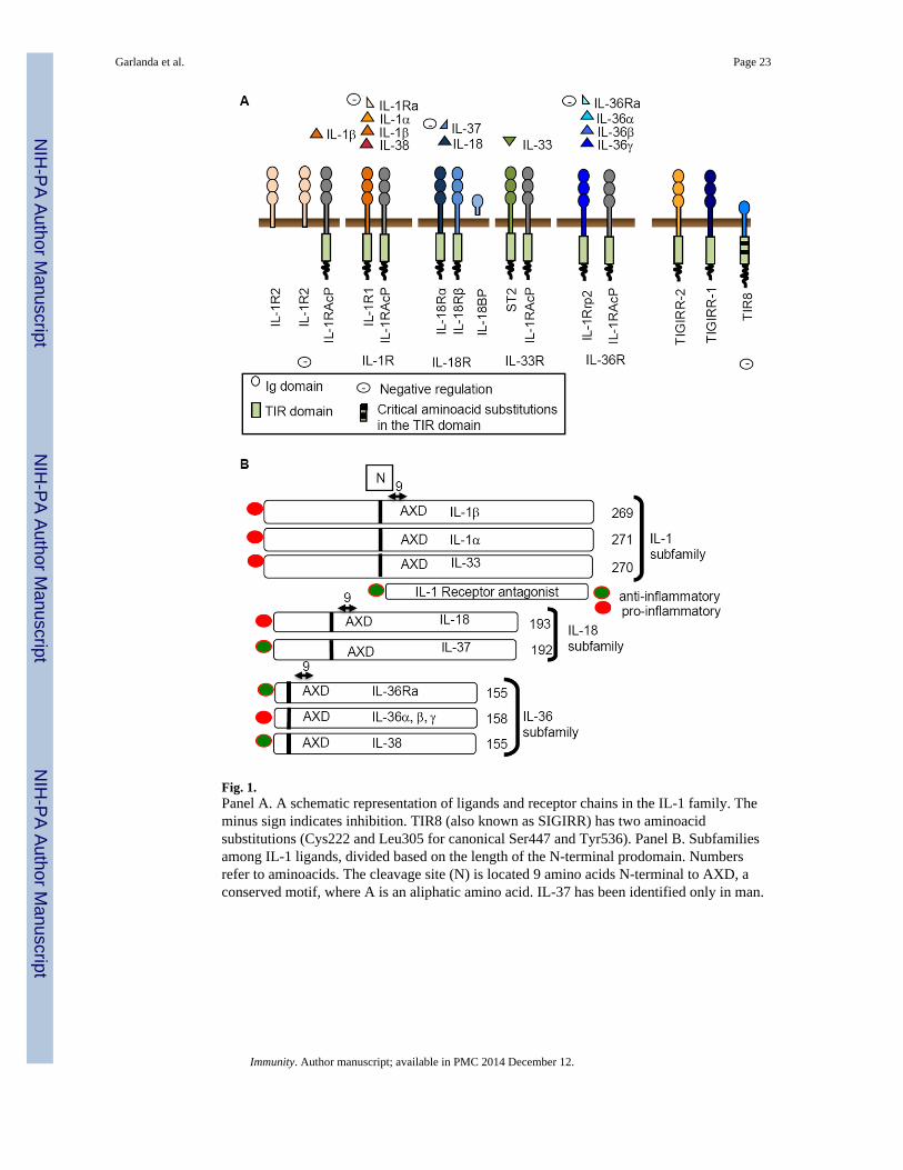
Fig. 1.Panel A. A schematic representation of ligands and receptor chains in the IL-1 family. Theminus sign indicates inhibition. TIR8 (also known as SIGIRR) has two aminoacidsubstitutions (Cys222 and Leu305 for canonical Ser447 and Tyr536). Panel B. Subfamiliesamong IL-1 ligands, divided based on the length of the N-terminal prodomain. Numbersrefer to aminoacids. The cleavage site (N) is located 9 amino acids N-terminal to AXD, aconserved motif, where A is an aliphatic amino acid. IL-37 has been identified only in man.
Garlanda et al. Page 23
Immunity. Author manuscript; available in PMC 2014 December 12.
NIH
-PA Author Manuscript
NIH
-PA Author Manuscript
NIH
-PA Author Manuscript

Fig. 2.A schematic representation of role of IL-1 (panel A) and IL-33 (panel B) in innate andadaptive immunity. “+IL-12” refers to costimulation by IL-33 + IL-12 of target cells. PRR,pattern recognition receptor; Nc Th1, non-conventional Th1; α1AT, α1 antitrypsin inhibitor;CRP, C reactive protein; PTX3, pentraxin 3; HPA, hypothalamus-pituitary-adrenal axis;FFA, free fatty acid.
Garlanda et al. Page 24
Immunity. Author manuscript; available in PMC 2014 December 12.
NIH
-PA Author Manuscript
NIH
-PA Author Manuscript
NIH
-PA Author Manuscript
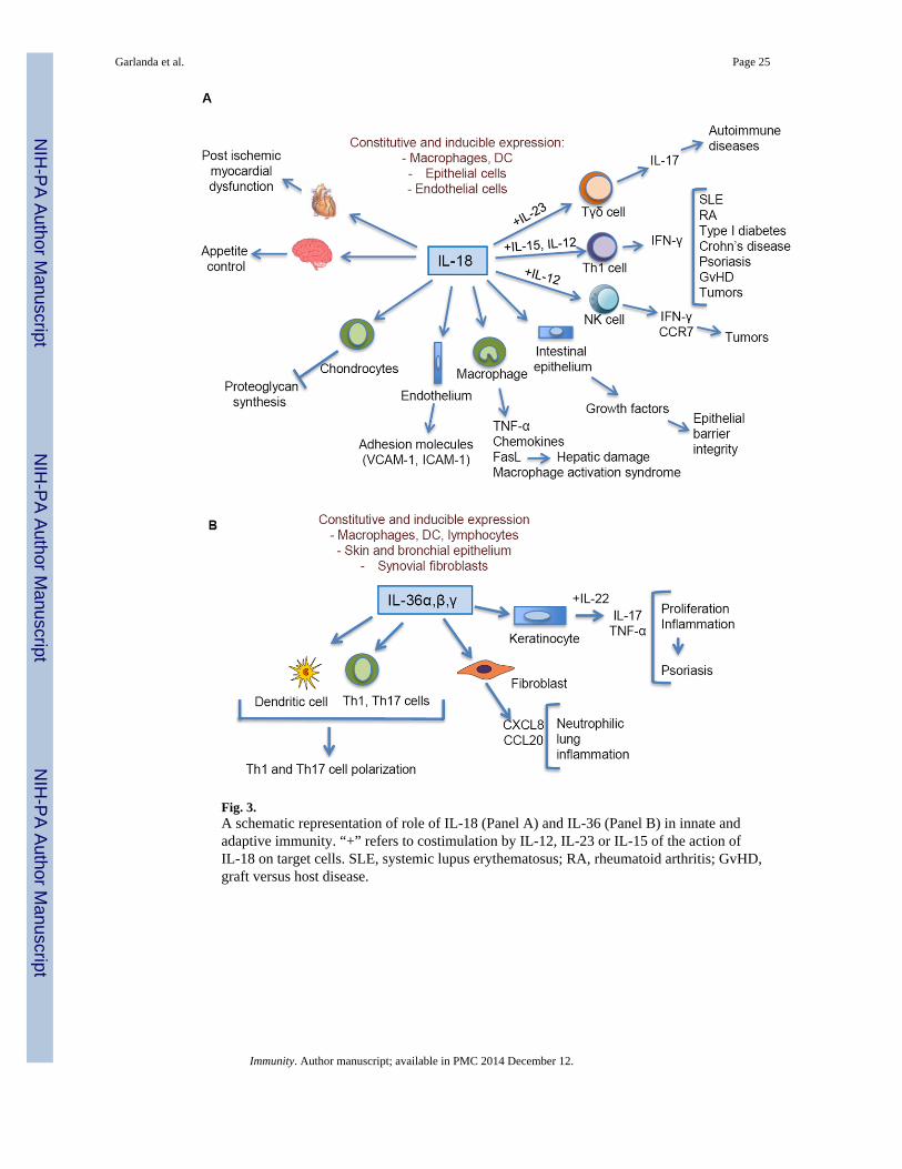
Fig. 3.A schematic representation of role of IL-18 (Panel A) and IL-36 (Panel B) in innate andadaptive immunity. “+” refers to costimulation by IL-12, IL-23 or IL-15 of the action ofIL-18 on target cells. SLE, systemic lupus erythematosus; RA, rheumatoid arthritis; GvHD,graft versus host disease.
Garlanda et al. Page 25
Immunity. Author manuscript; available in PMC 2014 December 12.
NIH
-PA Author Manuscript
NIH
-PA Author Manuscript
NIH
-PA Author Manuscript
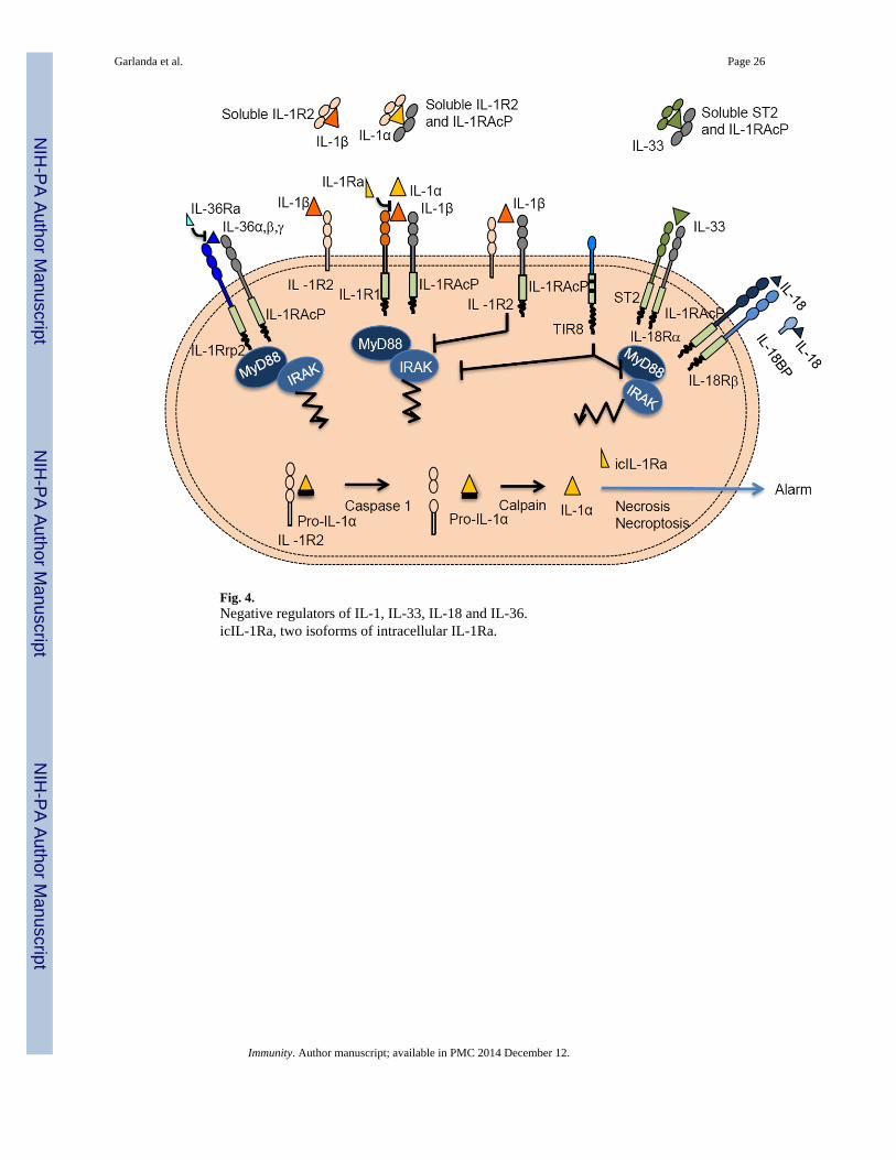
Fig. 4.Negative regulators of IL-1, IL-33, IL-18 and IL-36.icIL-1Ra, two isoforms of intracellular IL-1Ra.
Garlanda et al. Page 26
Immunity. Author manuscript; available in PMC 2014 December 12.
NIH
-PA Author Manuscript
NIH
-PA Author Manuscript
NIH
-PA Author Manuscript
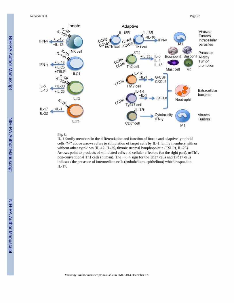
Fig. 5.IL-1 family members in the differentiation and function of innate and adaptive lymphoidcells. “+” above arrows refers to stimulation of target cells by IL-1 family members with orwithout other cytokines (IL-12, IL-25, thymic stromal lymphopoietin (TSLP), IL-23).Arrows point to products of stimulated cells and cellular effectors (on the right part). ncTh1,non-conventional Th1 cells (human). The → → sign for the Th17 cells and Tγδ17 cellsindicates the presence of intermediate cells (endothelium, epithelium) which respond toIL-17.
Garlanda et al. Page 27
Immunity. Author manuscript; available in PMC 2014 December 12.
NIH
-PA Author Manuscript
NIH
-PA Author Manuscript
NIH
-PA Author Manuscript

NIH
-PA Author Manuscript
NIH
-PA Author Manuscript
NIH
-PA Author Manuscript
Garlanda et al. Page 28
Tabl
e 1
Nom
encl
atur
e an
d m
ain
func
tions
of
IL-1
fam
ily m
embe
rs
Cyt
okin
eA
lter
nati
ve n
ame
Rec
epto
rC
o-re
cept
orP
ropo
sed
nom
encl
atur
e:A
ctiv
ity
Rec
epto
rC
o-re
cept
or
IL-1α
IL-1
F1IL
-1R
1IL
-1R
AcP
IL-1
R3
Ala
rmin
, inf
lam
mat
ion,
Th1
7 ce
ll re
spon
ses
IL-1
R2
IL-1β
IL-1
F2IL
-1R
1IL
-1R
AcP
IL-1
R3
Ant
imic
robi
al r
esis
tanc
e, in
flam
mat
ion,
Th1
7 ce
ll re
spon
ses
IL-1
R2
IL-1
Ra
IL-1
F3IL
-1R
1In
hibi
tion
of in
flam
mat
ion
IL-1
8IL
-1F4
IL-1
8Rα
IL-1
8Rβ
IL-1
R5
IL-1
R7
Infl
amm
atio
n, T
h1 c
ell r
espo
nses
IL-3
3IL
-1F1
1ST
2IL
-1R
AcP
IL-1
R4
IL-1
R3
Infl
amm
atio
n, T
h2 c
ell r
espo
nses
IL-3
6αIL
-1F6
IL-1
Rrp
2 (I
L-3
6R)
IL-1
RA
cPIL
-1R
6IL
-1R
3
Skin
and
lung
infl
amm
atio
nIL
-36β
IL-1
F7IL
-1R
rp2
(IL
-36R
)IL
-1R
AcP
IL-1
R6
IL-1
R3
IL-3
6γIL
-1F8
IL-1
Rrp
2 (I
L-3
6R)
IL-1
RA
cPIL
-1R
6IL
-1R
3
IL-3
6Ra
IL-1
F5IL
-1R
rp2
(IL
-36R
)IL
-1R
6In
hibi
tion
of in
flam
mat
ion
IL-3
7IL
-1F7
IL-1
8Rα
Inhi
bitio
n of
infl
amm
atio
n
IL-3
8IL
-1F1
0IL
-1R
rp2
(IL
-36R
)In
hibi
tion
of in
flam
mat
ion
TIR
8 (S
IGIR
R)
IL-1
R8
Inhi
bitio
n of
infl
amm
atio
n
TIG
IRR
-1 (
IL1R
APL
2)IL
-1R
9no
t kno
wn
TIG
IRR
-2IL
-1R
10no
t kno
wn
IL-1
R1
(?)
IL-1
RA
cPb
IL-1
R3b
Inhi
bitio
n of
infl
amm
atio
n
?: n
ot f
orm
ally
pro
ven
to in
tera
ct w
ith I
L-1
RA
cPb.
Immunity. Author manuscript; available in PMC 2014 December 12.
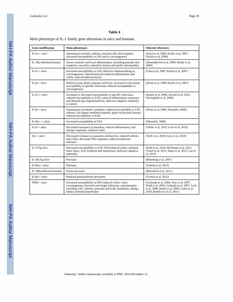
NIH
-PA Author Manuscript
NIH
-PA Author Manuscript
NIH
-PA Author Manuscript
Garlanda et al. Page 29
Table 2
Main phenotype of IL-1 family gene alterations in mice and humans.
Gene modification Main phenotypes Selected references
Il-1ra-/- mice Spontaneous arteritis, arthritis, psoriatic-like skin eruption,increased susceptibility to LPS and to carcinogenesis
(Horai et al, 2000; Krelin et al, 2007;Nicklin et al, 2000)
IL-1Ra-deficient humans Severe systemic and local inflammation, including pustular skineruptions, vasculitis, osteolytic lesions and sterile osteomyelitis
(Aksentijevich et al, 2009; Reddy et al,2009)
Il-1α-/- mice Increased susceptibility to LPS, defective immunoediting incarcinogenesis, reduced necrosis-induced inflammation andcolitis, reduced atherosclerosis
(Chen et al, 2007; Krelin et al, 2007)
Il-1β-/- mice Reduced acute phase response and fever, increased or decreasedsusceptibility to specific infections, reduced susceptibility tocarcinogenesis
(Horai et al, 1998; Krelin et al, 2007)
Il-1r1-/- mice Increased or decreased susceptibility to specific infections,reduced susceptibility to EAE, reduced inflammatory responsesand delayed type hypersensitivity, defective adaptive immunityto tumors
(Sutton et al, 2006; Joeckel et al, 2012;Ghiringhelli et al, 2009)
Il-18-/- mice Spontaneous metabolic syndrome, reduced susceptibility to LPS,arthritis, Fas ligand–mediated hepatitis, graft-versus-host disease,reduced susceptibility to EAE
(Netea et al, 2006; Dinarello, 2009)
Il-18rα -/- mice Increased susceptibility to EAE (Dinarello, 2009)
Il-33-/- mice Decreased resistance to parasites, reduced inflammatory andallergic responses, reduced colitis
(Oboki et al, 2010; Liew et al, 2010)
St2-/- mice Decreased resistance to parasites and bacteria, reduced arthritisand colitis, decreased Th2 responses, reduced endotoxintolerance
(Neill et al, 2010; Liew et al, 2010)
IL-37Tg mice Decreased susceptibility to LPS, DSS-induced colitis, ischemicheart injury, liver ischemia and reperfusion, defective adaptiveimmunity.
(Nold et al, 2010; McNamee et al, 2011;Yousif et al, 2011; Sakai et al, 2012; Luo etal, 2013)
IL-36αTg mice Psoriasis (Blumberg et al, 2007)
Il-36ra-/- mice Psoriasis (Tortola et al, 2012)
IL-36Ra-deficient humans Severe psoriasis (Marrakchi et al, 2011)
Il-36r-/- mice Reduced psoriasiformis dermatitis (Tortola et al, 2012)
TIR8-/- mice Increased susceptibility to DSS-induced colitis, coloncarcinogenesis, bacterial and fungal infections, autoimmunityincluding LES, arthritis, psoriasis and EAE, leukaemia, allergy,kidney ischemia/reperfusion
(Garlanda et al, 2004; Xiao et al, 2007;Wald et al, 2003; Garlanda et al, 2007; Lechet al, 2008; Bulek et al, 2009; Gulen et al,2010; Bertilaccio et al, 2011)
Immunity. Author manuscript; available in PMC 2014 December 12.