Inflammatory Bowel Disease and Mutations Affecting the Interleukin-10 Receptor
Journal o f Clinical Immunology, Vol. 12, No. 4, 1992
Special Article
Biological Properties of Interleukin 10
MAUREEN HOWARD,I'2 ANNE O'GARRA,1 HIROSHI ISHIDA,1 RENt~ DE WAAL MALEFYT, I and JAN DE VRIES 1
Accepted: March 10, 1992
INTRODUCTION
Intefleukin I0 (IL-10) was discovered in 1989 by Mosmann and colleagues as an activity produced by murine type 2 helper T cells which suppressed cytokine production by type 1 helper T cells (1). This activity, initially designated cytokine synthesis inhibitory factor (CSIF), was isolated by expression cloning by Moore and colleagues (2), who subse- quently identified and isolated a human analogue of CSIF by cross hybridization (3), using as their source a human helper T-cell clone produced by Roncarolo et al. (M.-G. Roncarolo and H. Spits, unpublished). While a search of the available se- quence data banks indicated that CSIF represented a novel cytokine, this search also revealed a re- markable homology between CSIF and a previously uncharacterized open reading frame, termed BCRF~, in the Epstein-Barr virus (EBV) genome (2-4). After further analysis of CSIF genomic Struc- ture, Moore and colleagues proposed the intriguing possibility that at some stage, EBV may have captured this mammalian gene, presumably to con- fer some survival advantage on itself.
The availability of recombinant CSIF, and neu- tralizing monoclonal antibodies that recognized it specifically (5), rapidly led to the discovery that this new cytokine accounted for two other novel activ-
1DNAX Research Institute, 901 California Avenue, Palo Alto, California 94304.
2To whom correspondence should be addressed.
ities under investigation at DNAX: (i) B-TCGF, a B cell-derived growth costimulator for thymocytes (6, 7); and (ii) MCGFIII, a B cell-derived growth co- stimulator for mast cells that could be distinguished from IL-3 and IL-4, the first two cytokines known to exhibit mast-cell stimulating function (8, 9). The realization that CSIF was a pleiotropic cytokine exerting effects on a diversity of cell types led to its renaming as interleukin I0 and initiated Studies which demonstrated that a number of immunostim- ulatory and immunosuppressive properties could be attributed to this cytokine (6, 7, 9-16). In addition to a diversity of cell types that respond to IL-10, it is now apparent that interleukin 10 can be produced by B cells (8, IT) and monocyte/macrophages (12, 14) in addition to the TH0 and TH2 subsets (re- viewed in Ref. 18) of helper T cells. In this review, we attempt to summarize what is currently known of the biological properties of IL-10 and speculate on its clinical potential and usefulness.
IMMUNOSUPPRESSIVE PROPERTIES OF IL-10 IN VITRO (TABLE I)
As indicated above, IL-10 was originally identi- fied as a product of murine T helper 2 (Th2) clones that inhibited the production of IL-2, IL-3, tumor necrosis factor beta (TNFI3), granulocyte-macro- phage colony-stimulating factor (GM-CSF), and in- terferon gamma (IFNT) by Thl clones following antigen presenting cell (APC)-dependent stimula- tion with antigen or soluble anti-CD3 monoclonal antibodies (I). The inhibitory effects of IL-10 on Thl cytokine synthesis were especially pronounced
239 0271-9142/92/0700-0239506,50/0 © 1992 Plenum Publishing Corporation

240 HOWARD ET AL.
Table I. Immunosuppressive Properties of IL-10 in Vitro
• Inhibits monocyte/macrophage-dependent cytokine synthesis by PBMNC, NK cells, and Thl clones
• Inhibits monocyte/macrophage-dependent Th0, Thl, and Th2 ~ proliferation
• Inhibits cytokine synthesis by activated macrophages/monocytes
• Inhibits monocyte class II MHC expression • Inhibits macrophage production of reactive nitrogen oxides • Inhibits macrophage intracellular and extracellular killing of
parasites
aSO far shown only in human system.
on the production of IFN~/and IL-3, both of which are normally synthesized late following activation of these clones. While murine-IL-10 is species spe- cific, human- and EBV-derived viral IL-10 (vlL-10) inhibit cytokine synthesis by both mouse Thl clones and human peripheral blood mononuclear cells (PBMNC) (3, 19). Human IL-10 inhibited strongly the production of IFN~/, GM-CSF, TNFtx, and TNFI3 by PBMNC following activation with phytohemagglutinin (PHA), anti-CD3 monoclonal antibodies, or IL-2. This inhibition of cytokine synthesis by murine Thl clones and human PBMNC has been shown to occur at both the protein and the mRNA level (1, 3).
The mechanism by which IL-I0 inhibits cytokine synthesis by helper T-cell clones or PBMNC has not been completely elucidated. Mosmann and col- leagues initially showed that m-IL-10 does not act directly on murine Thl clones, since IFN~/synthe- sis was unaffected when the T-cell clones were stimulated by immobilized anti-CD3 monoclonal antibodies or concanavalin A (Con A) in the ab- sence of accessory cells (1). It was subsequently shown by O'Garra and Fiorentino, using purified populations of splenic or peritoneal APC, that IL- 10 inhibited IFN~/synthesis by antigen-stimulated Thl clones only when purified macrophages were used as APC, and had no effect on Thl activation when antigen was presented by either conventional or Lyl + B cells (13). Additional studies to elucidate this inhibition of macrophage APC function showed that IL-10 affected the antigen presenting capacity of metabolically active macrophages only. Further- more, since macrophage-dependent Thl stimulation by superantigens was also inhibited by IL-10, it appeared that the mechanism of IL-10-mediated suppression was not only at the level of antigen uptake or processing (13). The direct effect of IL-10 on processing has not been addressed as yet. Ex- periments to date make it unlikely that IL-10 acts by
inhibiting the production of a soluble costimulatory activity or by inducing a suppressor activity by macrophages. Some effort has been focused on the possibility that IL-10 suppresses APC function of macrophages by down-regulation of class II major histocompatibility complex (MHC) expression on these cells. Indeed, Stein et al. (M. Stein and S. Gordon, personal communication) have demon- strated that IL-10 inhibits IFN~-induced class II expression on polyacrylamide bead-elicited perito- neal macrophages. It remains to be determined whether this effect is also observed on resident macrophages which are inhibited by IL-10 from stimulating TH1 clones. Regardless of this out- come, several observations indicate that IL-10- mediated suppression of macrophage APC function involves additional factors other than class I I MHC expression. First, IL-10 inhibited the antigen pre- senting capacity of an activated murine macrophage line without apparently decreasing its expression of class II antigens, although this could be due to slow turnover of class II (13). Second, IL-10 inhibited the IL-2-induced production of IFN~/by Thl cells (13), a mode of stimulation which is dependent on a Mac 1 + accessory cell but not on antigen or class II MHC molecules (20). Finally, increases in either APC or antigen dose did not affect IL-10 action (13), again implicating suppression of an activation path- way other than T-cell receptor [TCR]-MHC class II interactions.
In a concurrent investigation of the mechanism of IL-10-mediated immunosuppression, de Waal- Malefyt and colleagues have analyzed the ability of human IL-10 to inhibit the production of IFN~/by PHA- or IL-2-activated human PBMNC (3, 19). Consistent with the above murine experiments, the inhibition of cytokine production by PHA-activated peripheral blood T cells is indirectly mediated via inhibitory effects of IL-10 on the antigen presenting or accessory function of human monocytes (R. de Waal-Malefyt, in preparation). Similar results were obtained to explain IL-10-mediated inhibition of IFN-~ production by human natural killer (NK) cells stimulated with accessory cells plus IL-2 (19). In this system, IL-10 did not effect the small amounts of IFN~ produced by highly purified human NK cells stimulated with IL-2 in the absence of acces- sory cells. However, IL-10 caused profound sup- pression of the enhanced production of IFN~ and TNFet that occurs when purified monocytes are added to IL-2-stimulated NK cells. IL-10-mediated inhibition of NK cell derived-IFN-,! has also been
Journal o f Clinical Immunology, Vol. 12, No. 4, 1992

BIOLOGICAL PROPERTIES OF INTERLEUKIN 10 241
shown in a murine in vitro model by Bancroft and Webster (21).
In addition to its cytokine synthesis inhibitory activity, IL-10 inhibits accessory cell-dependent, antigen-specific proliferation of resting human T cells and human Th0-, Thl-, and Th2-"like" CD4 ÷ T-cell clones (11) and of mouse Thl clones (S. Macatonia and A. O'Garra, in preparation). IL-10 inhibited antigen-specific proliferation when mono- cytes, but not when EBV-lymphoblastoid cell line [LCL] were used as APC, indicating that IL-10 affected the antigen presenting capacity of mono- cytes but not transformed B cells (11). Inhibition of human T-cell proliferation in this system appears to be independent of antigen processing, since human IL-10 was equally as effective at inhibiting human T-cell proliferation induced by monocyte-presented peptide antigens which did not require processing. Consistent with the observation on CSIF activity in mouse systems, no evidence has been obtained in the human T-cell proliferation system forthe induc- tion of a monocyte suppressor factor by IL-10 or the inhibition of a soluble activator. However, down-regulation of class II MHC expression ap- pears to play a significant role in IL-10-mediated suppression of human monocyte APC function (I 1). Human IL-10 strongly down-regulated constitutive, IL-4, and IFN~/-induced expression of HLA DR/DP and HLA DQ antigens on human monocytes; fur- thermore, it rendered monocytes refractory to in- duction of class II MHC expression by IFN~/and IL-4. The down-regulation of class II MHC expres- sion correlated with the inhibition of proliferation by the T-cell clones, suggesting that IL-10 pre- vented the activation of the T-cell clones rather than actively suppressing their proliferation (I1). This notion was confirmed by the observation that Ca 2÷ fluxes were strongly reduced in T-cell clones following their activation with monocytes that had been pretreated with antigen and IL-10 and that IL-10 prevented the induction of the IL-2 receptor ~x chain (TAC) in T-cell cultures stimulated by antigen and monocytes. Importantly, IL-10-induced inhibi- tion of T-cell proliferation was only partially re- versed by the addition of high concentrations of IL-2, suggesting that the reduced proliferative re- sponses may not simply reflect reduced IL-2 pro- duction.
Extending this concept of IL-10-mediated sup- pression of human T-cell proliferation, Roncarolo et al. have shown that IL-10 also inhibits the prolifer- ation of class II MHC alloantigen-specific T-cell
clones when monocytes are used as APC (M.-G. Roncarolo, in preparation). Furthermore, IL-10 in- hibits the proliferation and cytokine production generated in primary one-way mixed leukocyte cul- tures (MLC) (M. T. Bejarano, submitted for publi- cation). Separation of responder and stimulator cells indicated that IL-10 strongly inhibited the proliferation when allogeneic monocytes were used as stimulators and purified T cells as responders. IL-10 did not inhibit the proliferation induced by allogeneic EBV-LCL. Although the total number of responder T cells was reduced in the cultures with IL-10, the ratio of CD4 + and CD8 ÷ T cells remained unaffected, indicating that IL-10 had no preferential inhibitory effects on CD4 + or CD8 + T cells. Thus, the effects of IL-10 on alloantigen-induced prolifer- ative responses are comparable to those on T-cell proliferation induced by soluble antigens presented by monocytes. IL-10 also inhibited the generation of allospecific cytotoxic T cells in primary one-way MLC in these studies. However, it is not yet clear whether IL-10 inhibits directly the generation of cytotoxic CD8 + T cells or that it affects the activa- tion of the CD4 + T helper cells which are required for the generation of the cytotoxic T cells. In murine systems, IL-10 did not affect proliferation in an MLC initiated by mouse dendritic cells or un- separated splenic APC, using unseparated or puri- fied CD4 + cell responders (S. Macatonia and A. O'Garra, in preparation). In these studies, resident splenic macrophages could not initiate an MLC (S. Macatonia and A. O'Garra, in preparation), a find- ing which is consistent with previous analyses of murine MLC (22). These differences in mouse and human systems may be due to the different sources of responder and stimulator cells used throughout, e.g., peripheral blood in the human versus splenic or lymph node cells in the mouse. Thus, more studies are needed to predict whether IL-10 will play a role in the induction or maintenance of tolerance following allogeneic bone marrow or or- gan transplantations in vivo.
In addition to its effects on the accessory/antigen presenting functions of monocytes/macrophages, IL-10 also inhibits other monocyte/macrophage functions in both mouse and man. The addition of IL-10 to cultures of monocytes or peritoneal mac- rophages results in changes in morphology and adhesion (14; R. de Waal-Malefyt, in preparation). The explanation of these morphological changes is not yet known, however, several considerations have been eliminated, e.g., IL-10 does not alter the
Journal of Clinical Immunology, Vol. 12, No. 4, 1992
242 HOWARD ET AL.
expression of a variety of adhesion molecules in- cluding CD11 abc, CD18, CD54, CD58, VLA-4, and VLA-5 on cultured monocytes (I1). In addition to these dramatic morphological changes, IL-10 inhib- its the production of numerous cytokines, e.g., IL-let, IL-6, IL-8, GM-CSF, G-CSF, and TNFot, by murine peritoneal macrophages, macrophage cell lines, and human monocytes stimulated with lipo- polysaccharide (LPS), IFN~/, or LPS and IFN7 (I4, 12, 23, 24). The substantial suppression of IL-let, GM-CSF, and TNFet production, in particular, pre- sumably reflects the relatively late production of these cytokines following monocyte activation. In vitro studies have shown that monocytes and mac- rophages can produce high levels of IL-10 following activation by a variety of stimulants, such as LPS (14, 12). Interestingly, in vitro activation of mono- cytes/macrophages in the presence of neutralizing anti-IL-10 monoclonal antibodies results in in- creased production of IL-la, IL-II3, IL-6, IL-8, GM-CSF, G-CSF, and TNFa, a strong induction of class II MHC expression by human monocytes (12), and enhanced IL-6 production by mouse peritoneal macrophages (14), suggesting that endogenously produced IL-10 normally suppresses cytokine pro- duction and class II MHC expression by these cells. These observations have now been extended by the recent finding that exogenous IL-10 inhibited IL-10 mRNA expression by LPS-activated monocytes, suggesting that production of this cytokine is itself regulated by a negative feedback mechanism (12). In addition to its effects on monokine production, IL-10 has also been shown to inhibit the production of reactive nitrogen oxides (NO) following IFN7 activation of thioglycollate-elicited peritoneal mac- rophages (23, 25, 26). The production of NO is involved in the IFN-f-mediated elimination of intra- cellular Toxoplasma gondii (25) and the extracellu- lar killing of schistosomula of Schistosoma mansoni (26) by macrophages. Since IL-10 is present follow- ing host infection by a variety of parasites (27), it may provide a way in which intra- and extracellular parasites escape from IFNT-dependent cell- mediated immunity. IL-10 does not completely sup- press all monocyte protein synthesis, since it has no effect on the constitutive expression of TGF[~ (12), and it up-regulates expression of the IL-1 receptor antagonist (IL-1 Ra), an important regulator of inflammatory reactions (de Waal-Malefyt, unpub- lished). Collectively, these in vitro data suggest that IL-10 may play an important role in regulating
Table H. Immunostimulatory Properties of IL-IO in Vitro
• Enhances B-cell class II MHC expression • Enhances B-cell viability • Enhances activated B-cell proliferation and Ig secretion • Growth costimulator for thymocytes • Enhances cytotoxic T-cell development • Growth costimulator for mast cells
inflammatory responses via inhibition of proinflam- matory cytokines and stimulation of IL-1RA.
IMMUNOSTIMULATORY PROPERTIES OF IL-10 IN VITRO
While IL-10 is a potent immunosuppressant of macrophage function, it exerts a wide array of immunostimulatory effects on B cells (summarized in Table II). IL-10 up-regulates class II expression and in vitro viability of highly purified small dense B cells obtained from the spleens of unstimulated mice (10). Previous studies have indicated that IL-4 is also capable of mediating these same effects on resting B cells (28-3I); however, neutralizing monoctonal antibodies that specifically recognize each of these cytokines suggest that the two cyto- kines work independently in these bioassays (10). Interestingly, B cells from xid mice were incapable of up-regulating class II expression in response to IL-10 stimulation (10), despite the apparent exis- tence of functional IL-10 receptors on these cells and their ability to up-regulate class II expression in response to IL-4 stimulation (N. Go and M. How- ard, unpubiished). This currently unexplained ob- servation raises the possibility that IL-10 in some way contributes to the immunodeficiency of xid mice, which are unresponsive to thymus-indepen- dent type II antigens in B-cell bioassays in vitro and susceptible to an array of microorganisms in vivo (32-36).
In addition to these effects on resting B cells, IL-10 augments proliferation of activated B cells and induces their differentiation into antibody se- creting cells. This has been demonstrated most convincingly using human B cells stimulated with anti-CD40 antibodies extensively cross-linked on FcR/L cell transfectants (37). In the presence of IL-4, this mode of activating human B cells induces their vigorous and long-term proliferation, with little differentiation into Ig secreting cells (38). The addition of IL-10 to these cultures augments DNA synthesis and expansion of total B cells and induces their differentiation into antibody producing cells,
Journal of Clinical Immunology, Vol. 12, No. 4, 1992
BIOLOGICAL PROPERTIES OF INTERLEUKIN 10 243
with subsequent expression of large amounts of IgM, IgG, and IgA antibodies (37). IgE can also be obtained in this system but has an absolute require- ment for IL-4. Similar results were obtained when human B lymphocytes were activated via their antigen receptors (37). More recently, these studies have been extended to demonstrate that IL-10 in- duces IgM secretion predominantly from surface IgD ÷ naive B cells stimulated with anti-CD40 anti- bodies and IgG secretion predominantly from sur- face IgD- "germinal center-like" B cells stimulated with anti-CD40 antibodies (39). In the latter studies, IL-I0 also appeared to be an important cofactor for driving naive B cells activated with anti-CIM0 anti- bodies and tumor growth factor beta (TGFI3) into IgA secretion (39).
In addition to its potent immunostimulatory ef- fects on B lymphocytes, IL-10 is a stimulant of numerous other cell types (summarized in Table II). It is capable of augmenting the in vitro proliferative response of mouse thymocytes and T cells which have been activated with IL-2 and IL-4 (6, 7) and may, therefore, play a role in T-cell development. In single-cell cultures containing IL-2, IL-10 en- hanced the frequency of murine cytotoxic T-cell precursors generated from Con A-activated fluores- cence-activated cell sorter (FACS)-purified CD8 + splenic T cells (16) and markedly expanded the clone size and lytic potential of these cells when assayed against appropriate target cells (16). The effect of IL-10 on other hemopoietic lineages is only starting to be investigated now. Rennick and col- leagues have shown that IL-10 sustains the viability of several mouse mast-ceU lines in vitro and syner- gizes with IL-3 and IL-4 to stimulate proliferation of these cell lines (9, 8). Perhaps more significantly, IL-10 markedly enhanced the total number and average size of mast-cell colonies which developed after 14 days of culture in the presence of IL-3 and IL-4 from mesenteric lymph node cells of nema- tode-infected mice.
A C T I O N O F M U R I N E IL-10 I N VIVO
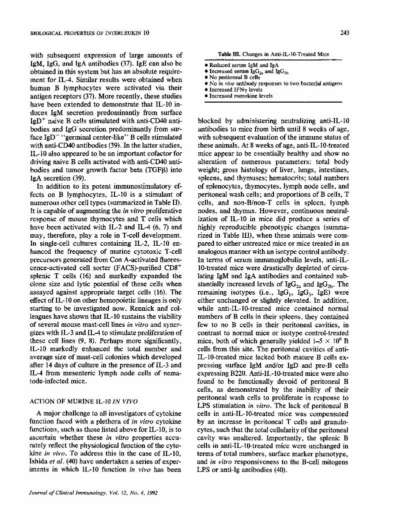
A major challenge to all investigators of cytokine function faced with a plethora of in vitro cytokine functions, such as those listed above for IL-10, is to ascertain whether these in vitro properties accu- rately reflect the physiological function of the cyto- kine in vivo. To address this in the case of IL-10, Ishida et al. (40) have undertaken a series of exper- iments in which IL-10 function in vivo has been
Table m . Changes in Anti-IL-10-Treated Mice
• Reduced serum IgM and IgA • Increased serum IgG2a and IgGEb • No peritoneal B cells • No in vivo antibody responses to two bacterial antigens • Increased IFN~/levels • Increased monokine levels
blocked by administering neutralizing anti-IL-10 antibodies to mice from birth until 8 weeks of age, with subsequent evaluation of the immune status of these animals. At 8 weeks of age, anti-IL-10-treated mice appear to be essentially healthy and show no alteration of numerous parameters: total body weight; gross histology of liver, lungs, intestines, spleens, and thymuses; hematocrits; total numbers of splenocytes, thymocytes, lymph node cells, and peritoneal wash cells; and proportions of B cells, T cells, and non-B/non-T cells in spleen, lymph nodes, and thymus. However, continuous neutral- ization of IL-10 in mice did produce a series of highly reproducible phenotypic changes (summa- rized in Table III), when these animals were com- pared to either untreated mice or mice treated in an analogous manner with an isotype control antibody. In terms of serum immunoglobulin levels, anti-IL- 10-treated mice were drastically depleted of circu- lating IgM and IgA antibodies and contained sub- stantially increased levels of IgGEa and IgG2b. The remaining isotypes (i.e., IgG1, IgG3, IgE) were either unchanged or slightly elevated. In addition, while anti-IL-10-treated mice contained normal numbers of B cells in their spleens, they contained few to no B cells in their peritoneal cavities, in contrast to normal mice or isotype control-treated mice, both of which generally yielded 1-5 x 10⁶ B cells from this site. The peritoneal cavities of anti- IL-10-treated mice lacked both mature B cells ex- pressing surface IgM and/or IgD and pre-B cells expressing B220. Anti-IL-10-treated mice were also found to be functionally devoid of peritoneal B cells, as demonstrated by the inability of their peritoneal wash cells to proliferate in response to LPS stimulation in vitro. The lack of peritoneal B cells in anti-IL-10-treated mice was compensated by an increase in peritoneal T cells and granulo- cytes, such that the total cellularity of the peritoneal cavity was unaltered. Importantly, the splenic B cells in antML-10-treated mice were unchanged in terms of total numbers, surface marker phenotype, and in vitro responsiveness to the B-cell mitogens LPS or anti-Ig antibodies (40).
Journal of Clinical Immunology, Vol. 12, No. 4, 1992

244 HOWARD ET AL.
Several other immunological modifications were found to be characteristic of anti-IL-10-treated mice. While developing perfectly normal primary and secondary antibody responses to the T-depen- dent antigen TNP-KLH, anti-IL-10-treated mice were found to be deficient in antibody production to two bacterial antigens, phosphorylcholine and ct- 1,3-dextran. Furthermore, anti-IL-10-treated mice contained substantial levels of circulating IFN~/and frequently expressed IL-6 in their sera, two cytokines which were not found in the circula- tion of either normal mice or isotype control-treated mice. The increased IL-6 expression was probably representative of a broader monokine up-regula- tion, since anti-IL-I0-treated mice were found to be 50-fold more susceptible to death by endotoxin- induced shock (H. Ishida and M. Howard, unpub- lished data), an inflammatory reaction resulting from elevation of numerous monokines (41-44).
These changes in anti-IL-10-treated mice, as summarized in Table III, can be interpreted as follows. The anti-IL-10-induced increase in IFN~/ and IL-6 expression implied by the aberrant pres- ence of these cytokines in the circulation of anti- IL-10 treated mice is consistent with the previously documented ability of IL-10 to suppress production of these cytokines in vitro. As outlined above, numerous in vitro studies have shown that IL-10 is capable of suppressing IFN~/production by helper T cells (I, 13) and NK cells (19, 21) via an indirect action on accessory cells. Similarly, IL-10 is a potent suppressant of monokine production in vitro (12, 14). The increase in serum IgG2a and IgG2b is presumably the result of IFN~/elevation in anti-IL- 10-treated mice, since IFN~/has been shown previ- ously to up-regulate expression of these immuno- globulin isotypes in vitro (45, 46). The remaining changes summarized in Table III, i.e., reduction of serum IgM and IgA, depletion of peritoneal B cells, and elimination of antibacterial antibody responses, can be explained by the selective elimination of a subpopulation of B cells, designated CD5 or Ly-1 B cells, from anti-IL-10-treated mice. CD5 B cells (47-51) are highly prevalent in the fetal and neonatal immune systems of mice and man but represent only a minor B-cell subpopulation in adulthood of both species. They have been studied extensively in the murine system, where numerous intriguing properties of CD5 B cells have been deduced from a variety of animal cell-transfer experiments where irradiated mice or SCID mice are selectively recon- stituted with either CD5 B cells or conventional B
cells. Such experiments have shown that murine CD5 B cells generate most circulating IgM (52-54), and contain the precursors of IgA secreting cells (55) and of all antibody forming cells specific for the bacterial antigens phosphorylcholine and ct-l,3- dextran (53, 56). In normal mice, CD5 B cells are almost undetectable in primary and secondary lym- phoid tissues but become greatly enriched in the peritoneal and pleural cavities. Thus, the observed depletion of peritoneal B cells, circulating IgM and IgA, and in vivo antibody responses to phosphoryl- choline and ot-l,3-dextran in anti-IL- 10-treated mice are highly suggestive of an elimination of CD5 B cells resulting from this treatment.
Several experiments were performed to explore further the mechanism of CD5 B-cell depletion in anti-IL-10-treated mice. We have eliminated the trivial explanation that anti-IL-10 antibodies are selectively cytotoxic for CD5 B cells by showing that injection of the same antibodies into the peri- toneal cavities of normal adult mice had no effect on the subsequent numbers or phenotypes of B cells recovered from this site 1, 2, or 3 days later. As alternative possible mechanisms, we have consid- ered whether CD5 B-cell depletion was the result of a secondary cytokine perturbation occurring in anti- IL-10-treated mice or, indeed, whether IL-10 acted as an autocrine growth factor for CD5 B cells. The latter theory initially seemed particularly attractive in light of our previous studies showing greatly elevated constitutive and inducible IL-10 produc- tion by CD5 B cells compared to conventional B cells (8, 17). Recent studies have indicated, how- ever, that CD5 B-cell depletion in anti-IL-10-treated mice is more likely to be another consequence of IFN~/elevation in these animals. When mice were treated from birth until 8 weeks of age with neutral- izing anti-IFN~/antibodies in addition to neutraliz- ing anti-IL-10 antibodies, they contained virtually normal numbers of peritoneal B cells (40), suggest- ing that neutralization of IFN~/prevented anti-IL- 10-induced depletion of CD5 B cells in these mice. Consistent with this notion, recent preliminary ex- periments have shown that IFN~/ suppresses in vitro proliferation of LPS-stimulated peritoneal cells but not LPS-stimulated spleen cells (A. O'Garra and M. Howard, unpublished observa- tions).
In summary, the anti-IL-10-treated mouse con- firms several of the immunosuppressive in vitro properties of IL-10 described above (see Table I). Further work is required to evaluate whether the
Journal of Clinical Immunology, Vol. 12, No. 4, 1992

BIOLOGICAL PROPERTIES OF INTERLEUKIN 10 245
immunostimulatory in vitro properties of IL-10 (see Table II) are also confirmed by the anti-IL-10- treated mouse; however, there is nothing yet to suggest that anti-IL-10 treatment decreases Ig se- cretion in vivo from the majority of conventional B cells, as would be anticipated from the B-cell stim- ulatory properties of IL-10. The phenotype of the anti-IL-10-treated mouse makes certain predictions for in vivo roles of IL-10. The elevation of mono- kines in these animals suggests that IL-10 may act as a potent antiinflammatory agent in vivo. Prelim- inary data indeed suggest that IL-10 is capable of directly protecting mice against death from endot- oxin-induced shock in vivo (M. Howard and S. Menon, unpublished data). It is more difficult to speculate on IL-10 function in vivo based on the depletion of CD5 B cells in anti-IL-10-treated mice, since the contribution of this numerically small subset of B lymphocytes to the immune system is still unclear. The various models of CD5 B-cell function that have been advanced based on the specificities of antibodies they produce include roles in antibacterial immunity (57, 58), in clearance of host cellular debris such as senescent erythro- cytes (59, 60), and in modulation of the antibody repertoire during development (61-64). Our recent finding that CD5 B cells are themselves potent producers of IL-10 (8, 17) indeed raises the possi- bility of a broader immunoregulatory role of CD5 B cells. Collectively, the observations that anti-IL-10- treated mice are deficient in IgM, IgA, specific antibacterial antibody responses, and a subpopula- tion of B cells believed to play a role in antibacterial immunity suggest that IL-10 may have the capacity to augment directly antibody-mediated antibacterial immunity. To this end, it will be interesting to evaluate the susceptibility of anti-IL-10-treated mice to a broad range of microorganisms.
P O T E N T I A L CLINICAL APPLICATIONS OF IL-10
In the absence of animal model experimentation or preclinical testing, it is difficult to predict accu- rately the clinical potential of IL-IO. Nevertheless, speculation may be justified on the basis of several of the striking biological properties described above. IL-IO is capable of suppressing IFN~/ and numerous monokines in a variety of in vitro assay systems. The elevation of these same cytokines in anti-IL-lO-treated mice strongly suggests that these immunosuppressive properties of IL-IO will occur in vivo. The suppression of monokine production
suggests that IL-10 may be a potent antiinflamma- tory reagent in a variety of clinical situations: bacterial sepsis, rheumatoid arthritis, psoriasis, etc. The ability of IL-10 to suppress a subset of T cell-derived cytokines (e.g., IFN-¢, IL-2, TNF[3) in addition to monokine suppression makes it an at- tractive candidate for prolonging allograft survival and in treating a variety of T cell-mediated autoim- mune diseases, e.g., type I diabetes, multiple scle- rosis. Indeed, animal models already exist for some of these clinical conditions where suppression of IFN% IL-2, TNFa, or IL-6 using monoclonal anti- bodies causes a marked improvement in pathology. More information as to whether IL-10 also inhibits accessory and antigen presenting cell functions and proinflammatory cytokine production by other cells that have been implicated in inflammation, such as endothelial cells, epithelial cells, keratinocytes, Langerhans, and dendritic cells, together with ad- ditional information about its antiinflammatory ef- fects and its ability to inhibit transplant rejection is required to determine whether IL-10 will indeed act as a bona fide antigen-nonspecific suppressor factor in vivo.
In other clinical situations, antagonism of IL-10 may be a more desirable choice. The expression of IL-10 activity (v-IL-10) by EBV presumably confers some survival advantage to the virus in terms of its ability to infect, replicate, and/or maintain itself within the host. The ability of (v) IL-10 to down- regulate IFN'y production by both T cells and NK cells, together with its B-ceU viability enhancing effects, suggests that (v) IL-10 can suppress antiviral immunity while at the same time enhancing the potential of EBV to transform human B cells. It is therefore feasible that IL-10 antagonists could effec- tively boost antiviral immunity in vivo to EBV and possibly other viruses. Similarly, IL-10 antagonists may also provide effective therapy in infectious disease situations where IL-10 has been shown to inhibit macrophage-mediated immunity against intra- and extracellular parasites. We therefore look for- ward with optimism to future experimentation where IL-10 is either administered directly or, alterna- tively, antagonized by neutralizing anti-IL-10 anti- bodies, in a variety of animal models of infectious disease, inflammation, autoimmunity, and transplan- tation. It is to be hoped that information obtained from these approaches will offer IL-10 numerous opportunities to diminish the clinical pathology as- sociated with these diverse disease situations.
Journal of Clinical Immunology, Vol. 12, No. 4, 1992

246 HOWARD ET AL.
R E F E R E N C E S
1. Fiorentino DF, Bond MW, Mosmann TR: Two types of mouse helper T cell. IV. Th2 clones secrete a factor that inhibits cytokine production by Thl clones. J Exp Med 170:2081-2095, 1989
2. Moore KW, Vieira P, Fiorentino DF, Trounstine ML, Khan TA, Mosmann TR: Homology of cytokine synthesis inhibi- tory factor OL-10) to the Epstein Barr virus gene BCRFI. Science 248:i230-1234, 1990
3. Vieira P, de waal-Malefyt R, Dang MN, Johnson KE, Kastelein R, Fiorentino DF, deVries JE, Roncarolo MG, Mosmann TR, Moore KW: Isolation and expression of human cytokine synthesis inhibitory factor (CSIF/IL10) eDNA clones: Homology to Epstein-Barr virus open reading frame BCRFI. Proc Natl Acad Sci USA 88:1172-1176, 1991
4. Hsu DH, de Waal Malefyt R, Fiorentino DF, Dang MN, Vieira P, de Vries J, Spits H, Mosmann TR, Moore KW: Expression of IL-10 activity by Epstein-Barr virus protein BCRFI. Science 250:830-832, 1990
5. Mosmann TR, Schumacher J, Fiorentino DF, Leverah J, Moore KW, Bond MW: isolation of monoclonal antibodies specific for ILA, IL5, IL6, and a new Th2-specific cytokine (IL-10), cytokine synthesis inhibitory factor, by using a solid phase radioimmunoadsorbent assay. J Immunol 145:2938- 2945, t990
6. Suda T, O'Garra A, MacNeil I, Fischer M, Bond M, Zlotnik A: Identification of a novel thymocyte growth promoting factor derived from B cell lymphomas. Cell Immunol 129: 228-240, 1990
7. MacNeil I, Suda T, Moore KW, Mosmann TR, Zlotnik A: IL-10: A novel cytokine growth cofactor for mature and immature T cells. J Immunol t45:4167-4173, 1990
8. O'Garra A, Stapleton G, Dhar V, Pearce M, Schumacher J, Rugo H, Barbis D, Stall A, Cupp J, Moore K, Vieira P, Mosmann T, Whitmore A, Arnold L, Haughton G, Howard M: Production of cytokines by mouse B cells: B lymphomas and normal B cells produce interleukin 10. Int Immunol 2:821-832, 1990
9. Thompson-Snipes L, Dhar V, Bond MW, Mosmann TR, Moore KW, Rennick D: Interleukin-10: A novel stimulatory factor for mast cells and their progenitors. J Exp Med 173:507-510, 1991
10. Go NF, Castle BE, Barrett R, Kastelein R, Dang W, Mosmann TR, Moore KW, Howard M: Interleukin 10 (IL- l0), a novel B cell stimulatory factor: Unresponsiveness of X chromosome-linked immunodeficiency B cells. J Exp Med 172:1625-1631, 1990
11. de Waal Malefyt R, Haanen J, Yssel H, Roncarolo MG, te Velde A, Figdor C, Johnson K, Kastelein R, Spits H, de Vries JE: IL-10 and v-IL-10 strongly reduce antigen specific human T cell responses by diminishing the antigen present- ing capacity of monocytes via down-regulation of class II MHC expression. J Exp Med 174:915-924, 1991
12. de Waal Malefyt R, Abrams J, Bennett B, Figdor C, de Vries JE: IL-10 inhibits cytokine synthesis by human monocytes: An autoregulatory role of IL-10 produced by monocytes. J Exp Med 174:1209-1220, 1991
13. Fiorentino DF, Zlotnik A, Vieira P, Mosmarm TR, Howard M, Moore KW, O'Garra A: IL-10 acts on the antigen- presenting cell to inhibit cytokine production by Thl cells. J Immunol 146:3444-3451, 1991
14. Fiorentino DF, Zlotnik A, Mosmann TR, Howard M, O'Garra A: IL-10 inhibits cytokine production by activated macrophages. J Immunol 147:3815-3822, 1991
15. Zlotnik A, Moore KW: Interleukin 10. Cytokines 3:366-371, 1991
16. Chert WF, Zlotnik A: Interleukin 10: A novel cytotoxic T cell differentiation factor. J Immunol 147:528-534, 1990
17. O'Garra A, Chang R, Hastings R, Go N, Haughton G, Howard M: Lyl B (B-I) cells are the main source of B-cell derived IL-10. Eur J Immunot 22:711-717, 1992
18. Mosmann TR, Coffman RL: Heterogeneity of cytokine secretion patterns and functions of helper T cells. In Adv. Immunol., FJ Dixon, KF Austen, LE Hood, JW Uhr (eds). San Diego, Academic Press, 1989, pp 111-147
19. Hsu DH, Moore KW, Spits H: Differentia/effects of inter- leukin-4 and-10 on interleukin-2-induced interferon--t syn- thesis and lymphokine-activated killer activity. Int Immunol (in press)
20. Dunn DE, Jin J, Lancki DW, Fitch FW: An alternative pathway of induction of lymphokine production by T lym- phocyte clones. J Immunol 142:3847-3856, 1989
21. Bancroft GJ, Webster G: Regulation of IFN-~t synthesis by natural killer cells: Differential effects of Thl (IL-2) and Th2 (IL-10) derived cytokines (submitted for publication)
22. Steinman RM, Nussenzweig MC: Dendritic cells: Features and functions. Immunol Rev 53:128-147, 1980
23. Bogdan C, Vodovotz Y, Nathan C: Macrophage deactiva- tion by interleukin-10. J Exp Med 174:1549-1555, 1991
24. Ralph P, Nakoinz I, Sampson-Johannes A, Fong S, Lowe D, Min HY, Lin L: IL-10, T lymphocyte inhibitor of human blood cell production of IL-1 and tumor necrosis factor~ J Immunol 148:808-814, 1992
25. Gazzinelli RT, Oswald IP, James SL, Sher A: IL-t0 inhibits parasite killing and nitric oxide production by IFN~ acti- vated macrophages. J Immunol 148:1792-1796, 1992
26. Oswald IP, Gazzinelli RT, Sher A, James SL: IL-10 syner- gizes with IL-4 and TGF-[3 to inhibit macrophag¢ cytotoxic activity. J Immunol (in press)
27. Sher A, Fiorentino D, Caspar P, Pearce E, Mosmann T: Production of IL-10 by CD4 T lymphocytes correlates with down-regulation of Thl cytokine synthesis in helminth in- fection. J Immunol 147:2713-2716, 1992
28. Hodgkin P, Go N, Cupp J, Howard M: Intedeukin-4 en- hances anti-IgM stimulation of B cells by improving cell viability and by increasing the sensitivity of B cells to the anti-IgM signal. Cell Immunol (in press)
29. Roehm NW, Leibson H J, Zlotnik A, Kappler J, Marrack P, Cambier JC: Interleukin-4-induced increase in Ia expression by normal mouse B cells. J Exp Med 160:679-694, 1984
30. Noelle R, Krammer PH, Ohara J, Uhr J, Vitetta ES: Increased expression of Ia antigens on resting B cells: An additional role for B-cell growth factor. Proc Natl Acad Sci USA 81:6149-6153, 1984
31. Alderson MR, Pike BL, Nossal GJV: Single cell studies on the rote of B-cell stimulatory factor 1 in B-cell activation. Proc Natl Acad SO USA 84:1389-1393, 1987
32. Scher I: The CBA/N mouse strain: An experimental model illustrating the influence of the X-chromosome on immunity. Adv Immunol 33:1-71, 1982
33. O'Brian A, Scher I, Campbell GH, MacDermott RP, Formal SB: Susceptibility of CBA/N mice to infection with Salmo-
Journal of Clinical Immunology, Vol. 12, No. 4, 1992

BIOLOGICAL PROPERTIES OF INTERLEUKIN 10 247
nella typhimurium: Influence of the X-linked gene control- ling B lymphocyte function. J Immunol 123:720-724, 1979
34. Briles D, Nahm M, Schroer K, Baker P, Davie J: Perspec- tives in Immunology. New York, Academic Press, 1980
35. Hunter K, Finkelman FD, Strickland GT, Sayles PC, Scher IJ: Defective resistance to Plasmodium yoelii in CBA/N mice. J Immunol 123:133-137, 1979
36. Mosier DE, Scher I, Paul WE: In vitro responses of CBA/N mice: Spleen cells of mice with an X-linked defect that precludes immune responses to several thymus-independent antigens can respond to TPN-lipopolysaccharide. J Immunol 117:1363-1369, 1976
37. Rousset F, Garcia E, Defrance T, Peronne C, Vezzio N, Hsu DH, Kastelein R, Moore KW, Banchereau J: IL-10 is a potent growth and differentiation factor for activated human B lymphocytes. Proc Natl Acad Sci 89:1890-1893, 1992
38. Banchereau J, de Paoli P, Valle A, Garcia E, Rousset F: Long-term human B cell lines dependent on interleukin-4 and antibody to CD40. Science 251:70-72, 1991
39. Defrance T, Vanbervliet B, Briere F, Durand I, Rousset F, Banchereau J: Interleukin 10 and transforming growth factor beta cooperate to induce anti-CD40-activated naive human B cells to secrete immunoglobulin A. J Exp Med 175:671-682, 1992
40, Ishida H, Hastings R, Kearney J, Howard M: Continuous anti-IL-10 antibody administration depletes mice of Ly-1 B ceils but not conventional B cells. J Exp Med 175:1213- 1220, 1992
41. Beutler B, Milsark IW, Cerami AC: Passive immunization against cachectin/tumor necrosis factor protects mice from lethal effect of endotoxin. Science 229:869-871, 1985
42. Beutler B, Cerami A: Cachectin and tumor necrosis factor as two sides of the same biological coin. Nature 320:584-588, 1986
43. Tracey KJ, Fong ¥, Hesse DG, Manogue KR, Lee T, Kuo GC, Lowry SF, Cerami A: Anti-cachectin/TNF monoclonal antibodies prevent septic shock during lethal bacteremia. Nature 330:662-664, 1987
44. Starnes HF, Pearce MK, Tewari A, Yim JH, Zou JC, Abrams JS: Anti-IL-6 monoclonal antibodies protect against lethal Escherichia coli infection and lethal tumor necrosis factor-a challenge in mice. J Immunol 145:4185- 4191, 1990
45. Coffman RL, Seymour B, Lebman D, Hiraki D, Christiansen J, Shrader B, Cherwinski H, Savelkoul H, Finkelman F, Bond M, Mosmann TR: The role of helper T cell products in mouse B cell differentiation and isotype regulation. Immunol Rev 102:5-28, 1988
46. Snapper CM, Paul WE: Interferon-gamma and B cell stim- ulatory factor-1 reciprocally regulate Ig isotype production. Science 236:944-947, 1987
47. Hayakawa K, Hardy RR, Herzenberg LA: Progenitors for Ly-I B cells are distinct from progenitors for other B cells. J Exp Med 161:1554-1568, 1985
48. Herzenberg LA, Stall AM, Lalor PA, Sidman C, Moore WA, Parks DR: The LY-1 B cell lineage. Immunol Rev 93:81, 1986
49. Hardy RR, Hayakawa K: Development and physiology of LY-I B and its human homolog, LEU-1 B. Immunol Rev 93:53-79, 1986
50. Hayakawa K, Hardy RR: Normal, autoimmune, and malig- nant CD5 ÷ B cells: The Ly-1 B lineage? Annu Rev Immunol 6:197-218, 1988
51. Kipps TJ: The CD5 B cell. Adv Immunol 47:117-185, 1989 52. Hayakawa K, Hardy RR, Parks DR, Herzenberg LA: The
"Ly-1 B" cell subpopulation in normal immunodefective, and autoimmune mice. J Exp Med 157:202-218, 1983
53. Forster I, Rajewsky K: Expansion and functional activity of Ly-1 ÷ B cells upon transfer of peritoneal cells into allotype- congenic newborn mice. Eur J Immunol 17:521-528, 1987
54. Hayakawa K, Hardy RR, Honda M, Herzenberg LA, Stein- berg AD, Herzenberg LA: Ly-1 B cells: functionally distinct lymphocytes that secrete IgM autoantibodies. Proc Natl Acad Sci USA 81:2494-2498, 1984
55. Kroese F, Butcher E, Stall A, Lalor P, Adams S, Herzen- berg LA: Many of the IgA producing plasma cells in murine gut are derived from self-replenishing precursors in the peritoneal cavity. Int Immunol 1:75-84, 1989
56. Masmoudi H, Mota-Santos T, Huetz F, Coutinho A, Casenave PA: All T15 Id-positive antibodies (but not the majority of VHT15+ antibodies) are produced by peritoneal CD5+ B lymphocytes. Intl Immunol 2:515-520, 1990
57. Carroll P, Stafford D, Schwartz RS, Stollar BD: Murine monoclonal anti-DNA antibodies bind to endogenous bacte- ria. J Immunol 135:1086-1090, 1985
58. Briles D, Schroer K, Dowie J, Baker P, Kearney J, Barletta R: Antiphosphocholine antibodies found in normal mouse serum are protective against intravenous infection type 3 Streptococcus pneumoniae. J Exp Med 153:694-705, 1981
59. Cunningham AJ: Large numbers of cells in normal mice produce antibody components of isologous erythrocytes. Nature 252:749-751, 1974
60. Grabar P: Autoantibodies and the physiological role of immunoglobulins. Immunol Today 4:337-340, 1983
61. Vakil M, Kearney JF: Functional characterization of mono- clonal auto-anti-idiotype antibodies isolated from the early B cell repertoire of BALB/c mice. Eur J Immunol 16:1151- 1158, 1986
62. Vakil M, Sauter H, Paige C, Kearney JF: In vivo suppres- sion of perinatal multispecific B cells results in a distortion of the adult B cell repertoire. Eur J Immunol 16:1159-1165, 1986
63. Martinez AC, Pereira P, Cazenave PA, Coutinho A: The mutual selective influences of T-cell and B-cell repertoires: The idiotypic network. Ann Inst Pasteur Immunol 137c:82- 84, 1986
64. Araujo PM, Holmberg D, Coutinho A: Idiotypic multireac- tivity of "natural" antibodies. "Natural" anti-idiotypes also inhibit helper cells with cross-reactive clonotypes. Scand J Immunol 25:497-505, 1987
Journal of Clinical Immunology, Vol. 12, No. 4, 1992
Copyright © 2022 FDOKUMEN