Anti-inflammatory properties of interleukin-10 administration in hapten-induced colitis
10
Ž . European Journal of Pharmacology 323 1997 245–254 Anti-inflammatory properties of interleukin-10 administration in hapten-induced colitis Karen A. Ribbons a , Jane H. Thompson a , Xiaoping Liu a , Ken Pennline b , David A. Clark a , Mark J.S. Miller a, ) a Department of Pediatrics, Louisiana State UniÕersity Medical Center, 1542 Tulane AÕenue, New Orleans, LA 70112, USA b Schering-Plough Research Institute,Kenilworth, NJ 07033, USA Received 14 October 1996; revised 20 December 1996; accepted 31 December 1996 Abstract Therapeutic efficacy of interleukin-10 administration in colonic inflammation was assessed in rats. Following intracolonic instillation Ž . Ž of 2,4,6-trinitrobenzene sulfonic acid TNBS , subcutaneous administration of 1–1000 mgrkg per day interleukin-10, or a placebo 0.9% . NaCl was commenced and continued for 5 days. Interleukin-10 administered at 1, 10 and 100 mgrkg per day significantly reduced myeloperoxidase activity by 34, 57, and 28%, respectively, compared to the placebo-treated group, which was paralleled by an Ž . attenuation of colonic tumor necrosis factor a TNF-a content. In contrast, the severity of mucosal necrosis was not affected by interleukin-10 administration at the dose range used. In addition, the 10-fold elevation in nitric oxide release, 5-fold rise in colonic nitrite production and enhanced expression of inducible nitric oxide synthase, associated with TNBS colitis, was not suppressed by interleukin-10. Interleukin-10 gene expression was elevated during the first 14 days of TNBS colitis. We conclude that 5 days administration of interleukin-10 in TNBS colitis displays mild anti-inflammatory properties which were not mediated via a nitric oxide-dependent pathway, but may involve TNF-a . Ž . Ž . Keywords: Colitis; Interleukin-10; Inflammation; Nitric oxide NO ; TNF-a tumor necrosis factor-a 1. Introduction Inflammatory bowel disease is associated with an in- creased production of a range of cytokines including tumor Ž . necrosis factor a TNF-a and interleukin-1b which dis- play potent pro-inflammatory actions thought to contribute to the pathogenesis of chronic intestinal inflammation Ž Murch et al., 1993; Olson et al., 1993; Reinecker et al., . 1993; Stevens et al., 1992 . A novel therapeutic approach for the treatment of such diseases is to down-regulate the expression or action of these cytokines using antibodies or agents which suppress their pro-inflammatory action. A range of endogenously expressed cytokines or soluble receptors, which display anti-inflammatory actions, have been identified including the interleukin-1 receptor antago- Ž nist, which suppresses the action of interleukin-1b Seitz et . al., 1995 , and interleukin-10, also known as cytokine ) Ž . Ž . Corresponding author. Tel.: 1-504 568-6230; Fax: 1-504 568-7532. Ž . synthesis inhibitory factor Fiorentino et al., 1991b . Inter- leukin-10 is produced by a range of cell types including macrophages, monocytes, T and B lymphocytes, keratino- Ž cytes and tumor cells see Goldman and Velu, 1995 for . review . The anti-inflammatory properties of interleukin-10 have been illustrated in a variety of cell types in vitro: interleukin-10 reduces the transcription and production of Ž interleukin-1b, TNF-a , interleukin-6 and interleukin-8 De Waal Malefyt et al., 1991; Fiorentino et al., 1991a; Cas- . satella et al., 1993; Seitz et al., 1995 , and increases the Ž release of interleukin-1 receptor antagonist Cassatella et . al., 1994; Seitz et al., 1995 . Interleukin-10 also attenuates inflammation by limiting cellular infiltration. B cell aggre- Ž gation and proliferation are suppressed by IL-10 Clinchy . Ž et al., 1994 as is macrophage infiltration Richter et al., . 1993 . This has also been illustrated in vivo, in the murine air pouch model, in which interleukin-10 administration reduces neutrophil accumulation into the air pouch ex- Ž . posed to interleukin-1b Perretti et al., 1995 . Endogenously expressed interleukin-10 is considered to act as an essential immunoregulator in the intestinal tract. 0014-2999r97r$17.00 Copyright q 1997 Elsevier Science B.V. All rights reserved. Ž . PII S0014-2999 97 00017-4
Transcript of Anti-inflammatory properties of interleukin-10 administration in hapten-induced colitis
Ž .European Journal of Pharmacology 323 1997 245–254
Anti-inflammatory properties of interleukin-10 administration inhapten-induced colitis
Karen A. Ribbons a, Jane H. Thompson a, Xiaoping Liu a, Ken Pennline b, David A. Clark a,Mark J.S. Miller a,)
a Department of Pediatrics, Louisiana State UniÕersity Medical Center, 1542 Tulane AÕenue, New Orleans, LA 70112, USAb Schering-Plough Research Institute,Kenilworth, NJ 07033, USA
Received 14 October 1996; revised 20 December 1996; accepted 31 December 1996
Abstract
Therapeutic efficacy of interleukin-10 administration in colonic inflammation was assessed in rats. Following intracolonic instillationŽ . Žof 2,4,6-trinitrobenzene sulfonic acid TNBS , subcutaneous administration of 1–1000 mgrkg per day interleukin-10, or a placebo 0.9%
.NaCl was commenced and continued for 5 days. Interleukin-10 administered at 1, 10 and 100 mgrkg per day significantly reducedmyeloperoxidase activity by 34, 57, and 28%, respectively, compared to the placebo-treated group, which was paralleled by an
Ž .attenuation of colonic tumor necrosis factor a TNF-a content. In contrast, the severity of mucosal necrosis was not affected byinterleukin-10 administration at the dose range used. In addition, the 10-fold elevation in nitric oxide release, 5-fold rise in colonic nitriteproduction and enhanced expression of inducible nitric oxide synthase, associated with TNBS colitis, was not suppressed byinterleukin-10. Interleukin-10 gene expression was elevated during the first 14 days of TNBS colitis. We conclude that 5 daysadministration of interleukin-10 in TNBS colitis displays mild anti-inflammatory properties which were not mediated via a nitricoxide-dependent pathway, but may involve TNF-a.
Ž . Ž .Keywords: Colitis; Interleukin-10; Inflammation; Nitric oxide NO ; TNF-a tumor necrosis factor-a
1. Introduction
Inflammatory bowel disease is associated with an in-creased production of a range of cytokines including tumor
Ž .necrosis factor a TNF-a and interleukin-1b which dis-play potent pro-inflammatory actions thought to contributeto the pathogenesis of chronic intestinal inflammationŽMurch et al., 1993; Olson et al., 1993; Reinecker et al.,
.1993; Stevens et al., 1992 . A novel therapeutic approachfor the treatment of such diseases is to down-regulate theexpression or action of these cytokines using antibodies oragents which suppress their pro-inflammatory action.
A range of endogenously expressed cytokines or solublereceptors, which display anti-inflammatory actions, havebeen identified including the interleukin-1 receptor antago-
Žnist, which suppresses the action of interleukin-1b Seitz et.al., 1995 , and interleukin-10, also known as cytokine
) Ž . Ž .Corresponding author. Tel.: 1-504 568-6230; Fax: 1-504 568-7532.
Ž .synthesis inhibitory factor Fiorentino et al., 1991b . Inter-leukin-10 is produced by a range of cell types includingmacrophages, monocytes, T and B lymphocytes, keratino-
Žcytes and tumor cells see Goldman and Velu, 1995 for.review . The anti-inflammatory properties of interleukin-10
have been illustrated in a variety of cell types in vitro:interleukin-10 reduces the transcription and production of
Žinterleukin-1b, TNF-a , interleukin-6 and interleukin-8 DeWaal Malefyt et al., 1991; Fiorentino et al., 1991a; Cas-
.satella et al., 1993; Seitz et al., 1995 , and increases theŽrelease of interleukin-1 receptor antagonist Cassatella et
.al., 1994; Seitz et al., 1995 . Interleukin-10 also attenuatesinflammation by limiting cellular infiltration. B cell aggre-
Žgation and proliferation are suppressed by IL-10 Clinchy. Žet al., 1994 as is macrophage infiltration Richter et al.,
.1993 . This has also been illustrated in vivo, in the murineair pouch model, in which interleukin-10 administrationreduces neutrophil accumulation into the air pouch ex-
Ž .posed to interleukin-1b Perretti et al., 1995 .Endogenously expressed interleukin-10 is considered to
act as an essential immunoregulator in the intestinal tract.
0014-2999r97r$17.00 Copyright q 1997 Elsevier Science B.V. All rights reserved.Ž .PII S0014-2999 97 00017-4
( )K.A. Ribbons et al.rEuropean Journal of Pharmacology 323 1997 245–254246
In mice rendered deficient in interleukin-10, by gene tar-geting techniques, a spontaneous enterocolitis developswhich shares some of the morphological characteristics of
Ž .Crohn’s disease Kuhn et al., 1993; Doyle et al., 1996 andŽ .appears to involve T helper 1 TH1 -mediated immune
Ž .mechanisms Davidson et al., 1996 . However, inflamma-tory bowel disease is not associated with a quantitative
Ž .deficiency of interleukin-10 Schreiber et al., 1995 ; in-stead endogenous levels are comparable to, or elevated
Žabove those detected in normal patients Niessner and.Volk, 1995 . Studies investigating the ability of exogenous
interleukin-10 to suppress intestinal inflammation haveŽbeen limited Dieleman et al., 1996; Grool et al., 1996;
Herfarth et al., 1996; Linevsky et al., 1996; Mitsuyama et.al., 1996 . Topical administration of exogenous inter-
leukin-10, to a relatively small cohort of ulcerative coliticpatients, by enema, has been shown to suppress TNF-aand interleukin-1b production by lamina propria polymor-
Ž .phonuclear cells ex vivo Schreiber et al., 1995 .In concert to pro-inflammatory cytokine release, chronic
intestinal inflammation is associated with an increasedproduction of nitric oxide, paralleled by expression ofinducible nitric oxide synthase. This has been demon-strated in inflammatory bowel disease and in a range of
Žexperimental models of intestinal inflammation Bough-ton-Smith et al., 1993; Ribbons et al., 1995; Miller et al.,
.1993, 1995 . Elevated levels of nitric oxide can induceintestinal injury by enhancing the formation of cytotoxic
Žnitrating species such as peroxynitrite Miller et al., 1995;.Sandoval et al., 1996 . Interleukin-10 has been shown to
modulate nitric oxide production in vitro and in vivoalthough the mechanism involved remains unclear. Anattenuation of pro-inflammatory cytokines by interleukin-10 administration is paralleled by a reduction in nitricoxide production and inducible nitric oxide synthase gene
Žexpression in stimulated keratinocytes Becherel et al.,. Ž .1995 and macrophages Cunha et al., 1992 . In vivo,
elevated macrophage nitric oxide production in Candidaalbicans infection is suppressed by interleukin-10 adminis-
Ž .tration Cenci et al., 1993 . However, these studies havenot been supported by confounding reports of a lack ofinhibitory effect of interleukin-10 administration on nitric
Žoxide production Chesbrown et al., 1994; Perretti et al.,.1995 . Indeed in murine macrophages, interleukin-10 ad-
ministration exacerbates nitric oxide release and inducibleŽnitric oxide synthase gene expression Corradin et al.,
.1993; Chesbrown et al., 1994 . To date, no studies havebeen undertaken to evaluate the ability of exogenous inter-leukin-10 to attenuate nitric oxide production in models ofchronic gut inflammation.
The objective of this study was to assess the potentialtherapeutic effect of exogenous interleukin-10 administra-tion in colonic inflammation. Using the trinitrobenzene
Ž .sulfonic acid TNBS model of colitis interleukin-10 wasadministered at a dose range from 1 to 1000 mgrkg perday. This model was chosen because of its similarities with
Crohn’s disease and our extensive experience with the roleof nitric oxide in this model allowed us to meaningfullyaddress the hypothesis. Therapeutic efficacy was based onan attenuation of mucosal necrosis, cellular infiltration andcolonic TNF-a content. The effect of interleukin-10 onnitric oxide production was also determined by measuringnitric oxide and nitrite release ex vivo and colonic in-ducible nitric oxide synthase gene expression.
2. Materials and methods
2.1. Induction of colitis
Ž .Female Sprague-Dawley rats 230–250 g , fasted for 24h, were anesthetized by an intramuscular injection of ke-
Ž . Žtamine hydrochloride 80 mgrkg Ketaset; Fort Dodge. ŽLaboratories, Fort Dodge, IO, USA and Xylazine 4
. Žmgrkg Tech-America Veterinary Products, Kansas City,.MO, USA . Under aseptic conditions, a mid-line laparo-
tomy was performed and the distal colon was isolated andgently flushed with 2 ml intraluminal saline followed byinstillation of 2 ml of air. At the position directly adjacentto the middle colic vein 500 ml of 50% ethanol containing
Ž . Ž30 mg 2,4,6-trinitrobenzene sulfonic acid TNBS Fluka.Biochemika, Buchs, Switzerland was injected transmu-
rally into the colonic lumen through a 27 gauge needle.The peritoneum was then closed with silk sutures and 300ml of sterile saline containing interleukin-10 at the follow-
Ž . Ž .ing doses: 0.5 mgrkg ns15 , 5 mgrkg ns11 , 50Ž . Ž .mgrkg ns14 , 500 mgrkg ns12 , or 0.9% NaCl
Ž .alone placebo, ns22 was injected subcutaneously intothe suprascapular region. Six hours later, animals werere-administered a subcutaneous injection of the appropriate
Ž .interleukin-10 dose or placebo saline . Sham-operatedŽ .animals ns12 underwent the same surgical procedure
but were administered saline instead of ethanol and TNBS,and were treated with 0.9% NaCl subcutaneously. Twice
Ždaily injections of placebo or interleukin-10 recombinanthuman interleukin-10, Schering-Plough, Kenilworth, NJ,
.USA was continued for 5 days. A longer treatment periodwas not assessed because of the limitations of interleukin-10 availability. However, some TNBS-treated rats wereallowed to proceed to day 10 or 14 before they were killed,in order to assess interleukin-10 gene expression.
After the treatment period all rats were fasted for 24 hand anesthetized, as described above. The colon was ex-cised from the middle colic vein to the pelvic bone. Beforethe animals were killed by anesthesia overdose, cardiacblood was drawn for quantification of peripheral leukocytecounts. The excised colonic segment was then slit openlongitudinally, rinsed in ice-cold saline and blotted dry.Colonic samples from representative animals from eachtreatment group were used in one of two collection proto-cols. In the first protocol, the colon was cut in halflongitudinally with one half placed in zinc formalin for 24
( )K.A. Ribbons et al.rEuropean Journal of Pharmacology 323 1997 245–254 247
h and processed for histological assessment, while theremaining colonic segment was minced and frozen forsubsequent myeloperoxidase enzyme analysis. In the sec-ond sacrifice procedure, 0.5 cm sections of the proximaland distal ends of the colonic segments were placed,mucosal side up, in a 5.5 cm diameter plastic Petri dish
Žcontaining RPMI medium Life Technologies, Grand Is-.land, NY, USA , pre-warmed to 378C, and used to assess
ex vivo nitric oxide release. The remaining colonic seg-ment was cut in half longitudinally. One half was mincedand snap frozen in liquid nitrogen for subsequent RNAextraction. The remaining segment was rinsed in sterilesaline and transferred to RPMI medium where it was
Ž .minced into 3 mm diameter cubes 25–30 pieces . Fivepieces were randomly selected and frozen in liquid nitro-gen for TNF-a analysis, while the remaining pieces weretransferred to a 96-well sterile culture plate and used toassess ex vivo nitrite production. To each colonic explant200 ml RPMI containing 2.5 mmolrl L-glutamine and 5%fetal bovine serum was added. Explants were incubated for3 h at 378C in a 95% O , 5% CO water jacketed2 2
Ž .incubator Forma Scientific, Marietta, OH, USA afterwhich time explant tissue from 5 wells was harvested,pooled, snap frozen in liquid nitrogen and stored at y808C.Medium from the 5 explants was collected and stored in asimilar manner.
2.2. Morphometry
Colonic segments fixed in zinc formalin were dehy-Ždrated using a Titertek automated tissue processor Miles
.Scientific, Napervile, IL, USA and embedded in paraffin.Ž .Transverse sections 3 mm of the entire length of the
colonic segment were cut using a Shandon AS-325 retrac-Ž .tion microtome Shandon, Cheshire, UK . Sections were
either stained with hematoxylin and eosin or processed forimmunohistochemistry.
ŽUsing a Nikon AFX-DX microscope Mellville, NY,.USA at 40=magnification, hematoxylin and eosin stained
sections were viewed and images of each field weretransferred to a Macintosh 800 PowerPC via a SonyDXC-151A video camera and MACII frame grabber data
Ž .acquisition board Data Translation, Marlboro, MA, USA .Starting at one end of the colonic segment, consecutivefields were captured until the entire length of the segmentwas retrieved. In each field the total length of the colon,measured along the mucosal luminal surface, and thelength of necroticrulcerated colonic mucosa were deter-mined using Image NIH-Shareware software. The totallength of necrotic colonic mucosa in the segment wasdetermined and expressed as a percentage of the totalsegment length.
Inducible nitric oxide synthase was localized in tissuesections immunohistochemically using a murine polyclonal
Ž .antibody as previously described Miller et al., 1995 .Visualization of the bound antibody was by the alkaline
phosphataseranti-alkaline phosphatase chromagen systemŽ .Vector Laboratories, Burlingame, CA, USA .
2.3. Cellular infiltration
Colonic myeloperoxidase activity was measured as anindex of tissue granulocyte content by a modification of
Ž .the method of Bradley et al. 1982 . In this model of gutinflammation this assay primarily reflects the neutrophil
Ž .content resident and infiltrating as the macrophage con-tent does not dramatically change in contrast to other
Ž .models of gut inflammation Seago et al., 1995 . Briefly,Ž .minced colonic tissue 300–400 mg collected at necropsy
was stored at y808C for no longer than 7 days. Tissue wasthawed and homogenized at 48C with a Brinkmann poly-
Ž .tron Brinkmann Instruments, Westbury, NY, USA for 1Ž .min in 50 mmolrl potassium phosphate buffer pH 6.0 .
The homogenate was centrifuged at 20 000=g for 20 minat 48C, and the resulting pellet was re-homogenized in 50
Ž .mmolrl potassium phosphate buffer pH 6.0 containing14 mM hexadecyltrimethylammonium bromide. The ho-mogenate was then sonicated for 30 s at 48C using a Vibra
Ž .cell sonicator Sonics and Materials, Danbury, CT, USA ,frozen, thawed and re-sonicated. This cycle of freeze-thaw-ing-sonicating was repeated twice. Homogenates were thencentrifuged at 20 000=g for 20 min at 48C and 100 ml ofthe supernatant was added to 2.9 ml of 5 mmolrl potas-
Ž .sium phosphate buffer pH 6.0 containing O-dianisidineŽ .dihydrochloride 0.167 mgrml and hydrogen peroxide
Ž y4 Ž ..5=10 % vrv . The change in absorbance at 460 nmover 1 min was measured using a Beckman DU-64
Žspectrophotometer Beckman Instruments, Fullerton, CA,.USA . One unit of myeloperoxidase activity was set as that
which degraded 1 mmolrl of H O in 1 min at 258C.2 2
Circulating leukocyte counts were performed from bloodcollected at necropsy in order to assess potential systemic
Ž .complications. Whole blood 10 ml was added to 90 ml of3% acetic acid and white blood cells were counted using aNeubauer chamber under light microscopy.
2.4. TNF-a tissue content
Colonic tissue was stored at y808C prior to analysisand tissue wet weights were determined on frozen samples.Colon samples were transferred to a metal tissue grinder,pre-chilled in liquid nitrogen, and pulverized to a finepowder. 0.9% NaCl was then added to ground frozen
Ž .samples 1 mlrmg and suspensions were thoroughly mixedand then centrifuged at 10 000=g for 20 min at 48C.Aliquots of the resulting supernatant were assayed forTNF-a content using a Cytimmune rat TNF-a enzyme-lin-
Ž .ked immunosorbent assay ELISA according to the manu-Ž .facturer’s instructions Biosource, Camarillo, CA, USA .
Results were expressed per mg wet weight tissue.
( )K.A. Ribbons et al.rEuropean Journal of Pharmacology 323 1997 245–254248
2.5. Nitric oxide production
Two methods were used to estimate the magnitude ofnitric oxide release from sham-operated and TNBS-treatedrats, including direct measurement of nitric oxide, using anelectrochemical microelectrode detection system, and theaccumulation of nitrite, a stable end product of nitric oxidemetabolism, in colonic explants ex vivo. To detect nitricoxide electrochemically, a Nafion-coated custom micro-electrode, sensitive to nitric oxide, was used as the work-ing electrode and was placed to contact the mucosal
Ž .surface of the colonic segment Miller et al., 1996 . Aplatinum wire auxiliary electrode and AgrAgCl referenceelectrode were positioned in the medium adjacent to thetissue. The current through the working electrode was then
Žrecorded with a BAS100B electrochemical analyzer Bio-.analytical Systems, West Lafayette, IN, USA and the
concentration of nitric oxide released was calculated bycomparison to current readings from authentic nitric oxidesolutions. Readings were taken at three distinct sites ineach segment collected with the highest recording used torepresent the colonic nitric oxide release from that animal.All ex vivo nitric oxide recordings were taken within 5min of dissecting the colonic segments and all measure-ments were performed in a Faraday cage.
Nitrite accumulation in RPMI medium of colonic ex-plants was assessed by the Greiss reaction as described by
Ž .Miller et al. 1993 . In brief, samples of culture mediumŽ .were added to 100 ml of 2.5% wrv H PO containing3 4
Ž . Ž .0.1% wrv N-1-naphthethylenediamine and 1% wrvŽ .sulfanidamide Sigma, St. Louis, MO, USA . Absorbance
Žwas measured using a Bio-Rad microplate reader model.3550; Bio-Rad, Hercules, CA, USA . Concentrations of
nitrite were determined by comparison to absorbance read-ings from solutions of known concentrations of sodium
Ž .nitrite J.T. Baker, Phillipsburg, NJ, USA and were ex-pressed per mg wet weight of colonic explant.
2.6. Inducible nitric oxide synthase gene expression
Total RNA was isolated from colonic samples by theacid guanidine thiocyanate-phenol-chloroform extractionmethod. Integrity of RNA extracts was assessed on a 1.5%agarose gel and visualization of RNA was by ethidiumbromide staining. First-strand complementary DNAs were
Žsynthesized from 1 mg RNA using random primers Boeh-.ringer-Mannheim, Indianapolis, IN, USA and SuperScript
ŽII RNAse Reverse Transcriptase Gibco, Grand Island,.NY, USA . The first-strand complementary DNA tem-
plates were amplified for glyceraldehyde-3-phosphate de-Ž .hydrogenase GAPDH and inducible nitric oxide synthase
Ž .by polymerase chain reaction PCR . The oligonucleotideprimers used to detect inducible nitric oxide synthase werebased on the sequence of a conserved region of mouse andhuman inducible nitric oxide synthase was provided by Dr.
Ž .Charles Rodi G.D. Searle, St. Louis, MO, USA as de-
Ž .scribed by Miller et al. 1995 . Sense and antisense primersŽ .for GAPDH Genbank accession No. M17701 were also
Ž .the same as those described by Miller et al. 1995 . PCRŽ .reactions from RNA derived from sham-operated ns4 ,
Ž .and TNBS-colitic rats treated with a placebo ns8 , orwith interleukin-10 at 1, 10, 100, 1000 mgrkg per dayŽ .ns7, 5, 5, 6, respectively were performed in addition toPCR analysis of pooled RNA samples from each treatmentgroup, derived from a mix of RNA from animals in eachgroup.
2.7. Interleukin-10 gene expression
With the same RNA extraction conditions outlinedabove for inducible nitric oxide synthase gene expression,interleukin-10 gene expression was evaluated. First-strandcomplimentary DNAs were synthesized from 1 mg of total
Ž . Ž .RNA using oligo dT Boehringer-Mannheim and Super-Ž .script II Reverse Transcriptase Gibco . The first-strand
complimentary DNA templates were amplified for glycer-aldehyde-3-phoshate dehydrogenase and interleukin-10 by
Ž .polymerase chain reaction PCR . The primers for inter-leukin-10 were as follows:
X X ŽSense 5 -CAT CCG GGG TGA CAA TAA CTG C-3 a.22-mer oligonucleotide at position 70
Antisense 5X-ACC TGC TCC ACT GCC TTG CTT T-3X
Ž .a 22-mer oligonucleotide at position 426 , giving rise toa 357-bp PCR product.The primers for glyceraldehyde-3-phosphate dehydroge-
nase used as an internal standard for rats were as follows:Sense 5X-ATT CTA CCC ACG GCA AGT TCA ATGG-3X
Antisense 5X-AGG GGC GGA GAT GAT GAC CC-3X
The PCR cycle was an initial step of 958C for 3 min,followed by 948C for 30 s; 608C for 45 s; 728C for 1 minwith 31 cycles and a final cycle of 728C for 4 min. Thenegative control was a cDNA reaction using water insteadof RNA.
2.8. Statistical analysis
Data are expressed as mean"standard error of themean, unless otherwise stated. Statistical comparisons be-tween treatment groups were made by one-way analysis ofvariance and Tukey’s post-hoc tests using the Instat soft-
Ž .ware package GraphPad Software, San Diego, CA, USA .
3. Results
3.1. Clinical presentation and morphology
Intracolonic instillation of TNBS into adult female ratsinduced a 10% reduction in body weight, assessed 5 daysafter colitis induction, in contrast to sham-operated animalswho displayed a 10% gain in body weight over this period.
( )K.A. Ribbons et al.rEuropean Journal of Pharmacology 323 1997 245–254 249
Table 1The effect of interleukin-10 administration on the severity of mucosalnecrosis in TNBS colitis
Ž .% Mucosal necrosis Weight change g
Ž . Ž .Placebo 76.5"10.10 7 y17.8"4.4 12
Ž .Interleukin-10 mgrkg per dayŽ . Ž .1 59.20"15.00 7 y28.7"2.6 15Ž . Ž .10 75.97"19.13 5 y19.4"3.0 11Ž . Ž .100 89.72"3.16 8 y27.1"3.8 14Ž . Ž .1000 80.97"5.47 6 y24.2"1.9 12
% Mucosal necrosis is the proportion of the colonic segment covered byulceratedrnecrotic mucosa and was determined using quantitative imageanalysis techniques. Numbers in parentheses show the number of animalsin each treatment group measured. The weight change from the pre-surgeryvalue in sham-operated animals was y2.2"3.1 g, which was signifi-cantly different from the rats treated with TNBS with or without addi-
Ž .tional interleukin-10 P -0.05 . There were no statistically significantdifferences in weight change amongst the colitic groups displayed.
The body weight loss associated with colitis induction wasnot affected by treatment with interleukin-10 at the dose
Ž .range assessed 1–1000 mgrkg per day, Table 1 . Diar-rhea was also associated with TNBS treatment; however,there was no obvious resolution of symptoms in any of theinterleukin-10-administered groups.
Morphologically, at 5 days the TNBS-treated colondisplayed severe mucosal necrosis and ulceration, transmu-ral inflammation, with the majority of the cellular infiltratecomprised of neutrophils and macrophages, and enlarge-ment of submucosal lymphoid aggregates. The extent ofmucosal necrosis, expressed as a percentage length of the
Žentire colonic segment from the middle colic vein to the. Žpelvic bone was high in placebo-treated colitic rats Table
.1 . Administration of interleukin-10 did not reduce theŽ .percentage of mucosal necrosis Table 1 , suggesting that 5
days treatment with interleukin-10 failed to enhance mu-cosal re-epithelialization or prevent colonic ulceration, in-duced by TNBS instillation.
3.2. Leukocytes
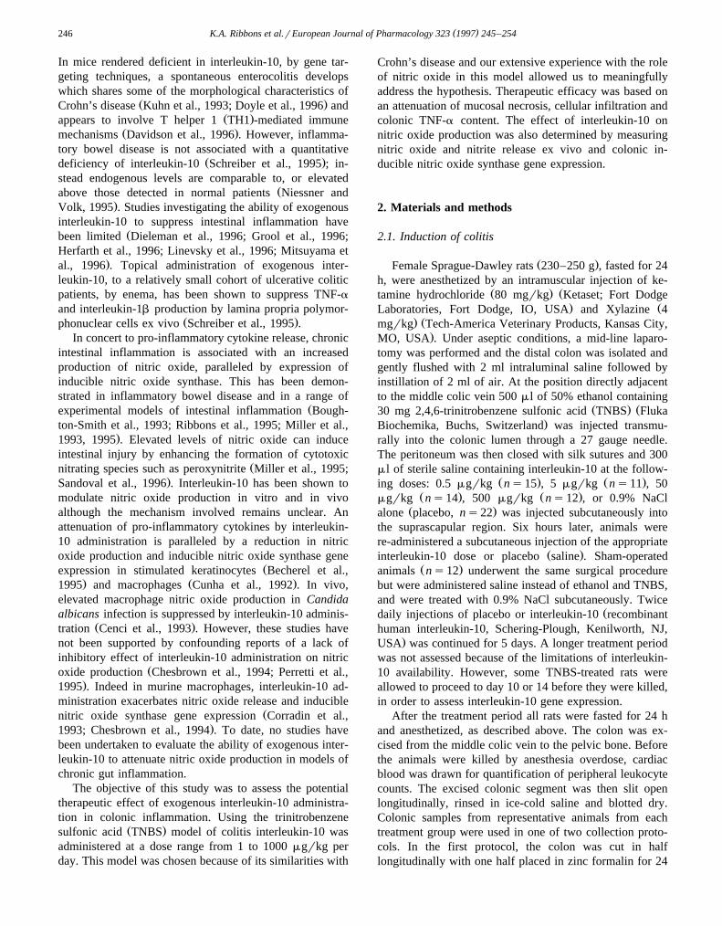
The severity of granulocyte infiltration was quantitatedby assessing colonic myeloperoxidase activity. Associatedwith the transmural colonic inflammatory response, inTNBS colitis, was a significant increase in colonicmyeloperoxidase activity, compared to the sham-operated
Ž .group Fig. 1 . Interleukin 10 administration induced asignificant reduction in colonic myeloperoxidase activitywhen given at doses of 1, 10 and 100 mgrkg per day for 5days compared to placebo-treated animals, although themagnitude of myeloperoxidase activity following inter-leukin-10 treatment was still significantly elevated abovethat observed for sham-operated controls. In contrast, thehighest dose of interleukin-10 administered, 1000 mgrkgper day, failed to attenuate the severity of granulocyteinfiltration and produced a colonic myeloperoxidase activ-
Fig. 1. Colonic myeloperoxidase activity in TNBS-treated rats and theeffects of 5 days interleukin-10 administration. Colonic MPO activity for
Ž . Ž .sham-operated 1st column ns6 , TNBS colitic rats treated with aŽ . Ž .placebo 2nd column ns11 , interleukin-10 administered at 1 mgrkgŽ . Ž . Ž . Ž .per day 3rd column ns8 , 10 mgrkg per day 4th column ns5 ,
Ž . Ž . Ž100 mgrkg per day 5th column ns7 , 1000 mgrkg per day 6th. Ž . )column ns6 are shown. P -0.001 versus sham-operated group;
† P -0.001versus placebo-treated TNBS colitic group and the 1000mgrkg per day interleukin-10-treated TNBS colitic group.
Ž .ity comparable to placebo-treated animals Fig. 1 . Theseresults suggest that interleukin-10 administration displaysanti-inflammatory properties in TNBS colitis by attenuat-
Žing granulocyte infiltration at the lower doses 1–100. Žmgrkg per day ; however, the highest dose assessed 1000.mgrkg per day was ineffective.
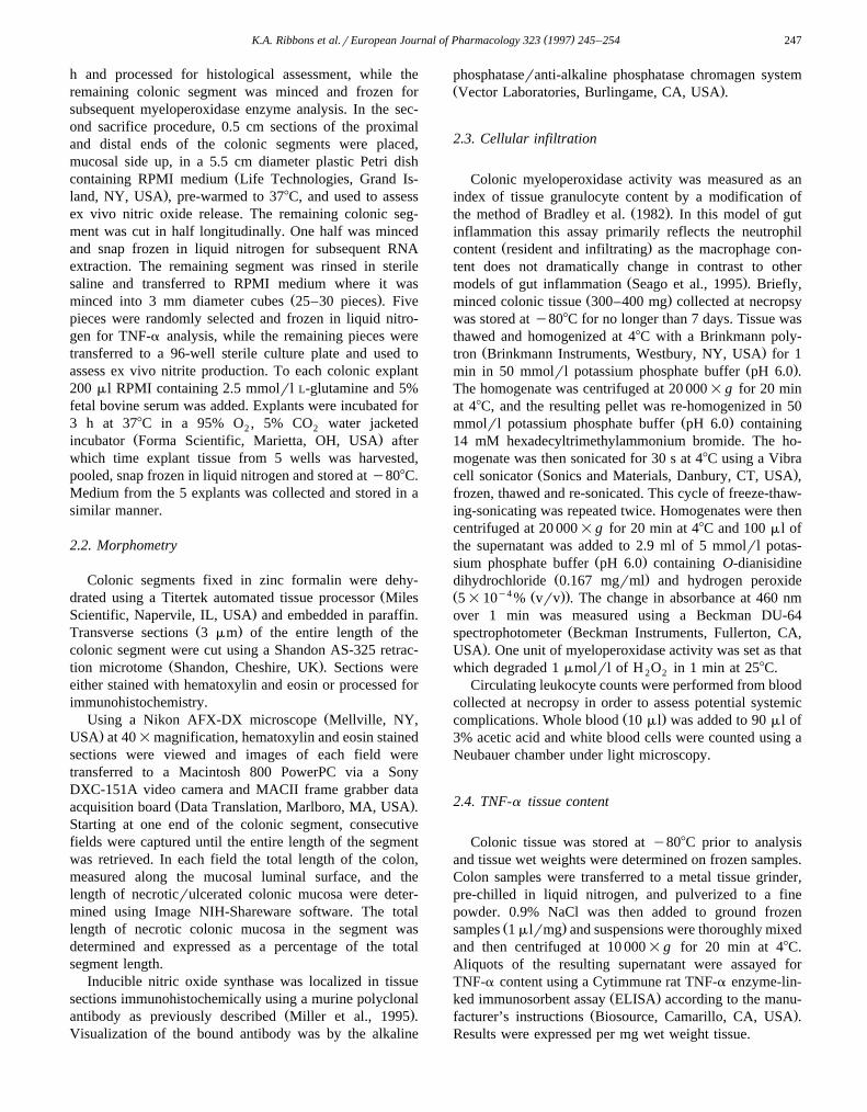
In contrast to the enhanced colonic inflammatory infil-trate, systemic leukocyte counts were not affected by the
Ž .induction of colitis by TNBS Fig. 2 . There was, however,a trend, albeit non-significant, for an increase in the sys-temic leukocyte count in TNBS-treated rats administered
Ž .10, 100 and 1000 mgrkg per day interleukin-10 Fig. 2 .
3.3. TNF-a content
Colonic TNF-a content was measured by ELISA spe-cific for rodent TNF-a. TNBS colitis was associated with
Fig. 2. Total systemic white blood cell count as influenced by TNBScolitis and interleukin-10 administration. Systemic leukocyte count for
Ž . Ž .sham-operated 1st column ns12 , TNBS colitic rats treated with aŽ . Ž .placebo 2nd column ns22 , interleukin-10 administered at 1 mgrkgŽ . Ž . Ž . Ž .per day 3rd column ns15 , 10 mgrkg per day 4th column ns11 ,
Ž . Ž . Ž100 mgrkg per day 5th column ns14 , 1000 mgrkg per day 6th. Ž .column ns12 are shown. No statistical significance was detected
between the groups.
( )K.A. Ribbons et al.rEuropean Journal of Pharmacology 323 1997 245–254250
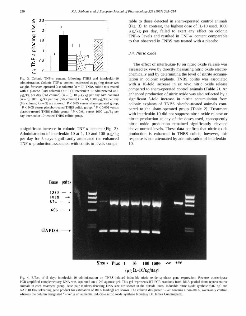
Fig. 3. Colonic TNF-a content following TNBS and interleukin-10administration. Colonic TNF-a content, expressed as pgrmg tissue wet
Ž . Ž .weight, for sham-operated 1st column ns5 , TNBS colitic rats treatedŽ . Ž .with a placebo 2nd column ns11 , interleukin-10 administered at 1Ž . Ž . Ž .mgrkg per day 3rd column ns8 , 10 mgrkg per day 4th column
Ž . Ž . Ž .ns6 , 100 mgrkg per day 5th column ns6 , 1000 mgrkg per dayŽ . Ž . )6th column ns5 are shown. P -0.05 versus sham-operated group;† P -0.05 versus placebo-treated TNBS colitic group; § P -0.001 versusplacebo-treated TNBS colitic group; ¶ P -0.01 versus 1000 mgrkg perday interleukin-10-treated TNBS colitic group.
Ž .a significant increase in colonic TNF-a content Fig. 2 .Administration of interleukin-10 at 1, 10 and 100 mgrkgper day for 5 days significantly attenuated the enhancedTNF-a production associated with colitis to levels compa-
rable to those detected in sham-operated control animalsŽ .Fig. 3 . In contrast, the highest dose of IL-10 used, 1000mgrkg per day, failed to exert any effect on colonicTNF-a levels and resulted in TNF-a content comparableto that observed in TNBS rats treated with a placebo.
3.4. Nitric oxide
The effect of interleukin-10 on nitric oxide release wasassessed ex vivo by directly measuring nitric oxide electro-chemically and by determining the level of nitrite accumu-lation in colonic explants. TNBS colitis was associatedwith a 10-fold increase in ex vivo nitric oxide release
Ž .compared to sham-operated control animals Table 2 . Anenhanced production of nitric oxide was also reflected by asignificant 5-fold increase in nitrite accumulation fromcolonic explants of TNBS placebo-treated animals com-
Ž .pared to the sham-operated group Table 2 . Treatmentwith interleukin-10 did not suppress nitric oxide release ornitrite production at any of the doses used, consequentlynitric oxide production remained significantly elevatedabove normal levels. These data confirm that nitric oxideproduction is enhanced in TNBS colitis; however, thisresponse is not attenuated by administration of interleukin-10.
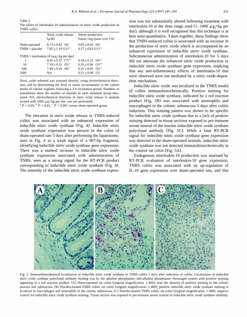
Fig. 4. Effect of 5 days interleukin-10 administration on TNBS-induced inducible nitric oxide synthase gene expression. Reverse transcriptasePCR-amplified complementary DNA was separated on a 2% agarose gel. This gel represents RT-PCR reactions from RNA pooled from representative
Ž .animals in each treatment group. Base pair markers denoting DNA size are shown in the outside lanes. Inducible nitric oxide synthase 907 bp andŽ .GAPDH housekeeping gene product for estimation of RNA loading are shown. The column designated ‘–ve’ contains a non-DNA, water-only control,
Ž .whereas the column designated ‘qve’ is an authentic inducible nitric oxide synthase courtesy Dr. James Cunningham .
( )K.A. Ribbons et al.rEuropean Journal of Pharmacology 323 1997 245–254 251
Table 2The effect of interleukin-10 administration on nitric oxide production inTNBS colitis
Nitric oxide release Nitrite productionŽ . Ž .mM nmolrmg tissue over 3 h
Ž . Ž .Sham-operated 0.73"0.43 6 0.05"0.02 6c aŽ . Ž .TNBSqplacebo 7.05"1.19 11 0.27"0.03 11
Ž .TNBSqinterleukin-10 mgrkg per dayb aŽ . Ž .1 4.20"0.37 7 0.34"0.12 4c bŽ . Ž .10 7.10"0.32 5 0.35"0.09 5c aŽ . Ž .100 7.48"0.36 8 0.31"0.05 6
Ž .1000 NA 0.21"0.08 5
Nitric oxide released was assessed directly, using electrochemical detec-tion, and by determining the level of nitrite accumulation in the culturemedia of colonic explants following a 3 h incubation period. Numbers inparentheses show the number of animals in each treatment group mea-sured. NA, electrochemical detection of nitric oxide release in animalstreated with 1000 mgrkg per day was not performed.a P -0.05, b P -0.01, c P -0.001 versus sham-operated group.
The elevation in nitric oxide release in TNBS-inducedcolitis was associated with an enhanced expression of
Ž .inducible nitric oxide synthase Fig. 4 . Inducible nitricoxide synthase expression was present in the colon ofsham-operated rats 5 days after performing the laparotomy,seen in Fig. 4 as a weak signal of a 907-bp fragment,identifying inducible nitric oxide synthase gene expression.There was a marked increase in inducible nitric oxidesynthase expression associated with administration ofTNBS, seen as a strong signal for the RT-PCR product
Ž .corresponding to inducible nitric oxide synthase Fig. 4 .The intensity of the inducible nitric oxide synthase expres-
sion was not substantially altered following treatment withŽinterleukin-10 at the dose range used 1–1000 mgrkg per
.day , although it is well recognised that this technique is atbest semi-quantitative. Taken together, these findings showthat TNBS-induced colitis is associated with an increase inthe production of nitric oxide which is accompanied by anenhanced expression of inducible nitric oxide synthase.Subcutaneous administration of interleukin-10 for 5 daysdid not attenuate the enhanced nitric oxide production orinducible nitric oxide synthase gene expression, implyingthat any anti-inflammatory effects of interleukin-10 thatwere observed were not mediated by a nitric oxide-depen-dent mechanism.
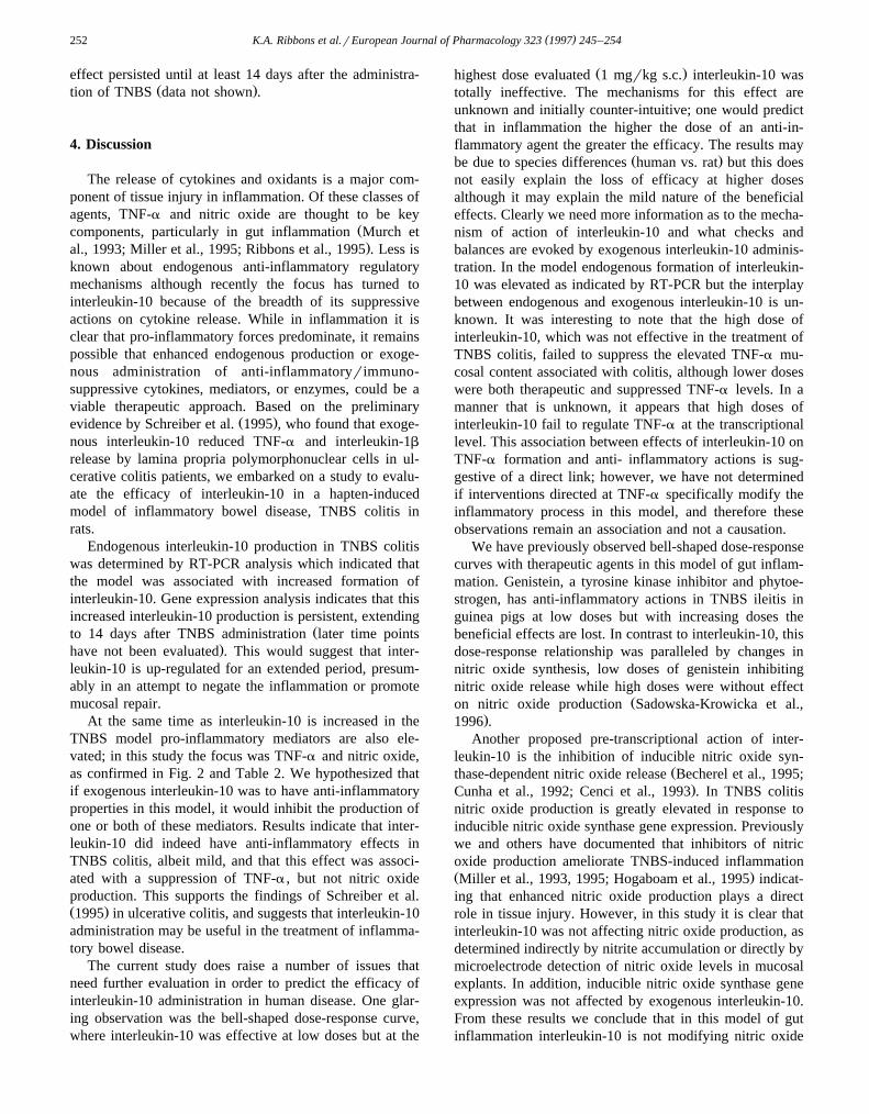
Inducible nitric oxide was localized in the TNBS modelof colitis immunohistochemically. Positive staining forinducible nitric oxide synthase, indicated by a red reaction
Ž .product Fig. 5B was associated with neutrophils andmacrophages in the colonic submucosa 5 days after colitisinduction. This staining pattern was shown to be specificfor inducible nitric oxide synthase due to a lack of positivestaining detected in tissue sections exposed to pre-immuneserum instead of the murine inducible nitric oxide synthase
Ž .polyclonal antibody Fig. 5C . While a faint RT-PCRsignal for inducible nitric oxide synthase gene expressionwas detected in the sham-operated animals, inducible nitricoxide synthase was not detected immunohistochemically in
Ž .the control rat colon Fig. 5A .Endogenous interleukin-10 production was assessed by
RT-PCR evaluation of interleukin-10 gene expression.TNBS colitis was associated with an up-regulation ofIL-10 gene expression over sham-operated rats, and this
Fig. 5. Immunohistochemical localization of inducible nitric oxide synthase in TNBS colitis 5 days after induction of colitis. Localization of induciblenitric oxide synthase polyclonal antibody binding was by the alkaline phosphataseranti-alkaline phosphatase chromagen system with positive staining
Ž . Ž .appearing as a red reaction product. A Sham-operated rat colon original magnification =400 ; note the absence of positive staining in the colonicŽ . Ž .mucosa and submucosa. B Placebo-treated TNBS colitic rat colon original magnification =400 ; positive inducible nitric oxide synthase staining is
Ž . Ž .localized in macrophages and neutrophils in the colonic submucosa. C Placebo-treated TNBS colitic rat colon original magnification =400 ; negativecontrol for inducible nitric oxide synthase staining. Tissue section was exposed to pre-immune serum instead of inducible nitric oxide synthase antibody.
( )K.A. Ribbons et al.rEuropean Journal of Pharmacology 323 1997 245–254252
effect persisted until at least 14 days after the administra-Ž .tion of TNBS data not shown .
4. Discussion
The release of cytokines and oxidants is a major com-ponent of tissue injury in inflammation. Of these classes ofagents, TNF-a and nitric oxide are thought to be key
Žcomponents, particularly in gut inflammation Murch et.al., 1993; Miller et al., 1995; Ribbons et al., 1995 . Less is
known about endogenous anti-inflammatory regulatorymechanisms although recently the focus has turned tointerleukin-10 because of the breadth of its suppressiveactions on cytokine release. While in inflammation it isclear that pro-inflammatory forces predominate, it remainspossible that enhanced endogenous production or exoge-nous administration of anti-inflammatoryrimmuno-suppressive cytokines, mediators, or enzymes, could be aviable therapeutic approach. Based on the preliminary
Ž .evidence by Schreiber et al. 1995 , who found that exoge-nous interleukin-10 reduced TNF-a and interleukin-1b
release by lamina propria polymorphonuclear cells in ul-cerative colitis patients, we embarked on a study to evalu-ate the efficacy of interleukin-10 in a hapten-inducedmodel of inflammatory bowel disease, TNBS colitis inrats.
Endogenous interleukin-10 production in TNBS colitiswas determined by RT-PCR analysis which indicated thatthe model was associated with increased formation ofinterleukin-10. Gene expression analysis indicates that thisincreased interleukin-10 production is persistent, extending
Žto 14 days after TNBS administration later time points.have not been evaluated . This would suggest that inter-
leukin-10 is up-regulated for an extended period, presum-ably in an attempt to negate the inflammation or promotemucosal repair.
At the same time as interleukin-10 is increased in theTNBS model pro-inflammatory mediators are also ele-vated; in this study the focus was TNF-a and nitric oxide,as confirmed in Fig. 2 and Table 2. We hypothesized thatif exogenous interleukin-10 was to have anti-inflammatoryproperties in this model, it would inhibit the production ofone or both of these mediators. Results indicate that inter-leukin-10 did indeed have anti-inflammatory effects inTNBS colitis, albeit mild, and that this effect was associ-ated with a suppression of TNF-a , but not nitric oxideproduction. This supports the findings of Schreiber et al.Ž .1995 in ulcerative colitis, and suggests that interleukin-10administration may be useful in the treatment of inflamma-tory bowel disease.
The current study does raise a number of issues thatneed further evaluation in order to predict the efficacy ofinterleukin-10 administration in human disease. One glar-ing observation was the bell-shaped dose-response curve,where interleukin-10 was effective at low doses but at the
Ž .highest dose evaluated 1 mgrkg s.c. interleukin-10 wastotally ineffective. The mechanisms for this effect areunknown and initially counter-intuitive; one would predictthat in inflammation the higher the dose of an anti-in-flammatory agent the greater the efficacy. The results may
Ž .be due to species differences human vs. rat but this doesnot easily explain the loss of efficacy at higher dosesalthough it may explain the mild nature of the beneficialeffects. Clearly we need more information as to the mecha-nism of action of interleukin-10 and what checks andbalances are evoked by exogenous interleukin-10 adminis-tration. In the model endogenous formation of interleukin-10 was elevated as indicated by RT-PCR but the interplaybetween endogenous and exogenous interleukin-10 is un-known. It was interesting to note that the high dose ofinterleukin-10, which was not effective in the treatment ofTNBS colitis, failed to suppress the elevated TNF-a mu-cosal content associated with colitis, although lower doseswere both therapeutic and suppressed TNF-a levels. In amanner that is unknown, it appears that high doses ofinterleukin-10 fail to regulate TNF-a at the transcriptionallevel. This association between effects of interleukin-10 onTNF-a formation and anti- inflammatory actions is sug-gestive of a direct link; however, we have not determinedif interventions directed at TNF-a specifically modify theinflammatory process in this model, and therefore theseobservations remain an association and not a causation.
We have previously observed bell-shaped dose-responsecurves with therapeutic agents in this model of gut inflam-mation. Genistein, a tyrosine kinase inhibitor and phytoe-strogen, has anti-inflammatory actions in TNBS ileitis inguinea pigs at low doses but with increasing doses thebeneficial effects are lost. In contrast to interleukin-10, thisdose-response relationship was paralleled by changes innitric oxide synthesis, low doses of genistein inhibitingnitric oxide release while high doses were without effect
Žon nitric oxide production Sadowska-Krowicka et al.,.1996 .
Another proposed pre-transcriptional action of inter-leukin-10 is the inhibition of inducible nitric oxide syn-
Žthase-dependent nitric oxide release Becherel et al., 1995;.Cunha et al., 1992; Cenci et al., 1993 . In TNBS colitis
nitric oxide production is greatly elevated in response toinducible nitric oxide synthase gene expression. Previouslywe and others have documented that inhibitors of nitricoxide production ameliorate TNBS-induced inflammationŽ .Miller et al., 1993, 1995; Hogaboam et al., 1995 indicat-ing that enhanced nitric oxide production plays a directrole in tissue injury. However, in this study it is clear thatinterleukin-10 was not affecting nitric oxide production, asdetermined indirectly by nitrite accumulation or directly bymicroelectrode detection of nitric oxide levels in mucosalexplants. In addition, inducible nitric oxide synthase geneexpression was not affected by exogenous interleukin-10.From these results we conclude that in this model of gutinflammation interleukin-10 is not modifying nitric oxide
( )K.A. Ribbons et al.rEuropean Journal of Pharmacology 323 1997 245–254 253
release, and that any observed anti-inflammatory actionsare nitric oxide-independent.
In summary, endogenous interleukin-10 gene expres-sion was elevated in TNBS colitis and exogenous inter-leukin-10 administration resulted in an attenuation of gran-ulocyte infiltration but did not affect the extent of mucosalnecrosis or weight loss associated with this model. Themild anti-inflammatory effects produced by interleukin-10may be due to the suppression of TNF-a production butwere clearly independent of effects on nitric oxide releaseand inducible nitric oxide synthase gene expression. Anunexpected finding was the loss of beneficial effects ofinterleukin-10 at high doses. These results are supportiveof a potential therapeutic application of exogenous inter-leukin-10 in human inflammatory bowel disease but addi-tional studies are needed to facilitate understanding of themechanism of action of interleukin-10 and the unusualdose-response relationships.
Acknowledgements
The authors wish to thank Dr. Xiao-Jing Zhang forassistance with immunohistochemistry. This work wasfunded in part from a grant awarded to M.J.S.M.: NIH R01HD 31885.
References
Becherel, P.A., L. Le Goff, S. Ktorza, F. Ouaaz, J.-M. Mencia-Huerta, B.Dugas, P. Debre, M.D. Mossalayi and M. Arock, 1995, Interleukin-10inhibits IgE-mediated nitric oxide synthase induction and cytokinesynthesis in normal human keratinocytes, Eur. J. Immunol. 25, 2992.
Boughton-Smith, N.K., S.M. Evans, C.J. Hawkey, A.T. Cole, M. Balsitis,N.J.R. Whittle and S. Moncada, 1993, Nitric oxide synthase activityin ulcerative colitis and Crohn’s disease, Lancet 341, 338.
Bradley, P.P., D.A. Priebat, R.D. Christensen and G. Rothstein, 1982,Measurement of cutaneous inflammation: estimation of neutrophilcontent with an enzyme marker, J. Invest. Dermatol. 78, 206.
Cassatella, M.A., L. Meda, S. Bonoro, M. Ceska and G. Constantin,Ž .1993, Interleukin 10 IL-10 inhibits the release of pro inflammatory
cytokines from human polymorphonuclear leukocytes. Evidence foran autocrine role of tumor necrosis factor and IL-1B in mediating theproduction of IL-8 triggered by lipopolysaccharide, J. Exp. Med. 178,2207.
Cassatella, M.A., L. Meda, S. Gasperini, F. Calzetti and S. Bonora, 1994,Ž .Interleukin 10 IL-10 upregulates IL-1 receptor antagonist production
from lipopolysaccharide-stimulated human polymorphonuclear leuko-cytes by delaying mRNA degradation, J. Exp. Med. 179, 1695.
Cenci, E., L. Romani, A. Mencacci, R. Spaccapelo, E. Schiafella, P.Puccetti and F. Bistoni, 1993, Interleukin-4 and interleukin-10 inhibitnitric oxide-dependent macrophage killing of Candida albicans, Eur.J. Immunol. 23, 1034.
Chesbrown, S.E., J. Monnier, G. Visner and H.S. Nick, 1994, Regulationof inducible nitric oxide synthase mRNA levels by LPS, IFN-g,TGF-b and IL-10 in murine macrophage cell lines and rat peritonealmacrophages, Biochem. Biophys. Res. Commun. 200, 126.
Clinchy, B., P. Bjorck, S. Paulie and G. Moller, 1994, Interleukin-10inhibits motility in murine and human lymphocytes, Immunology 82,376.
Corradin, S.B., N. Fasel, Y. Buchmuller-Rouiller, A. Ransijn, J. Smithand J. Mauel, 1993, Induction of macrophage nitric oxide productionby interferon-g and tumor necrosis factor-a is enhanced by inter-leukin-10, Eur. J. Immunol. 23, 2045.
Ž .Cunha, F.Q., S. Moncada and F.Y. Liew, 1992, Interleukin-10 IL-10inhibits the induction of nitric oxide synthase by interferon-g inmurine macrophages, Biochem. Biophys. Res. Commun. 182, 1155.
Davidson, N.J., M.W. Leach, M.M. Fort, L. Thompson-Snipes, R. Kuhn,W. Muller, D.J. Berg and D.M. Rennick, 1996, T helper cell 1-typeCD4q cells, but not B cells, mediated colitis in interleukin 10-defi-cient mice, J. Exp. Med. 184, 241.
De Waal Malefyt, R., J. Abrams, B. Bennet, C.G. Fidgor and J.E. DeŽ .Vries, 1991, Interleukin 10 IL-10 cytokine synthesis by human
monocytes: an autoregulatory role of IL-10 produced by monocytes,J. Exp. Med. 174, 1209.
Dieleman, L.A., Z. De Rover, E. Bloemena, M.B. Van der Ende, A.S.Pena and E.P. Van Rees, 1996, Effect of interleukin-10 in dextransulfate sodium-induced colitis, Gastroenterology 110, A895.
Doyle, J.S.G., L.D. Jewell, K.L. Madsen, T. Mosmann and R.N. Fedorak,1996, Intestinal histopathology in interleukin-10 gene knock-out miceresembles Crohn’s disease, Gastroenterology 110, A898.
Fiorentino, D.F., A. Zlotnik, P. Viera, T.R. Mosmann, M. Howard, K.W.Moore and A. O’Garra, 1991a, IL-10 acts on the antigen-presentingcell to inhibit cytokine production by Th1 cells, J. Immunol. 146,3444.
Fiorentino, D.F., A. Zlotnik, T.R. Mosmann, M. Howard and A. O’Garra,1991b, IL-10 inhibits cytokine production by activated macrophages,J. Immunol. 147, 3815.
Goldman, M. and T. Velu, 1995, Interleukin-10 and its implications forimmunopathology, Adv. Neph. 24, 79.
Grool, T.A., J. Meenan, H. Van Dullemen, F. Koster, F.J.W. Ten Kate,A. Lebeaut, G.N.J. Tytgat and S.J.H. Van Deventer, 1996, Theanti-inflammatory effect of interleukin 10 in a rabbit model of im-mune complex-induced colitis, Gastroenterology 110, A918.
Herfarth, H., R. Janardhanam, H.C. Rath and R.B. Sartor, 1996, In vivoIL-10 treatment suppresses experimental chronic granulomatous in-flammation and has an additive effect with corticosteroids, Gastroen-
Ž .terology 110 40 , A924.Hogaboam, C.M., R. Jacobsen, S.M. Collins and M.G. Blennerhassett,
1995, The selective beneficial effects of nitric oxide inhibition inexperimental colitis, Am. J. Physiol. 268, G673.
Kuhn, R., J. Lohler, D. Rennick, K. Rajewsky and W. Muller, 1993,Interleukin-10-deficient mice develop chronic enterocolitis, Cell 75,263.
Linevsky, J.K., G.G. Morton, I. Castagiuolo, C. Pothoulakis, S. Ketes,J.T. LaMont and C.P. Kelly, 1996, IL-10 inhibits Clostridium difficiletoxin A-induced enteritis, Gastroenterology 110, A950.
Miller, M.J.S., H. Sadowska-Krowicka, S. Chotinareumol, J.L. Kakkisand D.A. Clark, 1993, Amelioration of chronic ileitis by nitric oxidesynthase inhibition, J. Pharmacol. Exp. Ther. 264, 11.
Miller, M.J.S., J.H. Thompson, X.-J. Zhang, H. Sadowska-Krowicka, J.L.Kakkis, U.K. Munshi, M. Sandoval, J.L. Rossi, S. Eloby-Childress,J.S. Beckmann, Y.-Z. Ye, C.P. Rodi, P.T. Manning, M.G. Currie andD.A. Clark, 1995, Role of inducible nitric oxide synthase expressionand peroxynitrite formation in guinea pig ileitis, Gastroenterology109, 1475.
Miller, M.J.S., C.A. Voelker, S. Olister, J.H. Thompson, X.-J. Zhang, D.Rivera, S. Eloby-Childress, X. Liu, D.A. Clark and M.R. Pierce,1996, Fetal growth retardation in rats may result from apoptosis: roleof peroxynitrite, Free Radic. Biol. Med. 21, 619.
Mitsuyama, K., M. Tomoyose, A. Toyonaga, K. Tanikawa and H. Ishida,1996, Interleukin-10 is protective in a murine model of experimentalcolitis, Gastroenterology 110, A971.
Murch, S.H., C.P. Braegger, J.A. Walker-Smith and T.T. MacDonald,1993, Location of tumour necrosis factor a by immunohistochemistryin chronic inflammatory bowel disease, Gut 34, 1705.
Niessner, M. and B.A. Volk, 1995, Altered Th1rTh2 cytokine profiles in
( )K.A. Ribbons et al.rEuropean Journal of Pharmacology 323 1997 245–254254
the intestinal mucosa of patients with inflammatory bowel disease asassessed by quantitive reverse transcriptase polymerase chain reactionŽ .RT-PCR , Clin. Exp. Immunol. 101, 428.
Olson, A.D., M. Ayass and S. Chensue, 1993, Tumor necrosis factor andIL-1b expression in pediatric patients with inflammatory bowel dis-ease, J. Pediatr. Gastroenterol. Nutr. 16, 241.
Perretti, M., C. Szabo and C. Thiemermann, 1995, Effect of interleukin-4and interleukin-10 on leucocyte migration and nitric oxide productionin the mouse, Br. J. Pharmacol. 116, 2251.
Reinecker, H.C., M. Steffen, T. Witthoeft, I. Pflueger, S. Schreiber, R.P.MacDermott and A. Raedler, 1993, Enhanced secretion of tumornecrosis factor-alpha, IL-6, and IL-1 beta by isolated lamina propriamononuclear cells from patients with ulcerative colitis and Crohn’sdisease, Clin. Exp. Immunol. 94, 174.
Ribbons, K.A., X.-J. Zhang, J.H. Thompson, S.S. Greenberg, W.M.Moore, C.M. Kornmeier, M.G. Currie, N.W. Lerche, D.A. Clark andM.J.S. Miller, 1995, Increased nitric oxide synthesis in colitic rhesusmacaques, Gastroenterology 108, 705.
Richter, G., S. Krugger-Krasagakes, G. Hein, C. Huls, E. Schmitt, T.Diamantstein and T. Blankenstein, 1993, Interleukin 10 transfectedinto Chinese hamster ovary cells prevents tumor growth andmacrophage infiltration, Cancer Res. 53, 4134.
Sadowska-Krowicka, H., E.E. Mannick, P.D. Oliver, M. Sandoval, X.-J.Zhang, S. Eloby-Childress, D.A. Clark and M.J.S. Miller, 1996,Genistein and gut inflammation: role of nitric oxide, Proc. Soc. Exp.
Ž .Biol. Med. in press .Sandoval, M., X.-J. Zhang, X. Liu, E.E. Mannick, D.A. Clark and M.J.S.
Miller, 1996, Peroxynitrite-induced apoptosis in T84 and RAW 264.7Ž .cells: attenuation by ascorbic acid, Free Radic. Biol. Med. in press .
Schreiber, S., T. Heinig, H.-G. Thiele and A. Raedler, 1995,Immunoregu-latory role of interleukin 10 in patients with inflammatory boweldisease, Gastroenterology 108, 1434.
Seago, N.D., J.H. Thompson, X.-J. Zhang, S. Eloby-Childress, H. Sad-owska-Krowicka, J.L. Rossi, M.G. Currie, P.T. Manning, D.A. Clarkand M.J.S. Miller, 1995, Inducible nitric oxide synthase and guinea-pigileitis induced by adjuvant, Mediators Inflamm. 4, 19.
Seitz, M., P. Loetscher, B. Dewald, H. Towbin, H. Gallati and M.Baggiolini, 1995, Interleukin-10 differentially regulates cytokine in-hibitor and chemokine release from blood mononuclear cells andfibroblasts, Eur. J. Immunol. 25, 1129.
Stevens, C., G. Walz., C. Singaram, M.L. Lipman, B. Zanker, A. Muggia,D. Antonioli, M.A. Peppercorn and T.B. Strom, 1992, Tumor necrosisfactor-a , interleukin-1b, and interleukin-6 expression in inflammatorybowel disease, Dig. Dis. Sci. 37, 818.