miR-223 Deficiency Increases Eosinophil Progenitor Proliferation
Upload
independentCategory
view
1download
0
ORIGINAL ARTICLE
Eosinophil-mediated signalling attenuatesinflammatory responses in experimental colitis
Joanne C Masterson,1,2,3 Eóin N McNamee,2,3,4 Sophie A Fillon,1,2,3
Lindsay Hosford,1,2,3 Rachel Harris,1,2,3 Shahan D Fernando,1,2,3 Paul Jedlicka,3,5
Ryo Iwamoto,6 Elizabeth Jacobsen,7,8 Cheryl Protheroe,7,8 Holger K Eltzschig,2,3,4
Sean P Colgan,2,3,9 Makoto Arita,6,7 James J Lee,8 Glenn T Furuta1,2,3
▸ Additional material ispublished online only. To viewplease visit the journal online(http://dx.doi.org/10.1136/gutjnl-2014-306998).
For numbered affiliations seeend of article.
Correspondence toDr Glenn T Furuta, Section ofPediatric Gastroenterology,Hepatology and Nutrition,Department of Pediatrics,University of Colorado Schoolof Medicine, 13123 East16th Ave, B290, Aurora,CO 80045, USA;[email protected]
Received 11 February 2014Revised 18 August 2014Accepted 19 August 2014
To cite: Masterson JC,McNamee EN, Fillon SA,et al. Gut Published OnlineFirst: [please include DayMonth Year] doi:10.1136/gutjnl-2014-306998
ABSTRACTObjective Eosinophils reside in the colonic mucosaand increase significantly during disease. Although anumber of studies have suggested that eosinophilscontribute to the pathogenesis of GI inflammation, theexpanding scope of eosinophil-mediated activitiesindicate that they also regulate local immune responsesand modulate tissue inflammation. We sought to definethe impact of eosinophils that respond to acute phasesof colitis in mice.Design Acute colitis was induced in mice byadministration of dextran sulfate sodium, 2,4,6-trinitrobenzenesulfonic acid or oxazolone to C57BL/6J(control) or eosinophil deficient (PHIL) mice. Eosinophilswere also depleted from mice using antibodies againstinterleukin (IL)-5 or by grafting bone marrow from PHILmice into control mice. Colon tissues were collected andanalysed by immunohistochemistry, flow cytometry andreverse transcription PCR; lipids were analysed by massspectroscopy.Results Eosinophil-deficient mice developedsignificantly more severe colitis, and their colon tissuescontained a greater number of neutrophils, thancontrols. This compensatory increase in neutrophils wasaccompanied by increased levels of the chemokinesCXCL1 and CXCL2, which attract neutrophils. Lipidomicanalyses of colonic tissue from eosinophil-deficient miceidentified a deficiency in the docosahexaenoic acid-derived anti-inflammatory mediator 10, 17-dihydroxydocosahexaenoic acid (diHDoHE), namelyprotectin D1 (PD1). Administration of an exogenousPD1-isomer (10S, 17S-DiHDoHE) reduced the severity ofcolitis in eosinophil-deficient mice. The PD1-isomer alsoattenuated neutrophil infiltration and reduced levels oftumour necrosis factor-α, IL-1β, IL-6 and inducible NO-synthase in colons of mice. Finally, in vitro assaysidentified a direct inhibitory effect of PD1-isomer onneutrophil transepithelial migration.Conclusions Eosinophils exert a protective effect inacute mouse colitis, via production of anti-inflammatorylipid mediators.
INTRODUCTIONEosinophils synthesise and release a broad range ofbiologically active mediators, including antimicro-bial, proremodelling or anti-inflammatory asso-ciated factors (reviewed in1 2). An increasing bodyof evidence suggests that eosinophils play a primary
role in a number of diseases such as asthma3–5 andeosinophilic GI diseases.6–8 In these diseases, eosi-nophils increase within the mucosal surfaces wherethey may participate in processes that often contrib-ute to tissue damage9 10 and/or remodelling,11
either way leading to eventual organ dysfunction.Acute inflammatory/immune reparative responses
are necessary self-limited processes that participatein maintaining host health and tissue function.Mucosal surfaces use and amplify acute restorativeanti-inflammatory activities through a number ofmechanisms including anti-inflammatory lipid
Significance of this study
What is already known on this subject?▸ IBD is a disease characterised by increased
numbers of eosinophils.▸ The exact role of eosinophils during the onset
of inflammation and in chronic disease remainsunclear.
What are the new findings?▸ In the absence of eosinophils, mice succumb to
a greater colonic inflammation during acuteexperimental models of colitis.
▸ Eosinophil-deficient mice develop an exuberantand compensatory neutrophilic inflammation ofthe colon, accompanied by greater increases inlevels of proinflammatory molecules.
▸ Eosinophil-deficient mice have diminishedlevels of arachidonate-15-lipoxygenase, thelipid biosynthetic enzyme, and the proresolvinglipid mediator protectin D1 during acute colonicinflammation, and administration of protectinD1-isomer to eosinophil-deficient miceattenuates inflammatory indices of colitis,including neutrophilic infiltration.
How might it impact on clinical practice inthe foreseeable future?▸ Since eosinophils are normal residents of the
colonic mucosa, further studies are necessaryto determine eosinophil’s role in health anddisease. Protective factors released or regulatedby eosinophils may be beneficial to colonichealth.
Masterson JC, et al. Gut 2014;0:1–12. doi:10.1136/gutjnl-2014-306998 1
Inflammatory bowel disease Gut Online First, published on September 10, 2014 as 10.1136/gutjnl-2014-306998
Copyright Article author (or their employer) 2014. Produced by BMJ Publishing Group Ltd (& BSG) under licence.
group.bmj.com on September 10, 2014 - Published by gut.bmj.comDownloaded from

mediator synthesis.12 13 In this regard, omega-3 polyunsaturatedfatty acid (PUFA)-derived resolvins and protectins are potentanti-inflammatory lipid mediators. For example, mice ingestingchow rich in endogenous omega-3 fatty acids have less severecolitis.14 By contrast, increased consumption of dietary proin-flammatory omega-6 PUFA correlates with an increase in IBDincidence.15 16 Thus, dietary omega-3 fatty acid-derived anti-inflammatory molecules have emerged as potent options fortherapeutic intervention.17–20 Docosahexaenoic acid (DHA), anomega-3 PUFA abundant in fish oils, is converted to hydroxydo-cosahexaenoic acids (HDoHE), by enzymatic oxidation.Protectin D1 (PD1) (10,17- dihydroxydocosahexaenoic acid(DiHDoHE)) is biosynthesised by 15-lipoxygenase (LOX) inhumans and 12/15-LOX in mice, and present in inflamedtissues.21–24 Recent reports show that both human and mouseeosinophils express high levels of leucocyte-type 15-LOX and12/15-LOX, respectively, and are capable of producing PD1when activated in vitro.24 25 Other cells with the potential toexpress 12/15-LOX include dendritic cells, tissue-residentmacrophage, mast cells and epithelial cells.21 While the presenceof the enzyme is necessary, it is not always sufficient for PD1biosynthesis, as expression may be regulated by environmentalfactors, such as cytokines or cell differentiation/activationstates.21 Administration of exogenous PD1 to mouse models ofinflammation protects tissues from inflammatory damage,expediting phagocytic resolution of acute peritonitis and retinalinjury models, attenuating inflammation and airway hyper-responsiveness in mouse asthma models, while mouse eosino-phils contribute to the resolution of acute peritonitis in a12/15-LOX-dependent manner.12 22–24 26 To date, the receptorand, therefore, cellular target for PD1 has not been identified.
Expression of these lipid mediators, association with coloniceosinophils and their role in resolution responses during colonicinflammation is incompletely understood. The aim of our studywas to determine the role(s) of eosinophils in mouse models ofacute colitis. Here we provide evidence for a novel protectiverole of eosinophils and concurrently show that this protectionalso correlates with the induced levels of 12/15-LOX-derivedanti-inflammatory lipid mediators. Results from these studieshave broad implications on potential therapeutic options forpatients with IBD.
MATERIALS AND METHODSMiceAll studies were performed with female C57BL/6J mice (wildtype, WT) and PHIL.27 Eosinophil depletion studies were com-pleted using a monoclonal antibody specific for interleukin-5(TRFK-5; Cayman Chemical, Ann Arbor, Michigan, USA). Ageand sex-matched C57BL/6J littermate mice were used as con-trols. The University of Colorado and Mayo Clinic ArizonaIACUC approved studies.
Dextran sodium sulfate, 2,4,6-trinitrobenzenesulfonic acidand Oxazolone-induced colitisColitis was induced by administering dextran sodium sulfate(DSS) (2.5%–3.5%(w/v)) 36 000–50 000 kDa; (MP Biomedicals,Santa Ana, California, USA), trinitrobenzenesulfonic acid(TNBS) or oxazolone (OXA) (Sigma, St Louis, Missouri, USA) to10-week old mice for up to 6–8 days. Daily assessments ofdisease activity index (DAI) (ie, weight loss, stool consistency andfaecal blood) and postnecropsy colon lengths were measured aspreviously described.28
In some studies, the commercially available DHA-derivedPD1 isomer (10S, 17S-diHDoHE)23 29 (hereafter referred to as
PD1-isomer) (0.05 mg/kg) (Cayman Chemical, Ann Arbor,Michigan, USA) was administered to mice.
Generation of bone marrow chimeric miceBone marrow chimeric mice were generated by transferring iso-lated bone marrow from PHIL (PHIL->WT) or WT(WT->WT) donor mice into the retro-orbital plexus of subleth-ally irradiated WTrecipients, as described previously.30
Tissue processing and histological analysisWhole length colons were removed, processed and a globalinflammatory score was developed from H&E stained sectionsmodified to account for area of tissue affected from thatdescribed previously.28
Immunohistochemical assessment of eosinophil,myeloperoxidase or F4/80 cellular inflammationEosinophils were localised in mouse and human tissues(Institutional Review Board approved) by eosinophil major basicprotein-1 (MBP-1) and eosinophil peroxidase immunohisto-chemistry as previously described.11 31 Neutrophils were loca-lised in mouse tissues by myeloperoxidase (MPO)immunohistochemistry. Macrophage and other F4/80+ cellswere localised in mouse tissues by F4/80 immunohistochemistry.
In situ hybridisation assessment of CXCL1 productionFormalin-fixed paraffin-embedded sections were subjected toin situ hybridisation with probes targeted to the CXCL1 mRNAas per manufacturer’s recommendations (Advanced CellDiagnostics, Hayward, California, USA). Slight modificationsare detailed in online supplementary methods. Probes directedtowards Ubc and DapB were used as positive and negative con-trols, respectively.
Colonic lamina propria and bone marrow leucocyte isolationfor flow cytometryLeucocytes were isolated from colonic mucosa by collagenasedigestion as previously described.11 30 32 Bone marrow cellswere isolated from femurs and tibias.33 Viable single cell suspen-sions were subjected to flow cytometric analysis as previouslydescribed.32
RNA isolation and real-time RT-PCRTaqman gene expression assays (Applied Biosystems, Foster City,California, USA) were performed on whole colon RNA, pro-cessed as previously described.11 Data were normalised to 18Sand calculated as relative quantity (2−ΔΔCt, where Ct is cyclethreshold for each sample).
Colon lipidomic analysisColon tissues were harvested and processed for liquid chroma-tography tandem mass spectrometry (LC-MS/MS) based lipido-mics analyses as previously described.12 MS/MS analyses wereconducted in negative ion mode, and fatty acid metabolites wereidentified and quantified by multiple reaction monitoring.
Cell culture and in vitro transmigration assayCaco-2 human intestinal epithelial cells were cultured and poly-morphonuclear (PMN) cell transmigration towards a gradient of100 nM Formyl–Methionyl–Leucyl–Phenylalanine (FMLP;Sigma, St Louis, Missouri, USA) was performed as previouslydescribed.34 35 PMNs were added in the presence of 0.2 or20 nM of PD1-isomer or vehicle alone.
2 Masterson JC, et al. Gut 2014;0:1–12. doi:10.1136/gutjnl-2014-306998
Inflammatory bowel disease
group.bmj.com on September 10, 2014 - Published by gut.bmj.comDownloaded from

Statistical analysisStatistical analyses of data outcomes were performed by Studentt test, one-way analysis of variance or Log-rank (Mantel–Cox)test (conservative) survival curve where appropriate. Data areexpressed as means±SEM. A p value of ≤0.05 was considered asstatistical significance although in some cases higher levels ofsignificance are noted and described in the figure legendswhere applicable: *†p≤0.05, ††p≤0.01, †††p≤0.001.
Further detail can be found in online supplementary materialsand methods.
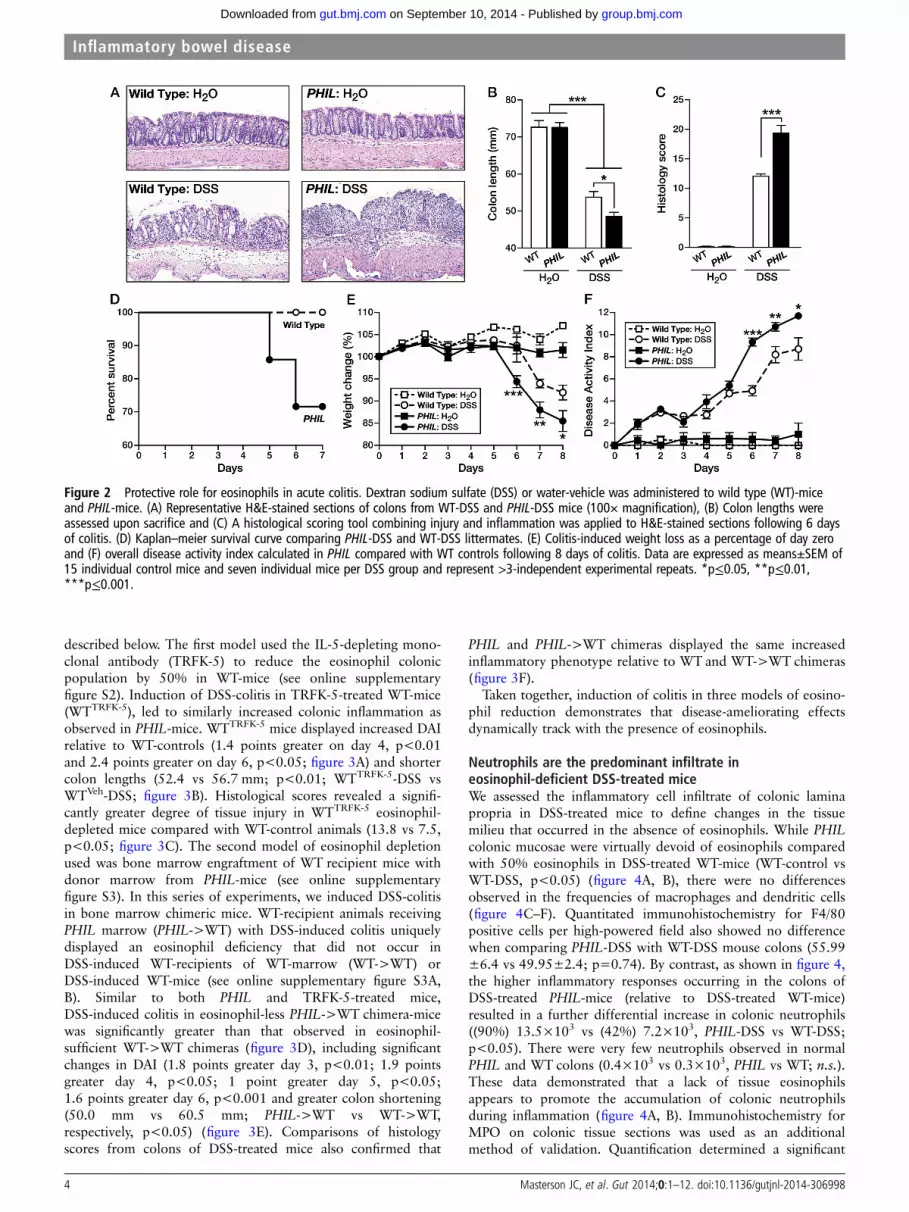
RESULTSProtective role for eosinophils in three models of acute colitisAnalysis of WT-mice and human tissues identified eosinophils as asignificant component of the acute inflammatory response inexperimental DSS-colitis and ulcerative colitis (figure 1A–C).Additionally, analysis of molecular responses identified thateosinophilic chemokines were significantly increased in experi-mental DSS-colitis (figure 1D). Except for the absence of eosino-phils, the normal colonic mucosa of WT-mice and eosinophil-less(PHIL)-mice appeared the same (figure 2A). We inducedDSS-colitis and monitored disease activity to determine the impactof eosinophils in acute experimental colitis. Upon harvest, coloniclength was found to be significantly shorter (48.4 vs 53.8 mm,PHIL-DSS vs WT-DSS; p<0.001), and in congruence, tissueinjury was significantly greater in PHIL-DSS compared withWT-DSS challenged mice, as evidenced by histological scoringof ulceration, oedema and leucocytic infiltration (19.5 vs 12.1,PHIL vs WT; p<0.001) (figure 2B, C). As shown in figure 2D,PHIL-mice experienced a greater mortality compared with
WT-mice (PHIL-DSS—29% death vs WT-DSS—0% at 7 days).Further studies, with lower DSS concentrations, determined thatPHIL-DSS mice developed significantly greater weight loss thanWT-DSS mice in a time-dependent fashion (8.2% greater lossday 6, p<0.001; 5.9% greater loss day 7, p<0.01; 6.4% greaterloss day 8, p<0.05; figure 2E). PHIL-DSS mice also had a higherDAI compared with WT-DSS mice (4.4 points greater day 6,p<0.001; 2.5 points greater day 7, p<0.01; 3 points greaterday 8, p<0.05; figure 2F).
We next induced TNBS-colitis and monitored disease activityas described above. Results from these studies were remarkablysimilar to those found in DSS-colitis. PHIL-mice compared withWT-mice, experienced a greater rate of weight loss (7% greaterloss day 2, p<0.05; 7% greater loss day 3, p<0.05), signifi-cantly shorter colon length (50 mm vs 61 mm, PHIL-TNBS vsWT-TNBS; p<0.001) and greater histological measure ofdisease (9.8 vs 3.5, PHIL-TNBS vs WT-TNBS; p<0.01) (seeonline supplementary figure S1).
In a third method of experimentally inducible colitis, WTandPHIL mice were treated with OXA-colitis. The death rate was89% in PHIL-OXA and 20% in WT-OXA mice at day 4 postin-stillation, p<0.0005 vs WT-OXA. No vehicle-treated mice ofeither genotype died. At 3 days postinstillation, maximumweight loss was observed (WT-OXA 88.29±2.3% vs PHIL-OXA78.833±3.9% (p=0.07)). By 4 days postinstillation, most PHIL
mice had died. Thus, consistent with findings in DSS and TNBSmodels of colitis, PHIL mice were more susceptible than theirWT littermate counterparts and did not survive the induction ofcolitis using the OXA methodology.
We used two other methods, antibody targeting and bonemarrow depletion, to address eosinophils’ role in colitis as
Figure 1 Eosinophilic inflammation in acute colitis. (A) H&E, and major basic protein immunohistochemistry on mouse colons following dextransodium sulfate (DSS)-colitis. (B) Eosinophil peroxidase immunohistochemistry on human colons from normal and ulcerative colitis subjects. (C) Wildtype whole mouse colon analysis by flow cytometry for infiltrating myeloid leucocytes. (D) Colon mRNA analysis for eosinophil-associatedchemokines CCL11, CCL24 and CCL5. Scale bars represent 100 and 50 μM, respectively. Data are expressed as means±SEM of 3–7 individual miceper group and represent >3-independent experimental repeats following 6 days of colitis. *p≤0.05.
Masterson JC, et al. Gut 2014;0:1–12. doi:10.1136/gutjnl-2014-306998 3
Inflammatory bowel disease
group.bmj.com on September 10, 2014 - Published by gut.bmj.comDownloaded from
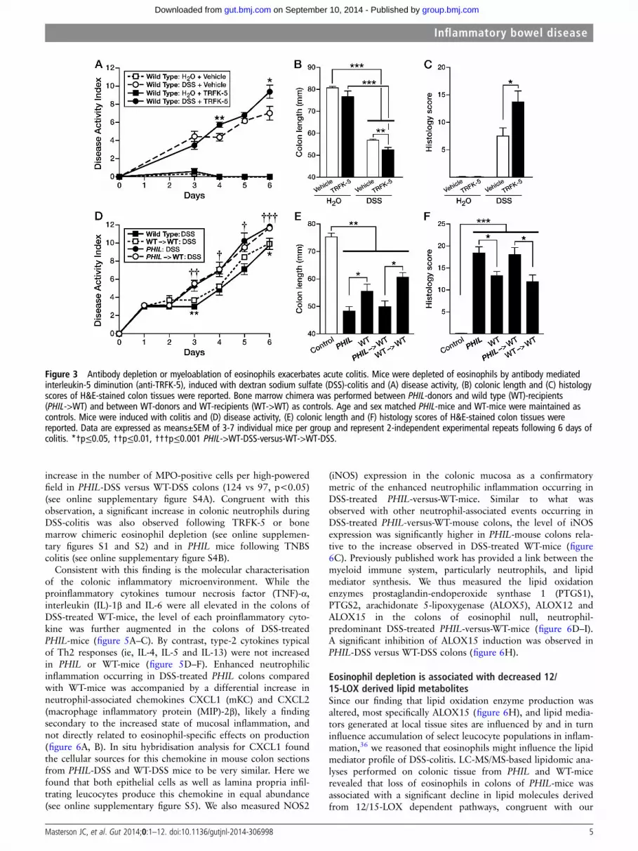
described below. The first model used the IL-5-depleting mono-clonal antibody (TRFK-5) to reduce the eosinophil colonicpopulation by 50% in WT-mice (see online supplementaryfigure S2). Induction of DSS-colitis in TRFK-5-treated WT-mice(WTTRFK-5), led to similarly increased colonic inflammation asobserved in PHIL-mice. WTTRFK-5 mice displayed increased DAIrelative to WT-controls (1.4 points greater on day 4, p<0.01and 2.4 points greater on day 6, p<0.05; figure 3A) and shortercolon lengths (52.4 vs 56.7 mm; p<0.01; WTTRFK-5-DSS vsWTVeh-DSS; figure 3B). Histological scores revealed a signifi-cantly greater degree of tissue injury in WTTRFK-5 eosinophil-depleted mice compared with WT-control animals (13.8 vs 7.5,p<0.05; figure 3C). The second model of eosinophil depletionused was bone marrow engraftment of WT recipient mice withdonor marrow from PHIL-mice (see online supplementaryfigure S3). In this series of experiments, we induced DSS-colitisin bone marrow chimeric mice. WT-recipient animals receivingPHIL marrow (PHIL->WT) with DSS-induced colitis uniquelydisplayed an eosinophil deficiency that did not occur inDSS-induced WT-recipients of WT-marrow (WT->WT) orDSS-induced WT-mice (see online supplementary figure S3A,B). Similar to both PHIL and TRFK-5-treated mice,DSS-induced colitis in eosinophil-less PHIL->WT chimera-micewas significantly greater than that observed in eosinophil-sufficient WT->WT chimeras (figure 3D), including significantchanges in DAI (1.8 points greater day 3, p<0.01; 1.9 pointsgreater day 4, p<0.05; 1 point greater day 5, p<0.05;1.6 points greater day 6, p<0.001 and greater colon shortening(50.0 mm vs 60.5 mm; PHIL->WT vs WT->WT,respectively, p<0.05) (figure 3E). Comparisons of histologyscores from colons of DSS-treated mice also confirmed that
PHIL and PHIL->WT chimeras displayed the same increasedinflammatory phenotype relative to WT and WT->WT chimeras(figure 3F).
Taken together, induction of colitis in three models of eosino-phil reduction demonstrates that disease-ameliorating effectsdynamically track with the presence of eosinophils.
Neutrophils are the predominant infiltrate ineosinophil-deficient DSS-treated miceWe assessed the inflammatory cell infiltrate of colonic laminapropria in DSS-treated mice to define changes in the tissuemilieu that occurred in the absence of eosinophils. While PHIL
colonic mucosae were virtually devoid of eosinophils comparedwith 50% eosinophils in DSS-treated WT-mice (WT-control vsWT-DSS, p<0.05) (figure 4A, B), there were no differencesobserved in the frequencies of macrophages and dendritic cells(figure 4C–F). Quantitated immunohistochemistry for F4/80positive cells per high-powered field also showed no differencewhen comparing PHIL-DSS with WT-DSS mouse colons (55.99±6.4 vs 49.95±2.4; p=0.74). By contrast, as shown in figure 4,the higher inflammatory responses occurring in the colons ofDSS-treated PHIL-mice (relative to DSS-treated WT-mice)resulted in a further differential increase in colonic neutrophils((90%) 13.5×10³ vs (42%) 7.2×10³, PHIL-DSS vs WT-DSS;p<0.05). There were very few neutrophils observed in normalPHIL and WT colons (0.4×10³ vs 0.3×10³, PHIL vs WT; n.s.).These data demonstrated that a lack of tissue eosinophilsappears to promote the accumulation of colonic neutrophilsduring inflammation (figure 4A, B). Immunohistochemistry forMPO on colonic tissue sections was used as an additionalmethod of validation. Quantification determined a significant
Figure 2 Protective role for eosinophils in acute colitis. Dextran sodium sulfate (DSS) or water-vehicle was administered to wild type (WT)-miceand PHIL-mice. (A) Representative H&E-stained sections of colons from WT-DSS and PHIL-DSS mice (100× magnification), (B) Colon lengths wereassessed upon sacrifice and (C) A histological scoring tool combining injury and inflammation was applied to H&E-stained sections following 6 daysof colitis. (D) Kaplan–meier survival curve comparing PHIL-DSS and WT-DSS littermates. (E) Colitis-induced weight loss as a percentage of day zeroand (F) overall disease activity index calculated in PHIL compared with WT controls following 8 days of colitis. Data are expressed as means±SEM of15 individual control mice and seven individual mice per DSS group and represent >3-independent experimental repeats. *p≤0.05, **p≤0.01,***p≤0.001.
4 Masterson JC, et al. Gut 2014;0:1–12. doi:10.1136/gutjnl-2014-306998
Inflammatory bowel disease
group.bmj.com on September 10, 2014 - Published by gut.bmj.comDownloaded from
increase in the number of MPO-positive cells per high-poweredfield in PHIL-DSS versus WT-DSS colons (124 vs 97, p<0.05)(see online supplementary figure S4A). Congruent with thisobservation, a significant increase in colonic neutrophils duringDSS-colitis was also observed following TRFK-5 or bonemarrow chimeric eosinophil depletion (see online supplemen-tary figures S1 and S2) and in PHIL mice following TNBScolitis (see online supplementary figure S4B).
Consistent with this finding is the molecular characterisationof the colonic inflammatory microenvironment. While theproinflammatory cytokines tumour necrosis factor (TNF)-α,interleukin (IL)-1β and IL-6 were all elevated in the colons ofDSS-treated WT-mice, the level of each proinflammatory cyto-kine was further augmented in the colons of DSS-treatedPHIL-mice (figure 5A–C). By contrast, type-2 cytokines typicalof Th2 responses (ie, IL-4, IL-5 and IL-13) were not increasedin PHIL or WT-mice (figure 5D–F). Enhanced neutrophilicinflammation occurring in DSS-treated PHIL colons comparedwith WT-mice was accompanied by a differential increase inneutrophil-associated chemokines CXCL1 (mKC) and CXCL2(macrophage inflammatory protein (MIP)-2β), likely a findingsecondary to the increased state of mucosal inflammation, andnot directly related to eosinophil-specific effects on production(figure 6A, B). In situ hybridisation analysis for CXCL1 foundthe cellular sources for this chemokine in mouse colon sectionsfrom PHIL-DSS and WT-DSS mice to be very similar. Here wefound that both epithelial cells as well as lamina propria infil-trating leucocytes produce this chemokine in equal abundance(see online supplementary figure S5). We also measured NOS2
(iNOS) expression in the colonic mucosa as a confirmatorymetric of the enhanced neutrophilic inflammation occurring inDSS-treated PHIL-versus-WT-mice. Similar to what wasobserved with other neutrophil-associated events occurring inDSS-treated PHIL-versus-WT-mouse colons, the level of iNOSexpression was significantly higher in PHIL-mouse colons rela-tive to the increase observed in DSS-treated WT-mice (figure6C). Previously published work has provided a link between themyeloid immune system, particularly neutrophils, and lipidmediator synthesis. We thus measured the lipid oxidationenzymes prostaglandin-endoperoxide synthase 1 (PTGS1),PTGS2, arachidonate 5-lipoxygenase (ALOX5), ALOX12 andALOX15 in the colons of eosinophil null, neutrophil-predominant DSS-treated PHIL-versus-WT-mice (figure 6D–I).A significant inhibition of ALOX15 induction was observed inPHIL-DSS versus WT-DSS colons (figure 6H).
Eosinophil depletion is associated with decreased 12/15-LOX derived lipid metabolitesSince our finding that lipid oxidation enzyme production wasaltered, most specifically ALOX15 (figure 6H), and lipid media-tors generated at local tissue sites are influenced by and in turninfluence accumulation of select leucocyte populations in inflam-mation,36 we reasoned that eosinophils might influence the lipidmediator profile of DSS-colitis. LC-MS/MS-based lipidomic ana-lyses performed on colonic tissue from PHIL and WT-micerevealed that loss of eosinophils in colons of PHIL-mice wasassociated with a significant decline in lipid molecules derivedfrom 12/15-LOX dependent pathways, congruent with our
Figure 3 Antibody depletion or myeloablation of eosinophils exacerbates acute colitis. Mice were depleted of eosinophils by antibody mediatedinterleukin-5 diminution (anti-TRFK-5), induced with dextran sodium sulfate (DSS)-colitis and (A) disease activity, (B) colonic length and (C) histologyscores of H&E-stained colon tissues were reported. Bone marrow chimera was performed between PHIL-donors and wild type (WT)-recipients(PHIL->WT) and between WT-donors and WT-recipients (WT->WT) as controls. Age and sex matched PHIL-mice and WT-mice were maintained ascontrols. Mice were induced with colitis and (D) disease activity, (E) colonic length and (F) histology scores of H&E-stained colon tissues werereported. Data are expressed as means±SEM of 3-7 individual mice per group and represent 2-independent experimental repeats following 6 days ofcolitis. *†p≤0.05, ††p≤0.01, †††p≤0.001 PHIL->WT-DSS-versus-WT->WT-DSS.
Masterson JC, et al. Gut 2014;0:1–12. doi:10.1136/gutjnl-2014-306998 5
Inflammatory bowel disease
group.bmj.com on September 10, 2014 - Published by gut.bmj.comDownloaded from

observation of inhibited ALOX15 induction. In particular, colonlevels of eosinophil-associated anti-inflammatory PD124 37 werevirtually absent in healthy-PHIL-mice (0.6 ng/g-colon) andDSS-treated PHIL-mice (1 ng/g-colon) relative to eosinophil suf-ficient healthy (3 ng/g-colon) and DSS-treated (11 ng/g-colon)WT-mice (PHIL-DSS vs WT-DSS; p<0.05) (figure 7-inset). No
difference in levels of the PD1 precursor DHA was observed ineither PHIL or WT colons prior to, or following, the inductionof colitis. However, 17-hydroxy-DHA, a marker of DHA utilisa-tion, was significantly decreased in PHIL-colons prior to andfollowing induction of colitis (p<0.001). Levels of lipid media-tors derived from the other major metabolic networks linked
Figure 4 Inflammatory infiltrate in eosinophil null mucosa is predominated by neutrophils. Whole colon flow cytometry analysis for infiltratingmyeloid leucocytes in wild type (WT)-colons and PHIL-colons in dextran sodium sulfate (DSS)-colitis. Derivation and quantification of relativeproportions of (A and B) neutrophils and eosinophils, (C and D) macrophage and (E and F) dendritic cell infiltrates comparing WT-DSS withPHIL-DSS-colitis. Data are expressed as means±SEM of seven individual mice per group and represent >3-independent experimental repeatsfollowing 6 days of colitis. **p≤0.01, Wild Type versus PHIL.
6 Masterson JC, et al. Gut 2014;0:1–12. doi:10.1136/gutjnl-2014-306998
Inflammatory bowel disease
group.bmj.com on September 10, 2014 - Published by gut.bmj.comDownloaded from

with cyclo-oxygenase (COX) and/or 5-LOX were not differentin PHIL and WT healthy or DSS-treated colons. Congruentwith our finding of increased neutrophils in inflamedeosinophil-deficient colons, a significant increase in theneutrophil-associated chemokine LTB4 was observed (figure 7).
Exogenous administration of PD1-isomer rescues PHIL-micefrom colitis and diminishes neutrophil chemotaxis andinflammationAdministration of PD1-isomer to DSS-treated PHIL-miceresulted in significant improvement in colonic inflammation(figure 8). Specifically, PD1-isomer treatment of PHIL-miceresulted in significant improvement in colon shortening (69.5mm vs 60.6 mm vs 52.8 mm, Control PHIL vs. PD1-isomer-treated PHIL vs vehicle-treated PHIL, p<0.01) and histologicalscores (9.4 vs 16.5, PD1-isomer-treated PHIL vs vehicle-treatedPHIL, p<0.01) (figure 8A, B). Representative photomicrographsof colonic mucosa from PD1-isomer-treated mice revealedimprovements in epithelial cell loss, inflammatory infiltrates,and architectural disruption (figure 8C). Consistent with thesefindings, proinflammatory molecules, including TNF-α, IL-1βand IL-6 decreased significantly (figure 8D).
PD1-isomer add-back also led to a significant decrease in neu-trophilic inflammation in DSS-treated PHIL-mice (figure 8E), as
well as a decrease in neutrophil-specific chemokines CXCL-1/KC and CXCL-2/MIP2-α and proinflammatory NOS2 (figure8D). Ex vivo chemotaxis studies further substantiated the anti-inflammatory effects of PD1-isomer leading to a concentration-dependent decrease in PMN chemotaxis across intestinal epithe-lial monolayers compared with control PMNs (figure 8G). Suchfindings identify PD1/PD1-isomers as a prominent molecularsignature that attenuates neutrophil accumulation duringongoing colonic inflammation.
DISCUSSIONAlthough eosinophil accumulation in tissues of patients withIBD has been described, the specific and unique contributionsof eosinophils to IBD has yet to be clearly delineated.2 6 8 38
Here, we demonstrate a protective role for eosinophils duringmouse colitis. We show that the physical, histological andmolecular features of acute colonic inflammation are signifi-cantly increased without these granulocytes. Our data demon-strates acute colitis in eosinophil-competent mice correlates withbiosynthesis of anti-inflammatory derivatives of omega-3 fattyacids, including PD1, a molecule known to dampen neutrophilinflux and proinflammatory cytokine production.23 The enzymenecessary for PD1 synthesis, 15-LOX was not induced inPHIL-mice and, thus, PD1 was barely detectable in PHIL-mice
Figure 5 Proinflammatory and type-2 cytokine expression profile in PHIL-mice during acute colitis. mRNA was isolated from whole colons andassessed by Taqman technologies. (A–C) Major acute proinflammatory cytokines tumour necrosis factor (TNF)-α, interleukin (IL)-1β and IL-6 as wellas (D–F) Type-2 cytokines IL-4, IL-5 and IL-13 were measured. Data are expressed as means±SEM of 7–10 individual mice per group and represent2-independent experimental repeats following 6 days of colitis. *p≤0.05, **p≤0.01, ***p≤0.001 versus genotype matched water controls.†p≤0.05, ††p≤0.01, †††p≤0.001 versus wild type-dextran sodium sulfate (DSS).
Masterson JC, et al. Gut 2014;0:1–12. doi:10.1136/gutjnl-2014-306998 7
Inflammatory bowel disease
group.bmj.com on September 10, 2014 - Published by gut.bmj.comDownloaded from

at baseline or during colitis suggesting an important role forthese leucocytes in PD1 synthesis in this myeloid predominantacute experimental colitis model. Bypassing the deficiency in the15-LOX enzyme, exogenous administration of PD1-isomer toeosinophil-deficient mice following colonic injury markedly atte-nuated weight loss, colon shortening, histological indices ofinjury and inflammation, PMN infiltration and proinflammatorycytokine production including TNF-α, IL-1β, IL-6 and induciblenitric oxide. These findings suggest a role for acute-phase-responding eosinophils, either directly or indirectly, in the
resolution of early inflammatory responses, possibly throughalterations in gene expression and leucocyte recruitment, suchas that mediated by the 12/15-LOX-derived lipid mediatorPD1/PD1-isomers.
To date, the role of eosinophils during IBD has remainedelusive. A number of studies suggest an end-stage destructiverole(s) for eosinophils in the pathogenesis of gut inflammationin addition to other mucosal organs.10 39 For instance, eosino-phil granule protein null mice were relatively protected fromcolitis indicating a pathological role for these proteins.10 39–41
Figure 6 Chemokine, iNOS and lipid oxygenation enzyme expression profile in PHIL-mice during acute colitis. mRNA was isolated from wholecolons and assessed by Taqman technologies. (A–C) Neutrophil-associated transcripts CXCL1/KC, CXCL2 and NOS2 were assessed. (D–H) Lipidoxygenation enzymes critical for lipid mediator biosynthesis PTGS1, PTGS2, ALOX5, ALOX12 and ALOX15 were also measured. Data are expressed asmeans±SEM of 7–10 individual mice per group and represent 2-independent experimental repeats following 6 days of colitis. *p≤0.05, **p≤0.01,***p≤0.001 versus genotype-matched water controls. †p≤0.05, ††p≤0.01, †††p≤0.001 versus wild type-dextran sodium sulfate (DSS).
8 Masterson JC, et al. Gut 2014;0:1–12. doi:10.1136/gutjnl-2014-306998
Inflammatory bowel disease
group.bmj.com on September 10, 2014 - Published by gut.bmj.comDownloaded from

The emerging dual role for eosinophils in contributing to hosthealth and to inflammation is a fascinating one. Eosinophilshave been reported as important for the maintenance of hosthealth in the retention of memory plasma cells to the bonemarrow.42 They are also important for the maintenance ofmicrobial homeostasis of commensal organisms in the intestinesduring health by retaining IgA-producing plasma cells in peyer’spatches.43 It is important to note that eosinophil-deficient miceare not devoid of IgA, however, as other mechanisms of IgAproduction were maintained. Both these studies implicate eosi-nophils as a primary source for the critical plasma cell chemo-kine, APRIL. In the setting of acute peritoneal inflammation,eosinophils have also been demonstrated as protective sentinels,producing anti-inflammatory lipids including PD1, promotingthe rate of resolution.24 Experimental models of colonic inflam-mation, such as the IL-10−/− mouse present with significantcolonic eosinophilia prior to and during disease.44 In thismodel, the absence of IL-4 and, thus, eosinophils resulted indiminished disease and led the authors to suggest important role(s) for eosinophils in the initiation of colitis. However, IL-4mediates significant influences on a number of other potentialpathogenic effector cells. In light of our findings, one could
speculate eosinophils’ increase prior to the onset of inflamma-tion, as observed in the IL-10−/− model, may be an earlyattempt to dampen inflammation, potentially via mechanismsincluding the production of anti-inflammatory lipid mediators,among others, for example, PD1. It is important to keep incontext that eosinophils may play a role, but are not the solemediators in the maintenance of intestinal health and homeosta-sis. It appears that eosinophils may react to acute insults inattempt to resolve and restore intestinal health.
Clinical studies have identified increased numbers of coloniceosinophils in disease states and a reduction following treatmentproviding circumstantial evidence for a pathogenic role for eosi-nophils in patients.45 Contrasting with this, others have foundan increase in eosinophils during disease remission.46 However,studies have not addressed the impact of eosinophils in theacute/early phase of colitis induction, including whether thebaseline population of these granulocytes may offer host benefitsas a part of innate host defence. Recent review speculates thateosinophils may mediate host health by antimicrobial and tissuerepair functions.2 When these cells are chronically present inexcess however, they may indeed participate in a deleteriousmanner towards organ dysfunction.
Figure 7 12/15-lipoxygenase (12/15-LOX) lipid derivative deficiency in colitis in PHIL-mice. Whole mouse colons from dextran sodium sulfate(DSS)-colitis and water control wild type (WT)-mice and PHIL-mice were assessed by LC-MS/MS-based mediator lipidomics following 6 days of colitis.Arachidonic acid (AA)-derived, eicosapentaenoic acid-derived and docosahexaenoic acid (DHA)-derived products were quantified by LC-MS/MS, andmultiple reaction monitoring. Levels of 12/15-LOX products are boxed in black or blue and 12/15-LOX biosynthetic end products with statisticallysignificant differences are in blue shaded boxes. Data are expressed as means±SEM of 3-H2O and 6-DSS individual mice. *p≤0.05, **p≤0.01,***p≤0.001 DSS versus genotype-matched water controls. ††p≤0.01, †††p≤0.001 PHIL-DSS versus WT-DSS.
Masterson JC, et al. Gut 2014;0:1–12. doi:10.1136/gutjnl-2014-306998 9
Inflammatory bowel disease
group.bmj.com on September 10, 2014 - Published by gut.bmj.comDownloaded from

Two previous studies have addressed the impact of eosinophilson the development of colitis.9 47 Comparison of those resultswith findings shown here, yielded different conclusions, thusdemonstrating the complexity of studies in mouse models ofdisease. For example, results from two models of eosinophildeficiency (PHIL and ΔdblGATA) at two locations (USA andBrazil) suggested that the loss of eosinophils leads to the ameli-oration of colitis.9 47 Potential factors that explain these conflict-ing results include technical aspects of experimental colitisinduction, institutional differences including dietary source andcomposition, age of mice and variability in eosinophil-deficientstrains. For example, Ahrens et al studied PHIL-mice at youngerages than our study and measured histology and colon length asthe primary readouts. Vieira et al examined the ΔdblGATA
mouse, deficient in basophils, in addition to eosinophils,48 andused a higher concentration of DSS than our studies (4% vs2.5%). Additionally, the dietary availability and ratio of the gen-erally considered proinflammatory omega-6 and anti-inflammatory omega-3 precursors of potent lipid mediators indifferent mouse chow will play a critical role in the balancebetween inflammation and its resolution. We suggest that theseexperimental differences are significant and contribute to thedisparity in our findings. More importantly, we propose thatthis likely reflects the multifaceted role of eosinophils in diseaseinitially contributing anti-inflammatory activities following acuteprovocation and then proinflammatory effects in chronic set-tings. Our ongoing studies of eosinophil deficiency in a varietyof environmental settings (eg, barrier facilities vs conventional
Figure 8 Protectin D1 (PD1)-isomer (PD1-i) treatment attenuates colitis in PHIL-mice via attenuation of neutrophil migration. PHIL- dextran sodiumsulfate (DSS)-colitis mice were treated with PD1-isomer or vehicle via intraperitoneal injection 0.05 mg/kg daily throughout the course of colitiswhich lasted 6 days. Upon sacrifice, colons were excised and (A) colon length measured. (B) Injury, inflammatory and total histology scores wereassessed and (C) representative H&E-stained sections from PHIL-DSS-mice treated with PD1-isomer or vehicle (100× magnification). (D)Proinflammatory cytokine and neutrophil-associated chemokine mRNA production was examined in PD1-isomer versus vehicle-treatedPHIL-DSS-mice. (E and F) Flow cytometric analysis of lamina propria infiltrates for neutrophil percent and absolute number comparing PD1-isomerwith vehicle-treated PHIL-DSS-controls. Data are expressed as means±SEM of 3–7 individual mice per group, and represent 2 independentexperimental repeats. (G) In vitro examination of the effects of PD1-isomer on Formyl–Methionyl–Leucyl–Phenylalanine-induced neutrophil-colonepithelial monolayer (Caco-2) transmigration; dose-dependence and time-dependence. Data are expressed as means±SEM of three individual donorsand three independent experimental repeats. *p≤0.05, **p≤0.01 0.2 nM PD1 versus controls. ††p≤0.01, †††p≤0.001 20 nM PD1-isomer versuscontrols.
10 Masterson JC, et al. Gut 2014;0:1–12. doi:10.1136/gutjnl-2014-306998
Inflammatory bowel disease
group.bmj.com on September 10, 2014 - Published by gut.bmj.comDownloaded from

housing venues)49 provide an additional confounding perspec-tive; the rapidly responsive microbiome and/or exposure toenvironmental toxicants (eg, endotoxins) present in the residentanimal facilities are likely to be key issues modulating theimmune/inflammatory responses linked with eosinophils inmouse models of disease.50 We suggest that the confoundinginterinstitutional issues are a consequence of these extrinsicmodulatory events, altering the proinflammatory versus anti-inflammatory roles of eosinophils in a given setting. It may bepossible that each, or any combination, of these factors may benecessary to stimulate the eosinophil population to act in a regu-latory manner. Thus, both conclusions may be correct, and thespecific circumstances surrounding a given model and protocolwill need to be noted in future studies with the multipleeosinophil-deficient mouse models now currently available.
Our study contributes to a growing body of basic and clinicalstudies that support a role for omega-3 PUFAs and their deriva-tives in IBD treatment. 17–20 51–53 While we do not rule out thepotential contributions of anti-inflammatory lipids derived fromCOX or 5-LO biosynthesis pathways,54 we specifically see aglobal deficiency in 12/15-LOX and its derived lipid mediatorsin the absence of eosinophils. Moreover, we do not discount thepotential anti-inflammatory utility of AA or eicosapentaenoicacid-derived 12/15-LOX mediated lipids, including5,15-diHETE and 15-HEPE, however 5,15-diHETE maypotentiate PMN degranulation and thus exacerbate inflamma-tion in this model.55 Our studies have focused on theDHA-derived, previously established eosinophil-associated, PD1molecule.24 37 The data reported here suggest a link betweeneosinophil activities and the levels of the DHA-derived PD1family of lipid mediators present in the initial stages of colonicinflammation, an attempt by the host to limit colonic inflamma-tion. However, in addition to eosinophils, 12/15-LOX-derivedbiosynthesis of PD1 and its isomers may occur through othercells present during DSS-colitis, such as tissue-resident macro-phages, dendritic cells, mast cells as well as epithelial cells.21
Our experiments do not rule out these other cell types as poten-tial contributors of PD1/PD1-isomers. Nonetheless, it is note-worthy that no differences were observed in the frequencies ofthese other cells between colitic PHIL and control mice, suggest-ing the critical role, specifically for the increased eosinophilinfiltrate in either directly or indirectly producing or inducing15-LOX, and the proresolving lipid mediator PD1/PD1-isomers.Thus, these findings are consistent with observations thatrecruitment of eosinophils and production of PD1/PD1-isomersare associated with reduced numbers of neutrophils in inflamedtissue and the resolution of inflammation.22 24 The importanceof eosinophil-derived PD1/PD1-isomer expression awaits futurestudies using eosinophil-specific 12/15-LOX knockout mice gen-erated by using 12/15-LOXflox/flox animals crossed witheosinophil-specific Cre-expressing mice.
Reconstitution studies, and in vitro modelling identify theimpact of PD1-isomer on reducing PMN inflammation in colitisas has been demonstrated in other organ systems.23 36 56 Ourdata extends these findings by showing that proinflammatorygene expression (TNF-α, IL-1β, IL-6, nitric oxide, CXCL1 andCXCL2) associated with PMN recruitment in colonic tissuesdecreases following PD1-isomer treatment. This reduction inproinflammatory cytokines and PMN infiltration, mediated, inpart, by pharmacological reconstitution with PD1-isomers, maybe an effective approach in the prevention and maintenance ofremission in patients with IBD.
In summary, the present study highlights a critical role foreosinophils in regulating inflammatory tone during acute
experimental mouse colitis. Mechanistically, we identify that theomega-3 PUFA-derived lipid mediator, PD1/PD1-isomer specif-ically limits colon PMN infiltration. Interestingly, complete abla-tion of neutrophil recruitment in animal models of colitis isitself deleterious, and may suggest that a balance between eosi-nophils and neutrophils may be necessary for resolution ofinflammation.57 58 These findings support a potentially protect-ive role for resident eosinophils in mouse colitis and, thus, chal-lenge the paradigm for a solely proinflammatory role in humandisease. We speculate that in certain circumstances, the increasedpresence of eosinophils in early stages of mucosal inflammationmay not necessarily be an indicator of disease, but rather a hostresponse, fostering mucosal healing and a pathway to return tohomoeostasis (figure 1). Thus, future translational studieslinking numbers of eosinophils in a given patient with observedhistopathology and clinical manifestation of disease are neces-sary to test our hypotheses.
Author affiliations1Section of Pediatric Gastroenterology, Hepatology and Nutrition, GastrointestinalEosinophilic Diseases Program, Department of Pediatrics; Digestive Health Institute,Children’s Hospital Colorado, University of Colorado School of Medicine, Aurora,Colorado, USA2Mucosal Inflammation Program, University of Colorado School of Medicine, Aurora,Colorado, USA3University of Colorado School of Medicine, Aurora, Colorado, USA4Department of Anesthesiology, University of Colorado School of Medicine, Aurora,Colorado, USA5Department of Pathology, University of Colorado School of Medicine, Aurora,Colorado, USA6Department of Health Chemistry, Graduate School of Pharmaceutical Sciences,University of Tokyo, Tokyo, Japan7Laboratory for Metabolomics, RIKEN Center for Integrative Medical Sciences (IMS),Yokohama, Japan8Department of Biochemistry and Molecular Biology, Mayo Clinic Scottsdale,Scottsdale, Arizona, USA9Department of Medicine, University of Colorado School of Medicine, Aurora,Colorado, USA
Acknowledgements The authors wish to thank the members of all theparticipating laboratories for insightful discussions and critical comments. We alsowish to acknowledge the invaluable assistance in animal husbandry and care atUniversity of Colorado School of Medicine (Kristann Magee), the Mayo Clinic Arizonamedical graphic artist (Marv Ruona), and the administrative support provided byLinda Mardel and Shirley (‘Charlie’) Kern. We gratefully thank Ms Michiko Kamio fortechnical assistance on LC-MS/MS analyses.
Contributors Study concept and design ( JCM, ENMcN, SPC, MA, JJL, GTF);acquisition of data ( JCM, ENMcN, SAF, LH, RH, SDF, PJ, RI, GTF); analysis andinterpretation of data ( JCM, ENMcN, SDF, PJ, RI, SPC, MA, JJL, GTF); drafting of themanuscript ( JCM, JJL, GTF); critical revision of the manuscript for importantintellectual content ( JCM, ENMcN, SPC, MA, JJL, GTF); statistical analysis ( JCM);obtained funding ( JCM, HKE, MA, JJL, GTF); technical or material support (EJ, CP);study supervision ( JCM, SPC, MA, JJL, GTF).
Funding Grants from the USA National Institutes of Health (R01-DK62245 andK24-DK 100303) (GTF), (R01-HL065228 and K26-RR0109709) ( JJL), and(DK097075, R01-HL0921, R01-DK083385, R01-HL098294 and POIHL114457-01)(HKE), Crohn’s and Colitis Foundation of America ( JCM, HKE), the North AmericanSociety for Pediatric Gastroenterology Hepatology and Nutrition ( JCM, GTF), theMayo Foundation for Medical Education and Research ( JJL), a Grant-in-aid from theJapan Science and Technology Agency Precursory Research for Embryonic Scienceand Technology (MA) and a Grant-in-aid from the Ministry of Education, Culture,Sports, Science, and Technology of Japan (MA) were the sources of funding used inthe performance of studies including data analysis and manuscript preparation.
Competing interests None.
Ethics approval Colorado Multiple Institutional Review Board.
Provenance and peer review Not commissioned; externally peer reviewed.
Data sharing statement Available data in this paper will be shared upon requestto the corresponding author.
Open Access This is an Open Access article distributed in accordance with theCreative Commons Attribution Non Commercial (CC BY-NC 3.0) license, which
Masterson JC, et al. Gut 2014;0:1–12. doi:10.1136/gutjnl-2014-306998 11
Inflammatory bowel disease
group.bmj.com on September 10, 2014 - Published by gut.bmj.comDownloaded from

permits others to distribute, remix, adapt, build upon this work non-commercially,and license their derivative works on different terms, provided the original work isproperly cited and the use is non-commercial. See: http://creativecommons.org/licenses/by-nc/3.0/
REFERENCES1 Rothenberg ME, Hogan SP. The eosinophil. Annu Rev Immunol 2006;24:147–74.2 Hogan SP, Waddell A, Fulkerson PC. Eosinophils in infection and intestinal
immunity. Curr Opin Gastroenterol 2013;29:7–14.3 Boyce JA, Bochner B, Finkelman FD, et al. Advances in mechanisms of asthma,
allergy, and immunology in 2011. J Allergy Clin Immunol 2012;129:335–41.4 Jacobsen EA, Ochkur SI, Lee NA, et al. Eosinophils and asthma. Curr Allergy
Asthma Rep 2007;7:18–26.5 Hogan SP, Rosenberg HF, Moqbel R, et al. Eosinophils: biological properties and
role in health and disease. Clin Exp Allergy 2008;38:709–50.6 Wedemeyer J, Vosskuhl K. Role of gastrointestinal eosinophils in inflammatory
bowel disease and intestinal tumours. Best Pract Res Clin Gastroenterol2008;22:537–49.
7 Hogan SP. Functional role of eosinophils in gastrointestinal inflammation. ImmunolAllergy Clin North Am 2009;29:129–40, xi.
8 Masterson JC, Furuta GT, Lee JJ. Update on clinical and immunological features ofeosinophilic gastrointestinal diseases. Curr Opin Gastroenterol 2011;27:515–22.
9 Ahrens R, Waddell A, Seidu L, et al. Intestinal macrophage/epithelial cell-derivedCCL11/eotaxin-1 mediates eosinophil recruitment and function in pediatriculcerative colitis. J Immunol 2008;181:7390–9.
10 Furuta GT, Nieuwenhuis EE, Karhausen J, et al. Eosinophils alter colonic epithelialbarrier function: role for major basic protein. Am J Physiol Gastrointest Liver Physiol2005;289:G890–7.
11 Masterson JC, McNamee EN, Jedlicka P, et al. CCR3 blockade attenuateseosinophilic ileitis and associated remodeling. Am J Pathol 2011;179:2302–14.
12 Arita M. Mediator lipidomics in acute inflammation and resolution. J Biochem2012;152:313–19.
13 Bannenberg G, Serhan CN. Specialized pro-resolving lipid mediators in theinflammatory response: an update. Biochim Biophys Acta 2010;1801:1260–73.
14 Hudert CA, Weylandt KH, Lu Y, et al. Transgenic mice rich in endogenous omega-3fatty acids are protected from colitis. Proc Natl Acad Sci USA 2006;103:11276–81.
15 Sakamoto N, Kono S, Wakai K, et al. Dietary risk factors for inflammatory boweldisease: a multicenter case-control study in Japan. Inflamm Bowel Dis2005;11:154–63.
16 Shoda R, Matsueda K, Yamato S, et al. Epidemiologic analysis of Crohn disease inJapan: increased dietary intake of n-6 polyunsaturated fatty acids and animalprotein relates to the increased incidence of Crohn disease in Japan. Am J Clin Nutr1996;63:741–5.
17 Bento AF, Claudino RF, Dutra RC, et al. Omega-3 fatty acid-derived mediators 17(R)-hydroxy docosahexaenoic acid, aspirin-triggered resolvin D1 and resolvin D2prevent experimental colitis in mice. J Immunol 2011;187:1957–69.
18 Ibrahim A, Mbodji K, Hassan A, et al. Anti-inflammatory and anti-angiogenic effectof long chain n-3 polyunsaturated fatty acids in intestinal microvascularendothelium. Clin Nutr 2011;30:678–87.
19 Matsunaga H, Hokari R, Kurihara C, et al. Omega-3 polyunsaturated fatty acidsameliorate the severity of ileitis in the senescence accelerated mice (SAM)P1/Yitmice model. Clin Exp Immunol 2009;158:325–33.
20 Varma S, Eskin MN, Bird R, et al. Potential of magnetic resonance spectroscopy inassessing the effect of fatty acids on inflammatory bowel disease in an animalmodel. Lipids 2010;45:843–54.
21 Kuhn H, O’Donnell VB. Inflammation and immune regulation by 12/15-lipoxygenases. Prog Lipid Res 2006;45:334–56.
22 Levy BD, Kohli P, Gotlinger K, et al. Protectin D1 is generated in asthma anddampens airway inflammation and hyperresponsiveness. J Immunol2007;178:496–502.
23 Serhan CN, Gotlinger K, Hong S, et al. Anti-inflammatory actions of neuroprotectinD1/protectin D1 and its natural stereoisomers: assignments of dihydroxy-containingdocosatrienes. J Immunol 2006;176:1848–59.
24 Yamada T, Tani Y, Nakanishi H, et al. Eosinophils promote resolution of acuteperitonitis by producing proresolving mediators in mice. FASEB J 2011;25:561–8.
25 Isobe Y, Kato T, Arita M. Emerging roles of eosinophils and eosinophil-derived lipidmediators in the resolution of inflammation. Front Immunol 2012;3:270.
26 Marcheselli VL, Mukherjee PK, Arita M, et al. Neuroprotectin D1/protectin D1stereoselective and specific binding with human retinal pigment epithelial cells andneutrophils. Prostaglandins Leukot Essent Fatty Acids 2010;82:27–34.
27 Lee JJ, Dimina D, Macias MP, et al. Defining a link with asthma in micecongenitally deficient in eosinophils. Science 2004;305:1773–6.
28 Smith P, Mangan NE, Walsh CM, et al. Infection with a helminth parasite preventsexperimental colitis via a macrophage-mediated mechanism. J Immunol2007;178:4557–66.
29 Chen P, Fenet B, Michaud S, et al. Full characterization of PDX, a neuroprotectin/protectin D1 isomer, which inhibits blood platelet aggregation. FEBS Lett2009;583:3478–84.
30 McNamee EN, Masterson JC, Jedlicka P, et al. Interleukin 37 expression protectsmice from colitis. Proc Natl Acad Sci USA 2011;108:16711–16.
31 Protheroe C, Woodruff SA, de Petris G, et al. A novel histologic scoring system toevaluate mucosal biopsies from patients with eosinophilic esophagitis. ClinGastroenterol Hepatol 2009;7:749–55. e11.
32 Masterson JC, McNamee EN, Hosford L, et al. Local hypersensitivity reaction intransgenic mice with squamous epithelial IL-5 overexpression provides a novelmodel of eosinophilic oesophagitis. Gut 2014;63:43–53.
33 Furuta GT, Ackerman SJ, Lu L, et al. Stem cell factor influences mast cell mediatorrelease in response to eosinophil-derived granule major basic protein. Blood1998;92:1055–61.
34 Aherne CM, Collins CB, Masterson JC, et al. Neuronal guidance molecule netrin-1attenuates inflammatory cell trafficking during acute experimental colitis. Gut2012;61:695–705.
35 Colgan SP, Dzus AL, Parkos CA. Epithelial exposure to hypoxia modulates neutrophiltransepithelial migration. J Exp Med 1996;184:1003–15.
36 Schwab JM, Chiang N, Arita M, et al. Resolvin E1 and protectin D1 activateinflammation-resolution programmes. Nature 2007;447:869–74.
37 Miyata J, Fukunaga K, Iwamoto R, et al. Dysregulated synthesis of protectin D1 ineosinophils from patients with severe asthma. J Allergy Clin Immunol2013;131:353–60. e2.
38 Woodruff SA, Masterson JC, Fillon S, et al. Role of eosinophils in inflammatorybowel and gastrointestinal diseases. J Pediatr Gastroenterol Nutr 2011;52:650–61.
39 Forbes E, Murase T, Yang M, et al. Immunopathogenesis of experimental ulcerativecolitis is mediated by eosinophil peroxidase. J Immunol 2004;172:5664–75.
40 Forbes E, Hulett M, Ahrens R, et al. ICAM-1-dependent pathways regulate coloniceosinophilic inflammation. J Leukoc Biol 2006;80:330–41.
41 Stevceva L, Pavli P, Husband A, et al. Eosinophilia is attenuated in experimentalcolitis induced in IL-5 deficient mice. Genes Immun 2000;1:213–18.
42 Chu VT, Frohlich A, Steinhauser G, et al. Eosinophils are required for the maintenanceof plasma cells in the bone marrow. Nature immunology 2011;12:151–9.
43 Chu VT, Beller A, Rausch S, et al. Eosinophils promote generation and maintenanceof immunoglobulin-A-expressing plasma cells and contribute to gut immunehomeostasis. Immunity 2014;40:582–93.
44 Specht S, Arriens S, Hoerauf A. Induction of chronic colitis in IL-10 deficient micerequires IL-4. Microbes Infect 2006;8:694–703.
45 Stasikowska-Kanicka O, Danilewicz M, Glowacka A, et al. Mast cells andeosinophils are involved in activation of ulcerative colitis. Adv Med Sci2012;57:230–6.
46 Lampinen M, Ronnblom A, Amin K, et al. Eosinophil granulocytes are activatedduring the remission phase of ulcerative colitis. Gut 2005;54:1714–20.
47 Vieira AT, Fagundes CT, Alessandri AL, et al. Treatment with a novelchemokine-binding protein or eosinophil lineage-ablation protects mice fromexperimental colitis. Am J Pathol 2009;175:2382–91.
48 Nei Y, Obata-Ninomiya K, Tsutsui H, et al. GATA-1 regulates the generation andfunction of basophils. Proc Natl Acad Sci USA 2013;110:18620–5.
49 Jacobsen EA, Lesuer WE, Willetts L, et al. Eosinophil Activities Modulate theImmune/Inflammatory Character of Allergic Respiratory Responses in Mice. Allergy2014;69(3):315–27.
50 Ma BW, Bokulich NA, Castillo PA, et al. Routine habitat change: a source ofunrecognized transient alteration of intestinal microbiota in laboratory mice. PLoSONE 2012;7:e47416.
51 Kromann N, Green A. Epidemiological studies in the Upernavik district, Greenland.Incidence of some chronic diseases 1950–1974. Acta Med Scand 1980;208:401–6.
52 Hillier K, Jewell R, Dorrell L, et al. Incorporation of fatty acids from fish oil and oliveoil into colonic mucosal lipids and effects upon eicosanoid synthesis in inflammatorybowel disease. Gut 1991;32:1151–5.
53 Katz MS, Thatch KA, Schwartz MZ. Hepatocyte growth factor and omega-3-enrichedfeeds have a synergistic effect on mucosal mass in an animal model ofinflammatory bowel disease. J Pediatr Surg 2012;47:194–8.
54 Ajuebor MN, Singh A, Wallace JL. Cyclooxygenase-2-derived prostaglandin D(2) isan early anti-inflammatory signal in experimental colitis. Am J Physiol GastrointestLiver Physiol 2000;279:G238–44.
55 O’Flaherty JT, Thomas MJ. Effect of 15-lipoxygenase-derived arachidonate metaboliteson human neutrophil degranulation. Prostaglandins Leukot Med 1985;17:199–212.
56 Marcheselli VL, Hong S, Lukiw WJ, et al. Novel docosanoids inhibit brainischemia-reperfusion-mediated leukocyte infiltration and pro-inflammatory geneexpression. J Biol Chem 2003;278:43807–17.
57 Campbell EL, Bruyninckx WJ, Kelly CJ, et al. Transmigrating neutrophils shape themucosal microenvironment through localized oxygen depletion to influenceresolution of inflammation. Immunity 2014;40:66–77.
58 Kuhl AA, Kakirman H, Janotta M, et al. Aggravation of different types ofexperimental colitis by depletion or adhesion blockade of neutrophils.Gastroenterology 2007;133:1882–92.
12 Masterson JC, et al. Gut 2014;0:1–12. doi:10.1136/gutjnl-2014-306998
Inflammatory bowel disease
group.bmj.com on September 10, 2014 - Published by gut.bmj.comDownloaded from

doi: 10.1136/gutjnl-2014-306998 published online September 10, 2014Gut
Joanne C Masterson, Eóin N McNamee, Sophie A Fillon, et al. colitisinflammatory responses in experimental Eosinophil-mediated signalling attenuates
http://gut.bmj.com/content/early/2014/09/10/gutjnl-2014-306998.full.htmlUpdated information and services can be found at:
These include:
Data Supplement http://gut.bmj.com/content/suppl/2014/09/10/gutjnl-2014-306998.DC1.html
"Supplementary Data"
References http://gut.bmj.com/content/early/2014/09/10/gutjnl-2014-306998.full.html#ref-list-1
This article cites 58 articles, 23 of which can be accessed free at:
Open Access
non-commercial. See: http://creativecommons.org/licenses/by-nc/3.0/terms, provided the original work is properly cited and the use iswork non-commercially, and license their derivative works on different license, which permits others to distribute, remix, adapt, build upon thisCreative Commons Attribution Non Commercial (CC BY-NC 3.0) This is an Open Access article distributed in accordance with the
P<P Published online September 10, 2014 in advance of the print journal.
serviceEmail alerting
the box at the top right corner of the online article.Receive free email alerts when new articles cite this article. Sign up in
(DOIs) and date of initial publication. publication. Citations to Advance online articles must include the digital object identifier citable and establish publication priority; they are indexed by PubMed from initialtypeset, but have not not yet appeared in the paper journal. Advance online articles are Advance online articles have been peer reviewed, accepted for publication, edited and
http://group.bmj.com/group/rights-licensing/permissionsTo request permissions go to:
http://journals.bmj.com/cgi/reprintformTo order reprints go to:
http://group.bmj.com/subscribe/To subscribe to BMJ go to:
group.bmj.com on September 10, 2014 - Published by gut.bmj.comDownloaded from

CollectionsTopic
(155 articles)Open access � Articles on similar topics can be found in the following collections
Notes
(DOIs) and date of initial publication. publication. Citations to Advance online articles must include the digital object identifier citable and establish publication priority; they are indexed by PubMed from initialtypeset, but have not not yet appeared in the paper journal. Advance online articles are Advance online articles have been peer reviewed, accepted for publication, edited and
http://group.bmj.com/group/rights-licensing/permissionsTo request permissions go to:
http://journals.bmj.com/cgi/reprintformTo order reprints go to:
http://group.bmj.com/subscribe/To subscribe to BMJ go to:
group.bmj.com on September 10, 2014 - Published by gut.bmj.comDownloaded from
Copyright © 2022 FDOKUMEN