A CD44v6 peptide reveals a role of CD44 in VEGFR-2 signaling and angiogenesis
Upload
independentCategory
view
3download
0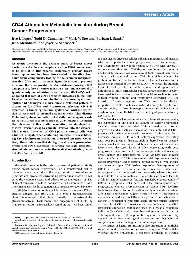
CD44 Attenuates Metastatic Invasion during Breast
Cancer Progression
Jose I. Lopez,1Todd D. Camenisch,
2,3Mark V. Stevens,
2Barbara J. Sands,
1
John McDonald,4and Joyce A. Schroeder
1,3
1Department of Molecular and Cellular Biology and Arizona Cancer Center; 2Department of Pharmacology and Toxicology and SteeleChildren’s Research Center; 3Bio5 Institute, University of Arizona, Tucson, Arizona; and 4Mayo Clinic Scottsdale, Scottsdale, Arizona
Abstract
Metastatic invasion is the primary cause of breast cancermortality, and adhesion receptors, such as CD44, are believedto be critical in this process. Historically, primary breasttumor epithelium has been investigated in isolation fromother tissue components, leading to the common interpreta-tion that CD44 and its primary ligand, hyaluronan, promoteinvasion. Here, we provide in vivo evidence showing CD44antagonism to breast cancer metastasis. In a mouse model ofspontaneously metastasizing breast cancer (MMTV-PyV mT),we found that loss of CD44 promotes metastasis to the lung.Localization studies, in combination with a novel hyaluronansynthase-GFP transgenic mouse, show a restricted pattern ofexpression for CD44 and hyaluronan. Whereas CD44 isexpressed in tumor epithelium, hyaluronan synthase expres-sion is restricted to stromal-associated cells. This distinctCD44 and hyaluronan pattern of distribution suggests a rolefor epithelial-stromal interaction in CD44 function. To definethe relevance of this spatial regulation, we developed anin vitro invasion assay to emulate invasion into the extracel-lular matrix. Invasion of CD44-positive tumor cells wasinhibited in hyaluronan-containing matrices, whereas block-ing CD44-hyaluronan association increased invasion. Collec-tively, these data show that during breast cancer progression,hyaluronan-CD44 dynamics occurring through epithelial-stromal interactions are protective against metastasis. (CancerRes 2005; 65(15): 6755-63)
Introduction
Metastatic invasion is the primary cause of patient mortalityduring breast cancer progression. For a transformed cell tometastasize to a distant site in the body, it must first lose adhesion,penetrate and invade the surrounding extracellular matrix (ECM),enter the vascular system, and adhere to distant organs (1). Theability of transformed cells to modulate their adhesion to the ECM isa key mechanism facilitating metastatic invasion to secondary sites.CD44 (also known as homing cellular adhesion molecule, PGP-1,
Hermes antigen, and HUTCH-1) is a type I transmembraneglycoprotein receptor that binds primarily to the extracellularglycosaminoglycan, hyaluronan. The engagement of CD44 byhyaluronan results in intracellular signaling that has been linked
to such diverse effects as cellular adhesion, migration, and invasionwhich are important in cancer progression, as well as hematopoi-etic development and wound healing (2–6). The wide variety ofresponses resulting from CD44-hyaluronan interactions may beattributed to the alternate expression of CD44 variant isoforms indifferent cell types and tissues. CD44 is a highly polymorphicprotein due to the potential insertion of 10 variant exons into theextracellular portion of the protein (CD44v). Whereas the standardform of CD44 (CD44s) is widely expressed and hyaluronan isubiquitous to most extracellular spaces, variant isoforms of CD44have restricted expression to specific conditions such as transfor-mation, wound healing, and lymphocyte activation (4). Theinsertion of variant regions into CD44 may confer distinctproperties to CD44v such as a reduced affinity for hyaluronanand the ability to form homotypic interactions with CD44 onneighboring cells in CD44v8-10, or the binding of growth factors byCD44v3 (7, 8).The last decade has produced varied observations concerning
the expression of CD44 and its variants in cancer progression.Several studies have associated CD44 expression with tumorprogression and metastasis, whereas others maintain that CD44-positive cells exhibit a favorable prognosis. Studies have foundincreased levels of CD44 variant isoform expression correlatingwith poor prognosis in diseases, such as thyroid cancer, prostatecancer, renal cell carcinoma, and breast cancer; whereas othershave shown decreased levels of CD44 correlating with goodprognosis in head and neck carcinomas, prostate, colon cancer,breast cancer, and neuroblastoma (9–20). In vitro data showingthat the effects of CD44 engagement with hyaluronan duringcancer progression and metastatic spread seem cell type specificand dependent upon CD44 isoform expression. Overexpression ofCD44s in colon carcinoma cell lines results in decreasedtumorigenicity and decreased liver metastasis, whereas transfec-tion of CD44v6 into nonmetastatic pancreatic cancer cells leads toa full metastatic phenotype (21, 22). Similarly, overexpression ofCD44s in lymphoma cells does not affect tumorigenesis andprogression, whereas overexpression of variant CD44 isoformsleads to accelerated tumor formation and lymph node metastasis(23). These observations suggest a role for CD44v expression inmetastatic spread even as CD44s may protect from metastasis incancers of epithelial or lymphatic origin. Human studies focusingon the role of CD44 in breast cancer have indicated that CD44expression cannot be confidently used as a reliable prognosticindicator (24). Collectively, these observations emphasize that thediffering ability of CD44 to promote migration or adhesion maydepend on isoform and ligand expression and highlight thecomplexity of cancer biology in the intact organism.The source of ligand production is also important as epithelial
versus stromal production of hyaluronan may alter CD44 activity.Whereas native hyaluronan is observed primarily in stromal
Note: J.I. Lopez and T.D. Camenisch contributed equally to this work.J. McDonald is currently at the School of Medicine, University of Nevada, Reno, NV
89557.Requests for reprints: Joyce A. Schroeder, Arizona Cancer Center, 1515 North
Campbell Avenue, Tucson, AZ 85724. Phone: 520-626-1384; Fax: 520-626-3764; E-mail:[email protected].
I2005 American Association for Cancer Research.
www.aacrjournals.org 6755 Cancer Res 2005; 65: (15). August 1, 2005
Research Article

compartments, studies driving exogenous expression of the threehyaluronan synthase enzymes (Has1-3) in fibroblasts and epithelialcells indicate a potential for transformation. Overexpression of anyof the mammalian hyaluronan synthases leads to increased cellulargrowth and migration in vitro , although clones expressing higherlevels of hyaluronan displayed inhibited cell growth (25–27).Whereas epithelial cell lines may retain the ability to producehyaluronan in vitro , the increased levels of hyaluronan found instromal compartments are most likely the product of fibroblastsbecause tumor cells can secrete factors to induce hyaluronanproduction by cells resident in the connective tissue (28–31). Thesestudies exemplify the complex nature of cell adhesion proteins andtheir ligands in cancer progression and make it clear that the roleof CD44 and hyaluronan in cell adhesion and migration can varygreatly when comparing studies on single cell populations done intwo-dimensional assays to those done with multiple cell types inthe presence of ECM.In this investigation, we define the in vivo role of CD44 in breast
cancer progression through the use of mouse models andcomplementary in vitro invasion studies. We have crossed CD44null mice (32) onto the mouse mammary tumor virus–drivenpolyoma middle T antigen (MMTV-PyVmT) transgenic mice (33) inan effort to understand the epithelial/stromal interactions ofCD44/hyaluronan in a physiologically intact and spontaneouslyarising tumor environment. MMTV-PyV mT mice develop stochas-tic, metastatic mammary gland tumors with an etiology andmolecular phenotype strongly resembling human disease (34). Bycrossing these MMTV-PyV mT mice onto a CD44�/� background,we have determined that CD44 plays a protective role againstmetastatic invasion to the lung. Further analysis shows that theprotective role of the CD44-hyaluronan interaction againstmetastasis may be based on an epithelial-stromal interaction thatinhibits the invasion of transformed epithelium.
Materials and Methods
Mice. CD44 null mice were a kind gift from Tak Mak (University of
Toronto; ref. 32) and maintained on a C57Bl/6 background. MMTV-PyV mTmice on both the FVB/N and C57Bl/6 background were a gift from William
Muller (McGill University) and Sandra Gender (Mayo Clinic Scottsdale). F1
generation–outbred mice (C57Bl/6 X FVB/N) were analyzed for tumor andmetastasis formation.
Has2-GFP mice were generated by microinjection of a GFP-reporter
construct under the direction of putative promoter/cis-acting elements of
the Has2 allele. Briefly, site-directed mutagenesis (QuickChange, Stratagene,La Jolla, CA) introduced a NotI restriction enzyme site into exon 2 of Has2
allele. The open reading frame (ORF) and bovine growth hormone
polyadenylate tail of green fluorescent protein (GFP, pQBI, Quantum
Technologies, Chelmsford, MA) had NotI linkers (New England Biolabs,Beverly, MA) placed onto NheI and DraIII exposed ends to allow cloning
into the site-directed fabricated NotI site within exon 2 of Has2. Identified
clones were sequence verified for in-frame placement of the GFP-ORF. The
construct was linearized and microinjected into fertilized FVB eggs andtransferred into psuedopregnant female mice by standard methodologies.
Seventeen of the GFP founder mice were confirmed by Southern blot
analysis to carry the transgene. Multiple founder lines demonstrating GFPfluorescence and germ line transmission of the transgene were established
and lines 54 and 64 were used in this study. These Has2-GFP mouse lines
recapitulate endogenous Has2 gene expression.5 Mice were housed in
microisolator cages and fed and watered ad libidum .
Cell lines. MDA-MB-231, MDA-MB-468, and BT-20 invasive breastcancer cell lines were obtained from American Type Culture Collection
(Manassas, VA) and maintained in DMEM (Life Technologies, Gaithers-
burg, MD) with 10% fetal bovine serum (Biomedia, Foster City, CA) and
1% pennstrep-glutamine (Invitrogen, San Diego, CA) at 5% CO2 in ahumidified chamber at 37jC.
Immunofluorescence. Tissues were dissected and fixed in 10% bufferedformalin, transferred to 70% ethanol, embedded in paraffin, and sectionedby the Tissue Acquisition and Cellular/Molecular Analysis Shared Service atthe Arizona Cancer Center. Sections (5 Am) were deparaffinized in xylenesand stained with the following: anti-CD44 PE-IM7.8.1 (1:100; PharMingen,San Diego, CA), hyaluronic acid–binding protein (HABP)-biotinylated(1:200; U.S. Biological, Swampscott, MA), and Living Colors GFP monoclonalantibody (BD Biosciences, San Jose, CA).
Green fluorescent protein analysis. Tissues were dissected andplaced in 1� PBS for visualizing and documenting GFP fluorescenceusing a Leica MZFLIII stereodissecting microscope and MagnafireImaging Software.
Protein analysis. Cells were lysed in Triton X-100 buffer [20 mmol/LHEPES (pH 8.0), 150 mmol/L NaCl, 1.0% Triton X-100, 2 mmol/L EDTA,2 mmol/L sodium orthovanadate, 50 Amol/L ammonium molybdate,10 mmol/L sodium fluoride, and Complete protease inhibitor (Sigma, St.Louis, MO)]. Protein concentrations were determined by bicinchoninicacid assay (Pierce, Rockford, IL) and proteins were separated by SDS-PAGE. Proteins were transferred to polyvinylidene difluoride membrane(Millipore, Billerica, MA) and immunoblotted with anti-CD44 antibody(H-300, Santa Cruz Biotechnology, Santa Cruz, CA).
Invasion assays. A collagen gel matrix [0.9 mg/mL Type I rat tailcollagen (BD Biosciences), 83.0% (v/v) M-199 medium (Life Technologies),0.18% NaHCO3 (Fisher, Hampton, NH), F0.75 Ag/mL hyaluronan recon-stituted in medium and boiled to inactivate contaminants (mean MW of757,000 kDa, Calbiochem-Novabiochem Co., San Diego, CA)] was pouredinto 24-well plates. Costar Transwell inserts (Corning, Cambridge, MA, 8-Ampore) were placed into the wells and gels were allowed to polymerize. Gelswere then hydrated with DMEM (Life Technologies) supplemented with20% fetal bovine serum (Biomedia) overnight. Cells were labeled with a5.0 AL/mL Vybrant DiO solution (Molecular Probes, Eugene, OR) in serum-free DMEM for 30 minutes. Cells were washed twice in serum-free medium,trypsinized, counted, and placed in the upper chamber of the transwellinsert and allowed to invade for the indicated time points. After invasion,the transwell inserts were removed from the plate and the quantity ofinvading cells into the gel matrix was determined by reading thefluorescence of DiO in a fluorescence plate reader at 485/538 nm(Spectramax Gemini, Molecular Devices, Sunnyvale, CA). CD44 blockingantibody (KM201, Southern Biotechnology) and IgG control (Rat IgG1,Southern Biotechnology) were used at 1.0 Ag/100,000 cells.
Metastasis evaluation. Lungs were excised and fixed in 10% bufferedformalin and transferred to 70% ethanol. Pulmonary lobes were dissectedand metastatic foci were enumerated under a dissection microscope. Toverify that these foci were lung metastases, lungs from eight independentmice were sectioned and stained with H&E. Metastatic foci wereenumerated by multiple observers naı̈ve to the genotype of the tissue,under 100� magnification and 100% concordance was obtained withcounting foci under the dissection microscope.
Results
Absence of CD44 does not affect tumor onset or size intransgenic mice. The metastatic MMTV-PyV mT transgenicmouse line (33) was crossed onto a CD44-deficient background(32) to understand how CD44 affects breast cancer progressionin vivo. MMTV-PyV mT is a transgenic mouse line that results inthe spontaneous development of multifocal, metastatic breastcancer due to the hyperactivation of a number of oncogenes,including Src, phosphatidylinositol-3 kinase, and Shc (33, 35).The MMTV-PyV mT mouse serves as an excellent model forhuman breast cancer progression because the MMTV-PyV mT5 T.D. Camenisch, data not shown.
Cancer Research
Cancer Res 2005; 65: (15). August 1, 2005 6756 www.aacrjournals.org

transgenic mammary gland undergoes a multistep progressiontowards malignancy similar to that observed in human disease(34).MMTV-PyV mT/CD44�/� mice (n = 23) and MMTV-PyV mT/
CD44+/� (n = 26) littermate controls were palpated once weeklyfor 16 weeks to assess mammary tumor onset. Mice wereconsidered to have developed tumors when a tumor of z0.5 cmformed and did not regress upon subsequent palpation. Tumorgrowth rates between the MMTV-PyV mT/CD44�/� and MMTV-PyV mT/CD44+/� mice were statistically similar (Fig. 1A), as weretumor sizes upon sacrifice (data not shown). Fifty percent of bothCD44+/� and CD44�/� mice developed mammary gland tumors at13.5 weeks (Fig. 1A), with no considerable differences at othertime points. The comparable onset of tumor growth ratesbetween these two populations suggest that CD44 does not playa crucial role in either the initiation of transformation or thegrowth of established tumors in this model.Absence of CD44 expression promotes metastasis in MMTV-
PyV mT transgenic mice. Female MMTV-PyV mT transgenicmice develop lung metastases with complete penetrance whenallowed to progress to advanced tumor development. Pulmonarymetastases were compared and contrasted between MMTV-PyVmT mice on a CD44�/� or CD44+/� background. We sacrificedboth MMTV-PyV mT/CD44�/� (n = 27) and MMTV-PyV mT/CD44+/� mice (n = 28) at 14 weeks of age and extracted thelungs. At this time point, only 60% of animals had formed tumorsof z0.5 cm, whereas all animals exhibited tumors of V0.5 cmaccompanied by glandular hyperplasia. This time point waschosen to amplify our power to detect an increase in metastasesin the absence of CD44 because typically, 10% to 15% of wild-typeMMTV-PyV mT mice have visible lung metastasis at this stage oftumor progression. We found that a striking 66% of MMTV-PyVmT/CD44�/� mice had developed lung metastasis at 14 weeks ofage (Fig. 1B), contrasted to only 11% of MMTV-PyV mT/CD44+/�
littermates having visible lung metastasis at this age. This 5.7-folddifference is highly significant (P = 0.0005) as determined by atwo-tailed Student’s t test. In addition to an increase in thepercentage of mice developing metastases at 14 weeks, MMTV-PyV mT/CD44�/� mice also developed a greater number ofmetastases per lung. MMTV-PyV mT/CD44�/� mice developed anaverage of 29 metastatic tumors per lung, whereas MMTV-PyVmT/CD44+/� developed an average of only 4.7 metastatic tumorsper lung (Fig. 1C). The number of metastasis in MMTV-PyV mT/CD44�/� mice was highly variable with mice possessing as manyas 99 pulmonary metastatic tumors, whereas MMTV-PyV mT/CD44+/� mice developed a maximum of only 12 pulmonarymetastatic tumors.As this initial study was done on a mixed genetic background
(FVB/N X C57Bl/6), we next repeated the study on a fully inbredC57Bl/6 background. We observed a similar increase in metastasisformation in the lungs of MMTV-PyV mT/CD44�/� (n = 9, 67%metastases) compared with MMTV-PyV mT/CD44+/� (n = 19, 16%metastases). This second study indicates that the high metastaticpotential of CD44 null mammary tumors is due to CD44 expressionitself, instead of genetic modifiers as a result of differences inbackground strain.Taken together, these results show a protective role for CD44
against breast cancer metastasis. Not only do fewer CD44-positivemice succumb to metastatic disease, but these mice also display alower number of metastatic tumors in the lung compared withCD44�/� mice.
Figure 1. Loss of CD44 promotes metastases in the lungs of MMTV-PyVmT transgenic mice. MMTV-PyV mT transgenic mice were crossed onto aCD44 null background (.) and tumor and metastasis formation were comparedwith MMTV-PyV mT/CD44+/- mice (x). A, mammary trees were palpatedweekly and mice with 0.5-cm nonregressing tumors were considered tumorbearing. B, at 14 weeks of age, animals were sacrificed, lungs fixed in10% buffered formalin, and pulmonary tumors were counted visually undera dissection microscope. Percentage of mice with a z1 mm. Pulmonary tumormass in MMTV-PyV mT/CD44�/� versus MMTV-PyV mT/CD44+/� groups.C, total numbers of metastatic tumors per mouse in shown from mice describedin (B ).
CD44 and Hyaluronan Interactions Inhibit Metastasis
www.aacrjournals.org 6757 Cancer Res 2005; 65: (15). August 1, 2005
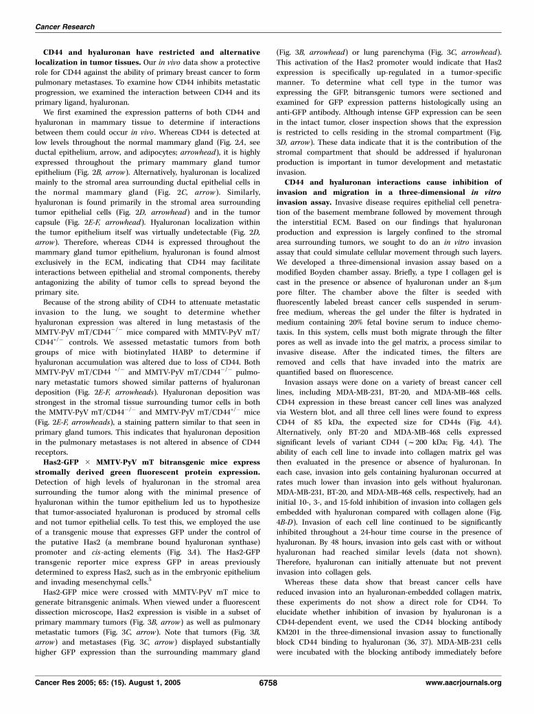
CD44 and hyaluronan have restricted and alternativelocalization in tumor tissues. Our in vivo data show a protectiverole for CD44 against the ability of primary breast cancer to formpulmonary metastases. To examine how CD44 inhibits metastaticprogression, we examined the interaction between CD44 and itsprimary ligand, hyaluronan.We first examined the expression patterns of both CD44 and
hyaluronan in mammary tissue to determine if interactionsbetween them could occur in vivo . Whereas CD44 is detected atlow levels throughout the normal mammary gland (Fig. 2A , seeductal epithelium, arrow, and adipocytes; arrowhead), it is highlyexpressed throughout the primary mammary gland tumorepithelium (Fig. 2B, arrow). Alternatively, hyaluronan is localizedmainly to the stromal area surrounding ductal epithelial cells inthe normal mammary gland (Fig. 2C, arrow ). Similarly,hyaluronan is found primarily in the stromal area surroundingtumor epithelial cells (Fig. 2D, arrowhead) and in the tumorcapsule (Fig. 2E-F, arrowhead). Hyaluronan localization withinthe tumor epithelium itself was virtually undetectable (Fig. 2D,arrow). Therefore, whereas CD44 is expressed throughout themammary gland tumor epithelium, hyaluronan is found almostexclusively in the ECM, indicating that CD44 may facilitateinteractions between epithelial and stromal components, therebyantagonizing the ability of tumor cells to spread beyond theprimary site.Because of the strong ability of CD44 to attenuate metastatic
invasion to the lung, we sought to determine whetherhyaluronan expression was altered in lung metastasis of theMMTV-PyV mT/CD44�/� mice compared with MMTV-PyV mT/CD44+/� controls. We assessed metastatic tumors from bothgroups of mice with biotinylated HABP to determine ifhyaluronan accumulation was altered due to loss of CD44. BothMMTV-PyV mT/CD44 +/� and MMTV-PyV mT/CD44�/� pulmo-nary metastatic tumors showed similar patterns of hyaluronandeposition (Fig. 2E-F, arrowheads). Hyaluronan deposition wasstrongest in the stromal tissue surrounding tumor cells in boththe MMTV-PyV mT/CD44�/� and MMTV-PyV mT/CD44+/� mice(Fig. 2E-F, arrowheads), a staining pattern similar to that seen inprimary gland tumors. This indicates that hyaluronan depositionin the pulmonary metastases is not altered in absence of CD44receptors.Has2-GFP � MMTV-PyV mT bitransgenic mice express
stromally derived green fluorescent protein expression.Detection of high levels of hyaluronan in the stromal areasurrounding the tumor along with the minimal presence ofhyaluronan within the tumor epithelium led us to hypothesizethat tumor-associated hyaluronan is produced by stromal cellsand not tumor epithelial cells. To test this, we employed the useof a transgenic mouse that expresses GFP under the control ofthe putative Has2 (a membrane bound hyaluronan synthase)promoter and cis-acting elements (Fig. 3A). The Has2-GFPtransgenic reporter mice express GFP in areas previouslydetermined to express Has2, such as in the embryonic epitheliumand invading mesenchymal cells.5
Has2-GFP mice were crossed with MMTV-PyV mT mice togenerate bitransgenic animals. When viewed under a fluorescentdissection microscope, Has2 expression is visible in a subset ofprimary mammary tumors (Fig. 3B, arrow) as well as pulmonarymetastatic tumors (Fig. 3C, arrow). Note that tumors (Fig. 3B,arrow) and metastases (Fig. 3C, arrow) displayed substantiallyhigher GFP expression than the surrounding mammary gland
(Fig. 3B, arrowhead) or lung parenchyma (Fig. 3C, arrowhead).This activation of the Has2 promoter would indicate that Has2expression is specifically up-regulated in a tumor-specificmanner. To determine what cell type in the tumor wasexpressing the GFP, bitransgenic tumors were sectioned andexamined for GFP expression patterns histologically using ananti-GFP antibody. Although intense GFP expression can be seenin the intact tumor, closer inspection shows that the expressionis restricted to cells residing in the stromal compartment (Fig.3D, arrow). These data indicate that it is the contribution of thestromal compartment that should be addressed if hyaluronanproduction is important in tumor development and metastaticinvasion.CD44 and hyaluronan interactions cause inhibition of
invasion and migration in a three-dimensional in vitroinvasion assay. Invasive disease requires epithelial cell penetra-tion of the basement membrane followed by movement throughthe interstitial ECM. Based on our findings that hyaluronanproduction and expression is largely confined to the stromalarea surrounding tumors, we sought to do an in vitro invasionassay that could simulate cellular movement through such layers.We developed a three-dimensional invasion assay based on amodified Boyden chamber assay. Briefly, a type I collagen gel iscast in the presence or absence of hyaluronan under an 8-Ampore filter. The chamber above the filter is seeded withfluorescently labeled breast cancer cells suspended in serum-free medium, whereas the gel under the filter is hydrated inmedium containing 20% fetal bovine serum to induce chemo-taxis. In this system, cells must both migrate through the filterpores as well as invade into the gel matrix, a process similar toinvasive disease. After the indicated times, the filters areremoved and cells that have invaded into the matrix arequantified based on fluorescence.Invasion assays were done on a variety of breast cancer cell
lines, including MDA-MB-231, BT-20, and MDA-MB-468 cells.CD44 expression in these breast cancer cell lines was analyzedvia Western blot, and all three cell lines were found to expressCD44 of 85 kDa, the expected size for CD44s (Fig. 4A).Alternatively, only BT-20 and MDA-MB-468 cells expressedsignificant levels of variant CD44 (f200 kDa; Fig. 4A). Theability of each cell line to invade into collagen matrix gel wasthen evaluated in the presence or absence of hyaluronan. Ineach case, invasion into gels containing hyaluronan occurred atrates much lower than invasion into gels without hyaluronan.MDA-MB-231, BT-20, and MDA-MB-468 cells, respectively, had aninitial 10-, 3-, and 15-fold inhibition of invasion into collagen gelsembedded with hyaluronan compared with collagen alone (Fig.4B-D). Invasion of each cell line continued to be significantlyinhibited throughout a 24-hour time course in the presence ofhyaluronan. By 48 hours, invasion into gels cast with or withouthyaluronan had reached similar levels (data not shown).Therefore, hyaluronan can initially attenuate but not preventinvasion into collagen gels.Whereas these data show that breast cancer cells have
reduced invasion into an hyaluronan-embedded collagen matrix,these experiments do not show a direct role for CD44. Toelucidate whether inhibition of invasion by hyaluronan is aCD44-dependent event, we used the CD44 blocking antibodyKM201 in the three-dimensional invasion assay to functionallyblock CD44 binding to hyaluronan (36, 37). MDA-MB-231 cellswere incubated with the blocking antibody immediately before
Cancer Research
Cancer Res 2005; 65: (15). August 1, 2005 6758 www.aacrjournals.org

their addition to the upper chamber of the assay. Wheninvading into gels containing hyaluronan, the addition of theblocking antibody results in increased invasiveness. By 24hours, KM201-treated cells invaded into gels containinghyaluronan more readily than into gels without hyaluronan(Fig. 4E), whereas the addition of an IgG isotype control
antibody does not affect invasion (Fig. 4F). Note that invasioninto gels lacking hyaluronan does not differ in the presence orabsence of KM201 (Fig. 4E). This shows that hyaluronan is notsimply providing steric hindrance to prevent cellular invasionbut that the effect is due to a specific ligand-receptorinteraction. This ties the role of hyaluronan during invasion
Figure 2. CD44 and hyaluronan have distinct but separate localization within MMTV-PyV mT breast tumors. MMTV-PyV mT nontransformed mammary glands (A andC) and tumors (B and D ) were fixed, sectioned, and probed with anti-CD44 antibodies (A and B) or HABP (C and D ). A, ductal epithelia are shown (arrow )surrounded by mammary gland adipocytes (arrowhead) and ECM. B, tumor epithelium (arrow ) and ECM (arrowhead) are designated. C, stromal area surroundingductal epithelial cells (arrow ) and the surrounding parenchyma (arrowhead). D, stromal tissue (arrowhead) surrounding the tumor epithelium (arrow ). MMTV-PyVmT/CD44�/� (E) and MMTV-PyV mT/CD44+/� (F ) pulmonary metastatic tumors were stained with HABP. Sections were counterstained with either DAPI (A-D ) orhematoxylin (E-F ) to show nuclei. Arrow, tumor epithelium; arrowhead, ECM.
CD44 and Hyaluronan Interactions Inhibit Metastasis
www.aacrjournals.org 6759 Cancer Res 2005; 65: (15). August 1, 2005

directly to the binding of CD44. Importantly, whereashyaluronan may inhibit cellular invasion in the presence ofCD44, the absence of CD44 may promote hyaluronan-mediatedinvasion.
Discussion
To determine the precise role of CD44 in tumor growth andmetastasis, we have crossed the MMTV-PyV mT onto a CD44�/�
background. The absence of CD44 in this model results in a striking5.7-fold increase in pulmonary metastases when compared withcontrol mice, whereas not affecting either primary tumor formationor growth rate. Additionally, there is a 5-fold increase in the numberof metastases per lung of the CD44 null mice compared with CD44-expressing mice. Based on epithelial localization of CD44 andstromal localization of hyaluronan, we developed an in vitroapproach to analyze these interactions. Three-dimensional in vitroinvasion assays show that CD44 in breast tumor epithelial cellsinhibits the ability of cells to invade hyaluronan-containing collagenmatrices. These data strongly suggest that epithelial-stromalinteractions in vivo need to be taken into account when identifyingroles for adhesion proteins in cancer progression.Hyaluronan can interact with several receptors in addition to
CD44, including the receptor for hyaluronic acid–mediated motility(RHAMM), LYVE-1, HARE, Layilin, and Toll-4 (2). Of these receptors,only CD44 and RHAMM are reported to be expressed in invasive
breast cancer (2, 38). RHAMM responds to hyaluronan binding byinducing cellular motility and transformation in vitro and ininjectable tumor models (39). Hence, hyaluronan activity in vivomay vary depending upon the receptor it interacts with in thetransformed state. The action of hyaluronan may be that ofhomotypic adhesion when binding to the CD44 receptor (as occursin tumor growth and encapsulation), while binding to an alternatereceptor, such as RHAMM or LYVE-1 on lymphatic endothelium,may induce an entirely different phenotype, that of invasion. Thesestudiesmay be pertinent to our findings with the CD44 null model. Itis possible that in the absence of CD44, hyaluronan interactions withan alternate receptor such as RHAMM are not balanced by the anti-invasive effects of CD44 binding.In regard to CD44 isoform expression, the CD44�/� mouse
model represents cells lacking all forms of CD44, whereas breastcancer cell lines typically express both standard and variantisoforms. In vitro studies have provided strong evidence to suggestthat variant CD44 expression on cancer cells may facilitate diseaseprogression and metastasis. Isoforms such as CD44v6, CD44v3,8-10, and CD44v10 have enhanced cellular metastasis when trans-fected into colon and breast cancer cell lines, whereas transfectionof CD44s into colorectal cells decreases metastasis (21, 22, 40, 41).Additional studies have also identified a number of partners thatCD44 may interact with to promote cell signaling that may lead totumor progression such as c-Src, ErbB2, ankyrin, or the ERMproteins (42–44). Taking this into account, two of the cell lines
Figure 3. Has2-GFP transgenic mice reveal restricted activation of hyaluronan synthase enzymes in MMTV-PyV mT mammary gland tumors. A, Has2-GFP constructshowing insertion of the Has2 promoter region before GFP. GFP detection is shown for a (B ) MMTV-PyV mT/Has2-GFP bitransgenic mouse mammary tumor and(C) pulmonary metastasis (arrows ). Surrounding tissue parenchyma (arrowheads ). D, MMTV-PyV mT/Has2-GFP bitransgenic mammary gland tumors were sectionedand treated with anti-GFP antibody. Arrows, GFP-positive cells in the stromal capsule surrounding the tumor; arrowheads, GFP-negative tumor epithelium. Tissuesections were counterstained with hematoxylin.
Cancer Research
Cancer Res 2005; 65: (15). August 1, 2005 6760 www.aacrjournals.org

used in our studies (BT-20 and MDA-MB-468) express variantisoforms; yet invasion in both of these cell lines are inhibited byhyaluronan-embedded collagen. Further studies examining the roleof CD44v in hyaluronan binding may clarify this issue.
In the CD44-deficient mouse model used in this study, neitherCD44s nor CD44v are expressed, and the only effect of completeCD44 absence can be evaluated. Transformed fibroblasts fromthe CD44�/� mice were previously shown to form significantly
Figure 4. CD44 required to reduce invasion of breast cancer cells into an hyaluronan (HA )–embedded collagen matrix. A, protein lysates from MDA-MB-231 (lane 1),BT20 (lane 2), and MDA-MB-468 (lane 3) metastatic breast cancer cell lines were separated by SDS-PAGE and immunoblotted with anti-CD44 antibodies(H-300). Invasion of MDA-MB-231 (B ), BT20 (C), or MDA-MB-468 (D ) cells in the presence (.) or absence (4) of hyaluronan. E , MDA-MB-231 cells were pretreatedwith either PBS (open symbols ) or KM201 blocking antibody (closed symbols ) and invaded into collagen gels either with (triangles ) or without (circles ) hyaluronan.F, MDA-MB-231 cells treated as described in (E), except IgG isotype control was used in place of KM201. B-F, cells were treated as described in Materials andMethods and invasion was measured as a fluorescent value. Three or more independent experiments were done for each invasion assay and each time point was donein triplicate in each experiment.
CD44 and Hyaluronan Interactions Inhibit Metastasis
www.aacrjournals.org 6761 Cancer Res 2005; 65: (15). August 1, 2005

larger tumors which develop at a much faster rate when injectedinto nude mice (32). Although we found no effect on primarytumor growth in our model, this disparity is probably due to thedifferences between injectable and transgenic models. Further-more, studies done by Weber et al. (45) using this model ofCD44 ablation (32) indicate that CD44 can promote metastaticinvasion in a p53-dependent model of osteosarcoma, while notaffecting primary tumor growth. This may be due to thealternative ECM components available in bone or perhapsalternative isoforms of CD44 expressed in osteosarcomascompared with breast cancer.Studies in our MMTV-PyV mT mouse model, crossed onto both
the CD44�/� and the Has2-GFP transgenic, provide compellingevidence for a protective role for CD44 against breast cancermetastasis. Whereas hyaluronan is highly expressed and eveninduced in the ECM of these tumors, loss of the primary receptorfor hyaluronan results in an increase in tumor metastasis. Thisstrongly implicates an adhesive role for the CD44/hyaluronaninteraction. Strikingly, our in vitro model precisely recapitulatedthis CD44/hyaluronan mechanism. Addition of the CD44 blockingantibody, essentially removing CD44/hyaluronan interactions,causes an increase in invasion over IgG treatment controls.Importantly, this only happens in the presence of hyaluronan notin collagen gels without hyaluronan. This indicates that in theabsence of CD44 expression, hyaluronan is prometastatic. Previous
studies show that high levels of hyaluronan in breast tumorstroma are correlated with poor patient survival (46). Our datasuggest that this correlation may be due to a loss of CD44sexpression in this patient group. Alternatively, hyaluronandeposition in vivo has been shown to inhibit tumor growth, ass.c. injection of hyaluronan into breast cancer xenografts inducedtumor regression (47). These findings suggest that stromallylocalized hyaluronan can also serve a protective role in breastcancer progression. These previous studies, in addition to ourcurrent study, indicate that the tumor microenvironment cansignificantly alter the effect of CD44 and hyaluronan expression intumor progression. In sum, these data indicate that epithelial-stromal interactions as well as ligand and isotype expressionprofiles should be considered in their entirety when evaluating arole for CD44 in breast cancer progression.
Acknowledgments
Received 3/15/2005; revised 5/3/2005; accepted 5/18/2005.Grant support: Susan G. Komen Breast Cancer Foundation grant for Basic,
Clinical and Translational Research BCTR0402597 (J. Schroeder).The costs of publication of this article were defrayed in part by the payment of page
charges. This article must therefore be hereby marked advertisement in accordancewith 18 U.S.C. Section 1734 solely to indicate this fact.
We thank Graeme Dougherty, Ray Nagle, Mamata Pochampalli, Rachid el Bejjani,Wendy Knowlton, and Andrew Chang for helpful comments on the article; WilliamMuller and Sandra Gendler for the use of the MMTV-PyVmT transgenic mice; and TakMak for the use of the CD44 null mice.
References1. Hanahan D, Weinberg RA. The hallmarks of cancer.Cell 2000;100:57–70.
2. Toole BP. Hyaluronan promotes the malignant phe-notype. Glycobiology 2002;12:37–42R.
3. Kosaki R, Watanabe K, Yamaguchi Y. Overproductionof hyaluronan by expression of the hyaluronan synthaseHas2 enhances anchorage-independent growth andtumorigenicity. Cancer Res 1999;59:1141–5.
4. Naor D, Nedvetzki S, Golan I, Melnik L, Faitelson Y.CD44 in cancer. Crit Rev Clin Lab Sci 2002;39:527–79.
5. Iida N, Bourguignon LY. New CD44 splice variantsassociated with human breast cancers. J Cell Physiol1995;162:127–33.
6. Naor D, Sionov RV, Ish-Shalom D. CD44: structure,function, and association with the malignant process.Adv Cancer Res 1997;71:241–319.
7. Droll A, Dougherty ST, Chiu RK, et al. Adhesiveinteractions between alternatively spliced CD44 iso-forms. J Biol Chem 1995;270:11567–73.
8. Bennett KL, Jackson DG, Simon JC, et al. CD44isoforms containing exon V3 are responsible for thepresentation of heparin-binding growth factor. J CellBiol 1995;128:687–98.
9. Kurozumi K, Nakao K, Nishida T, Nakahara M, OginoN, Tsujimoto M. Significance of biologic aggressivenessand proliferating activity in papillary thyroid carcinoma.World J Surg 1998;22:1237–42.
10. Daniel L, Lechevallier E, Giorgi R, et al. CD44s andCD44v6 expression in localized T1-T2 conventionalrenal cell carcinomas. J Pathol 2001;193:345–9.
11. Gotoda T, Matsumura Y, Kondo H, et al. Expressionof CD44 variants and its association with survival inpancreatic cancer. Jpn J Cancer Res 1998;89:1033–40.
12. de Alava E, Panizo A, Sola I, Rodriguez-Rubio FI,Pardo-Mindan FJ. CD44v6 expression is related toprogression in renal epithelial tumours. Histopathology1998;33:39–45.
13. Bankfalvi A, Terpe HJ, Breukelmann D, et al. Gainsand losses of CD44 expression during breast carcino-genesis and tumour progression. Histopathology 1998;33:107–16.
14. Bankfalvi A, Terpe HJ, Breukelmann D, et al.Immunophenotypic and prognostic analysis of E-
cadherin and h-catenin expression during breastcarcinogenesis and tumour progression: a comparativestudy with CD44. Histopathology 1999;34:25–34.
15. Kanke M, Fujii M, Kameyama K, et al. Role of CD44variant exon 6 in invasion of head and neck squamouscell carcinoma. Arch Otolaryngol Head Neck Surg2000;126:1217–23.
16. De Marzo AM, Bradshaw C, Sauvageot J, Epstein JI,Miller GJ. CD44 and CD44v6 downregulation in clinicalprostatic carcinoma: relation to Gleason grade andcytoarchitecture. Prostate 1998;34:162–8.
17. Aaltomaa S, Lipponen P, Ala-Opas M, Kosma VM.Expression and prognostic value of CD44 standard andvariant v3 and v6 isoforms in prostate cancer. Eur Urol2001;39:138–44.
18. Takahashi K, Stamenkovic I, Cutler M, Saya H,Tanabe KK. CD44 hyaluronate binding influencesgrowth kinetics and tumorigenicity of human coloncarcinomas. Oncogene 1995;11:2223–32.
19. Shtivelman E, Bishop JM. Expression of CD44 isrepressed in neuroblastoma cells. Mol Cell Biol 1991;11:5446–53.
20. Benard J. Genetic alterations associated with meta-static dissemination and chemoresistance in neuroblas-toma. Eur J Cancer 1995;31A:560–4.
21. Choi SH, Takahashi K, Eto H, Yoon SS, Tanabe KK.CD44s expression in human colon carcinomas influencesgrowth of liver metastases. Int J Cancer 2000;85:523–6.
22. Gunthert U, Hofmann M, Rudy W, et al. A new variantof glycoprotein CD44 confers metastatic potential to ratcarcinoma cells. Cell 1991;65:13–24.
23. Wallach-Dayan SB, Grabovsky V, Moll J, et al. CD44-dependent lymphoma cell dissemination: a cell surfaceCD44 variant, rather than standard CD44, supportsin vitro lymphoma cell rolling on hyaluronic acidsubstrate and its in vivo accumulation in the peripherallymph nodes. J Cell Sci 2001;114:3463–77.
24. Herrera-Gayol A, Jothy S. Adhesion proteins in thebiology of breast cancer: contribution of CD44. Exp MolPathol 1999;66:149–56.
25. Itano N, Atsumi F, Sawai T, et al. Abnormalaccumulation of hyaluronan matrix diminishes contactinhibition of cell growth and promotes cell migration.Proc Natl Acad Sci U S A 2002;99:3609–14.
26. Itano N, Sawai T, Miyaishi O, Kimata K. Relationship
between hyaluronan production and metastatic poten-tial of mouse mammary carcinoma cells. Cancer Res1999;59:2499–504.
27. Itano N, Sawai T, Atsumi F, et al. Selective expressionand functional characteristics of three mammalianhyaluronan synthases in oncogenic malignant transfor-mation. J Biol Chem 2004;279:18679–87.
28. Yeo TK, Nagy JA, Yeo KT, Dvorak HF, Toole BP.Increased hyaluronan at sites of attachment to mesen-tery by CD44-positive mouse ovarian and breast tumorcells. Am J Pathol 1996;148:1733–40.
29. Knudson W, Biswas C, Toole BP. Interactionsbetween human tumor cells and fibroblasts stimulatehyaluronate synthesis. Proc Natl Acad Sci U S A 1984;81:6767–71.
30. Asplund T, Versnel MA, Laurent TC, Heldin P.Human mesothelioma cells produce factors that stim-ulate the production of hyaluronan by mesothelial cellsand fibroblasts. Cancer Res 1993;53:388–92.
31. Heldin P, Asplund T, Ytterberg D, Thelin S, LaurentTC. Characterization of the molecular mechanisminvolved in the activation of hyaluronan synthetase byplatelet-derived growth factor in human mesothelialcells. Biochem J 1992;283:165–70.
32. Schmits R, Filmus J, Gerwin N, et al. CD44 regulateshematopoietic progenitor distribution, granuloma for-mation, and tumorigenicity. Blood 1997;90:2217–33.
33. Guy CT, Cardiff RD, Muller WJ. Induction ofmammary tumors by expression of polyomavirusmiddle T oncogene: a transgenic mouse model formetastatic disease. Mol Cell Biol 1992;12:954–61.
34. Maglione JE, Moghanaki D, Young LJ, et al. Trans-genic polyoma middle-T mice model premalignantmammary disease. Cancer Res 2001;61:8298–305.
35. Webster MA, Hutchinson JN, Rauh MJ, et al.Requirement for both Shc and phosphatidylinositol 3Vkinase signaling pathways in polyomavirus middle T-mediated mammary tumorigenesis. Mol Cell Biol1998;18:2344–59.
36. Monaghan M, Mulligan KA, Gillespie H, et al.Epidermal growth factor up-regulates CD44-depen-dent astrocytoma invasion in vitro . J Pathol 2000;192:519–25.
37. Miyake K, Medina KL, Hayashi S, Ono S, Hamaoka T,Kincade PW. Monoclonal antibodies to Pgp-1/CD44
Cancer Research
Cancer Res 2005; 65: (15). August 1, 2005 6762 www.aacrjournals.org

block lympho-hemopoiesis in long-term bone marrowcultures. J Exp Med 1990;171:477–88.
38. Jackson DG, Prevo R, Clasper S, Banerji S. LYVE-1,the lymphatic system and tumor lymphangiogenesis.Trends Immunol 2001;22:317–21.
39. Hall CL, Yang B, Yang X, et al. Overexpression ofthe hyaluronan receptor RHAMM is transforming andis also required for H-ras transformation. Cell 1995;82:19–26.
40. Bourguignon LY, Gunja-Smith Z, Iida N, et al.CD44v(3,8-10) is involved in cytoskeleton-mediatedtumor cell migration and matrix metalloproteinase(MMP-9) association in metastatic breast cancer cells.J Cell Physiol 1998;176:206–15.
41. Iida N, Bourguignon LY. Coexpression of CD44variant (v10/ex14) and CD44S in human mammaryepithelial cells promotes tumorigenesis. J Cell Physiol1997;171:152–60.
42. Bourguignon LY, Zhu H, Chu A, Iida N, Zhang L,Hung MC. Interaction between the adhesion receptor,CD44, and the oncogene product, p185HER2, promoteshuman ovarian tumor cell activation. J Biol Chem1997;272:27913–8.
43. Bourguignon LY, Zhu H, Shao L, Chen YW. CD44interaction with c-Src kinase promotes cortactin-mediated cytoskeleton function and hyaluronic acid-dependent ovarian tumor cell migration. J Biol Chem2001;276:7327–36.
44. Tsukita S, Oishi K, Sato N, Sagara J, Kawai A. ERMfamily members as molecular linkers between the cellsurface glycoprotein CD44 and actin-based cytoskele-tons. J Cell Biol 1994;126:391–401.
45. Weber GF, Bronson RT, Ilagan J, et al. Absence of theCD44 gene prevents sarcoma metastasis. Cancer Res2002;62:2281–6.
46. Auvinen P, Tammi R, Parkkinen J, et al. Hyaluronan inperitumoral stroma and malignant cells associates withbreast cancer spreading and predicts survival. Am JPathol 2000;156:529–36.
47. Herrera-Gayol A, Jothy S. Effect of hyaluronan onxenotransplanted breast cancer. Exp Mol Pathol 2002;72:179–85.
CD44 and Hyaluronan Interactions Inhibit Metastasis
www.aacrjournals.org 6763 Cancer Res 2005; 65: (15). August 1, 2005
Copyright © 2022 FDOKUMEN