A Mandibular Advancement Device Attenuates the Abnormal ...
20
Page 1/20 A Mandibular Advancement Device Attenuates the Abnormal Morphology and Function of Mitochondria from the Genioglossus in OSAHS Rabbits Chunyan Liu Hebei Medical University Shilong Zhang Hebei Medical University Dechao Zhu Hebei Medical University https://orcid.org/0000-0002-8237-1731 Dengying Fan Hebei Medical University Yahui Zhu Hebei Medical University Wenjing Kang Hebei Medical University Haiyan Lu ( [email protected] ) Hebei Medical University Jie Wang Hebei Medical University Research Keywords: Obstructive sleep apnea-hypopnea syndrome, Mandibular advancement device, Genioglossus, Mitochondria Posted Date: August 5th, 2021 DOI: https://doi.org/10.21203/rs.3.rs-622336/v1 License: This work is licensed under a Creative Commons Attribution 4.0 International License. Read Full License
-
Upload
khangminh22 -
Category
Documents
-
view
2 -
download
0
Transcript of A Mandibular Advancement Device Attenuates the Abnormal ...

Page 1/20
A Mandibular Advancement Device Attenuates theAbnormal Morphology and Function ofMitochondria from the Genioglossus in OSAHSRabbitsChunyan Liu
Hebei Medical UniversityShilong Zhang
Hebei Medical UniversityDechao Zhu
Hebei Medical University https://orcid.org/0000-0002-8237-1731Dengying Fan
Hebei Medical UniversityYahui Zhu
Hebei Medical UniversityWenjing Kang
Hebei Medical UniversityHaiyan Lu ( [email protected] )
Hebei Medical UniversityJie Wang
Hebei Medical University
Research
Keywords: Obstructive sleep apnea-hypopnea syndrome, Mandibular advancement device, Genioglossus,Mitochondria
Posted Date: August 5th, 2021
DOI: https://doi.org/10.21203/rs.3.rs-622336/v1
License: This work is licensed under a Creative Commons Attribution 4.0 International License. Read Full License

Page 2/20
AbstractBackground: To examine the morphology and function of mitochondria from the genioglossus in a rabbitmodel of obstructive sleep apnea-hypopnea syndrome (OSAHS), as well as these factors after insertionof a mandibular advancement device (MAD).
Methods: Thirty male New Zealand white rabbits were randomized into three groups: control, OSAHS andMAD, with 10 rabbits in each group. Animals in Group OSAHS and Group MAD were induced to developOSAHS by injection of gel into the submucosal muscular layer of the soft palate. The rabbits in GroupMAD were �tted with a MAD. The animals in the control group were not treated. Further,polysomnography (PSG) and CBCT scan were used to measure MAD effectiveness. CBCT of the upperairway and PSG suggested that MAD was effective. Rabbits in the three groups were induced to sleep for4–6 hours per day for 8 consecutive weeks. The genioglossus was harvested and detected by opticalmicroscopy and transmission electron microscopy. The mitochondrial membrane potential wasdetermined by laser confocal microscopy and �ow cytometry. Mitochondrial complex I and IV activitieswere detected by mitochondrial complex assay kits.
Results: OSAHS-like symptoms were induced successfully in Group OSAHS and rescued by MADtreatment. The relative values of the mitochondrial membrane potential, mitochondrial complex I activityand complex IV activity were signi�cantly lower in Group OSAHS than in the control group; however, therewas no signi�cant difference between Group MAD and the control group. The OSAHS-induced injury andthe dysfunctional mitochondria of the genioglossus muscle were reduced by MAD treatment.
Conclusion: Damaged mitochondrial structure and function were induced by OSAHS and could beattenuated by MAD treatment.
Contributions To The Literature1. This study suggested that MAD treatment attenuated the dysfunction of genioglossus mitochondria
caused by OSAHS, which could expand our understanding of the effect of MAD on the genioglossusduring OSAHS treatment.
2. This study will provide further evidence for the treatment of OSAHS with appliance in clinicalpractice.
3. This study will enrich our knowledge on the advantages of MAD treatment for OSAHS, to furtherguide clinicians' treatment options for OSAHS.
BackgroundObstructive sleep apnea-hypopnea syndrome (OSAHS) is a serious, potentially life-threatening conditioncharacterized by loud snoring and recurrent episodes of partial or complete upper airway (UA) collapseoccurring during sleep(1). OSAHS is the most common respiratory sleep disorder, and its prevalence is
Page 3/20
estimated to be up to 10% in middle-aged men and 3% in middle-aged women(2). The UA is susceptibleto collapse during sleep due to its lack of rigid bony support(3). Its lumen size depends on the balancebetween the UA dilator tension and upper airway negative pressure. Normally, the UA dilator muscles areresponsible for maintaining UA patency, especially the genioglossus, which is the most importantpharyngeal dilator muscle and plays a key role in the maintenance of UA patency during sleep(4).
Treatment options for OSAHS include behavioral modi�cation, continuous positive airway pressure(CPAP), mandibular advancement device (MAD)(5), surgical procedures(6), electrical stimulation(7) andpharmacological treatments. CPAP is recommended as the �rst-line therapy according to the treatmentguidelines for patients with moderate-to-severe OSAHS(8). CPAP can improve snoring, theapnea/hypopnea index (AHI) and daytime sleepiness through an air splint to maintain UA patency(9).However, CPAP is not accepted by all patients; thus, the effectiveness is unsatisfactory in some patientsdue to the limited adherence to this treatment(10). MAD has the advantages of being noninvasive, lowcost, comfortable and easy to carry. Therefore, this option is very popular for most OSAHS patients,especially for those who are neither able nor willing to tolerate CPAP therapy or surgery. To date, scholarspay more attention on changes in subjective symptoms, sleep quality and airway structure during MADtherapy for OSAHS. Our previous studies(11) found that abnormal contractility and �ber type distributionof the genioglossus could be caused by OSAHS. However, whether the mitochondria participate in thesetissue changes is unclear. As skeletal muscle, the function of genioglossus is closely related to �ber typesand energy metabolism(11). Thus, OSAHS-induced abnormality of genioglossus function may correlatewith mitochondrial dysfunction. Therefore, the aim of this study was to investigate whether OSAHSinduces mitochondrial structural damage and dysfunction. Moreover, considering the effectiveness ofMAD in treating patients with mild-to-moderate OSAHS, we investigated whether MAD treatment could beeffective against OSAHS-induced mitochondrial structural damage and dysfunction.
MethodsAll experimental protocols and animal studies were submitted to and approved by local animalcommittee. The �owchart of the study was shown in Fig. 1. All methods in this study were performed inaccordance with the medical ethics committee, and an additional �le shows this in more details [seeAdditional �le 1]. Animal use and care was in accordance with the guidelines of the medical ethicscommittee for the housing and care of animals bred, supplied and used for scienti�c purposes. Allexperiments were performed in accordance with relevant guidelines and regulations. The study wascarried out in compliance with the ARRIVE guidelines.
Animal Model DevelopmentThirty New Zealand white rabbits (initial weight, 3 kg–3.5 kg) were equally divided into three experimentalgroups, namely the control group, Group OSAHS and Group MAD, with 10 animals in each group. Theanimals were kept at 22°C–25°C with free access to food and water. The animal models were developedas described in our previous studies(11, 13, 14). Brie�y, OSAHS was induced by Medical Sodium

Page 4/20
Hyaluronate Gel (Shanghai Qisheng Biological Preparation Co., Ltd. Shanghai, China) injection via thesubmucosal muscular layer at the center of the soft palate, approximately 1.5 cm away from the junctionof the hard and soft palates. The animals in Group MAD were given MAD to alleviate the OSAHSsymptoms. Animals in the control group were not given any treatment.
Con�rmation of induced OSAHSCone-beamcomputedtomography (CBCT) and polysomnography (PSG) were conducted. A CBCT scan ofthe UA was performed with a CBCT machine (KaVo 3D eXam, USA). The parameters were set as follows:the current size of 5 mA, the voltage magnitude of 120 kV, the scan time of 17.8 s and the layer thicknessof 0.3 mm. Then, 3D reconstruction by the manufacturer was conducted, and the retropalatal space in theUA was examined from the top, 1/4, 1/2, and 3/4 levels to the bottom of the soft palate(15).
PSG (Rembrandt Embla Polysomnography System, Reykjavik, Iceland) was used to monitor the sleepparameters. PSG recordings were conducted as described in our previous studies(11, 13). Brie�y, therabbits were equipped with surface electrodes taped on the skull, face, and chest for monitoring theelectroencephalogram, electrooculogram and respiration. Nasal air �ow, respiratory movements, bloodoxygen saturation (SaO2), and AHI were scored for all animals, the scoring method was the same as thatwe previously used(11, 13, 15).
OSAHS rabbits in Group MAD were given the MAD. This device was made of self-curing composite resinand bonded to the upper incisors with glass ionomer, with a 30° inclined plane to the long axis of theupper incisors(11, 13). The mandible was guided forward by 3–4 mm. After 3–5 days of adaptation, norabbits had di�culty eating or had signs of distress.
No animals showed di�culties eating or drinking. Chloral hydrate was infused orally to induce sleep in asupine position for 4–6 hours per day for the next eight weeks. Basic health data, including body weightand behavior, were recorded at 2 weeks and 8 weeks.
Preparation of the genioglossus muscle tissuesThe observation period lasted for 8 weeks. Finally, the genioglossus was harvested and �xed in 10%buffered formalin for 24 hours and then routinely embedded in para�n. The sections were stained withhematoxylin/eosin and examined under light microscopy (Olympus, Japan). A portion of thegenioglossus was quickly dissected into a 1 mm × 1 mm× 3 mm3 piece, which was �xed in a 4%cryopreservation glutaraldehyde solution and then underwent preparation for conventional transmissionelectron microscopy to examine the ultrastructure.
Mitochondrial functions of the genioglossusMitochondrial isolation of the genioglossus was performed in strict accordance with the instructions forthe mitochondria extraction kit using a buffer containing 180 mM KCl, 10 mM EDTA(Ethylene DiamineTetraacetic Acid), and 0.5% albumin at pH 7.4. The mitochondrial membrane potential (ΔΨm) wasestimated with a membrane electrical potential assay kit. After JC-1( CBIC2) �uorescent probe loading,

Page 5/20
�ow cytometric analysis was conducted to quantitatively determine the relative value of themitochondrial membrane potential. The �uorescence signals of the JC-1 monomer (red �uorescence) andpolymer (green �uorescence) were obtained in the �uorescence 1 (FL1), and �uorescence 2 (FL2)detectors. A �ow diagram was obtained from �ow cytometry and analyzed by Exp032ADC analysissoftware. Comparisons of the �uorescence intensities of red �uorescence and green �uorescence couldre�ect the mitochondrial membrane potential. The mitochondrial membrane potential was qualitativelydetermined by laser confocal microscopy. The excitation wavelengths were 488 nm and 525 nm. Theappearance of green �uorescence or a decreased intensity of red �uorescence indicated that themitochondrial membrane potential decreased.
Mitochondrial complex I and IV activitiesThe quantitative detection was in strict accordance with the mitochondrial respiratory chain complex Iactivity and complex IV activity assay kit. The activities of the samples were calculated according to theformula shown in the kit.
The results were analyzed with SPSS 22.0 software (SPSS, Chicago, USA). Different parameters wereassessed for normally distributed data. All data are expressed as the mean ± SD. The statisticalsigni�cance of differences was assessed by analysis of variance (ANOVA) after normality and varianceequality were tested, and the LSD test was used to compare the differences among three groups. A pvalue < 0.05 was considered statistically signi�cant.
Results
Rabbit behaviorsAt baseline,2 weeks and 8 weeks after modeling, there was no signi�cant difference in body weight andfood intake among the three groups (Fig. 2).
The induced OSAHS-like symptomsOSAHS-like symptoms were induced successfully in Group OSAHS, which showed snoring and apnea orhypopnea during supine sleep, accompanied by interrupted sleep, but these symptoms were not detectedin the animals in Group MAD and the controls.
The retropalatal space in the UA was signi�cantly decreased in Group OSAHS (p < 0.05), and MADenlarged the retropalatal UA (Fig. 3). PSG also showed signi�cantly higher AHI and lower SaO2 levels inGroup OSAHS than in Group MAD and the control group (p < 0.05) (Fig. 4). All of these results suggestedthat OSAHS was successfully induced and that MAD effectively alleviated the OSAHS-like symptoms.
The morphology of the genioglossus

Page 6/20
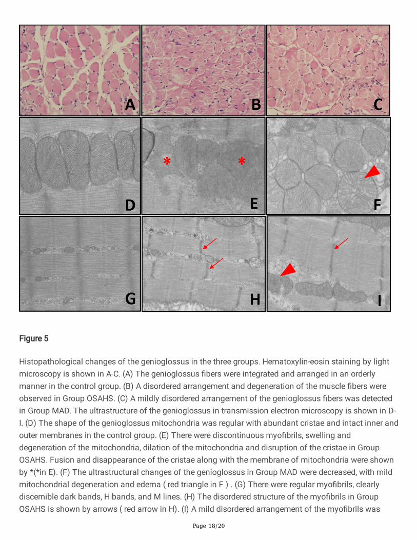
HE staining showed that varying degrees of disordered arrangement of the muscle �bers in GroupOSAHS. Disorders and degeneration of the genioglossus �bers were not detected in Group MAD and inthe control group (Fig. 5).
As for the ultrastructure of the genioglossus, the control group showed regular myo�brils. In GroupOSAHS, discontinuous myo�brils, swelling and degeneration of the mitochondria, dilation of themitochondria and disruption of the cristae were detected. Some mitochondria dissolved or evendisappeared. Compared with Group OSAHS, the ultrastructural changes of genioglossus in Group MADwere less severe, with a mildly disordered arrangement of the myo�brils and mild mitochondrialdegeneration and edema (Fig. 5).
Mitochondrial functions of the genioglossusFlow cytometric analysis and laser scanning confocal microscopy showed that the relative value of themitochondrial membrane potential was signi�cantly lower in Group OSAHS than that in the control group.There was no signi�cant difference in the relative value of the mitochondrial membrane potentialbetween Group MAD and the control group (Fig. 6).
Mitochondrial complex I activity and complex IV activity were signi�cantly decreased in Group OSAHScompared with the control group (p < 0.05). However, MAD treatment attenuated the effect of OSAHS oncomplex I activity and complex IV activities (Fig. 7).
DiscussionIn this study, we detected genioglossus injury, dysfunctional mitochondria and decreased mitochondrialrespiratory chain complex I and
IV activities of the genioglossus in a rabbit model of OSAHS, these results described a possiblemechanism, supporting of our previous reports on genioglossus fatigue. As in other diseases, researchbased on animal models is crucially important in OSAHS(11, 13). The model of intermittent hypoxia wasa widely documented method of inducing OSAHS(17). Although this model appeared to demonstratesimilar symptoms to OSAHS patients, it was obviously limited by the absence of recurrent UAobstructions, apnea, increased inspiratory effort and sleep fragmentation(18). In these cases, the aimwas to obtain an OSAHS model induced by UA obstruction and demonstrate all the typical characteristicsof OSAHS, such as intermittent hypoxia, increased inspiratory effort and sleep fragmentation. Anapproach to induce OSAHS and insert MAD in rabbits was developed in our laboratory. Technical successwas achieved, and the MAD was well tolerated in the rabbits, as shown by the body weight and foodintake.
This study found that OSAHS caused abnormal morphology of genioglossus, such as degeneration ofthe genioglossus �bers and disordered mitochondrial ultrastructure, including discontinuous myo�brils,dilation of the mitochondria and disruption of the cristae. Accordingly, we previously found that OSAHSresulted in genioglossus fatigue in vitro(11). However, the detailed molecular mechanisms remain to be

Page 7/20
elucidated. Previous reports demonstrated that the genioglossus may be more vulnerable to fatigue inOSAHS patients(19, 20) and animal models of OSAHS than in controls(21–23). Therefore, identi�cationof an underlying contributory mechanism of genioglossus fatigue is important and has therapeuticimplications. The function of skeletal muscle is intimately linked to the proper function of mitochondriabecause mitochondria constitute the main energy supply for contraction of skeletal muscle. We intendedto examine whether genioglossus fatigue was related to mitochondria. Consistent with our hypothesis,mitochondrial abnormalities, such as decreased mitochondrial membrane potential and decreasedmitochondrial respiratory chain complex activity, as well as dysfunctional mitochondrial ultrastructure ofgenioglossus, were revealed in Group OSAHS. These �ndings could explain why the abnormal changes inthe structure and contractile properties of the genioglossus were observed in our previous studies.
With regard to ΔΨm, our data showed that the mitochondria isolated from the animals with OSAHS had alower ΔΨm than those of the controls. Since mitochondrial membrane potential is a key indicatorre�ecting mitochondrial function and ΔΨm provides reliable information on muscle function anddysfunction(24), the data suggested that OSAHS could indeed cause mitochondrial dysfunction.However, a simple analysis of mitochondrial membrane potential is insu�cient to determine themechanisms underlying the damage to genioglossus function and the effectiveness of the MADtreatment. Since respiratory chain complexes I, III and IV generate ΔΨm as a result of energy transferthrough the electron transport chain(25), to further clarify the genioglossus mitochondrial function, weevaluated the mitochondrial respiratory chain complexes in the present study. We found that thesecomplexes were also severely affected by OSAHS; thus, we con�rmed that OSAHS clearly affectsgenioglossus mitochondrial function.
To the best of our knowledge, this is the �rst study examining genioglossus mitochondrial functionality inOSAHS models and after MAD treatment. We hypothesize that the mitochondrial dysfunction andmorphological abnormalities observed in the animals with OSAHS are due to one or more of the followingcauses. First, chronic intermittent hypoxia during repeated apnea or hypopnea results in mitochondrialdysfunction. UA closure during sleep is associated with oxygen desaturation, which terminates when anarousal transiently interrupts sleep. Then, apneas can recur as sleep resumes, contributing to thepathogenesis of chronic intermittent hypoxia. A study showed that hypoxia in OSAHS patients impairedUA muscle activity(26). Another report demonstrated that hypoxia could increase oxidative stress andimpair mitochondrial function in mouse skeletal muscle because hypoxia affected both the mitochondrialphosphorylation e�ciency and the coupling between respiration and ATP synthesis. Similarly, a previousstudy showed that the hypoxia-induced mitochondrial dysfunction and the inner and outer mitochondrialmembrane integrity were signi�cantly affected by hypoxia exposure(28). Therefore, mitochondrialdysfunction may be closely related to chronic intermittent hypoxia, the most basic physical characteristicof OSAHS. Second, repeated bursts of forceful contraction may lead to mitochondrial abnormalities ingenioglossus. Genioglossus actively compensate for the narrowed upper airway in OSAHS duringwakefulness, which is supported by the research that OSAHS patients have increased GG activationrelative to controls during wakefulness(29) and there was greater reduction in GG activation in OSApatients than in controls after CPAP treatment, implying that the enhanced activity is a compensatory
Page 8/20
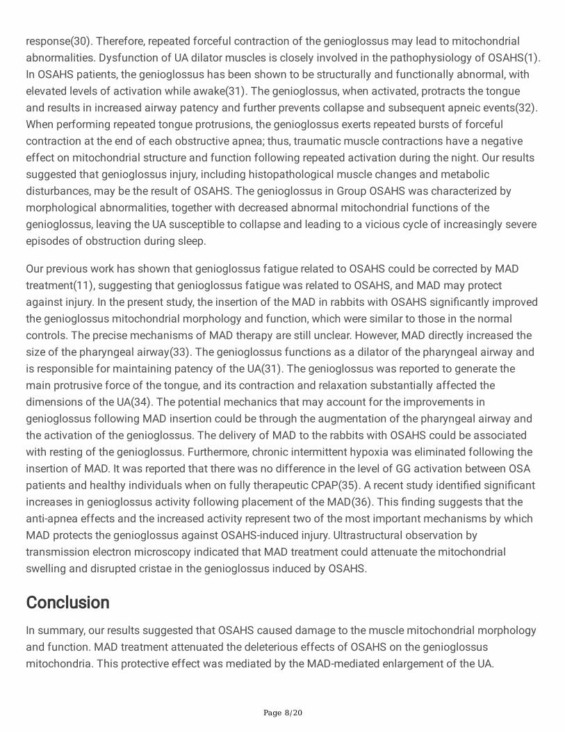
response(30). Therefore, repeated forceful contraction of the genioglossus may lead to mitochondrialabnormalities. Dysfunction of UA dilator muscles is closely involved in the pathophysiology of OSAHS(1).In OSAHS patients, the genioglossus has been shown to be structurally and functionally abnormal, withelevated levels of activation while awake(31). The genioglossus, when activated, protracts the tongueand results in increased airway patency and further prevents collapse and subsequent apneic events(32).When performing repeated tongue protrusions, the genioglossus exerts repeated bursts of forcefulcontraction at the end of each obstructive apnea; thus, traumatic muscle contractions have a negativeeffect on mitochondrial structure and function following repeated activation during the night. Our resultssuggested that genioglossus injury, including histopathological muscle changes and metabolicdisturbances, may be the result of OSAHS. The genioglossus in Group OSAHS was characterized bymorphological abnormalities, together with decreased abnormal mitochondrial functions of thegenioglossus, leaving the UA susceptible to collapse and leading to a vicious cycle of increasingly severeepisodes of obstruction during sleep.
Our previous work has shown that genioglossus fatigue related to OSAHS could be corrected by MADtreatment(11), suggesting that genioglossus fatigue was related to OSAHS, and MAD may protectagainst injury. In the present study, the insertion of the MAD in rabbits with OSAHS signi�cantly improvedthe genioglossus mitochondrial morphology and function, which were similar to those in the normalcontrols. The precise mechanisms of MAD therapy are still unclear. However, MAD directly increased thesize of the pharyngeal airway(33). The genioglossus functions as a dilator of the pharyngeal airway andis responsible for maintaining patency of the UA(31). The genioglossus was reported to generate themain protrusive force of the tongue, and its contraction and relaxation substantially affected thedimensions of the UA(34). The potential mechanics that may account for the improvements ingenioglossus following MAD insertion could be through the augmentation of the pharyngeal airway andthe activation of the genioglossus. The delivery of MAD to the rabbits with OSAHS could be associatedwith resting of the genioglossus. Furthermore, chronic intermittent hypoxia was eliminated following theinsertion of MAD. It was reported that there was no difference in the level of GG activation between OSApatients and healthy individuals when on fully therapeutic CPAP(35). A recent study identi�ed signi�cantincreases in genioglossus activity following placement of the MAD(36). This �nding suggests that theanti-apnea effects and the increased activity represent two of the most important mechanisms by whichMAD protects the genioglossus against OSAHS-induced injury. Ultrastructural observation bytransmission electron microscopy indicated that MAD treatment could attenuate the mitochondrialswelling and disrupted cristae in the genioglossus induced by OSAHS.
ConclusionIn summary, our results suggested that OSAHS caused damage to the muscle mitochondrial morphologyand function. MAD treatment attenuated the deleterious effects of OSAHS on the genioglossusmitochondria. This protective effect was mediated by the MAD-mediated enlargement of the UA.
Page 9/20
AbbreviationsOSAHS: Obstructive sleep apnea-hypopnea syndrome
MAD: Mandibular advancement device
PSG: Polysomnography
UA: Upper airway
CPAP: Continuous positive airway pressure
CBCT: Cone-beamcomputedtomography
SaO2: Blood oxygen saturation
FL1: Fluorescence 1
FL2: Fluorescence 2
DeclarationsAcknowledgements
We gratefully acknowledge study participants who took their valuable time to participate in this study.
Funding
Funding/Support: This study was funded by Natural Science Foundation of China (NSFC) (GrantNo.81701010 ), the Training Program of Clinical Medicine Talents Funded by the Government of HebeiProvince (Grant No. 361029) , Youth Foundation of Hebei Education Department (Grant No. QN2017109 )and Natural Science Foundation of Hebei Province(Grant No. H2021206431)
Availability of data and materials
The data underlying this article will be shared on reasonable request to the corresponding author.
Authors' information
A�liations
Chunyan Liu, Shilong Zhang, Dechao Zhu, Dengying Fan, Yahui Zhu, Wenjing Kang, Haiyan Lu
Department of Orthodontics, School and Hospital of Stomatology, Hebei Medical University & Hebei KeyLaboratory of Stomatology & Hebei Clinical Research Center for Oral Diseases, Shijiazhuang, 050017, PRChina.

Page 10/20
Jie Wang
Department of Oral Pathology, School and Hospital of Stomatology, Hebei Medical University & Hebei KeyLaboratory of Stomatology & Hebei Clinical Research Center for Oral Diseases, Shijiazhuang, 050017, PRChina.
Authors' Contributions
Chunyan Liu, Haiyan Lu and Jie Wang led the conception and design of the study, was closely involved indata analysis and interpretation. Chunyan Liu, Shilong Zhang and Dechao Zhu were responsible forraising animals. Chunyan Liu, Shilong Zhang, Dechao Zhu, Dengying Fan, Yahui Zhu, Wenjing Kang wereresponsible for establishing animal models. Dechao Zhu, Dengying Fan, Yahui Zhu, Wenjing Kang wereresponsible for all the experiments and acquiring data. All authors reviewed the manuscript.
Corresponding author
Correspondence to Haiyan Lu and Jie Wang
Ethics declarations
Ethics approval and consent to participate
Approval to conduct the study was gained from the medical ethics committee in Hospital of Stomatology,Hebei Medical University.
Consent for publication
Yes
Competing interests
The authors declare that they have no competing interests.
References1. Patil SP, Schneider H, Schwartz AR, Smith PL. Adult obstructive sleep apnea: pathophysiology and
diagnosis. CHEST. [Journal Article; Research Support, N.I.H., Extramural; Review]. 2007 2007-07-01;132(1):325-37.
2. Peppard PE, Young T, Barnet JH, Palta M, Hagen EW, Hla KM. Increased prevalence of sleep-disordered breathing in adults. AM J EPIDEMIOL. [Journal Article; Research Support, N.I.H.,Extramural]. 2013 2013-05-01;177(9):1006-14.
3. Horner RL. The neuropharmacology of upper airway motor control in the awake and asleep states:implications for obstructive sleep apnoea. Respir Res. [Journal Article; Research Support, Non-U.S.Gov't; Review]. 2001 2001-01-20;2(5):286-94.

Page 11/20
4. Remmers JE, DeGroot WJ, Sauerland EK, Anch AM. Pathogenesis of upper airway occlusion duringsleep. J Appl Physiol Respir Environ Exerc Physiol. [Journal Article; Research Support, U.S. Gov't,P.H.S.]. 1978 1978-06-01;44(6):931-8.
5. John CR, Gandhi S, Sakharia AR, James TT. Maxillomandibular advancement is a successfultreatment for obstructive sleep apnoea: a systematic review and meta-analysis. Int J Oral MaxillofacSurg. [Journal Article; Meta-Analysis; Systematic Review]. 2018 2018-12-01;47(12):1561-71.
�. Epstein LJ, Kristo D, Strollo PJ, Friedman N, Malhotra A, Patil SP, et al. Clinical guideline for theevaluation, management and long-term care of obstructive sleep apnea in adults. J CLIN SLEEPMED. [Journal Article; Practice Guideline]. 2009 2009-06-15;5(3):263-76.
7. Certal VF, Zaghi S, Riaz M, Vieira AS, Pinheiro CT, Kushida C, et al. Hypoglossal nerve stimulation inthe treatment of obstructive sleep apnea: A systematic review and meta-analysis. LARYNGOSCOPE.[Journal Article; Meta-Analysis; Review; Systematic Review]. 2015 2015-05-01;125(5):1254-64.
�. Qaseem A, Holty JE, Owens DK, Dallas P, Starkey M, Shekelle P. Management of obstructive sleepapnea in adults: A clinical practice guideline from the American College of Physicians. ANN INTERNMED. [Journal Article; Practice Guideline]. 2013 2013-10-01;159(7):471-83.
9. McDaid C, Duree KH, Gri�n SC, Weatherly HL, Stradling JR, Davies RJ, et al. A systematic review ofcontinuous positive airway pressure for obstructive sleep apnoea-hypopnoea syndrome. SLEEP MEDREV. [Comparative Study; Journal Article; Research Support, Non-U.S. Gov't; Review; SystematicReview]. 2009 2009-12-01;13(6):427-36.
10. Hirshkowitz M, Sharafkhaneh A. Positive airway pressure therapy of OSA. Semin Respir Crit CareMed. [Journal Article; Review]. 2005 2005-02-01;26(1):68-79.
11. Liu CY, Lu HY, Dong FS, Ma WS, Wang J, Hu XY, et al. Effects of a mandibular advancement deviceon genioglossus in obstructive sleep apnoea hypopnea syndrome. Eur J Orthod. [Comparative Study;Journal Article]. 2015 2015-06-01;37(3):290-6.
12. Pathi B, Kinsey ST, Locke BR. In�uence of reaction and diffusion on spatial organization ofmitochondria and effectiveness factors in skeletal muscle cell design. BIOTECHNOL BIOENG.[Journal Article; Research Support, U.S. Gov't, Non-P.H.S.]. 2011 2011-08-01;108(8):1912-24.
13. Lu HY, Dong F, Liu CY, Wang J, Liu Y, Xiao W. An animal model of obstructive sleep apnoea-hypopneasyndrome corrected by mandibular advancement device. Eur J Orthod. [Journal Article]. 2015 2015-06-01;37(3):284-9.
14. Liu C, Kang W, Zhang S, Qiao X, Yang X, Zhou Z, et al. Mandibular Advancement Devices Prevent theAdverse Cardiac Effects of Obstructive Sleep Apnea-Hypopnea Syndrome (OSAHS). Sci Rep. [JournalArticle; Research Support, Non-U.S. Gov't]. 2020 2020-02-25;10(1):3394.
15. Zhu D, Kang W, Zhang S, Qiao X, Liu J, Liu C, et al. Effect of mandibular advancement devicetreatment on HIF-1alpha, EPO and VEGF in the myocardium of obstructive sleep apnea-hypopneasyndrome rabbits. Sci Rep. [Journal Article; Research Support, Non-U.S. Gov't]. 2020 2020-08-06;10(1):13261.
Page 12/20
1�. Kemnitz JW, Schultz-Darken N, Tapscott SJ. An IACUC perspective on animal models of sleep-disordered breathing. ILAR J. [Journal Article]. 2009 2009-01-20;50(3):312-3.
17. Farre R, Montserrat JM, Navajas D. Morbidity due to obstructive sleep apnea: insights from animalmodels. CURR OPIN PULM MED. [Journal Article; Research Support, Non-U.S. Gov't; Review]. 20082008-11-01;14(6):530-6.
1�. Nacher M, Farre R, Montserrat JM, Torres M, Navajas D, Bulbena O, et al. Biological consequences ofoxygen desaturation and respiratory effort in an acute animal model of obstructive sleep apnea(OSA). SLEEP MED. [Journal Article; Research Support, Non-U.S. Gov't]. 2009 2009-09-01;10(8):892-7.
19. Eckert DJ, Lo YL, Saboisky JP, Jordan AS, White DP, Malhotra A. Sensorimotor function of the upper-airway muscles and respiratory sensory processing in untreated obstructive sleep apnea. J ApplPhysiol (1985). [Journal Article; Research Support, N.I.H., Extramural; Research Support, Non-U.S.Gov't]. 2011 2011-12-01;111(6):1644-53.
20. McSharry D, O'Connor C, McNicholas T, Langran S, O'Sullivan M, Lowery M, et al. Genioglossusfatigue in obstructive sleep apnea. Respir Physiol Neurobiol. [Journal Article]. 2012 2012-08-15;183(2):59-66.
21. Bradford A, McGuire M, O'Halloran KD. Does episodic hypoxia affect upper airway dilator musclefunction? Implications for the pathophysiology of obstructive sleep apnoea. Respir PhysiolNeurobiol. [Journal Article; Research Support, Non-U.S. Gov't; Review]. 2005 2005-07-28;147(2-3):223-34.
22. Fuller DD, Fregosi RF. Fatiguing contractions of tongue protrudor and retractor muscles: in�uence ofsystemic hypoxia. J Appl Physiol (1985). [Journal Article; Research Support, U.S. Gov't, P.H.S.]. 20002000-06-01;88(6):2123-30.
23. Pae EK, Wu J, Nguyen D, Monti R, Harper RM. Geniohyoid muscle properties and myosin heavy chaincomposition are altered after short-term intermittent hypoxic exposure. J Appl Physiol (1985).[Comparative Study; Journal Article; Research Support, N.I.H., Extramural; Research Support, Non-U.S.Gov't; Research Support, U.S. Gov't, P.H.S.]. 2005 2005-03-01;98(3):889-94.
24. Chen LB. Mitochondrial membrane potential in living cells. Annu Rev Cell Biol. [Journal Article;Research Support, U.S. Gov't, P.H.S.; Review]. 1988 1988-01-19;4:155-81.
25. Huttemann M, Helling S, Sanderson TH, Sinkler C, Samavati L, Mahapatra G, et al. Regulation ofmitochondrial respiration and apoptosis through cell signaling: cytochrome c oxidase andcytochrome c in ischemia/reperfusion injury and in�ammation. Biochim Biophys Acta. [JournalArticle; Research Support, N.I.H., Extramural; Research Support, Non-U.S. Gov't; Review]. 2012 2012-04-01;1817(4):598-609.
2�. Okabe S, Hida W, Kikuchi Y, Kurosawa H, Midorikawa J, Chonan T, et al. Upper airway muscle activityduring sustained hypoxia in awake humans. J Appl Physiol (1985). [Journal Article]. 1993 1993-10-01;75(4):1552-8.
27. Magalhaes J, Ascensao A, Soares JM, Ferreira R, Neuparth MJ, Marques F, et al. Acute and severehypobaric hypoxia increases oxidative stress and impairs mitochondrial function in mouse skeletal

Page 13/20
muscle. J Appl Physiol (1985). [Journal Article]. 2005 2005-10-01;99(4):1247-53.
2�. Magalhaes J, Ferreira R, Neuparth MJ, Oliveira PJ, Marques F, Ascensao A. Vitamin E preventshypobaric hypoxia-induced mitochondrial dysfunction in skeletal muscle. Clin Sci (Lond). [JournalArticle; Research Support, Non-U.S. Gov't]. 2007 2007-12-01;113(12):459-66.
29. Mezzanotte WS, Tangel DJ, White DP. Waking genioglossal electromyogram in sleep apnea patientsversus normal controls (a neuromuscular compensatory mechanism). J CLIN INVEST. [JournalArticle; Research Support, U.S. Gov't, Non-P.H.S.; Research Support, U.S. Gov't, P.H.S.]. 1992 1992-05-01;89(5):1571-9.
30. Fogel RB, Malhotra A, Pillar G, Edwards JK, Beauregard J, Shea SA, et al. Genioglossal activation inpatients with obstructive sleep apnea versus control subjects. Mechanisms of muscle control. Am JRespir Crit Care Med. [Journal Article; Research Support, Non-U.S. Gov't; Research Support, U.S. Gov't,P.H.S.]. 2001 2001-12-01;164(11):2025-30.
31. Fogel RB, Trinder J, White DP, Malhotra A, Raneri J, Schory K, et al. The effect of sleep onset on upperairway muscle activity in patients with sleep apnoea versus controls. J Physiol. [Comparative Study;Journal Article; Research Support, N.I.H., Extramural; Research Support, U.S. Gov't, P.H.S.]. 2005 2005-04-15;564(Pt 2):549-62.
32. Fogel RB, Malhotra A, Pillar G, Edwards JK, Beauregard J, Shea SA, et al. Genioglossal activation inpatients with obstructive sleep apnea versus control subjects. Mechanisms of muscle control. Am JRespir Crit Care Med. [Journal Article; Research Support, Non-U.S. Gov't; Research Support, U.S. Gov't,P.H.S.]. 2001 2001-12-01;164(11):2025-30.
33. Ryan CF, Love LL, Peat D, Fleetham JA, Lowe AA. Mandibular advancement oral appliance therapyfor obstructive sleep apnoea: effect on awake calibre of the velopharynx. THORAX. [Journal Article;Research Support, Non-U.S. Gov't]. 1999 1999-11-01;54(11):972-7.
34. Alonso JF, Mananas MA, Rojas M, Bruce EN. Coordination of respiratory muscles assessed by meansof nonlinear forecasting of demodulated myographic signals. J Electromyogr Kinesiol. [JournalArticle]. 2011 2011-12-01;21(6):1064-73.
35. Jordan AS, Wellman A, Heinzer RC, Lo YL, Schory K, Dover L, et al. Mechanisms used to restoreventilation after partial upper airway collapse during sleep in humans. THORAX. [Journal Article;Research Support, N.I.H., Extramural; Research Support, Non-U.S. Gov't]. 2007 2007-10-01;62(10):861-7.
3�. Johal A, Gill G, Ferman A, McLaughlin K. The effect of mandibular advancement appliances onawake upper airway and masticatory muscle activity in patients with obstructive sleep apnoea. ClinPhysiol Funct Imaging. [Clinical Trial; Journal Article]. 2007 2007-01-01;27(1):47-53.
Figures

Page 14/20
Figure 1
Flowchart of the experimental design. OSAHS was induced by gel injection via the soft palate of NewZealand white rabbits, and then, the rabbits in Group MAD had the MAD inserted. There were threegroups: control, OSAHS, and MAD. CBCT scans of the UA and PSG were used to evaluate the effect ofMAD treatment. All animals were induced to sleep in a supine position for 4–6 hours per day. After 8weeks, the genioglossus samples were harvested for evaluation of mitochondrial morphology andfunction.

Page 15/20
Figure 2
Animal modeling and supine sleep. (A) Diagram of the injection site. (B) The arrow indicates the injectionsite of the soft palate, approximately 1.5 cm from the junction of the hard and soft palate. UI meansUpper incisors. LI means Lower incisors. J means The junction of the hard and soft palate. T meansTongue. (C) The sleeping rabbits in the supine position were placed in the homemade experimental setup.(D) Body weight of the animals in the three groups. There were no signi�cant differences among the three

Page 16/20
groups. (E) Food intake of the animals in the three groups. There were no signi�cant differences amongthe three groups.
Figure 3
CBCT of the upper airway. Representative 3D images of the UA of the control group (A and D), GroupOSAHS (B and E), and Group MAD (C and F). (G) The UA volume signi�cantly decreased in Group OSAHScompared with the control group, and MAD could increase the airway volume. (H) The cross-sectionalarea signi�cantly decreased in Group OSAHS compared with the control group, and MAD increased thecross-sectional area at the upper, 1/4, 2/4 and 3/4 levels. (I) The sagittal diameter of the UA signi�cantlydecreased in Group OSAHS compared with the control group, and MAD increased the sagittal diameter atthe 1/4, 2/4, 3/4 and 4/4 levels. *p<0.05.

Page 17/20
Figure 4
PSG of the three groups. A representative pattern of the 30 s recordings in the control group (A), GroupOSAHS (B), and Group MAD (C). There was a cease in nasal air�ow (apnea) with an associated increasein respiratory effort in Group OSAHS (B). The breathing movements and air�ow were even and smooth inthe control group (A) and Group MAD (C). LOC and ROC: left and right electrooculograms. C3/A2, C4/A1,O1/A2: electroencephalogram leads. FLOW: nasal air�ow. CHEST and ABDM: chest and abdominal straingauges. Snore: microphone recordings of snoring. (C) There was a signi�cant increase in the AHI and adecrease in the minimum SaO2 (E) and average SaO2 (F) in Group OSAHS compared with the controlgroup (p < 0.05). The decreased min SaO2 and average SaO2 in Group OSAHS were signi�cantly rescuedby MAD treatment (E and F). PSG showed that MAD signi�cantly rescued the abnormal respiratoryfunction and sleep parameters. *p<0.05 vs the controls.
Page 18/20
Figure 5
Histopathological changes of the genioglossus in the three groups. Hematoxylin-eosin staining by lightmicroscopy is shown in A-C. (A) The genioglossus �bers were integrated and arranged in an orderlymanner in the control group. (B) A disordered arrangement and degeneration of the muscle �bers wereobserved in Group OSAHS. (C) A mildly disordered arrangement of the genioglossus �bers was detectedin Group MAD. The ultrastructure of the genioglossus in transmission electron microscopy is shown in D-I. (D) The shape of the genioglossus mitochondria was regular with abundant cristae and intact inner andouter membranes in the control group. (E) There were discontinuous myo�brils, swelling anddegeneration of the mitochondria, dilation of the mitochondria and disruption of the cristae in GroupOSAHS. Fusion and disappearance of the cristae along with the membrane of mitochondria were shownby *(*in E). (F) The ultrastructural changes of the genioglossus in Group MAD were decreased, with mildmitochondrial degeneration and edema ( red triangle in F ) . (G) There were regular myo�brils, clearlydiscernible dark bands, H bands, and M lines. (H) The disordered structure of the myo�brils in GroupOSAHS is shown by arrows ( red arrow in H). (I) A mild disordered arrangement of the myo�brils was

Page 19/20
observed in the MAD group( red arrow in I). These results suggested that the genioglossus injuryassociated with OSAHS can be rescued by MAD treatment.
Figure 6
Laser scanning confocal microscopy and �ow cytometric analysis. (A) The mitochondrial membranepotential (△Ψm) of the genioglossus was high in the control group, with mostly red �uorescence. (B)△Ψm of the genioglossus was decreased in Group OSAHS, with green �uorescence. (C) △Ψm of thegenioglossus was high in Group MAD, and green �uorescence was rare, with mostly red �uorescence. The�ow diagram shows that cells labeled with green �uorescence could be observed in Q3 of the graphics,and cells labeled with red �uorescence could be observed in Q1 of the graphics. (D)△Ψm of thegenioglossus was signi�cantly lower in Group OSAHS than in the control group, and there was nosigni�cant difference between the control group and Group MAD. The asterisk * represents p<0.05compared with the controls. (E) There was a high percentage of cells labeled with red �uorescence in thecontrol group. (F) There was a high percentage of cells labeled with green �uorescence in Group OSAHS.(G) There was a high percentage of cells labeled with red �uorescence in Group MAD.

Page 20/20
Figure 7
Complex I and complex IV activities of the genioglossus in the three groups. Complex I and complex IVactivities of the genioglossus were signi�cantly lower in Group OSAHS than in the control group, andthere was no signi�cant difference between the control group and Group MAD. The asterisk * representsp<0.05 compared with the controls, the bars in the �gures are SDs
Supplementary Files
This is a list of supplementary �les associated with this preprint. Click to download.
Additional�le1.doc
Additional�le2.doc