The potential protective role of grape seed proanthocyanidin ...
Upload
khangminh22Category
view
2download
0
�����������������
Citation: Calabriso, N.; Massaro, M.;
Scoditti, E.; Verri, T.; Barca, A.;
Gerardi, C.; Giovinazzo, G.;
Carluccio, M.A. Grape Pomace
Extract Attenuates Inflammatory
Response in Intestinal Epithelial and
Endothelial Cells: Potential Health-
Promoting Properties in Bowel
Inflammation. Nutrients 2022, 14,
1175. https://doi.org/10.3390/
nu14061175
Academic Editor: Rosa Casas
Received: 17 February 2022
Accepted: 9 March 2022
Published: 11 March 2022
Publisher’s Note: MDPI stays neutral
with regard to jurisdictional claims in
published maps and institutional affil-
iations.
Copyright: © 2022 by the authors.
Licensee MDPI, Basel, Switzerland.
This article is an open access article
distributed under the terms and
conditions of the Creative Commons
Attribution (CC BY) license (https://
creativecommons.org/licenses/by/
4.0/).
nutrients
Article
Grape Pomace Extract Attenuates Inflammatory Response inIntestinal Epithelial and Endothelial Cells: PotentialHealth-Promoting Properties in Bowel InflammationNadia Calabriso 1,* , Marika Massaro 1, Egeria Scoditti 1 , Tiziano Verri 2 , Amilcare Barca 2 ,Carmela Gerardi 3, Giovanna Giovinazzo 3 and Maria Annunziata Carluccio 1,*
1 National Research Council (CNR) Institute of Clinical Physiology (IFC), 73100 Lecce, Italy;[email protected] (M.M.); [email protected] (E.S.)
2 Department of Biological and Environmental Sciences and Technologies (DISTEBA), University of Salento,73100 Lecce, Italy; [email protected] (T.V.); [email protected] (A.B.)
3 National Research Council (CNR) Institute of Sciences of Food Production (ISPA), 73100 Lecce, Italy;[email protected] (C.G.); [email protected] (G.G.)
* Correspondence: [email protected] (N.C.); [email protected] (M.A.C.)
Abstract: Inflammatory bowel disease (IBD) implies the chronic inflammation of the gastrointestinaltract, combined with systemic vascular manifestations. In IBD, the incidence of cardiovascular diseaseappears to be related to an increase of oxidative stress and endothelial dysfunction. Grape pomacecontains high levels of anti-oxidant polyphenols that are able to counteract chronic inflammatorysymptoms. The aim of this study was to determine whether grape pomace polyphenolic extract(GPE) was able to mitigate the overwhelming inflammatory response in enterocyte-like cells and toimprove vascular function. Intestinal epithelial Caco-2 cells, grown in monolayers or in co-culturewith endothelial cells (Caco-2/HMEC-1), were treated with different concentrations of GPE (1, 5,10 µg/mL gallic acid equivalents) for 2 h and then stimulated with lipopolysaccharide (LPS) andtumor necrosis factor (TNF)-α for 16 h. Through multiple assays, the expression of intestinal andendothelial inflammatory mediators, intracellular reactive oxygen species (ROS) levels and NF-κBactivation, as well as endothelial-leukocyte adhesion, were evaluated. The results showed that GPEsupplementation prevented, in a concentration-dependent manner, the intestinal expression and re-lease of interleukin (IL)-6, monocyte chemoattractant protein (MCP)-1, and matrix metalloproteinases(MMP)-9 and MMP-2. In Caco-2 cells, GPE also suppressed the gene expression of several pro-inflammatory markers, such as IL-1β, TNF-α, macrophage colony-stimulating factor (M-CSF), C-X-Cmotif ligand (CXCL)-10, intercellular adhesion molecule (ICAM)-1, vascular cell adhesion molecule(VCAM)-1, and cyclooxygenase (COX)-2. The GPE anti-inflammatory effect was mediated by theinhibition of NF-κB activity and reduced intracellular ROS levels. Furthermore, transepithelial GPEsuppressed the endothelial expression of IL-6, MCP-1, VCAM-1, and ICAM-1 and the subsequentadhesion of leukocytes to the endothelial cells under pro-inflammatory conditions. In conclusion, ourfindings suggest grape pomace as a natural source of polyphenols with multiple health-promotingproperties that could contribute to the mitigation of gut chronic inflammatory diseases and improvevascular endothelial function.
Keywords: gut inflammation; endothelial dysfunction; pro-inflammatory markers; leukocyte adhe-sion; grape pomace; polyphenols; oxidative stress; gene expression
1. Introduction
Cardiovascular diseases (CVD) represent the main cause of mortality and morbid-ity worldwide, due to the continuous increase in the prevalence of cardiovascular riskfactors. Several studies have shown heightened risk of cardiovascular complications inchronic inflammatory disorders, especially those affecting the gastrointestinal tract, such as
Nutrients 2022, 14, 1175. https://doi.org/10.3390/nu14061175 https://www.mdpi.com/journal/nutrients

Nutrients 2022, 14, 1175 2 of 20
inflammatory bowel diseases (IBD) [1–3]. IBD and CVD share similar immune responsein chronic systemic inflammation and atherogenesis [4]. In the course of IBD, increasingconcentrations of pro-inflammatory cytokines may lead to endothelial dysfunction andCVD development [5]. Endothelial dysfunction is a systemic disorder characterized byimbalanced vasodilation and vasoconstriction, elevated reactive oxygen species (ROS), andpro-inflammatory factors, as well as deficiency of nitric oxide bioavailability [6,7]. In par-ticular, in both CVD and IBD, pro-inflammatory angiogenesis is recognized as a commontrait, sustaining both atherosclerotic plaque growth and intestinal inflammation [8,9]. Theinflamed intestinal mucosa itself seems to play an important role in promoting arterialdisease [10]. IBD patients have a disrupted mucosal barrier, and, as a consequence, pro-inflammatory and pro-angiogenic products through the gut lining may enter the circulationand directly promote inflammation by activating immune cells and endothelial cells, knowntriggers in the onset and progression of CVD [11]. Finally, during IBD flares the adhesionof circulating monocytes to the intestinal microvascular endothelial cells, as well as theirinfiltration and transformation into macrophages occur, in close analogy to what occursin the early phases of atherosclerosis [12,13]. The occurrence of endothelial dysfunctiondisrupts the endothelial barrier permeability, which is part of the inflammatory response inthe development of CVD. The boosted expression of endothelial adhesion molecules, suchas vascular cell adhesion molecule (VCAM)-1, intercellular adhesion molecule (ICAM)-1,and of chemoattractants, is involved in the recruitment of monocytes/macrophages toendothelial cells, which is an obliged step in atherosclerotic development and progres-sion [14].
The Mediterranean diet, characterized by a high intake of vegetables and fruits, exhibitshealthy properties and provides significant reduction of chronic disease risk, attributable tothe synergistic actions of anti-oxidant and anti-inflammatory compounds [15,16]. Adherenceto the Mediterranean diet is low in IBD patients [17]. Therefore, additional or alternativestrategies to reduce cardiovascular risk and curb intestinal and systemic inflammationare highly desirable. Grapes are one of the largest fruit crops in the world, and almostall of the production (about 80% of the yield) is used for wine making. Due to the wideavailability of grape skins, the utilization of grape by-products has attracted increasingattention for their potential health benefits not only for their antioxidant activity, but alsofor their antibacterial, anti-inflammatory, and anticarcinogenic properties [18,19]. Thesebiological properties are believed to be due to the functions of polyphenols and dietaryfibers still contained in grape pomace after grape fermentation. Among grape pomacecompounds with high nutraceutical value [20,21], polyphenols such as phenolic acids,flavanols, proanthocyanidins, flavonols, anthocyanins, and stilbenes are the most interest-ing due to their antioxidant, anti-inflammatory, anti-neurodegenerative, anti-microbial,anti-cancer, and cardioprotective activities [19,22]. For these reasons, grape pomace couldbe exploited as a natural source of functional polyphenolic compounds for food or pharma-cology use [21,23,24]. The incorporation of these remnants into food products or their useas food supplements could be useful for health promotion and chronic disease prevention,including IBD [25].
Our group has already shown anti-oxidant properties of those polyphenols andpolyphenolic extracts from skin pomace in non-intestinal in vitro models. These dietarypolyphenols were able to attenuate endothelial dysfunction and reduce leukocyte adhesionto the endothelium [26,27]. Moreover, recent evidence reports grape pomace to improvethe gut microbiota by increasing the beneficial bacteria and decreasing the harmful bac-teria, as well as reducing the level of pro-inflammatory cytokines [19,28]. However, thebiological health potential of grape pomace-derived polyphenols on intestinal and vascularinflammation and the underlying mechanisms of action are not entirely clear.
The aim of the present study was to analyze whether a polyphenol extract of grapemarc inhibits intestinal and endothelial inflammation. For this purpose, we used an in vitroinflammatory model consisting of intestinal epithelial cells and endothelial cells that be-come dysfunctional upon pro-inflammatory stimuli. As intestinal cells, we used Caco-2

Nutrients 2022, 14, 1175 3 of 20
cells, which are able to spontaneously differentiate into an enterocyte-like phenotype [29,30].Differentiated Caco-2 cells were stimulated with pathological concentrations of lipopolysac-charide (LPS) and tumor necrosis factor-α (TNF-α) to mimic the gut inflammatory milieu.The effects of grape pomace extract (GPE) on inflammatory markers in intestinal cells wereevaluated, and the underlying mechanism of action was explored assessing intracellularROS levels and NF-κB activation. Moreover, the crosstalk between intestinal and endothe-lial cells was investigated by analyzing the transepithelial effect of GPE on endothelialdysfunction. Finally, the leukocyte adhesion to inflamed endothelium was evaluated.
2. Materials and Methods2.1. Reagents
Reagents were acquired from various suppliers: cyanidin 3-O-glucoside chloride,rutin (quercetin 3-O-rutinoside), and chlorogenic acid (5-caffeoylquinic acid) were pur-chased from Extrasynthèse (Genay, France); gallic acid, Folin–Ciocalteu phenol reagent,Trolox ((S)-(-)-6-hydroxy-2,5,7,8 tetramethylchroman-2-carboxylic acid), fluorescein dis-odium, ABTS (2,2′-azino-bis (3-ethylbenzothiazoline-6-sulfonic acid)), AAPH (2,2′-azobis(2-methyl-propionamide)), acetonitrile, formic acid, ethanol, and organic acids (all HPLC-grade) were acquired from Sigma-Aldrich (St. Louis, MO, USA). Milli-Q water was used(Merck Millipore, Darmstadt, Germany).
2.2. Grape Pomace Polyphenolic Extract
Grape pomace (Vitis vinifera L., cv Negramaro) was obtained from a winemakingfacility (Azienda Agricola Cantele, Guagnano, Lecce, Italy). The wet pomace was dried in anoven at 50 ◦C until constant weight. Subsequently, the skins were manually recovered fromthe pomace samples and stored at room temperature in the dark until further processing.
Polyphenol compounds were extracted from a fine powder of grape pomace obtainedby freezing the samples in liquid nitrogen and grinding with a blender. The samples (1 g)were treated with 10 mL of methanol/ethanol (80:20, v/v) in an ultrasound bath (LabsonicFalc, LBS1-H3) at 35 kHz and 88 W for 5 min. Samples were extracted at room temperaturefor 16 h in the dark under continuous stirring. Extraction mixtures were centrifuged(4000× g) for 5 min, the supernatants were collected, and the solvent was evaporated underN2 flow. Dried grape pomace extracts (GPE) were solubilized in 70% ethanol and stored at−20 ◦C until analysis.
2.3. High-Performance Liquid Chromatography (HPLC) Characterization of Polyphenols
To quantify the polyphenolic molecules in alcoholic extracts, we performed HPLCanalysis using an Agilent-1100 liquid chromatograph equipped with a DAD detector(Agilent 1100 HPLC system, Santa Clara, CA, USA) as described by Gerardi et al. [21]. Thechromatographic analysis was performed by comparing each peak retention time with theretention time and UV-visible spectra of external standards.
2.4. Folin–Ciocalteu Assay
A rapid method [31] was used to measure the total phenols in alcoholic and waterextracts from dried whole and skin pomace in 96-well plates (Corning) using a microplatereader (Infinite 200 Pro, Tecan, Männedorf, Switzerland). Folin–Ciocalteu reagent (1:5, v/v,50 µL) and sodium hydroxide solution (0.35 mol/L, 100 µL) were added to each well. Theabsorbance value at 760 nm was recorded after 5 min incubation. Gallic acid was usedto obtain a calibration curve in the range from 2.5 to 40.0 mg/L (R ≥ 0.9997). Gallic acidequivalents (GAE) were used to express the total phenol content of different samples.
2.5. Trolox Equivalent Antioxidant Capacity (TEAC) Assay
The TEAC assay was performed as previously reported [21]. The ABTS radical, dilutedin PBS (pH 7.4), showed an absorbance value of 0.4 (read at 734 nm). A volume of 200 µLof diluted ABTS was added to 10 µL of extract. Then, the absorbance value was recorder at

Nutrients 2022, 14, 1175 4 of 20
734 nm after 6 min using a plate reader (Infinite 200 Pro, Tecan, Männedorf, Switzerland).TEAC values were obtained considering the percentage inhibition at 734 nm with Troloxas standard (0–16 µmol/L were used to obtain a standard curve). TEAC values wereexpressed as Trolox equivalents (µmol/g).
2.6. Oxygen Radical Absorbance Capacity (ORAC) Assay
The ORAC procedure was accomplished as per Gerardi et al. [21]. Briefly, the re-action was performed using a 96-well plate reader (Infinite 200 Pro, Tecan, Männedorf,Switzerland) in a 75 mmol/L phosphate buffer (pH 7.4), in 200 µL final reaction volume.The mixture of dried grape pomace extracts (20 µL) and fluorescein solutions (120 µL;70 nmol/L) was heated at 37 ◦C for 15 min. Then, 2,2′-azobis-(2-methylpropionamidine)dihydrochloride was added, and the fluorescence was recorded (excitation and emissionwavelengths of 485 and 527 nm, respectively) every minute for 60 min. A blank usingphosphate buffer instead of the sample was carried out in each assay and all the reactionmixtures were assessed in triplicate. Decay curves (fluorescence intensity vs. time) wererecorded and the net area under the curve was calculated by subtracting the blank valuefrom that of sample or standard. The antioxidant capacity was quantified using the an-tioxidant Trolox as a standard. Final ORAC values were expressed as Trolox equivalents(µmol/g) of dried grape pomace extract.
2.7. Cell Cultures
The human colorectal adenocarcinoma-derived intestinal epithelial cell line Caco-2was obtained from the American Tissue Culture Collection (Rockville, MD, USA) andcultured in Dulbecco’s modified Eagle’s medium (DMEM) supplemented with 10% heat-inactivated fetal bovine serum (FBS), 2 mmol/L glutamine, 1% non-essential amino acids,100 U/mL penicillin, and 100 µg/mL streptomycin, in monolayers at 37 ◦C in a humidifiedatmosphere of 5% CO2 as previously described [32]. The human microvascular endothelialcell line (HMEC-1), obtained from Dr. Thomas J. Lawley, was cultured in MCBD 131 sup-plemented with 10% FBS, 2 mmol/L glutamine, and 100 U/mL penicillin and 100 µg/mLstreptomycin, and grown at 37 ◦C in a humidity-controlled 5% CO2 cell culture incubator,as previously described [33,34]. Human monocytoid THP-1 cells were obtained from theAmerican Tissue Culture Collection (Rockville, MD, USA) and maintained in RPMI 1640medium supplemented with 10% FBS, 2 mmol/L glutamine, and 100 U/mL penicillin and100 µg/mL streptomycin in a 5% CO2 humidified atmosphere at 37 ◦C.
2.8. Cell Viability
Cell viability was determined by a 3-(4,5-dimethylthiazol-2-yl)-2,5-diphenyl tetra-zolium bromide (MTT) assay, which is a commonly used method to evaluate cell survival,on the basis of the ability of viable cells to convert MTT, a soluble tetrazolium salt, intoan insoluble formazan precipitate, which is then quantitated spectrophotometrically [35].Briefly, cells were seeded on the 96-well culture plates and treated with various concentra-tion of GPE (1, 5, 10, and 25 µg/mL GAE) for 24 h. Then, cells were incubated with MTT(0.5 mg/mL) for 4 h. After that, the formazan products were dissolved in isopropanol andabsorbance was measured at 540 nm using a microplate reader. The results were expressedas percentage compared to untreated cells.
2.9. Treatments of Caco-2 Cell Monolayers
For the experiments, Caco-2 cells were grown on 12-well plates for 21 days to obtainspontaneous differentiation towards enterocyte-like cells, replacing the medium every2 days, as previously described [32]. After differentiation, cell culture medium was shiftedto 3% FBS, and then Caco-2 cells were treated with different concentrations of GPE (1, 5, and10 µg/mL GAE) for 2 h; after that, cells were stimulated with LPS 10 µg/mL and TNF-α10 ng/mL for 16 h (Figure 1A). Cells exposed to LPS plus TNF-α only were consideredinflamed controls (positive), whereas those without any treatment were considered negative

Nutrients 2022, 14, 1175 5 of 20
controls. After incubation, culture media were collected in sterile microtubes and the cellswere lysed with TRIzol reagent solution (Thermo Fisher Scientific, Waltham, MA, USA)or specific lysis buffer to obtain RNA or nuclear protein, respectively. Culture media,as well as cell lysates, were stored at −80 ◦C until analysis. Three biological replicateswere performed.
Nutrients 2022, 14, x FOR PEER REVIEW 5 of 20
days, as previously described [32]. After differentiation, cell culture medium was shifted
to 3% FBS, and then Caco-2 cells were treated with different concentrations of GPE (1, 5,
and 10 μg/mL GAE) for 2 h; after that, cells were stimulated with LPS 10 µg/mL and TNF-
α 10 ng/mL for 16 h (Figure 1A). Cells exposed to LPS plus TNF-α only were considered
inflamed controls (positive), whereas those without any treatment were considered nega-
tive controls. After incubation, culture media were collected in sterile microtubes and the
cells were lysed with TRIzol reagent solution (Thermo Fisher Scientific, Waltham, MA,
USA) or specific lysis buffer to obtain RNA or nuclear protein, respectively. Culture me-
dia, as well as cell lysates, were stored at −80 °C until analysis. Three biological replicates
were performed.
Figure 1. Experimental design. Caco-2 cell monolayers (A); Caco-2/HMEC-1 co-culture system (B)
and subsequent adhesion of human leukocytes to HMEC-1 (C). GPE: Grape pomace extract; LPS:
Lipopolysaccharide; TNF-α: Tumor necrosis factor-α.
2.10. Caco-2/HMEC-1 Co-Culture System and Treatments
For the co-culture experiments, Caco-2 cells were grown on semipermeable filters
over 21 days to differentiate and develop an enterocyte-like phenotype, as previously de-
scribed [36–38]. Then, Caco-2 cells were moved to 6- or 12-well plates, containing HMEC-
1 cells on the bottom compartment. GPE (1, 5, and 10 μg/mL GAE) was added on the
apical compartment for 2 h. Afterwards, LPS (10 μg/mL) and TNF-α (10 ng/mL) were ap-
plied on the apical compartment, and TNF-α (10 ng/mL) on the basolateral compartment
for 16 h (Figure 1B).
2.11. Leukocyte Adhesion Assay
After the above-described treatments, trans-well inserts containing Caco-2 monolay-
ers were removed and HMEC-1 was washed with DPBS before leukocyte assay (Figure
1C). THP-1 cells were labeled with 1 μmol/L calcein AM (Molecular Probes, a brand of
Thermo Fisher Scientific, Waltham, MA, USA) for 30 min in DMEM medium containing
3% FBS. In a co-culture system, labelled THP-1 cells were seeded at 5 × 10⁵ cell density
onto the HMEC-1 monolayer and incubated under rotating conditions (63 rpm) at 21 °C,
as previously described [26]. After a gentle wash to remove unattached cells, the adherent
THP-1 cells were observed under fluorescence microscope. Alternatively, the fluorescence
intensity was measured in a microplate reader with an excitation/emission wavelength of
485/530 nm.
2.12. Detection of Endothelial Cell Surface Molecules
After the treatments described above, the trans-well inserts containing Caco-2 mon-
olayers were removed, and HMEC-1 was used for the detection of endothelial cell surface
molecules VCAM-1 and ICAM-1 by cell surface enzyme immunoassays (EIA), as de-
scribed previously [26].
2.13. Measure of Inflammation Marker Release
The conditioned media from Caco-2 monolayers and the basolateral compartment of
Caco-2/HMEC-1 co-culture were collected, and the levels of secreted IL-6, MCP-1, MMP-
Figure 1. Experimental design. Caco-2 cell monolayers (A); Caco-2/HMEC-1 co-culture system(B) and subsequent adhesion of human leukocytes to HMEC-1 (C). GPE: Grape pomace extract; LPS:Lipopolysaccharide; TNF-α: Tumor necrosis factor-α.
2.10. Caco-2/HMEC-1 Co-Culture System and Treatments
For the co-culture experiments, Caco-2 cells were grown on semipermeable filtersover 21 days to differentiate and develop an enterocyte-like phenotype, as previouslydescribed [36–38]. Then, Caco-2 cells were moved to 6- or 12-well plates, containing HMEC-1 cells on the bottom compartment. GPE (1, 5, and 10 µg/mL GAE) was added on the apicalcompartment for 2 h. Afterwards, LPS (10 µg/mL) and TNF-α (10 ng/mL) were appliedon the apical compartment, and TNF-α (10 ng/mL) on the basolateral compartment for16 h (Figure 1B).
2.11. Leukocyte Adhesion Assay
After the above-described treatments, trans-well inserts containing Caco-2 monolayerswere removed and HMEC-1 was washed with DPBS before leukocyte assay (Figure 1C).THP-1 cells were labeled with 1 µmol/L calcein AM (Molecular Probes, a brand of ThermoFisher Scientific, Waltham, MA, USA) for 30 min in DMEM medium containing 3% FBS. In aco-culture system, labelled THP-1 cells were seeded at 5 × 10⁵ cell density onto the HMEC-1 monolayer and incubated under rotating conditions (63 rpm) at 21 ◦C, as previouslydescribed [26]. After a gentle wash to remove unattached cells, the adherent THP-1 cellswere observed under fluorescence microscope. Alternatively, the fluorescence intensity wasmeasured in a microplate reader with an excitation/emission wavelength of 485/530 nm.
2.12. Detection of Endothelial Cell Surface Molecules
After the treatments described above, the trans-well inserts containing Caco-2 mono-layers were removed, and HMEC-1 was used for the detection of endothelial cell surfacemolecules VCAM-1 and ICAM-1 by cell surface enzyme immunoassays (EIA), as describedpreviously [26].
2.13. Measure of Inflammation Marker Release
The conditioned media from Caco-2 monolayers and the basolateral compartment ofCaco-2/HMEC-1 co-culture were collected, and the levels of secreted IL-6, MCP-1, MMP-9,and MMP-2 were determined using the corresponding enzyme-linked immunosorbentassay (ELISA) kits, according to the manufacturer’s instructions.
2.14. RNA Isolation and Real-Time Quantitative PCR
Total RNA was extracted from Caco-2 monolayers and HMEC-1 in co-culture systems,using the TRIzol reagent (Thermo Fisher Scientific, Waltham, MA, USA), and quantifiedspectrophotometrically. Total RNA (1 µg) was converted into first-strand cDNA using

Nutrients 2022, 14, 1175 6 of 20
the High-Capacity cDNA Reverse Transcription Kit (Applied Biosystems, Monza, Italy).Quantitative RT-PCR was performed in CFX384 Touch Real-Time PCR Detection System(Bio-Rad Laboratories, Milan, Italy) using a SYBR Green PCR Master Mix (Bio-Rad Labora-tories, Milan, Italy) and the synthesized primers (Thermo Fisher Scientific, Waltham, MA,USA) reported in Table 1. All reactions were assessed in triplicate. The mRNA quantitywas calculated by comparative critical threshold method [38]. As endogenous referenceribosomal RNA 18 S and GAPDH was simultaneously quantified for each sample, andthe data were normalized accordingly. Results are expressed as fold increase relative tounstimulated control (=1).
Table 1. Oligonucleotides used for quantitative real-time PCR analysis.
Gene Name Accession Number Forward Primer Reverse Primer Size (bp)
IL1B NM_000576.2 5′-CTGTCCTGCGTGTTGAAAGA-3′ 5′-AGTTATATCCTGGCCGCCTT-3′ 228IL6 NM_000600.3 5′-AGGAGACTTGCCTGGTGAAA-3′ 5′-CAGGGGTGGTTATTGCATCT-3′ 180TNF NM_000594.2 5′-CCTGTGAGGAGGACGAACAT-3′ 5′-AGGCCCCAGTTTGAATTCTT-3′ 240CXCL10 NM_001565.2 5′-CAAGGATGGACCACACAGAG-3′ 5′-GCAGGGTCAGAACATCCACT-3′ 248CCL2/MCP1 NM_002982.3 5′-CCCCAGTCACCTGCTGTTAT-3′ 5′-TCCTGAACCCACTTCTGCTT-3′ 166CSF1/MCSF NM_000757.4 5′-TGGACGCACAGAACAGTCTC-3′ 5′-CCTCCAGGGCTCACAATAAA-3′ 235PTGS2/COX-2 NM_000963.2 5′-TGCTGTGGAGCTGTATCCTG-3′ 5′-GAAACCCACTTCTCCACCA-3′ 176VCAM1 NM_00107B.3 5′-CATGGAATTCGAACCCAAAC-3′ 5′-CCTGGCTCAAGCATGTCATA-3′ 140ICAM1 NM_000201.2 5′-AGACATAGCCCCACCATGAG-3′ 5′-CAAGGGTTGGGGTCAGTAGA-3′ 190MMP9 NM_004994.2 5′-AAAGCCTATTTCTGCCAGGAC-3′ 5′-GTGGGGATTTACATGGCACT-3′ 157MMP2 NM_004530.4 5′-CACTTTCCTGGGCAACAAAT-3′ 5′-TGATGTCATCCTGGGACAGA-3′ 257TIMP1 NM_003254.2 5′-TGACATCCGGTTCGTCTACA-3′ 5′-CTGCAGTTTTCCAGCAATGA-3′ 103TIMP2 NM_003255.4 5′-CCAAGCAGGAGTTTCTCGAC-3′ 5′-TTTCCAGGAAGGGATGTCAG-3′ 121GAPDH NM_002046.3 5′-ATCACTGCCACCCAGAAGAC-3′ 5′-TTCTAGACGGCAGGTCAGGT-3′ 21018 ribosomal RNA NR_003286.2 5′-AAACGGCTACCACATCCAAG-3′ 5′-CCTCCAATGGATCCTCGTTA-3′ 155
2.15. Preparation of Nuclear Protein Extracts and NF-κB Activation Assay
Caco-2 cells were treated with GPE for 2 h and stimulated with LPS and TNF-α for anadditional 2 h, after which nuclear proteins were purified by using a kit from Active Motif(Carlsbad, CA, USA). Briefly, cell monolayers were collected in ice-cold PBS containingphosphatase inhibitors, then centrifuged at 300× g for 5 min. Pellets were resuspended in ahypotonic buffer and centrifuged at 14,000× g for 30 s. Nuclear proteins were solubilized inlysis buffer containing proteasome inhibitors, and protein concentrations were determinedby Bio-Rad protein assay (Bio-Rad Laboratories, Milan, Italy). The DNA-binding activityof NF-κB (p65) was quantitated using the Active Motif’s ELISA-based “TransAM kit”following the manufacturer’s instructions.
2.16. Detection of Intracellular ROS Production
Cellular ROS levels were assessed using a carboxy-2,7-dichlorofluorescein diacetate(CM-H2DCFDA) probe, as described previously [34]. CM-H2DCFDA is hydrolyzed inthe cytosol to form the DCFH carboxylate anion. Oxidation results in the formation offluorescent DCF, which is maximally excited at 495 nm and emits at 520 nm. DifferentiatedCaco-2 cells were incubated with GPE for 2 h and stimulated with LPS and TNF-α foran additional 2 h, and, after that, loaded with the probe CM-H2DCFDA (10µmol/L) for45 min at 37 ◦C in the dark. Following CM-H2DCFDA incubation, monolayers were gentlywashed twice in PBS; then, phenol red-free medium was added, and fluorescence wasmonitored by spectrofluorimetric analysis.
2.17. Statistical Analysis
Data are expressed as mean ± standard deviation (SD) of at least three independentexperiments. Differences between two groups were determined by unpaired Student’st-test. Multiple comparisons were performed by one-way analysis of variance (ANOVA),and individual differences were then tested by the Fisher’s protected least-significantdifference test after the demonstration of significant inter-group differences by ANOVA. Ap value < 0.05 was considered to be statistically significant.

Nutrients 2022, 14, 1175 7 of 20
3. Results3.1. Total Phenolic Content and Antioxidant Potential of GPE
In the present study, we used a polyphenolic extract from the grape pomace of the Ne-gramaro cv (GPE), by applying an eco-sustainable and safe strategy previously developedby our research group [21]. Results for GPE antioxidant activity (TEAC and ORAC), totalphenolic (TP) content, and polyphenolic profile are reported in Table 2 and Figure 2. Wefound that GPE exhibited an antioxidant activity and phenolic content comparable to ourprevious data [21]. The main components of GPE were oenin, epicatechin, catechin, gallicacid, quercetin, and quercetin-3-glucoside (Figure 2). Oenin and epicatechin were foundto be the most representative constituents (with resulting amounts of 4.9 and 3.7 mg/g,respectively) (Figure 2).
Table 2. Antioxidant activity and total phenols of GPE.
TEAC ORAC TP
µmolTE/g µmolTE/g mgGAE/g
794.98 ± 39.75 801.99 ± 40.09 61.40 ± 1.03Data are expressed as mean value ± standard deviation and are representative of three independent experiments.TEAC: Trolox equivalent antioxidant capacity; ORAC: Oxygen radical absorbance capacity; TP: Total phenols.
Nutrients 2022, 14, x FOR PEER REVIEW 8 of 20
Figure 2. Characterization of different phenolic compounds occurring in Negramaro grape pomace
extract (GPE) suspended in ethanol 70% and used for bio-activity assays.
3.2. GPE Prevents the Stimulated Expression of Proinflammatory Mediators in Caco-2 Cells
We next investigated the effects of GPE on intestinal inflammatory parameters using
Caco-2 cell monolayers. We preliminarily assessed the effects of GPE on cell viability us-
ing the MTT assay. To this aim, Caco-2 cells were exposed to increasing concentrations (1,
5, 10, and 25 µg/mL) of GPE for 24 h (Figure 3). The MTT assay results suggest that con-
centrations equal to 25 µg/mL started to be toxic for Caco-2 cells (toxicity >20%) compared
to the vehicle control. On the basis of these results, in the following experiments, we used
GPE concentrations of 10 µg/mL or less, which were not cytotoxic and could represent
plausible concentrations in the human gut [39].
Figure 3. GPE effect on Caco-2 cell viability. Caco-2 cells were seeded into a 96-well plate and treated
with increasing concentrations of GPE (1, 5, 10, and 25 µg/mL) for 24 h. Cell viability was determined
using MTT assay. Data are reported as percentage of untreated control cells (mean ± SD), and they
are representative of three independent experiments. * p < 0.05 versus untreated control.
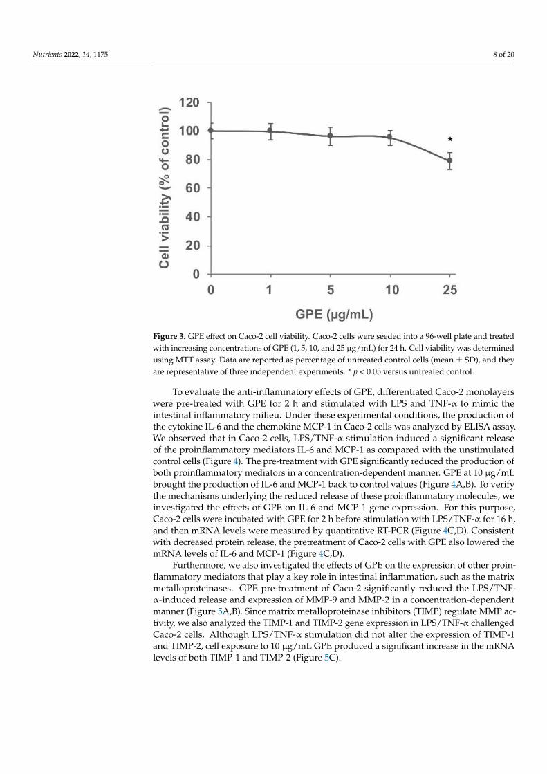
To evaluate the anti-inflammatory effects of GPE, differentiated Caco-2 monolayers
were pre-treated with GPE for 2 h and stimulated with LPS and TNF-α to mimic the in-
testinal inflammatory milieu. Under these experimental conditions, the production of the
cytokine IL-6 and the chemokine MCP-1 in Caco-2 cells was analyzed by ELISA assay. We
0
1
2
3
4
5
6
mg
/g
Figure 2. Characterization of different phenolic compounds occurring in Negramaro grape pomaceextract (GPE) suspended in ethanol 70% and used for bio-activity assays.
3.2. GPE Prevents the Stimulated Expression of Proinflammatory Mediators in Caco-2 Cells
We next investigated the effects of GPE on intestinal inflammatory parameters usingCaco-2 cell monolayers. We preliminarily assessed the effects of GPE on cell viabilityusing the MTT assay. To this aim, Caco-2 cells were exposed to increasing concentrations(1, 5, 10, and 25 µg/mL) of GPE for 24 h (Figure 3). The MTT assay results suggestthat concentrations equal to 25 µg/mL started to be toxic for Caco-2 cells (toxicity >20%)compared to the vehicle control. On the basis of these results, in the following experiments,we used GPE concentrations of 10 µg/mL or less, which were not cytotoxic and couldrepresent plausible concentrations in the human gut [39].
Nutrients 2022, 14, 1175 8 of 20
Nutrients 2022, 14, x FOR PEER REVIEW 8 of 20
Figure 2. Characterization of different phenolic compounds occurring in Negramaro grape pomace
extract (GPE) suspended in ethanol 70% and used for bio-activity assays.
3.2. GPE Prevents the Stimulated Expression of Proinflammatory Mediators in Caco-2 Cells
We next investigated the effects of GPE on intestinal inflammatory parameters using
Caco-2 cell monolayers. We preliminarily assessed the effects of GPE on cell viability us-
ing the MTT assay. To this aim, Caco-2 cells were exposed to increasing concentrations (1,
5, 10, and 25 µg/mL) of GPE for 24 h (Figure 3). The MTT assay results suggest that con-
centrations equal to 25 µg/mL started to be toxic for Caco-2 cells (toxicity >20%) compared
to the vehicle control. On the basis of these results, in the following experiments, we used
GPE concentrations of 10 µg/mL or less, which were not cytotoxic and could represent
plausible concentrations in the human gut [39].
Figure 3. GPE effect on Caco-2 cell viability. Caco-2 cells were seeded into a 96-well plate and treated
with increasing concentrations of GPE (1, 5, 10, and 25 µg/mL) for 24 h. Cell viability was determined
using MTT assay. Data are reported as percentage of untreated control cells (mean ± SD), and they
are representative of three independent experiments. * p < 0.05 versus untreated control.
To evaluate the anti-inflammatory effects of GPE, differentiated Caco-2 monolayers
were pre-treated with GPE for 2 h and stimulated with LPS and TNF-α to mimic the in-
testinal inflammatory milieu. Under these experimental conditions, the production of the
cytokine IL-6 and the chemokine MCP-1 in Caco-2 cells was analyzed by ELISA assay. We
0
1
2
3
4
5
6
mg
/g
Figure 3. GPE effect on Caco-2 cell viability. Caco-2 cells were seeded into a 96-well plate and treatedwith increasing concentrations of GPE (1, 5, 10, and 25 µg/mL) for 24 h. Cell viability was determinedusing MTT assay. Data are reported as percentage of untreated control cells (mean ± SD), and theyare representative of three independent experiments. * p < 0.05 versus untreated control.
To evaluate the anti-inflammatory effects of GPE, differentiated Caco-2 monolayerswere pre-treated with GPE for 2 h and stimulated with LPS and TNF-α to mimic theintestinal inflammatory milieu. Under these experimental conditions, the production ofthe cytokine IL-6 and the chemokine MCP-1 in Caco-2 cells was analyzed by ELISA assay.We observed that in Caco-2 cells, LPS/TNF-α stimulation induced a significant releaseof the proinflammatory mediators IL-6 and MCP-1 as compared with the unstimulatedcontrol cells (Figure 4). The pre-treatment with GPE significantly reduced the production ofboth proinflammatory mediators in a concentration-dependent manner. GPE at 10 µg/mLbrought the production of IL-6 and MCP-1 back to control values (Figure 4A,B). To verifythe mechanisms underlying the reduced release of these proinflammatory molecules, weinvestigated the effects of GPE on IL-6 and MCP-1 gene expression. For this purpose,Caco-2 cells were incubated with GPE for 2 h before stimulation with LPS/TNF-α for 16 h,and then mRNA levels were measured by quantitative RT-PCR (Figure 4C,D). Consistentwith decreased protein release, the pretreatment of Caco-2 cells with GPE also lowered themRNA levels of IL-6 and MCP-1 (Figure 4C,D).
Furthermore, we also investigated the effects of GPE on the expression of other proin-flammatory mediators that play a key role in intestinal inflammation, such as the matrixmetalloproteinases. GPE pre-treatment of Caco-2 significantly reduced the LPS/TNF-α-induced release and expression of MMP-9 and MMP-2 in a concentration-dependentmanner (Figure 5A,B). Since matrix metalloproteinase inhibitors (TIMP) regulate MMP ac-tivity, we also analyzed the TIMP-1 and TIMP-2 gene expression in LPS/TNF-α challengedCaco-2 cells. Although LPS/TNF-α stimulation did not alter the expression of TIMP-1and TIMP-2, cell exposure to 10 µg/mL GPE produced a significant increase in the mRNAlevels of both TIMP-1 and TIMP-2 (Figure 5C).

Nutrients 2022, 14, 1175 9 of 20
Nutrients 2022, 14, x FOR PEER REVIEW 9 of 20
observed that in Caco-2 cells, LPS/TNF-α stimulation induced a significant release of the
proinflammatory mediators IL-6 and MCP-1 as compared with the unstimulated control
cells (Figure 4). The pre-treatment with GPE significantly reduced the production of both
proinflammatory mediators in a concentration-dependent manner. GPE at 10 µg/mL
brought the production of IL-6 and MCP-1 back to control values (Figure 4A,B). To verify
the mechanisms underlying the reduced release of these proinflammatory molecules, we
investigated the effects of GPE on IL-6 and MCP-1 gene expression. For this purpose,
Caco-2 cells were incubated with GPE for 2 h before stimulation with LPS/TNF-α for 16
h, and then mRNA levels were measured by quantitative RT-PCR (Figure 4C,D). Con-
sistent with decreased protein release, the pretreatment of Caco-2 cells with GPE also low-
ered the mRNA levels of IL-6 and MCP-1 (Figure 4C,D).
Figure 4. Inhibitory effects of GPE on the expression of IL-6 and MCP-1 in inflamed Caco-2 cells.
Differentiated Caco-2 cells were pre-treated with increasing concentrations of GPE (1, 5, and 10
µg/mL) for 2 h, followed by incubation with LPS (10 μg/mL) and TNF-α (10 ng/mL) for 4 h (C,D) or
24 h (A,B). IL-6 and MCP-1 release in culture medium was analyzed by ELISA assay. Results are
shown as percentage of unstimulated control (mean ± SD). IL-6 and MCP-1 mRNA levels were de-
termined by quantitative RT-PCR and are expressed as fold over unstimulated control (mean ± SD).
The white histograms correspond to the untreated control (CTR), while the colored histograms cor-
respond to the different treated groups (black for LPS/TNF-α and orange for GPE+LPS/TNF-α). Data
are representative of three independent experiments. # p < 0.01 versus control; * p < 0.05 versus
LPS/TNF-α alone.
Furthermore, we also investigated the effects of GPE on the expression of other pro-
inflammatory mediators that play a key role in intestinal inflammation, such as the matrix
metalloproteinases. GPE pre-treatment of Caco-2 significantly reduced the LPS/TNF-α-
induced release and expression of MMP-9 and MMP-2 in a concentration-dependent man-
ner (Figure 5A,B). Since matrix metalloproteinase inhibitors (TIMP) regulate MMP activ-
ity, we also analyzed the TIMP-1 and TIMP-2 gene expression in LPS/TNF-α challenged
Caco-2 cells. Although LPS/TNF-α stimulation did not alter the expression of TIMP-1 and
Figure 4. Inhibitory effects of GPE on the expression of IL-6 and MCP-1 in inflamed Caco-2 cells. Dif-ferentiated Caco-2 cells were pre-treated with increasing concentrations of GPE (1, 5, and 10 µg/mL)for 2 h, followed by incubation with LPS (10 µg/mL) and TNF-α (10 ng/mL) for 4 h (C,D) or24 h (A,B). IL-6 and MCP-1 release in culture medium was analyzed by ELISA assay. Results areshown as percentage of unstimulated control (mean ± SD). IL-6 and MCP-1 mRNA levels were de-termined by quantitative RT-PCR and are expressed as fold over unstimulated control (mean ± SD).The white histograms correspond to the untreated control (CTR), while the colored histogramscorrespond to the different treated groups (black for LPS/TNF-α and orange for GPE+LPS/TNF-α).Data are representative of three independent experiments. # p < 0.01 versus control; * p < 0.05 versusLPS/TNF-α alone.
Nutrients 2022, 14, x FOR PEER REVIEW 10 of 20
TIMP-2, cell exposure to 10 μg/mL GPE produced a significant increase in the mRNA lev-
els of both TIMP-1 and TIMP-2 (Figure 5C).
Figure 5. GPE effects on the expression of matrix metalloproteinases and their inhibitors in inflamed
Caco-2 cells. Differentiated Caco-2 cells were pre-treated with various concentrations of GPE (5 and
10 µg/mL) for 2 h, followed by incubation with LPS (10 μg/mL) and TNF-α (10 ng/mL) for 16 h (B,C)
or 24 h (A). MMP-9 and MMP-2 release in culture medium was analyzed by ELISA assay. Results
are shown as percentage of unstimulated control (mean ± SD). MMP-9 and MMP-2 (B) as well as
TIMP-1 and TIMP-2 (C) mRNA levels were determined by quantitative RT-PCR and are expressed
as fold over unstimulated control (mean ± SD). The white histograms correspond to the untreated
control, while the colored histograms correspond to the different treated groups (black for LPS/TNF-
α and orange for GPE+LPS/TNF-α). Data are representative of three independent experiments. # p
< 0.01 versus control. * p < 0.05 versus LPS/TNF-α alone.
Moreover, GPE was able to significantly reduce the LPS/TNF-α-induced mRNA lev-
els of several inflammatory markers including the cytokines IL-1β and TNF-α, the chem-
okines CXCL-10 and M-CSF, the inflammatory enzyme cyclooxygenase-2 (COX-2), and
the adhesion molecules VCAM-1 and ICAM-1 (Table 3). These data suggest a possible
protective effect of grape pomace polyphenols in the gut by inhibiting the expression and
release of key inflammatory mediators.
Table 3. GPE effect on the gene expression of inflammatory mediators in Caco-2 cells under inflam-
matory conditions.
Gene Name LPS/TNF-α GPE + LPS/TNF-α
IL1B 9.3 ± 0.8 7.5 ± 0.7 *
TNF 4.3 ± 0.7 0.9 ± 0.6 **
CXCL10 14.6 ± 1.2 5.9 ± 0.8 **
CSF1/MCSF 4.0 ± 0.6 1.6 ± 0.7 **
PTGS2/COX-2 3.8 ± 0.9 1.7 ± 0.6 *
VCAM1 15.1 ± 2.3 2.3 ± 0.8 **
ICAM1 1.56 ± 0.3 1.0 ± 0.2 *
The expression levels of genes were analyzed by quantitative RT-PCR. Data are reported as fold
over unstimulated control (mean ± SD) and are representative of three independent experiments. *
p < 0.05, ** p < 0.01 versus LPS/TNF-α alone.
3.3. GPE Reduced the Stimulated Activation of NF-κB and the Intracellular ROS Levels in
Caco-2 Cells
The expression of inflammatory genes is mainly regulated by NF-κB signaling path-
ways. Under resting conditions, NF-κB is relegated in cytoplasm in an inactive form;
Figure 5. GPE effects on the expression of matrix metalloproteinases and their inhibitors in inflamedCaco-2 cells. Differentiated Caco-2 cells were pre-treated with various concentrations of GPE (5and 10 µg/mL) for 2 h, followed by incubation with LPS (10 µg/mL) and TNF-α (10 ng/mL) for

Nutrients 2022, 14, 1175 10 of 20
16 h (B,C) or 24 h (A). MMP-9 and MMP-2 release in culture medium was analyzed by ELISA assay.Results are shown as percentage of unstimulated control (mean ± SD). MMP-9 and MMP-2 (B) aswell as TIMP-1 and TIMP-2 (C) mRNA levels were determined by quantitative RT-PCR and areexpressed as fold over unstimulated control (mean ± SD). The white histograms correspond to theuntreated control, while the colored histograms correspond to the different treated groups (blackfor LPS/TNF-α and orange for GPE+LPS/TNF-α). Data are representative of three independentexperiments. # p < 0.01 versus control. * p < 0.05 versus LPS/TNF-α alone.
Moreover, GPE was able to significantly reduce the LPS/TNF-α-induced mRNAlevels of several inflammatory markers including the cytokines IL-1β and TNF-α, thechemokines CXCL-10 and M-CSF, the inflammatory enzyme cyclooxygenase-2 (COX-2),and the adhesion molecules VCAM-1 and ICAM-1 (Table 3). These data suggest a possibleprotective effect of grape pomace polyphenols in the gut by inhibiting the expression andrelease of key inflammatory mediators.
Table 3. GPE effect on the gene expression of inflammatory mediators in Caco-2 cells under inflam-matory conditions.
Gene Name LPS/TNF-α GPE + LPS/TNF-α
IL1B 9.3 ± 0.8 7.5 ± 0.7 *TNF 4.3 ± 0.7 0.9 ± 0.6 **CXCL10 14.6 ± 1.2 5.9 ± 0.8 **CSF1/MCSF 4.0 ± 0.6 1.6 ± 0.7 **PTGS2/COX-2 3.8 ± 0.9 1.7 ± 0.6 *VCAM1 15.1 ± 2.3 2.3 ± 0.8 **ICAM1 1.56 ± 0.3 1.0 ± 0.2 *
The expression levels of genes were analyzed by quantitative RT-PCR. Data are reported as fold over unstimulatedcontrol (mean ± SD) and are representative of three independent experiments. * p < 0.05, ** p < 0.01 versusLPS/TNF-α alone.
3.3. GPE Reduced the Stimulated Activation of NF-κB and the Intracellular ROS Levels inCaco-2 Cells
The expression of inflammatory genes is mainly regulated by NF-κB signaling path-ways. Under resting conditions, NF-κB is relegated in cytoplasm in an inactive form;inflammatory stimuli activate NF-κB, which translocates to the nucleus, where it binds topromoters of target genes, such as those codifying for proinflammatory proteins [40]. InCaco-2 cells, we verified the effect of GPE on LPS/TNF-α-stimulated NF-κB activation byan ELISA-based method, which measures the DNA-binding activity of the NF-κB subunitp65. The activation of NF-κB was evidenced by the increased levels of p65 subunit in thenuclear extracts of Caco-2 cells treated with LPS/TNF-α for 2 h, whereas GPE pre-treatmentprevented, in a concentration-dependent manner, LPS/TNF-α-induced nuclear transloca-tion of p65 (Figure 6A). These results suggest that GPE is able to modulate the activation ofNF-κB in response to inflammatory stimuli in intestinal epithelial cells.
Changes in the cellular redox balance may mediate the activation of NF-κB, which canbe inhibited by treatment with antioxidants [41]. Because GPE showed strong antioxidantpotential in cell-free systems (Table 2), we examined its antioxidant capacity in Caco-2cells triggered with LPS/TNF-α. The LPS/TNF-α stimulation of Caco-2 cells produceda significant increase in the intracellular levels of ROS, as assessed by the ROS-sensitivecarboxy-H2DCFDA probe. Cell treatment with GPE, before LPS/TNF-α stimulation, signif-icantly reduced ROS levels in a concentration-dependent manner (Figure 6B,C), indicatingmeaningful intracellular antioxidant effects by GPE in our experimental conditions.

Nutrients 2022, 14, 1175 11 of 20
Nutrients 2022, 14, x FOR PEER REVIEW 11 of 20
inflammatory stimuli activate NF-κB, which translocates to the nucleus, where it binds to
promoters of target genes, such as those codifying for proinflammatory proteins [40]. In
Caco-2 cells, we verified the effect of GPE on LPS/TNF-α-stimulated NF-κB activation by
an ELISA-based method, which measures the DNA-binding activity of the NF-κB subunit
p65. The activation of NF-κB was evidenced by the increased levels of p65 subunit in the
nuclear extracts of Caco-2 cells treated with LPS/TNF-α for 2 h, whereas GPE pre-treat-
ment prevented, in a concentration-dependent manner, LPS/TNF-α-induced nuclear
translocation of p65 (Figure 6A). These results suggest that GPE is able to modulate the
activation of NF-κB in response to inflammatory stimuli in intestinal epithelial cells.
Changes in the cellular redox balance may mediate the activation of NF-κB, which
can be inhibited by treatment with antioxidants [41]. Because GPE showed strong antiox-
idant potential in cell-free systems (Table 2), we examined its antioxidant capacity in Caco-
2 cells triggered with LPS/TNF-α. The LPS/TNF-α stimulation of Caco-2 cells produced a
significant increase in the intracellular levels of ROS, as assessed by the ROS-sensitive
carboxy-H2DCFDA probe. Cell treatment with GPE, before LPS/TNF-α stimulation, sig-
nificantly reduced ROS levels in a concentration-dependent manner (Figure 6B,C), indi-
cating meaningful intracellular antioxidant effects by GPE in our experimental conditions.
Figure 6. Inhibitory effects of GPE on NF-κB activation and intracellular ROS levels in inflamed
Caco-2 cells. Differentiated Caco-2 cells were pre-treated with various concentrations of GPE (5 and
10 µg/mL) for 2 h, followed by incubation with LPS (10 μg/mL) and TNF-α (10 ng/mL) for 2 h. NF-
κB activation was assessed in nuclear proteins by an ELISA-based method measuring the DNA-
binding activity of NF-κB (A). Intracellular ROS were analyzed by using carboxy-H2DCFDA stain-
ing by fluorescence plate reader (B). The white histograms correspond to the untreated control
(CTR), while the colored histograms correspond to the different treated groups (black for LPS/TNF-
α and orange for GPE+LPS/TNF-α). Each experiment was executed in triplicate. Data are reported
as unstimulated control percentage (mean ± SD). # p < 0.01 versus control; * p < 0.05 versus LPS/TNF-
α alone.
3.4. GPE Inhibited Endothelial Activation and Monocyte Adhesion in Intestinal
Epithelial/Endothelial Co-Culture Model
To mimic intestinal epithelial–endothelial cell interactions in vitro, we used a co-cul-
ture system that places endothelial cells in close proximity, but not in contact, to intestinal
epithelial cells. In this model, we evaluated if intestinal anti-inflammatory action of GPE
was associated with improved endothelial function, assessed as endothelial activation and
monocyte recruitment.
To this aim, differentiated Caco-2 monolayers, grown on the upper side of semiper-
meable trans-well inserts, were placed in close proximity to HMEC-1 grown on the bottom
of the wells (see Figure 1 for details). GPE was added on the apical compartment for 2 h;
then, LPS and TNF-α were applied on the apical side, and TNF-α on the basolateral side.
Figure 6. Inhibitory effects of GPE on NF-κB activation and intracellular ROS levels in inflamedCaco-2 cells. Differentiated Caco-2 cells were pre-treated with various concentrations of GPE (5 and10 µg/mL) for 2 h, followed by incubation with LPS (10 µg/mL) and TNF-α (10 ng/mL) for 2 h. NF-κBactivation was assessed in nuclear proteins by an ELISA-based method measuring the DNA-bindingactivity of NF-κB (A). Intracellular ROS were analyzed by using carboxy-H2DCFDA staining byfluorescence plate reader (B). The white histograms correspond to the untreated control (CTR), whilethe colored histograms correspond to the different treated groups (black for LPS/TNF-α and orangefor GPE+LPS/TNF-α). Each experiment was executed in triplicate. Data are reported as unstimulatedcontrol percentage (mean ± SD). # p < 0.01 versus control; * p < 0.05 versus LPS/TNF-α alone.
3.4. GPE Inhibited Endothelial Activation and Monocyte Adhesion in Intestinal Epithelial/EndothelialCo-Culture Model
To mimic intestinal epithelial–endothelial cell interactions in vitro, we used a co-culture system that places endothelial cells in close proximity, but not in contact, to intestinalepithelial cells. In this model, we evaluated if intestinal anti-inflammatory action of GPEwas associated with improved endothelial function, assessed as endothelial activation andmonocyte recruitment.
To this aim, differentiated Caco-2 monolayers, grown on the upper side of semiperme-able trans-well inserts, were placed in close proximity to HMEC-1 grown on the bottom ofthe wells (see Figure 1 for details). GPE was added on the apical compartment for 2 h; then,LPS and TNF-α were applied on the apical side, and TNF-α on the basolateral side. After16 h of inflammatory stimulation, the release of IL-6 and MCP-1 in the conditioned mediaof the basolateral side was evaluated by ELISA.
The results showed that inflammatory stimuli significantly intensified the release of IL-6 and MCP-1 in co-culture system as compared with unstimulated cells (Figure 7A,B). Pre-treatment of Caco-2 with GPE dose-dependently reduced the release of both inflammatorymediators in co-culture system (Figure 7A,B), confirming and strengthening the dataobtained in Caco-2 monoculture.
To analyze the specific contribution of endothelial cells in the release of IL-6 andMCP-1 in Caco-2/HMEC-1 co-culture model, mRNA levels of both inflammatory me-diators were analyzed in HMEC-1. The results showed that inflammatory stimulationinduced a significant increase in the mRNA levels of IL-6 and MCP-1 that was reduced in aconcentration-dependent manner by pre-treatment with GPE (Figure 7C,D).

Nutrients 2022, 14, 1175 12 of 20
Nutrients 2022, 14, x FOR PEER REVIEW 12 of 20
After 16 h of inflammatory stimulation, the release of IL-6 and MCP-1 in the conditioned
media of the basolateral side was evaluated by ELISA.
The results showed that inflammatory stimuli significantly intensified the release of
IL-6 and MCP-1 in co-culture system as compared with unstimulated cells (Figure 7A,B).
Pre-treatment of Caco-2 with GPE dose-dependently reduced the release of both inflam-
matory mediators in co-culture system (Figure 7A,B), confirming and strengthening the
data obtained in Caco-2 monoculture.
To analyze the specific contribution of endothelial cells in the release of IL-6 and
MCP-1 in Caco-2/HMEC-1 co-culture model, mRNA levels of both inflammatory media-
tors were analyzed in HMEC-1. The results showed that inflammatory stimulation in-
duced a significant increase in the mRNA levels of IL-6 and MCP-1 that was reduced in a
concentration-dependent manner by pre-treatment with GPE (Figure 7C,D).
In co-cultured endothelial cells, pro-inflammatory stimuli significantly induced the
expression of VCAM-1 and ICAM-1 at the protein and mRNA levels, as evaluated by cell
surface EIA and quantitative RT-PCR, respectively (Figure 8A,B). The results showed that
GPE were able to downregulate the endothelial cell adhesion molecule expression in a
concentration-dependent manner (Figure 8A,B), without affecting the expression of the
constitutive endothelial surface antigen E1/1 (data not shown).
Figure 7. Inhibitory effects of GPE on IL-6- and MCP-1-stimulated expression in a Caco-2/HMEC-1
co-culture model. Differentiated Caco-2 monolayers were grown on the upper side of the inserts
and placed in proximity to HMEC-1 grown on the bottom of the wells. GPE (5 and 10 μg/mL) was
added on the apical compartment for 2 h, after which LPS (10 μg/mL) and TNF-α (10 ng/mL) were
applied on the apical compartment and TNF-α (10 ng/mL) on the basolateral compartment for 16 h
(C,D) or 24 h (A,B). The protein release of IL-6 and MCP-1 was assessed in basolateral culture me-
dium by ELISA assay. Results are shown as percentage of unstimulated control (mean ± SD). In
endothelial cells, IL-6 and MCP-1 mRNA levels were measured by quantitative RT-PCR and are
expressed as fold over unstimulated control (mean ± SD). The white histograms correspond to the
untreated control (CTR), while the colored histograms correspond to the different treated groups
(black for LPS/TNF-α and pink for GPE+LPS/TNF-α). Data are representative of three independent
experiments. # p < 0.01 versus control; * p < 0.05 versus LPS/TNF-α alone.
Figure 7. Inhibitory effects of GPE on IL-6- and MCP-1-stimulated expression in a Caco-2/HMEC-1co-culture model. Differentiated Caco-2 monolayers were grown on the upper side of the inserts andplaced in proximity to HMEC-1 grown on the bottom of the wells. GPE (5 and 10 µg/mL) was addedon the apical compartment for 2 h, after which LPS (10 µg/mL) and TNF-α (10 ng/mL) were appliedon the apical compartment and TNF-α (10 ng/mL) on the basolateral compartment for 16 h (C,D) or24 h (A,B). The protein release of IL-6 and MCP-1 was assessed in basolateral culture medium byELISA assay. Results are shown as percentage of unstimulated control (mean ± SD). In endothelialcells, IL-6 and MCP-1 mRNA levels were measured by quantitative RT-PCR and are expressed as foldover unstimulated control (mean ± SD). The white histograms correspond to the untreated control(CTR), while the colored histograms correspond to the different treated groups (black for LPS/TNF-αand pink for GPE+LPS/TNF-α). Data are representative of three independent experiments. # p < 0.01versus control; * p < 0.05 versus LPS/TNF-α alone.
In co-cultured endothelial cells, pro-inflammatory stimuli significantly induced theexpression of VCAM-1 and ICAM-1 at the protein and mRNA levels, as evaluated by cellsurface EIA and quantitative RT-PCR, respectively (Figure 8A,B). The results showed thatGPE were able to downregulate the endothelial cell adhesion molecule expression in aconcentration-dependent manner (Figure 8A,B), without affecting the expression of theconstitutive endothelial surface antigen E1/1 (data not shown).

Nutrients 2022, 14, 1175 13 of 20
Nutrients 2022, 14, x FOR PEER REVIEW 13 of 20
Endothelial expression of adhesion molecules is responsible for leukocyte adhesion
to vascular endothelium and migration into the subendothelial space. In order to confirm
the inhibitory activity of GPE on endothelial activation in Caco-2/HMEC-1 co-culture, the
adhesion of human leukocytes THP-1 on HMEC-1 was performed as described in Section
2.11. As shown in Figure 8C, the number of leukocytes adhering to the stimulated endo-
thelial cells was higher than that observed in unstimulated control. Leukocyte adhesion
appeared to be reduced in a concentration-dependent manner by Caco-2 pre-treatment
with GPE, demonstrating that grape pomace polyphenols were able to reduce intestinal
inflammation and prevent the consequent endothelial activation and leukocyte adhesion.
Figure 8. Inhibitory effects of GPE on the stimulated expression of endothelial adhesion molecules
and on the endothelial monocyte adhesion. Differentiated Caco-2 monolayers were grown on the
upper side of the inserts and placed in proximity to HMEC-1 grown on the bottom of the wells. GPE
(5 and 10 μg/mL) was added on the apical compartment for 2 h, after which LPS (10 μg/mL) and
TNF-α (10 ng/mL) were applied on the apical compartment and TNF-α (10 ng/mL) on the basolat-
eral compartment for 16 h (B) or 12 h. (A,C) Endothelial cell surface expression of VCAM-1 and
ICAM-1 was assessed by cell surface EIA and expressed as percentage of unstimulated control. (B)
VCAM-1 and ICAM-1 mRNA levels were analyzed by quantitative RT-PCR and expressed as fold
over unstimulated control (mean ± SD). (C) HMEC-1 were co-cultured with labeled THP-1 cells for
1 h. The number of adherent THP-1 cells was monitored by fluorescence microscope or measured
by the fluorescence plate reader. Each experiment was performed in triplicate. The white histograms
correspond to the untreated control (CTR), while the colored histograms correspond to the different
treated groups (black for LPS/TNF-α and pink for GPE+LPS/TNF-α). # p < 0.01 versus control; * p <
0.05 versus LPS/TNF-α alone.
4. Discussion
Agroindustrial waste from food production is now reviewed as a valuable product
due to its high content of phytochemicals with purported health benefits. During wine-
making, about 20–25% of the grape weight remains as waste material [42]. Although
mainly used in the past as livestock feed, soil fertilizer, or fuel, they can be also exploited
in the production of new nutraceuticals and functional foods to promote health and pre-
vent disease [25]. Grape pomace composes mainly of skins and seeds, which, due to poor
Figure 8. Inhibitory effects of GPE on the stimulated expression of endothelial adhesion moleculesand on the endothelial monocyte adhesion. Differentiated Caco-2 monolayers were grown on theupper side of the inserts and placed in proximity to HMEC-1 grown on the bottom of the wells.GPE (5 and 10 µg/mL) was added on the apical compartment for 2 h, after which LPS (10 µg/mL)and TNF-α (10 ng/mL) were applied on the apical compartment and TNF-α (10 ng/mL) on thebasolateral compartment for 16 h (B) or 12 h. (A,C) Endothelial cell surface expression of VCAM-1and ICAM-1 was assessed by cell surface EIA and expressed as percentage of unstimulated control.(B) VCAM-1 and ICAM-1 mRNA levels were analyzed by quantitative RT-PCR and expressed as foldover unstimulated control (mean ± SD). (C) HMEC-1 were co-cultured with labeled THP-1 cells for1 h. The number of adherent THP-1 cells was monitored by fluorescence microscope or measured bythe fluorescence plate reader. Each experiment was performed in triplicate. The white histogramscorrespond to the untreated control (CTR), while the colored histograms correspond to the differenttreated groups (black for LPS/TNF-α and pink for GPE+LPS/TNF-α). # p < 0.01 versus control;* p < 0.05 versus LPS/TNF-α alone.
Endothelial expression of adhesion molecules is responsible for leukocyte adhesion tovascular endothelium and migration into the subendothelial space. In order to confirm theinhibitory activity of GPE on endothelial activation in Caco-2/HMEC-1 co-culture, the ad-hesion of human leukocytes THP-1 on HMEC-1 was performed as described in Section 2.11.As shown in Figure 8C, the number of leukocytes adhering to the stimulated endothelialcells was higher than that observed in unstimulated control. Leukocyte adhesion appearedto be reduced in a concentration-dependent manner by Caco-2 pre-treatment with GPE,demonstrating that grape pomace polyphenols were able to reduce intestinal inflammationand prevent the consequent endothelial activation and leukocyte adhesion.

Nutrients 2022, 14, 1175 14 of 20
4. Discussion
Agroindustrial waste from food production is now reviewed as a valuable productdue to its high content of phytochemicals with purported health benefits. During winemak-ing, about 20–25% of the grape weight remains as waste material [42]. Although mainlyused in the past as livestock feed, soil fertilizer, or fuel, they can be also exploited in theproduction of new nutraceuticals and functional foods to promote health and preventdisease [25]. Grape pomace composes mainly of skins and seeds, which, due to poor extrac-tion during winemaking, still contain high levels of phytochemical compounds, especiallypolyphenols [43], which are the main secondary metabolites of plants and an integral partof the human diet. It has been reported that grape pomace contains a great variety ofpolyphenols including anthocyanins, catechins, flavonols, alcohols, stilbenes, and benzoic(gallic, protocatechuic, 4-hydroxybenzoic) and cinnamic (p-coumaric) acids [44,45]. In thepresent study, we confirm our previous findings regarding the polyphenolic profile ofNegramaro grape pomace [21], showing that the main components found in GPE were ac-tive polyphenols such as oenin, epicatechin, catechin, quercetin and quercetin-3-glucoside,gallic acid, caftaric acid, and resveratrol, which are responsible for GPE antioxidant activity.The intestinal epithelium is constantly exposed to dietary phenolic compounds, which arepoorly absorbed and reach high concentrations in the lumen, where they can exert healthbenefits by protecting the intestinal mucosa from oxidative damage and helping to improveantioxidant capacity [46]. A continuous crosstalk between the different cell types present inthe gut, including endothelial cells, influences the inflammatory process and the immuno-logical response of the gut, which is crucial for preserving gut health and functioning aswell as for preventing CVD [47,48]. A dysregulation of intestinal inflammation can leadto severe gut disorders such as IBD, which is characterized by overproduction of severalinflammatory mediators, including TNF-α and LPS, implicated in the initiation and flares ofIBD, as well as in systemic inflammatory reaction [49,50]. In vivo and in vitro studies haveshown that polyphenols are effective in preventing and relieving IBD symptoms, regulatingthe intestinal ecosystem, and reducing the level of pro-inflammatory cytokines [46,51–53].Since nutritional interventions based on high polyphenol intake have a lower toxic effectthan pharmaceutical approaches [54], ongoing efforts are aimed at developing new dietarypolyphenol-based strategies targeting oxidative stress and inflammatory signaling events.The aim of the present study was therefore to explore the effect of polyphenolic extract ofgrape pomace on intestinal inflammation and the transepithelial impact of GPE compoundson endothelial dysfunction.
Since GPE could act directly on intestinal epithelial cells before crossing the blood–intestinal barrier, we first investigated the potential anti-inflammatory properties of GPEin Caco-2 cell monolayers, a model of human intestinal epithelium. These cells, derivedfrom a human colon adenocarcinoma, once in culture undergo a process of spontaneousdifferentiation into normal mature enterocytes [29,30]. To try and mimic the intestinal in-flammatory milieu detectable during IBD, we exposed Caco-2 cells to TNF-α and LPS. Ourresults in Caco-2 cells showed that LPS and TNF-α activated intracellular cascades leadingto increased transcriptional activity and secretion of the cytokines IL-6 and MCP-1 andthe matrix metalloproteinases MMP-9 and MMP-2. We also observed an overall boostedgene expression of other inflammatory markers, such as the cytokines IL-1β and TNF-α,the chemokines CXCL-10 and M-CSF, the pro-inflammatory enzyme COX-2, and the adhe-sion molecules VCAM-1 and ICAM-1. In our experimental model, the anti-inflammatoryproperty of GPE against LPS/TNF-α stimulation was demonstrated in terms of IL-6 andMCP-1 decrease, at protein and mRNA levels, occurring in a concentration-dependentmanner. This finding can be relevant to counteract gut inflammation in IBD. Indeed, IL-6and MCP-1 are upregulated in IBD patients and play an important role in mucosal im-mune responses [55,56]. The 10 µg/mL concentration of GPE provided the most consistentresults across all measures, as it was also associated with the highest suppression of pro-inflammatory mediators including IL-1β and TNF-α, CXCL-10 and M-CSF, COX-2, andVCAM-1 and ICAM-1. GPE concentrations equal to or less than 10 µg/mL, as used in this

Nutrients 2022, 14, 1175 15 of 20
study, were not toxic in Caco-2 cells. These values, on the other hand, represent biologicallyrelevant concentrations in the human gut lumen [39], where polyphenols may reach highconcentrations after dietary consumption [46]. Moreover, GPE pre-treatment significantlyreduced the LPS/TNF-α-induced release and expression of MMP-9 and MMP-2 in Caco-2cells, in a concentration-dependent manner, while it augmented the mRNA levels of met-alloproteinase inhibitors TIMP-1 and TIMP-2. Extracellular MMPs are essential factorsinvolved in the development, modification, and healing of inflammatory lesions. MMP-9is suggested to be the key factor determining mucous membrane damage in IBD [57,58];thus, it can be a potential therapeutic target. Indeed, therapeutic strategies with infliximab(anti-TNF-α) enhance mucosal healing by reducing MMP-9 activity through an increasedexpression of TIMP-1, the most potent natural inhibitor of MMP-9 [59]. To the best of ourknowledge, these results show for the first time that polyphenolic extracts from grapepomace can improve gut health by restoring the balance between MMP-9/2 and TIMP1/2, which is altered by the intestinal inflammatory environment. In this study, we alsoinvestigated the molecular mechanism of GPE action in inflamed Caco-2 monolayer. Wefound that GPE was able to revert the LPS/TNF-α-induced activation of NF-κB, whichplays a pivotal role during intestinal inflammatory responses. Thus, the ability of GPE tocounteract the activation of NF-kB signaling pathway may explain the anti-inflammatoryactivities of wine pomace polyphenols at the intestinal level. This effect was associatedwith decreased intracellular ROS content in inflamed Caco-2 cells, which was related to theantioxidant capacity of GPE. Intestine is a key source of ROS due to necessary exposure toforeign substances and microbial pathogens. Disproportionate generation and long-termexposure to ROS lead to various inflammatory intestinal diseases, such as IBD [49]. GPE, bydumping intracellular ROS, may protect gut health and mitigate oxidative stress-induceddamage. Our results confirm and extend previous studies showing significant protection bythe polyphenolic components of grape marc against inflammation, oxidative stress [60,61],and NF-κB activation [62,63], thus restoring tight junction barrier function in Caco-2 coloncells [64]. Similar effects have been also reported for pure polyphenols. In particular,malvidin-3-glucoside has been shown to have anti-inflammatory properties in a murine col-itis model through the modulation of the integrity of the colon epithelium, gut microbiota,and gut metabolism [65,66]. Catechins and epicatechins influenced cellular response tooxidative stress and exerted intestinal anti-inflammatory properties by modulating severalcell signaling pathways, such as NF-κB, mitogen-activated protein kinases (MAPKs), andtranscription factor nuclear factor (erythroid-derived 2)-like 2 (Nrf2) [67,68]. Similarly,kaempferol counteracted gut inflammation by inhibiting the activation of the NF-κB signal-ing pathway in intestinal epithelial–endothelial co-culture model [69]. In intestinal in vitroand in vivo models, both quercetin and its metabolites, including rutin, exhibited anti-inflammatory properties by inhibiting the activity of NF-κB and reducing the productionof several proinflammatory mediators [70–72]. Although the in vitro effects of resveratrolon intestinal inflammation are contradictory [70], animal studies have shown that resvera-trol can reduce the severity of intestinal inflammation in IBD models by downregulatingseveral intestinal immunity mediators and affecting key components of the inflammatorycascade, mainly the inhibition of NF-κB activation and the attenuation of reactive speciesproduction [73]. Moreover, findings showed that gallic acid protected Caco-2 cells againstoxidative stress [60] and inhibited inflammation in dextran sulfate sodium-induced colitisin mice through the suppression of p65-NF-κB [74]. In vitro digestion studies revealed thatphenolic acids, in addition to quercetin, were the polyphenols most resistant to digestion,and could be the most relevant to explain the biological activity of digested foods [75].
Overall, our data on GPE are in accordance with previous in vitro and in vivo re-sults on the intestinal anti-oxidant and anti-inflammatory signaling pathways of purepolyphenols, although further investigations are necessary to fully clarify the underlyingmechanisms of action.
Since oxidative stress and immune response are common mechanisms in both IBD andCVD, our findings suggest that GPE could be an effective strategy to improve gut health

Nutrients 2022, 14, 1175 16 of 20
and prevent CVD. Recent evidence shows that gut inflammation in IBD is an independentrisk factor for endothelial dysfunction and related CVD. In this contest, to assess thegut–vascular axis, we analyzed the impact of transepithelial GPE on inflamed endothelialcells and leukocyte adhesion. We tested the biological activity of GPE in a well-validatedco-culture model of intestinal epithelial/endothelial cells [30,36–38,76,77], analyzing thetransepithelial effects in HMEC-1 cells under inflamed conditions. We found, for the firsttime, that GPE pre-treatment of Caco-2 in co-culture system reduced endothelial activationin terms of release and expression of IL-6 and MCP-1, as well as the endothelial expressionof VCAM-1 and ICAM-1. As a functional implication, GPE anti-inflammatory effects wererelated with a reduced endothelial leukocyte recruitment in response to inflammatorystimuli, which can suggest an additional protective effect of GPE in attenuating bothvascular and intestinal inflammation with potential joint beneficial impact on IBD andCVD. Our findings regarding the multiple anti-inflammatory effects at the intestinal andendothelial levels extend previous studies showing that grape polyphenols, includingresveratrol, kaempferol, or anthocyanins, exert a protective effect in endothelial cellsagainst oxidative stress and inflammation, which are closely connected with cardiovasculardiseases [37,38,69].
The beneficial actions of polyphenols are largely dependent on their bioavailability atthe target tissues [70]. This bioavailability depends on the absorption of polyphenols atthe gastrointestinal level, as well as on their metabolism [70]. The intestinal absorption ofpolyphenols depends on their chemical structures; some glycoside forms, mainly glucosides,interact with proteins, including the glucose transporters, the cytosolic b-glucosidase, orthe membrane-bound lactase-phlorizin hydrolase, while aglycone forms seem to undergopassive diffusion only [78,79]. Typically, polyphenolic compounds can be subjected tocatabolism by the intestinal microbiota, which breaks down the polyphenolic compound,producing smaller molecules, including phenolic acids and hydroxycinnamates, whichare absorbed by the colon. Before passage into the blood stream, absorbed compoundscan undergo some degree of phase II metabolism, forming sulfate, glucuronide, and/ormethylated metabolites through specific metabolic enzymes [79]. In particular, resveratrolundergoes excessive metabolism by intestinal epithelial cells, and very low amounts ofunconjugated resveratrol circulate in the blood, while sulphate and glucuronate conjugatesare present in higher amounts [80,81]. These conjugates have also been found in studiesconducted in Caco-2 cells treated with resveratrol or kaempferol [38,82], thus confirmingthe value of using an intestinal cell culture in the study of polyphenol absorption andbiological effects. Other previous data showed that dietary polyphenols, including caffeicacid, gallic acid, quercetin, rutin, and resveratrol, were poorly absorbed by Caco-2 cells,and their transepithelial transports occurred mainly by passive diffusion [83]. A recentstudy reported that anthocyanins from red wine, including malvidin-3-glucoside, weretransported through Caco-2 cells [84]. In vivo study reported that malvidin-3-glucosidewas found in plasma and urine after ingestion of anthocyanin-containing beverages, andits plasma concentration correlated with the amount of the anthocyanin ingested [85].
Overall, our research shows that GPE polyphenols are biologically active and effectivein counteracting excessive inflammation by acting on both intestinal and endothelial cells.Therefore, GPE could be exploited as a “green source” of nutraceuticals for food supplementor components of functional food to improve the nutritional status of subjects with IBD,prevent intestinal inflammation, and assist in therapeutic strategies.
5. Conclusions
In conclusion, our results suggest that protective effects of grape pomace polyphe-nols may begin in the gut, where they counteract the excessive inflammatory response byexhibiting ROS scavenging ability and inhibiting the activation of redox-sensitive transcrip-tion factors. We show for the first time that physiological concentrations of grape pomacepolyphenols ameliorate the inflammatory response in intestinal cells by downregulatingthe expression of pro-inflammatory cytokines, chemokines, adhesion molecules, and matrix

Nutrients 2022, 14, 1175 17 of 20
metalloproteinases, which are all crucial mediators of the oxidative and inflammatoryprocess in IBD. Furthermore, GPE exhibit bioactivity on inflamed endothelial cells byreducing endothelial activation and subsequent leukocyte adhesion. Although it is difficultto directly transfer these data to the in vivo conditions, our results suggest that grapepomace is a natural source of polyphenols with multiple healthy properties that couldcontribute to the development of new ingredients and nutraceuticals able to relieve chronicgut inflammatory diseases and improve vascular endothelial functions.
Author Contributions: Conceptualization, N.C., M.A.C. and G.G.; methodology, N.C., M.M., E.S.,G.G., C.G., A.B. and M.A.C.; validation, N.C., M.M. and E.S.; investigation, N.C. and C.G.; resources,C.G., G.G. and T.V.; writing—original draft preparation, N.C. and M.A.C.; writing—review andediting, N.C., M.M., E.S., G.G., C.G., A.B., T.V. and M.A.C.; supervision, N.C. and M.A.C.; projectadministration, M.A.C., G.G. and T.V.; funding acquisition, M.A.C. and G.G. All authors have readand agreed to the published version of the manuscript.
Funding: This research was partially funded by CNR project NUTR-AGE (FOE2019, DSB.AD004.271)and ValBioVit-SNSvS NP-2.75 project- “2.2 Circular Economy” MITE-Italy.
Institutional Review Board Statement: Not applicable.
Informed Consent Statement: Not applicable.
Acknowledgments: The authors are grateful for technical support to D’Amico L., to Pascali A.M., toCagnazzo G., and to Azienda Cantine Cantele for providing the grape pomaces.
Conflicts of Interest: The authors declare no conflict of interest.
References1. Zhang, Y.Z.; Li, Y.Y. Inflammatory bowel disease: Pathogenesis. World J. Gastroenterol. 2014, 20, 91–99. [CrossRef] [PubMed]2. Wu, P.; Jia, F.; Zhang, B.; Zhang, P. Risk of cardiovascular disease in inflammatory bowel disease. Exp. Ther. Med. 2017, 13,
395–400. [CrossRef] [PubMed]3. Biondi, R.B.; Salmazo, P.S.; Bazan, S.G.Z.; Hueb, J.C.; de Paiva, S.A.R.; Sassaki, L.Y. Cardiovascular Risk in Individuals with
Inflammatory Bowel Disease. Clin. Exp. Gastroenterol. 2020, 13, 107–113. [CrossRef] [PubMed]4. Bunu, D.M.; Timofte, C.E.; Ciocoiu, M.; Floria, M.; Tarniceriu, C.C.; Barboi, O.B.; Tanase, D.M. Cardiovascular Manifestations of
Inflammatory Bowel Disease: Pathogenesis, Diagnosis, and Preventive Strategies. Gastroenterol. Res. Pract. 2019, 2019, 3012509.[CrossRef] [PubMed]
5. Cibor, D.; Domagala-Rodacka, R.; Rodacki, T.; Jurczyszyn, A.; Mach, T.; Owczarek, D. Endothelial dysfunction in inflammatorybowel diseases: Pathogenesis, assessment and implications. World J. Gastroenterol. 2016, 22, 1067–1077. [CrossRef] [PubMed]
6. Davignon, J.; Ganz, P. Role of endothelial dysfunction in atherosclerosis. Circulation 2004, 109, III27–32. [CrossRef] [PubMed]7. Xu, S.; Ilyas, I.; Little, P.J.; Li, H.; Kamato, D.; Zheng, X.; Luo, S.; Li, Z.; Liu, P.; Han, J.; et al. Endothelial Dysfunction in
Atherosclerotic Cardiovascular Diseases and Beyond: From Mechanism to Pharmacotherapies. Pharmacol. Rev. 2021, 73, 924–967.[CrossRef] [PubMed]
8. Danese, S.; Sans, M.; de la Motte, C.; Graziani, C.; West, G.; Phillips, M.H.; Pola, R.; Rutella, S.; Willis, J.; Gasbarrini, A.;et al. Angiogenesis as a novel component of inflammatory bowel disease pathogenesis. Gastroenterology 2006, 130, 2060–2073.[CrossRef] [PubMed]
9. Moreno, P.R.; Purushothaman, M.; Purushothaman, K.R. Plaque neovascularization: Defense mechanisms, betrayal, or a war inprogress. Ann. N. Y. Acad. Sci. 2012, 1254, 7–17. [CrossRef] [PubMed]
10. Schicho, R.; Marsche, G.; Storr, M. Cardiovascular complications in inflammatory bowel disease. Curr. Drug Targets 2015, 16,181–188. [CrossRef] [PubMed]
11. Zanoli, L.; Rastelli, S.; Inserra, G.; Lentini, P.; Valvo, E.; Calcagno, E.; Boutouyrie, P.; Laurent, S.; Castellino, P. Increasedarterial stiffness in inflammatory bowel diseases is dependent upon inflammation and reduced by immunomodulatory drugs.Atherosclerosis 2014, 234, 346–351. [CrossRef] [PubMed]
12. Chidlow, J.H., Jr.; Shukla, D.; Grisham, M.B.; Kevil, C.G. Pathogenic angiogenesis in IBD and experimental colitis: New ideas andtherapeutic avenues. Am. J. Physiol. Gastrointest. Liver Physiol. 2007, 293, G5–G18. [CrossRef] [PubMed]
13. Bain, C.C.; Mowat, A.M. Macrophages in intestinal homeostasis and inflammation. Immunol. Rev. 2014, 260, 102–117. [CrossRef][PubMed]
14. Liao, J.K. Linking endothelial dysfunction with endothelial cell activation. J. Clin. Investig. 2013, 123, 540–541. [CrossRef][PubMed]
15. Martinez-Gonzalez, M.A.; Gea, A.; Ruiz-Canela, M. The Mediterranean Diet and Cardiovascular Health. Circ. Res. 2019, 124,779–798. [CrossRef] [PubMed]

Nutrients 2022, 14, 1175 18 of 20
16. Widmer, R.J.; Flammer, A.J.; Lerman, L.O.; Lerman, A. The Mediterranean diet, its components, and cardiovascular disease. Am.J. Med. 2015, 128, 229–238. [CrossRef] [PubMed]
17. Vrdoljak, J.; Vilovic, M.; Zivkovic, P.M.; Tadin Hadjina, I.; Rusic, D.; Bukic, J.; Borovac, J.A.; Bozic, J. Mediterranean Diet Adherenceand Dietary Attitudes in Patients with Inflammatory Bowel Disease. Nutrients 2020, 12, 3429. [CrossRef] [PubMed]
18. Olejar, K.J.; Ricci, A.; Swift, S.; Zujovic, Z.; Gordon, K.C.; Fedrizzi, B.; Versari, A.; Kilmartin, P.A. Characterization of anAntioxidant and Antimicrobial Extract from Cool Climate, White Grape Marc. Antioxidants 2019, 8, 232. [CrossRef] [PubMed]
19. Gerardi, C.; Pinto, L.; Baruzzi, F.; Giovinazzo, G. Comparison of Antibacterial and Antioxidant Properties of Red (cv. Negramaro)and White (cv. Fiano) Skin Pomace Extracts. Molecules 2021, 26, 5918. [CrossRef]
20. Bordiga, M.; Travaglia, F.; Locatelli, M. Valorisation of grape pomace: An approach that is increasingly reaching its maturity—areview. Int. J. Food Sci. Tech. 2019, 54, 933–942. [CrossRef]
21. Gerardi, C.; D’Amico, L.; Migoni, D.; Santino, A.; Salomone, A.; Carluccio, M.A.; Giovinazzo, G. Strategies for Reuse of SkinsSeparated From Grape Pomace as Ingredient of Functional Beverages. Front. Bioeng. Biotechnol. 2020, 8, 645. [CrossRef]
22. Georgiev, V.; Ananga, A.; Tsolova, V. Recent advances and uses of grape flavonoids as nutraceuticals. Nutrients 2014, 6, 391–415.[CrossRef] [PubMed]
23. Urquiaga, I.; D’Acuna, S.; Perez, D.; Dicenta, S.; Echeverria, G.; Rigotti, A.; Leighton, F. Wine grape pomace flour improvesblood pressure, fasting glucose and protein damage in humans: A randomized controlled trial. Biol. Res. 2015, 48, 49. [CrossRef][PubMed]
24. Costabile, G.; Vitale, M.; Luongo, D.; Naviglio, D.; Vetrani, C.; Ciciola, P.; Tura, A.; Castello, F.; Mena, P.; Del Rio, D.; et al. Grapepomace polyphenols improve insulin response to a standard meal in healthy individuals: A pilot study. Clin. Nutr. 2019, 38,2727–2734. [CrossRef] [PubMed]
25. Antonic, B.; Jancikova, S.; Dordevic, D.; Tremlova, B. Grape Pomace Valorization: A Systematic Review and Meta-Analysis. Foods2020, 9, 1627. [CrossRef] [PubMed]
26. Calabriso, N.; Scoditti, E.; Massaro, M.; Pellegrino, M.; Storelli, C.; Ingrosso, I.; Giovinazzo, G.; Carluccio, M.A. Multipleanti-inflammatory and anti-atherosclerotic properties of red wine polyphenolic extracts: Differential role of hydroxycinnamicacids, flavonols and stilbenes on endothelial inflammatory gene expression. Eur. J. Nutr. 2016, 55, 477–489. [CrossRef] [PubMed]
27. Calabriso, N.; Massaro, M.; Scoditti, E.; Pellegrino, M.; Ingrosso, I.; Giovinazzo, G.; Carluccio, M.A. Red Grape Skin PolyphenolsBlunt Matrix Metalloproteinase-2 and -9 Activity and Expression in Cell Models of Vascular Inflammation: Protective Role inDegenerative and Inflammatory Diseases. Molecules 2016, 21, 1147. [CrossRef] [PubMed]
28. Ramos-Romero, S.; Martinez-Maqueda, D.; Hereu, M.; Amezqueta, S.; Torres, J.L.; Perez-Jimenez, J. Modifications of GutMicrobiota after Grape Pomace Supplementation in Subjects at Cardiometabolic Risk: A Randomized Cross-Over ControlledClinical Trial. Foods 2020, 9, 1279. [CrossRef] [PubMed]
29. Van De Walle, J.; Hendrickx, A.; Romier, B.; Larondelle, Y.; Schneider, Y.J. Inflammatory parameters in Caco-2 cells: Effect ofstimuli nature, concentration, combination and cell differentiation. Toxicol. In Vitro 2010, 24, 1441–1449. [CrossRef]
30. Grootaert, C.; Kamiloglu, S.; Capanoglu, E.; Van Camp, J. Cell Systems to Investigate the Impact of Polyphenols on CardiovascularHealth. Nutrients 2015, 7, 9229–9255. [CrossRef]
31. Magalhaes, L.M.; Santos, F.; Segundo, M.A.; Reis, S.; Lima, J.L. Rapid microplate high-throughput methodology for assessment ofFolin-Ciocalteu reducing capacity. Talanta 2010, 83, 441–447. [CrossRef] [PubMed]
32. Serreli, G.; Melis, M.P.; Corona, G.; Deiana, M. Modulation of LPS-induced nitric oxide production in intestinal cells byhydroxytyrosol and tyrosol metabolites: Insight into the mechanism of action. Food Chem. Toxicol. 2019, 125, 520–527. [CrossRef][PubMed]
33. Ades, E.W.; Candal, F.J.; Swerlick, R.A.; George, V.G.; Summers, S.; Bosse, D.C.; Lawley, T.J. HMEC-1: Establishment of animmortalized human microvascular endothelial cell line. J. Investig. Dermatol. 1992, 99, 683–690. [CrossRef] [PubMed]
34. Calabriso, N.; Gnoni, A.; Stanca, E.; Cavallo, A.; Damiano, F.; Siculella, L.; Carluccio, M.A. Hydroxytyrosol AmelioratesEndothelial Function under Inflammatory Conditions by Preventing Mitochondrial Dysfunction. Oxid. Med. Cell Longev. 2018,2018, 9086947. [CrossRef] [PubMed]
35. Mosmann, T. Rapid colorimetric assay for cellular growth and survival: Application to proliferation and cytotoxicity assays. J.Immunol. Methods 1983, 65, 55–63. [CrossRef]
36. Maaser, C.; Schoeppner, S.; Kucharzik, T.; Kraft, M.; Schoenherr, E.; Domschke, W.; Luegering, N. Colonic epithelial cells induceendothelial cell expression of ICAM-1 and VCAM-1 by a NF-kappaB-dependent mechanism. Clin. Exp. Immunol. 2001, 124,208–213. [CrossRef] [PubMed]
37. Kuntz, S.; Asseburg, H.; Dold, S.; Rompp, A.; Frohling, B.; Kunz, C.; Rudloff, S. Inhibition of low-grade inflammation byanthocyanins from grape extract in an in vitro epithelial-endothelial co-culture model. Food Funct. 2015, 6, 1136–1149. [CrossRef][PubMed]
38. Toaldo, I.M.; Van Camp, J.; Gonzales, G.B.; Kamiloglu, S.; Bordignon-Luiz, M.T.; Smagghe, G.; Raes, K.; Capanoglu, E.;Grootaert, C. Resveratrol improves TNF-alpha-induced endothelial dysfunction in a coculture model of a Caco-2 with anendothelial cell line. J. Nutr. Biochem. 2016, 36, 21–30. [CrossRef] [PubMed]
39. Manach, C.; Scalbert, A.; Morand, C.; Remesy, C.; Jimenez, L. Polyphenols: Food sources and bioavailability. Am. J. Clin. Nutr2004, 79, 727–747. [CrossRef] [PubMed]

Nutrients 2022, 14, 1175 19 of 20
40. Liu, T.; Zhang, L.; Joo, D.; Sun, S.C. NF-kappaB signaling in inflammation. Signal. Transduct. Target. Ther. 2017, 2. [CrossRef][PubMed]
41. van den Berg, R.; Haenen, G.R.M.M.; van den Berg, H.; Bast, A. Transcription factor NF-kappa B as a potential biomarker foroxidative stress. Br. J. Nutr. 2001, 86, S121–S127. [CrossRef] [PubMed]
42. Garcia-Lomillo, J.; Gonzalez-SanJose, M.L. Applications of Wine Pomace in the Food Industry: Approaches and Functions. Compr.Rev. Food Sci. Food Saf. 2017, 16, 3–22. [CrossRef] [PubMed]
43. Kammerer, D.; Claus, A.; Carle, R.; Schieber, A. Polyphenol screening of pomace from red and white grape varieties (Vitis viniferaL.) by HPLC-DAD-MS/MS. J. Agric. Food Chem. 2004, 52, 4360–4367. [CrossRef] [PubMed]
44. Yu, J.M.; Ahmedna, M. Functional components of grape pomace: Their composition, biological properties and potentialapplications. Int. J. Food Sci. Tech. 2013, 48, 221–237. [CrossRef]
45. Ky, I.; Teissedre, P.L. Characterisation of Mediterranean grape pomace seed and skin extracts: Polyphenolic content andantioxidant activity. Molecules 2015, 20, 2190–2207. [CrossRef] [PubMed]
46. Wan, M.L.Y.; Co, V.A.; El-Nezami, H. Dietary polyphenol impact on gut health and microbiota. Crit. Rev. Food Sci. Nutr. 2021, 61,690–711. [CrossRef] [PubMed]
47. Martin, G.R.; Wallace, J.L. Gastrointestinal inflammation: A central component of mucosal defense and repair. Exp. Biol. Med.2006, 231, 130–137. [CrossRef] [PubMed]
48. Shao, Y.; Saredy, J.; Yang, W.L.Y.; Sun, Y.; Lu, Y.F.; Saaoud, F.; Drummer, C.; Johnson, C.; Xu, K.M.; Jiang, X.H.; et al. VascularEndothelial Cells and Innate Immunity. Arterioscl. Throm. Vas. 2020, 40, E138–E152. [CrossRef] [PubMed]
49. Biasi, F.; Leonarduzzi, G.; Oteiza, P.I.; Poli, G. Inflammatory bowel disease: Mechanisms, redox considerations, and therapeutictargets. Antioxid Redox Signal. 2013, 19, 1711–1747. [CrossRef] [PubMed]
50. Sanchez-Munoz, F.; Dominguez-Lopez, A.; Yamamoto-Furusho, J.K. Role of cytokines in inflammatory bowel disease. World J.Gastroenterol. 2008, 14, 4280–4288. [CrossRef] [PubMed]
51. Roth, S.; Spalinger, M.R.; Gottier, C.; Biedermann, L.; Zeitz, J.; Lang, S.; Weber, A.; Rogler, G.; Scharl, M. Bilberry-DerivedAnthocyanins Modulate Cytokine Expression in the Intestine of Patients with Ulcerative Colitis. PLoS ONE 2016, 11, e0154817.[CrossRef] [PubMed]
52. Shanmugam, S.; Thangaraj, P.; Lima, B.D.; Trindade, G.G.G.; Narain, N.; Silva, A.M.D.E.; Santin, J.R.; Broering, M.F.; Serafini, M.R.;Quintans, L.J.; et al. Protective effects of flavonoid composition rich P. subpeltata Ortega. on indomethacin induced experimentalulcerative colitis in rat models of inflammatory bowel diseases. J. Ethnopharmacol. 2020, 248, 112350. [CrossRef] [PubMed]
53. Scarano, A.; Chieppa, M.; Santino, A. Plant Polyphenols-Biofortified Foods as a Novel Tool for the Prevention of Human GutDiseases. Antioxidants 2020, 9, 1225. [CrossRef] [PubMed]
54. Cory, H.; Passarelli, S.; Szeto, J.; Tamez, M.; Mattei, J. The Role of Polyphenols in Human Health and Food Systems: A Mini-Review.Front. Nutr. 2018, 5, 87. [CrossRef] [PubMed]
55. McCormack, G.; Moriarty, D.; O’Donoghue, D.P.; McCormick, P.A.; Sheahan, K.; Baird, A.W. Tissue cytokine and chemokineexpression in inflammatory bowel disease. Inflamm. Res. 2001, 50, 491–495. [CrossRef]
56. Mudter, J.; Neurath, M.F. Il-6 signaling in inflammatory bowel disease: Pathophysiological role and clinical relevance. Inflamm.Bowel Dis. 2007, 13, 1016–1023. [CrossRef]
57. Naito, Y.; Yoshikawa, T. Role of matrix metalloproteinases in inflammatory bowel disease. Mol. Asp. Med. 2005, 26, 379–390.[CrossRef]
58. Garg, P.; Vijay-Kumar, M.; Wang, L.; Gewirtz, A.T.; Merlin, D.; Sitaraman, S.V. Matrix metalloproteinase-9-mediated tissue injuryoverrides the protective effect of matrix metalloproteinase-2 during colitis. Am. J. Physiol. Gastrointest. Liver Physiol. 2009, 296,G175–184. [CrossRef]
59. Di Sabatino, A.; Pender, S.L.; Jackson, C.L.; Prothero, J.D.; Gordon, J.N.; Picariello, L.; Rovedatti, L.; Docena, G.; Monteleone, G.;Rampton, D.S.; et al. Functional modulation of Crohn’s disease myofibroblasts by anti-tumor necrosis factor antibodies.Gastroenterology 2007, 133, 137–149. [CrossRef]
60. Wang, S.; Mateos, R.; Goya, L.; Amigo-Benavent, M.; Sarria, B.; Bravo, L. A phenolic extract from grape by-products and its mainhydroxybenzoic acids protect Caco-2 cells against pro-oxidant induced toxicity. Food Chem. Toxicol. 2016, 88, 65–74. [CrossRef]
61. Carullo, G.; Governa, P.; Spizzirri, U.G.; Biagi, M.; Sciubba, F.; Giorgi, G.; Loizzo, M.R.; Di Cocco, M.E.; Aiello, F.; Restuccia, D.Sangiovese cv Pomace Seeds Extract-Fortified Kefir Exerts Anti-Inflammatory Activity in an In Vitro Model of IntestinalEpithelium Using Caco-2 Cells. Antioxidants 2020, 9, 54. [CrossRef] [PubMed]
62. Martins, I.M.; Macedo, G.A.; Macedo, J.A. Biotransformed grape pomace as a potential source of anti-inflammatory polyphenolics:Effects in Caco-2 cells. Food Biosci. 2020, 35, 100607. [CrossRef]
63. Martins, I.M.; Roberto, B.S.; Blumberg, J.B.; Chen, C.O.; Macedo, G.A. Enzymatic biotransformation of polyphenolics increasesantioxidant activity of red and white grape pomace. Food Res. Int. 2016, 89, 533–539. [CrossRef] [PubMed]
64. Nallathambi, R.; Poulev, A.; Zuk, J.B.; Raskin, I. Proanthocyanidin-Rich Grape Seed Extract Reduces Inflammation and OxidativeStress and Restores Tight Junction Barrier Function in Caco-2 Colon Cells. Nutrients 2020, 12, 1623. [CrossRef]
65. Liu, F.; Wang, T.T.Y.; Tang, Q.; Xue, C.; Li, R.W.; Wu, V.C.H. Malvidin 3-Glucoside Modulated Gut Microbial Dysbiosis and GlobalMetabolome Disrupted in a Murine Colitis Model Induced by Dextran Sulfate Sodium. Mol. Nutr. Food Res. 2019, 63, e1900455.[CrossRef] [PubMed]

Nutrients 2022, 14, 1175 20 of 20
66. Noe, V.; Penuelas, S.; Lamuela-Raventos, R.M.; Permanyer, J.; Ciudad, C.J.; Izquierdo-Pulido, M. Epicatechin and a cocoapolyphenolic extract modulate gene expression in human Caco-2 cells. J. Nutr. 2004, 134, 2509–2516. [CrossRef] [PubMed]
67. Phan, M.A.T.; Bucknall, M.; Arcot, J. Effect of Different Anthocyanidin Glucosides on Lutein Uptake by Caco-2 Cells, and TheirCombined Activities on Anti-Oxidation and Anti-Inflammation In Vitro and Ex Vivo. Molecules 2018, 23, 35. [CrossRef] [PubMed]
68. Fan, F.Y.; Sang, L.X.; Jiang, M. Catechins and Their Therapeutic Benefits to Inflammatory Bowel Disease. Molecules 2017, 22, 484.[CrossRef] [PubMed]
69. Bian, Y.; Dong, Y.; Sun, J.; Sun, M.; Hou, Q.; Lai, Y.; Zhang, B. Protective Effect of Kaempferol on LPS-Induced Inflammation andBarrier Dysfunction in a Coculture Model of Intestinal Epithelial Cells and Intestinal Microvascular Endothelial Cells. J. Agric.Food Chem. 2020, 68, 160–167. [CrossRef] [PubMed]
70. Romier, B.; Schneider, Y.J.; Larondelle, Y.; During, A. Dietary polyphenols can modulate the intestinal inflammatory response.Nutr. Rev. 2009, 67, 363–378. [CrossRef]
71. Darband, S.G.; Kaviani, M.; Yousefi, B.; Sadighparvar, S.; Pakdel, F.G.; Attari, J.A.; Mohebbi, I.; Naderi, S.; Majidinia, M. Quercetin:A functional dietary flavonoid with potential chemo-preventive properties in colorectal cancer. J. Cell Physiol. 2018, 233, 6544–6560.[CrossRef] [PubMed]
72. Zhang, X.A.; Zhang, S.; Yin, Q.; Zhang, J. Quercetin induces human colon cancer cells apoptosis by inhibiting the nuclearfactor-kappa B Pathway. Pharm. Mag. 2015, 11, 404–409. [CrossRef]
73. Nunes, S.; Danesi, F.; Del Rio, D.; Silva, P. Resveratrol and inflammatory bowel disease: The evidence so far. Nutr. Res. Rev. 2018,31, 85–97. [CrossRef] [PubMed]
74. Pandurangan, A.K.; Mohebali, N.; Esa, N.M.; Looi, C.Y.; Ismail, S.; Saadatdoust, Z. Gallic acid suppresses inflammation in dextransodium sulfate-induced colitis in mice: Possible mechanisms. Int. Immunopharmacol. 2015, 28, 1034–1043. [CrossRef] [PubMed]
75. Lingua, M.S.; Theumer, M.G.; Kruzynski, P.; Wunderlin, D.A.; Baroni, M.V. Bioaccessibility of polyphenols and antioxidantproperties of the white grape by simulated digestion and Caco-2 cell assays: Comparative study with its winemaking product.Food Res. Int. 2019, 122, 496–505. [CrossRef] [PubMed]
76. Kamiloglu, S.; Grootaert, C.; Capanoglu, E.; Ozkan, C.; Smagghe, G.; Raes, K.; Van Camp, J. Anti-inflammatory potential of blackcarrot (Daucus carota L.) polyphenols in a co-culture model of intestinal Caco-2 and endothelial EA.hy926 cells. Mol. Nutr. FoodRes. 2017, 61, 1600455. [CrossRef] [PubMed]
77. Ferrari, D.; Cimino, F.; Fratantonio, D.; Molonia, M.S.; Bashllari, R.; Busa, R.; Saija, A.; Speciale, A. Cyanidin-3-O-GlucosideModulates the In Vitro Inflammatory Crosstalk between Intestinal Epithelial and Endothelial Cells. Mediat. Inflamm. 2017, 2017,3454023. [CrossRef] [PubMed]
78. Scalbert, A.; Williamson, G. Dietary intake and bioavailability of polyphenols. J. Nutr. 2000, 130, 2073S–2085S. [CrossRef][PubMed]
79. Del Rio, D.; Rodriguez-Mateos, A.; Spencer, J.P.; Tognolini, M.; Borges, G.; Crozier, A. Dietary (poly)phenolics in human health:Structures, bioavailability, and evidence of protective effects against chronic diseases. Antioxid. Redox Signal. 2013, 18, 1818–1892.[CrossRef] [PubMed]
80. Saiko, P.; Szakmary, A.; Jaeger, W.; Szekeres, T. Resveratrol and its analogs: Defense against cancer, coronary disease andneurodegenerative maladies or just a fad? Mutat. Res. 2008, 658, 68–94. [CrossRef]
81. Menet, M.C.; Cottart, C.H.; Taghi, M.; Nivet-Antoine, V.; Dargere, D.; Vibert, F.; Laprevote, O.; Beaudeux, J.L. Ultra highperformance liquid chromatography-quadrupole-time of flight analysis for the identification and the determination of resveratroland its metabolites in mouse plasma. Anal. Chim. Acta 2013, 761, 128–136. [CrossRef] [PubMed]
82. Barrington, R.; Williamson, G.; Bennett, R.N.; Davis, B.D.; Brodbelt, J.S.; Kroon, P.A. Absorption, Conjugation and Efflux of theFlavonoids, Kaempferol and Galangin, Using the Intestinal CACO-2/TC7 Cell Model. J. Funct. Foods 2009, 1, 74–87. [CrossRef][PubMed]
83. Rastogi, H.; Jana, S. Evaluation of physicochemical properties and intestinal permeability of six dietary polyphenols in humanintestinal colon adenocarcinoma Caco-2 cells. Eur. J. Drug Metab. Pharm. 2016, 41, 33–43. [CrossRef] [PubMed]
84. Han, F.; Oliveira, H.; Bras, N.F.; Fernandes, I.; Cruz, L.; De Freitas, V.; Mateus, N. In vitro gastrointestinal absorption of red wineanthocyanins—Impact of structural complexity and phase II metabolization. Food Chem. 2020, 317, 126398. [CrossRef] [PubMed]
85. Bub, A.; Watzl, B.; Heeb, D.; Rechkemmer, G.; Briviba, K. Malvidin-3-glucoside bioavailability in humans after ingestion of redwine, dealcoholized red wine and red grape juice. Eur. J. Nutr. 2001, 40, 113–120. [CrossRef] [PubMed]
Copyright © 2022 FDOKUMEN