Visceral vasculature in the family Cordylidae (Reptilia: Squamata)
Upload
archaeologysouthwestCategory
view
1download
0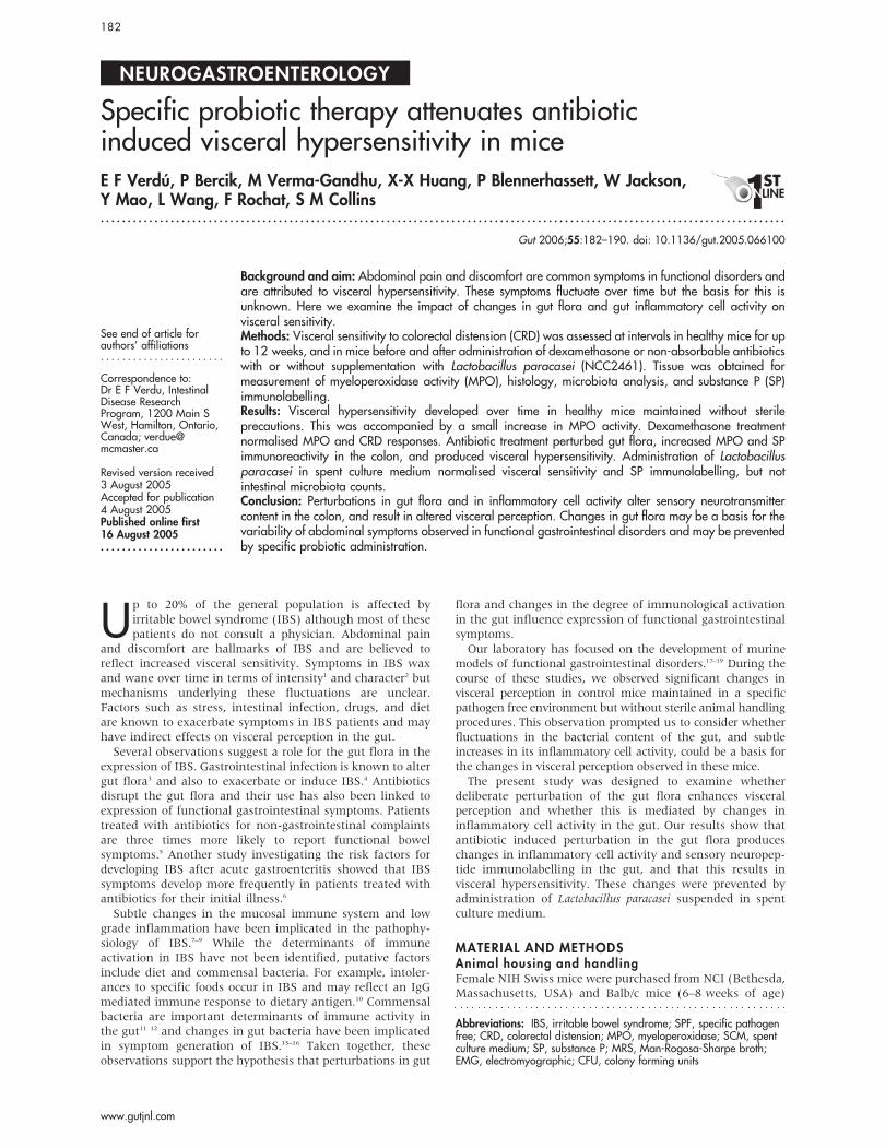
NEUROGASTROENTEROLOGY
Specific probiotic therapy attenuates antibioticinduced visceral hypersensitivity in miceE F Verdu, P Bercik, M Verma-Gandhu, X-X Huang, P Blennerhassett, W Jackson,Y Mao, L Wang, F Rochat, S M Collins. . . . . . . . . . . . . . . . . . . . . . . . . . . . . . . . . . . . . . . . . . . . . . . . . . . . . . . . . . . . . . . . . . . . . . . . . . . . . . . . . . . . . . . . . . . . . . . . . . . . . . . . . . . . . . . . . . . . . . . . . . . . . . .
See end of article forauthors’ affiliations. . . . . . . . . . . . . . . . . . . . . . .
Correspondence to:Dr E F Verdu, IntestinalDisease ResearchProgram, 1200 Main SWest, Hamilton, Ontario,Canada; [email protected]
Revised version received3 August 2005Accepted for publication4 August 2005Published online first16 August 2005. . . . . . . . . . . . . . . . . . . . . . .
Gut 2006;55:182–190. doi: 10.1136/gut.2005.066100
Background and aim: Abdominal pain and discomfort are common symptoms in functional disorders andare attributed to visceral hypersensitivity. These symptoms fluctuate over time but the basis for this isunknown. Here we examine the impact of changes in gut flora and gut inflammatory cell activity onvisceral sensitivity.Methods: Visceral sensitivity to colorectal distension (CRD) was assessed at intervals in healthy mice for upto 12 weeks, and in mice before and after administration of dexamethasone or non-absorbable antibioticswith or without supplementation with Lactobacillus paracasei (NCC2461). Tissue was obtained formeasurement of myeloperoxidase activity (MPO), histology, microbiota analysis, and substance P (SP)immunolabelling.Results: Visceral hypersensitivity developed over time in healthy mice maintained without sterileprecautions. This was accompanied by a small increase in MPO activity. Dexamethasone treatmentnormalised MPO and CRD responses. Antibiotic treatment perturbed gut flora, increased MPO and SPimmunoreactivity in the colon, and produced visceral hypersensitivity. Administration of Lactobacillusparacasei in spent culture medium normalised visceral sensitivity and SP immunolabelling, but notintestinal microbiota counts.Conclusion: Perturbations in gut flora and in inflammatory cell activity alter sensory neurotransmittercontent in the colon, and result in altered visceral perception. Changes in gut flora may be a basis for thevariability of abdominal symptoms observed in functional gastrointestinal disorders and may be preventedby specific probiotic administration.
Up to 20% of the general population is affected byirritable bowel syndrome (IBS) although most of thesepatients do not consult a physician. Abdominal pain
and discomfort are hallmarks of IBS and are believed toreflect increased visceral sensitivity. Symptoms in IBS waxand wane over time in terms of intensity1 and character2 butmechanisms underlying these fluctuations are unclear.Factors such as stress, intestinal infection, drugs, and dietare known to exacerbate symptoms in IBS patients and mayhave indirect effects on visceral perception in the gut.
Several observations suggest a role for the gut flora in theexpression of IBS. Gastrointestinal infection is known to altergut flora3 and also to exacerbate or induce IBS.4 Antibioticsdisrupt the gut flora and their use has also been linked toexpression of functional gastrointestinal symptoms. Patientstreated with antibiotics for non-gastrointestinal complaintsare three times more likely to report functional bowelsymptoms.5 Another study investigating the risk factors fordeveloping IBS after acute gastroenteritis showed that IBSsymptoms develop more frequently in patients treated withantibiotics for their initial illness.6
Subtle changes in the mucosal immune system and lowgrade inflammation have been implicated in the pathophy-siology of IBS.7–9 While the determinants of immuneactivation in IBS have not been identified, putative factorsinclude diet and commensal bacteria. For example, intoler-ances to specific foods occur in IBS and may reflect an IgGmediated immune response to dietary antigen.10 Commensalbacteria are important determinants of immune activity inthe gut11 12 and changes in gut bacteria have been implicatedin symptom generation of IBS.13–16 Taken together, theseobservations support the hypothesis that perturbations in gut
flora and changes in the degree of immunological activationin the gut influence expression of functional gastrointestinalsymptoms.
Our laboratory has focused on the development of murinemodels of functional gastrointestinal disorders.17–19 During thecourse of these studies, we observed significant changes invisceral perception in control mice maintained in a specificpathogen free environment but without sterile animal handlingprocedures. This observation prompted us to consider whetherfluctuations in the bacterial content of the gut, and subtleincreases in its inflammatory cell activity, could be a basis forthe changes in visceral perception observed in these mice.
The present study was designed to examine whetherdeliberate perturbation of the gut flora enhances visceralperception and whether this is mediated by changes ininflammatory cell activity in the gut. Our results show thatantibiotic induced perturbation in the gut flora produceschanges in inflammatory cell activity and sensory neuropep-tide immunolabelling in the gut, and that this results invisceral hypersensitivity. These changes were prevented byadministration of Lactobacillus paracasei suspended in spentculture medium.
MATERIAL AND METHODSAnimal housing and handlingFemale NIH Swiss mice were purchased from NCI (Bethesda,Massachusetts, USA) and Balb/c mice (6–8 weeks of age)
Abbreviations: IBS, irritable bowel syndrome; SPF, specific pathogenfree; CRD, colorectal distension; MPO, myeloperoxidase; SCM, spentculture medium; SP, substance P; MRS, Man-Rogosa-Sharpe broth;EMG, electromyographic; CFU, colony forming units
182
www.gutjnl.com
from Harlan (Indianapolis, Indiana, USA). Mice were keptunder specific pathogen free (SPF) conditions at McMasterUniversity Central Animal Care Facility. Sentinel SPF micewere screened according to the tracking profile of CharlesRiver’s Diagnostic laboratories (www.criver.com).
In the first set of experiments, mice were exposed tounfiltered air during colorectal distension (CRD) recordings.Prior to use, intrarectal balloons were thoroughly cleaned butnot sterilised. In the experiments using antibiotics, a ‘‘sterileprotocol’’ in which mice were exposed to HEPA filtered airwas applied. Mice were placed in restrainers inside level Bhoods and kept in custom made sterile HEPA filtered cagesduring CRD recordings. Fistulas and cables were sterilised atthe Central Sterilisation Unit at McMaster University.Intrarectal balloons and instruments were sterilised usingCIDEX OPA, and rinsed in sterile distilled water prior to use.
All experiments were approved by the McMaster UniversityAnimal Care Committee and the Canadian Council onAnimal Care.
Overall designTo confirm preliminary observations of the spontaneousdevelopment of visceral hypersensitivity in mice, we mea-sured CRD in Balb/c mice at day 0, and weeks 4, 8, 10, and 12.To examine the strain specificity of time dependent changesin visceral sensitivity, we also studied a group of NIH Swissmice at day 0 and at week 6. To investigate whether changesin visceral perception were accompanied by an increase ininflammatory cell activity, we measured myeloperoxidase(MPO) activity in colonic samples in additional Balb/c micethat were sacrificed at day 0 and at week 10. To establish anassociation between inflammatory cell activity and changesin visceral sensitivity, responses to CRD and MPO activitywere measured after intraperitoneal administration ofplacebo (sterile saline 100 ml) or dexamethasone (0.5 mg/kg) at week 12.
To determine whether changes in gut flora result in alteredvisceral perception, NIH Swiss mice received by gavage acombination of non-absorbable antibiotics or drinking water(placebo) for 10 days. Responses to CRD were measuredbefore and at 10 and 30 days after starting the antibiotics.Samples of colonic tissue and lumen content were obtainedat the different time points for MPO activity and for bacterialcounts. We used a combination of antibiotics that had beenwell characterised in terms of dose, drug concentration, andtissue toxicity in the mouse.20 21 Bacitracin and neomycinwere diluted in sterile deionised water and the pH of thesolution was adjusted to 4.0 to prevent inactivation ofbacitracin. Primaricin was added to the antibiotic solutionto prevent yeast overgrowth. Mice received bacitracin4 mg/ml, neomycin 4 mg/ml, and primaricin 0.2 g/ml ofdrinking water during the first five days. Antibiotic concen-trations in drinking water were reduced to 2 mg/ml forbacitracin and neomycin and to 0.1 g/ml for primaricinduring the last five days of treatment.
To examine whether probiotic therapy could protectagainst the antibiotic induced changes in visceral sensitivity,we administered the above combination of antibiotics plus100 ml of 1010 L paracasei NCC2461/ml in spent culturemedium, or placebo, for 10 days by oral gavage. L paracaseiwas chosen based on results from previous experiments in amodel of post-infective IBS showing that this probiotic strainattenuated post-infective hypercontractility in part by exert-ing an anti-inflammatory effect. The beneficial effect wasalso observed with its spent culture medium (SCM) devoid oflive bacteria.22 Responses to CRD were investigated beforeantibiotic therapy (day 0) and on day 10. Colonic content andtissue were obtained for histology (haematoxylin-eosinstain), for substance P (SP) immunohistochemistry, as well
as for measurement of total lactobacilli counts and forspecific detection of L paracasei NCC 2461.
To test the effect of L paracasei on normal visceralperception in mice that did not receive antibiotics, additionalmice (n = 6) were investigated at day 0 after a 10 day gavagewith Man-Rogosa-Sharpe broth (MRS; Becton Dickinson,Sparks, USA) and eight weeks later after a 10 day gavagewith MRS, followed by a 10 day gavage with L paracasei.
To discriminate between the effect of distensions or ofhandling of mice using an unsterile technique on MPOactivity, additional groups of mice were sham distended. Forthis, mice were handled in level B hoods and placed in HEPAfiltered restrainers during experiments. A sterile balloon wasinserted intrarectally but was not distended. Groups of micewere sacrificed at day 0, week 10, and week 12 (n = 5/group).
To determine whether dexamethasone or the antibioticcombination exerted direct effects on visceral sensitivity,responses to CRD were measured in a separate group of micethat received either dexamethasone or the antibiotic combi-nation directly into the colon via a double lumen catheter(n = 12). Local administration of saline or histamine wasused as negative and positive controls, respectively. Awashout period of three days was respected betweenexperiments. Dexamethasone (0.5 mg/kg; 0.2 ml), neomycin(4 mg/ml) + bacitracin (4 mg/ml) + primaricin (0.2 mg/ml)(0.2 ml), histamine (1026 mM; 0.2 ml), or saline (0.2 ml)were administered intrarectally after the third distension.
Pseudoaffective response to colorectal distensionElectromyographic (EMG) electrodes were surgically implantedunder sterile conditions in the anterior abdominal wall muscle
AUC
0
1
2
3
4A
Day 0
30 mm Hg
**
*
4 weeks 8 weeks Day 0
60 mm Hg4 weeks 8 weeks
AUC
0
1
2
3
4B
Day 0
30 mm Hg
*
6 weeks Day 0
60 mm Hg6 weeks
Figure 1 Visceral sensitivity to colorectal distension to 30 and60 mm Hg in (A) Balb/c mice (n = 8/group). At 30 mm Hg, *p = 0.02versus day 0; at 60 mm Hg, **p = 0.0008 versus day 0. (B) NIH Swissmice (n = 10/group). At 60 mm Hg, *p = 0.02 versus day 0. AUC, areaunder the curve for electromyographic responses to colorectal distension.Data are presented as box plots.
Probiotics and visceral sensitivity 183
www.gutjnl.com
of mice and a chronic fistula was exteriorised. Mice were thenallowed to recover for a period of at least seven days.
The response to CRD was assessed using a methoddescribed previously.19 CRD was performed in a stepwisefashion. Each 10 second distension was followed by a fiveminute resting period. Each level of distension (30 and60 mm Hg) was repeated three times. EMG activity of theabdominal muscle was continuously recorded using custo-mised software (Acquire 5.0; A Bayatti). The area under thecurve was calculated for five seconds before and after thebeginning of each distension period using customised soft-ware (GrafView 4.1; A Bayatti).
For experiments using the double lumen catheter, a singlenon-painful level of distension (40 mm Hg) was chosen andCRD responses were recorded before and 15 minutes afterdrug administration. The level of distension was chosenbased on previous studies demonstrating robust CRDresponses to 40 mm Hg.19
L paracasei culture and spent culture mediumL paracasei was chosen based on previous results showing thatthis strain attenuated post-infective muscle hypercontractility
by attenuating the inflammatory response to infection. Thiseffect was also observed with its SCM devoid of live bacteria.22 Lparacasei NCC2461 was obtained from the Nestle CultureCollection (Lausanne, Switzerland) and grown under anaerobicconditions in MRS. After 48 hours at 37 C, the number ofbacteria was estimated by measuring optical density at 600 nm(1 OD600 = 108 bacteria/ml). Bacterial cells were pelleted bycentrifugation for 15 minutes at 5000 g at 4 C, furtherresuspended at a concentration of 1010/ml in its SCM, and keptin frozen aliquots until use.
Intestinal microbiota countsAnalysis of a portion of the intestinal microbiota wasperformed before, on day 10 during antibiotic therapy, andon day 30 after discontinuation of antibiotics. Total lactoba-cilli counts were also performed before and on day 10 duringantibiotic therapy in mice treated with placebo and supple-mented with L paracasei. The presence of L paracasei wasspecifically investigated in the latter group. As no significantdifferences in intestinal microbiota were observed in controlsamples at different time points (days 0, 10, and 30), thesesamples were pooled in one single control group.
MPO
(U/g
)
4
2
1
3
0
A
Week 10 Week 12Day 0
PlaceboDexamethasone
***
***
††
AUC
4
2
1
3
0
C
Week 10
Distension: 60 mm Hg
Week 12Day 0
*†
AUC
4
2
1
3
0
B
Week 10
Distension: 30 mm Hg
Week 12Day 0
*†
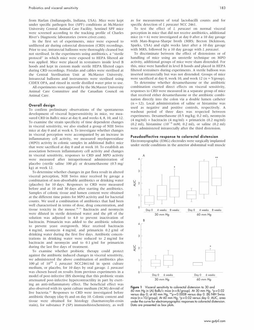
Figure 2 (A) Myeloperoxidase (MPO) activity for Balb/c mice at day 0 (n = 5), week 10 (n = 5), and week 12 (placebo n = 5; dexamethasone n = 8).***p,0.001 versus day 0, ��p = 0.01 versus week 12 placebo. Data are means (SD). (B) Visceral sensitivity to colorectal distension to 30 mmHg onday 0, week 10, and week 12 (n = 8). Dexamethasone (n = 8) or placebo (n = 5) was administered at week 11 (arrow). *p = 0.02 versus placebo;�p = 0.03 versus week 10. Data are presented as box plots. (C) Visceral sensitivity to colorectal distension to 60 mm Hg on day 0, week 10, and week12. *p = 0.07 versus placebo; �p = 0.04 versus week 10. AUC, area under the curve for electromyographic responses to colorectal distension. Data arepresented as box plots.
Day 30
CRD
res
pons
es (%
)
MPO
U/g
2.0
1.6
1.2
0.8
0.4
0
C500
400
300
200
100
0
A B
Day 10Day 0Placebo
30 mm HgAntibiotic Placebo
Day 10
60 mm HgAntibiotic
PlaceboATB
*†
**
Placebo
30 mm HgAntibiotic Placebo
Day 30
60 mm HgAntibiotic
*
*†
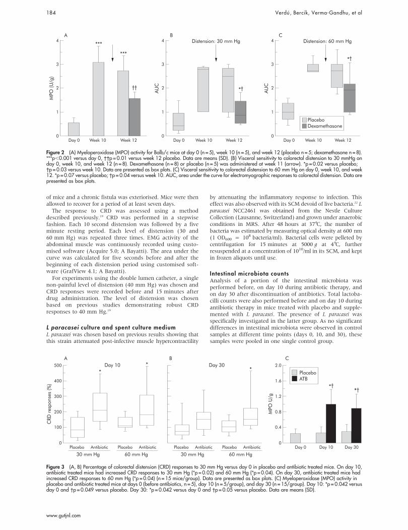
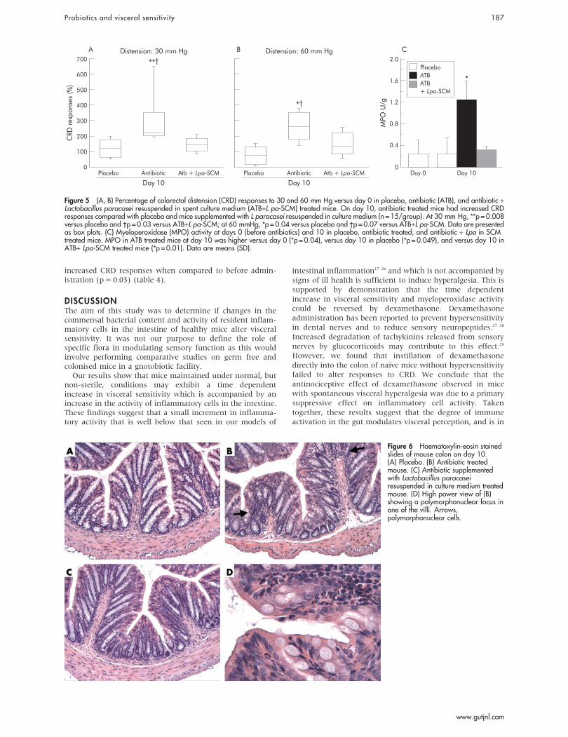
Figure 3 (A, B) Percentage of colorectal distension (CRD) responses to 30 mm Hg versus day 0 in placebo and antibiotic treated mice. On day 10,antibiotic treated mice had increased CRD responses to 30 mm Hg (*p = 0.02) and 60 mm Hg (*p = 0.04). On day 30, antibiotic treated mice hadincreased CRD responses to 60 mm Hg (*p = 0.04) (n = 15 mice/group). Data are presented as box plots. (C) Myeloperoxidase (MPO) activity inplacebo and antibiotic treated mice at days 0 (before antibiotics, n = 5), day 10 (n = 5/group), and day 30 (n = 15/group). Day 10: *p = 0.042 versusday 0 and �p = 0.049 versus placebo. Day 30: *p = 0.042 versus day 0 and �p = 0.05 versus placebo. Data are means (SD).
184 Verdu , Bercik, Verma-Gandhu, et al
www.gutjnl.com

Colonic segments (2 cm) were obtained under sterileconditions. Contents were pooled with 1 ml of 0.9% NaCl-10% glycerol used to wash the lumen. Tissues were ground in2 ml of 0.9% NaCL-10% glycerol using a polytron(Kinematica, Littau-Lucerne, Switzerland). Samples werestored at 270 C until analysis. On plating on semi-selectivemedia,23 bacterial populations were estimated by countingcolony forming units (CFU). Lactobacilli and Bacteroides wereincubated anaerobically (AnaeroGen; Oxoid, Basingstoke,UK) at 37 C for 48 hours. Enterobacteria, enterococci, andyeast were incubated aerobically at 37 C for 24 hours.Bacterial counts were expressed in log10 CFU/g faeces ortissue. Presence of L paracasei NCC2461 was monitored byrandom amplification of polymorphism DNA (RAPD) finger-print as described previously.22
Immunohistochemistry for substance PSP is a neurotransmitter demonstrated in the terminals ofprimary afferent nerves and in enteric neurones of bothanimals and humans.24 25 To detect SP we used immunohis-tochemistry in colonic frozen sections from mice treated withantibiotics, antibiotics plus L paracasei, or placebo. Tissue wasobtained before (day 0) and at day 10 during administrationof antibiotics.
As a primary antibody, we used a rabbit anti-substance P(1:2000) (1:1000) antiserum (Chemicon International,Temecula, California, USA). Negative controls were performedby omitting the primary antibody or by blocking antibody/protein complex formation for SP (peptide concentration10 mM). For the latter, SP blocking peptide (Sigma-Aldrich,
Oakville, Canada) was incubated with the primary antibodybefore its application on the slides. Vectastain Elite ABC kit wasused for secondary antibody and reporter solutions (Vector,Burlingame, California, USA).
Tissue sections were analysed using light microscopy(DMLS, Leica, Germany), and quantification of immuno-staining was performed on computer using public domainsoftware (image J 1.32, http:rsb.info.nih.gov/ij/) selecting thearea of the submucous plexus and muscularis externa, andpositive staining was expressed as percentage of total tissuearea.
Data presentation and statistical analysisParametric data are presented as means (SD) and non-parametric data as box plots (box median; 25%, 75%percentiles; whiskers 5th and 95th percentiles). EMGresponses to CRD are expressed as AUC or as percentage ofchange versus day 0 (day 0 = 100%) for each distension level.In experiments using the double lumen catheter, EMGresponses to CRD are presented as percentage of changeversus the first distension (1st distension = 100%).
Parametric data were analysed using the paired orunpaired t test, as appropriate. ANOVA was used for multiplecomparisons. Paired comparisons of non-parametric datawere performed by the Mann-Whitney U test. For multiplecomparisons of non-parametric data, the Friedman testfollowed by Wilcoxon-Wilcox was used.
RESULTSSpontaneous changes in visceral hypersensitivityVisceral sensitivity increased with time at four and eightweeks compared with day 0 in Balb/c mice (fig 1A). A 270%and a 140% increase in CRD responses to 30 and 60 mm Hg,respectively, was observed at eight weeks compared with day0. This observation was not strain specific as a 100% increasein CRD responses to 60 mm Hg was also observed in NIHSwiss mice at six weeks compared with day 0 (fig 1B).
To determine whether the time dependent increase invisceral perception was accompanied by changes in inflam-matory cell activity in the gut, we next measured MPO atweek 10 in Balb/c mice. As shown in fig 2A, there was a mildelevation in MPO values at week 10 in the hyperalgesic mice.
Effect of dexamethasoneTo determine whether the increased inflammatory cellactivity was associated with the emergence of visceralhypersensitivity, we measured responses to CRD before andafter dexamethasone treatment. As shown in fig 2A, MPOactivity increased at weeks 10 and 12 compared with day 0. Incontrast with placebo, dexamethasone reduced MPO activityat week 12 compared with week 10. This was accompanied bya 70% and a 25% drop in CRD responses to 30 and 60 mm Hg,respectively, at 12 weeks (fig 2B).
Table 1 Median (interquartile range) area under the curve values in placebo andantibiotic treated mice
Placebo Antibiotic
0 mm Hg 30 mm Hg 60 mm Hg 0 mm Hg 30 mm Hg 60 mm Hg
Day 0 0.2 1.2 1.7 0.2 0.7 1.3(0.2–0.3) (0.6–1.3) (1.0–2.0) (0.2–0.6) (0.5–1.4) (1.0–2.0)
Day 10 0.2 1.1 2.2 0.4 1.6** 2.8*�(0.1–0.3) (0.5–1.4) (1.0–2.4) (0.1–0.6) (0.6–2.0) (2.0–3.1)
Day 30 0.3 0.7 1.6 0.4 1.0 2.1**(0.2–0.4) (0.5–1.5) (1.4–2.3) (0.3–0.4) (0.5–1.5) (1.4–2.4)
*p,0.05 and **p = 0.08 versus day 0; �p,0.05 versus placebo.
*** ***
*** ***
3
4
5
6
7
8
9
Tissue
Content
Controls ATBday 10
ATBday 30
Log
CFU
/g
Figure 4 Total lactobacilli populations from colonic content and tissueon days 10 and 30 in antibiotic treated mice (ATB, n = 5 per group) andin controls. All placebo controls (day 0, day 10, and day 30) werepooled into one single group (n = 15). ***p,0.0001 versus controls.Data are means (SD). CFU, colony forming units.
Probiotics and visceral sensitivity 185
www.gutjnl.com
Effect of perturbation of the intestinal microbiotaTo determine whether minor increases in MPO activity wereassociated with the distension procedure itself or to exposureof mice to unfiltered air, additional groups of mice were shamdistended. We minimised the risk of bacterial contaminationof these mice by adopting sterile techniques and avoidingexposure of these mice to unfiltered air. MPO activity in thecolon was 0.2 (0.2), 0.3 (0.2), and 0.14 (0.1) on day 0, week10, and week 12, respectively (p.0.05).
To perturb the gut flora, mice were gavaged with acombination of non-absorbable antibiotics. Controls receivedphosphate buffered saline (placebo). MPO activity in placebotreated mice was 0.1 (0.01) at day 0, 0.5 (0.01) at day 10, and0.3 (0.01) at day 30 (both p.0.05 versus day 0). In addition,no significant increase in CRD responses was observed inplacebo treated mice at days 10 and 30 compared with day 0(fig 3A, 3B; table 1).
As shown in fig 3A and 3B, a 10 day course of antibioticswas associated with the emergence of visceral hypersensti-tivity to CRD. This was associated with a mild but significantincrease in MPO activity on day 10 and 30 after antibiotictherapy (fig 3C).
To evaluate the impact of the antibiotic combination usedin this study on selected gut microbiota, Bacteroides, entero-bacteria, enterococci, and lactobacilli counts were performed.Ten days after administration of antibiotics, we were not ableto culture lactobacilli from colonic content or tissue (,3.0 logCFU/g). As shown in fig 4, on day 30, lactobacilli populationswere still markedly reduced in mice previously treated withantibiotics compared with placebo treated controls.
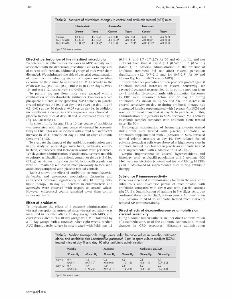
Table 2 shows the effect of antibiotics on enterobacteria,Bacteroides, and enterococci populations. Bacteroides andenterococci decreased significantly on day 10 during anti-biotic therapy. On day 30, increases in enterobacteria andBacteroides were observed with respect to control values.However, enterococci counts remained lower than controlvalues on day 30.
Effect of probioticsTo investigate the effect of L paracasei administration ofvisceral perception in untreated mice, visceral sensitivity wasmeasured in six mice after a 10 day gavage with MRS, andeight weeks later after a 10 day gavage with MRS followed bya 10 day gavage with L paracasei. After eight weeks, medianAUC (interquartile range) in mice treated with MRS was 1.1
(0.7–1.8) and 1.7 (0.7–2.7) for 30 and 60 mm Hg, and notdifferent from that at day 0 (1.1 (0.6–2.0); 1.5 (0.6–1.8))(table 3). L paracasei administration in the absence ofantibiotic treatment did not affect visceral perceptionsignificantly (1.2 (0.7–2.1) and 1.4 (0.7–2.5) for 30 and60 mm Hg; both p.0.05 versus MRS).
To test whether probiotics or their products protect againstantibiotic induced increases in visceral sensitivity, wegavaged L paracasei resuspended in its culture medium fromday 1 until day 10 concomitantly with antibiotics. Responsesto CRD were measured before and on day 10 duringantibiotics. As shown in fig 5A and 5B, the increase invisceral sensitivity on day 10 during antibiotic therapy wasattenuated in mice supplemented with L paracasei in SCM andwas not different than that at day 0. In parallel with this,administration of L paracasei in SCM decreased MPO activityin colonic samples compared with antibiotic alone treatedmice (fig 5C).
Histological examination of haematoxylin-eosin stainedslides from mice treated with placebo, antibiotics, orantibiotics supplemented with L paracasei in SCM revealednormal colonic structure at day 10. Few isolated foci ofpolymorphonuclear cells were observed at high power view inantibiotic treated mice but not in placebo or antibiotic treatedmice supplemented with L paracasei in SCM (fig 6).
Despite improvement in visceral hypersensitivity andhistology, total lactobacilli populations and L paracasei NCC2461 were undetectable (content and tissue ,3.0 log 10 CFU/g) in L paracasei/SCM supplemented mice during antibiotictherapy.
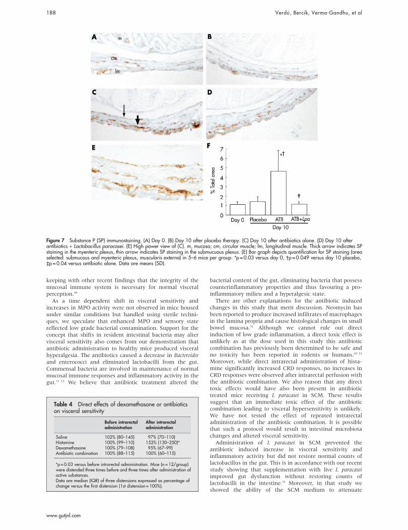
Substance P immunoreactivityThere was increased immunostaining for SP in the area of thesubmucous and myenteric plexus of mice treated withantibiotics compared with day 0 and with placebo controls(fig 7A, B). Quantification of staining in 5–6 slides per groupconfirmed these results (fig 7, bottom panel). Administrationof L paracasei in SCM to antibiotic treated mice markedlyreduced SP immunostaining.
Direct effects of dexamethasone or antibiotics onvisceral sensitivityUsing a double lumen catheter, neither direct administrationof dexamethasone, or of the antibiotic combination, causedchanges in CRD responses. Histamine administration
Table 2 Markers of microbiota changes in control and antibiotic treated (ATB) mice
Enterobacteria Bacteroides Enterococci
Content Tissue Content Tissue Content Tissue
Control 4.1 (0.3) 4.0 (0.0) 6.9 (1.1) 5.0 (1.0) 5.3 (1.2) 4.5 (0.7)Day 10 ATB 4.0 (0.0) 4.0 (0.0) 4.0 (0.0)* 4.0 (0.0) 4.0 (0.0)* 4.0 (0.0)Day 30 ATB 5.3 (1.1)* 4.8 (1.2)* 7.0 (2.4) 6.1 (2.0)* 4.08 (0.0)* 4.0 (0.0)
*p,0.05 versus control.
Table 3 Median (interquartile range) area under the curve values in placebo, antibioticalone, and antibiotic plus Lactobacillus paracasei (L pa) in spent culture medium (SCM)treated mice at day 0 and day 10 after antibiotic administration
Placebo Antibiotic Antibiotic+L pa/SCM
30 mm Hg 60 mm Hg 30 mm Hg 60 mm Hg 30 mm Hg 60 mm Hg
Day 0 0.7 1.5 0.6 1.5 0.8 1.7(0.5–1.1) (0.7–1.7) (0.4–0.8) (1.0–1.6) (0.4–1.1) (0.7–2.0)
Day 10 0.7 1.7 1.5* 2.6* 1.2 2.3(0.2–1.5) (1.0–2.5) (0.9–2.1) (1.6–3.3) (0.5–1.6) (1.0–3.1)
*p,0.05 versus day 0.
186 Verdu , Bercik, Verma-Gandhu, et al
www.gutjnl.com
increased CRD responses when compared to before admin-istration (p = 0.03) (table 4).
DISCUSSIONThe aim of this study was to determine if changes in thecommensal bacterial content and activity of resident inflam-matory cells in the intestine of healthy mice alter visceralsensitivity. It was not our purpose to define the role ofspecific flora in modulating sensory function as this wouldinvolve performing comparative studies on germ free andcolonised mice in a gnotobiotic facility.
Our results show that mice maintained under normal, butnon-sterile, conditions may exhibit a time dependentincrease in visceral sensitivity which is accompanied by anincrease in the activity of inflammatory cells in the intestine.These findings suggest that a small increment in inflamma-tory activity that is well below that seen in our models of
intestinal inflammation17 26 and which is not accompanied bysigns of ill health is sufficient to induce hyperalgesia. This issupported by demonstration that the time dependentincrease in visceral sensitivity and myeloperoxidase activitycould be reversed by dexamethasone. Dexamethasoneadministration has been reported to prevent hypersensitivityin dental nerves and to reduce sensory neuropeptides.27 28
Increased degradation of tachykinins released from sensorynerves by glucocorticoids may contribute to this effect.29
However, we found that instillation of dexamethasonedirectly into the colon of naıve mice without hypersensitivityfailed to alter responses to CRD. We conclude that theantinociceptive effect of dexamethasone observed in micewith spontaneous visceral hyperalgesia was due to a primarysuppressive effect on inflammatory cell activity. Takentogether, these results suggest that the degree of immuneactivation in the gut modulates visceral perception, and is in
CRD
res
pons
es (%
)
MPO
U/g
2.0
1.6
1.2
0.8
0.4
0
C700
500
600
400
300
200
100
0
A B
Day 10Day 0Placebo
Day 10
Distension: 30 mm Hg
*†
**†
*
Atb + Lpa-SCMAntibiotic Placebo
Day 10
Distension: 60 mm Hg
Atb + Lpa-SCMAntibiotic
PlaceboATBATB+ Lpa-SCM
Figure 5 (A, B) Percentage of colorectal distension (CRD) responses to 30 and 60 mm Hg versus day 0 in placebo, antibiotic (ATB), and antibiotic +Lactobacillus paracasei resuspended in spent culture medium (ATB+L pa-SCM) treated mice. On day 10, antibiotic treated mice had increased CRDresponses compared with placebo and mice supplemented with L paracasei resuspended in culture medium (n = 15/group). At 30 mm Hg, **p = 0.008versus placebo and �p = 0.03 versus ATB+L pa-SCM; at 60 mmHg, *p = 0.04 versus placebo and �p = 0.07 versus ATB+L pa-SCM. Data are presentedas box plots. (C) Myeloperoxidase (MPO) activity at days 0 (before antibiotics) and 10 in placebo, antibiotic treated, and antibiotic + Lpa in SCMtreated mice. MPO in ATB treated mice at day 10 was higher versus day 0 (*p = 0.04), versus day 10 in placebo (*p = 0.049), and versus day 10 inATB+ Lpa-SCM treated mice (*p = 0.01). Data are means (SD).
Figure 6 Haematoxylin-eosin stainedslides of mouse colon on day 10.(A) Placebo. (B) Antibiotic treatedmouse. (C) Antibiotic supplementedwith Lactobacillus paracaseiresuspended in culture medium treatedmouse. (D) High power view of (B)showing a polymorphonuclear focus inone of the villi. Arrows,polymorphonuclear cells.
Probiotics and visceral sensitivity 187
www.gutjnl.com
keeping with other recent findings that the integrity of themucosal immune system is necessary for normal visceralperception.30
As a time dependent shift in visceral sensitivity andincreases in MPO activity were not observed in mice housedunder similar conditions but handled using sterile techni-ques, we speculate that enhanced MPO and sensory statereflected low grade bacterial contamination. Support for theconcept that shifts in resident intestinal bacteria may altervisceral sensitivity also comes from our demonstration thatantibiotic administration to healthy mice produced visceralhyperalgesia. The antibiotics caused a decrease in Bacteroidesand enterococci and eliminated lactobacilli from the gut.Commensal bacteria are involved in maintenance of normalmucosal immune responses and inflammatory activity in thegut.11 12 We believe that antibiotic treatment altered the
bacterial content of the gut, eliminating bacteria that possesscounterinflammatory properties and thus favouring a pro-inflammatory milieu and a hyperalgesic state.
There are other explanations for the antibiotic inducedchanges in this study that merit discussion. Neomycin hasbeen reported to produce increased infiltrates of macrophagesin the lamina propria and cause histological changes in smallbowel mucosa.31 Although we cannot rule out directinduction of low grade inflammation, a direct toxic effect isunlikely as at the dose used in this study this antibioticcombination has previously been determined to be safe andno toxicity has been reported in rodents or humans.20 21
Moreover, while direct intrarectal administration of hista-mine significantly increased CRD responses, no increases inCRD responses were observed after intrarectal perfusion withthe antibiotic combination. We also reason that any directtoxic effects would have also been present in antibiotictreated mice receiving L paracasei in SCM. These resultssuggest that an immediate toxic effect of the antibioticcombination leading to visceral hypersensitivity is unlikely.We have not tested the effect of repeated intrarectaladministration of the antibiotic combination. It is possiblethat such a protocol would result in intestinal microbiotachanges and altered visceral sensitivity.
Administration of L paracasei in SCM prevented theantibiotic induced increase in visceral sensitivity andinflammatory activity but did not restore normal counts oflactobacillus in the gut. This is in accordance with our recentstudy showing that supplementation with live L paracaseiimproved gut dysfunction without restoring counts oflactobacilli in the intestine.23 Moreover, in that study weshowed the ability of the SCM medium to attenuate
Figure 7 Substance P (SP) immunostaining. (A) Day 0. (B) Day 10 after placebo therapy. (C) Day 10 after antibiotics alone. (D) Day 10 afterantibiotics + Lactobacillus paracasei. (E) High power view of (C). m, mucosa; cm, circular muscle; lm, longitudinal muscle. Thick arrow indicates SPstaining in the myenteric plexus, thin arrow indicates SP staining in the submucuous plexus. (E) Bar graph depicts quantification for SP staining (areaselected: submucous and myenteric plexus, muscularis externa) in 5–6 mice per group. *p = 0.03 versus day 0, �p = 0.049 versus day 10 placebo,`p = 0.04 versus antibiotic alone. Data are means (SD).
Table 4 Direct effects of dexamethasone or antibioticson visceral sensitivity
Before intrarectaladministration
After intrarectaladministration
Saline 102% (80–145) 97% (70–110)Histamine 100% (99–110) 152% (130–250)*Dexamethasone 100% (79–108) 95% (67–99)Antibiotic combination 100% (88–115) 100% (60–115)
*p = 0.03 versus before intrarectal administration. Mice (n = 12/group)were distended three times before and three times after administration ofactive substances.Data are median (IQR) of three distensions expressed as percentage ofchange versus the first distension (1st distension = 100%).
188 Verdu , Bercik, Verma-Gandhu, et al
www.gutjnl.com

inflammation induced changes in muscle contractility fol-lowing primary infection by the nematode Trichinella spiralis.23
Taken together with our previous results, and because Lparacasei was undetectable on day 10 after antibiotic therapy,we suggest that the beneficial effect after antibiotic therapymay be mediated by a soluble product of L paracasei present inthe SCM in which the bacteria were resuspended.Alternatively, bacterial fragments released as a consequenceof antibiotic therapy may be involved, as probiotic DNA hasbeen shown to attenuate inflammation in models ofexperimental colitis.32
In the present study, we showed that the antibioticinduced change in visceral sensitivity was accompanied byincreased SP immunoreactivity, which was localised primar-ily in the myenteric and submucous plexus. It is likely thatthe change in sensory neurotransmitter content was second-ary to the increase in inflammatory activity, as previousstudies have shown increases in enteric SP followingexposure of the myenteric plexus to interleukin 1b33 and inexperimental colitis.34 However, we cannot exclude thepossibility that bacteria directly influence neurotransmittercontent. Previous studies have suggested that pathogenicmicrobes can upregulate SP in infected tissue.35 36 Directcommunication between commensals and the enteric ner-vous system was suggested in the study of Hooper et al inwhich expression of genes encoding enteric neural transmis-sion differed in germ free and colonised mice.37 In addition,Kamm et al showed changes in the localisation patterns ofneuronal markers in myenteric neurones of the pig jejunumof Saccharomyces boulardi treated animals.38 While an oligo-peptidase produced by L paracasei has been shown tohydrolyse the Pro-Gln, Gln-Phe, and Phe-Gly bonds of SP,39
we consider this an unlikely explanation for its prevention ofantibiotic induced increases in SP and visceral sensitivity asthis action would not account for the decrease in MPOactivity.
There are several implications of our results. Firstly, thehygiene status of mice maintained in conventional facilitiesmay influence responsiveness to colorectal distension, andthis may be important in disease models, which are notassociated with overt inflammation. Secondly, the notionthat subtle changes in inflammatory activity in the gut altervisceral perception may have bearing on the finding that IBSsymptoms are less prevalent in those patients receiving oralcorticosteroids for other indications,40 and on the associationbetween antibiotic usage and expression of IBS.5 6 41 Thirdly,these findings provide a rationale for the use of selectedprobiotics in the management of IBS.
ACKNOWLEDGEMENTSThe authors thank G Bergonzelli for helpful discussions andG Reuteler for technical assistance with the bacterial cultures.
Authors’ affiliations. . . . . . . . . . . . . . . . . . . . .
E F Verdu, P Bercik, M Verma-Gandhu, X-X Huang, P Blennerhassett,W Jackson, Y Mao, L Wang, S M Collins, IDRP, McMaster University,Hamilton, CanadaF Rochat, Department of Nutrition and Health, Nestle Research Centre,Lausanne, Switzerland
We acknowledge the Canadian Institutes of Health Research (grant toSMC) and the Canadian Association of Gastroenterology and AstraZeneca, Canada for scholarship support (EFV).
Conflict of interest: None declared.
REFERENCES1 Agreus L, Svardsudd K, Talley NJ, et al. Natural history of gastroesophageal
reflux disease and functional abdominal disorders: a population-based study.Am J Gastroenterol 2001;96:2905–11.
2 Locke GR III. Natural history of irritable bowel syndrome and durability of thediagnosis. Rev Gastroenterol Disord 2003;S12:12–17.
3 Guarner F, Malagelada JR. Gut flora in health and disease. Lancet2003;361:512–19.
4 Spiller RC. Postinfectious irritable bowel syndrome. Gastroenterology2003;124:1662–71.
5 Alun-Jones V, Wilson AJ, Hunter JO, et al. The aetiological role of antibioticprophylaxis with hysterectomy in irritable bowel syndrome. J Obstet Gynaecol1984;5:S22–3.
6 Gwee KA, Graham JC, McKendrick MW, et al. Psychometric scores andpersistence of irritable bowel after infectious diarrhea. Lancet1996;347:150–3.
7 Rodriguez LA, Ruigomez A. Increased risk of irritable bowel syndrome afterbacterial gastroenteritis: cohort study. BMJ 1999;318:565–6.
8 Collins SM, Piche T, Rampal P. The putative role of inflammation in irritablebowel syndrome. Gut 2001;49:743–5.
9 Chadwick VS, Chen W, Shu D, et al. Activation of the mucosal immune systemin irritable bowel syndrome. Gastroenterology 2002;122:1778–83.
10 Atkinson W, Sheldon TA, Shaath N, et al. Food elimination based on IgGantibodies in irritable bowel syndrome: a randomized controlled trial. Gut2004;53:1459–64.
11 Cebra JJ, Periwal SB, Lee G, et al. Development and maintenance of the gut-associated lymphoid tissue (GALT): the roles of enteric bacteria and viruses.Dev Immunol 1998;6:13–18.
12 Macpherson AJ, Harris NL. Interactions between commensal intestinalbacteria and the immune system. Nat Rev Immunol 2004;4:478–85.
13 King TS, Elia M, Hunter JO. Abnormal colonic fermentation in irritable bowelsyndrome. Lancet 1998;352:1187–9.
14 Madden JA, Hunter JO. A review of the role of the gut microflora in irritablebowel syndrome and the effects of probiotics. Br J Nutr 2002;88:S67–72.
15 Nobaek S, Johansson ML, Molin G, et al. Alteration of intestinal microflora isassociated with reduction in abdominal bloating and pain in patients withirritable bowel syndrome. Am J Gastroenterol 2000;95:1231–8.
16 Pimentel M, Chow EJ, Lin HC. Eradication of small intestinal bacterialovergrowth reduces symptoms of irritable bowel syndrome. Am J Gastroenterol2000;95:3503–6.
17 Barbara G, Vallance BA, Collins SM. Persistent intestinal neuromusculardysfunction after acute nematode infection in mice. Gastroenterology1997;113:1224–32.
18 Barbara G, De Giorgio R, Deng Y, et al. Role of immunologic factors andcyclooxygenase 2 in persistent postinfective enteric muscle dysfunction inmice. Gastroenterology 2001;120:1729–36.
19 Bercik P, Wang L, Verdu EF, et al. Visceral hyperalgesia and intestinaldysmotlity in a mouse model of post infective gut dysfunction.Gastroenterology 2004;127:179–87.
20 Van Der Waaij D, Sturm CA. Antibiotic decontamination of the digestive tractof mice. Technical procedures. Lab Anim Care 1968;18:1–10.
21 Van Der Waaij D, Berghuis-De Vries JM, Korthals Altes C, et al. Oral doseand faecal concentrations of antibiotics during antibiotic decontamination inmice and in a patient. J Hyg Camb 1974;73:197–203.
22 Verdu EF, Bercik P, Bergonzelli G, et al. Lactobacillus paracasei normalizesmuscle hypercontractility in a murine model of post-infective gut dysfunction.Gastroenterology 2004;127:826–37.
23 Guigoz Y, Rochat F, Perruisseau-Carrier G, et al. Effects of oligosaccharide onthe faecal flora and non-specific immune system in elderly people. Nutr Res,22:13–25.
24 Kamm K, Hoppes S, Breves G, et al. Effect of the probiotic yeastSaccaromyces boulardii on the neurochemistry of myenteric neurons in pigjejunum. Neurogastroenterol Motil 2004;16:53–60.
25 Schneider J, Jehle EC, Starlinger MJ, et al. Neurotransmitter coding of entericneurons in the submucous plexus is changed in non-inlfamed rectum ofpatients with Crohn’s disease. Neurogastroenterol Motil 2001;13:255–64.
26 Verdu EF, Deng Y, Bercik P, et al. Modulatory effects of estrogen in two murinemodels of experimental colitis. Am J Physiol Gastrointest Liver Physiol2002;283:G27–36.
27 Barron RP, Benoliel R, Zeltser R, et al. Effect of dexamethasone and dypironeon lingual and inferior alveolar nerve hypersensitivity following third molarextractions: preliminary report. J Orofac Pain 2004;18:62–8.
28 Hong D, Byers MR, Oswald RJ. Dexamethasone treatment reduces sensoryneuropeptides and nerve sprouting reactions in injured teeth. Pain1993;55:171–81.
29 Piedimonte G, McDonald DM, Nadel JA. Endopeptidase and kininase IImediate glucocorticoid inhibition of neurogenic inflammation in the rattrachea. J Clin Invest 1991;88:40–4.
30 Verma-Gandhu M, Bercik P, Blennerhassett P, et al. Immunodeficiency andvisceral hyperalgesia: A putative mechanisms for abdominal pain in AIDSpatients. Gastroenterology 2004;126:A161.
31 Dobins III WO, Herrero BA, Mansbach CM. Morphologic alterations associatedwith neomycin induced malabsorption. Am J Med Sci 1968;255:63–77.
32 Rachmilewitz D, Katakura K, Karmeli F, et al. Toll-like receptor 9 signalingmediates the anti-inflammatory effects of probiotics in murine experimentalcolitis. Gastroenterology 2004;126:520–8.
33 Hurst SM, Stanisz AM, Sharkey KA, et al. Interleukin 1 beta-induced increasein substance P in rat myenteric plexus. Gastroenterology 1993;105:1754–60.
34 Miampamba M, Sharkey KA. Distribution of calcitonin gene-related peptide,somatostatin, substance P and vasoactive intestinal polypeptide inexperimental colitis in rats. Neurogastroenterol Motil 1998;10:315–29.
35 Gonkowski S, Kaminska B, Bossowska A, et al. The influence of experimentalBacteroides fragilis infection on substance P and somatostatin-immunoreactiveneural elements in the porcine ascending colon - a preliminary report. FoliaMorphol (Warsz) 2003;62:455–57.
Probiotics and visceral sensitivity 189
www.gutjnl.com
36 Piedimonte G, Rodriguez, MM, King KA, et al. Respiratory syncytial virusupregulates expression of the substance P receptor in rat lungs. Am J Physiol1999;277:L831–40.
37 Hooper LV, Wong MH, Thelin A, et al. Molecular analysis of commensal host-microbial relationships in the intestine. Science 2001;291:881–4.
38 Kamm K, Hoppe S, Breves G, et al. Effects of the probiotic yeastSaccharomyces boulardii on the neurochemistry of myenteric neurones in pigjejunum. Neurogastroenterol Motil 2004;16:53–60.
39 Tobiassen RO, Sorhaug T, Stepaniak L. Characterization of an intracellularoligopeptidase from Lactobacillus paracasei. Appl Environ Microbiol1997;63:1284–7.
40 Huerta C, Garcia Rodriguez LA, Wallander MA, et al. Users of oral steroidsare at a reduced risk of developing irritable bowel syndrome.Pharmacoepidemiol Drug Saf 2003;12:583–8.
41 Maxwell PR, Rink E, Kumar D, et al. Antibiotics increase functional abdominalsymptoms. Am J Gastroenterol 2002;97:104–8.
EDITOR’S QUIZ: GI SNAPSHOT . . . . . . . . . . . . . . . . . . . . . . . . . . . . . . . . . . . . . . . . . . . . . . . . . . . . . . . . . . . . . . . . . .
doi: 10.1136/gut.2005.073270
AnswerFrom question on page 181Computed tomography scan showed signs of ileal obstruc-tion. The terminal ileal loop presented a stenosis withoedematous wall (fig 1 (ii)) and a foreign body stuck in thestenosis (fig 1 (i)).
The patient was interviewed again and she admittedhaving swallowed a medlar seed 24 hours before hersymptoms began. After a preliminary elimination of aprobable infectious causes of terminal ileitis (includingtuberculosis), Crohn’s disease was suspected and she wastreated with hydrocortisone 300 mg/day. Her symptomsresolved 72 hours later, and the seed was recovered in thestools.
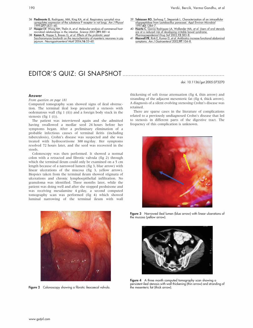
Colonoscopy was then performed. It showed a normalcolon with a retracted and fibrotic valvula (fig 2) throughwhich the terminal ileum could only be examined on a 5 cmlength because of a narrowed lumen (fig 3, blue arrow) withlinear ulcerations of the mucosa (fig 3, yellow arrow).Biopsies taken from the terminal ileum showed stigmata ofulcerations and chronic lymphoepithelial infiltration. Nogranuloma was identified. Three months later, while thepatient was doing well and after she stopped prednisone andwas receiving mesalamine 4 g/day, a second computedtomography scan was performed (fig 4) which showedluminal narrowing of the terminal ileum with wall
thickening of soft tissue attenuation (fig 4, thin arrow) andstranding of the adjacent mesenteric fat (fig 4, thick arrow).A diagnosis of a silent evolving stenosing Crohn’s disease wasretained.
There are sparse cases in the literature of complicationsrelated to a previously undiagnosed Crohn’s disease that ledto stenosis in different parts of the digestive tract. Thefrequency of this complication is unknown.
Figure 2 Colonoscopy showing a fibrotic ileocaecal valvula.
Figure 3 Narrowed ileal lumen (blue arrow) with linear ulcerations ofthe mucosa (yellow arrow).
Figure 4 A three month computed tomography scan showing apersistent ileal stenosis with wall thickening (thin arrow) and stranding ofthe mesenteric fat (thick arrow).
190 Verdu , Bercik, Verma-Gandhu, et al
www.gutjnl.com
Copyright © 2022 FDOKUMEN