Ameliorative Function of a Probiotic Bacterium, Lactobacillus ...
Upload
khangminh22Category
view
12download
0
University of South Florida University of South Florida
Digital Commons @ University of South Florida Digital Commons @ University of South Florida
USF St. Petersburg campus Faculty Publications USF Faculty Publications
2020
Antimicrobial and Antibiofilm Activities of Probiotic Lactobacilli Antimicrobial and Antibiofilm Activities of Probiotic Lactobacilli
on Antibiotic-Resistant Proteus mirabilis on Antibiotic-Resistant Proteus mirabilis
Mona Shaaban
Ola A. Abd El-Rahman
Bashair Al-Qaidi
Hossam M. Ashour University of South Florida St. Petersburg, [email protected]
Follow this and additional works at: https://digitalcommons.usf.edu/fac_publications
Recommended Citation Recommended Citation Shaaban, M., Abd El-Rahman, O. A., Al-Qaidi, B., & Ashour, H. M. (2020). Antimicrobial and Antibiofilm Activities of Probiotic Lactobacilli on Antibiotic-Resistant Proteus mirabilis. Microorganisms, 8(6), 960. doi:10.3390/microorganisms8060960
This Article is brought to you for free and open access by the USF Faculty Publications at Digital Commons @ University of South Florida. It has been accepted for inclusion in USF St. Petersburg campus Faculty Publications by an authorized administrator of Digital Commons @ University of South Florida. For more information, please contact [email protected].

microorganisms
Article
Antimicrobial and Antibiofilm Activities of ProbioticLactobacilli on Antibiotic-Resistant Proteus mirabilis
Mona Shaaban 1,† , Ola A. Abd El-Rahman 2,† , Bashair Al-Qaidi 3 and Hossam M. Ashour 4,5,*1 Department of Microbiology and Immunology, Faculty of Pharmacy, Mansoura University,
Mansoura 35516, Egypt2 Department of Microbiology and Immunology, Faculty of Pharmacy (Girls), Al-Azhar University,
Cairo 11651, Egypt3 Madinah Maternity and Children Hospital, Madinah 42319, Saudi Arabia4 Department of Biological Sciences, College of Arts and Sciences, University of South Florida St. Petersburg,
St. Petersburg, FL 33701, USA5 Department of Microbiology and Immunology, Faculty of Pharmacy, Cairo University, Cairo 11562, Egypt* Correspondence: [email protected]† Both authors contributed equally to this work.
Received: 6 June 2020; Accepted: 23 June 2020; Published: 26 June 2020�����������������
Abstract: The emergence of biofilm-forming, multi-drug-resistant (MDR) Proteus mirabilis infections isa serious threat that necessitates non-antibiotic therapies. Antibiotic susceptibility and biofilm-formingactivity of P. mirabilis isolates from urine samples were assessed by disc diffusion and crystal violetassays, respectively. Antimicrobial activities of probiotic Lactobacilli were evaluated by agar diffusion.Antibiofilm and anti-adherence activities were evaluated by crystal violet assays. While most P. mirabilisisolates were antibiotic-resistant to varying degrees, isolate P14 was MDR (resistant to ceftazidime,cefotaxime, amoxicillin-clavulanic acid, imipenem, ciprofloxacin, and amikacin) and formed strongbiofilms. Cultures and cell-free supernatants of Lactobacillus casei and Lactobacillus reuteri exhibitedantimicrobial and antibiofilm activities. The 1/16 concentration of untreated supernatants of L. caseiand L. reuteri significantly reduced mature biofilm formation and adherence of P14 by 60% and 72%,respectively (for L. casei), and by 73% each (for L. reuteri). The 1/8 concentration of pH-adjustedsupernatants of L. casei and L. reuteri significantly reduced mature biofilm formation and adherenceof P14 by 39% and 75%, respectively (for L. casei), and by 73% each (for L. reuteri). Scanning electronmicroscopy (SEM) confirmed eradication of P14’s biofilm by L. casei. L. casei and L. reuteri could beutilized to combat Proteus-associated urinary tract infections.
Keywords: Proteus mirabilis; antibiofilm; anti-adherence; Lactobacillus
1. Introduction
Proteus mirabilis is one of the most common pathogens associated with urinary tract infections(UTI) [1,2]. In addition, it can cause surgical wound infections, biliary tract infections, wound infections,and nosocomial infections [3]. It is part of the normal gut flora and can cause opportunistic infectionsin immunocompromised and elderly patients [3]. The emergence of antibiotic resistance in clinicalisolates of P. mirabilis is a major healthcare issue [4,5]. P. mirabilis poses a major challenge in infectionmanagement as it produces AmpC β-lactamases and extended spectrum β-lactamases [6]. Furthermore,it can develop complex biofilms with accumulated layers of polysaccharides in which sessile cells areembedded, which adds to the severity of the infection [7]. The severity, chronicity, and disseminationof Proteus infections have been mainly attributed to its ability to form biofilms [7].
Probiotics are living microorganisms that belong to Lactobacillus and Bifidobacterium genera [8,9].The intestinal probiotic Lactobacillus strains have been recognized for their antimicrobial activities
Microorganisms 2020, 8, 960; doi:10.3390/microorganisms8060960 www.mdpi.com/journal/microorganisms
Microorganisms 2020, 8, 960 2 of 13
against enteric bacterial pathogens [10]. Different groups showed that Lactobacillus casei producesbacteriocins and anti-adherence biosurfactant proteins against Staphylococcus aureus, Bacillus subtilis,and Micrococcus roseus [11,12]. Human gastrointestinal Lactobacillus reuteri produces a broad-spectrumantimicrobial called reuterin, which possesses activity against Gram-positive and Gram-negativeenteric pathogens [13]. Probiotic Lactobacilli can also suppress virulence and dissemination ofinfectious pathogens [14]. This is typically accomplished through the production of organic acids andantimicrobials, such as bacteriocins, lipopeptides, and surface proteins [11,15,16].
We have previously shown that probiotic lactobacilli inhibited growth, biofilm formation, and geneexpression of Streptococcus mutans [17]. The anti-infective and anti-colonization properties of probioticlacobacilli are of paramount importance in combating various bacterial infections [18,19]. Due to theirantimicrobial properties, probiotics may be considered for treatment and prevention of infectiousdiseases caused by oral, enteric, and urogenital pathogens [20,21]. Lactobacilli have been specificallyshown to prevent recurrent urinary tract infections [22]. This indicates their promise as anti-Proteusagents. Given the antibiotic resistance problem, it is important to develop Lactobacillus-based approachesto combat Proteus mirabilis-induced urinary tract infections and catheter-associated infections.
In order to be able to better manage P. mirabilis infections, we investigated the potential inhibitoryactivities of L. casei and L. reuteri on bacterial growth, mature biofilm formation, and adhesion propertiesof multi-drug-resistant (MDR) Proetus mirabilis clinical isolates.
We tested pH-adjusted supernatants of Lactobacillus, which is a commonly used approach inLactobacillus probiotic studies to reduce the acidity of the Lactobacillus supernatants and thus assess ifthe antimicrobial activity of Lactobacillus products is pH-dependent [23–27]. This is significant giventhat studies showed that the acidic pH (due to lactic acid secretion) contributed only a small part of theactivity [28], or did not contribute any additional activity (activity was pH-independent) [29]. In ourstudy, the pH adjustment was important to reveal how much of the anti-Proteus activity of probioticLactobacillus supernatants was related to non-acidic products and whether the antimicrobial activity ofthe supernatant was pH-dependent.
2. Materials and Methods
2.1. Microorganism and Growth Conditions
P. mirabilis isolates were obtained from urine samples collected from several hospitals in Madinah,KSA (Ohud Hospital, King Fahad Hospital, Al-Ansar Hospital, and Madina Maternity and ChildrenHospital). P. mirabilis isolates were identified using biochemical assays and confirmed as P. mirabilisusing the VITEK compact system. Assays included Gram stain, growth characteristics on MacConkeyagar, CLED agar, and triple sugar iron media [30].
Two probiotic strains (L. casei DSM 20011 and L. reuteri DSM 20016) were purchased from MERCEN,Egypt. Lactobacillus isolates were grown on deMan, Rogosa and Sharpe medium (MRS) and wereanaerobically (AnaeroGen 2.5 L sachets in 2.5 L AnaeroJar AG25, Oxoid Ltd., Hampshire, UK) incubatedat 37 ◦C [31].
Ethical approvals were obtained from the Institutional Review Boards of the hospitals, ethicscommittees of the college of pharmacy, Taibah University, KSA and Faculty of Pharmacy, MansouraUniversity, Egypt. Ethical approval code is TUCODRE/20151025/ALQAIDI (in October 2015).
2.2. Antimicrobial Susceptibility of P. mirabilis Isolates
P. mirabilis isolates were tested for their resistance to different antimicrobials using the diskdiffusion method [32]. The antimicrobials used were amoxicillin-clavulanic acid (AMC; 30 mg),imipenem (IMP; 10 mg), cefoxitin (FOX; 30 mg), ceftazidime (CAZ; 30 mg), ciprofloxacin (CIP; 5 mg),cefotaxime; (CTX; 30 mg), and amikacin (AK; 30 mg) (Bioanalyse, Ankara, Turkey).

Microorganisms 2020, 8, 960 3 of 13
2.3. Detection of Biofilm Formation by P. mirabilis Isolates
The mature biofilm of P. mirabilis isolates was formed in flat-bottomed 96-well microtiter plates.The overnight culture of each isolate was adjusted to 0.5 McFarland (1.5 × 10⁸ CFU/mL) using MullerHinton broth. Then, 200 µL of the diluted cultures was distributed in each well and incubated at 37 ◦Cfor 48 h. The planktonic cells were removed and the attached cells were gently washed twice withsterile physiological saline. Then, 200 µL of methanol (99%) was added to each well and retainedfor 15 min to fix the sessile cells. The methanol was discarded, and the plate was left until completedryness. To stain the adherent cells, 200 µL of 2% w/v crystal violet solution was added to each well andwas left for 20 min. The wells were washed gently and left to dry. Glacial acetic acid (200 µL; 33% w/v)was added to release the bound dye and the absorbance was measured at OD540 nm using ELISAmicroplate reader (MR-960, Perlong Medical Equipment Co. Ltd., Nanjing, China). Depending on theoptical density (OD) generated by the bacterial biofilms, the tested P. mirabilis isolates were classifiedinto non-biofilm producers, weak biofilm producers, moderate biofilm producers, and strong biofilmproducers [33]. The cut-off OD of the negative control (ODc) was determined by adding the meanof the negative control to three standard deviations of it. The lack of biofilm formation is indicatedby ODs ≤ ODc for the tested isolates. Weak biofilm production is indicated by ODc < OD ≤ (2 ODc).Moderate biofilm formation is indicated by (2 ODc) < O.D. ≤ (4 ODc). Strong biofilm formation isindicated by (4 ODc) < O.D. Tests were conducted in triplicates.
2.4. Preparation of Cell-Free Supernatant from Lactobacillus Strains
In order to prepare the Lactobacillus culture, the MRS medium was inoculated with Lactobacillusstrains with inoculum size 1% v/v and was incubated anaerobically at 37 ◦C for 48 h. The grown culturewas centrifuged at 6000 rpm for 15 min to separate all cells and the supernatant was filtered through a0.22 µm membrane filter. The cell-free supernatant was labeled as untreated supernatant (U) and storedat −20 ◦C. The Lactobacillus supernatant was highly acidic due to lactic acid production. To adjust thepH of the supernatant to pH 6.5–7.0, 1N NaOH was used and this fraction of the supernatant waslabeled as treated supernatant (T) and stored at −20 ◦C [34].
2.5. Antimicrobial Activity of Lactobacillus Supernatants
The antimicrobial activity of Lactobacillus supernatants (treated or untreated) was assessed againstthe P. mirabilis isolates P2, P4, P14, P15, P23, P24 and P25 using microtiter plate assays. The antibacterialactivity was assayed by the agar diffusion method [35]. The P. mirabilis isolates were incubated in MullerHinton broth at 37 ◦C for 24 h. The culture inoculum was adjusted to 0.5 McFarland (1.5 × 10⁸ CFU/mL)and was used to inoculate melted Muller Hinton agar at 50 ◦C. After medium solidification, wells werecut in agar using a cork borer. Wells were filled with 100 µL of the whole cell culture of Lactobacillus orcell-free supernatants (treated or untreated), and the plates were incubated aerobically at 37 ◦C for 24 h.Inhibition zones were measured to indicate the antimicrobial activity of the corresponding Lactobacillus.
Stock solutions of treated or untreated Lactobacillus supernatants were maintained, and differentconcentrations 1/2, 1/4, 1/18, 1/16, and 1/32 were prepared.
2.6. Effect of Lactobacillus Supernatant on Mature Biofilms
The mature biofilms of P. mirabilis isolates P2, P4, P14, P15, P23, P24, and P25 were formed inflat-bottomed 96-well microtiter plates. The overnight culture of each isolate was adjusted to 0.5McFarland using Muller Hinton broth. Then, 100 µL of the diluted Proteus cultures was added to eachwell and incubated at 37 ◦C for 48 h. The planktonic cells were removed and the attached cells weregently washed twice with sterile physiological saline. Untreated or treated supernatants of Lactobacilluswere then added to the wells and the plates were incubated at 37 ◦C for 24 h. The biofilm of eachProteus isolate without Lactobacillus supernatants was used as a positive control. The influence ofdifferent concentrations of Lactobacillus supernatants (treated or untreated) on the mature biofilm was
Microorganisms 2020, 8, 960 4 of 13
detected using crystal violet microtiter plate assays. The treated and the mature Proteus biofilms werewashed, fixed using methanol, and stained with crystal violet as described before [36].
The effects of different concentrations (1/2, 1/4, 1/18, 1/16, and 1/32) of treated and untreatedsupernatants of L. casei and L. reuteri on the mature biofilms of the P. mirabilis isolate P14 werealso evaluated.
2.7. Anti-Adherence Effect of Lactobacillus Supernatants
In order to study the effect of Lactobacillus on biofilm formation, 100 µL of treated or untreatedsupernatants of Lactobacillus was mixed at low concentrations (1/8, 1/16 and 1/32) with 100 µLof the P. mirabilis isolate P14 (with inoculum size 1.5 × 10⁸ CFU/mL). The microtiter plate wasincubated for 48 h at 37 ◦C and the biofilm formation was detected using the crystal violet microtiterplate method [17]. Wells containing the Proteus isolate P14 in contact with different concentrationsof Lactobacillus supernatant (At) were compared to wells containing Proteus culture without theLactobacillus supernatant (control, Ac). The percent reduction in biofilm formation was calculated asfollows = ((Ac − At)/Ac) ×100.
2.8. Scanning Electron Microscopy (SEM)
The overnight culture of P14 was diluted to 0.5 McFarland using tryptic soy broth (TSB). Similarly,cultures of the tested Lactobacillus strains were diluted in MRS and co-cultured with equal volumes ofP14 (1:1) in sterile six well plates (Greiner Bio-One, Kremsmünster, Austria) at 37 ◦C for 24 h. The controlwells containing P14 only with MRS and TSB media were also prepared. A clean sterile cover slidewas added to each well. The slides were removed and washed gently with phosphate buffered saline(PBS) to remove the planktonic cells. The biofilm was fixed and prepared for examination by SEM(JSM-7600F, JEOL USA, INC., Peabody, MA, USA) as previously described [37].
2.9. Statistical Analysis
Statistical analysis was performed using one-way ANOVA in order to compare the effect ofLactobacillus, cell-free, and treated supernatants on the mature biofilm and on the adhesion of P. mirabilisisolates. A p value of <0.05 indicated statistical significance.
3. Results
3.1. Antimicrobial Susceptibility and Biofilm Formation of P. mirabilis isolates
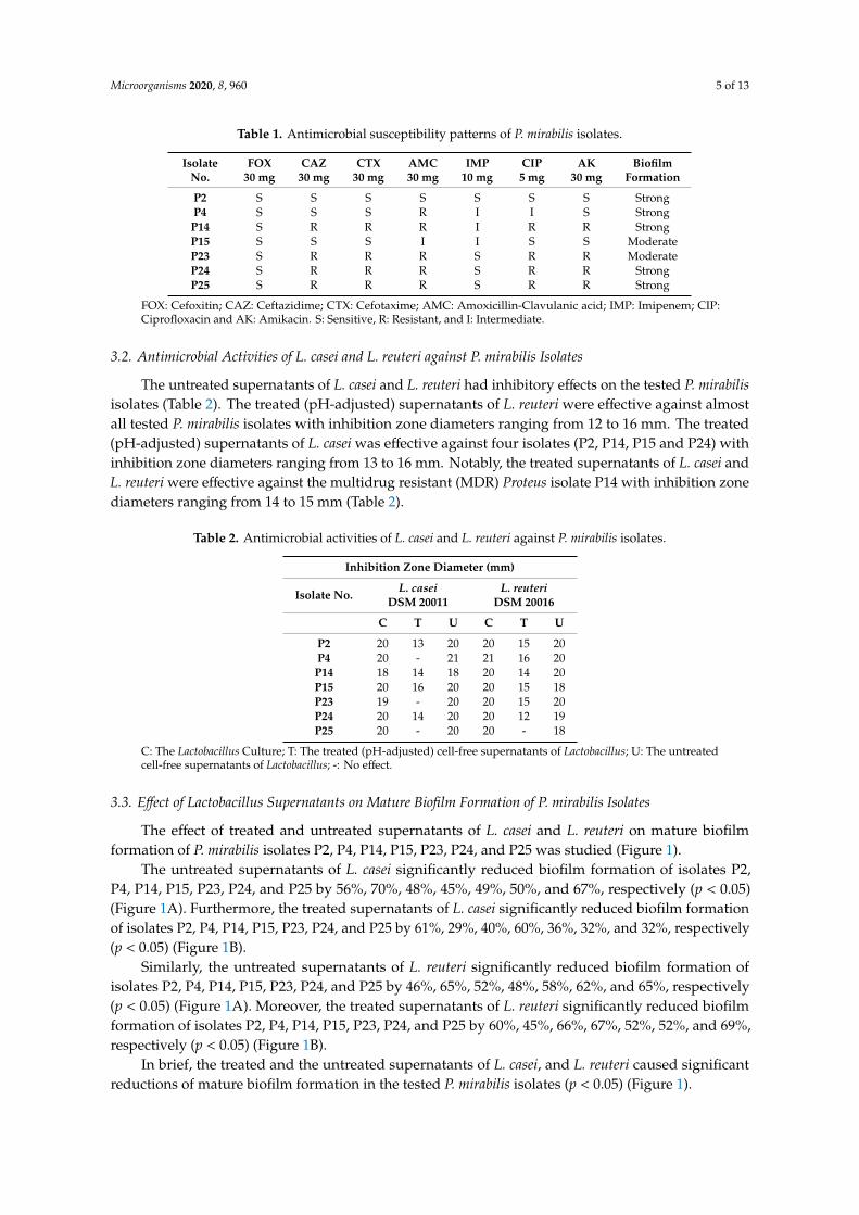
Activity of the tested antimicrobial agents against P. mirabilis isolates was evaluated accordingto CLSI standards [32]. As shown in Table 1, 86% of the tested P. mirabilis isolates were resistantto amoxicillin-clavulanic, 57% were resistant to cefotaxime, 57% were resistant to ceftazidime, 71%were resistant to ciprofloxacin, 57% were resistant to amikacin, 43% were resistant to imipenem, and0% were resistant to cefoxitin. Furthermore, isolate P14 was resistant to cefotaxime, ceftazidime,amoxicillin-clavulanic acid, ciprofloxacin, and amikacin and intermediately resistant to imipenem(Table 1). In contrast, isolate P2 was sensitive to all the assessed antimicrobials (Table 1).
P. mirabilis isolates P2, P4, P14, P24 and P25 showed strong biofilm formation. Isolates P15 andP23 showed moderate biofilm formation. Isolate P14 showed the strongest biofilm formation and wasresistant to the tested antimicrobials. Isolate P2 showed a strong biofilm formation, but was sensitiveto all assessed antimicrobials.
Microorganisms 2020, 8, 960 5 of 13
Table 1. Antimicrobial susceptibility patterns of P. mirabilis isolates.
IsolateNo.
FOX30 mg
CAZ30 mg
CTX30 mg
AMC30 mg
IMP10 mg
CIP5 mg
AK30 mg
BiofilmFormation
P2 S S S S S S S StrongP4 S S S R I I S Strong
P14 S R R R I R R StrongP15 S S S I I S S ModerateP23 S R R R S R R ModerateP24 S R R R S R R StrongP25 S R R R S R R Strong
FOX: Cefoxitin; CAZ: Ceftazidime; CTX: Cefotaxime; AMC: Amoxicillin-Clavulanic acid; IMP: Imipenem; CIP:Ciprofloxacin and AK: Amikacin. S: Sensitive, R: Resistant, and I: Intermediate.
3.2. Antimicrobial Activities of L. casei and L. reuteri against P. mirabilis Isolates
The untreated supernatants of L. casei and L. reuteri had inhibitory effects on the tested P. mirabilisisolates (Table 2). The treated (pH-adjusted) supernatants of L. reuteri were effective against almostall tested P. mirabilis isolates with inhibition zone diameters ranging from 12 to 16 mm. The treated(pH-adjusted) supernatants of L. casei was effective against four isolates (P2, P14, P15 and P24) withinhibition zone diameters ranging from 13 to 16 mm. Notably, the treated supernatants of L. casei andL. reuteri were effective against the multidrug resistant (MDR) Proteus isolate P14 with inhibition zonediameters ranging from 14 to 15 mm (Table 2).
Table 2. Antimicrobial activities of L. casei and L. reuteri against P. mirabilis isolates.
Inhibition Zone Diameter (mm)
Isolate No. L. caseiDSM 20011
L. reuteriDSM 20016
C T U C T U
P2 20 13 20 20 15 20P4 20 - 21 21 16 20
P14 18 14 18 20 14 20P15 20 16 20 20 15 18P23 19 - 20 20 15 20P24 20 14 20 20 12 19P25 20 - 20 20 - 18
C: The Lactobacillus Culture; T: The treated (pH-adjusted) cell-free supernatants of Lactobacillus; U: The untreatedcell-free supernatants of Lactobacillus; -: No effect.
3.3. Effect of Lactobacillus Supernatants on Mature Biofilm Formation of P. mirabilis Isolates
The effect of treated and untreated supernatants of L. casei and L. reuteri on mature biofilmformation of P. mirabilis isolates P2, P4, P14, P15, P23, P24, and P25 was studied (Figure 1).
The untreated supernatants of L. casei significantly reduced biofilm formation of isolates P2,P4, P14, P15, P23, P24, and P25 by 56%, 70%, 48%, 45%, 49%, 50%, and 67%, respectively (p < 0.05)(Figure 1A). Furthermore, the treated supernatants of L. casei significantly reduced biofilm formationof isolates P2, P4, P14, P15, P23, P24, and P25 by 61%, 29%, 40%, 60%, 36%, 32%, and 32%, respectively(p < 0.05) (Figure 1B).
Similarly, the untreated supernatants of L. reuteri significantly reduced biofilm formation ofisolates P2, P4, P14, P15, P23, P24, and P25 by 46%, 65%, 52%, 48%, 58%, 62%, and 65%, respectively(p < 0.05) (Figure 1A). Moreover, the treated supernatants of L. reuteri significantly reduced biofilmformation of isolates P2, P4, P14, P15, P23, P24, and P25 by 60%, 45%, 66%, 67%, 52%, 52%, and 69%,respectively (p < 0.05) (Figure 1B).
In brief, the treated and the untreated supernatants of L. casei, and L. reuteri caused significantreductions of mature biofilm formation in the tested P. mirabilis isolates (p < 0.05) (Figure 1).

Microorganisms 2020, 8, 960 6 of 13
Microorganisms 2020, 8, x FOR PEER REVIEW 6 of 13
6
The effect of treated and untreated supernatants of L. casei and L. reuteri on mature biofilm formation of P. mirabilis isolates P2, P4, P14, P15, P23, P24, and P25 was studied (Figure 1).
The untreated supernatants of L. casei significantly reduced biofilm formation of isolates P2, P4, P14, P15, P23, P24, and P25 by 56%, 70%, 48%, 45%, 49%, 50%, and 67%, respectively (p < 0.05) (Figure 1A). Furthermore, the treated supernatants of L. casei significantly reduced biofilm formation of isolates P2, P4, P14, P15, P23, P24, and P25 by 61%, 29%, 40%, 60%, 36%, 32%, and 32%, respectively (p < 0.05) (Figure 1B).
Similarly, the untreated supernatants of L. reuteri significantly reduced biofilm formation of isolates P2, P4, P14, P15, P23, P24, and P25 by 46%, 65%, 52%, 48%, 58%, 62%, and 65%, respectively (p < 0.05) (Figure 1A). Moreover, the treated supernatants of L. reuteri significantly reduced biofilm formation of isolates P2, P4, P14, P15, P23, P24, and P25 by 60%, 45%, 66%, 67%, 52%, 52%, and 69%, respectively (p < 0.05) (Figure 1B).
In brief, the treated and the untreated supernatants of L. casei, and L. reuteri caused significant reductions of mature biofilm formation in the tested P. mirabilis isolates (p < 0.05) (Figure 1).
Figure 1. Effect of supernatants of L. casei and L. reuteri on mature biofilm formation of Proteus mirabilis isolates P2, P4, P14, P15, P23, P24, and P25 (* p < 0.05). (A) Effect of untreated supernatants of L. casei and L. reuteri on mature biofilm formation of Proteus mirabilis isolates P2, P4, P14, P15, P23, P24, and P25 (* p < 0.05). (B) Effect of treated supernatants of L. casei and L. reuteri on mature biofilm formation of Proteus mirabilis isolates P2, P4, P14, P15, P23, P24, and P25 (* p < 0.05).
Next, we tested various concentrations of treated and untreated supernatants of L. casei and L. reuteri on mature biofilm formation of the MDR isolate P14. The diluted untreated supernatants (1/4, 1/8, and 1/16) of L. casei caused a significant reduction of biofilm formation in the P14 isolate by 61%, 61%, and 60%, respectively (p < 0.05) (Figure 2A). Similarly, the diluted treated supernatants (1/2, 1/4, 1/8) of L. casei significantly reduced biofilm formation in the P14 isolate by 55%, 33%, and 39%, respectively (p < 0.05) (Figure 2B.).
The 1/16 and 1/32 concentrations of the untreated supernatants of L. reuteri caused a significant reduction in biofilm formation of the P14 isolate by 73% and 32%, respectively (p < 0.05) (Figure 2C). The 1/8 concentration of the treated supernatants of L. reuteri significantly reduced biofilm formation of the P14 isolate by 73% (p < 0.05) (Figure 2D).
0
0.1
0.2
0.3
0.4
0.5
0.6
0.7
L. casei L. reuteri Control
OD
540
nm
P2
P4
P14
P15
P23
P24
P25
0
0.1
0.2
0.3
0.4
0.5
0.6
0.7
L. casei L. reuteri Control
OD
540
nm
P2
P4
P14
P15
P23
P24
P25
* *
A. B.
Figure 1. Effect of supernatants of L. casei and L. reuteri on mature biofilm formation of Proteus mirabilisisolates P2, P4, P14, P15, P23, P24, and P25 (* p < 0.05). (A) Effect of untreated supernatants of L. caseiand L. reuteri on mature biofilm formation of Proteus mirabilis isolates P2, P4, P14, P15, P23, P24, andP25 (* p < 0.05). (B) Effect of treated supernatants of L. casei and L. reuteri on mature biofilm formationof Proteus mirabilis isolates P2, P4, P14, P15, P23, P24, and P25 (* p < 0.05).
Next, we tested various concentrations of treated and untreated supernatants of L. casei andL. reuteri on mature biofilm formation of the MDR isolate P14. The diluted untreated supernatants(1/4, 1/8, and 1/16) of L. casei caused a significant reduction of biofilm formation in the P14 isolate by61%, 61%, and 60%, respectively (p < 0.05) (Figure 2A). Similarly, the diluted treated supernatants (1/2,1/4, 1/8) of L. casei significantly reduced biofilm formation in the P14 isolate by 55%, 33%, and 39%,respectively (p < 0.05) (Figure 2B.). Microorganisms 2020, 8, x FOR PEER REVIEW 7 of 13
7
Figure 2. Effect of supernatants of L. casei and L. reuteri on mature biofilm formation of the P. mirabilis isolate P14 (* p < 0.05). (A) Effect of untreated supernatants of L. casei on mature biofilm formation of the Proteus isolate P14 (* p < 0.05). (B) Effect of treated supernatants of L. casei on mature biofilm formation of the Proteus isolate P14 (* p < 0.05). (C) Effect of untreated supernatants of L. reuteri on mature biofilm formation of the Proteus isolate P14 (* p < 0.05). (D) Effect of treated supernatants of L. reuteri on mature biofilm formation of the Proteus isolate P14 (* p < 0.05).
3.4. Anti-Adherence Effect of Lactobacillus Supernatants
The anti-adherence effect of diluted Lactobacillus supernatants (treated and untreated) on the P14 isolate was assessed. As in Figure 3, the untreated supernatants of L. casei and L. reuteri significantly reduced the adhesion of the P14 isolate (1/16 concentration) by 72% and 73%, respectively (p < 0.01) (Figure 3A, C). The anti-adherence effect of the untreated supernatants on the P14 isolate decreased by further diluting the supernatant.
As in Figure 3, the treated supernatants of L. casei and L. reuteri significantly reduced biofilm formation of the P14 isolate (1/8 concentration) by 75% and 73%, respectively (p < 0.01) (Figure 3B,D).
0
0.1
0.2
0.3
0.4
0.5
0.6
0.7
0.8
P14 1/8 conc 1/16 conc
OD
540
nm
Concentartion of treated supernatant of Lactobacillus reuteri
0
0.1
0.2
0.3
0.4
0.5
0.6
0.7
0.8
P14 1/16 conc 1/32 conc
OD
540
nm
Concentartion of untreated supernatant of Lactobacillus reuteri
0
0.1
0.2
0.3
0.4
0.5
0.6
0.7
0.8
P14 1/2 conc 1/4 conc 1/8 conc
OD
540
nm
Concentartion of treated supernatant of Lactobacillus casei
0
0.1
0.2
0.3
0.4
0.5
0.6
0.7
0.8
P14 1/4 conc 1/8 conc 1/16 conc
OD
540
nm
Concentartion of untreated supernatant of Lactobacillus casei
*
C.
*
*
A.
* * *
B.
*
* *
D.
Figure 2. Effect of supernatants of L. casei and L. reuteri on mature biofilm formation of the P. mirabilisisolate P14 (* p < 0.05). (A) Effect of untreated supernatants of L. casei on mature biofilm formationof the Proteus isolate P14 (* p < 0.05). (B) Effect of treated supernatants of L. casei on mature biofilmformation of the Proteus isolate P14 (* p < 0.05). (C) Effect of untreated supernatants of L. reuteri onmature biofilm formation of the Proteus isolate P14 (* p < 0.05). (D) Effect of treated supernatants ofL. reuteri on mature biofilm formation of the Proteus isolate P14 (* p < 0.05).

Microorganisms 2020, 8, 960 7 of 13
The 1/16 and 1/32 concentrations of the untreated supernatants of L. reuteri caused a significantreduction in biofilm formation of the P14 isolate by 73% and 32%, respectively (p < 0.05) (Figure 2C).The 1/8 concentration of the treated supernatants of L. reuteri significantly reduced biofilm formationof the P14 isolate by 73% (p < 0.05) (Figure 2D).
3.4. Anti-Adherence Effect of Lactobacillus Supernatants
The anti-adherence effect of diluted Lactobacillus supernatants (treated and untreated) on the P14isolate was assessed. As in Figure 3, the untreated supernatants of L. casei and L. reuteri significantlyreduced the adhesion of the P14 isolate (1/16 concentration) by 72% and 73%, respectively (p < 0.01)(Figure 3A,C). The anti-adherence effect of the untreated supernatants on the P14 isolate decreased byfurther diluting the supernatant.
Microorganisms 2020, 8, x FOR PEER REVIEW 8 of 13
8
Figure 3. Adherence of the Proteus isolate P14 in the presence of supernatants of L. casei and L. reuteri (* p < 0.05). (A) Adherence of the Proteus isolate P14 in the presence of untreated supernatants of L. casei (* p < 0.05). (B) Adherence of the Proteus isolate P14 in the presence of treated supernatants of L. casei (* p < 0.05). (C) Adherence of the Proteus isolate P14 in the presence of untreated supernatants of L. reuteri (* p < 0.05). (D) Adherence of the Proteus isolate P14 in the presence of treated supernatants of L. reuteri (* p < 0.05).
3.5. Scanning Electron Micrographs
As in Figure 4A, there is a dense mass of biofilm-forming P. mirabilis P14 (as a positive control). Co-culture of L. casei with P. mirabilis P14 caused the complete elimination of biofilm formation by P14 (Figure 4B). Co-culture of L. reuteri with P. mirabilis P14 showed scattered cells and a loose biofilm architecture but no dense aggregates (Figure 4C).
0
0.2
0.4
0.6
0.8
1
P14 1/8 conc 1/16 conc
OD
540
nm
Concentration of treated supernatant of Lactobacillus casei
0
0.2
0.4
0.6
0.8
1
P14 1/16 conc 1/32 conc
OD
540
nm
Concentration of untreated supernatant of Lactobacillus casei
0
0.1
0.2
0.3
0.4
0.5
0.6
0.7
0.8
P14 1/8 conc 1/16 conc
OD
540
nm
Concentration of treated supernatant of Lactobacillus reuteri
* *
* *
A. B.
C. D.
0
0.1
0.2
0.3
0.4
0.5
0.6
0.7
0.8
P14 1/16 conc 1/32 conc
OD
540
nm
Concentration of untreated supernatant of Lactobacillus reuteri
Figure 3. Adherence of the Proteus isolate P14 in the presence of supernatants of L. casei and L. reuteri(* p < 0.05). (A) Adherence of the Proteus isolate P14 in the presence of untreated supernatants ofL. casei (* p < 0.05). (B) Adherence of the Proteus isolate P14 in the presence of treated supernatants ofL. casei (* p < 0.05). (C) Adherence of the Proteus isolate P14 in the presence of untreated supernatantsof L. reuteri (* p < 0.05). (D) Adherence of the Proteus isolate P14 in the presence of treated supernatantsof L. reuteri (* p < 0.05).
As in Figure 3, the treated supernatants of L. casei and L. reuteri significantly reduced biofilmformation of the P14 isolate (1/8 concentration) by 75% and 73%, respectively (p < 0.01) (Figure 3B,D).

Microorganisms 2020, 8, 960 8 of 13
3.5. Scanning Electron Micrographs
As in Figure 4A, there is a dense mass of biofilm-forming P. mirabilis P14 (as a positive control).Co-culture of L. casei with P. mirabilis P14 caused the complete elimination of biofilm formation by P14(Figure 4B). Co-culture of L. reuteri with P. mirabilis P14 showed scattered cells and a loose biofilmarchitecture but no dense aggregates (Figure 4C).
Microorganisms 2020, 8, x FOR PEER REVIEW 8 of 13
8
Figure 3. Adherence of the Proteus isolate P14 in the presence of supernatants of L. casei and L. reuteri (* p < 0.05). (A) Adherence of the Proteus isolate P14 in the presence of untreated supernatants of L. casei (* p < 0.05). (B) Adherence of the Proteus isolate P14 in the presence of treated supernatants of L. casei (* p < 0.05). (C) Adherence of the Proteus isolate P14 in the presence of untreated supernatants of L. reuteri (* p < 0.05). (D) Adherence of the Proteus isolate P14 in the presence of treated supernatants of L. reuteri (* p < 0.05).
3.5. Scanning Electron Micrographs
As in Figure 4A, there is a dense mass of biofilm-forming P. mirabilis P14 (as a positive control). Co-culture of L. casei with P. mirabilis P14 caused the complete elimination of biofilm formation by P14 (Figure 4B). Co-culture of L. reuteri with P. mirabilis P14 showed scattered cells and a loose biofilm architecture but no dense aggregates (Figure 4C).
0
0.2
0.4
0.6
0.8
1
P14 1/8 conc 1/16 conc
OD
540
nm
Concentration of treated supernatant of Lactobacillus casei
0
0.2
0.4
0.6
0.8
1
P14 1/16 conc 1/32 conc
OD
540
nm
Concentration of untreated supernatant of Lactobacillus casei
0
0.1
0.2
0.3
0.4
0.5
0.6
0.7
0.8
P14 1/8 conc 1/16 conc
OD
540
nm
Concentration of treated supernatant of Lactobacillus reuteri
* *
* *
A. B.
C. D.
0
0.1
0.2
0.3
0.4
0.5
0.6
0.7
0.8
P14 1/16 conc 1/32 conc
OD
540
nm
Concentration of untreated supernatant of Lactobacillus reuteri
Figure 4. Scanning Electron micrographs (SEM) (magnification: × 15,000). (A) Biofilm formation of theP14 isolate in the absence of Lactobacillus spp. (control). (B) Biofilm formation of the P14 isolate in thepresence of L. casei. (C) Biofilm formation of the P14 isolate in the presence of L. reuteri.
4. Discussion
Bacterial resistance is one of the major public health problems. It includes the emergence of MDRpathogens, such as resistant isolates of Proteus mirabilis, which have been associated with urinarytract infections and nosocomial infections worldwide [4,38]. P. mirabilis isolates have been reportedto be resistant to penicillins [39]. They also show a high incidence of resistance to cephalosporins,carbapenems, and quinolones [4,40]. In the current study, a high percentage of the isolates was resistantor intermediately resistant to amoxicillin-clavulanic acid and cefotaxime. There was also significantresistance to ceftazidime, ciprofloxacin, and amikacin (Table 1). It is noteworthy that the isolate P14was resistant to three different groups of antibiotics with various mechanisms of action (Table 1).Cefotaxime-resistant P. mirabilis has been previously described [41]. Kwiecinska-Piróg et al. reportedthat P. mirabilis showed resistance against ceftazidime and ciprofloxacin [42]. Amikacin resistance hasbeen shown to be associated with extended spectrum beta-lactamase (ESBL(-producing isolates ofP. mirabilis, which were resistant to amikacin (85.1%) [43].
Urinary tract infection with P. mirabilis is associated with biofilm formation and accumulation ofthe polysaccharide matrix [44]. This is followed by urease production, increase in the pH, attractionof calcium and magnesium ions, and development of crystals [44]. The deposition of crystalswithin the biofilm can cause catheter blockage and urinary retention. This is because P. mirabilispossesses various virulence factors (lipopolysaccharide, quorum sensing autoinducers, pili, adhesin,and other proteins) that enhance adhesion and crystalline biofilm formation on the abiotic surfacesof urinary catheters [45]. It has been reported that 48% of the isolated proteus species were biofilmproducers [42]. The Proteus-biofilm assembly is dangerous, as it interferes with microbial penetration,increases antimicrobial resistance, and renders therapeutic treatments ineffective, which encouragesthe development and chronicity of infections [46].
Moreover, pathogenicity of P. mirabilis is enhanced by the complex architecture of its biofilm,which is characterized by a high ability for adapting to different environmental conditions, biocides,and antimicrobials [47,48]. Isolates in this study were able to form strong or moderate biofilms. This wastrue even for the antibiotic-sensitive isolate P2, which was observed to have a strong biofilm-formingability (Table 1). Typically, the biofilm-producing isolates are more resistant to antibiotics than thenon-biofilm producing isolates [49]. The reason is that the development of complex biofilm structures
Microorganisms 2020, 8, 960 9 of 13
can convey protection to the internal cells from antimicrobials [50]. It can also support the persistenceof P. mirabilis in the host cells [50].
Given the above, alternative treatments are required for the management of P. mirabilis infectionsand the disruption of its biofilm architecture. Probiotics as Lactobacilli, have been used for the treatmentof burn infections and have been shown to interfere with activity of Pseudomonas aeruginosa [51,52].Lactobacilli have also been used for the management of recurrent urinary tract infections causedby E. coli [53]. The antimicrobial activity of L. casei and L. reuteri against various enteropathogenicinfections have been previously reported [10]. Their antimicrobial activities against uropathogens havealso been reported [20]. L. reuteri has been shown to be effective against Escherichia coli and Listeriamonocytogenes [54]. Thus, it was important to examine the antimicrobial and antibiofilm activities ofLactobacillus on P. mirabilis clinical isolates. We examined the cultures and the cell-free supernatants ofL. casei, and L. reuteri against P. mirabilis isolates.
In our study, the treated supernatants of L. casei and L. reuteri retained antimicrobial activitiesagainst the sensitive P2 isolate and the other resistant Proteus mirabilis isolates (Table 2). Notably, treatedsupernatants of L. casei and L. reuteri retained significant antibiofilm and anti-adherence activitiesagainst P14 at 1/8 concentration (Figures 2 and 3). In addition, L. casei and L. reuteri had significantinhibitory effects on the typical biofilm formed by the MDR isolate P14, as detected by SEM (Figure 4).In another study, the untreated supernatants of fecal Lactobacilli caused an 85 to 95% reduction inbiofilm formation of Vibrio cholerae isolates [27]. Similarly, the pH-adjusted supernatants of fecalLactobacilli significantly reduced biofilm formation of Vibrio cholerae by 50–75% [27]. Additionally,Koohestani and colleagues showed that the untreated supernatants of L. casei significantly reducedbiofilm formation of S. aureus [55]. Probiotic lactobacilli have been shown to negatively impact growthand biofilm formation of Streptococcus mutans [34], pathogens in the oral cavity [56], and Salmonellaenterica serovar Typhimurium [28]. Moreover, probiotic lactobacilli inhibited cancer cells of the humancolonic carcinoma cell line HT-29 [23]. Lastly, Lactobacillus reuteri DPC16 supernatants have been shownto exhibit activity against Escherichia coli, S. aureus, Salmonella derby, and Listeria monocytogenes. This isconsistent with results in our study in which the untreated supernatants of L. casei reduced biofilmformation of P. mirabilis isolates by 45–67%.
It is important to mention that the use of probiotic Lactobacillus cell cultures and supernatants wasproposed as an alternative to traditional antibiotic therapy against P. mirabilis. In other words, it wasproposed as a way to avoid the adverse effects of antibiotic therapy and to circumvent the impactof antibiotic resistance developed by the microbe. Indeed, we saw a significant inhibitory effect ofLactobacillus cell cultures and supernatants on P. mirabilis biofilm formation and adherence. Futurestudies could examine the effect of Lactobacillus supernatants on the susceptibility of P. mirabilis to thetested antimicrobials. In this case, the focus would be on the possible synergistic effects of combiningprobiotic Lactobacillus supernatants with anti-Proteus agents.
The antimicrobial, antibiofilm, and anti-adherence activities of probiotic L. casei and L. reuteriused in this study could be attributed to their secreted biosurfactants [15], S-layer proteins [57,58],surface-acting proteins (such as enolase) [59], and peptidoglycan-binding proteins [60].
5. Conclusions
The pathogenicity and virulence of Proteus infections includes a biofilm-forming ability that enablesserious urinary tract infection. Isolates in this study showed multidrug resistance (MDR) againstmore than one antimicrobial agent, highlighting the importance of the development of alternative andadjuvant treatments for the efficient management of P. mirabilis infections. Using cell cultures, cell-freesupernatants, and pH-adjusted supernatants, we have shown that L. casei DSM 20011 and L. reuteriDSM 20016 exhibit antimicrobial, anti-adherence, and antibiofilm activities against MDR P. mirabilis.Thus, L. casei and L. reuteri could be utilized to combat Proteus-associated urinary tract infections.
Microorganisms 2020, 8, 960 10 of 13
Author Contributions: M.S., O.A.A.E.-R., B.A.-Q., and H.M.A. contributed to the conception of the study,experimental design, execution, data analysis, writing of the original draft, revision of the manuscript, and finalpublication. All authors have read and agreed to the published version of the manuscript.
Funding: This research received no external funding.
Conflicts of Interest: The authors confirm that there are no conflict of interest.
References
1. Armbruster, C.E.; Smith, S.N.; Johnson, A.O.; DeOrnellas, V.; Eaton, K.A.; Yep, A.; Mody, L.; Wu, W.;Mobley, H.L.T. The Pathogenic Potential of Proteus mirabilis Is Enhanced by Other Uropathogens duringPolymicrobial Urinary Tract Infection. Infect. Immun. 2017, 85. [CrossRef]
2. Armbruster, C.E.; Mobley, H.L.T.; Pearson, M.M. Pathogenesis of Proteus mirabilis Infection. Ecosal. Plus2018, 8. [CrossRef]
3. Chen, C.Y.; Chen, Y.H.; Lu, P.L.; Lin, W.R.; Chen, T.C.; Lin, C.Y. Proteus mirabilis urinary tract infection andbacteremia: Risk factors, clinical presentation, and outcomes. J. Microbiol. Immunol. Infect. 2012, 45, 228–236.[CrossRef]
4. Pal, N.; Hooja, S.; Sharma, R.; Maheshwari, R.K. Phenotypic Detection and Antibiogram of beta-lactamase-producing Proteus Species in a Tertiary Care Hospital, India. Ann. Med. Health Sci. Res. 2016, 6, 267–273.[CrossRef]
5. Adamus-Bialek, W.; Zajac, E.; Parniewski, P.; Kaca, W. Comparison of antibiotic resistance patterns incollections of Escherichia coli and Proteus mirabilis uropathogenic strains. Mol. Biol. Rep. 2013, 40, 3429–3435.[CrossRef]
6. Gajdacs, M.; Urban, E. Comparative Epidemiology and Resistance Trends of Proteae in Urinary TractInfections of Inpatients and Outpatients: A 10-Year Retrospective Study. Antibiotics (Basel) 2019, 8, 91.[CrossRef]
7. Nucleo, E.; Fugazza, G.; Migliavacca, R.; Spalla, M.; Comelli, M.; Pagani, L.; Debiaggi, M. Differencesin biofilm formation and aggregative adherence between beta-lactam susceptible and beta-lactamasesproducing P. mirabilis clinical isolates. New Microbiol. 2010, 33, 37–45.
8. Mercenier, A.; Pavan, S.; Pot, B. Probiotics as biotherapeutic agents: Present knowledge and future prospects.Curr. Pharm. Des. 2003, 9, 175–191. [CrossRef]
9. Singh, V.P.; Sharma, J.; Babu, S.; Rizwanulla, S.A.; Singla, A. Role of probiotics in health and disease: A review.J. Pak. Med. Assoc. 2013, 63, 253–257.
10. Lievin-Le Moal, V.; Servin, A.L. Anti-infective activities of lactobacillus strains in the human intestinalmicrobiota: From probiotics to gastrointestinal anti-infectious biotherapeutic agents. Clin. Microbiol. Rev.2014, 27, 167–199. [CrossRef]
11. Sharma, D.; Singh Saharan, B. Simultaneous Production of Biosurfactants and Bacteriocins by ProbioticLactobacillus casei MRTL3. Int. J. Microbiol. 2014, 2014, 698713. [CrossRef] [PubMed]
12. Golek, P.; Bednarski, W.; Brzozowski, B.; Dziuba, B. The obtaining and properties of biosurfactants synthesizedby bacteria of the genusLactobacillus. Ann. Microbiol. 2009, 59, 119–126. [CrossRef]
13. Spinler, J.K.; Taweechotipatr, M.; Rognerud, C.L.; Ou, C.N.; Tumwasorn, S.; Versalovic, J. Human-derivedprobiotic Lactobacillus reuteri demonstrate antimicrobial activities targeting diverse enteric bacterialpathogens. Anaerobe 2008, 14, 166–171. [CrossRef]
14. Servin, A.L. Antagonistic activities of lactobacilli and bifidobacteria against microbial pathogens. FEMSMicrobiol. Rev. 2004, 28, 405–440. [CrossRef] [PubMed]
15. Kanmani, P.; Satish Kumar, R.; Yuvaraj, N.; Paari, K.A.; Pattukumar, V.; Arul, V. Probiotics and its functionallyvaluable products-a review. Crit. Rev. Food Sci. Nutr. 2013, 53, 641–658. [CrossRef] [PubMed]
16. Satpute, S.K.; Kulkarni, G.R.; Banpurkar, A.G.; Banat, I.M.; Mone, N.S.; Patil, R.H.; Cameotra, S.S.Biosurfactant/s from Lactobacilli species: Properties, challenges and potential biomedical applications.J. Basic Microbiol. 2016, 56, 1140–1158. [CrossRef] [PubMed]
17. Wasfi, R.; Abd El-Rahman, O.A.; Zafer, M.M.; Ashour, H.M. Probiotic Lactobacillus sp. inhibit growth,biofilm formation and gene expression of caries-inducing Streptococcus mutans. J. Cell Mol. Med. 2018,22, 1972–1983. [CrossRef]

Microorganisms 2020, 8, 960 11 of 13
18. Shokryazdan, P.; Sieo, C.C.; Kalavathy, R.; Liang, J.B.; Alitheen, N.B.; Faseleh Jahromi, M.; Ho, Y.W. Probioticpotential of Lactobacillus strains with antimicrobial activity against some human pathogenic strains. Biomed.Res. Int. 2014, 2014, 927268. [CrossRef]
19. Karska-Wysocki, B.; Bazo, M.; Smoragiewicz, W. Antibacterial activity of Lactobacillus acidophilus andLactobacillus casei against methicillin-resistant Staphylococcus aureus (MRSA). Microbiol. Res. 2010,165, 674–686. [CrossRef]
20. Velraeds, M.M.; van de Belt-Gritter, B.; Busscher, H.J.; Reid, G.; van der Mei, H.C. Inhibition of uropathogenicbiofilm growth on silicone rubber in human urine by lactobacilli—A teleologic approach. World J. Urol. 2000,18, 422–426. [CrossRef]
21. Pascual, L.M.; Daniele, M.B.; Ruiz, F.; Giordano, W.; Pajaro, C.; Barberis, L. Lactobacillus rhamnosus L60, apotential probiotic isolated from the human vagina. J. Gen. Appl. Microbiol. 2008, 54, 141–148. [CrossRef][PubMed]
22. Grin, P.M.; Kowalewska, P.M.; Alhazzan, W.; Fox-Robichaud, A.E. Lactobacillus for preventing recurrenturinary tract infections in women: Meta-analysis. Can. J. Urol. 2013, 20, 6607–6614. [PubMed]
23. Chen, Z.Y.; Hsieh, Y.M.; Huang, C.C.; Tsai, C.C. Inhibitory Effects of Probiotic Lactobacillus on the Growth ofHuman Colonic Carcinoma Cell Line HT-29. Molecules 2017, 22, 107. [CrossRef] [PubMed]
24. Hernandez, A.; Larsson, C.U.; Sawicki, R.; van Niel, E.W.J.; Roos, S.; Hakansson, S. Impact of the fermentationparameters pH and temperature on stress resilience of Lactobacillus reuteri DSM 17938. AMB Express 2019,9, 66. [CrossRef] [PubMed]
25. Palmfeldt, J.; Hahn-Hagerdal, B. Influence of culture pH on survival of Lactobacillus reuteri subjected tofreeze-drying. Int. J. Food Microbiol. 2000, 55, 235–238. [CrossRef]
26. Sgouras, D.N.; Panayotopoulou, E.G.; Martinez-Gonzalez, B.; Petraki, K.; Michopoulos, S.; Mentis, A.Lactobacillus johnsonii La1 attenuates Helicobacter pylori-associated gastritis and reduces levels ofproinflammatory chemokines in C57BL/6 mice. Clin. Diagn Lab. Immunol. 2005, 12, 1378–1386. [CrossRef]
27. Kaur, S.; Sharma, P.; Kalia, N.; Singh, J.; Kaur, S. Anti-biofilm Properties of the Fecal Probiotic LactobacilliAgainst Vibrio spp. Front. Cell. Infect. Microbiol. 2018, 8. [CrossRef]
28. Fayol-Messaoudi, D.; Berger, C.N.; Coconnier-Polter, M.H.; Lievin-Le Moal, V.; Servin, A.L. pH-, Lacticacid-, and non-lactic acid-dependent activities of probiotic Lactobacilli against Salmonella enterica SerovarTyphimurium. Appl. Environ. Microbiol. 2005, 71, 6008–6013. [CrossRef]
29. Bian, L.; Molan, A.-L.; Maddox, I.; Shu, Q. Antimicrobial activity of Lactobacillus reuteri DPC16 supernatantsagainst selected food borne pathogens. World J. Microbiol. Biotechnol. 2011, 27, 991–998. [CrossRef]
30. Cheesbrough, M. Medical Laboratory Manual for Tropical Countries; Cheesbrough, M., Ed.; 14 Bevills Close;PE15 OTT: Doddington/Cambridgeshire, UK, 1981; Volume 1.
31. Ding, Y.; Wang, W.; Fan, M.; Tong, Z.; Kuang, R.; Jiang, W.; Ni, L. Antimicrobial and anti-biofilm effect ofBac8c on major bacteria associated with dental caries and Streptococcus mutans biofilms. Peptides 2014,52, 61–67. [CrossRef]
32. Performance Standards for Antimicrobial Susceptibility Testing, 29th ed.; CLSI supplement M100; Clinical andLaboratory Standards Institute: Wayne, PA, USA, 2019.
33. Stepanovic, S.; Vukovic, D.; Dakic, I.; Savic, B.; Svabic-Vlahovic, M. A modified microtiter-plate test forquantification of staphylococcal biofilm formation. J. Microbiol. Methods 2000, 40, 175–179. [CrossRef]
34. Lin, X.; Chen, X.; Chen, Y.; Jiang, W.; Chen, H. The effect of five probiotic lactobacilli strains on the growthand biofilm formation of Streptococcus mutans. Oral. Dis. 2015, 21, e128–e134. [CrossRef] [PubMed]
35. Schillinger, U.; Lucke, F.K. Antibacterial activity of Lactobacillus sake isolated from meat. Appl. Environ.Microbiol. 1989, 55, 1901–1906. [CrossRef]
36. Wasfi, R.; Abd El-Rahman, O.A.; Mansour, L.E.; Hanora, A.S.; Hashem, A.M.; Ashour, M.S. Antimicrobialactivities against biofilm formed by Proteus mirabilis isolates from wound and urinary tract infections.Indian J. Med. Microbiol. 2012, 30, 76–80. [CrossRef] [PubMed]
37. Wu, C.C.; Lin, C.T.; Wu, C.Y.; Peng, W.S.; Lee, M.J.; Tsai, Y.C. Inhibitory effect of Lactobacillus salivarius onStreptococcus mutans biofilm formation. Mol. Oral. Microbiol. 2015, 30, 16–26. [CrossRef]
38. Lin, M.F.; Liou, M.L.; Kuo, C.H.; Lin, Y.Y.; Chen, J.Y.; Kuo, H.Y. Antimicrobial Susceptibility and MolecularEpidemiology of Proteus mirabilis Isolates from Three Hospitals in Northern Taiwan. Microb. Drug Resist.2019. [CrossRef]
Microorganisms 2020, 8, 960 12 of 13
39. Stock, I. Natural antibiotic susceptibility of Proteus spp., with special reference to P. mirabilis and P. penneristrains. J. Chemother. 2003, 15, 12–26. [CrossRef]
40. Mathai, D.; Jones, R.N.; Pfaller, M.A.; America, S.P.G.N. Epidemiology and frequency of resistance amongpathogens causing urinary tract infections in 1510 hospitalized patients: A report from the SENTRYAntimicrobial Surveillance Program (North America). Diagn. Microbiol. Infect. Dis. 2001, 40, 129–136.[CrossRef]
41. Horiguchi, Y.; Hashikita, G.; Oka, Y.; Takahashi, S.; Yamazaki, T.; Maesaki, S.; Ishii, Y. Study of resistancemechanism on cefotaxime resistant Proteus mirabilis isolated from clinical specimens and its clinicalbackground. Kansenshogaku Zasshi 2004, 78, 1–9. [CrossRef]
42. Kwiecinska-Piróg, J.; Skowron, K.; Zniszczol, K.; Gospodarek, E. The assessment of Proteus mirabilissusceptibility to ceftazidime and ciprofloxacin and the impact of these antibiotics at subinhibitoryconcentrations on Proteus mirabilis biofilms. Biomed. Res. Int. 2013, 2013, 930876. [CrossRef]
43. De Champs, C.; Bonnet, R.; Sirot, D.; Chanal, C.; Sirot, J. Clinical relevance of Proteus mirabilis in hospitalpatients: A two year survey. J. Antimicrob. Chemother. 2000, 45, 537–539. [CrossRef]
44. Wilks, S.A.; Fader, M.J.; Keevil, C.W. Novel insights into the Proteus mirabilis crystalline biofilm usingreal-time imaging. PLoS ONE 2015, 10, e0141711. [CrossRef]
45. Hori, K.; Matsumoto, S. Bacterial adhesion: From mechanism to control. Biochem. Eng. J. 2010, 48, 424–434.[CrossRef]
46. Srivastava, D.; Srivastava, S.; Singh, P.C.; Kumar, A. Mechanisms of Biofilm Development, AntibioticResistance and Tolerance and Their Role in Persistent Infections. In Antibacterial Drug Discovery to CombatMDR; Springer: Berlin, Germany, 2019; pp. 115–130.
47. Jacobsen, S.M.; Shirtliff, M.E. Proteus mirabilis biofilms and catheter-associated urinary tract infections.Virulence 2011, 2, 460–465. [CrossRef] [PubMed]
48. Pelling, H.; Nzakizwanayo, J.; Milo, S.; Denham, E.L.; MacFarlane, W.M.; Bock, L.J.; Sutton, J.M.; Jones, B.V.Bacterial biofilm formation on indwelling urethral catheters. Lett. Appl. Microbiol. 2019, 68, 277–293.[CrossRef] [PubMed]
49. Shikh-Bardsiri, H.; Shakibaie, M.R. Antibiotic resistance pattern among biofilm producing and non producingProteus strains isolated from hospitalized patients; matter of hospital hygiene and antimicrobial stewardship.Pak. J. Biol. Sci. 2013, 16, 1496–1502. [CrossRef]
50. Stickler, D.J. Clinical complications of urinary catheters caused by crystalline biofilms: Something needs tobe done. J. Intern. Med. 2014, 276, 120–129. [CrossRef]
51. Valdez, J.C.; Peral, M.C.; Rachid, M.; Santana, M.; Perdigon, G. Interference of Lactobacillus plantarum withPseudomonas aeruginosa in vitro and in infected burns: The potential use of probiotics in wound treatment.Clin. Microbiol. Infect. 2005, 11, 472–479. [CrossRef]
52. Shokri, D.; Khorasgani, M.R.; Mohkam, M.; Fatemi, S.M.; Ghasemi, Y.; Taheri-Kafrani, A. The Inhibition Effectof Lactobacilli Against Growth and Biofilm Formation of Pseudomonas aeruginosa. Probiotics Antimicrob.Proteins 2018, 10, 34–42. [CrossRef]
53. Gupta, V.; Nag, D.; Garg, P. Recurrent urinary tract infections in women: How promising is the use ofprobiotics? Indian J. Med. Microbiol. 2017, 35, 347–354. [CrossRef]
54. Eslami, G.; Karimiravesh, R.; Taheri, S.; Azargashb, E. Inhibitory Effect of Lactobacillus reuteri on SomePathogenic Bacteria Isolated from Women with Bacterial Vaginosis. Avicenna J. Clin. Microbiol. Infect. 2014,1, 19908. [CrossRef]
55. Koohestani, M.; Moradi, M.; Tajik, H.; Badali, A. Effects of cell-free supernatant of Lactobacillus acidophilusLA5 and Lactobacillus casei 431 against planktonic form and biofilm of Staphylococcus aureus. Vet. Res.Forum 2018, 9, 301–306. [CrossRef] [PubMed]
56. Donelli, G. Activity of Probiotics on Biofilm-Growing Pathogens of the Oral Cavity. Microb. Ecol. Health Dis.2013, 24.
57. Spurbeck, R.R.; Arvidson, C.G. Lactobacillus jensenii surface-associated proteins inhibit Neisseria gonorrhoeaeadherence to epithelial cells. Infect. Immun. 2010, 78, 3103–3111. [CrossRef]
58. Velraeds, M.; Van der Mei, H.; Reid, G.; Busscher, H.J. Inhibition of initial adhesion of uropathogenic Enterococcusfaecalis by biosurfactants from Lactobacillus isolates. Appl. Environ. Microbiol. 1996, 62, 1958–1963. [CrossRef]
Microorganisms 2020, 8, 960 13 of 13
59. Ren, D.; Li, C.; Qin, Y.; Yin, R.; Li, X.; Tian, M.; Du, S.; Guo, H.; Liu, C.; Zhu, N. Inhibition of Staphylococcusaureus adherence to Caco-2 cells by lactobacilli and cell surface properties that influence attachment. Anaerobe2012, 18, 508–515. [CrossRef]
60. Kang, M.S.; Lim, H.S.; Oh, J.S.; Lim, Y.J.; Wuertz-Kozak, K.; Harro, J.M.; Shirtliff, M.E.; Achermann, Y.Antimicrobial activity of Lactobacillus salivarius and Lactobacillus fermentum against Staphylococcus aureus.Pathog. Dis. 2017, 75. [CrossRef]
© 2020 by the authors. Licensee MDPI, Basel, Switzerland. This article is an open accessarticle distributed under the terms and conditions of the Creative Commons Attribution(CC BY) license (http://creativecommons.org/licenses/by/4.0/).
Copyright © 2022 FDOKUMEN