Ameliorative Function of a Probiotic Bacterium, Lactobacillus ...
Upload
khangminh22Category
view
3download
0
The beneficial effects of the Lab4
probiotic consortium in atherosclerosis
Victoria O’Morain BSc (Hons)
Primary Supervisor Professor Dipak P. Ramji
A dissertation presented for the degree of Doctor of Philosophy
2019
Cardiff School of Biosciences
Cardiff University
Museum Avenue
Cardiff CF10 3AX


i
Declaration
This work has not been submitted in substance for any other degree or award at this or
any other university or place of learning, nor is being submitted concurrently in
candidature for any degree or other award.
Signed……………………………………… (Candidate) Date…………………
STATEMENT 1
This thesis is being submitted in partial fulfilment of the requirements for the degree of
Doctor of Philosophy.
Signed……………………………………… (Candidate) Date…………………
STATEMENT 2
This thesis is the result of my own independent work/investigation, except where
otherwise stated. Other sources are acknowledged by explicit references. The views
expressed are my own.
Signed……………………………………… (Candidate) Date…………………
STATEMENT 3
I hereby give consent for my thesis, if accepted, to be available for photocopying and for
inter-library loan, and for the title and summary to be made available to outside
organisations.
Signed……………………………………… (Candidate) Date…………………
09/08/2019
09/08/2019
09/08/2019
09/08/2019

ii
List of contents Declaration i
List of contents ii
List of figures vii
List of tables xi
Abstract xii
Acknowledgements xiii
Abbreviations xiv
CHAPTER 1 –General Introduction 1
1.2.1 Initiation 5
1.2.2 Foam cell formation 8
1.2.3 Disease progression 12
1.4.1 The effect of probiotics on key atherosclerosis associated risk factors 19
CHAPTER 2 - Materials and Methods 33
2.2.1 Production 36
2.2.2 Processing and storage 36
2.3.1 Maintenance of cell lines and cultures 37
2.3.2 Establishment and storage of cell lines 39
2.3.3 Cell counting 39
2.4.1 Cell viability 39
2.4.2 Proliferation 40
2.4.3 Measurement of reactive oxygen species 42
2.4.4 Monocyte migration 43
2.4.5 Vascular smooth muscle cell invasion 44
2.4.6 Mitochondrial bioenergetic profile 45
2.4.7 Phagocytosis 46
1.1 Atherosclerosis - prevalence and aetiology 2
1.2 Pathophysiology 4
1.3 Current and emerging therapies 14
1.4 Probiotics in atherosclerosis 17
1.5 The Lab4 probiotic consortium 26
1.6 Project aims 30
2.1 Materials 34
2.2 Production of bacterial conditioned media 36
2.3 Cell culture techniques 37
2.4 Cell based assays 39
2.5 Molecular techniques 47

iii
2.5.1 RNA extraction 47
2.5.2 Agarose gel electrophoresis 48
2.5.3 Reverse transcriptase polymerase chain reaction (RT-PCR) 48
2.5.4 Real time quantitative PCR (qPCR) 49
2.6.1 Protein extraction 52
2.6.2 Protein quantification 52
2.6.3 Enzyme linked immunosorbent assay (ELISA) 53
2.7.1 Oxidised LDL uptake 54
2.7.2 Macropinocytosis 54
2.7.3 Cholesterol efflux 55
2.7.4 Cholesterol metabolism 55
2.8.1 Animals and feeding 57
2.8.2 Blood and tissue collection 57
2.8.3 Plasma lipid quantification 58
2.8.4 Immunophenotyping of bone marrow cell populations 60
2.8.5 Plaque morphometric analysis 64
2.8.6 Liver gene expression 66
CHAPTER 3 – The effect of probiotic on CM key processes in atherosclerosis 68
3.1.1 Normalisation of probiotic CM batches 70
3.1.2 Cell viability 70
3.1.3 Key processes in atherosclerosis 71
3.1.4 Anti-inflammatory effects in atherosclerosis 77
3.3.1 Normalisation of probiotic CM to total protein concentration 82
3.3.2 Probiotic CM has no detrimental effect on the viability of cell culture model systems used for in vitro studies 83
3.3.3 Monocyte migration is reduced in the presence of probiotic CM 84
3.3.4 Probiotic CM exerts varying effects on the production of ROS by THP-1 monocytes and macrophages 87
3.3.5 Macropinocytosis was attenuated in the presence of several different concentrations of probiotic CM 92
3.3.6 Probiotic CM has a strain specific effect on mitochondrial function 94
3.3.7 Phagocytic activity of THP-1 macrophages was enhanced in the presence of probiotic CM 96
3.3.8 Probiotic CM can attenuate proliferation of key cell types involved in atherogenesis 97
2.6 Protein methods 52
2.7 Lipid methods 54
2.8 In vivo methods 57
2.9 Statistical analysis 67
3.1 Introduction 69
3.2 Experimental design 79
3.3 Results 82

iv
3.3.9 Probiotic CM displays a strain specific effect on the invasion of HASMCs 103
3.3.10 Activation of the NLRP3 inflammasome was enhanced in the presence of probiotic CM 104
3.3.11 Probiotic CM displays a number of strain specific effects on inflammatory gene expression in cytokine stimulated macrophages 105
3.4.1 Probiotic CM attenuates MCP-1-stimulated migration of monocytes 108
3.4.2 Measurement of ROS as an indicator of antioxidant capacity 108
3.4.3 Uptake of modified LDL in macrophages via macropinocytosis 109
3.4.4 The effect of probiotic CM on mitochondrial function 110
3.4.5 Phagocytic activity of macrophages was enhanced in the presence of probiotic CM 110
3.4.6 Differential effects of probiotic CM on the proliferation of cell types involved in atherosclerosis 111
3.4.7 Invasion of SMCs 112
3.4.8 The effects of probiotic CM on common inflammatory markers 113
3.4.9 Future directions 114
CHAPTER 4 - The effects of probiotic CM on foam cell formation 115
4.1.1 Uptake of modified LDL 117
4.1.2 Cholesterol efflux 118
4.1.3 Intracellular cholesterol metabolism 120
4.2 Experimental aims 121
4.3.1 Probiotic CM attenuates receptor-mediated uptake of modified LDL in human macrophages 125
4.3.2 Expression of macrophage scavenger receptors was attenuated following treatment with probiotic CM 127
4.3.3 Probiotic CM enhances the efflux of cholesterol from human macrophage foam cells 130
4.3.4 Probiotic CM has strain specific effects on the expression of key genes involved in cholesterol efflux 132
4.3.5 Cholesterol metabolism 134
4.3.6 Probiotic CM has strain specific effects on the expression of genes involved in intracellular cholesterol metabolism 136
4.4.1 Modified LDL uptake 138
4.4.2 Cholesterol efflux 139
4.4.3 Intracellular cholesterol metabolism 141
4.4.4 Summary and future work 143
CHAPTER 5 – The effects of Lab4 and CUL66 combination in vivo 145
5.1.1 Study design 146
3.4 Discussion 108
4.1 Introduction 116
4.3 Results 125
4.4 Discussion 138
5.1 Introduction 146

v
5.3.1 Lab4/CUL66 supplementation had no effect on the body weight of LDLr-/- mice fed a high fat diet 149
5.3.2 The effect of Lab4/CUL66 supplementation on mouse body fat and organ weight 150
5.3.3 The effect of Lab4/CUL66 supplementation on plasma cholesterol levels in mice fed a high fat diet 151
5.3.4 Triglycerides were increased in the plasma of probiotic mice compared to the control 154
5.3.5 Plaque area and lipid content was reduced with Lab4/CUL66 supplementation 154
5.3.6 The effects of Lab4/CUL66 supplementation on macrophage, SMC and T cell content of plaques 157
5.4.1 The effect of Lab4/CUL66 supplementation on mouse body and organ weights 162
5.4.2 Lab4/CUL66 supplementation has beneficial effects on plasma lipid composition 163
5.4.3 Lab4/CUL66 supplementation reduces plaque size and lipid content in LDLr-/- mice 167
5.4.4 The effect of Lab4/CUL66 supplementation on plaque composition 168
5.4.5 Summary and future work 171
CHAPTER 6 – Mechanisms underlying the effect of Lab4/CUL66 supplementation in atherosclerosis 173
6.3.1 The effects of Lab4/CUL66 supplementation on the expression of key genes involved in atherosclerosis disease 179
6.3.2 The effect of Lab4/CUL66 supplementation on key bone marrow cell populations 189
6.4.1 Lab4/CUL66 supplementation significantly attenuated the expression of key genes involved in cell adhesion 194
6.4.2 The effect of Lab4/CUL66 supplementation on the expression of genes involved in the regulation of extracellular matrix 196
6.4.3 Lipid transport and metabolism 198
6.4.4 Lab4/CUL66 supplementation attenuates gene expression of key pro-inflammatory cytokines 199
6.4.5 The effect of Lab4/CUL66 supplementation on key cell populations in the bone marrow of LDLr-/- mice 201
6.4.6 Summary 204
CHAPTER 7 – General discussion 206
5.2 Experimental aim 147
5.3 Results 149
5.4 Discussion 162
6.1 Introduction 174
6.2 Experimental aim 176
6.3 Results 179
6.4 Discussion 194

vi
7.3.1 Plasma lipid modulation 214
7.3.2 Foam cell formation 215
7.3.3 Monocyte recruitment 217
7.3.4 Additional mechanisms of action 218
APPENDIX 225
REFERENCES 227
7.1 Introduction 207
7.2 Summary of key findings 208
7.3 Mechanisms of action 213
7.4 Further work and future perspective 220
7.5 Conclusions 223

vii
List of figures
Figure 1.1 - Overview of atherosclerosis disease development. ............................................................ 5
Figure 1.2 – Recruitment of monocytes across the activated endothelium ........................................... 7
Figure 1.3 - Schematic representation of processes involved in macrophage foam cell formation ........ 9
Figure 1.4 - Overview of reverse cholesterol transport from peripheral tissues to the liver for
excretion .....................................................................................................................................11
Figure 1.5 - Probiotics beneficially modulate a number of atherosclerosis-associated
cardiovascular risk factors...........................................................................................................19
Figure 1.6 - Overview of the project aims .............................................................................................32
Figure 2.1 – Illustration of the modified Boyden chamber experimental set up used in the
investigation of monocyte migration ..........................................................................................43
Figure 2.2 - Illustration of the modified Boyden chamber experimental set up used for the
investigation of SMC invasion .....................................................................................................45
Figure 2.3 - Example of quality of RNA by size-fractionation ................................................................48
Figure 2.4 - Illustration of fractions obtained for the quantification of total cholesterol, free
cholesterol, HDL and LDL/VLDL using the Abcam HDL and LDL/VLDL Cholesterol Assay Kit ........59
Figure 3.1 – Illustration of the ETC components specifically targeted by respiratory modulators
in the Seahorse Mito Stress Test .................................................................................................75
Figure 3.2 – Seahorse XFp Cell Mito Stress Test profile showing key parameters of
mitochondrial respiration ...........................................................................................................75
Figure 3.3 - Experimental strategy for the investigation of the effects of probiotic CM on key
cellular processes in atherosclerosis ...........................................................................................80
Figure 3.4 - Experimental strategy for the investigation of the effect of probiotic CM on the
proliferation of key cell types in atherosclerosis .........................................................................81
Figure 3.5 - Experimental strategy for the investigation of the anti-inflammatory effects of
probiotic CM ...............................................................................................................................81
Figure 3.6 - Viability of THP-1 macrophages assessed using the LDH Assay Kit .....................................83
Figure 3.7 - Probiotic CM has no detrimental effect on the viability of cell culture model
systems utilised in the study .......................................................................................................84
Figure 3.8 - Migration of THP-1 monocytes in response to MCP-1 was attenuated by various
concentrations of probiotic CM...................................................................................................86
Figure 3.9 - Migration of monocytes in response to MCP-1 was attenuated in the presence of
probiotic CM. ..............................................................................................................................87
Figure 3.10 – The effect of probiotic CM on TBHP-induced ROS production in THP-1 monocytes .........89
Figure 3.11 - Probiotic CM increases TBHP-induced ROS production in THP-1 macrophages ................90
Figure 3.12 – The effect of probiotic CM on TBHP-induced ROS production in THP-1 monocytes .........91
Figure 3.13 - Probiotic CM increases TBHP-induced ROS production in THP-1 macrophages ................92

viii
Figure 3.14 - Probiotic CM attenuates macropinocytosis in PMA-differentiated macrophages. ...........93
Figure 3.15 - Probiotic CM attenuates macropinocytosis in THP-1 macrophages ..................................94
Figure 3.16 - Oxygen consumption rate measured following the serial injection of respiration
modulators .................................................................................................................................95
Figure 3.17 - Probiotic CM has strain specific effects on mitochondrial function. .................................96
Figure 3.18 - Probiotic CM increases phagocytosis in THP-1 macrophages ...........................................97
Figure 3.19 - Probiotic CM reduces the proliferation of THP-1 monocytes over a 7-day period ............99
Figure 3.20 - Probiotic CM reduces the proliferation of THP-1 monocytes over a 7-day period. ...........99
Figure 3.21 - Proliferation of macrophages was reduced in the presence of probiotic CM ................. 100
Figure 3.22 - Probiotic CM has a strain-specific effect on the proliferation rate of THP-1
macrophages ............................................................................................................................ 101
Figure 3.23 - Probiotic CM reduces the proliferation of HASMCs over a 7-day period ........................ 102
Figure 3.24 - Probiotic CM has a strain-specific effect on the proliferation rate of HASMCs ............... 103
Figure 3.25 - Invasion of HASMCs was attenuated in the presence of probiotic CM in a strain-
specific manner ......................................................................................................................... 104
Figure 3.26 - Inflammasome activation was significantly increased in the presence of probiotic
CM ............................................................................................................................................ 105
Figure 3.27 - Probiotic CM has strain specific effects on inflammatory gene expression in
cytokine stimulated macrophages ............................................................................................ 107
Figure 4.1 - Schematic representation of processes involved in macrophage foam cell formation ..... 116
Figure 4.2 - Experimental strategy for the investigation of the effect of probiotic CM on the
expression of key genes in foam cell formation. ....................................................................... 122
Figure 4.3 – Experimental strategy for the investigation of the effect of probiotic CM on the
uptake of oxLDL. ....................................................................................................................... 122
Figure 4.4 – Experimental strategy for the investigation of the effect of probiotic CM on
cholesterol efflux from macrophage foam cells. ....................................................................... 123
Figure 4.5 – Experimental strategy for the investigation of the effect of probiotic CM on
macrophage intracellular cholesterol metabolism. ................................................................... 124
Figure 4.6 - Receptor-mediated uptake of modified lipoproteins was attenuated in the presence
of probiotic CM ......................................................................................................................... 126
Figure 4.7 - Probiotic CM inhibits the uptake of modified lipoproteins in primary human
macrophages ............................................................................................................................ 126
Figure 4.8 – Gene expression of macrophage scavenger receptors was inhibited by probiotic CM
treatment ................................................................................................................................. 128
Figure 4.9 - Probiotic CM attenuates the expression of scavenger receptor genes in primary
human macrophages ................................................................................................................ 129
Figure 4.10 - Treatment with probiotic CM enhances efflux of cholesterol from THP-1
macrophages ............................................................................................................................ 131

ix
Figure 4.11 - Probiotic CM has a strain specific effect on cholesterol efflux in primary human
macrophages ............................................................................................................................ 131
Figure 4.12 - Probiotic CM has strain specific effects on the expression of key genes involved
cholesterol efflux ...................................................................................................................... 133
Figure 4.13 - Probiotic CM increases the expression of key genes involved in cholesterol efflux
from primary human macrophage foam cells ........................................................................... 134
Figure 4.14 - The effect of probiotic CM on the incorporation of [14C] acetate into major lipid
classes ....................................................................................................................................... 135
Figure 4.15 – Probiotic CM has strain specific effects on the expression of key genes involved in
cholesterol metabolism ............................................................................................................ 137
Figure 5.1 - Experimental strategy used to assess the effect of probiotic treatment on plaque
formation and lipid profile in vivo ............................................................................................. 148
Figure 5.2 - Probiotics have no effect on mouse body weights compared to the control group .......... 149
Figure 5.3 – Mouse fat and organ weight recorded at study end. ....................................................... 150
Figure 5.4 - Increased HDL and decreased LDL cholesterol levels in probiotic mice compared to
the control ................................................................................................................................ 152
Figure 5.5 – Probiotic treatment has beneficial effects on cholesterol ratios compared to the
control ...................................................................................................................................... 153
Figure 5.6 - Plasma triglyceride levels were increased in the probiotic compared to the control
group ........................................................................................................................................ 154
Figure 5.7 - H&E and Oil Red O staining of the aortic sinus in the control and probiotic groups ......... 155
Figure 5.8 - Plaque area was significantly reduced in probiotic mice compared to the control ........... 156
Figure 5.9 – Lipid content was significantly reduced in the aortic sinus of probiotic mice
compared to the control ........................................................................................................... 156
Figure 5.10 - Immunofluorescent staining of macrophages present in sections of the aortic sinus..... 158
Figure 5.11 - Immunofluorescent staining of T cells present in sections of the aortic sinus ................ 159
Figure 5.12 - Immunofluorescent staining of SMCs present in sections of the aortic sinus ................. 160
Figure 5.13 – Macrophage, SMC and T cell content was significantly reduced in the aortic sinus
of probiotic mice compared to the control................................................................................ 161
Figure 6.1 - Simplified representation of bone marrow cell populations ............................................ 175
Figure 6.2 - Experimental strategy for the analysis of liver gene expression and bone marrow
cell populations. ....................................................................................................................... 177
Figure 6.3 – Representative flow plots outlining the gating strategy utilised in the analysis of
haematopoietic cell populations by flow cytometry ................................................................. 178
Figure 6.4 - Volcano plot showing changes in gene expression with Lab4/CUL66
supplementation compared to control ..................................................................................... 185
Figure 6.5 – Heatmaps showing a visual representation of fold-change in gene expression with
Lab4/CUL66 supplementation ................................................................................................... 187

x
Figure 6.6 – Related genes show significant change in expression with Lab4/CUL66
supplementation compared to the control ............................................................................... 188
Figure 6.7 – Treatment with Lab4/CUL66 had no significant effect on white blood cell counts in
the bone marrow ...................................................................................................................... 191
Figure 6.8 – The effect of Lab4/CUL66 supplementation on bone marrow stem cell populations ...... 191
Figure 6.9 - The effect of Lab4/CUL66 supplementation on bone marrow CLP progenitor cell
populations ............................................................................................................................... 192
Figure 6.10 - The effect of Lab4/CUL66 supplementation on bone marrow lineage differentiated
cell populations ........................................................................................................................ 193
Figure 6.11 - Bone marrow cell populations showing significant changes with probiotic
supplementation ...................................................................................................................... 202
Figure 7.1 - Key anti-atherosclerotic effects of Lab4, Lab4b, CUL66 and Lab4/CUL66. ........................ 209
Figure 7.2 - Probiotics exert effects in a number of key stages in atherosclerosis development ......... 213
xi
List of tables
Table 1.1 – The athero-protective effects of probiotic bacteria ............................................................20
Table 1.2 – The composition of Lab4 and Lab4b probiotic consortia ....................................................27
Table 1.3 - Completed trials of Lab4 and Lab4b probiotic consortia .....................................................29
Table 2.1 - Materials and reagents used during the course of the study ...............................................34
Table 2.2 – Concentrations of compounds used in the Seahorse Cell Mito Stress Test .........................46
Table 2.3 - Composition of cDNA master mix used in the reverse transcription process ......................49
Table 2.4 - Intron-spanning primer sequences used in qPCR ................................................................51
Table 2.5 - Composition of 1X master mix used in qPCR and microarray reactions...............................51
Table 2.6 - Amplification steps for qPCR and microarray reactions ......................................................52
Table 2.7 - Composition of antibody cocktails used in the immunophenotyping of bone marrow
cell populations ..........................................................................................................................62
Table 2.8 - Markers used in the immunophenotyping of bone marrow cell populations ......................63
Table 2.9 - Antibodies used for immunofluorescent staining of sections ..............................................66
Table 5.1 - Bacteria species included in the Lab4/CUL66 probiotic supplement ................................. 147
Table 6.1 - Change in expression of each gene tested using the Atherosclerosis RT2 Profiler PCR
Arrays ....................................................................................................................................... 181
Table 6.2 – Atherosclerosis-associated genes showing significant changes in expression. ................. 184
Table 6.3 - Further genes showing significant changes in expression and their role in
atherosclerosis .......................................................................................................................... 201
Table 7.1 - Summary of key findings from in vitro investigations of the effects of Lab4, Lab4b
and CUL66 on key processes in atherosclerosis. ........................................................................ 212
Table 7.2 - Summary of key findings from in vivo investigation of the effect of Lab4/CUL66
supplementation in LDLr-/- mice. ............................................................................................... 212
Appendix 1 - Full list of genes included in the RT2 Atherosclerosis Array ............................................ 225

xii
Abstract
Background
Cardiovascular disease (CVD) is currently the leading cause of mortality world-wide,
responsible for approximately one-third of all global deaths. Atherosclerosis, the
primary cause of CVD, is a chronic inflammatory disease characterised by lipid
accumulation in the arterial wall. Despite the success of current therapies, the incidence
of CVD continues to rise and the search continues for alternative therapeutic agents.
The Lab4, Lab4b and CUL66 probiotic consortia have been shown to possess lipid-
lowering and immunomodulatory capabilities, highlighting their anti-atherogenic
potential. The aim of this project was to assess the anti-atherogenic effects of Lab4,
Lab4b and CUL66 probiotic consortia using in vitro and in vivo model systems.
Results
Experiments using in vitro model systems demonstrated probiotic-induced attenuation
of macrophage foam cell formation at the cellular and molecular levels. In addition,
probiotics reduced monocyte migration, increased phagocytosis, and reduced
proliferation of key atherosclerosis-associated cell types. Mice supplemented with a live
combination of Lab4 and CUL66 demonstrated reduced atherosclerotic plaque
formation, and an improved plasma lipid profile compared to the control. In addition,
numbers of macrophages and T-cells were reduced in both the plaque and the bone
marrow of probiotic-treated mice. Liver gene expression analysis revealed probiotic-
induced attenuation of several key pro-atherogenic genes.
Conclusion
Findings from this study demonstrate many anti-atherogenic effects of Lab4, Lab4b and
CUL66 probiotic consortia in both in vitro and in vivo model systems, and highlight their
potential as candidates for inclusion in clinical trials and future atherosclerosis
intervention strategies. Underlying mechanisms of action have also been investigated
and proposed, thereby contributing to the understanding of probiotic action.

xiii
Acknowledgements
Firstly, I would like to thank my supervisor Prof. Dipak Ramji for the fantastic opportunity
to complete this PhD in his lab. His patience, advice and guidance over the past three
years has proven invaluable in the success of this project. From him I have learned how
good robust scientific research should be conducted, for which I will be forever grateful.
I would like to thank Dr. Daryn Michael, Dr. Irina Guschina and Dr. Timothy Hughes who
have provided valuable guidance throughout this project. I also give a special thank you
to Dr. Jessica Williams and Miss Yee Chan, who have provided much appreciated
assistance, and who’s friendship has made this project such an enjoyable experience.
And to all of my lab mates, past and present, thank you for what has been a truly
unforgettable research experience.
Last but not least, I would like to thank all of my family and friends for their continuous
support. Thank you to my children, Ethan and Evan, who have been a constant source
of encouragement. And most of all I thank my husband Chris, who’s tireless support
during the last three years has made completion of this PhD possible.
Knowledge Economy Skills Scholarships (KESS) is a pan-Wales higher level skills initiative led by Bangor University on
behalf of the HE sector in Wales. It is part funded by the Welsh Government’s European Social Fund (ESF) convergence
programme for West Wales and the Valleys.

xiv
Abbreviations
Abbreviation Full name
ABC ATP-binding cassette
ACAT1 Acyl-CoA acyltransferase 1
ACE Angiotensin-converting enzyme
AcLDL Acetylated LDL
AF Alexa Fluor
ANOVA One-way analysis of variance
ANSA 8-anilino-4-naphthosulphonic acid
Apo Apolipoprotein
ATP Adenosine triphosphate
BMDMs Bone marrow derived macrophages
BrdU 5-Bromo-2′-deoxy-uridine
BSA Bovine serum albumin
BSH Bile salt hydrolase
CAD Coronary artery disease
CCL2 C-C motif chemokine ligand 2
CCR2 C-C motif chemokine receptor 2
CE Cholesterol esters
CETP Cholesterol ester transfer protein
CFLAR CASP8 And FADD like apoptosis regulator
CFU Colony forming units
CHD Coronary heart disease
CLP Common lymphoid progenitor
CM Conditioned media
CMP Common myeloid progenitor
CVD Cardiovascular disease
DCFDA 2’7’-dichlorofluorescin diacetate
DGLA Dihomo-γ-linolenic acid
DHA Docosahexaenoic acid
ECM Extracellular matrix
ECs Endothelial cells
ELISA Enzyme-linked immunosorbent assay
eNOS Endothelial nitric oxide synthase
EPA Eicosapentaenoic acid
ER Endoplasmic reticulum
ERK Extracellular signal-regulated kinase
ETC Electron transport chain
FACS Flow assisted cell sorting
FAD Fatty acid desaturase
FC Free cholesterol
FCCP Carbonyl cyanide-4 (trifluoromethoxy) phenylhydrazone
FFA Free fatty acid

xv
Abbreviation Full name
FH Familial hypercholesterolaemia
FITC Fluorescein isothiocyanate
GAPDH Glyceraldehyde 3-phosphate dehydrogenase
GDC Genomic DNA control
GMP Granulocyte-macrophage progenitor
H&E Haematoxylin & eosin
HASMCs Human aortic smooth muscle cells
HBEGF Heparin-binding EGF-like growth factor
HDL High density lipoprotein
HFD High fat diet
HI-FCS Heat-inactivated foetal calf serum
HMDM Human monocyte-derived macrophages
HMG CoA reductase 3-hydroxy-3-methylglutaryl-CoA reductase
HPC II Haematopoietic progenitor cells II
HPF High power fields
HRP Horseradish-peroxidase
HSC Haematopoietic stem cell
HSPCs Haematopoietic stem/progenitor cells
ICAM Intercellular adhesion molecule
IDL Intermediate density lipoproteins
IFN Interferon
IL Interleukin
JNK Janus kinase
LAL Lysosomal acid lipase
LCAT Lecithin cholesterol acyltransferase
LDH Lactate dehydrogenase
LDL Low density lipoprotein
LDLr LDL receptor
LK Lin- c-Kit+
LPA Lysophosphatiditic acid
LPC Lysophosphatidylcholine
LPL Lipoprotein lipase
LPS Lipopolysaccharide
LXR Liver X receptor
LY Lucifer yellow CH dilithium salt
LYPLA1 Lysophospholipase A1
Mac-1 Macrophage-1-antigen
MCP Monocyte chemotactic protein
M-CSF Macrophage colony-stimulating factor
MDSC Myeloid derived suppressor cells
MEP Megakaryocyte-erythroid progenitor
MMLV Moloney murine leukaemia virus
MMP Matrix metalloproteinase
MPP Multipotent progenitor cells
NCEH Neutral cholesterol ester hydrolase
NF-κB Nuclear factor-kappa B

xvi
Abbreviation Full name
NLRP3 NACHT, LRR and PYD domains-containing protein 3
NPC1L1 Niemann-Pick C1-like protein
OCR Oxygen consumption rate
OxLDL Oxidised LDL
PAI-1 Plasminogen activator inhibitor 1
PAMPs Pathogen-associated molecular patterns
PBS Phosphate buffered saline
PC Phosphorylcholine
PCSK9 Proprotein convertase subtilisin/kexin type 9
PDGF Platelet-derived growth factor
PDGFR Platelet-derived growth factor receptor
PE Phycoerythrin
PFA Paraformaldyhyde
PMA Phorbol-12-myristate-13-acetate
PPAR Peroxisome proliferator-activated receptor
PPC Positive PCR control
PRR Pattern recognition receptor
PSGL-1 P-selectin glycoprotein ligand-1
qPCR Quantitative polymerase chain reaction
RCT Reverse cholesterol transport
ROS Reactive oxygen species
RTC Reverse transcription control
SCFA Short chain fatty acids
SELE E-selectin
SLAM Signalling lymphocytic activation molecule
SLpA S-layer protein A
SR Scavenger receptor
TBE Tris/borate/EDTA
TBHP Tert-butyl hydroperoxide
TC Total cholesterol
TD Tangier disease
TIMP Tissue inhibitors of MMP
TG Triacylglycerol
TGF Transforming growth factor
TLC Thin layer chromatography
TLRs Toll-like receptors
TMAO Trimethylamine N-oxide
TNF Tumour necrosis factor
Tregs Regulatory T cells
VCAM Vascular cell adhesion molecule
VEGF Vascular endothelial growth factor
VLDL Very low density lipoproteins
VSMC Vascular smooth muscle cells
WHO World Health Organisation

CHAPTER 1 –
General Introduction General Introduction

Chapter 1 - Introduction
2
1.1 Atherosclerosis - prevalence and aetiology
Cardiovascular disease (CVD) is a class of diseases involving the heart and the circulatory
system including coronary artery diseases (CAD), stroke and peripheral artery disease.
Together CVD is the leading cause of mortality worldwide with one in three deaths
attributed to the disease (World Health Organization, 2018). In 2016, the total number
of global deaths due to CVD was estimated to be 17.9 million, representing 31% of all
global deaths (World Health Organization, 2018). Furthermore, the prevalence of CVD
presents a tremendous financial burden, costing the UK economy an estimated 19 billion
annually (BHF, 2018).
Atherosclerosis is the primary cause underlying CVD-related morbidity and mortality.
Atherosclerosis is a chronic inflammatory disease featuring slow onset, and the
prevalence of atherosclerosis-associated CVD in people under 50 years of age is
reported to be less than 1%. However, this figure rises to 25% between the ages of 70
and 79, and 40% above the age of 80 (Hinton et al., 2018). Risk of atherosclerosis disease
progression is largely determined by common modifiable cardiovascular risk factors
including dyslipidaemia, tobacco smoking (Lubin et al., 2017), hypertension (Petruski-
Ivleva et al., 2016), diabetes and obesity (Groh et al., 2018; Lee et al., 2017). One of the
most important causal agents of atherosclerosis is apolipoprotein-B (apoB)-containing
lipoproteins, which include chylomicron remnants, very low density lipoproteins (VLDL),
low density lipoproteins (LDL) and intermediate density lipoproteins (IDL) (Shapiro and
Fazio, 2017). These apoB-containing lipoproteins are highly atherogenic and LDL
cholesterol (LDL-C) in particular has long been regarded as the principle driver in the
initiation and progression of the atherosclerotic plaque (Tabas et al., 2007). LDL is the
most abundant atherogenic lipoprotein in fasting blood, and while most lipoproteins are
capable of promoting plaque formation, LDL is the most prominent driver of circulating
cholesterol accumulation in the arterial wall (Shapiro and Fazio, 2017) where an
inflammatory response is initiated (Linton et al., 2000). In fact, in a recent evaluation of
evidence from a variety of meta-analyses of genetic, epidemiological and clinical studies,
authors reported an unequivocal causality between LDL cholesterol and atherosclerosis-
associated CVD (Ference et al., 2017).

Chapter 1 - Introduction
3
In addition to modifiable lifestyle factors, individual risk of developing atherosclerosis
also depends on unmodifiable factors including age above 50 (Nanayakkara et al., 2018),
male gender (Kouvari et al., 2018), ethnicity and familial predisposition (Gijsberts et al.,
2015). A number of genetic defects are known to result in a predisposition to early onset
atherosclerosis and CVD including a group of disorders known as primary
dyslipidaemias. Primary or familial hypercholesterolaemias (FH) are dyslipidaemias
caused by mutations in genes involved in the hepatic uptake of low density lipoprotein
(LDL), resulting in elevated LDL cholesterol and high risk of premature atherosclerosis
disease (Averna et al., 2017). Advances in genetic testing has enabled the screening and
diagnosis of many mutations known to cause FH, with early intervention improving
prognosis for these patients. However, many dyslipidaemias remain largely undiagnosed
and untreated (Averna et al., 2017). Tangier disease is a rare autosomal recessive
disease characterised by severely low levels of high density lipoprotein (HDL) cholesterol
(HDL-C), resulting from mutations in the ATP-binding cassette transporter A1 (ABCA1)
gene involved in cholesterol transport. ABCA1 is a transmembrane protein involved in
cholesterol efflux and reverse cholesterol transport (RCT), a process by which
cholesterol is transported from the vessel wall back to the liver for biliary excretion
(discussed later) (Yin et al., 2010). Tangier disease is rare, however extremely low HDL-
C is a strong risk factor for atherosclerosis and CVD, and patients with TD suffer a
predisposition to early onset of CVD and clinical complications (Hovingh et al., 2004).
Genetic mutations in genes involved in lipid homeostasis, as in FH and TD, have provided
valuable insight into the aetiology and pathophysiology of atherosclerosis. Additionally,
mouse models commonly used in atherosclerosis research have also significantly
improved our understanding of the pathology of atherosclerosis. Wild-type mice lack
the cholesterol ester transfer protein (CETP) which promotes the transfer of cholesterol
esters from HDL to LDL/VLDL, and consequently carry the majority of cholesterol via HDL
particles (Oppi et al., 2019). This high level of HDL-C together with low levels of LDL-C
confers a natural resistance to atherosclerosis in wild-type mice, and current
atherosclerosis models are based on alteration of lipid metabolism via dietary or genetic
manipulation (Zadelaar et al., 2007). The most commonly used models are the
apolipoprotein E (apoE)-deficient mice (ApoE-/-) and the LDL receptor-deficient mice
(LDLr-/-). ApoE, which is secreted by the liver and macrophages, is a major component of

Chapter 1 - Introduction
4
plasma lipoproteins and functions to promote the uptake of atherogenic lipoproteins
from the circulation (Greenow et al., 2005). Homozygous deletion of the APOE gene
therefore results in a deleterious increase in plasma LDL/VLDL levels, and spontaneous
development of atherosclerotic lesions resembling those found in humans (Zadelaar et
al., 2007). The LDL receptor is a widely expressed membrane protein involved in the
clearance of both apoB and apoE-containing lipoproteins. Similar to ApoE-/- mice, those
lacking the LDLR gene exhibit elevated plasma cholesterol levels in comparison to wild-
type mice even on a standard chow diet. However, due to a relatively mild increase in
atherosclerotic lipoproteins compared to the ApoE-/- model, this model does not
spontaneously develop atherosclerotic lesions and is most often utilised in combination
with a high-fat, high-cholesterol ‘western’ type diet (Getz and Reardon, 2015). Upon
feeding with high fat diet (HFD) LDLr-/- mice show highly elevated plasma cholesterol
levels and rapid development of atherosclerotic lesions (Zadelaar et al., 2007).
In addition to the traditional risk factors associated with atherosclerosis and CVD, clonal
haematopoiesis of indeterminate potential (CHIP) is an emerging age-related risk factor,
reported to correlate with atherosclerotic cardiovascular disease (Jaiswal et al., 2017).
Over time, haematopoietic stem cells in the bone marrow accumulate mutations, some
of which may confer a survival advantage. Clonal expansion of these mutant stem cells
is considered premalignant, and can be detected in peripheral blood. CHIP is the
presence of these clonal haematopoietic cells exceeding >2% of peripheral blood cells
count. (Libby and Ebert, 2018) Recently, Jaiswal and colleagues reported a significant
association between the presence of CHIP and cardiovascular risk in human participants,
supporting the hypothesis that somatic mutations in haematopoietic stem cells may
contribute to the development of atherosclerosis (Jaiswal et al., 2017).
1.2 Pathophysiology
Atherosclerosis is a disease of the medium and large arteries occurring predominantly
at sites of low shear stress and disturbed lamina flow (Groh et al., 2018), and is
characterised by chronic low-grade inflammation. Atherosclerosis involves a complex
interplay of lipids, inflammatory responses and cell signalling molecules to orchestrate

Chapter 1 - Introduction
5
initiation, development and progression of the disease. Figure 1.1 shows an overview of
each stage of atherosclerosis development.
Figure 1.1 - Overview of atherosclerosis disease development. 1. Circulating LDL that has become trapped in the arterial intima triggers an inflammatory response and activation of the endothelium. Activated endothelial cells express a variety of adhesion molecules and chemokines which aid in the recruitment of circulating monocytes to the site. Through a process of adherence and rolling, monocytes transmigrate into the intima, where they differentiate into macrophages. 2. Uptake of LDL via the LDL receptor is regulated via a negative feedback mechanism. However, uptake of modified LDL via macrophage scavenger receptors occurs autonomously in an unregulated manner, which when coupled with ineffective metabolism and efflux of cholesterol, results in the formation of lipid-laden foam cells – the hallmark of atherosclerosis. 3. Cholesterol-induced cytotoxicity results in increased apoptotic and necrotic cell death, which in addition to reduced efferocytosis, leads to the accumulation of apoptotic cells and necrotic debris. 4. As the atherosclerotic plaque advances, a sustained inflammatory response together with continuous accumulation of apoptotic cells and debris, pro-atherogenic lipoproteins and lipoprotein remnants, leads to secondary necrosis and the formation of a lipid-rich necrotic core. 5. VSMCs migrate from the media to the intima and contribute to extracellular matrix (ECM) remodelling and formation of a protective fibrous cap between the necrotic core and the lumen, which functions to stabilise the plaque. 6. In advanced disease and under an enhanced inflammatory setting, protease action degrades the ECM, compromising the integrity of the protective cap. Plaque vulnerability and eventual rupture results in the release of plaque contents into the lumen, thrombosis and subsequent clinical complications.
1.2.1 Initiation
Initial stages of atherosclerosis disease involve endothelial cell activation or dysfunction
in response to environmental insult and vascular injury. Activation of the endothelium
leads to the recruitment, attachment and migration of monocytes into the arterial
intima. Monocytes differentiate into macrophages which via aberrant uptake of
modified lipoproteins develop into lipid laden foam cells, which accumulate resulting in
the formation of a fatty streak – the hallmark of early atherosclerosis (Crowther, 2005).
Endothelial cells are sensitive and susceptible to shear stress induced by laminar blood
flow, particularly at sites of arterial branching and curvature where disturbed flow

Chapter 1 - Introduction
6
contributes to subendothelial accumulation of apoB-containing lipoproteins and lesion
initiation (McLaren et al., 2011a; Mestas and Ley, 2008). Hypertension and
hyperlipidaemia increase the risk of atherosclerosis where increased shear stress or
greater levels of circulating LDL may lead to increased accumulation at susceptible sites.
Accumulation of LDL in the vessel wall induces endothelial cell activation and triggers a
chronic inflammatory response, resulting in the expression of a large variety of pro-
inflammatory molecules and recruitment of circulating immune cells to the site of
activation (McLaren et al., 2011a).
Monocyte recruitment represents one of the earliest events in atherosclerosis lesion
formation. Vascular injury and activation of endothelial cells results in the upregulation
of the expression of chemokines and cell adhesion molecules involved in the
recruitment of circulating monocytes (Mestas and Ley, 2008). A simplified depiction of
monocyte recruitment is shown in Figure 1.2. Monocytes initially roll and adhere to
surface molecules expressed by activated endothelial cells such as E- and P-selectins,
which mediate tethering and rolling through binding to P-selectin glycoprotein ligand-1
(PSGL-1) expressed on all monocytes (Gerhardt and Ley, 2015). Selectins signal through
PSGL-1 to activate integrins; heterodimeric cell surface receptors that further support
rolling and adhesion of monocytes via association with additional adhesion molecules,
including vascular cell adhesion molecule-1 (VCAM-1) and intercellular adhesion
molecule-1 (ICAM-1). VCAM-1 and ICAM-1 are members of a superfamily of cell
adhesion molecules, expressed on the surface of cytokine-stimulated endothelium.
While VCAM-1 mediates the firm adhesion of monocytes, ICAM-1 is believed to be
involved in adhesion, thereby strengthening transendothelial migration (Mestas and
Ley, 2008). In addition to adhesion molecules, activated endothelial cells express many
cytokines and chemokines that participate in the recruitment and transendothelial
migration of monocytes, including macrophage colony-stimulating factor (M-CSF),
interleukin-8 (IL-8), tumour necrosis factor-α (TNF-α) and junctional adhesion molecules
(JAMs) (McLaren et al., 2011a; Weber et al., 2007). One of the most characterised is the
chemokine monocyte chemoattract protein-1 (MCP-1), also known as CC chemokine
ligand-2 (CCL2), and its cognate receptor CCR2 which are known to play a key role in
monocyte recruitment. Studies in atherosclerosis mouse models deficient in MCP-1 or
its receptor have demonstrated decreased subendothelial monocyte accumulation and

Chapter 1 - Introduction
7
protection against lesion development (Combadiere et al., 2008; Mestas and Ley, 2008;
Raghu et al., 2017). Following slow rolling and arrest, the monocytes spread, polarise
and undergo intraluminal ‘crawling’ to the nearest junction where migration takes place
(Gerhardt and Ley, 2015). Intraluminal crawling is dependent upon endothelial adhesion
molecules ICAM-1 and ICAM-2 as well as macrophage-1-antigen (Mac-1), and blocking
of these molecules has been shown to disrupt crawling and therefore disable
transmigration (Schenkel et al., 2004).
Figure 1.2 – Recruitment of monocytes across the activated endothelium. Activated endothelial cells express a number of cell adhesion molecules in addition to cytokines and chemokines including MCP-1, M-CSF, IL-8 and TNF-α, that participate in the recruitment and transendothelial migration of monocytes. Circulating monocytes initially roll and adhere to surface molecules present on activated endothelial cells including E- and P- selectins. Further binding via ICAM1 and VCAM1 mediates firm adhesion. Monocytes then undergo intraluminal crawling to the nearest junction, a process mediated by ICAM1 and Mac-1. JAMs then mediate transmigration of monocytes across the endothelium to the arterial intima where they differentiate into macrophages. Uptake of modified LDL by monocyte-differentiated and resident macrophages occurs in an unregulated manner leading to the formation lipid-laden foam cells. Macrophage foam cells lead to the production of further pro-inflammatory cytokines and chemokines such as MCP-1 and TNF-α, thereby sustaining the recruitment of monocytes from the lumen into the arterial intima. Abbreviations: ICAM1, intercellular adhesion molecule 1; VCAM1, vascular cell adhesion molecule 1; Mac-1, macrophage-1-antigen; JAM, junctional adhesion molecule; MCP-1, monocyte chemotactic protein 1; M-CSF, macrophage colony stimulating factor; IL-8, interleukin 8; TNF-α, tumour necrosis factor alpha.
Once across the endothelium, monocytes differentiate into macrophages which
undergo differential polarisation into a wide spectrum of macrophage phenotypes
(Shaikh et al., 2012). The process of macrophage polarisation is subject to the influence
of the intimal micro-environment resulting a heterogenous range of macrophage
subsets, characterised at the extremes by either M1 or M2 polarised phenotypes (Colin
et al., 2014). M1, or classically activated macrophages, are phenotypically different to

Chapter 1 - Introduction
8
M2 macrophages where the M1 phenotype is considered pro-inflammatory, and the M2
phenotype is considered anti-inflammatory (Nagenborg et al., 2017). M1 macrophages,
which correlate with atherosclerosis progression, are associated with an elevated level
of cytokines and chemokines, including MCP-1 and TNF-α, which further promote
monocyte recruitment to the site (Bai et al., 2017) and contribute to an increased and
sustained inflammatory response (Colin et al., 2014). At the opposite end of the
macrophage spectrum, M2 ‘alternatively’ activated macrophages are associated with
tissue repair, phagocytosis, fibrosis and angiogenesis and are considered anti-
inflammatory and anti-atherogenic (Colin et al., 2014; Nagenborg et al., 2017). Under
atherosclerotic conditions, monocyte-derived macrophages exhibit many morphological
changes, including increased expression of surface proteins and enhanced sensitivity to
cell signalling molecules (Bobryshev et al., 2016). Furthermore, macrophages residing in
atherosclerotic lesions exhibit decreased ability to migrate, a feature which contributes
to the failure of inflammation resolution and to plaque progression (Randolph, 2014).
M1 polarised pro-inflammatory macrophages represent the most abundant immune
cells residing in atherosclerotic plaques, originating either from local proliferation or
from transmigration and differentiation of circulating monocytes (Groh et al., 2018).
Monocyte-derived macrophages represent a large portion of the macrophage
population present in atherosclerotic lesions. However, local proliferation of resident
macrophages contributes significantly to lesional macrophage accumulation (Robbins et
al., 2013), and recent studies have now demonstrated the direct involvement of local
macrophage proliferation in the formation and progression of atherosclerotic plaques
(Sukhovershin et al., 2016; Yamada et al., 2018).
1.2.2 Foam cell formation
A small proportion of foam cells originate from ECs with a significant proportion
originating from vascular smooth muscle cells (VSMCs) (Sun et al., 2016), however the
majority are believed to originate from macrophages (Chistiakov et al., 2017). The
formation of foam cells occurs as a result of a disruption of the normal homeostatic
mechanisms governing macrophage cholesterol metabolism, which results in the
excessive uptake of modified lipoproteins, particularly oxidised LDL (oxLDL; see below),
together with decreased efflux of cholesterol and the accumulation of intracellular

Chapter 1 - Introduction
9
cholesterol esters (Chistiakov et al., 2017). A simplified depiction of macrophage foam
cell formation is shown in Figure 1.3. Foam cells contribute to the progression of the
atherosclerotic plaque via a variety of mechanisms, including the secretion of high levels
of pro-inflammatory cytokines and chemokines, as well as matrix metalloproteinases
(MMPs) which contribute to plaque destabilisation, and by driving disease progression
and necrotic core formation (Groh et al., 2018).
Figure 1.3 - Schematic representation of processes involved in macrophage foam cell formation. OxLDL is internalised and trafficked to the endosome where the cholesterol ester (CE) portion is converted to free cholesterol (FC) via lysosomal acid lipase (LAL). FC is then either transported out of the cell via ABC cassette transporters to apoA1 or HDL, or trafficked to the ER and re-esterified via ACAT enzymes. This re-esterified cholesterol is then either stored within the cytoplasm as lipid droplets, or hydrolysed to FC by NCEH enzymes and transported from the cell. Image created using BioRender. Abbreviations: OxLDL, oxidised LDL; SR-A, scavenger receptor-A; ACAT, acyl-CoA cholesterol acyltransferase; NCEH, neutral cholesterol ester hydrolase; ER, endoplasmic reticulum; ABCA1/G1, ATP-binding cassette transporter A1/G1.
Once trapped in the subendothelial space, LDL undergoes enzymatic modification by
processes such as glycation, acetylation, aggregation or oxidation. Oxidation represents
one of the most common modifications leading to the formation of oxLDL, a highly pro-
inflammatory and pro-atherogenic molecule (Bobryshev et al., 2016) and a key
instigator of atherogenesis. During the oxidation of LDL, oxidation-specific neoepitopes
are generated which are not only immunogenic but prominent targets of pattern
recognition receptors (PRRs) (Chou et al., 2008). Macrophage scavenger receptors are
classic PRRs which readily recognise these oxidation-specific epitopes on oxLDL particles
(Chou et al., 2008). Macrophages ingest native LDL via LDL receptors in a process which

Chapter 1 - Introduction
10
is negatively regulated by an increase in intracellular cholesterol levels (McLaren et al.,
2011a). In contrast, the ingestion of modified LDL, namely oxLDL, via macrophage
scavenger receptors is unregulated, rapid and excessive (Di Pietro et al., 2016).
Receptor-mediated uptake and degradation of modified LDL by macrophages is
mediated primarily via CD36 and SR-A (Jiang et al., 2012), two of the most characterised
scavenger receptors since their discovery in the 1970s (Goldstein et al., 1979). OxLDL
represents the major form of modified LDL internalised via scavenger receptors
(Plüddemann et al., 2007). Absence of CD36 and/or SR-A has been shown to prevent
foam cell formation and protect against atherosclerosis in a number of studies using
ApoE-/- hyperlipidaemic mice (Febbraio et al., 2004; Kuchibhotla et al., 2008; Makinen et
al., 2010). Scavenger receptor-mediated uptake represents the most characterised
mode of modified lipoprotein uptake by macrophages (Michael et al., 2013). However,
uptake via receptor-independent processes including macropinocytosis, a form of fluid-
phase endocytosis, has also been shown to contribute significantly to foam cell
formation (Kruth et al., 2002; Michael et al., 2013).
Further to the aberrant uptake of modified lipoproteins, mechanisms governing
cholesterol efflux are also disrupted in atherosclerosis, leading to reduced efflux and
subsequently the accumulation of excess intracellular cholesterol. Under normal
conditions, the efflux machinery functions to transport excess intracellular cholesterol
out of the cell either by HDL-mediated passive diffusion, or to extracellular lipid
acceptors for hepatic removal via RCT. RCT is a process responsible for the removal of
excess cholesterol from peripheral tissues, where it may be reused or transported to the
liver and intestine for excretion (Litvinov et al., 2016). An overview of RCT is shown in
Figure 1.4. In the initial stages, free cholesterol is transferred out of the cell to lipid-free
apoA1, forming nascent HDL. Free cholesterol is esterified on the surface of HDL
particles by lecithin cholesterol acyltransferase (LCAT), and as further cholesterol is
acquired, mature HDL particles are formed. HDL cholesterol may then be delivered to
the liver directly via SR-B1, where it is prepared for excretion. Alternatively, HDL
cholesterol may be delivered to the liver indirectly via CETP-mediated transfer. During
this process, CEs are transferred to apoB-containing LDL/VLDL particles in exchange for
triglycerides, and are subsequently delivered to the liver via LDL receptors. (Tosheska
Trajkovska and Topuzovska, 2017)

Chapter 1 - Introduction
11
Figure 1.4 - Overview of reverse cholesterol transport from peripheral tissues to the liver for excretion. Excess intracellular cholesterol is transported out of the cell via cholesterol efflux machinery including ABCA1/G1. Lipid-free apoA1 binds FC to form nascent HDL particles. FC is then esterified to CE on the surface of HDL particles via the enzyme LCAT, and as further FC is acquired nascent HDL particles become larger and mature. CE may also be exchanged for triglycerides via CETP-mediated transfer to LDL/VLDL. HDL cholesterol may be transported directly to the liver for excretion via SR-B1, or indirectly transported via LDL/VLDL particles and transferred to the liver through LDLr. Abbreviations: ABCA1/G1, ABC cassette transporter A1/G1; FC, free cholesterol; CE, cholesterol esters; HDL, high density lipoprotein; ApoA1, apolipoprotein A1; SR-B1, scavenger receptor class B type 1; LDLr, LDL receptor; LCAT, lecithin cholesterol acyltransferase; CETP, cholesteryl ester transfer protein.
The ATP-binding cassette transporters ABCA1 and ABCG1 are integral membrane
proteins that mediate ATP-dependent transport of cholesterol across the membrane to
apoA1 or HDL cholesterol respectively (Favari et al., 2015). ABCA1 and similarly ABCG1
are localised both at the plasma membrane and at intracellular compartments, cycling
between locations and thereby promoting the flow of intracellular cholesterol to the
plasma membrane (Favari et al., 2015). ABCA1 and ABCG1 are key transporters involved
in cholesterol efflux and their expression is upregulated primarily in response to
cholesterol enrichment via liver X receptors (LXRs), which sense intracellular cholesterol
accumulation (Bobryshev et al., 2016). Although additional cholesterol-responsive ABC
transporters have been described, their relevance in atherogenic foam cell formation
and RCT requires further investigation (Favari et al., 2015; Fu et al., 2013). ApoE is a
lipoprotein component synthesised by many cell types, including macrophages in
atherosclerotic lesions, in response to stimuli such as cytokines and lipid enrichment
(Favari et al., 2015). ApoE secreted from cholesterol-rich macrophages is known to
promote cholesterol efflux in the absence of cholesterol acceptors (Huang et al., 2001).
Furthermore, genetic deficiency of apoE in mice leads to the development of

Chapter 1 - Introduction
12
atherosclerosis, while re-expression of the protein reduces the severity of the disease
(Greenow et al., 2005). As discussed previously the ApoE-/- mouse is commonly utilised
as a model for atherosclerosis study in vivo. Additionally the class B scavenger receptor,
SR-B1, contributes to cholesterol efflux; however, it also internalises modified LDL and
its role in atherosclerosis is complex (Huang et al., 2019; Yancey et al., 2003).
An imbalance between cholesterol uptake and efflux is largely responsible for the
formation of macrophage foam cells in atherosclerotic lesions; however, aberrant
intracellular cholesterol metabolism has also been shown to play a role. Under normal
conditions, lipoproteins are internalised and degraded in lysosome compartments.
Cholesterol esters are hydrolysed via neutral cholesteryl ester hydrolase 1 (NCEH1) and
lysosomal acid lipase (LAL) to form free cholesterol and fatty acids (Chistiakov et al.,
2017). Free cholesterol may then be either transported out of the cell or trafficked to
the ER where it is re-esterified via the acetyl-coenzyme A cholesterol acetyltransferase
1 (ACAT1) enzyme (Chistiakov et al., 2016). Cholesterol esters then coalesce to form
membrane-bound cytoplasmic lipid droplets (Tabas, 2009). Under atherosclerotic
conditions, cholesterol efflux may be ineffective due to compromised efflux machinery,
increased esterification via ACAT1 and/or decreased hydrolysis via NCEH1, leading to
increased esterification and the accumulation of large numbers of lipid droplets. This
accumulation of lipid droplets is responsible for the foamy appearance of foam cells
when observed under the microscope. Furthermore, the excessive accumulation of
cholesterol esters in the ER membrane leads to cholesterol-induced cytotoxicity and
apoptosis in macrophages (Feng et al., 2003), which when coupled with ineffective
clearance of these apoptotic cells propagates disease progression.
1.2.3 Disease progression
Efferocytosis is the clearance of apoptotic cells by phagocytes, including macrophages,
which are the predominant phagocytes in the atherosclerotic lesion (Tabas, 2009).
Under normal conditions apoptosis occurs at a very high rate, and apoptotic cells are
rapidly cleared via efferocytosis. In early lesions, the numbers of apoptotic macrophages
balance with effective efferocytosis leading to reduced plaque cellularity. However, in
advanced lesions efferocytosis is ineffective, resulting in an accumulation of apoptotic

Chapter 1 - Introduction
13
and necrotic cells and debris and subsequently leading to secondary necrosis. The
contents of dying macrophages are released and a large accumulation of lipids, together
with apoptotic cells and debris, leads to the formation of a lipid-rich necrotic core
(Tabas, 2009). Via increased apoptotic cell death coupled with impaired efferocytosis,
dead and dying macrophages are largely responsible for necrotic core formation, which
is characteristic of advanced atherosclerotic plaques (Bobryshev et al., 2016).
A further essential process in disease progression involves growth factor-induced
migration of VSMCs from the media into the arterial intima, where they secrete a variety
of extracellular matrix molecules (ECMs) (Fernandez-Hernando et al., 2009). ECM
proteins contribute to plaque progression, plaque stabilisation and to the formation of
a protective fibrous cap (Vassiliadis et al., 2013). In the healthy artery, VSMCs reside in
a largely quiescent state within the medial layer of the vessel wall. However, under
atherosclerotic conditions, stimulated cells migrate through to the intima where they
function to remodel the ECM. As a result of this remodelling, a dense protective cap of
collagenous tissue is deposited between the endothelium and the lipid-rich necrotic
core (Rohwedder et al., 2012) providing a protective barrier between thrombogenic
plaque and the bloodstream. The maintenance of a thick fibrous cap is achieved through
a balance between the generation and degradation of ECM components, and is a key
determinant of long-term disease prognosis (Rohwedder et al., 2012). When
degradation exceeds the generation of ECM proteins, the fibrous cap is compromised
and the plaque becomes unstable and vulnerable to rupture, leading to thrombus
formation and subsequent clinical complications. The exact mechanisms of fibrous cap
thinning remain unclear; however, a number of factors including increased production
of MMPs, have been implicated in the degradation and loss of ECM proteins.
Macrophage foam cells are an abundant source of serine proteases and MMPs (Johnson
and Newby, 2009) that actively degrade ECM components, including collagens, elastins
and ECM glycoproteins (Newby et al., 2009). Additionally, the expansion of the necrotic
core is associated with increased VSMC death and reduced production of ECM proteins.
According to the plaque disruption theory, only a minority of plaques become unstable
and rupture, and this process is associated more with the size of the necrotic core than
the size of the plaque itself (Tabas, 2009).

Chapter 1 - Introduction
14
1.3 Current and emerging therapies
Cost effective interventions identified by the World Health Organisation include a
number of possible population-wide and individual interventions. Population-wide
interventions include tobacco control, taxation of foods high in fat or sugar, building
walking and cycling paths to encourage physical activity and providing healthy school
meals for children (World Health Organization, 2018). At an individual level, primary
intervention aims to prevent first cardiovascular events in individuals at increased risk
of CVD, while secondary prevention strategies in those with established disease involve
pharmacological intervention (World Health Organization, 2018). Angiotensin-
converting enzyme (ACE) inhibitors, statins and calcium channel blockers were recently
reported to be the most commonly prescribed pharmaceutical agents (Hinton et al.,
2018).
Statins represent first-line lipid-lowering therapy in global treatment guidelines (Khunti
et al., 2018). Statins belong to a class of cholesterol-lowering pharmaceutical agents and
are known for their ability to inhibit 3-hydroxy-3-methylglutaryl-CoA (HMG CoA)
reductase, an enzyme involved in the rate limiting step of cholesterol biosynthesis.
Inhibition of HMG CoA reductase results in a reduction of circulating LDL-C,
subsequently lowering cardiovascular risk. In addition to these LDL-C-dependent effects,
statins have also been reported to exert LDL-C-independent (pleiotropic) effects
including anti-inflammatory effects (Oesterle et al., 2017). However, owing to the over-
shadowing effect of cholesterol reduction on cardiovascular risk, the clinical significance
of these pleiotropic effects remains controversial (Oesterle et al., 2017). The maximum
reduction in cardiovascular mortality that can be attributed to statin therapy is
approximately 22% per 1 mmol/L reduction in LDL cholesterol (Bohula et al., 2017), and
a substantial residual cardiovascular risk is associated with statin therapy as reported by
a large number of studies (Bertrand and Tardif, 2017; Cannon et al., 2015; Sampson et
al., 2012; Wong et al., 2017). Furthermore, of those patients receiving statins a small
subset are unable to achieve target plasma cholesterol levels even at the highest
possible dose, while further subsets suffer intolerable statin-associated side effects such
as statin-associated myopathy (Whayne, 2013). Due to the limitations of statin therapy,
a number of statin co-therapies with non-statin agents have been developed. One co-

Chapter 1 - Introduction
15
therapy involves statins in combination with the non-statin lipid-lowering agent
ezetimibe, designed to reduce cholesterol absorption in the intestine (Cannon et al.,
2015). In IMPROVE-IT (Improved Reduction of Outcomes: Vytorin Efficacy International
Trial), the addition of ezetimibe to statin therapy in the long-term treatment of patients
following acute coronary syndrome led to a significant reduction in cardiovascular
events (Cannon et al., 2015). Furthermore, a recent comparative meta-analysis reported
that statin-ezetimibe co-therapy was more effective in reducing the incidence of CVD in
comparison to statin monotherapy (Miao et al., 2019). Proprotein convertase
subtilisin/kexin type 9 (PCSK9)-inhibitors have also shown potential as efficient lipid-
lowering agents (Saborowski et al., 2018). PCSK9 is a serine protease which binds to the
LDL receptor, inducing intracellular degradation and thereby reducing clearance of LDL
cholesterol. Monoclonal antibodies targeting PCSK9, namely alirocumab and
evolocumab, have shown success in lowering LDL cholesterol and are currently
approved for use in hypercholesterolaemic patients who otherwise fail to respond to
statin therapy (Preiss and Baigent, 2017; Saborowski et al., 2018). However, due to the
high expense of these treatments, they are restricted to high risk patients such as those
with homo or heterozygous FH (Weintraub and Gidding, 2016).
In addition to lipid-lowering agents, a number of alternative therapies have been
investigated, including HDL elevating agents and anti-inflammatory agents. Low levels
of HDL-C are known to be associated with high cardiovascular risk as demonstrated in
patients with Tangier disease, while increasing levels of HDL-C is known to lower the risk
of CVD (Mahdy Ali et al., 2012; Singh and Rohatgi, 2018). The beneficial effects of HDL
and its negative correlation with CVD is thought to be due to its role in RCT of excess
cholesterol from macrophages to the liver for biliary excretion (Farrer, 2018). In
prospective epidemiologic studies, every 1 mg/dL increase in HDL was associated with a
2-3% decrease in cardiovascular risk, independent of LDL-C and TG levels (Farrer, 2018;
Lloyd-Jones, 2014). HDL-C represents a promising target for pharmacological
intervention; however, studies report conflicting results. Niacin has been shown to
reduce CVD risk by both lowering LDL and elevating HDL cholesterol (Farrer, 2018).
However, in the trial Heart Protection Study 2—Treatment of HDL to Reduce the
Incidence of Vascular Events (HPS2-THRIVE), niacin treatment did not significantly
reduce major vascular events and was associated with adverse events (McCarthy, 2014).

Chapter 1 - Introduction
16
Given the inflammatory nature of atherosclerosis disease, anti-inflammatory agents
represent a promising therapeutic strategy for the reduction of cardiovascular risk;
however many promising candidates have failed clinical trials. In patients with high
levels of the inflammatory marker C-reactive protein (CRP), treatment with
canakinumab, a monoclonal antibody that inhibits inflammation by blocking IL-1β,
resulted in significantly reduced incidence of atherosclerotic events than placebo in the
CANTOS (Canakinumab Anti-inflammatory Thrombosis Outcomes Study) trial (Dolgin,
2017). However, patients also became prone to infection and so treatment may be
restricted to high risk patients. Despite the promising success of IL-1β blockers to date,
trials of alternative anti-inflammatory agents have reported fewer encouraging
outcomes. For example, one potential anti-inflammatory which is currently used in the
treatment of rheumatoid arthritis is methotrexate. However, the CIRT (Cardiovascular
Inflammation Reduction Trial) trial of methotrexate failed to reduce the incidence of
cardiovascular events in patients with hyperglycaemia and high levels of CRP (Mullard,
2018; Ridker et al., 2019). Additionally, the highly anticipated lipoprotein-associated
phospholipase A2 (Lp-PLA2) inhibitor darapladib, developed by pharmaceutical giant
GlaxoSmithKline, failed to reduce cardiovascular risk in two separate clinical trials;
STABILITY (Stabilisation of Atherosclerotic Plaque by Initiation of Darapladib Therapy)
and SOLID-TIMI 52 (The Stabilisation Of pLaques usIng Darapladib-Thrombolysis In
Myocardial Infarction 52) (Mullard, 2014). The potential of anti-inflammatory agents to
reduce the incidence of CVD remains the subject of investigation as the search continues
for effective anti-inflammatory therapies.
An alternative strategy for atherosclerosis intervention involves the use of
nutraceuticals, defined as foods or dietary supplements with health benefits beyond
their basic nutritional value. A number of nutraceuticals have demonstrated anti-
atherogenic effects in preclinical and human trials and are reviewed in detail in Moss
and Ramji, 2016. Unlike pharmaceuticals, nutraceuticals are derived from natural
compounds and are therefore considered safe for use over an extended period of time.
Among the most studied in human trials are omega-3 polyunsaturated fatty acids
(PUFAs), hydroxytyrosol, flavanols, phytosterols, butyrate and vitamin C and E (Moss
and Ramji, 2016). One nutraceutical that has shown particular promise is
hydroxytyrosol, a polyphenol compound found in olive oil, which is known to have anti-

Chapter 1 - Introduction
17
inflammatory properties (Moss and Ramji, 2016). A large number of clinical trials have
reported anti-atherogenic benefits of hydroxytyrosol, including reduced serum oxLDL
levels, increased HDL-C levels, reduced expression of inflammatory markers and
improved endothelial function (Fitó et al., 2005, 2008; Gimeno et al., 2007; Valls et al.,
2015). Overall, clinical studies have been successful in highlighting hydroxytyrosol as a
promising anti-atherosclerotic nutraceutical. Similarly flavanols, plant metabolites
commonly found in fruits and vegetables, have also been shown to have anti-
atherogenic properties with a number of human studies reporting reduced LDL-C,
reduced expression of pro-inflammatory markers and enhanced endothelial function
(Hsu et al., 2007; Matsuyama et al., 2008; Sansone et al., 2015). Omega-3 PUFAs have
also been held in high regard in previous decades for their beneficial effects, where their
dietary supplementation has been shown to reduce the number of cardiovascular
events in several preclinical and human trials (Moss and Ramji, 2016; Yokoyama et al.,
2007); however, additional studies have reported no change in the number of
cardiovascular events or overall mortality rate (Enns et al., 2014; Hooper et al., 2006;
Myung et al., 2012; Rizos et al., 2012). Recently, the clinical trial The Reduction of
Cardiovascular Events with Icosapent Ethyl–Intervention Trial (REDUCE-IT), which was
designed to address residual cardiovascular risk in statin-treated patients with elevated
triglycerides, demonstrated success in further reducing cardiovascular risk (Bhatt, 2019).
In this study icosapent ethyl, a highly purified ethyl ester of eicosapentaenoic acid (EPA)
omega-3 PUFA, was found to significantly reduce the risk of major adverse
cardiovascular events by 25% (Bhatt, 2019). In addition to these nutraceuticals, recent
studies have revealed an association between atherosclerosis-associated CVD and gut
microbial dysbiosis, and probiotic bacteria have been highlighted as potential
candidates for atherosclerosis intervention (Lau et al., 2017). The role of probiotics in
atherosclerosis is discussed in the next section.
1.4 Probiotics in atherosclerosis
Composed of approximately 1 x 10¹⁴ bacteria (Lubomski et al., 2019), the gut microbiota
is an essential mediator in health and disease and can be influenced by many factors,
including host genetics, diet, stress and disease state (Feng et al., 2018). The intestinal
barrier is an epithelial monolayer which forms a primary interface preventing the

Chapter 1 - Introduction
18
diffusion of potentially injurious factors from the intestinal lumen into the tissue and
systemic circulation (Rao and Samak, 2013). Dysbiosis in the gut compromises intestinal
barrier function leading to the leakage of lipopolysaccharide (LPS) and other bacterial
components into the circulation, triggering an inflammatory response. Toll-like
receptors (TLRs) are a family of recognition receptors for pathogen associated molecular
patterns (PAMPs), activated in response to bacterial components (Chan et al., 2016b).
Upon activation of TLRs, an inflammatory response is orchestrated via the expression of
a variety of pro-inflammatory cytokines and chemokines. Furthermore, microbiota-
derived metabolites, including atherogenic molecules such as choline and
trimethylamine N-oxide (TMAO) (Li and Tang, 2017) link the gut microbiome to disease.
Dysbiosis in the host gut has causative links to atherosclerosis, and microbial
immunogens originating from the gut have been implicated as instigators of chronic
inflammation which drives atherosclerosis (Chan et al., 2016b; Lau et al., 2017).
Probiotics are defined as microorganisms that when ingested in adequate amounts,
confer a health benefit to the host. Probiotics are ‘good’ bacteria that may be exploited
for their ability to combat dysbiosis and promote gut health. Probiotic bacteria have
beneficial effects on the host by producing vitamins K and B2, together with short chain
fatty acids (SCFA) such as acetate or propionate, which are used as fuel by intestinal flora
and coloncytes (Moludi et al., 2018). Importantly, probiotics are known to improve gut
barrier function via strengthening of the epithelial tight junctions (Rao and Samak,
2013). By reducing gut leakage, probiotic bacteria strengthen immunological and non-
immunological gut barrier function, and reducing the translocation of microbial
immunogens (Mennigen and Bruewer, 2009). The implication of host gut microbiota in
disease and the ability of probiotics to promote overall gut health has led to an explosion
of research showing therapeutic benefits in a vast range of disease states. Indeed,
probiotics are currently used in clinics for the treatment and prevention of inflammatory
bowel diseases, irritable bowel syndrome, gluten intolerance, gastroenteritis and
antibiotic-associated diarrhoea (Rao and Samak, 2013). More recent data implicates the
gut microbiota in a diverse range of diseases via the gut-brain axis (Dinan and Cryan,
2017; Kennedy et al., 2017; Mehrian-Shai et al., 2019; Niemarkt et al., 2019), gut-lung
axis (Dang and Marsland, 2019; Dumas et al., 2018; Liu et al., 2019), gut-liver axis (Bajaj

Chapter 1 - Introduction
19
and Hays, 2019; Denny et al., 2019; Ohtani and Kawada, 2019) and gut-vascular axis (Bu
and Wang, 2018; Li et al., 2017) among others (Feng et al., 2018).
1.4.1 The effect of probiotics on key atherosclerosis associated risk factors
Probiotic supplementation has been shown to beneficially modify a number of major
atherosclerosis-associated cardiovascular risk factors, including hypercholesterolaemia,
dyslipidaemia, hypertension and chronic inflammation (Figure 1.5). The anti-
atherogenic effects of several different probiotic strains as reported by human and
animal studies are summarised in Table 1.1.
Figure 1.5 - Probiotics beneficially modulate a number of atherosclerosis-associated cardiovascular risk factors. Abbreviations: HDL-C, HDL cholesterol; BP, blood pressure; TMAO, trimethylamine N-oxide; TG, triglycerides; LDL-C, LDL cholesterol.

Chapter 1 - Introduction
20
Table 1.1 – The athero-protective effects of probiotic bacteria
Probiotic Anti-atherogenic effects Study group References
VSL#3 Reduced lesion development; reduced
vascular inflammation ApoE-/- mice
(Chan et al., 2016b)
L. rhamnosus GG Reduced lesion development; reduced plasma cholesterol, E-selectin, ICAM-
1, VCAM-1 and endotoxin ApoE-/- mice
(Chan et al., 2016a)
E. faecium CRL183
Increased HDL-C; reduced triglycerides; no change in plaque size
Hypercholesterolemic rabbits
(Cavallini et al., 2009)
Pediococcus acidilactici R037
Reduced lesion development; reduced production of pro-inflammatory
cytokines and CD4+ T cells ApoE-/- mice
(Mizoguchi et al., 2017)
L. acidophilus 145 and B. longum
913
Increased HDL cholesterol; reduced LDL/HDL ratio
Human (Kiessling et
al., 2002)
L. acidophilus ATCC 4356
Reduced lesion development; reduced plasma cholesterol; reduced serum
oxLDL and TNF-α, increased serum IL-10
ApoE-/- mice (Chen et al., 2013; Huang et al., 2014)
L. plantarum ZDY04
Reduced TMAO-induced lesion development; reduced serum TMAO
ApoE-/- mice (Qiu et al.,
2018)
L. acidophilus and B. bifidum
Reduced TC, HDL-C and LDL-C Human
(Rerksuppaphol and
Rerksuppaphol, 2015)
VSL#3 Reduced TC, LDL-C, TG, hsCRP;
increased HDL-C; increased insulin sensitivity
Human (Rajkumar et
al., 2014)
S. thermophiles, L. bulgaricus, L. acidophilus LA5,
B. lactis BB12
Reduced TC, LDL-C; reduced insulin resistance; reduced postprandial
blood glucose; reduced fasting insulin Human
(Madjd et al., 2016)
L. plantarum LRCC 5273
Reduced TC and LDL-C; upregulation of LXR-α expression
C57BL/6 mice
(Heo et al., 2018)
S. cerevisiae ARDMC1
Reduced TC, LDL-C, TG Wister rats (Saikia et al.,
2018)
L. plantarum ECGC 13110402
Reduced TC, LDL-C, TG; increased HDL-C
Human (Costabile et
al., 2017)

Chapter 1 - Introduction
21
Probiotic Anti-atherogenic effects Study group References
B. lactis HN019 Reduced TC and LDL-C; reduced BMI;
reduced TNF-α and IL-6 Human
(Bernini et al., 2016)
L. casei W8 Reduced TG Human (Bjerg et al.,
2015)
L. acidophilus 74-2 and B. animalis subsp lactis DGCC
420
Reduced TG; increased phagocytic activity of granulocytes and
monocytes Human
(Klein et al., 2008)
BSH-active L. reuteri NCIMB
30242 Reduced TC, LDL-C; reduced apoB-100 Human
(Jones et al., 2012)
Probiotic yogurt and Cucurbita
ficifolia
Reduced TG; increased HDL-C; Reduced blood glucose, HbA1c,
hsCRP; reduced systolic and diastolic blood pressure
Human (Bayat et al.,
2016)
L. acidophilus La5 and B. lactis Bb12
Reduced TC, LDL-C; reduced TC/HDL-C and LDL-C/HDL-C ratios
Human (Ejtahed et al.,
2011)
L. acidophilus, L. casei, L. lactis, B.
bifidum, B. longum and B.
infantis
Reduced fasting insulin; reduced HbA1c
Human (Firouzi et al.,
2017)
L. acidophilus (LA‐5) and B. animalis subsp. Lactis (BB‐
12)
Reduced TC, TG, oxLDL; reduced blood glucose
Hamster (Stancu et al.,
2014)
Human and animal studies showing the beneficial effects of probiotic supplementation on
common atherogenic risk factors. VSL#3 is composed of Bifidobacterium longum,
Bifidobacterium infantis, Bifidobacterium breve, Lactobacillus acidophilus, Lactobacillus casei,
Lactobacillus delbrueckii subsp. bulgaricus, Lactobacillus plantarum, and Streptococcus
salivarius subsp. Thermophilus. Abbreviations: TC, total cholesterol; LDL-C, low density
lipoprotein cholesterol, HDL-C, high density lipoprotein cholesterol; ICAM-1, intercellular
adhesion molecule-1; VCAM-1, vascular cell adhesion molecule-1; oxLDL, oxidised LDL; IL-10,
interleukin-10; TMAO, trimethylamine-N-oxide; hsCRP, high sensitivity C-reactive protein; BMI,
body mass index; HbA1c, haemoglobin A1c.

Chapter 1 - Introduction
22
1.4.1.1 Lesion formation
Few studies have investigated the effect of probiotic supplementation on
atherosclerotic plaque formation. VSL#3 is a consortium of 8 lyophilised lactic acid
bacterial strains (Bifidobacterium longum, Bifidobacterium infantis, Bifidobacterium
breve, Lactobacillus acidophilus, Lactobacillus casei, Lactobacillus delbrueckii subsp.
bulgaricus, Lactobacillus plantarum, and Streptococcus salivarius subsp. thermophilus)
(Corthesy et al., 2007). Chan and colleagues have demonstrated significantly reduced
high fat diet (HFD)-induced lesion development in ApoE-/- mice supplemented with the
VSL#3 consortium, in addition to reduced vascular inflammation and significant
reductions in the levels of serum ICAM-1, VCAM-1, E-selectin and MCP-1 (Chan, 2012;
Chan et al., 2016b). A previous study investigated the effect of two Lactobacillus strains
(L. acidophilus ATCC 4356 and 4962) on atherosclerosis development in ApoE-/- mice
(Huang et al., 2014). Authors reported a dramatic reduction in atherosclerotic lesion
area in the L.4356 group; however, no significant effect was observed in the L.4962
group. In the same study, serum levels of TC and non-HDL-C were significantly reduced
and a significant reduction in cholesterol absorption was achieved. In a separate study,
L. acidophilus ATCC 4356 was again shown to reduce atherosclerosis lesion development
in ApoE-/- mice, in addition to reduced serum oxLDL and TNF-α levels, and increased
serum IL-10 indicating a beneficial effect on inflammatory markers (Chen et al., 2013).
1.4.1.2 Dyslipidaemia
The beneficial effects of probiotic supplementation on plasma lipids is well documented
and a large number of meta-analyses concur that probiotics are associated with a
significant reduction in TC and LDL-C. One meta-analysis of the effects of probiotic
supplementation on lipid profiles of normal and hypercholesterolaemic individuals
included 26 clinical studies utilising fermented milk products and probiotic supplements
(Shimizu et al., 2015). Probiotic supplementation resulted in a significant reduction in TC
and LDL-C, with no change in HDL-C or triglycerides. Subgroup analysis revealed a
statistically greater reduction in TC and LDL-C with long-term (>4 weeks) probiotic
intervention. Authors highlighted L. acidophilus as the strain most effective in reducing
TC and LDL-C. A further meta-analysis included 15 clinical studies with 788 participants

Chapter 1 - Introduction
23
(Sun and Buys, 2015). Significant pooled effects of probiotics were achieved for the
reduction of TC, LDL-C, body mass index (BMI) and inflammatory markers. Subgroup
analysis revealed statistically greater reductions in TC and LDL-C with long-term (>8
weeks) intervention, and with multiple versus single probiotic strains. Again L.
acidophilus was highlighted as the most effective strain in reducing LDL-C (Sun and Buys,
2015). A more recent meta-analysis of 32 clinical trials and 1971 patients also reported
a significant reduction in TC with probiotic supplementation (Wang et al., 2018).
Subgroup analysis suggested that a difference in baseline TC as well as the duration of
intervention may significantly impact results; however, the probiotic strain and the dose
were found to have no significant influence. There is a confounding body of evidence
that probiotic bacteria are able to influence host lipid profiles, however the exact
mechanisms of action remain to be ascertained.
1.4.1.3 Inflammation
Probiotics are known to modify host immune responses (Li et al., 2019). However,
interactions between probiotic bacteria, the gut and the host immune systems is highly
complex and despite increasingly growing clinical evidence, remains poorly understood
(Moludi et al., 2018). In a recent study, a reduction in atherosclerotic lesion
development was accompanied by the suppression of IFN-γ-producing CD4+ T cells and
pro-inflammatory cytokine production in Pediococcus acidilactici R037-treated mice
(Mizoguchi et al., 2017). In addition to a reduction in pro-inflammatory T cells, probiotic
bacteria have been shown to reduce inflammation via an increase in regulatory T cells
(Tregs) (Karimi et al., 2009; Shah et al., 2012; Smits et al., 2005). DNA from the VSL#3
consortium has been shown to limit epithelial proinflammatory responses in vivo and in
vitro, and to attenuate systemic release of TNF-α in response to Escherichia coli DNA
injection (Jijon et al., 2004). In a separate study, VSL#3 DNA was shown to exert anti-
inflammatory effects via TLR9 signalling (Rachmilewitz et al., 2004); interestingly, the
authors concluded that the protective anti-inflammatory effects of probiotics are
mediated via their own DNA rather than metabolites, and that TLR9 signalling is essential
in mediating this effect.

Chapter 1 - Introduction
24
1.4.1.4 Hypertension and hyperglycaemia
In a meta-analysis of the effect of probiotics on hypertension, the authors reported a
significant reduction in systolic and diastolic blood pressure (BP), particularly with a
baseline blood pressure of >130/85 mmHg (Khalesi et al., 2014). Subgroup analysis
revealed a greater reduction achieved with long-term treatment duration (>8 weeks),
where durations of <8 weeks showed no significant reduction. Additionally, the inclusion
of multiple compared to single strains, and daily consumption of doses >1011 CFU, were
associated with significant reductions in both systolic and diastolic BP. Furthermore, a
recent meta-analysis of 11 clinical studies also reported beneficial effects of probiotic
supplementation on both hypertension and dyslipidaemia in diabetic patients
(Hendijani and Akbari, 2018). Pooled data demonstrated significantly reduced systolic
and diastolic BP in addition to serum LDL-C, TC and TG. Similarly, studies investigating
probiotic supplementation in relation to type 2 diabetes and insulin resistance have
shown success (Gijsbers et al., 2016; Rad et al., 2017; Rezaei et al., 2017). In a meta-
analysis of 22 cohort studies and 579,832 patients, total dairy consumption was
inversely associated with type 2 diabetes risk, where yogurt consumption was reported
to be particularly effective (Gijsbers et al., 2016). A separate meta-analysis of 10 clinical
trials reported significantly reduced fasting blood glucose in addition to reduced systolic
and diastolic BP, reduced serum TC, LDL-C and TG in type 2 diabetic patients with
probiotic supplementation (He et al., 2017). Further studies have demonstrated
probiotic-associated reduction in blood glucose levels and/or insulin resistance with
various strains of Lactobacillus, Bifidobacterium and Streptococcus (Firouzi et al., 2017;
Madjd et al., 2016; Rajkumar et al., 2014). However, the association between probiotic
treatment and diabetes is less clear with a small number of studies showing no such
association (Bernini et al., 2016; Sabico et al., 2017). In a recent clinical trial, type 2
diabetic patients receiving a consortium of 8 different probiotic strains exhibited
significantly higher median glucose levels, in addition to higher circulating TC and LDL-C
(Sabico et al., 2017). Several additional studies have similarly reported no beneficial
effect of probiotic supplementation on blood glucose levels (Bernini et al., 2016; Hütt et
al., 2015).

Chapter 1 - Introduction
25
1.4.1.5 TMAO
In addition to their effects on atherogenic risk factors, probiotic bacteria have been
shown to affect the production of potentially atherogenic metabolites. One metabolite
currently receiving attention for its strong association with atherosclerosis is TMAO. The
bacterial metabolite TMA is produced by gut microbiota from dietary choline, then
oxidised to TMAO in the liver and released into the circulation (Ufnal et al., 2015). It has
been suggested that TMAO contributes to atherosclerosis in part by promoting
macrophage foam cell formation in atherosclerotic lesions as well as ineffective RCT
(Bennett et al., 2013; Moludi et al., 2018) and disruption of lipid homeostasis (Ufnal et
al., 2015). Probiotic treatment has been shown to reduce levels of TMAO in correlation
with reduced atherosclerosis development (Martin et al., 2008); however, this effect is
strain specific. A recent study investigated potential TMAO-lowering property of five
different probiotic strains (Qiu et al., 2018). Only L. plantarum ZDY04 was able to
significantly reduce serum TMAO, and it was suggested that this effect was achieved via
the remodelling of the gut microbiota. Furthermore, L. plantarum ZDY04 significantly
attenuated the development of TMAO-induced atherosclerosis in ApoE-/- choline-fed
mice (Qiu et al., 2018). In a similar study, serum TMAO was significantly reduced in
choline-fed mice treated with Enterobacter aerogenes ZDY01, and a similar remodelling
of gut microbiota was demonstrated (Qiu et al., 2017). However, the beneficial effect of
probiotic bacteria on TMAO production is strain specific and a separate study
investigating Streptococcus thermophilus (KB19), Lactobacillus acidophilus (KB27), and
Bifidobacteria longum (KB31), reported no change in TMAO levels (Borges et al., 2018).
A large body of evidence suggests a beneficial role for probiotic bacteria in the
management of many atherogenic risk factors. L. acidophilus in particular has shown
promise in many human and animal studies where a variety of strains have had
significant beneficial effects on atherosclerotic plaque development, plasma lipid
profile, pro-inflammatory markers, and even insulin resistance and blood glucose levels
(Bubnov et al., 2017; Chen et al., 2013; Hong et al., 2016; Huang et al., 2010, 2014; Song
et al., 2015). However, these anti-atherogenic effects are subject to strain specific
variation and a number of further studies have shown no effect, or even pro-atherogenic
effects of probiotic treatment (Hove et al., 2015; Hütt et al., 2015; Sabico et al., 2017).

Chapter 1 - Introduction
26
Moreover, little is understood about the mechanisms underlying the observed effects
of probiotics on host health. The cholesterol-lowering effect of certain probiotics is
thought to be a result of bile salt hydrolase (BSH) activity and its interaction with the
enterohepatic circulation (Michael et al., 2017). BSHs deconjugate conjugated bile acids
(CBAs) forming deconjugated bile salts, which are less soluble and are less efficiently
reabsorbed from the intestine. Deconjugated bile salts are therefore excreted in the
faeces, which creates greater demand for the de novo synthesis of bile acids to replace
those lost in the faeces (Ishimwe et al., 2015). As cholesterol is a precursor of bile salts,
this deconjugation and loss of bile acids results in a cholesterol-lowering effect.
Deconjugated bile acids are also less efficient in the solubilisation and absorption of
lipids in the gut leading to reduced absorption of cholesterol from the intestinal lumen
(Kumar et al., 2012).
Mechanisms of immunomodulation have also been investigated and thought to be
achieved via changes in cytokine production and modulation of signalling pathways in
intestinal epithelial and immune cells (Cambeiro-Pérez et al., 2018; Hemarajata and
Versalovic, 2013). This has been demonstrated via probiotic release of bioactive
metabolites and immunomodulatory factors; for example, histamine derived from L.
reuteri. It has previously been shown that L. reuteri-derived histamine suppresses pro-
inflammatory TNF production via transcriptional regulation through protein kinase A
(PKA) and extracellular signal-regulated kinase (ERK) signalling (Thomas et al., 2012).
Additionally, S-layer protein A (SlpA) produced by the probiotic L. acidophilus NCFM has
been shown to bind to the intestinal dendritic cell surface receptor DC-specific ICAM-3-
grabbing nonintegrin (DC-SIGN), inducing the production of anti-inflammatory IL-10 in a
dose-dependent manner (Konstantinov et al., 2008).
1.5 The Lab4 probiotic consortium
The Lab4 probiotic consortium developed by Cultech Ltd (Port Talbot, UK) is composed
of distinct populations of Lactobacillus spp. and Bifidobacterium spp. In addition to Lab4,
Lab4b is a similar consortium also developed by Cultech Ltd featuring alternative
Lactobacillus spp. The exact composition of Lab4 and Lab4b consortia is given in Table
1.2.

Chapter 1 - Introduction
27
Table 1.2 – The composition of Lab4 and Lab4b probiotic consortia
Lab4 Lab4b
Lactobacillus acidophilus CUL21 (NCIMB 30156)
Lactobacillus salivarius CUL61 (NCIMB 30211)
Lactobacillus acidophilus CUL60 (NCIMB 30157)
Lactobacillus paracasei CUL08 (NCIMB 30154)
Bifidobacterium bifidum CUL20 (NCIMB 30153)
Bifidobacterium bifidum CUL20 (NCIMB 30153)
Bifidobacterium animalis subsp. lactis CUL34 (NCIMB 30172)
Bifidobacterium animalis subsp. lactis CUL34 (NCIMB 30172)
Species of lactic acid bacteria (Lactobacillus, Lactococcus, Streptococcus), as well as
Bifidobacterium, have a long history of safe use as probiotics. Lactobacillus, as included
in Lab4 and Lab4b consortia, are rod-shaped gram-positive bacteria with a low guanine-
cytosine (G+C) content. Similarly, Bifidobacterium are also gram-positive rods, with a
high G+C content (de Melo Pereira et al., 2018). In line with selection criteria guidelines
from the Food and Agriculture Organization of the United Nations (FAO)/WHO (Araya et
al., 2002), a potential probiotic strain is subject to a systematic series of tests. Stress
tolerance, to survive the stresses of the human digestive tract; adhesion ability, to
colonise the gastrointestinal tract; anti-pathogenic activity, through the production of
antimicrobial components, competition for nutrients, coaggregation or immune system
activation; safety assessment, to confirm absence of virulence, infectivity, toxicity and
transferable antibiotic resistance genes (de Melo Pereira et al., 2018).
Over the previous 15 years, clinical studies have shown the benefit of Lab4 and Lab4b
consortia in a variety of functions, including digestive health, immune health, mood and
cognition, in adults, children and babies (Table 1.3). The Sheffield Irritable Bowel
Syndrome (IBS) Trial investigated the effect of Lab4 supplementation on symptoms of
IBS, reporting significant improvement in symptoms compared to the placebo (Williams
et al., 2009). In 2013, The Adult Immune Response Study examined the effects of Lab4
on ex-vivo pro-inflammatory and anti-inflammatory cytokine production by peripheral
blood mononuclear cells extracted from healthy volunteers (Hepburn et al., 2013). A
significant increase in the production of the anti-inflammatory cytokines IL-10 and TGF-

Chapter 1 - Introduction
28
β was observed over a 12 week period. Furthermore, in the presence of pro-
inflammatory bacterial toxin, the production of pro-inflammatory cytokines IL-6 and IL-
1β was significantly reduced, highlighting the potential immunomodulatory benefits of
Lab4 probiotics (Hepburn et al., 2013). The benefits of Lab4 supplementation also
extends to children. The ProChild Study investigated the efficacy of Lab4 probiotics plus
vitamin C in reducing symptoms of coughs and colds in a randomised, double-blind
placebo controlled study involving 57 children aged 3-6 years (Garaiova et al., 2015).
Compared to placebo, children taking Lab4 with vitamin C exhibited a significant 49%
reduction in the duration and a 33% reduction in the incidence of cough and cold
symptoms. In addition, the study reported a 30% reduction in absenteeism from school,
and a 43% reduction in GP visits. Details on completed trials of Lab4 and Lab4b consortia
are listed in Table 1.3.
A further probiotic bacteria of interest to Cultech Ltd is L. plantarum CUL66 (NCIMB
30280). It has previously been demonstrated that CUL66 exhibits BSH activity and the
ability to assimilate cholesterol from culture media (Michael et al., 2016). Furthermore,
Caco-2 epithelial cells treated with CUL66 exhibited significantly reduced uptake and
efflux of cholesterol, and reduced expression of Niemann-Pick C1-Like 1 (NPC1L1). As
discussed previously, NPC1L1 expressed on intestinal epithelial cells is not only critical
for intestinal cholesterol absorption but is the target of cholesterol-lowering drug
ezetimibe (Garcia-Calvo et al., 2005). Together, these results highlight CUL66 as an
effective cholesterol-lowering probiotic strain. These results are corroborated by
additional studies reporting reduced cholesterol uptake via down-regulation of NPC1L1
in response to L. plantarum strains (Yoon et al., 2013). Further to in vitro studies, the
anti-cholesterolaemic effect of short-term Lab4/CUL66 feeding was investigated in
C57BL/6J mice fed high fat diet (HFD) or HFD plus Lab4/CUL66 for a period of 2 weeks
(Michael et al., 2017). Lab4/CUL66 supplementation resulted in a significant reduction
in TC, in addition to significant reduction in weight gain, and a significant increase in
unconjugated bile salts present in the faeces. Results from these preliminary studies are
promising and when considered in conjunction with previously demonstrated
immunomodulatory effects, highlight the potential benefit of Lab4, Lab4b or CUL66
supplementation as potential athero-protective agents.

Chapter 1 - Introduction
29
Table 1.3 - Completed trials of Lab4 and Lab4b probiotic consortia
Study Study number Outcomes References
The Cambridge Clostridium
difficile Study 138
Reduction in the incidence of Clostridium difficile diarrhoea in
hospitalised patients; Reduction in the presence of the Clostridium difficile
toxin
(Plummer et al., 2004)
The Cambridge Probiotic/Antibi
otic Trial 1 22
Reduction in the overgrowth of undesirable and potentially harmful bacteria both during and following
antibiotic therapy
(Madden et al., 2005)
The Cambridge Probiotic/Antibi
otic Trial 2 155
Reduction in gut microbiota disruption; Reduction in the level of antibiotic resistance within the re-
growth microbiota
(Plummer et al., 2005)
The Sheffield IBS Trial
52 Improvement of total IBS symptoms (Williams et
al., 2009)
The Safety in Newborns Study
454 Tolerance of Lab4b in late pregnancy
and early infancy (Allen et al.,
2010)
The Adult Immune
Response Study 20
Significant enhancement of production of the anti-inflammatory cytokines IL-10 and TGF-β; Significant decrease in production of the pro-inflammatory
cytokines IL-6 and IL-1β
(Hepburn et al., 2013)
The Cambridge IBS Study
12 Reduction in yeast overgrowth in IBS
patients receiving antibiotic treatment (Plummer et
al., 2013)
The Swansea Baby Study
308 Reduction in development of atopic
eczema and atopic sensitisation (Allen et al.,
2014)
The Keele Study 50 Reduction in anxiety over a 6 week
period; Increased continuity of attention
(Owen et al., 2014)
The ProChild Study
57 Reduction in duration and incidence of
cough and cold symptoms (Garaiova et
al., 2015)
The Hertfordshire
Study 30
Reduction in gut symptom severity score during training; Reduction in endotoxin levels; Reduction in race
finish times
(Roberts et al., 2016)

Chapter 1 - Introduction
30
1.6 Project aims
Despite the success of current therapies, CVD remains the leading cause of death
worldwide and represents a major economic burden. Probiotics have been highlighted
as potential anti-atherogenic agents owing to their demonstrated ability to influence a
number of atherogenic risk factors, including cholesterol homeostasis, plasma lipid
levels and the host immune response. Given the cholesterol-lowering and
immunomodulatory effects of Lab4, Lab4b and CUL66, these specific consortia
represent potential novel anti-atherogenic agents. Several clinical studies have
demonstrated the beneficial effects of Lab4 and Lab4b consortia in a variety of
conditions (Davies et al., 2018; Garaiova et al., 2015; Michael et al., 2017, 2019);
however, their role in atherosclerosis has not previously been investigated.
The overall aim of this project was to assess the anti-atherogenic effects of the Lab4
probiotic consortium on key processes in atherosclerosis development. Further work
aimed to investigate the mechanisms underlying these effects, and to provide valuable
insight into probiotic mechanisms of action. Initial investigations were carried out in
vitro using CM from Lab4, Lab4b and CUL66 cultures, where the effect of probiotic CM
on key atherogenic processes was investigated in human monocytes, macrophages and
VSMCs. Key findings from in vitro investigations on monocyte/macrophage cell lines
were then confirmed in primary human macrophages isolated from human buffy coats.
The project then progressed to determine the anti-atherogenic effects of a Lab4/CUL66
combined live supplementation in vivo in an atherosclerosis mouse model. A schematic
diagram of the project outline is shown in Figure 1.5, and the main project objectives
are outlined below.
• Chapter 3 aimed to broadly investigate potential anti-atherosclerotic effects of
Lab4, Lab4b and CUL66 across a number of key cellular processes in
atherosclerosis initiation and progression, using in vitro model systems. Probiotic
CM was first optimised and then used to assess the effects of each probiotic in
human monocytes, macrophages and VSMCs. Key findings were confirmed in
primary human macrophages isolated from human buffy coats.

Chapter 1 - Introduction
31
• In Chapter 4, the effects of Lab4, Lab4b and CUL66 on macrophage foam cell
formation was investigated in vitro at the cellular and molecular level, and key
findings confirmed in primary human macrophages.
• Chapter 5 presents initial findings from the in vivo study of Lab4/CUL66
supplementation in the LDLr-/- mouse model. The effect of Lab4/CUL66 on plaque
formation and plaque composition are presented in addition to changes in
plasma lipid profiles.
• Chapter 6 continues the investigation of the in vivo effects with analysis of
changes in atherosclerosis-associated gene expression and bone marrow cell
populations, in an attempt to elucidate possible underlying mechanisms of
observed anti-atherogenic effects.

Chapter 1 - Introduction
32
Figure 1.6 - Overview of the project aims. Details on the specific methods and the rationale for each investigation are described through Chapters 2 – 6.

Chapter 2 – Materials and Methods
33
CHAPTER 2 -
Materials and Methods Materials & Methods

Chapter 2 – Materials and Methods
34
2.1 Materials
A list of materials and reagents used in this study is provided in Table 2.1.
Table 2.1 - Materials and reagents used during the course of the study
Supplier Material or reagent
Abcam, UK DCFDA Cellular ROS Detection Assay Kit; Eosin Y; HDL and LDL/VLDL Cholesterol Assay Kit; Haematoxylin; Oil Red O Staining Kit; Triglyceride Assay Kit
Agilent, UK Seahorse cell culture microplates; Seahorse XF calibrant solution; Seahorse XF Cell Mito Stress Test Kit
Alfa Aesar, UK Acetylated LDL, human; DiI-labelled oxidised LDL, human; Oxidised LDL, human
BD Bioscience, UK FITC-conjugated anti-mouse CD34
Biolegend, USA APC-conjugated anti-mouse c-Kit; PE-conjugated anti-mouse Sca-1; FITC-conjugated anti-mouse CD48; PE/Cy7-conjugated anti-mouse CD150; APC/Cy7-conjugated anti-mouse Sca-1; PE/Cy7-conjugated anti-mouse CD16/32; BV650-conjugated anti-mouse CD127
Cultech Limited, UK Lab4, Lab4b, Lactobacillus plantarum CUL66
Fisher Scientific, UK Agarose; MRS broth (Oxoid); Pierce Coomassie Plus Assay Kit
GE Health Care, UK [4-14C] cholesterol
Helena Biosciences, UK 0.22 μM pore syringe filters; Cell scrapers
Jackson Laboratory, USA LDLR-/- mice
Life Technologies, UK Foetal calf serum (FCS)
Lonza, UK Dulbecco's Modified Eagle Medium (DMEM); RPMI 1640 with L-Glutamine medium
Peprotech, UK IFN-γ; IL-1β; MCP-1; M-CSF; PDGF-BB; TNF-α
Promega, UK MMLV Reverse Transcriptase; MMLV RT 5x buffer; Random primers; RNasin ribonuclease inhibitor
Qiagen, UK Atherosclerosis RT2 Profiler PCR Arrays

Chapter 2 – Materials and Methods
35
Supplier Material or reagent
R&D Systems, UK Human IL-1β DuoSet ELISA
Sigma-Aldrich, UK 1, 2-Propanediol; 12/24/96-well plates; 10/25 mL strippets; 15/50 mL Falcon tubes; 25/75 cm2 tissue culture flasks; 5-Bromo-2′-deoxy-uridine Labeling and Detection Kit III; Accuspin tubes; Acetic acid; Acetone; Apolipoprotein A-I; Bovine serum albumin (BSA); Carbonyl cyanide 4-(trifluoromethoxy)phenylhydrazone (FCCP); Chloroform; Crystal violet; DAPI nuclear stain; DPX Mountant; Donkey serum; Matrigel ECM gel; Ethanol; Ethidium bromide; Fluoroshield™ with DAPI; Hexane; Isopropanol; Lucifer yellow CH dipotassium salt (LY); Methanol; Oligomycin A; Penicillin-Streptomycin 10,000 U/mL; Phorbol-12-myristate-13-acetate (PMA); Phosphate buffered saline (PBS) tablets; Primers; Protease inhibitor cocktail; RIPA lysis buffer; Smooth Muscle Cell Growth Medium; SYBR Green Jumpstart Taq Readymix; Sodium hydroxide; Sudan black B; THP-1 human acute monocytic leukemia cell line; Tris-borate EDTA (TBE)
Special Diets Services, UK High fat diet [21% (w/w) pork lard and 0.15% (w/w) cholesterol]
Starlabs, UK 96-well PCR plates for Roche® Lightcycler®; Polypropylene PCR sealing film strips; Reagent reservoirs
Stemcell Technologies, UK Ammonium chloride solution; Lymphoprep™
Thermo Scientific, UK Ambion™ nuclease-free water; Cryoprotected tissue freezing reagent (OCT); Cryomolds; Diethyl ether; Hank's Balanced Salt Solution (HBSS); Paraformaldehyde (PFA); Pierce™ Coomassie protein assay kit; Pierce™ LDH Cytotoxicity Assay Kit; RiboRuler high range RNA ladder; Ribozol; RNA loading dye (2X); Streptavidin; Trypsin-EDTA (0.05%); Vybrant® Phagocytosis Assay Kit; Xylene, Honeywell™
VWR Jencons, UK Falcon® cell culture inserts (8 μm pore size); Falcon® 12-well companion plates; Microscope slides
Abbreviations: DCFDA, 2’7’-dichlorofluorescin diacetate; ROS, reactive oxygen species; HDL,
high density lipoprotein; LDL, low density lipoprotein; VLDL, very low density lipoprotein; APC,
allophycocyanin; Cy7, cyanine7; FITC, fluorescein isothiocyanate; PE, phycoerythrin; LDLr, LDL
receptor; IFN, interferon; IL, interleukin; MCP-1, monocyte chemotactic protein-1; M-CSF,
macrophage colony-stimulating factor; PDGF, platelet-derived growth factor; TNF, tumour
necrosis factor; MMLV, moloney murine leukaemia virus; LDH, lactate dehydrogenase.

Chapter 2 – Materials and Methods
36
2.2 Production of bacterial conditioned media
Lab4, Lab4b and CUL66 were provided by Cultech Limited as premixed, freeze dried
stock and stored as aliquots at -80°C. Stock was removed as required and used in the
production of probiotic conditioned media (CM).
2.2.1 Production
Probiotic stock was cultured in MRS broth (0.1 mg/mL) for 18 hours at 37°C under
anaerobic and non-agitating conditions (80% N2, 10% CO2, 10% O2, v/v). Bacteria were
then pelleted by centrifugation (10 minutes, 1,000 x g), washed in 1X PBS and
resuspended in the same volume of cell culture media (RPMI-1640 or Dulbecco’s
Modified Eagle Media (DMEM)). Suspensions were incubated for a further 5 hours at
37°C under anaerobic and non-agitating conditions (80% N2, 10% CO2, 10% O2, v/v). The
bacteria were pelleted by centrifugation (10 minutes, 1,000 x g) and the now
conditioned cell culture media was transferred to new tubes for further processing.
2.2.2 Processing and storage
Probiotic CM was pH adjusted to 7.4 using 0.5 M sodium hydroxide (NaOH), then treated
with Penicillin-Streptomycin (both 10,000 U/mL), and filter sterilised using 0.22 µM pore
filters. Protein quantification was carried out on each batch of CM using the Pierce
Coomassie Plus Assay Kit (Section 2.6.2) prior to the addition of either 10% (v/v) heat-
activated (56°C for 30 minutes) foetal calf serum (HI-FCS), or 0.2% (w/v) bovine serum
albumin (BSA) as required. CM was then aliquoted into 1.5 mL Eppendorf tubes and
stored at -80°C until required. Once defrosted, CM was not subjected to any further
freeze/thaw cycles. For quality control purposes, probiotic cultures were assessed for
the presence of any microbial contamination. During CM production, 1 mL aliquots of
bacterial suspension were transferred to Eppendorf tubes and stored in 20% (v/v)
glycerol at -80°C until collected and cultured by Cultech Limited.

Chapter 2 – Materials and Methods
37
2.3 Cell culture techniques
2.3.1 Maintenance of cell lines and cultures
2.3.1.1 THP-1
The THP-1 cell line is a non-adherent human monocytic leukemia cell line. Upon
differentiation with phorbol 12-myristate 13-acetate (PMA), THP-1 cells become
adherent and display many physiological and biochemical properties of human
monocyte-derived macrophages (HMDMs). Furthermore, responses observed in THP-1
cells are conserved in primary cultures (McLaren et al., 2010). It is therefore one of the
most extensively utilised models for the in vitro investigation of monocytes and
macrophages in the cardiovascular system and in atherogenesis (Chanput et al., 2014;
Qin, 2012).
Cells were cultured in RPMI-1640 medium with L-glutamine, supplemented with 10%
(v/v) HI-FCS and Penicillin-Streptomycin (both 10,000 U/mL) and maintained in a
humidified incubator at 37°C, 5% (v/v) CO2. Cell culture media was used supplemented
unless otherwise stated. Cells were cultured in 75 cm3 tissue culture flasks to a
confluency of 60% before sub-culturing by pelleting cells (5 minutes, 250 x g), and
resuspending in fresh culture media at a ratio of 1:3. Cells between passage 4 and 10
were used at a concentration of 1 x 10⁶ cells per mL unless otherwise stated. THP-1
monocytes were differentiated into monocyte-derived macrophages by incubation with
0.16 µM PMA for 24 hours at 37°C, 5% (v/v) CO2. After 24 hours, the cells were observed
under a microscope for typical changes in morphological features to ensure
differentiation had occurred.
2.3.1.2 Human aortic smooth muscle cells
Primary Human Aortic Smooth Muscle Cells (HASMCs) were utilised as a popular
vascular SMC model commonly employed in the study of cardiovascular function and
disease (Boehme et al., 2018; Jiang et al., 2009). Cells were cultured in Smooth Muscle
Cell Growth Medium (Sigma-Aldrich). Sub-culturing was performed at 80% confluency.

Chapter 2 – Materials and Methods
38
To sub-culture, cells were washed with Hank’s Balanced Salt Solution (HBSS), incubated
briefly in 10 mL Trypsin-EDTA then resuspended by gently scraping. To inactivate the
trypsin enzyme, 10 mL of fresh DMEM was added. The cells were then pelleted (5
minutes, 220 x g) and resuspended in fresh culture media at a ratio of 1:3. Cells between
passage 4 and 10 were used at a concentration of 1 x 10⁴ cells per mL unless otherwise
stated.
2.3.1.3 Human monocyte-derived macrophages
THP-1 monocyte-derived macrophages display properties of primary human
macrophages, and experimental results are often conserved in primary human cells
(McLaren et al., 2010). However, to ensure that results were not due to the transformed
nature of the cell line, key findings were confirmed in primary cultures of human
monocyte-derived macrophages (HMDMs). HMDMs were isolated from human buffy
coats provided by the National Blood Service Wales, using the protocol detailed below.
Ethical approval was granted by the National Blood Serve Wales for use in research.
The buffy coat was removed to 50 mL tubes and diluted 1:1 with Dulbecco’s PBS at room
temperature (RT). Buffy coat was then layered on top of Lymphoprep™ density gradient
medium and centrifuged for 30 minutes at RT, 250 x g with no brake. The peripheral
blood mononuclear cell (PBMC) layer was carefully collected and transferred to new
tubes. To lyse red blood cell contamination, PBMCs were incubated for 3 minutes in 1
mL ammonium chloride and then pelleted by centrifugation for 7 minutes at 250 x g,
4°C. PBMCs were washed in cold PBS a further 6 – 8 times at 4°C to remove platelet
contamination, and then plated at a concentration of 2 x 10⁶ cells per mL in
supplemented RPMI-1640 media containing human macrophage colony stimulating
factor (HM-CSF; 20 ng/mL). Plates were incubated in a humidified incubator at 37°C, 5%
(v/v) CO2 for 4 days to allow differentiation to occur. Once differentiated, HMDM cells
were washed thoroughly in culture media and then utilised in experiments.

Chapter 2 – Materials and Methods
39
2.3.2 Establishment and storage of cell lines
Upon removal from storage, cells were defrosted and immediately added to 10 mL
complete culture media. Cells were then pelleted (5 minutes, 250 x g), resuspended in 5
mL fresh complete culture media, transferred to a 25 cm3 tissue culture flask and placed
in a humidified incubator at 37°C, 5% (v/v) CO2. Culture media was changed regularly
until the cell cultures were established, and then maintained and sub-cultured as
described in Section 2.3.1.
For the generation of frozen stocks, cells of passage 3 or less were grown to 80%
confluency, and then pelleted as described in Section 2.3.1 and resuspended in cell
freezing medium-DMSO (ThermoFisher Scientific). Cells were then transferred to 1 mL
cryotubes and stored in a Nalgene™ Cryo 1°C freezing container overnight at -80°C. Cell
stocks were transferred to liquid nitrogen for long term storage.
2.3.3 Cell counting
To obtain the correct number of cells required for the experimental set up, the
concentration of cells in suspension was calculated using a glass haemocytometer with
a 5 x 5 grid (Helena Biosciences, UK). Cells were pelleted as described in Section 2.3.1
and resuspended in 5 mL fresh culture media. Then, 7.5 µL of cell suspension was placed
on a haemocytometer and a cover slip applied. The average number of cells per square
of the 5 x 5 grid was calculated and multiplied by 104 to obtain number of cells per mL.
2.4 Cell based assays
2.4.1 Cell viability
The Pierce LDH Cytotoxicity Assay Kit exploits LDH enzyme activity to create a simple
colourimetric method of quantifying cytotoxicity in a 96-well format. LDH is an enzyme
normally present in the cytosol, which upon damage to the plasma membrane is
released into the cell culture media. LDH catalyses the conversion of lactate to pyruvate
via the reduction of NAD+ to NADH, which is subsequently used by an enzyme

Chapter 2 – Materials and Methods
40
diaphorase in the reduction of a tetrazolium salt 2-(4iodophenyl)-3-94-nitrophenyl)-5-
phenyl-2H-tetrazolium chloride (INT) to formazan; a red product that can be measured
at 490 nm. The level or formazan produced by this coupled enzymatic reaction is directly
proportional to the level of LDH released into the media, and hence a reliable measure
of cytotoxic effect.
The effect of probiotic CM on cell viability was assessed for each cell system using the
Pierce LDH Cytotoxicity Assay Kit according to manufacturer’s guidelines. Cells were
seeded in 96-well plates and treated with either vehicle control or probiotic CM for 24
hours at 37°C, 5% (v/v) CO2. A treatment time of 24 hours was chosen as the maximum
length of time cells were treated during in vitro investigations. From this point the assay
was carried out according to manufacturer’s instructions. At 45 minutes prior to the
assay start point, lysis buffer (provided in the kit) was added to the positive control wells
(to lyse the cells) and the plate returned to the incubator until assay start. Then, 50 µL
of supernatant from each well was transferred to a new 96-well plate and 50 µL of
reaction mix (components provided in the kit) was added. The plate was returned to the
incubator for a further 30 minutes before adding 50 µL of stop solution (provided in the
kit) to each well. The absorbance was then read in a microplate reader at 490 nm.
2.4.2 Proliferation
The effect of probiotic CM on cell proliferation was assessed in THP-1 monocytes, THP-
1 macrophages and HASMCs. Three different methods were used to assess proliferation
including simple cell counting (THP-1 monocytes), crystal violet staining (THP-1
macrophages and HASMCs) and the 5-Bromo-2′-deoxy-uridine (BrdU) Labeling and
Detection Kit III (THP-1 macrophages and HASMCs).
2.4.2.1 Cell counting
Cell counting is the most simple and reliable method for determining cell numbers over
a period of time. To assess the effect of probiotic CM on proliferation of THP-1
monocytes, cells were seeded in 12-well plates at a concentration of 1 x 10⁶ cells per mL

Chapter 2 – Materials and Methods
41
in either vehicle or CM. The plate was incubated for 7 days at 37°C, 5% (v/v) CO2 and
cells counted daily using a haemocytometer as described in section 2.3.3.
2.4.2.2 Crystal violet staining
Crystal violet is a potent DNA binding dye which freely enters cells, staining DNA purple.
Upon cell lysis the stained DNA is released into solution, where the level of purple
staining is proportional the amount of DNA and therefore cell number. Crystal violet
staining is a reliable method for quantifying cell number which when measured over a
period of time, also allows assessment of cell proliferation. THP-1 macrophages and
HASMCs were treated with either vehicle or CM and total proliferation over a 7 day
period determined by daily measurement of cell number using the crystal violet staining
method. Cells were stained for 5 minutes with crystal violet (0.2% (w/v) crystal violet in
10% (v/v) ethanol), then washed thoroughly 3 times with 1X PBS. The cells were then
lysed by rocking for 30 minutes in solubilisation buffer (0.1 M NaH2PO4 in 50% (v/v)
ethanol). The absorbance was then read in a microplate reader at 570 nm. Cell
proliferation was assessed according to changes in cell number from day 0 to day 7. Cells
were monitored for changes in viability using the LDH assay kit as described in Section
2.4.1.
2.4.2.3 5-Bromo-2′-deoxyuridine labelling
The rate of proliferation was assessed using the BrdU Labeling and Detection Kit III. 5-
Bromo-2′-deoxyuridine is a synthetic nucleoside which is incorporated into cellular DNA
during the DNA synthesis stage of replication, by substitution in place of a thymidine
base. The greater the level of cell proliferation that occurs, the greater the incorporation
of BrdU into cellular DNA. The BrdU Labelling Detection Kit uses a standard enzyme-
linked immunosorbent assay (ELISA) protocol to quantify the level of BrdU incorporated
into DNA. BrdU is specifically targeted using monoclonal anti-BrdU labelled with
peroxidase, which following addition of substrate, catalyses the formation of a coloured
reaction product detectable at 405 nm.

Chapter 2 – Materials and Methods
42
THP-1 macrophages and HASMCs were incubated at 37°C, 5% (v/v) CO2 with vehicle
control or probiotic CM for 24 hours (THP-1), or for 7 days (HASMCs) with fresh culture
media added on days 2, 5 and 7 to maintain culture conditions. The rate of proliferation
following this incubation period was then assessed using the BrdU Labeling and
Detection Kit according to manufacturer’s guidelines (Sigma-Aldrich). Unless otherwise
stated, all reagents were provided in the kit and reconstituted according to these
instructions. Cells were incubated with 10 µM bromouridine labelling solution for 3
hours at 37°C, 5% (v/v) CO2 for incorporation to occur. Cells were then washed with
culture media and fixed for 30 minutes at -20°C in ice cold fixative (0.5 M HCl in 70%
(v/v) ethanol). Once fixed, the cells were washed a further 3 times with culture media
and then incubated with nuclease solution for 30 minutes at 37°C in a non-CO2
incubator. Cells were again washed in culture media and incubated with anti-BrdU Fab
fragments solution for 30 minutes at 37°C, 5% (v/v) CO2. Finally, cells were washed with
wash buffer and peroxidase substrate solution added for 30 minutes at RT in the dark.
Colour product was then measured in a microplate reader at 405 nm with a reference
wave-length at 490 nm.
2.4.3 Measurement of reactive oxygen species
The effect of probiotic CM on tert-butyl hydroperoxide (TBHP)-induced ROS production
was assessed using the DCFDA Cellular ROS Detection Assay Kit according to
manufacturer’s guidelines (Abcam). The kit utilises a fluorogenic dye,
2’7’dichlorofluorescin diacetate (DCFDA), which following diffusion into the cell is
deacetylated by cellular esterases into a non-fluorescent compound. This compound is
then oxidised by cellular ROS to form the highly fluorescent 2’7’dichlorofluorescin (DCF),
detectable by fluorescence spectroscopy at a level proportional to ROS generation in
the cell. The ROS inducer TBHP (provided in the kit) was used to stimulate ROS
production in the cells in order to assess the ability of probiotic CM to lower its
production. THP-1 monocytes or THP-1 macrophages were stained with 20 µM DCFDA
for 45 minutes at 37°C, 5% (v/v) CO2. Cells were then washed with 1X buffer and
resuspended in either vehicle alone (negative control), vehicle with 55 µM TBHP (vehicle
control) or probiotic CM with 55 µM TBHP. Cells were then seeded in 96-well plates and

Chapter 2 – Materials and Methods
43
incubated for 3 hours at 37°C, 5% (v/v) CO2. Fluorescence was measured in a microplate
reader at excitation/emission (Ex/Em) 490/535 nm.
2.4.4 Monocyte migration
The effect of probiotic CM on monocyte migration was investigated using a modified
Boyden chamber method as previously used in the laboratory (Gallagher, 2016; Moss et
al., 2016), in which monocytes are allowed to migrate across a membrane mimicking
the arterial endothelium as illustrated in Figure 2.1. For this, 8 µM pore Falcon® cell
culture inserts were housed in Falcon® 12-well companion plates, where an 8 µM pore
membrane represents the arterial endothelium. THP-1 monocytes were added to the
inserts in either vehicle control (Negative and Vehicle controls) or probiotic CM. Then, 1
mL of culture media was added to the bottom well either alone (Negative control) or
with the chemoattractant MCP-1 (20 ng/mL; Vehicle control and probiotic CM wells).
Plates were then incubated for 3 hours at 37°C, 5% (v/v) CO2 to allow the cells to migrate.
Cells in the bottom layer were then counted using a haemocytometer as described in
Section 2.3.3, and migration calculated as a percentage of cells added to the cell culture
insert.
Figure 2.1 – Illustration of the modified Boyden chamber experimental set up used in the investigation of monocyte migration. THP-1 monocytes were suspended in either vehicle control or probiotic CM and added to the cell culture inserts (upper chamber). Cells are allowed to migrate through the 8 µM pore membrane to the bottom layer (lower chamber) in the presence of MCP-1. Migrated cells in the lower chamber were counted and percentage migration calculated. (A) Negative control representing physiological migration in the absence of chemoattractant stimulus, with cells plus vehicle in the upper chamber and media only in the lower chamber. (B) Vehicle control, with cells plus vehicle in the upper chamber and media plus MCP-1 in the lower chamber. (C) Treatment wells, with cells plus probiotic CM in the upper chamber and media plus MCP-1 in the lower chamber.
A B C

Chapter 2 – Materials and Methods
44
2.4.5 Vascular smooth muscle cell invasion
The effect of probiotic CM on the invasion of SMCs was investigated using HASMCs in a
modified Boyden chamber set up similar to that used for monocyte migration in Section
2.4.4. The experimental set up is illustrated in Figure 2.2. In place of MCP-1
chemoattractant, HASMC invasion was stimulated using human platelet-derived growth
factor-BB (PDGF-BB), a potent mitogen for SMCs in atherosclerotic conditions. To mimic
invasion through the basement membrane, cell culture inserts were coated with
Matrigel (Sigma-Aldrich) extracellular matrix (ECM) gel, forming an ECM layer on top of
the insert membrane. Coating of the inserts was carried out at 4°C to prevent setting of
the gel, then the plate was incubated at 37°C, 5% (v/v) CO2 for 30 minutes to allow the
gel to set. Once set, HASMCs were added to the insert at a concentration of 1 x 10⁴ cells
per mL in 500 µL culture media and returned to the incubator for 24 hours to allow the
cells to adhere. The culture media was then replaced with serum-free DMEM, and the
plate returned to the incubator for a further 48 hours to allow the cells to become
quiescent. After 48 hours, the serum-free DMEM was replaced with either vehicle
control (Negative and Vehicle controls) or probiotic CM, and 1 mL culture media was
added to the bottom wells either alone (Negative control) or with PDGF (20 ng/mL;
Vehicle control and probiotic CM wells). The plate was incubated for a further 4 hours
to allow invasion to occur. Supernatant was then removed from the inserts and loose
cells removed from the topside with a cotton swab. Membranes were carefully removed
and mounted on slides with Fluoroshield™ mountant containing DAPI. Cells were
counted and an average taken over 5 high power fields (HPF) using an Olympus BX61
microscope with DAPI filter.

Chapter 2 – Materials and Methods
45
Figure 2.2 - Illustration of the modified Boyden chamber experimental set up used for the investigation of SMC invasion. HASMCs suspended in either vehicle or CM in cell culture inserts (upper chamber) invade through the ECM gel layer to the underside of the 8 µM pore membrane (lower chamber), either in the absence of (Negative control) or the presence of PDGF-BB (Vehicle control and probiotic CM wells). Membranes were then removed and mounted on slides with Fluoroshield™ mountant containing DAPI. Cells were counted and an average taken over 5 high power fields (HPF) using an Olympus BX61 microscope with DAPI filter. (A) Negative control representing unstimulated invasion to the underside of the membrane, with cells plus vehicle in the upper chamber and media alone in the lower chamber. (B) Vehicle control, with cells plus vehicle in the upper chamber and media plus PDGF-BB in the lower chamber. (C) Treatment wells, with cells plus probiotic CM in the upper chamber, and media plus PDGF-BB in the lower chamber.
2.4.6 Mitochondrial bioenergetic profile
To investigate the effect of probiotic CM on mitochondrial bioenergetic profile, the
Agilent Seahorse XFp Cell Mito Stress Test was used. The Mito Stress Test directly
measures oxygen consumption rate (OCR) in response to the serial injection of
compounds involved in the modulation of components of the electron transport chain
(ETC), revealing information on key parameters of mitochondrial function.
THP-1 monocytes were plated in Seahorse 96-well plates at a concentration of 250,000
cells/mL (cell number as previously optimised in the laboratory) and differentiated to
macrophages using PMA as described previously (Section 2.3.1.1). Cells were then
incubated with either vehicle or probiotic CM for a further 24 hours at 37°C, 5% (v/v)
CO2. A Seahorse utility plate was hydrated with Seahorse calibrant (200 µL per well) and
incubated overnight at 37°C in a non-CO2 incubator. The cells were then washed twice
with pre-warmed Seahorse assay medium (serum-free RPMI-1640 supplemented with
L-Glutamine (2 mM), sodium pyruvate (1 mM) and glucose (10 mM); pH 7.4 at 37°C),
and then incubated in assay medium (175 µL per well) for 1 hour at 37°C, in a non-CO2
incubator. During this time, Stress Test compounds oligomycin, carbonyl cyanide-4
(trifluoromethoxy) phenylhydrazone (FCCP) and a mix of rotenone and antimycin A were
A B C

Chapter 2 – Materials and Methods
46
prepared in assay medium according to manufacturer’s instructions (Agilent). The
working concentration of each compound was previously optimised in the laboratory
according to manufacturer’s guidelines (Agilent). Stock and final working concentrations
used for each compound are shown in Table 2.2. The compounds were then loaded into
corresponding ports of the hydrated sensor cartridge (20 µL per well).
Table 2.2 – Concentrations of compounds used in the Seahorse Cell Mito Stress Test
Compound Stock (µM) Working (µM)
Oligomycin 100 1
FCCP 100 4
Rotenone + Antimycin A 50 0.5
The Mito Stress Test was carried out on an Agilent Seahorse FX96 analyser according to
the manufacturer’s pre-loaded program. The prepared utility plate with sensor cartridge
was loaded into the analyser to allow calibration of the instrument, after which the
utility plate was removed and the Seahorse cell culture plate inserted. Assay data was
exported to Microsoft Excel for analysis. Upon removal from the Seahorse FX96
analyser, total protein concentration of cells in each well was determined as outlined in
Section 2.6.2 and used in the normalisation of OCR values.
2.4.7 Phagocytosis
The effect of probiotic CM on phagocytic capability of macrophages was assessed in
THP-1 macrophages using the Vybrant® Phagocytosis Assay Kit according to
manufacturer’s instructions (Thermo Scientific). The assay utilises fluorescein-labelled
inactivated E-coli (K-12 strain) which when internalised by cells can be detected in a
microplate reader at Ex/Em 480/520 nm. THP-1 macrophages were incubated with
either vehicle or probiotic CM for 3 hours at 37°C, 5% (v/v) CO2. Supernatant was then
replaced with E. coli BioParticles® and the plate returned to the incubator for a further
2 hours. BioParticles were then removed and the cells stained for 1 minute with trypan
blue to limit fluorescence from non-internalised BioParticles. Trypan blue was then
removed, and the fluorescence read on a microplate reader at Ex/Em 480/520 nm.
Phagocytosis was calculated as a percentage of the vehicle control after subtraction of
background signal.

Chapter 2 – Materials and Methods
47
2.5 Molecular techniques
2.5.1 RNA extraction
RNA was extracted from both cells (THP-1 macrophages, HMDMs) and tissue (mouse
liver) for the purpose of gene expression analysis, using methods adapted from those
previously used in the laboratory (Moss, 2018). To minimise RNase activity, all steps
were carried out on ice unless otherwise stated. Cells were washed with 1X PBS and
then suspended in 1 mL RiboZol™ for 10 minutes for cell lysis to occur. Tissues were
directly homogenised in RiboZol™ to minimise RNase activity. Following cell lysis/tissue
homogenisation, suspensions were transferred to Eppendorf tubes and 200 µL
chloroform added. Tubes were shaken vigorously for 15 seconds and then incubated at
room temperature for 3 minutes. For phase separation, tubes were centrifuged for 15
minutes (12,000 x g at 4°C), creating a lower phenol-chloroform phase, a white
interphase, and an upper aqueous phase containing the RNA. To minimise
contamination approximately 80% of the aqueous phase was transferred to new RNase-
free Eppendorf tubes ensuring that the interphase was not disturbed. The RNA was then
precipitated by adding an equal volume of isopropanol and freezing for at least 1 hour
(up to a maximum of 48 hours for low RNA yields) at -80°C. RNA was then pelleted (10
minutes, 12,000 x g, 4°C), washed in 75% (v/v) ethanol and pelleted again (5 minutes,
7,500 x g, 4°C). The pellet was washed a minimum of 3 times and then left to air dry
before resuspending in nuclease-free water, and heating for 10 minutes at 60°C to re-
dissolve the RNA.
2.5.1.1 RNA quality assessment
The quality and concentration of extracted RNA was determined using a NanoDrop™
ND2000 spectrophotometer. The absorbance ratios A260/A280 and A230/A260 were used
to assess RNA purity where values of 1.8 – 2.0 were considered acceptable. To assess
RNA integrity, an aliquot was size-fractioned by agarose gel electrophoresis as detailed
below (Section 2.5.2). RNA of acceptable integrity was judged when distinct bands
representing the 28S and 18S ribosomal subunits were visible. Additionally the 28S band
was twice the intensity of the 18S, together with an absence of a high molecular weight

Chapter 2 – Materials and Methods
48
band indicative of genomic DNA contamination, or smearing indicative of RNA
degradation. An example of quality RNA as determined by size-fractionation is shown in
Figure 2.3.
Figure 2.3 - Example of quality of RNA by size-fractionation showing the relative positions of 28S and 18S ribosomal subunits. The 28S band is twice the intensity of the 18S band and evidence of RNA degradation or genomic DNA contamination is absent.
2.5.2 Agarose gel electrophoresis
Agarose (final concentration of 1.5%, w/v) was dissolved in 1X Tris-borate-EDTA (TBE;
89 mM Tris-borate, 89 mM boric acid, 2 mM EDTA, pH 8.3) buffer by heating in a
microwave until fully dissolved. For visualisation, the nucleic acid stain ethidium
bromide (20 µM/mL) was added to the gel prior to setting. To prepare samples, 2X RNA
loading dye (ThermoFisher Scientific) was added to RNA extracts in equal volumes and
the samples denatured by heating for 10 minutes at 70°C and then placed on ice.
Electrophoresis was carried out in a horizontal gel tank at 70 V for 60 minutes in 1X TBE.
RiboRuler High Range RNA ladder was included to indicate the size of RNA products. The
gel was then visualised under UV light using a Sygene Gel Documentation system.
2.5.3 Reverse transcriptase polymerase chain reaction (RT-PCR)
RNA was reverse transcribed into complimentary DNA (cDNA) by the process of RT-PCR.
Thus, 200 pmol of random primers were added to 1 µg RNA and made up to a total
volume of 14 µL with nuclease-free water. The samples were incubated for 5 minutes at
70°C and placed on ice. cDNA master mix (Table 2.3) was then added to a final volume

Chapter 2 – Materials and Methods
49
of 20 µL and the samples incubated for 60 minutes at 37°C. This was followed by a final
incubation for 5 minutes at 90°C and then cooling to 4°C. To monitor for the presence
of DNA contamination, a negative RT control was also included in the reaction.
Table 2.3 - Composition of cDNA master mix used in the reverse transcription process
Reagent Master mix (X1) (µL)
100 mM dNTP mix (dATP, dGTP, dCTP, dTTP) 1.0
M-MLV buffer (5X) 4.0
RNase inhibitor (40 U/µL) 0.5
M-MLV reverse transcriptase (200 U/µL) 0.5
cDNA samples were made up to a concentration of 10 ng/µL with nuclease-free water
and stored at -20°C for use in subsequent gene expression analysis.
2.5.4 Real time quantitative PCR (qPCR)
Gene expression was quantified using real time quantitative PCR (qPCR) on a Roche light
cycler, using the popular fluorescent DNA binding dye SYBR Green to quantify DNA
product in real time. SYBR Green intercalates into double-stranded DNA and
fluorescence is measured and recorded at the end of each cycle. Since the dye only
fluoresces when bound to DNA, the strength of emission is proportional to the amount
of cDNA template. Threshold (CT) values for the gene of interest are then compared to
reference values using housekeeping genes of stable expression, and the relative gene
expression calculated.
In this study qPCR was performed using gene-specific, intron-spanning primers (primer
sequences shown in Table 2.4) or PCR microarray plates (Atherosclerosis RT2 Profiler
PCR Arrays from Qiagen). All primer sequences were designed by members of the
laboratory to be intron spanning for the purpose of detecting any genomic DNA
contamination if present, where the amplified genomic DNA products are larger due to
the presence of the intron. Microarray plates contained a panel of 84 atherosclerosis-
related genes, five different housekeeping genes plus positive, negative and genomic
DNA controls. Due to the high expense of microarray plates, cDNA used in the
microarray format was subjected to an additional pre-check using the housekeeping

Chapter 2 – Materials and Methods
50
gene β-actin to assess the efficacy of the reaction prior to running microarray plates.
qPCR reaction mix and amplification conditions are shown in Tables 2.5 and 2.6
respectively. Standard qPCR involved a 3-step amplification process to include melting,
annealing and extension over 40 cycles. However, for qPCR microarrays, the
amplification process involved only 2 steps, melting and annealing, over 45 cycles.
Based on previous optimisation studies in the laboratory the housekeeping gene GAPDH
was used as a reference gene for standard qPCR (Gallagher, 2016). For qPCR
microarrays, an average of five stable housekeeping genes was used (Actb (actin, beta),
B2m (beta-2 microglobulin), Gapdh, Gusb (glucuronidase, beta), and Hsp90ab1 (heat
shock protein 90 alpha, class B member 1)). Following amplification, melting curve
analysis was carried out on individual samples where a single peak representing
amplification of a single product was acceptable. Relative gene expression was then
calculated using the ΔΔCT method (Rao et al., 2013).

Chapter 2 – Materials and Methods
51
Table 2.4 - Intron-spanning primer sequences used in qPCR
Target gene Forward Sequence (5’-3’) Reverse Sequence (5’-3’)
GAPDH CTTTTGCGTCGCCAGCCGAG GCCCAATACGACCAAATCCGTTGACT
MCP-1 CGCTCAGCCAGATGCAATCAATG TGGTCTTGAAGATCACAGCTTCTTTGG
ICAM-1 GACCAGAGGTTGAACCCCAC GCGCCGGAAAGCTGTAGAT
SR-A GTCCAATAGGTCCTCCGGGT CCCACCGACCAGTCGAAC
CD36 AGCCATTTTAAAGATAGCTTTCC AAGCTCTGGTTCTTATTCACA
LPL GAGATTTCTCTGTATGGCACC CTGCAAATGAGACACTTTCTC
ACAT1 ATACTCAGCCCTCTGCGACC TCTTATTTCCTGCACCAGCCT
NCEH1 CTGGTCACCTTCAGATGAAAT TTGTGGCCCGTACAACATCA
LXR-α CCTTCAGAACCCACAGAGATCC ACGCTGCATAGCTCGTTCC
LXR-β GCTAACAGCGGCTCAAGAACT GGAGCGTTTGTTGCACTGC
ABCA1 AGTGGAAACAGTTAATGACCAG GCAGCTGACATGTTTGTCTTC
ABCG1 GGTGGACGAAGAAAGGATACAAGACC ATGCCCGTCTCCCTGTATCCA
APOE CAGGAGCCGACTGGCCAATC ACCTTGGCCTGGCATCCTG
Abbreviations: GAPDH, glyceraldehyde 3-phosphate dehydrogenase; MCP-1, monocyte
chemotactic protein-1; ICAM-1, intercellular adhesion molecule-1; SR-A, scavenger receptor-A;
LPL, lipoprotein lipase; ACAT1, acetyl-CoA acetyltransferase 1; NCEH1, neutral cholesterol ester
hydrolase 1; LXR, liver X receptor; ABCA1, ATP-binding cassette transporter A1; ABCG1, ATP-
binding cassette transporter G1; APOE, apolipoprotein E.
Table 2.5 - Composition of 1X master mix used in qPCR and microarray reactions
Reagent qPCR volume (µL)
Microarray volume (µL)
SYBR Green Jumpstart™ Taq Readymix™ 12.5 12.5
Forward primer (2.5 µM) 0.5 -
Reverse primer (2.5 µM) 0.5 -
cDNA (10 ng/µL) 1 1
Nuclease-free H2O 10.5 11.5
TOTAL 25 25

Chapter 2 – Materials and Methods
52
Table 2.6 - Amplification steps for qPCR and microarray reactions
qPCR qPCR microarray
PCR step Temp (°C) Time (s) Temp (°C) Time (s)
Preincubation 94 120 95 600
Melting 95 30 95 15
Annealing 60 60 60 60
Extension 72 60
2.6 Protein methods
2.6.1 Protein extraction
To extract proteins, the cells were washed with 1X PBS and then lysed by incubating for
3 minutes with RIPA lysis buffer (Sigma-Aldrich) supplemented with protease inhibitor
cocktail (X1; Sigma-Aldrich). The suspension was transferred to Eppendorf tubes and
pelleted for 3 minutes (10,000 x g, 4°C). The supernatant was then transferred to clean
tubes and stored at -20°C.
2.6.2 Protein quantification
Total protein was quantified using the Pierce™ Coomassie Protein Assay Kit according to
manufacturer’s instructions. Coomassie is a brown dye which binds to proteins causing
an immediate shift from brown to a blue colour with absorbance at 595nm. Protein
concentration was determined by reference to a range of BSA protein standards (1 – 50
µg/mL). In this study, protein quantification of either cell extracts or aliquots of probiotic
CM was carried out in a 96-well plate, where 150 µL of Coomassie dye was added to 100
µL of sample or BSA protein standards. The plate was then incubated for 10 minutes at
RT and the absorbance read at 595nm in a microplate reader. Absorbance values for the
BSA protein standards were used to construct a standard curve from which the
concentration of the samples was determined.

Chapter 2 – Materials and Methods
53
2.6.3 Enzyme linked immunosorbent assay (ELISA)
Activation of the NLRP3 inflammasome in response to a pro-inflammatory stimulus may
be assessed according to the level of IL-1β production, measurable by standard ELISA
methods (Moss, 2018). Activation of the inflammasome in vitro requires priming
followed by a second stimulus using lipopolysaccharide (LPS), followed by cholesterol
crystals. However, in the current study the use of PMA to differentiate THP-1 monocytes
to macrophages acts as the first stimulus, and therefore LPS was not required. To
determine the effect of probiotic CM on inflammasome activation, THP-1 macrophages
treated with either vehicle or probiotic CM were stimulated with cholesterol crystals (1
mg/mL) for 24 hours at 37°C, 5% (v/v) CO2. Supernatant was then transferred to
Eppendorf tubes and the cholesterol crystals removed by centrifugation at 9,000 x g for
5 minutes. The concentration of IL-1β was then determined using a standard ELISA
protocol according to manufacturer’s instructions (R&D Systems).
All materials and reagents were provided in the kit unless otherwise stated. ELISA plates
were coated with human IL-1β capture antibody (4 µg/mL) and incubated overnight at
RT. The next day, the plates were washed 3 times in wash buffer and incubated for 2
hours with reagent diluent. Plates were washed a further 3 times. Samples and a series
of IL-1β standards (1 – 250 pg/mL) were prepared using reagent diluent and added to
the plate. The plate was incubated for 2 hours, wash steps repeated and then incubated
for a further 2 hours with biotinylated IL-1β antibody (200 ng/mL). Wash steps were
repeated, Streptavidin-HRP added for 20 minutes and then the plate washed again. The
substrate solution was then added and the incubation continued for an additional 20
minutes. Finally, stop solution was added and the absorbance read on a microplate
reader at 450 nm with reference values at 540 nm. Absorbance values of the IL-1β
standards were used to prepare a standard curve from which the IL-1β concentration of
the samples was determined.

Chapter 2 – Materials and Methods
54
2.7 Lipid methods
2.7.1 Oxidised LDL uptake
Dil-conjugated oxidised low density lipoprotein (Dil-oxLDL) is a fluorescently labelled
human oxLDL that can be taken up by macrophages in an unregulated manner via cell
surface receptors (scavenger receptor-mediated uptake). The level of oxLDL uptake can
then be determined by flow cytometry following a similar method to that described by
Moss, 2018. The effect of probiotic CM on the uptake of Dil-oxLDL was assessed in THP-
1 macrophages. In this and subsequent studies involving analysis of lipid homeostasis,
the culture media was supplemented with 0.2% (w/v) fatty acid free BSA in place of HI-
FCS due to the ability of serum to enhance efflux of cholesterol (Gallagher, 2016). The
cells were treated with either vehicle or probiotic CM plus Dil-oxLDL (10 µg/mL) and
incubated for 24 hours at 37°C, 5% (v/v) CO2. After 24 hours, the supernatant was
discarded and the cells detached following 30 minutes incubation with Trypsin-EDTA
(0.05%; Sigma-Aldrich). The cell suspension was transferred to Eppendorf tubes and
pelleted at 9,000 x g for 5 minutes. The supernatant was discarded and the cells
resuspended in 2% (w/v) paraformaldehyde (PFA). Dil-oxLDL uptake was then measured
by flow cytometry using a BD FACS Canto Flow Cytometer where 10,000 events were
recorded for each sample.
2.7.2 Macropinocytosis
Lucifer yellow CH dilithium salt (LY) is a highly fluorescent dye taken up by macrophages
via macropinocytosis, a form of fluid-phase endocytosis (McLaren et al., 2010). The
effect of probiotic CM on macropinocytosis was assessed in THP-1 macrophages. THP-1
macrophages were treated with either vehicle or probiotic CM plus LY (100 µg/mL) and
incubated for 24 hours at 37°C, 5% (v/v) CO2. The supernatant was then discarded and
the cells detached following 30 minutes incubation with Trypsin-EDTA (0.05%; Sigma-
Aldrich). The cell suspension was then transferred to Eppendorf tubes and pelleted at
9,000 x g for 5 minutes. The supernatant was discarded and the cells resuspended in 2%
(w/v) PFA. LY incorporation was then measured by flow cytometry using a BD FACS
Canto Flow Cytometer where 10,000 events were recorded for each sample.

Chapter 2 – Materials and Methods
55
2.7.3 Cholesterol efflux
Cholesterol efflux from THP-1 macrophages or HMDMs was assessed using a traditional
method previously utilised within the laboratory (Gallagher, 2016; McLaren et al., 2010;
Moss, 2018). Cholesterol efflux from macrophages can be measured following
transformation into foam cells using acLDL, in the presence of radioactively labelled [4-
14C] cholesterol, which is incorporated into the cells. Efflux of cholesterol is then
stimulated by apolipoprotein A1 (apoA1), a component of HDL particles, and the
radioactivity measured in both cells and supernatant by scintillation counting. Cells were
cultured in 12-well plates for 24 hours with cell culture media (BSA) containing acLDL
(25 µg/mL) and 0.5 µCi/mL [4-14C] cholesterol at 37°C, 5% (v/v) CO2. The cells were then
washed and incubated for a further 24 hours in vehicle alone (negative control), with
vehicle plus apoA1 (10 µg/mL; vehicle control), or probiotic CM plus apoA1 (10 µg/mL).
After 24 hours, 500 µL of supernatant was transferred to polyethylene scintillation vials
with 10 mL OptiFluor® scintillation fluid. The cells were then washed and solubilised by
shaking in 0.2% (w/v) NaOH for 30 minutes, and 500 µL cell suspension transferred to
scintillation vials with 10 mL OptiFluor®. Both supernatant and cellular fractions were
then analysed on a liquid scintillation counter (TriCarb 2800TR, Perkin Elmer), and
radioactivity recorded as the number of disintegrations per minute (dpm). Cholesterol
efflux was calculated as the percentage of radioactivity present in the supernatant
versus total radioactivity (supernatant plus cellular fractions).
2.7.4 Cholesterol metabolism
The effect of probiotic CM on intracellular metabolism of cholesterol was assessed in
THP-1 macrophage foam cells by measuring the incorporation of [14C] acetate (1 μCi/mL)
into major lipid classes, using a method adapted from Gallagher, 2016. Cells were
seeded in 6-well plates at a concentration of 2 x 10⁶ cells per mL, and incubated for 24
hours at 37°C, 5% (v/v) CO2 to allow cells to adhere. The cells were then incubated for a
further 24 hours with either vehicle plus [14C] acetate (1 μCi/mL; negative control),
vehicle plus [14C] acetate (1 μCi/mL) and 25 µg/mL acLDL (vehicle control) or probiotic
CM plus [14C] acetate (1 μCi/mL) and 25 µg/mL acLDL. Lipids were then extracted
(Section 2.7.4.1) and separated into major lipid classes by TLC (Section 2.7.4.2). The

Chapter 2 – Materials and Methods
56
incorporation of [14C] acetate into individual fractions was measured by liquid
scintillation counting as described in Section 2.7.3.
2.7.4.1 Lipid extraction
Lipids were extracted following a procedure similar to Folch’s method (Folch et al.,
1957). Cells were washed with 1X PBS, scraped and transferred to Eppendorf tubes. The
cells were then pelleted (5 minutes, 9,000 x g) and the supernatant discarded. Cells were
transferred to glass tubes with 2 mL of chloroform: methanol (2: 1) solution and
incubated for 15 minutes at RT. Then, 1 mL chloroform and 2 mL Garbus solution (2 M
potassium chloride (KCl) in 0.5 M potassium phosphate buffer, pH 7.6) (Garbus et al.,
1963) was added, vortexed and separation of layers achieved by centrifugation for 3
minutes at 220 x g. The chloroform layer containing the lipids was then transferred to
clean glass tubes, evaporated under nitrogen gas and reconstituted in 50 µL chloroform.
Samples were used immediately for separation of lipids or stored at -20°C.
2.7.4.2 Thin layer chromatography (TLC)
Separation of lipid classes was achieved by one-dimensional TLC, where the following
lipid classes were obtained: total polar lipids (TPL), triglycerides (TG), free fatty acids
(FFA) and cholesterol esters (CE). Lipid extracts were applied to 10 x 10 cm silica gel
plates (Merck) housed vertically in a glass tank containing hexane: diethyl ether: acetic
acid (80:20:1, v/v/v). Chromatography was carried out until the solvent front reached
approximately 1 mm from the top of the plates. The plates were then removed and
allowed to air dry. The lipids were visualised under UV light by spraying plates with a
0.05% (v/v) solution of 8-anilino-4-naphthosulphonic acid (ANSA) in methanol.
Separated lipid fractions were identified, marked on the plate, and then scraped into
scintillation vials for quantification of radioactivity present in each separated lipid
fraction using a liquid scintillation counter as described in Section 2.7.3.

Chapter 2 – Materials and Methods
57
2.8 In vivo methods
2.8.1 Animals and feeding
Investigation of the effect of probiotic supplementation in vivo was carried out using
LDLr-/- mice homozygous for the LDLrtm1Her mutation and backcrossed to the C57BL/6J
strain (Jackson Laboratory). All studies and protocols were approved by the Cardiff
University Institutional Ethics Review Committee and the United Kingdom Home Office,
and experiments were performed in accordance with the Guide for the Care and Use of
Laboratory Animals (NIH Publication No. 85–23, revised 1996; Experimental licence
30/3365).
Male-8 week-old LDLr-/- mice were housed in a ventilated cabinet (Scantainer) in a light
and temperature-controlled facility (12-hour light/dark cycle, 22°C) with free access to
water and food. The mice (30) were randomly distributed between two groups (15 per
group based on previous studies in the laboratory) and fed high fat diet (HFD) [21 %
(w/w) pork lard and 0.15 % (w/w) cholesterol] or HFD supplemented with probiotic
bacteria (1 x 10⁸ CFU/g; 100 billion CFU/day equivalent human dose as decided by
Cultech Ltd), for a period of 12 weeks. For both the control and probiotic groups, food
was prepared in advance by Cultech Ltd and stored at 4°C until required for feeding. To
maintain viability of the probiotic bacteria, mice were fed approximately every 2 days
for the duration of the study. Mouse body weight was measured using an electronic
scale and recorded at each feeding. The weight of food remaining together with food
supplied was also recorded for the purpose of monitoring of approximate food
consumed per mouse per day. At the end of the study, all mice were sacrificed by
increasing the levels of CO2 and death confirmed by an absence of a pulse.
2.8.2 Blood and tissue collection
Whole blood from cardiac puncture was collected into Eppendorf tubes containing
heparin (5000 U/mL) to prevent clotting, and the heart flushed with PBS. Plasma was
obtained by centrifugation for 5 minutes at 12,000 x g. Subcutaneous fat, gonadal fat,
spleen, thymus and liver were dissected, weighed, snap frozen in dry ice and stored at -

Chapter 2 – Materials and Methods
58
80°C. The heart, brachiocephalic artery and a section of the liver were removed and
stored in cryomolds with optimum cutting temperature formulation (OCT) at -80°C. The
descending aorta was also isolated from the aortic arch to the subclavian arteries and
stored in 1X PBS at 4°C. Rear legs were removed and stored in 1X PBS supplemented
with 2% (v/v) HI-FCS for subsequent analysis of cell populations in the bone marrow.
2.8.3 Plasma lipid quantification
The effect of probiotic treatment on plasma lipids was assessed using cholesterol and
triglyceride kits according to manufacturer’s instructions (Abcam).
2.8.3.1 Plasma cholesterol
The concentration of cholesterol in mouse plasma samples was determined using the
Abcam HDL and LDL/VLDL Cholesterol Assay Kit. Following the separation of lipoprotein
fractions (HDL, LDL, VLDL), the enzyme cholesterol oxidase produces a product from free
cholesterol which on addition of a probe, reacts to produce colour detectable at 570
nm. In addition, the kit uses cholesterol esterase to hydrolyse cholesterol esters into
free cholesterol, allowing the differential detection of both free cholesterol and
cholesterol esters. The concentration of different cholesterol fractions is then
determined by reference to a range of cholesterol standards. Preparation of all fractions
for cholesterol quantification is illustrated in Figure 2.4. All reagents were provided in
the kit and prepared according to manufacturer’s instructions unless otherwise stated.
For total cholesterol and free cholesterol fractions no additional preparation of mouse
plasma was required. To separate the HDL and LDL/VLDL fractions, the plasma was first
mixed with precipitation buffer and incubated for 10 minutes at RT. Separation was
achieved by centrifugation for 10 minutes at 2,000 x g. The upper supernatant
containing the HDL fraction was transferred to clean tubes, and the lower fraction
containing LDL/VLDL resuspended in 1X PBS. Total cholesterol (including cholesterol
esterase) and free cholesterol (no cholesterol esterase) reaction mixes were prepared
according to manufacturer’s instructions. Total cholesterol reaction mix was added to
all of the standards and HDL, LDL/VLDL, and total cholesterol fractions. Free cholesterol
reaction mix was added to the free cholesterol fractions only. The plate was incubated

Chapter 2 – Materials and Methods
59
for 60 minutes at 37°C to allow colour to develop, and the absorbance read in a
microplate reader at 570 nm. Absorbance values for the cholesterol standards were
used to construct a standard curve from which the concentration of cholesterol in all
the samples was determined.
Figure 2.4 - Illustration of fractions obtained for the quantification of total cholesterol, free cholesterol, HDL and LDL/VLDL using the Abcam HDL and LDL/VLDL Cholesterol Assay Kit. Plasma was used directly for total and free cholesterol fractions. For HDL and LDL/VLDL fractions the plasma was mixed with the precipitation buffer (PPT buffer) and separated by centrifugation for 10 minutes at 2,000 x g. The HDL fraction was then transferred to a new tube and the remaining LDL/VLDL fraction resuspended in 1X PBS.
2.8.3.2 Plasma triglyceride levels
TG levels in mouse plasma were quantified using the Abcam Triglyceride Quantification
Assay Kit according to manufacturer’s instructions. During the assay, triglycerides are
converted to free fatty acids and glycerol. The glycerol is then oxidised to a product,
which on addition of a probe reacts to produce a colour detectable at 570 nm. TG
concentration was then determined by reference to a range of standards. All reagents
were provided in the kit and prepared according to manufacturer’s instructions unless
otherwise stated. TG standards were prepared (0 – 10 nmol) and the samples diluted
within range of the standards as determined from initial pilot experiments. Each
Plasma
Plasma
Plasma

Chapter 2 – Materials and Methods
60
standard and sample were then incubated with the lipase enzyme for 20 minutes at RT.
A master mix of TG probe, enzyme and assay buffer was prepared according to
manufacturer’s instructors and added to all standards/samples for 1 hour at RT to allow
the colour to develop. The absorbance was then read on a microplate reader at 570 nm.
Absorbance values for the TG standards were used to construct a standard curve from
which the concentration of the samples were determined.
2.8.4 Immunophenotyping of bone marrow cell populations
Analysis of bone marrow cell populations was carried out using a method adapted from
that previously used in the laboratory (Moss, 2018). As soon as possible following the
collection of rear legs, the tibia and fibia were crushed in a pestle and mortar in 1X PBS
supplemented with 2% HI-FCS (v/v), referred to as 2% PBS-FCS from here on.
Homogenised marrow was then passed through a sterile 70 µM filter into a Falcon tube
and made up to a volume of 30 mL with 2% PBS-FCS. Then, 1 mL of cell suspension was
transferred to a separate tube and pelleted for 5 minutes at 220 x g. Red blood cells
were lysed by resuspending in 500 µL ammonium chloride and incubating for 5 minutes
before pelleting the cells again and resuspending in 1 mL 2% PBS-FCS for counting. Cell
counting was carried out as described in Section 2.3.3.
To analyse the signalling lymphocytic activation molecule (SLAM) and progenitor cell
populations, 10 or 8 million cells respectively were transferred from the cell suspension
to new tubes, and the cells pelleted for 5 minutes at 5,000 x g, 4°C. All centrifugation
steps were carried out on this setting unless otherwise stated. Cells were then
resuspended in a mix of lineage marker fluorochrome-conjugated antibodies present
within the SLAM and progenitor populations as detailed in Table 2.7, and incubated for
30 minutes at 4°C. Cells were then washed with 2% PBS-FCS, pelleted and resuspended
in PerCP-Cy5.5 conjugated streptavidin (2%; v/v)) for a further 15 minutes at 4°C. Cells
were again washed in 2% PBS-FCS, pelleted and resuspended in fresh 2% PBS-FCS ready
for FACS analysis.
For the preparation of lineage cell populations, 1 mL of cell suspension was transferred
to a clean tube. Cells were pelleted and resuspended in lineage marker antibodies as

Chapter 2 – Materials and Methods
61
detailed in Table 2.7, then incubated for 20 minutes at 4°C. Cells were then washed in
2% PBS-FCS, pelleted, and resuspended in fresh 2% PBS-FCS for FACS analysis. Samples
incubated with a single lineage marker antibody representing each fluorochrome (plus
DAPI and unstained controls) were prepared simultaneously alongside lineage cell
populations, to be used in the calibration of fluorochromes during FACS analysis.
Once all of SLAM, progenitor, lineage and single cell fluorochrome preparations were
complete, samples were filtered into FACS tubes using sterile 40 µM pore filters and
stored at 4°C. Samples were analysed on a BD FACS Tosseta flow cytometer.
Immediately prior to analysis, 0.5 μg/mL DAPI nuclear stain was added to identify viable
cells (SLAM, progenitor, lineage and DAPI control only), and the cells vortexed. For SLAM
and progenitor populations, samples were analysed for 5 minutes or until no cells
remained. Lineage samples were analysed for 1 minute, and single cell stains were
analysed until 10,000 counts were reached. In order to avoid cross-over between
fluorochromes, compensation was performed for each sample using single cell
fluorochrome stains. Gating of cell populations was then carried out according to cell
specific markers as detailed in Table 2.8. To ensure the accuracy of gating a backgating
strategy was applied, in which final gated populations were overlay with preceding
parent populations. Analysis of cell populations was carried out using FlowJo v.10
software. A schematic representation of the gating strategy employed is shown in Figure
6.3, and full details on population-specific gating is provided in Chapter 6.
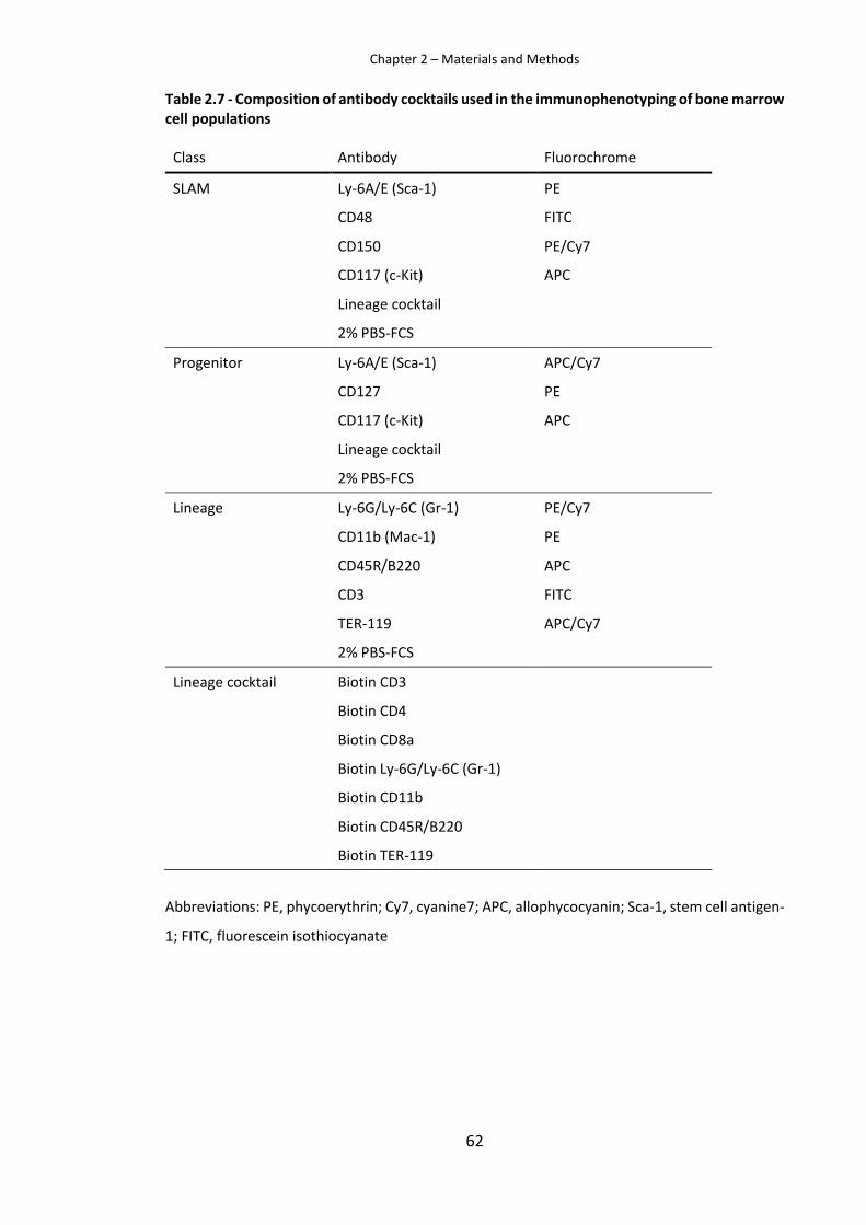
Chapter 2 – Materials and Methods
62
Table 2.7 - Composition of antibody cocktails used in the immunophenotyping of bone marrow cell populations
Class Antibody Fluorochrome
SLAM Ly-6A/E (Sca-1) PE
CD48 FITC
CD150 PE/Cy7
CD117 (c-Kit) APC
Lineage cocktail
2% PBS-FCS
Progenitor Ly-6A/E (Sca-1) APC/Cy7
CD127 PE
CD117 (c-Kit) APC
Lineage cocktail
2% PBS-FCS
Lineage Ly-6G/Ly-6C (Gr-1) PE/Cy7
CD11b (Mac-1) PE
CD45R/B220 APC
CD3 FITC
TER-119 APC/Cy7
2% PBS-FCS
Lineage cocktail Biotin CD3
Biotin CD4
Biotin CD8a
Biotin Ly-6G/Ly-6C (Gr-1)
Biotin CD11b
Biotin CD45R/B220
Biotin TER-119
Abbreviations: PE, phycoerythrin; Cy7, cyanine7; APC, allophycocyanin; Sca-1, stem cell antigen-
1; FITC, fluorescein isothiocyanate

Chapter 2 – Materials and Methods
63
Table 2.8 - Markers used in the immunophenotyping of bone marrow cell populations
Class Cell type Identifier
SLAM Lineage -
LSK Lin- Sca-1+ c-Kit+
HSC CD150+ CD48-
MPP CD150- CD48-
HPC I CD150- CD48+
HPC II CD150+ CD48+
Progenitor Lineage -
CLP CD127+
Lineage Lineage +
Granulocyte GR1+ Mac1-
MDSC GR1+ Mac1+
Macrophages GR1- Mac1+
B-Cell B220+
T-Cell CD3+
Abbreviations: SLAM, signalling lymphocyte activation molecule; HSC, haematopoietic stem cell;
MPP, multipotent progenitors; HPC, hematopoietic progenitor cell; CLP, common lymphoid
progenitor; MDSC, myeloid-derived suppressor cells.

Chapter 2 – Materials and Methods
64
2.8.5 Plaque morphometric analysis
2.8.5.1 Sectioning
Cryosections of OCT-embedded aortic roots were obtained by microtome-cryostat
sectioning at -20°C, where 10 μM sections were cut serially from the aortic root site such
that all three leaflets of the aortic valves were visible. Sections were collected onto poly-
L-lysine coated slides and air-dried for 1 hour before storage at -80°C.
2.8.5.2 Haematoxylin & Eosin
Haematoxylin and eosin (H&E) staining is a standard histological staining technique used
to impart contrast to tissues for microscopy. Haematoxylin is a dark blue/purple stain,
which due to its basic nature stains basophilic structures such as DNA in the nuclei,
resulting in blue/purple stained nuclei. The red stain eosin, used as a counterstain, is
acidic in nature resulting in the staining of positive structures such as cytoplasmic
proteins in a contrasting red colour.
Thawed sections were rinsed in distilled water for 5 minutes and then stained in 5%
(w/v) Gill’s haematoxylin (0.1% (w/v) haematoxylin, 0.02% (v/v) sodium iodate and 2%
(v/v) glacial acetic acid) for 5 minutes. Slides were then rinsed in running tap water until
clear (approximately 10 minutes). Slides were counterstained in Eosin for 10 minutes,
then dehydrated by passing through 80%, 95% and 100% ethanol for 5 minutes each.
Finally, the slides were incubated in xylene for 5 minutes and then mounted using DPX
mountant. Images were captured using a Leica DMRB brightfield microscope with
ProgRes® CapturePro 2.8.8 software at X40 magnification (X4 objective).
2.8.5.3 Oil Red O
Oil Red O is a red lipid soluble dye used in the visualisation of lipids in tissue sections. In
atherosclerosis, analysis of red pigment in stained sections allows quantification of
plaque area and lipid content. Thawed sections were fixed in 4% (w/v) PFA for 15
minutes and then washed 3 times in distilled water and counterstained in Gill’s

Chapter 2 – Materials and Methods
65
haematoxylin for 3 minutes. Slides were rinsed under running tap water. To dispel
water, slides were placed in absolute propylene glycol before staining in Oil red O for 15
minutes at 60°C. Slides were then regressed in 85% propylene glycol for 5 minutes,
washed three times in distilled water and mounted using DPX mountant. Images were
captured using a Leica DMRB brightfield microscope with ProgRes® CapturePro 2.8.8
software at X40 magnification (X4 objective). Oil red O stained sections were analysed
and plaque area and lipid content measured using ImageJ software.
2.8.5.4 Immunohistochemistry
Immunofluorescent (IF) staining of sections was carried out using cell surface marker
antibodies to detect macrophages (anti-mouse MOMA-2), SMCs (anti-mouse αSMA) and
T cells (anti-mouse CD3), which were then photographed by fluorescent microscopy and
analysed using ImageJ software. Details of antibodies used, stock concentrations and
working dilutions are shown in Table 2.9. Antibodies were used in accordance with
previous optimisation in the laboratory and diluted in 0.1% (w/v) BSA in PBS. Thawed
sections were first fixed in acetone for 15 minutes and then washed twice in PBS.
Sections were marked using a hydrophobic pen and then blocked for 30 minutes with
5% (v/v) serum in 5% (w/v) BSA/PBS. Blocking serum was chosen in accordance with the
species of secondary antibody used in order to limit non-specific binding. Primary
antibodies were then applied to sections and the slides incubated overnight at 4°C. The
isotype controls (rabbit IgG or rat IgG) were included to monitor specificity and control
for background staining. Following overnight incubation in primary antibody, the slides
were washed twice in PBS and the corresponding secondary antibodies applied for 1
hour in the dark. Slides were then washed a further twice and counterstained for 20
minutes in 0.3% (w/v) Sudan Black in order to limit auto fluorescence of the sections.
Slides were finally washed three times in PBS and mounted in Fluoroshield™ with DAPI
nuclear stain.

Chapter 2 – Materials and Methods
66
Table 2.9 - Antibodies used for immunofluorescent staining of sections
Antibody Species Stock (mg/mL)
Working dilution
Anti-αSMA Rabbit 0.2 1/100
Anti-CD3 Rabbit 0.2 1/100
Anti-MOMA-2 Rat 0.5 1/100
Rabbit IgG Rabbit 0.5 1/100
Rat IgG Rat 0.5 1/100
Chicken anti-rat IgG AF-594 Chicken 2 1/500
Donkey anti-rabbit IgG AF-488 Donkey 2 1/500
All antibodies were purchased from Abcam, UK. Abbreviations: MOMA-2, monocyte and
macrophage antibody; αSMA, alpha smooth muscle Actin; AF, Alexa Fluor®
Images were captured using an Olympus BX61 microscope with analySIS v3.2 software
at X40 magnification (X4/0.13 objective). DAPI, FITC and AF-488 filters were applied for
imaging of DAPI, AF-594 and AF-488 fluorophores respectively. All images were captured
using consistent exposure, intensity and contrast settings for comparable downstream
analyses. Regions of staining were quantified using ImageJ software.
2.8.6 Liver gene expression
Liver gene expression was analysed using the Qiagen Atherosclerosis RT2 Profiler PCR
Arrays. The PCR array plates contain a panel of 84 atherosclerosis-related genes which
are listed in Appendix 1. Liver samples (50 mg) were homogenised directly in 1 mL
RiboZol™ with a pestle and mortar. Homogenate was transferred to RNase-free
Eppendorf tubes and RNA extraction continued as described in Section 2.5.1. RNA
quality and integrity was examined (Section 2.5.1.1) and RNA reverse transcribed to
cDNA (Section 2.5.3). As an additional check, qPCR was performed for each cDNA sample
using the housekeeping gene Gapdh, and analysis of CT values and melting curves
performed to ensure good amplification efficiency prior to the processing of
Atherosclerosis RT2 Profiler PCR Arrays as outlined in Section 2.5.4.

Chapter 2 – Materials and Methods
67
2.9 Statistical analysis
For all data sets, values outside of 2 standard deviations of the mean were removed as
outliers prior to statistical analysis. Data sets were tested for normality using Q-Q plots
in addition to Shapiro-Wilk test and histograms. To compare the means of two data sets,
either a Student’s t-test (where data displays normal distribution) or Mann Whitney U
test (where normality could not be achieved) was performed. For the comparison of
means between three or more data sets, a One-way analysis of variants (ANOVA) was
performed. Homogeneity of variances was tested using Levene’s statistic and post-hoc
tests selected according to whether equal variances were observed. The Dunnett 2-
sided post-hoc test was performed where equal variances were present, or Dunnett T3
in the case of unequal variances. All statistical analysis was performed using IBM SPSS
Statistics 23 software unless otherwise stated. Significance was defined as p ≤0.05.

CHAPTER 3 –
The effect of probiotic on key processes in atherosclerosis The effect of probiotics on
key processes in atherosclerosis

Chapter 3 – Probiotic effects on key processes
69
3.1 Introduction
Supplementation with a variety of probiotic strains has been shown to beneficially
modify a number of atherosclerosis risk factors including hypercholesterolaemia
(Bernini et al., 2016), dyslipidaemia (Rerksuppaphol and Rerksuppaphol, 2015),
hypertension (Bayat et al., 2016) and chronic inflammation (Mizoguchi et al., 2017).
Indeed, select strains of probiotic bacteria have been shown to inhibit atherosclerotic
plaque formation in ApoE-/- mice (Chan et al., 2016b, 2016a). Clinical trials have reported
beneficial effects of Lab4 and Lab4b supplementation in a number of conditions,
including antibiotic-associated diarrhoea (Madden et al., 2005), irritable bowel
syndrome (IBS) (Williams et al., 2009), atopic sensitisation in infants (Allen et al., 2014),
and the severity and incidence of coughs and colds in children (Garaiova et al., 2015).
Furthermore, CUL66 has been shown to exert cholesterol-lowering effects in vitro
(Michael et al., 2016), and in vivo in combination with the Lab4 consortium (Michael et
al., 2017). In addition to their cholesterol-lowering potential, Lab4 and Lab4b have also
demonstrated immunomodulatory effects. The Adult Immune Study carried out by
Cultech Ltd reported a significant reduction in the pro-inflammatory cytokines IL-6 and
IL-1β, with a significant increase in production of the anti-inflammatory cytokines IL-10
and TGF-β (Hepburn et al., 2013). Together, results from previous studies have
highlighted potential anti-atherogenic effects of Lab4, Lab4b and CUL66; however, the
effects of these consortia have not previously been investigated in relation to
atherosclerosis.
The overall aim of studies presented in this chapter was to investigate the effects of
Lab4, Lab4b and CUL66 treatment across a range of key processes in atherosclerosis
development, thereby providing an initial assessment of the potential anti-
atherosclerotic effects of these probiotic consortia. Initial investigations were carried
out in vitro using human monocytes, macrophages and SMCs treated with probiotic CM.
Initially, probiotic CM was normalised to total protein concentration, then the
concentration was optimised for use in further experiments. Investigations then
continued to assess the effect of each probiotic CM on monocyte migration, ROS
production, lipoprotein uptake via macropinocytosis, VSMC invasion, phagocytosis,
proliferation of key cell types and inflammatory processes.

Chapter 3 – Probiotic effects on key processes
70
3.1.1 Normalisation of probiotic CM batches
Batch effects may be introduced by technical sources of variation during processing.
While it’s not possible to completely eliminate batch effects, the production of CM
batches was optimised in order to minimise variation and to ensure a robust protocol
and production of consistent, reproducible results. However, due to the use of live
bacteria in this study, additional batch effects in the form of biological variation had to
be considered. Biological variation was reduced by ensuring that the probiotic bacteria
were thoroughly mixed (Lab4 or Lab4b) and accurately weighed before culturing. A
simple measure of CFU is often used for normalisation. However, further variation
created during bacterial culturing is unavoidable, particularly with regards to Lab4 and
Lab4b where metabolic interaction between the different bacteria strains can occur
(Kort et al., 2015). Therefore, it was considered necessary for greater confidence of
results, to control for batch effects by normalising probiotic CM treatment to a
measurable parameter. Owing to ease of measurement, high reproducibility and low
assay cost, total protein concentration was selected as the most suitable parameter for
this purpose. For each probiotic CM, cell viability was assessed using a range of protein
concentrations. The optimal concentration was then determined as the highest protein
concentration at which cells maintain >85% viability.
3.1.2 Cell viability
Prior to the start of in vitro investigations, the effect of each CM on the viability of cells
was assessed for each individual cell culture model system utilised in the study. This was
carried out to ensure that results were not influenced by cell death and that firm
conclusions were made. Cell viability was assessed using the Pierce LDH Cytotoxicity
Assay Kit as described in Section 2.4.1. Damage to the cell membrane results in the
release of the cytosolic enzyme LDH into the culture media where it catalyses the
conversion of lactate to pyruvate via the reduction of NAD+ to NADH, which is
subsequently used by the enzyme diaphorase in the reduction of a tetrazolium salt 2-
(4iodophenyl)-3-94-nitrophenyl)-5-phenyl-2H-tetrazolium chloride (INT) to formazan; a
red coloured product that can be measured at 490 nm. The level or formazan produced

Chapter 3 – Probiotic effects on key processes
71
by this coupled enzymatic reaction is directly proportional to the level of LDH released
into the culture media.
Following cell viability assays, initial in vitro investigations were performed in a dose-
dependent manner in order to test the effectiveness of probiotic CM at a range of
concentrations, with the aim of selecting a single optimal concentration of each
probiotic CM for use in the remainder of in vitro studies.
3.1.3 Key processes in atherosclerosis
3.1.3.1 Monocyte recruitment and migration
One of the key initiating processes in atherosclerosis is the recruitment and migration
of circulating monocytes from the lumen into the arterial wall. Activated endothelial
cells express a number of chemokines and adhesion molecules involved in the capture
and immobilisation of monocytes followed by their transmigration across the
endothelium in response to a range of chemoattractants (Ramji and Davies, 2015). MCP-
1 (also known as CCL2) is a key pro-inflammatory chemokine fundamental in the
regulation of monocyte recruitment and migration (Deshmane et al., 2009) and a potent
monocyte chemotactic factor implicated in the development of atherosclerosis (Aiello
et al., 1999). Studies in ApoE knockout mice have shown MCP-1 deficiency to result in
smaller, less extensive lesion development (Gosling et al., 1999), while increased
expression of MCP-1 by leukocytes has been shown to increase atherosclerosis lesion
development (Aiello et al., 1999). Attenuation of MCP-1 stimulated monocyte migration
by probiotic CM would therefore represent a potential anti-atherogenic effect. A
previous study has demonstrated the ability of probiotic Enterococcus faecalis to
significantly downregulate the expression of MCP-1 in C57BL/6 mice (Chen et al., 2017).
However, this is the first study to investigate the effects of probiotics, or CM derived
from them, on monocyte migration in atherosclerosis. In this study, a modified Boyden
chamber method was used to assess the effect of probiotic CM treatment on migration
of monocytes through an 8 µM pore membrane, towards a chemoattractant (MCP-1)
stimulus, where the membrane mimics the arterial endothelial layer.

Chapter 3 – Probiotic effects on key processes
72
3.1.3.2 Modification of lipoproteins by ROS
ROS are a group of small highly reactive oxygen derivatives such as hydrogen peroxide
(H2O2) and the superoxide anion (O2-), involved in the normal functioning of biological
processes and cellular metabolic control (Goncharov et al., 2015). While ROS are critical
for healthy vascular homeostasis, excess production results in a state of imbalance
known as oxidative stress (Kattoor et al., 2017). ROS production and oxidative stress is
increased in many conditions known to be risk factors for atherosclerosis including
diabetes, hypertension, smoking and lipid dysregulation (Giacco and Brownlee, 2010;
Kattoor et al., 2017; Steffen et al., 2012). ROS are considered to be pro-atherogenic
factors integral in the pathogenesis of atherosclerosis (Goncharov et al., 2015). In fact,
the oxidation hypothesis suggests that oxidised LDL is a major factor driving the
development of atherosclerosis (Choi et al., 2017). Oxidative modification of LDL to form
oxLDL induces the expression of adhesion molecules that enhance leukocyte
recruitment and their migration into the arterial wall (Goncharov et al., 2015).
Differentiated macrophages express scavenger receptors which internalise these
modified forms of LDL resulting in the formation of macrophage foam cells, the hallmark
of atherosclerosis. Additionally, foam cells release proinflammatory cytokines further
promoting ROS production (Kattoor et al., 2017). In conjunction with growth factors,
ROS stimulate the migration of VSMCs, the expression of VSMC scavenger receptors and
subsequent formation of VSMC-derived foam cells (Kattoor et al., 2017). Furthermore
in advanced atherosclerosis, the production of MMPs induced by ROS contributes to the
degradation of the fibrous cap and to plaque instability (Kattoor et al., 2017). A number
of pharmacological agents have been shown to have beneficial effects on atherogenesis
by modulating oxidative stress, including ACE inhibitors and angiotensin II receptor type
1 (AT1 receptor) blockers (ARBs) (Saso et al., 2017). In this study, probiotic CM was
tested for its effect on cellular ROS production in the presence of potent ROS stimulant
H2O2, using the Abcam Cellular ROS Detection Assay Kit.
3.1.3.3 Macropinocytosis
The unregulated uptake of excessive amounts of modified LDL by macrophages is a key
event in the formation of macrophage foam cells, and essential to atherosclerosis
Chapter 3 – Probiotic effects on key processes
73
development. Uptake of modified LDL by macrophages is regarded as being largely
receptor-mediated; however, a significant portion is taken up by a non-receptor-
mediated process of fluid-phase endocytosis known as macropinocytosis (Kruth et al.,
2002). During this process, actin-dependent ruffling of the plasma membrane is followed
by fusion of the membrane to itself, leading to the internalisation of the surrounding
contents in fluid-filled intracellular vacuoles (Michael et al., 2013). This process has been
shown to contribute substantially to the uptake of both unmodified and modified LDL
and to the formation of macrophage foam cells (Michael et al., 2013). While the effect
of probiotics on macropinocytosis hasn’t yet been studied, previous studies have shown
that inhibiting macropinocytosis in macrophages decreases foam cell formation
(Gallagher, 2016; Yao et al., 2009). In this study, the effect of probiotic CM on
macropinocytosis was assessed in macrophages using the fluorescent molecule
luciferase yellow LY as described in Section 2.7.2. LY is a popular dye that has been
successfully utilised for the measurement of macropinocytosis in macrophages in
previous studies (McLaren et al., 2011b; Michael et al., 2013; Swanson, 1989).
3.1.3.4 Mitochondrial function
Mitochondria are well known as the energy producing powerhouse of the cell, though
the role of mitochondria goes beyond their energy producing capacity. As mentioned
previously, mitochondrial ROS production is a popular target for the reduction of
oxidative stress (Kattoor et al., 2017) and mitochondrial dysfunction, and mitochondrial
oxidative stress (mitoOS) has been shown to strongly correlate with atherosclerotic
lesion progression (Wang et al., 2017d). Mitochondrial respiratory function is routinely
assessed by Agilent Seahorse technology, where the OCR is measured in response to the
serial injection of respiratory modulators (Hujber et al., 2018; Tan et al., 2015) targeting
specific components of the electron transport chain (Figure 3.1). The effect of probiotic
bacteria on mitochondrial function has not yet been studied, therefore this study aims
to investigate the effect of probiotic CM on mitochondrial function using the Seahorse
Mito Stress Test as described in Section 2.4.6.
The Seahorse Mito Stress Test measures OCR following serial injection of oligomycin at
15 minutes, FCCP at 34 minutes and rotenone/antimycin A at 54 minutes as illustrated

Chapter 3 – Probiotic effects on key processes
74
in Figure 3.2. Initial OCR measurements represent basal respiration prior to the injection
of compounds, i.e. the energy demands of the cell under baseline conditions. Oligomycin
inhibits ATP synthase (Figure 3.1) and so the decrease in OCR following injection at 15
minutes correlates to ATP production (Figure 3.2). FCCP is an uncoupling agent which
targets the inner mitochondrial membrane, causing disruption of the mitochondrial
membrane potential and uninhibited flow of electrons through the ETC (Figure 3.1). The
peak in OCR following the injection of FCCP therefore represents the maximum
respiration achievable where the ETC is operating at full capacity, and by deduction of
basal respiration allows calculation of spare respiratory capacity (Figure 3.1). Spare
respiratory capacity is an essential parameter giving a measure of the cells capacity to
deal with increased energy demand and a sign of cellular health. The final injection is a
mix of rotenone and antimycin, which together inhibits the remaining ETC complexes
leading to complete cessation of mitochondrial respiration (Figure 3.1). The remaining
OCR therefore represents non-mitochondrial respiration (Figure 3.2). Finally, the OCR
following the cessation of respiration is deducted from the OCR following oligomycin
injection to provide a measure of proton leak (Figure 3.2). Proton leak is the remaining
basal respiration that is not coupled to ATP production, and is a sign of mitochondrial
damage. In this study, mitochondrial function was assessed in THP-1 macrophages
treated with either vehicle or probiotic CM by measurement of basal respiration,
maximal respiration, spare respiratory capacity, ATP production, non-mitochondrial
respiration and proton leak.

Chapter 3 – Probiotic effects on key processes
75
Figure 3.1 – Illustration of the ETC components targeted by respiratory modulators in the Seahorse Mito Stress Test. Oligomycin targets ATP synthase (complex V), inhibiting cellular ATP production. FCCP is an uncoupling agent that targets the inner mitochondrial membrane, causing collapse of the proton gradient and uninhibited flow of electrons through the ETC. As a result oxygen is maximally consumed by complex IV. Rotenone/antimycin shuts down mitochondrial respiration via the inhibition of complexes I and III respectively. Image taken from the Agilent Seahorse Cell Mito Stress Test User Guide (Agilent Technologies, 2019).
Figure 3.2 – Seahorse XFp Cell Mito Stress Test profile showing key parameters of mitochondrial respiration. OCR is measured during serial injection of respiratory modulators oligomycin, FCCP and rotenone/antimycin, thereby allowing the determination of basal respiration, ATP production, maximal respiration, spare respiratory capacity, proton leak and non-mitochondrial respiration. *, proton leak. Abbreviations: OCR, oxygen consumption rate; FCCP, carbonyl cyanide-4 (trifluoromethoxy) phenylhydrazone.
*

Chapter 3 – Probiotic effects on key processes
76
3.1.3.5 Phagocytosis
In addition to the uptake of oxLDL, macrophages play an important role in phagocytosis
of apoptotic cells and cellular debris which would otherwise accumulate, contributing
to the formation of a necrotic core (Schrijvers, 2005) and advanced plaque progression.
Phagocytosis by macrophages is known to be reduced in human atherosclerotic plaques
(Kojima et al., 2016). Recent studies have demonstrated that reduced macrophage
phagocytosis is associated with increased uptake of oxLDL and increased plaque
formation, while increased phagocytosis is associated with the opposite effect (Laguna-
Fernandez et al., 2018). The effect of probiotics on macrophage phagocytic activity has
not yet been investigated. This study aims to elucidate the effect of probiotic CM on
phagocytosis in human macrophages using the Vybrant Phagocytosis Assay Kit as
described in Section 2.4.7.
3.1.3.6 Proliferation of key cell types involved in atherosclerosis
Macrophage proliferation is a key aspect involved in the progression of atherosclerosis,
believed to be more relevant to lesion advancement than monocyte recruitment, and
has been identified as a potential therapeutic target for disease intervention (Robbins
et al., 2013). A study in ApoE-/- mice has demonstrated a reduction in macrophage
proliferation corresponding to a reduction in lesion size irrespective of the level of
monocyte recruitment (Robbins et al., 2013). Another study involving the disruption of
cell cycle regulators at the genetic level, demonstrated increased arterial macrophage
and VSMC proliferation with corresponding acceleration of atherosclerosis in ApoE-/-
mice (Diez-Juan et al., 2004). This study aims to investigate the effect of probiotic
treatment on the proliferation of human macrophages using crystal violet staining and
a BrdU ELISA method as described in Section 2.4.2.2. Briefly, proliferation was initially
assessed periodically over a 7-day period using crystal violet staining in conjunction with
cell viability testing to ensure results were not influenced by cell death. Additionally, the
rate of proliferation was assessed at 48 hours using the BrdU Labeling and Detection Kit.
Further to macrophages, the proliferation of VSMCs was also assessed in a similar
manner using HASMCs treated with either vehicle or probiotic CM. The proliferation of
monocytes was also assessed by simple cell counting over a 7-day period with

Chapter 3 – Probiotic effects on key processes
77
concurrent assessment of cell viability. This allows for a more complete picture of the
effect of probiotics on the proliferation of key cell types involved in atherosclerosis.
3.1.3.7 VSMC invasion
Following on from VSMC proliferation, this study investigates the effect of probiotic CM
on VSMC invasion. VSMCs migrate from the media towards the arterial intima in
response to vascular injury, a complex process driven by many cytokines, chemokines
and growth factors (Kim et al., 2014), including platelet-derived growth factor (PDGF).
PDGF is a potent mitogen and chemoattractant for SMCs (Sano et al., 2001) and inducer
of VSMC proliferation and migration (Ricci and Ferri, 2015). When physiological
migration of VSMCs becomes dysregulated, it becomes pathological and is often
referred to as VSMC invasion, an important factor in atherogenesis (Louis and Zahradka,
2010). As described in Chapter 1, migrated SMCs together with their secreted ECM
proteins form a protective cap surrounded by a collagen-rich matrix, which functions to
provide plaque stability (Louis and Zahradka, 2010). However, the proliferation and
invasion of SMCs remains closely associated with atherosclerosis progression and has
been shown to play a key role in neointima formation (Chen et al., 2018; Zhang, 2009).
To investigate the effect of probiotic CM on VSMC invasion, this study uses a modified
Boyden chamber method as described in Section 2.4.5, where HASMCs treated with
either vehicle or CM were allowed to migrate towards a PDGF stimulus.
3.1.4 Anti-inflammatory effects in atherosclerosis
Atherosclerosis is now widely regarded as an inflammatory disorder with anti-
inflammatory agents receiving much attention in the search for anti-atherosclerotic
therapeutics. More recently probiotics, including many different Lactobacilli strains,
have been shown to have beneficial anti-inflammatory actions in atherosclerosis, with a
large range of strains investigated in the literature (Ding et al., 2017). However, this
effect is highly strain specific with as many strains showing no effect or even pro-
inflammatory actions (Ding et al., 2017). In a strain-specific manner, probiotics have
been shown to exert many immunomodulatory effects including the promotion of anti-
inflammatory M2 macrophage polarisation (Ukibe et al., 2015), reduction of T-

Chapter 3 – Probiotic effects on key processes
78
lymphocyte helper 1 (Th1) cell numbers and pro-inflammatory cytokine production
(Smelt et al., 2013), and an increase in regulatory T cells (Tregs) (Ding et al., 2017; Smits
et al., 2005). This study aims to investigate the potential anti-inflammatory effects of
probiotic CM treatment on the inflammatory response of human macrophages to pro-
inflammatory cytokine stimulation.
3.1.4.1 Activation of the NLRP3 inflammasome
The inflammasome is a multi-protein complex formed by the activation of an innate
immune system pattern recognition receptors (PRR). NLRP3 is the most well defined
inflammasome responsible for the maturation of proinflammatory IL-1 family cytokines;
IL-1β and IL-18 (Jo et al., 2016) and has been implicated in a variety of inflammatory
diseases including atherosclerosis (Ozaki et al., 2015). Activation of NLRP3 in response
to a pro-inflammatory stimulus can be assessed by the level of IL-1β production,
measurable by standard ELISA methods. This study investigates the effect of probiotic
CM on the activation of the NLRP3 inflammasome by IL-1β ELISA as described in Section
2.6.3.
3.1.4.2 Inflammatory gene expression
Many classes of cytokines are present in atherosclerotic plaques with all atherosclerosis-
associated cell types capable of secreting and responding to them (Ramji and Davies,
2015). Key cytokines are very highly expressed in atherosclerotic regions including the
pro-inflammatory IL-1β, IFN-γ and TNF)-α. Of these, IFN-γ is considered a master
regulator of atherosclerosis; highly involved in many stages of disease progression and
a popular target for anti-atherosclerotic therapies (McLaren and Ramji, 2009). One study
demonstrated that the attenuation of IFN-γ action by administration of an inhibitory
protein prevented plaque progression in ApoE-/- mice, in addition to the downregulation
of pro-inflammatory cytokine expression (Koga et al., 2007). Further studies, including
the previously mentioned CANTOS trial (Dolgin, 2017) have demonstrated that the
inhibition or blocking of IL-1β leads to a corresponding suppression of atherosclerosis
progression in ApoE-/- mice (Bhaskar et al., 2011; Ramji and Davies, 2015), while TNF-α
knockout significantly reduced fatty streak formation with corresponding

Chapter 3 – Probiotic effects on key processes
79
downregulation in expression of pro-inflammatory factors (Xiao et al., 2009). The effect
of various probiotic bacteria on cytokine production has been investigated with
conflicting results. Many studies show beneficial effects with specific strains attenuating
the production of pro-inflammatory cytokines, including those mentioned above, while
inducing the production of anti-inflammatory cytokines such as IL-10 (Kekkonen et al.,
2008; Singh et al., 2018; Štofilova et al., 2017). However, the exact effects are highly
strain-specific with some strains having no effect or even a pro-inflammatory influence
(Meyer et al., 2007). MCP-1 and ICAM1 are both robust markers of inflammation and
play an essential role in the continued migration of monocytes across the endothelium.
Their expression is known to be induced by a range of pro-inflammatory cytokines,
including IFN-γ (Moss et al., 2016), IL-1β (Lappas, 2017) and TNF-α (Jiang et al., 2015),
thereby making these genes strong markers for the investigation of cytokine-induced
inflammatory effects. This study therefore investigates the effect of probiotic CM
treatment on cytokine-induced expression of inflammatory markers MCP-1 and ICAM1
in human macrophages stimulated with either IFN-γ, IL-1β or TNF-α.
3.2 Experimental design
Prior to the start of in vitro investigations, probiotic CM batches were normalised to total
protein concentration and confirmed to have no detrimental effects on cell viability.
Initial assays were performed in a dose response format to determine an optimal
concentration of the probiotic CM for further studies. Following optimisation, Lab4,
Lab4b and CUL66 CM was used at concentrations of 8, 1.5 and 5 µg/mL respectively.
Experiments then continued to investigate the effect of probiotic CM on key processes
associated with atherosclerosis development as outlined in Figures 3.3 – 3.5. Specific
methods for each experimental aim are detailed in Chapter 2.

Chapter 3 – Probiotic effects on key processes
80
Figure 3.3 - Experimental strategy for the investigation of the effects of probiotic CM on key cellular processes in atherosclerosis. Abbreviations: HASMCs, human aortic smooth muscle cells; SMC, smooth muscle cells; ROS, reactive oxygen species.

Chapter 3 – Probiotic effects on key processes
81
Figure 3.4 - Experimental strategy for the investigation of the effect of probiotic CM on the proliferation of key cell types in atherosclerosis. Abbreviations: HASMCs, human aortic smooth muscle cells; BrdU, 5-Bromo-2′-deoxy-uridine
Figure 3.5 - Experimental strategy for the investigation of the anti-inflammatory effects of probiotic CM. Abbreviations: AcLDL, acetylated LDL; ELISA, enzyme-linked immunosorbent assay.

Chapter 3 – Probiotic effects on key processes
82
3.3 Results
3.3.1 Normalisation of probiotic CM to total protein concentration
To control for batch effects, each probiotic CM was normalised according to the total
protein concentration, and the optimal concentration estimated as the maximum
concentration at which the cells remain viable. Probiotic CM was prepared and the total
protein quantified as described in Sections 2.2 and 2.6.2 respectively. Cell viability was
then assessed using the LDH Assay Kit (Section 2.4.1), using a range of known protein
concentrations for each CM at 100% then approximately 80%, 60% and 40% dilution.
For Lab4 CM, the protein concentrations of 21, 17, 13 and 8 µg/mL were tested where
21 µg/mL represents 100% CM. Similarly for Lab4b and CUL66, concentrations of 3, 2.5,
1.5 and 1 µg/mL (Lab4b) and 5, 4, 3 and 2 µg/mL (CUL66) were tested where 3 µg/mL
and 5 µg/mL represent 100% CM respectively. THP-1 macrophages were treated for 24
hours with either vehicle or probiotic CM at the range of concentrations detailed above.
Cell viability was determined as a percentage of the vehicle control, and a cell viability
of 85% or over was considered acceptable.
For Lab4 CM, higher concentrations of 21 and 17 µg/mL led to reductions in cell viability
below 85% (Figure 3.6A), indicating a detrimental effect and so were considered
unsuitable for use in experiments. All other tested concentrations of Lab4 displayed less
than 15% reduction in cell viability and were therefore considered suitable for use in
experiments. For some batches of Lab4 CM, total protein concentration was measured
at a maximum of 8 µg/mL, therefore this concentration was selected as the optimal
concentration for use in experiments. For Lab4b, cell viability remained at or above 85%
following treatment with all tested concentrations (Figure 3.6B). However, as with Lab4,
a concentration of 1.5 µg/mL was selected to reflect the minimum protein concentration
measured in any batch of Lab4b CM. Similarly, all tested concentrations of CUL66
resulted in greater than 85% cell viability (Figure 3.6C) and so the maximum
concentration of 5 µg/mL was selected for use in future experiments.

Chapter 3 – Probiotic effects on key processes
83
Figure 3.6 - Viability of THP-1 macrophages assessed using the LDH Assay Kit. THP-1 macrophages were treated with either the vehicle control or probiotic CM at a range of known protein concentrations. LDH assays were performed and cell viability determined as a percentage of the vehicle control which was set to 100%. Dashed line denotes acceptable level of viability at 85% and above. (A) Cells treated with Lab4 CM of 21, 17, 13 and 8 µg/mL protein concentration. (B) Cells treated with Lab4b CM of 3, 2.5, 1.5 and 1 µg/mL protein concentration. (C) Cells treated with CUL66 CM of 5, 4, 3 and 2 µg/mL protein concentration. The data are presented as mean ± SEM from three independent experiments.
3.3.2 Probiotic CM has no detrimental effect on the viability of cell culture
model systems used for in vitro studies
The effect of each probiotic CM on cell viability was then assessed for each cell culture
model system to be utilised in the study. Cells were incubated for 24 hours with vehicle
or probiotic CM and cell viability assessed using the LDH Assay Kit (Section 2.4.1). Results
are shown in Figure 3.7. Treatment with probiotic CM resulted in a significant increase
in the viability of cells, but no detrimental effect on the viability of THP-1 macrophages,
HASMCs or HMDMs.
Vehicle 8 13 17 210
20
40
60
80
100
120
Protein concentration (g/mL)
Ce
ll v
iab
ilit
y (
% o
f v
eh
icle
)
Vehicle 1 1.5 2.5 30
20
40
60
80
100
Protein concentration (g/mL)
Ce
ll v
iab
ilit
y (
% o
f v
eh
icle
)
Vehicle 1.5 3 4.5 60
20
40
60
80
100
120
Protein concentration (g/mL)
Ce
ll v
iab
ilit
y (
% o
f v
eh
icle
)
(A) (B)
(C)

Chapter 3 – Probiotic effects on key processes
84
Figure 3.7 - Probiotic CM has no detrimental effect on the viability of cell culture model systems utilised in the study. (A) THP-1 macrophages, (B) HASMCs and (C) HMDMs were treated for 24 hours with the vehicle control or Lab4 (8 µg/mL), Lab4b (1.5 µg/mL) or CUL66 (5 µg/mL) CM, and cell viability assessed using the LDH Assay Kit. Cell viability was determined as a percentage relative to the vehicle control which was set to 100%. Data are presented as mean ± SEM from three independent experiments. Statistical analysis was performed using a One-way ANOVA with Dunnett T3 post-hoc test where *p <0.05, **p <0.01 and *** p <0.001.
3.3.3 Monocyte migration was reduced in the presence of probiotic CM
Monocyte migration was assessed using a modified Boyden chamber method (Section
2.4.4). Initially, the assay was performed in a concentration dependent manner using
protein concentrations of 8, 6 and 4 µg/mL for Lab4 CM; 1.5, 1 and 0.5 µg/mL for Lab4b
CM; and 5, 4 and 3 µg/mL for CUL66 CM. Results are shown in Figure 3.8. In all cases,
the migration of monocytes was significantly increased (p <0.001) in the presence of
MCP-1 compared to vehicle alone (no MCP-1 control). In the presence of Lab4 CM, the
MCP-1-induced migration was attenuated by 0.58-fold (p <0.001), 0.73-fold (p =0.002)
or 0.65-fold (p <0.001) with concentrations of 8, 6 or 4 µg/mL respectively (Figure 3.8A).
Similarly, with Lab4b treatment the MCP-1-induced migration was attenuated in a
concentration dependent manner by 0.53-fold (p <0.001), 0.57-fold (p <0.001) or 0.68-
fold (p <0.001) with concentrations of 1.5, 1 or 0.5 µg/mL respectively. CUL66 treatment
also attenuated MCP-1-driven migration in a concentration dependent manner showing
Vehicle Lab4 Lab4b CUL660
50
100
150***
*****
Cell v
iab
ilit
y (%
)
Vehicle Lab4 Lab4b CUL660
50
100
150
******
Ce
ll v
iab
ilit
y (
%)
Vehicle Lab4 Lab4b CUL660
50
100
150 ***
*
Cell v
iab
ilit
y (%
)
(A) (B)
(C)

Chapter 3 – Probiotic effects on key processes
85
reductions of 0.73-fold (p =0.004), 0.7-fold (p =0.002) or 0.6-fold (p <0.001) with
concentrations of 5, 4 or 3 µg/mL respectively.

Chapter 3 – Probiotic effects on key processes
86
Figure 3.8 - Migration of THP-1 monocytes in response to MCP-1 was attenuated by various concentrations of probiotic CM. The effect of each probiotic CM on monocyte migration was assessed in a concentration dependent manner using the Boyden chamber method. THP-1 monocytes were allowed to migrate in the presence of MCP-1 and either vehicle (Vehicle control) or probiotic CM. Cells treated with vehicle but in the absence of MCP-1 were included for comparative purposes (No MCP-1). (A) Lab4 CM used at concentrations of 8, 6 and 4 µg/mL. (B) Lab4b CM used at concentrations of 1.5, 1 and 0.5 µg/mL (C) CUL66 CM used at concentrations of 5, 4 and 3 µg/mL. Migration was determined as a percentage of the total cells, and displayed as fold-change in migration relative to the Vehicle control which was arbitrarily set to 1. Data are presented as mean ± SEM from five (A and B), or three (C) independent experiments. Statistical analysis was performed using a One-way ANOVA with Dunnett (2-sided) post-hoc test where ** p <0.01 and *** p <0.001.
No MCP-1 Vehicle 4 g/mL 6 g/mL 8 g/mL0.0
0.2
0.4
0.6
0.8
1.0
1.2
1.4***
********
MCP-1 - + +++
Mo
no
cy
te m
igra
tio
n (
fold
ch
an
ge
)
No MCP-1 Vehicle 0.5 g/mL1 g/mL 1.5 g/mL0.0
0.2
0.4
0.6
0.8
1.0
1.2
1.4***
*********
MCP-1 - + +++
Mo
no
cy
te m
igra
tio
n (
fold
ch
an
ge
)
No MCP-1 Vehicle 3 g/mL 4 g/mL 5 g/mL0.0
0.2
0.4
0.6
0.8
1.0
1.2
1.4**
********
MCP-1 - + +++
Mo
no
cy
te m
igra
tio
n (
fold
ch
an
ge
)
(A)
(B)
(C)
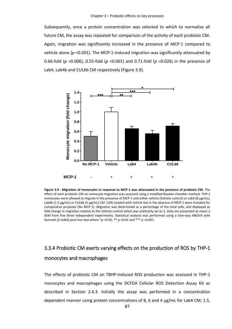
Chapter 3 – Probiotic effects on key processes
87
Subsequently, once a protein concentration was selected to which to normalise all
future CM, the assay was repeated for comparison of the activity of each probiotic CM.
Again, migration was significantly increased in the presence of MCP-1 compared to
vehicle alone (p <0.001). The MCP-1-induced migration was significantly attenuated by
0.66-fold (p =0.006), 0.55-fold (p <0.001) and 0.71-fold (p =0.026) in the presence of
Lab4, Lab4b and CUL66 CM respectively (Figure 3.9).
Figure 3.9 - Migration of monocytes in response to MCP-1 was attenuated in the presence of probiotic CM. The effect of each probiotic CM on monocyte migration was assessed using a modified Boyden chamber method. THP-1 monocytes were allowed to migrate in the presence of MCP-1 and either vehicle (Vehicle control) or Lab4 (8 µg/mL), Lab4b (1.5 µg/mL) or CUL66 (5 µg/mL) CM. Cells treated with vehicle but in the absence of MCP-1 were included for comparative purposes (No MCP-1). Migration was determined as a percentage of the total cells, and displayed as fold-change in migration relative to the Vehicle control which was arbitrarily set to 1. Data are presented as mean ± SEM from five three independent experiments. Statistical analysis was performed using a One-way ANOVA with Dunnett (2-sided) post-hoc test where *p <0.05, ** p <0.01 and *** p <0.001.
3.3.4 Probiotic CM exerts varying effects on the production of ROS by THP-1
monocytes and macrophages
The effects of probiotic CM on TBHP-induced ROS production was assessed in THP-1
monocytes and macrophages using the DCFDA Cellular ROS Detection Assay Kit as
described in Section 2.4.3. Initially the assay was performed in a concentration
dependent manner using protein concentrations of 8, 6 and 4 µg/mL for Lab4 CM; 1.5,
No MCP-1 Vehicle Lab4 Lab4b CUL660.0
0.2
0.4
0.6
0.8
1.0
1.2
1.4*
********
MCP-1 - + +++
Mo
no
cy
te m
igra
tio
n (
fold
ch
an
ge
)

Chapter 3 – Probiotic effects on key processes
88
1 and 0.5 µg/mL for Lab4b CM; and 5, 4 and 3 µg/mL for CUL66 CM. Results of
investigations in both THP-1 monocytes and macrophages are shown in Figures 3.10 and
3.11 respectively. TBHP significantly induced ROS production in both monocytes and
macrophages compared to vehicle alone (p <0.001). In THP-1 monocytes, no significant
change in TBHP-induced ROS production was observed with any probiotic CM at any
tested concentration, with the exception of the lowest concentration of Lab4b (0.5
µg/mL) which produced a significant 1.3-fold (p =0.003) increase in ROS production
(Figure 3.10B). Each probiotic CM at all tested concentrations increased the TBHP-
induced ROS production in THP-1 macrophages (Figure 3.11). Thus, treatment with Lab4
CM resulted in a similar degree of increase in ROS production across all concentrations
tested showing 1.5-fold (p =0.003), 1.46-fold (p =0.010) or 1.47-fold (p =0.003) increase
with 8, 6 or 4 µg/mL of protein respectively (Figure 3.11A). Similarly with Lab4b
treatment ROS production was increased by 1.73-fold (p =0.016), 1.52-fold (p <0.001)
and 1.81-fold (p <0.001) with 1.5, 1 or 0.5 µg/mL of protein respectively (Figure 3.11B).
In the presence of CUL66 CM, the TBHP-induced ROS production displayed an increase
in a concentration dependent manner from 1.43-fold (p <0.001), to 1.52-fold (p =0.002)
and 1.64-fold (p <0.001) with 5, 4 or 3 µg/mL of protein respectively (Figure 3.11C).

Chapter 3 – Probiotic effects on key processes
89
Figure 3.10 – The effect of probiotic CM on TBHP-induced ROS production in THP-1 monocytes. The effect of each probiotic CM on TBHP-induced ROS production was assessed in a concentration dependent manner using the DCFDA Cellular ROS Detection Assay Kit. THP-1 monocytes were treated with TBHP and either vehicle (Vehicle control) or the indicated concentration of probiotic CM. Cells treated with vehicle in the absence of TBHP were included for comparative purposes (No TBHP control). (A) Lab4 CM used at concentrations of 8, 6 and 4 µg/mL. (B) Lab4b CM used at concentrations of 1.5, 1 and 0.5 µg/mL. (C) CUL66 CM used at concentrations of 5, 4 and 3 µg/mL. ROS production is displayed as fold-change to the Vehicle control which was arbitrarily set to 1. Data are presented as mean ± SEM from five independent experiments. Statistical analysis was performed using a One-way ANOVA with Dunnett T3 post-hoc test where ** p <0.01, *** p <0.001.
No TBHP Vehicle 4 g/mL 6 g/mL 8 g/mL0.0
0.5
1.0
1.5
TBHP - + +++
NS
NS***NS
RO
S p
rod
ucti
on
(fo
ld c
han
ge)
No TBHP Vehicle 0.5 g/mL 1 g/mL 1.5 g/mL0.0
0.5
1.0
1.5
NS
****NS
TBHP - + +++
RO
S p
rod
ucti
on
(fo
ld c
han
ge)
No TBHP Vehicle 3 g/mL 4 g/mL 5 g/mL0.0
0.5
1.0
1.5
TBHP - + +++
NS
NS**NS
RO
S p
rod
ucti
on
(fo
ld c
han
ge)
(A)
(B)
(C)

Chapter 3 – Probiotic effects on key processes
90
Figure 3.11 - Probiotic CM increases TBHP-induced ROS production in THP-1 macrophages. The effect of each probiotic CM on TBHP-induced ROS production was assessed in a concentration dependent manner using the DCFDA Cellular ROS Detection Assay Kit. THP-1 macrophages were treated with TBHP and either vehicle (Vehicle control) or the indicated concentration of probiotic CM. Cells treated with vehicle in the absence of TBHP were included for comparative purposes (No TBHP control). (A) Lab4 CM used at concentrations of 8, 6 and 4 µg/mL. (B) Lab4b CM used at concentrations of 1.5, 1 and 0.5 µg/mL. (C) CUL66 CM used at concentrations of 5, 4 and 3 µg/mL. ROS production is displayed as fold-change to the Vehicle control which was arbitrarily set to 1. Data are presented as mean ± SEM from five independent experiments. Statistical analysis was performed using a One-way ANOVA with Dunnett T3 post-hoc test where * p <0.05, ** p <0.01 and *** p <0.001.
No TBHP Vehicle 4 g/mL 6 g/mL 8 g/mL0.0
0.5
1.0
1.5
2.0
TBHP - + +++
**
******
RO
S p
rod
ucti
on
(fo
ld c
han
ge)
No TBHP Vehicle 0.5 g/mL 1 g/mL 1.5 g/mL0.0
0.5
1.0
1.5
2.0
2.5
TBHP - + +++
*
*********
RO
S p
rod
ucti
on
(fo
ld c
han
ge)
No TBHP Vehicle 3 g/mL 4 g/mL 5 g/mL0.0
0.5
1.0
1.5
2.0
TBHP - + +++
***
*****
***
RO
S p
rod
ucti
on
(fo
ld c
han
ge)
(A)
(B)
(C)

Chapter 3 – Probiotic effects on key processes
91
Subsequently, once a protein concentration was selected to which to normalise all
future CM, the assay was repeated for comparison of the activity of each CM. For both
THP-1 monocytes and macrophages, ROS production was increased in the presence of
TBHP compared to vehicle alone (p <0.001). In agreement with results obtained during
the dose dependent investigations, no significant change in ROS production was
observed in THP-1 monocytes (Figure 3.12), while ROS production was significantly
increased in THP-1 macrophages with Lab4 (2.4-fold; p <0.001), Lab4b (1.66-fold; p
<0.001) and CUL66 (2.5-fold; p =0.028) treatment (Figure 3.13).
Figure 3.12 – The effect of probiotic CM on TBHP-induced ROS production in THP-1 monocytes. The effect of each probiotic CM on TBHP-induced ROS production was assessed using the DCFDA Cellular ROS Detection Assay Kit. THP-1 monocytes were treated with TBHP and either vehicle (Vehicle control) or Lab4 (8 µg/mL), Lab4b (1.5 µg/mL) or CUL66 (5 µg/mL) CM. Cells treated with vehicle in the absence of TBHP were included for comparative purposes (No TBHP control). ROS production is displayed as fold-change relative to the Vehicle control which was arbitrarily set to 1. Data are presented as mean ± SEM from three independent experiments. Statistical analysis was performed using a One-way ANOVA with Dunnett T3 post-hoc test where *** p <0.001.
No TBHP Vehicle Lab4 Lab4b CUL660.0
0.5
1.0
1.5
2.0
TBHP - + +++
***
RO
S p
rod
uc
tio
n (
fold
ch
an
ge
)

Chapter 3 – Probiotic effects on key processes
92
Figure 3.13 - Probiotic CM increases TBHP-induced ROS production in THP-1 macrophages. The effect of each probiotic CM on TBHP-induced ROS production was assessed using the DCFDA Cellular ROS Detection Assay Kit. THP-1 monocytes were treated with TBHP and either vehicle (Vehicle control) or Lab4 (8 µg/mL), Lab4b (1.5 µg/mL) or CUL66 (5 µg/mL) CM. Cells treated with vehicle in the absence of TBHP were included for comparative purposes (No TBHP control). ROS production is displayed as fold-change relative to the Vehicle control which was arbitrarily set to 1. Data are presented as mean ± SEM from three independent experiments. Statistical analysis was performed using a One-way ANOVA with Dunnett T3 post-hoc test where *** p <0.001, * p <0.05.
3.3.5 Macropinocytosis was attenuated in the presence of several different
concentrations of probiotic CM
The effect of each probiotic CM on macropinocytosis was assessed using the fluorescent
dye lucifer yellow as described in Section 2.7.2. Initially, the assay was performed in a
dose dependent manner using protein concentrations of 8, 6 and 4 µg/mL for Lab4 CM;
1.5, 1 and 0.5 µg/mL for Lab4b CM; and 5, 4 and 3 µg/mL for CUL66 CM. As shown in
Figure 3.14, macropinocytosis was inhibited by each probiotic CM at all concentrations,
with a greater effect generally obtained at the higher concentration. Treatment with
Lab4 CM resulted in reductions of 78% (p <0.001), 76% (p <0.001) and 73% (p <0.001)
with concentrations of 8, 6 or 4 µg/mL respectively. Similarly, Lab4b treatment resulted
in reductions of 83% (p <0.001), 73% (p <0.001) and 66% (p <0.001) with concentrations
of 1.5, 1 or 0.5 µg/mL respectively. In the presence of CUL66 CM, macropinocytosis was
reduced by 76% (p <0.001), 72% (p <0.001) and 66% (p <0.001) with concentrations of
5, 4 or 3 µg/mL respectively.
No TBHP Vehicle Lab4 Lab4b CUL660
1
2
3
4
TBHP - + +++
*
*********
RO
S p
rod
ucti
on
(fo
ld c
han
ge)

Chapter 3 – Probiotic effects on key processes
93
Figure 3.14 - Probiotic CM attenuates macropinocytosis in THP-1 macrophages. The effect of probiotic CM on macropinocytosis in macrophages was determined following incubation with LY and either vehicle (Vehicle control) or the indicated concentration of probiotic CM. (A) Lab4 CM used at concentrations of 8, 6 and 4 µg/mL; (B) Lab4b CM used at concentrations of 1.5, 1 and 0.5 µg/mL; and (C) CUL66 CM used at concentrations of 5, 4 and 3 µg/mL. Macropinocytosis was determined as a percentage relative to the vehicle control which was arbitrarily set to 100% following background subtraction. Data are presented as mean ± SEM from three independent experiments. Statistical analysis was performed using a One-way ANOVA with Dunnett 2-sided post-hoc test where *** p <0.001.
Vehicle 4 g/mL 6 g/mL 8 g/mL0
50
100
150
LY - + ++
***
******
Lu
cif
er
yello
w u
pta
ke (
%)
Vehicle 0.5 g/mL 1 g/mL 1.5 g/mL0
50
100
150 ***
******
LY - + ++
Lu
cif
er
yello
w u
pta
ke (
%)
Vehicle 3 g/mL 4 g/mL 5 g/mL0
50
100
150 ***
******
LY - + ++
Lu
cif
er
yello
w u
pta
ke (
%)
(A)
(B)
(C)

Chapter 3 – Probiotic effects on key processes
94
Subsequently, once a protein concentration was selected to which to normalise all
future CM, the assay was repeated for comparison of the activity of each probiotic CM.
In agreement with results of dose dependent investigations, macropinocytosis was
significantly attenuated in the presence of Lab4, Lab4b and CUL66 CM by 72% (p <0.001),
78% (p <0.001) and 71% (p <0.001) respectively (Figure 3.15).
Figure 3.15 - Probiotic CM attenuates macropinocytosis in THP-1 macrophages. The effect of probiotic CM on macropinocytosis in THP-1 macrophages was determined following incubation with LY and either vehicle (Vehicle control) or Lab4 (8 µg/mL), Lab4b (1.5 µg/mL) or CUL66 (5 µg/mL) CM. Macropinocytosis was determined as a percentage relative to the Vehicle control which was arbitrarily set to 100% following background subtraction. Data are presented as mean ± SEM from three independent experiments. Statistical analysis was performed using a One-way ANOVA with Dunnett 2-sided post-hoc test where *** p <0.001.
3.3.6 Probiotic CM has a strain-specific effect on mitochondrial function
The effect of probiotic CM on mitochondrial function was assessed in THP-1
macrophages treated with vehicle or probiotic CM using the Agilent Seahorse Mito
Stress Test (Section 2.4.6), where OCR was measured in response to the serial injection
of oligomycin, FCCP and rotenone/antimycin (Rot/Ant) at 15, 34 and 54 minutes
respectively (Figure 3.16). For each experiment a consistent protein concentration was
confirmed across all samples. OCR values were then exported to Excel and used to
determine the effect of probiotic CM on key parameters as follows (Figure 3.17):
Vehicle Lab4 Lab4b CUL660
50
100
150 ***
******
LY - + ++
Lu
cif
er
yello
w u
pta
ke (
%)

Chapter 3 – Probiotic effects on key processes
95
• Basal respiration; significant increase with Lab4 (p =0.042) and CUL66 (p =0.014)
CM, no significant change with Lab4b CM;
• ATP production; significant increase with CUL66 CM (p =0.034), no significant
change with Lab4 or Lab4b CM;
• Maximal respiration; significant increase with Lab4 (p =0.027) and CUL66 (p
=0.003) CM, no significant change with Lab4b CM;
• Spare respiratory capacity; significant increase with CUL66 CM (p =0.002), no
significant change with Lab4 or Lab4b CM;
• Proton leak; no significant change with Lab4, Lab4b or CUL66 CM.
• Non-mitochondrial respiration; significant increase with Lab4 (p =0.029), no
significant change with Lab4b or CUL66 CM.
Figure 3.16 - Oxygen consumption rate measured following the serial injection of respiration modulators. THP-1 macrophages were treated with either vehicle control or probiotic CM and OCR measured at intervals following the injection of Oligomycin at 15 minutes, FCCP at 34 minutes and Rot/Ant at 54 minutes. Black square, Vehicle; red diamond, Lab4; blue circle, Lab4b; green triangle, CUL66. Data are presented as mean OCR ± SEM from three independent experiments. Abbreviations: FCCP, carbonyl cyanide-4 (trifluoromethoxy) phenylhydrazone; Rot/Ant, Rotenone plus antimycin A.
0 10 20 30 40 50 60 70 800
40
80
120
160Vehicle
Lab4
Lab4b
CUL66
FCCPOligomycin Rot/Ant
Time (min)
OC
R (
pm
ol/m
in)

Chapter 3 – Probiotic effects on key processes
96
Figure 3.17 - Probiotic CM has strain specific effects on mitochondrial function. THP-1 macrophages were treated with either vehicle control, Lab4 (8 µg/mL), Lab4b (1.5 µg/mL) or CUL66 (5 µg/mL) CM and key parameters of respiration assessed using the Seahorse Cell Mito Stress Test. Data are presented as mean OCR ± SEM from three independent experiments. Statistical analysis was performed using a One-way ANOVA and Dunnett T3 post-hoc test where *p <0.05 and **p <0.01.
3.3.7 Phagocytic activity of THP-1 macrophages was enhanced in the presence
of probiotic CM
The effect of each probiotic CM on phagocytosis was assessed in THP-1 macrophages
using the Vybrant Phagocytosis Assay Kit as described in Section 2.4.7. Phagocytosis was
calculated as a percentage relative to the vehicle control which was set to 100% after
background subtraction. Treatment with Lab4 CM resulted in a 1.4-fold increase (p
Basal Respiration
Vehicle Lab4 Lab4b CUL660
20
40
60 **
OC
R (
pm
ol/m
in)
ATP Production
Vehicle Lab4 Lab4b CUL660
10
20
30
40
50 NS
*NS
OC
R (p
mo
l/m
in)
Maximal Respiration
Vehicle Lab4 Lab4b CUL660
20
40
60
80
100
120 ***
OC
R (p
mo
l/m
in)
Proton Leak
Vehicle Lab4 Lab4b CUL660
2
4
6
8
10NS
NS
NS
OC
R (p
mo
l/m
in)
Spare Resp Capacity
Vehicle Lab4 Lab4b CUL660
10
20
30
40
50
60
70NS
**NS
OC
R (p
mo
l/m
in)
Non-mitochondrial Resp
Vehicle Lab4 Lab4b CUL660
10
20
30
40
50 *
NS
NS
OC
R (
pm
ol/m
in)
(A)
(F) (E)
(D) (C)
(B)

Chapter 3 – Probiotic effects on key processes
97
<0.001) in phagocytosis, Lab4b in a 1.6-fold increase (p =0.035) and CUL66 in a 1.3-fold
increase (p =0.025) relative to the vehicle control (Figure 3.18).
Figure 3.18 - Probiotic CM increases phagocytosis in THP-1 macrophages. The effect of CM on phagocytic activity was assessed in THP-1 macrophages treated with Vehicle or Lab4 (8 µg/mL), Lab4b (1.5 µg/mL) or CUL66 (5 µg/mL) CM, using the Vybrant Phagocytosis Assay Kit. Phagocytosis was calculated as a percentage of the vehicle control after background subtraction, and presented relative to Vehicle which was set to 100%. Data are presented as mean ± SEM from three independent experiments. Statistical analysis was performed using a One-way ANOVA and Dunnett T3 post-hoc test where * p <0.05 and *** p <0.001.
3.3.8 Probiotic CM can attenuate proliferation of key cell types involved in
atherogenesis
The effect of probiotic CM on cellular proliferation was assessed in a number of cell
types involved in atherosclerosis, including monocytes, macrophages and VSMCs using
representative THP-1 monocytes, THP-1 macrophages and HASMCs respectively.
Several methods were used as detailed in Section 2.4.2 and illustrated in Figure 3.4.
3.3.8.1 THP-1 monocytes
THP-1 monocytes treated with either vehicle or probiotic CM were counted daily for 7
days and proliferation determined as the percentage change in cell number. Linear
regression was performed, and the gradient of the slopes compared by One-way ANOVA
using GraphPad Prism software (Figure 3.19). Results show a significant reduction in
Vehicle Lab4 Lab4b CUL660
50
100
150
200 ****
*P
hag
ocyto
sis
(%
)

Chapter 3 – Probiotic effects on key processes
98
monocyte proliferation over the 7-day period when treated with Lab4 CM (p <0.05),
Lab4b CM (p <0.05) or CUL66 CM (p <0.001) compared to the vehicle control. In addition,
the change in cell number was assessed on individual days 2, 5 and 7 and proliferation
determined as percentage change in cell number relative to the vehicle control. On day
2, treatment with either Lab4, Lab4b or CUL66 CM had resulted in non-significant
reductions of 33%, 36% and 24% respectively (Figure 3.20A). By day 5, treatment with
Lab4, Lab4b or CUL66 CM resulted in significant reductions of 63% (p <0.001), 49% (p
=0.001) and 59% (p <0.001) respectively (Figure 3.20B). By day 7, treatment with Lab4
or Lab4b showed non-significant reductions of 63% and 49% respectively, while CUL66
CM showed a significant reduction of 59% (p =0.023) (Figure 3.20C).
3.3.8.2 THP-1 macrophages
THP-1 macrophages were treated with either vehicle or probiotic CM for 48 hours and
cellular proliferation assessed by crystal violet staining as described in Section 2.4.2. To
control for any changes in cell viability, the LDH assay was performed simultaneously as
described in Section 2.4.1. Proliferation was determined as the change in cell number
from day 0, where the cell number was proportional to the level of crystal violet staining.
Treatment with Lab4, Lab4b or CUL66 CM significantly attenuated proliferation of THP-
1 macrophages by 49% (p <0.001), 35% (p <0.001) and 28% (p <0.001) respectively
(Figure 3.21A). No detrimental effect on cell viability was observed following 48-hour
treatment with probiotic CM (Figure 3.21B). Subsequently, the BrdU Labeling and
Detection Kit was used to assess the rate of proliferation after 48-hour treatment with
probiotic CM. Lab4 CM significantly reduced the rate of proliferation by 33% (p =0.002),
whereas Lab4b treatment resulted in a trend of reduction of 14% (p =0.053) and CUL66
had no effect (Figure 3.22).

Chapter 3 – Probiotic effects on key processes
99
Figure 3.19 - Probiotic CM reduces the proliferation of THP-1 monocytes over a 7-day period. THP-1 monocytes treated with either vehicle control or probiotic CM were counted daily over a 7-day period. Data are presented as linear regression of percentage change in cell number from three independent experiments. Linear regression was performed using GraphPad Prism followed by a One-way ANOVA with Dunnett (2-sided) post-hoc test where * p <0.05 and *** p <0.001.
Figure 3.20 - Probiotic CM reduces the proliferation of THP-1 monocytes over a 7-day period. THP-1 monocytes were treated with either vehicle control or Lab4 (8 µg/mL), Lab4b (1.5 µg/mL) or CUL66 (5 µg/mL) CM and the cell number counted daily for 7 days. Cell numbers are displayed as a percentage relative to the Vehicle control at (A) day 2, (B) day 5 and (C) day 7, where Vehicle control was set to 100%. Data are presented as mean ± SEM from three independent experiments. Statistical analysis was performed using a One-way ANOVA and Dunnett (2-sided) post-hoc test where * p <0.05, ** p <0.01 and *** p <0.001.
0 2 4 6 80
100
200
300
400
500
*****
Vehicle Lab4 Lab4b CUL66
Day
Cell
pro
life
rati
on
(%
of
day 0
)
Vehicle Lab4 Lab4b CUL660
50
100
150 NS
NS
NS
Cell n
um
ber
(% o
f veh
icle
)
Vehicle Lab4 Lab4b CUL660
50
100
150 ***
*****
Cell n
um
ber
(% o
f veh
icle
)
Vehicle L4 LB LP0
50
100
150*
NS
NS
Cell n
um
ber
(% o
f veh
icle
)
(A) (B)
(C)

Chapter 3 – Probiotic effects on key processes
100
Figure 3.21 - Proliferation of macrophages was reduced in the presence of probiotic CM. THP-1 macrophages were treated for 48 hours with either the vehicle control or Lab4 (8 µg/mL), Lab4b (1.5 µg/mL) or CUL66 (5 µg/mL) CM. (A) Change in proliferation from day 0 was determined as a percentage relative to the vehicle control which was set to 100%. (B) Cell viability at end-point was assessed using the LDH Assay Kit, determined as a percentage relative to the vehicle control which was set to 100%. Data are presented as mean ± SEM from three independent experiments. Statistical analysis was performed using a One-way ANOVA and Dunnett T3 post-hoc test where ** p <0.01 and *** p <0.001.
Vehicle Lab4 Lab4b CUL660
50
100
150
200
250***
******
Cell p
rolife
rati
on
(%
of
day 0
)
Vehicle Lab4 Lab4b CUL660
50
100
150NS
**NS
Cell v
iab
ilit
y (
% o
f veh
icle
)
(A)
(B)

Chapter 3 – Probiotic effects on key processes
101
Figure 3.22 - Probiotic CM has a strain-specific effect on the proliferation rate of THP-1 macrophages. The proliferation rate of THP-1 macrophages was determined using the BrdU Labeling and Detection Kit following 48 hours treatment with either the vehicle control or Lab4 (8 µg/mL), Lab4b (1.5 µg/mL) or CUL66 (5 µg/mL) CM. Proliferation rate was determined as a percentage of the vehicle control which was set to 100%. Data are presented as mean ± SEM from three independent experiments. Statistical analysis was performed using a One-way ANOVA and Dunnett (2-sided) post-hoc test where ** p <0.01.
3.3.8.3 HASMCs
HASMCs were treated with either vehicle or probiotic CM and cellular proliferation
assessed by crystal violet staining over a 7-day period. To ensure that the results were
not affected by changes in cell viability, the LDH assay was performed on days 2, 5 and
7. Linear regression was performed followed by One-way ANOVA to compare
percentage change in cell number for each probiotic CM or the vehicle control over the
7-day period (Figure 3.23). Results show a non-significant reduction in proliferation with
Lab4 and Lab4b, whereas CUL66 treatment resulted in a trend of reduction (p =0.08).
Figure 3.23 also shows the percentage change in cell numbers at day 7, where treatment
with Lab4, Lab4b or CUL66 CM resulted in a 15% (p <0.001), 7% (p =0.003) and 16% (p
<0.001) reduction in cell proliferation respectively (Figure 3.23B). Although CUL66
treatment showed a significant reduction in cell viability (p <0.001), this reduction was
small and overall there was no detrimental effect on cell viability with either probiotic
CM (Figure 3.23C). Subsequently, the BrdU Labeling and Detection Kit was used to assess
the rate of proliferation after 2, 5 and 7 days treatment with probiotic CM. After 2 days
treatment with Lab4b CM, the proliferation rate was reduced significantly by 59% (p
<0.001) (Figure 3.24). No change was seen following treatment with Lab4 or CUL66 CM.
Vehicle Lab4 Lab4b CUL660
50
100
150
NS
**p =0.053
Pro
life
rati
on
rate
(%
)

Chapter 3 – Probiotic effects on key processes
102
After 5 days, no significant change was seen with any CM treatment. By 7 days, Lab4 and
Lab4b treatment resulted in no change, whereas CUL66 treatment resulted in a 28% (p
=0.010) increase in proliferation rate.
Figure 3.23 - Probiotic CM reduces the proliferation of HASMCs over a 7-day period. HASMCs were treated with either vehicle or Lab4 (8 µg/mL), Lab4b (1.5 µg/mL) or CUL66 (5 µg/mL) CM and cellular proliferation assessed by crystal violet staining over a 7-day period. (A) Linear regression of percentage change in cell numbers from day 0. (B) Cell proliferation at day 7 determined by crystal violet staining method and displayed as a percentage relative to the vehicle control which was set to 100%. (C) Cell viability at day 7 determined using the LDH Assay Kit, determined as a percentage relative to the vehicle control which was set to 100%. Data are presented as mean ± SEM from three independent experiments. Statistical analysis was performed using a One-way ANOVA with Dunnett (2-sided) post-hoc test where ** p <0.01; *** p <0.001.
0 2 4 6 880
100
120
140
160
180
200
Vehicle Lab4 Lab4b CUL66
NS
NS
P =0.08
Day
Cell
pro
life
rati
on
(%
of
day 0
)
Vehicle Lab4 Lab4b CUL660
50
100
150 ***
*****
Cell p
rolife
rati
on
(%
of veh
icle
)
Vehicle Lab4 Lab4b CUL660
50
100
150***
NS
NS
Cell v
iab
ilit
y (
% o
f veh
icle
)
(A)
(B) (C)

Chapter 3 – Probiotic effects on key processes
103
Figure 3.24 - Probiotic CM has a strain-specific effect on the proliferation rate of HASMCs. HASMCs were treated for (A) 2, (B) 5 or (C) 7 days with either vehicle control or Lab4 (8 µg/mL), Lab4b (1.5 µg/mL) or CUL66 (5 µg/mL) CM. The proliferation rate was then measured using the BrdU Labeling and Detection Kit, where data are presented as a percentage relative to the vehicle control which was set to 100%. Data are presented as mean ± SEM from three independent experiments. Statistical analysis was performed using a One-way ANOVA and Dunnett T3 (A) or Dunnett (2-sided) (B) and (C) post-hoc tests where * p <0.05 and *** p <0.001.
3.3.9 Probiotic CM displays a strain-specific effect on the invasion of HASMCs
The effect of probiotic CM on smooth muscle cell invasion was assessed in HASMCs using
a modified Boyden chamber method as described in Section 2.4.5. Briefly, HASMCs were
treated with either vehicle control or probiotic CM and allowed to migrate in response
to PDGF-BB. Cells treated with vehicle control in the absence of PDGF-BB were included
for comparative purposes (No PDGF control). The number of migrated cells were
counted and averaged from five high power fields (HPF), and expressed as fold-change
to the vehicle control (Figure 3.25). HASMC invasion was significantly attenuated with
Lab4 and Lab4b CM treatment by 0.48-fold (p <0.001) and 0.5-fold (p <0.001)
respectively, whereas treatment with CUL66 CM had no significant effect on SMC
invasion (Figure 3.25).
Vehicle Lab4 Lab4b CUL660
50
100
150
NS
NS***
Pro
life
rati
on
rate
(%
of veh
icle
)
Vehicle Lab4 Lab4b CUL660
50
100
150
NS
NS
NS
Pro
life
rati
on
rate
(%
of veh
icle
)
Vehicle Lab4 Lab4b CUL660
50
100
150
*NS
NS
Pro
life
rati
on
rate
(%
of veh
icle
)
(A) (B)
(C)

Chapter 3 – Probiotic effects on key processes
104
Figure 3.25 - Invasion of HASMCs was attenuated in the presence of probiotic CM in a strain-specific manner. The effect of probiotic CM on the invasion of VSMCs was assessed using HASMCs incubated with PDGF-BB and either vehicle control (Vehicle) or Lab4 (8 µg/mL), Lab4b (1.5 µg/mL) or CUL66 (5 µg/mL) CM. Cells incubated with the vehicle in the absence of PDGF-BB (No PDGF) were included for comparative purposes. The number of migrated cells were counted and averaged per five HPF, and presented as fold-change to the vehicle control which was arbitrarily set to 1.0. Data are presented as mean ± SEM from three independent experiments. Statistical analysis was performed using a One-way ANOVA and Dunnett (2-sided) post-hot test where *** p <0.001.
3.3.10 Activation of the NLRP3 inflammasome was enhanced in the presence
of probiotic CM
The effect of probiotic CM on the activation of NLRP3 inflammasomes by cholesterol
crystals was assessed in THP-1 macrophages using a standard ELISA protocol as outlined
in Section 2.6.3. Treatment with Lab4, Lab4b or CUL66 CM resulted in a significant 47-
fold (p <0.001), 29-fold (p =0.001) and 55-fold (p <0.001) induction of IL-1β production
respectively (Figure 3.26).
No PDGF Vehicle Lab4 Lab4b CUL660.0
0.2
0.4
0.6
0.8
1.0
1.2
1.4
PDGF - + +++
NS
*********
SM
C in
va
sio
n (
fold
ch
an
ge
)

Chapter 3 – Probiotic effects on key processes
105
Figure 3.26 - Inflammasome activation was significantly increased in the presence of probiotic CM. THP-1 macrophages were stimulated with cholesterol crystals and treated with vehicle (Vehicle control) or Lab4 (8 µg/mL), Lab4b (1.5 µg/mL) or CUL66 (5 µg/mL) CM. Cells treated with vehicle control in the absence of cholesterol crystal stimulus (No C/C) were included for comparative purposes. IL-1β concentration was determined by ELISA and expressed as fold-change to the vehicle control which was arbitrarily set to 1. Data are presented as mean ± SEM from three independent experiments. Statistical analysis was performed using a One-way ANOVA with Dunnett (2-sided) post-hoc test where **p <0.01 and ***p <0.001. C/C, cholesterol crystals.
3.3.11 Probiotic CM displays a number of strain-specific effects on
inflammatory gene expression in cytokine stimulated macrophages
The effect of probiotic CM on inflammatory gene expression was assessed in THP-1
macrophages stimulated with either IFN-γ (250 U/mL), IL-1β (1000 U/mL) or TNF-α (100
ng/mL) in the presence of vehicle control or probiotic CM. RNA was then extracted,
reverse transcribed to cDNA and the expression levels of both MCP-1 and ICAM1
measured by qPCR as described in Section 2.5.
Macrophages showed a significant increase in the expression of both MCP-1 (86-fold; p
<0.001) and ICAM1 (4.6-fold; p <0.001) with IFN-γ stimulus compared to vehicle alone
(Figure 3.27A and B). Treatment of IFN-γ-stimulated macrophages with Lab4, Lab4b or
CUL66 CM had no further effect on MCP-1 expression, whereas expression of ICAM1
was significantly increased by 2-fold (Lab4; p =0.020), 4.1-fold (Lab4b; p <0.001) and 2.4-
fold (CUL66; p =0.010).
No C/C Vehicle Lab4 Lab4b CUL660
20
40
60
80***
***NS**
C/C - + +++
IL-1
co
nc
en
tra
tio
n (
fold
ch
an
ge
)

Chapter 3 – Probiotic effects on key processes
106
Following IL-1β stimulation, macrophages showed a significant increase in the
expression of both MCP-1 (6-fold; p <0.001) and ICAM1 (4.5-fold; p <0.001) (Figure 3.27C
and D). In the presence of Lab4b CM, IL-1β-induced expression of MCP-1 was
significantly increased by 4.9-fold (p <0.001), whereas no significant change was
observed in the presence of Lab4 or CUL66 CM. IL-1β-induced expression of ICAM1
showed no significant change with any probiotic CM treatments.
In vehicle treated macrophages, TNF-α stimulus had no effect on MCP-1 expression but
resulted in a 1.9-fold increase in ICAM1 expression (p =0.001) (Figure 3.27E and F). The
TNF-α-induced expression of MCP-1 was significantly increased by 3.2-fold with Lab4b
treatment (p <0.001), whereas no significant changes were observed in the presence of
Lab4 or CUL66 CM. The TNF-α-induced expression of ICAM1 was significantly reduced
with Lab4 and CUL66 CM by 1.9-fold (p =0.001) and 2.3-fold (p <0.001) respectively, and
non-significantly reduced by 1.5-fold (p =0.075) in the presence of Lab4b.

Chapter 3 – Probiotic effects on key processes
107
Figure 3.27 - Probiotic CM has strain specific effects on inflammatory gene expression in cytokine stimulated macrophages. IFN-γ (A and B), IL-1β (C and D) or TNF-α (E and F) stimulated THP-1 macrophages were treated with either vehicle control (Vehicle) or Lab4 (8 µg/mL), Lab4b (1.5 µg/mL) or CUL66 (5 µg/mL) CM. Unstimulated cells treated with the vehicle control were included for comparative purposes (No cytokine). The expression of MCP-1 and ICAM1 was determined by qPCR using the ΔΔCT method. Data are presented as box and whisker plots of log2 fold-change in expression relative to the vehicle control, where whiskers represent min to max fold-change from three independent experiments. Statistical analysis was performed using a One-way ANOVA and Dunnett T3 post-hoc test where *p <0.05, **p <0.01 and ***p <0.001.
No IFN- Vehicle Lab4 Lab4b CUL66
-5
0
5***
NS
NS
NS
IFN- - + +++
Rela
tive M
CP
-1 e
xp
ressio
n (
log
2)
No IFN- Vehicle Lab4 Lab4b CUL66-4
-2
0
2
4
***
*
****
IFN- - + +++
Rela
tive IC
AM
1 e
xp
ressio
n (
log
2)
No IL-1 Vehicle Lab4 Lab4b CUL66-4
-2
0
2
4***
NS
NS***
IL-1 - + +++
Rela
tive M
CP
-1 e
xp
ressio
n (
log
2)
No IL-1 Vehicle Lab4 Lab4b CUL66-4
-2
0
2
4 ***
NS
NS
NS
IL-1 - + +++
Rela
tive IC
AM
1 e
xp
ressio
n (
log
2)
No TNF- Vehicle Lab4 Lab4b CUL66
-2
0
2
4NS
NS
***NS
TNF- - + +++
Rela
tive M
CP
-1 e
xp
ressio
n (
log
2)
No TNF- Vehicle Lab4 Lab4b CUL66-3
-2
-1
0
1
2p =0.075
** **
***
TNF- - + +++
Rela
tive IC
AM
1 e
xp
ressio
n (
log
2)
(A) (B)
(C) (D)
(E) (F)

Chapter 3 – Probiotic effects on key processes
108
3.4 Discussion
Prior to the start of in vitro investigations, it was decided to limit batch effects by
normalising CM to the same protein concentration throughout all experiments. The
maximum protein concentration at which cells remain viable was determined as 8
µg/mL for Lab4, 1.5 µg/mL for Lab4b and 5 µg/mL for CUL66. Although Lab4b and CUL66
showed no detrimental effect on cell viability, higher concentrations of Lab4 CM clearly
had a detrimental effect on cell survival (Figure 3.6), highlighting the importance of
assessing cell viability prior to beginning in vitro investigations, and maintaining
confidence in the data obtained. Furthermore, the total protein concentration was
noted to vary between batches of CM, despite the same mass of starting probiotics,
highlighting the importance of controlling for batch effects and normalising treatment
throughout all experiments. Initial investigations were performed in a dose-dependent
manner to determine an optimal concentration for each probiotic CM to continue for
the remaining in vitro studies.
3.4.1 Probiotic CM attenuates MCP-1-stimulated migration of monocytes
Results show that the migration of monocytes was significantly reduced at all tested
concentrations of each probiotic CM (Figure 3.8). In the comparison of probiotic activity,
each probiotic CM significantly attenuated MCP-1-stimulated monocyte migration
(Figure 3.9). These findings suggest a role for probiotic treatment in the inhibition of
monocyte migration, an important process in the initiation of atherosclerosis. Given that
previous studies have shown the ability of probiotic bacteria to downregulate MCP-1
expression (Chen et al., 2017), future work may involve more detailed investigation of
the effects of probiotic CM on pathways involved in the induced expression of MCP-1
and its actions.
3.4.2 Measurement of ROS as an indicator of antioxidant capacity
Treatment with either Lab4, Lab4b or CUL66 CM had no significant effect on TBHP-
induced cellular ROS production in THP-1 monocytes (Figures 3.10 and 3.12). In contrast,
in THP-1 macrophages ROS production was significantly increased with all

Chapter 3 – Probiotic effects on key processes
109
concentrations of each probiotic CM (Figures 3.11 and 3.13). Overall, results suggest that
in the current setup, probiotic CM has no beneficial effect on TBHP-induced ROS
production. However, while the reduction of ROS by antioxidant agents has previously
been considered beneficial in atherosclerosis, more elaborate current therapeutic
strategies target not only excess ROS but a large variety of modulators of oxidative stress
(Kattoor et al., 2017; Saso et al., 2017) such as mitochondrial ROS (Mercer et al., 2012),
endothelial nitric oxide synthase (eNOS) (Antonopoulos et al., 2012) or the major ROS
producing enzymes NADPH oxidases (NOX) (Kattoor et al., 2017; Li et al., 2012). Owing
to the strong link between ROS and oxidative modification of LDL, it may be more
relevant in future studies to investigate more directly the ability of probiotics to mitigate
LDL oxidation using a method described by Amarowicz and Pegg (2017). LDL is isolated
from human plasma and oxidation induced by Cu2+ ions or 2,2′-azobis(2-
methylpropionamidine) dihydrochloride (AAPH), in the presence or absence of probiotic
treatment.
3.4.3 Uptake of modified LDL in macrophages via macropinocytosis
Following on from monocyte migration and ROS production, the effect of probiotic CM
on macropinocytosis in macrophages was initially assessed using a range of
concentrations. Results show significant attenuation of LY uptake with all
concentrations of Lab4, Lab4b and CUL66 CM (Figure 3.14). Notably, while effects
achieved at all concentrations maintain statistical significance, a dose-dependent
response is notable for each probiotic CM treatment, where attenuation of LY uptake
becomes less effective at lower concentrations (Figure 3.14). These data suggest that
the highest tested concentration of 8 µg/mL for Lab4, 1.5 µg/mL for Lab4b and 5 µg/mL
for CUL66 were the most effective and were therefore selected for continued use in in
vitro investigations.
In the comparison of probiotic activity, each probiotic CM was able to attenuate
macropinocytosis by a similar degree (Figure 3.15), suggesting that all of Lab4, Lab4b
and CUL66 effectively attenuate macropinocytosis in macrophages. The excessive
uptake of modified LDL is a key event in the formation of foam cells and in
atherosclerosis progression. Uptake via macropinocytosis occurs independently of

Chapter 3 – Probiotic effects on key processes
110
receptor-mediated uptake and constitutes a substantial contribution of total lipid
uptake by macrophage foam cells (Michael et al., 2013). While the effect of probiotic
treatment on macropinocytosis has not previously been studied, these results represent
a possible mechanism leading to an attenuation of foam cell formation during
atherosclerosis development.
3.4.4 The effect of probiotic CM on mitochondrial function
Basal respiration was significantly increased with Lab4 or CUL66 treatment compared to
the vehicle control (Figure 3.17A). This result correlates well with the significant increase
in ROS production demonstrated in Section 3.3.4. Increased basal respiration has been
shown to correlate with increased proton leakage and reduced mitochondrial health
(Koczor et al., 2013). Both Lab4 and CUL66 treatment resulted in a significant increase
in maximal respiration (Figure 3.17C) and subsequently an increase in spare respiratory
capacity (Figure 3.17E), suggesting that although basal respiration is increased, cells
have an increased capacity to deal with the energy demand. Lab4b showed a non-
significant increase in basal respiration, maximal respiration and spare respiratory
capacity suggesting a similar trend with all CM treatments. In terms of mitochondrial
function, results from this series of investigations suggest that while Lab4b has no effect,
Lab4 shows no overall positive or negative effect and CUL66 has an overall positive
effect. Studies in LDLr-/- mice have demonstrated a correlation between increased
mitochondrial stress and atherosclerosis progression (Dorighello et al., 2018; Wang et
al., 2017d), with one study showing accelerated lesion development with excessive
macrophage mitochondrial stress (Wang et al., 2014). Further research is required to
gain a more in depth understanding of the effect of these probiotics on mitochondrial
function; however, these preliminary results indicate that CUL66 in particular may exert
positive effects on the mitochondrial bioenergetic profile in human macrophages.
3.4.5 Phagocytic activity of macrophages was enhanced in the presence of
probiotic CM
Phagocytic activity of human macrophages was assessed in THP-1 macrophages treated
with either the vehicle control or probiotic CM, using the Vybrant Phagocytosis Assay

Chapter 3 – Probiotic effects on key processes
111
Kit. Phagocytic activity was significantly increased in the presence of all of Lab4, Lab4b
and CUL66 CM (Figure 3.18). Few studies have investigated the effect of probiotic
bacteria on macrophage phagocytic activity. A recent study has demonstrated increased
macrophage phagocytic activity in vitro in response to a specific Lactobacillus strain
(Jaffar et al., 2018). This follows a previous study that demonstrated increased BMDM
phagocytic activity in response to a novel strain of Bacteroides (Deng et al., 2016).
However, these previous studies were not conducted in the context of atherosclerosis.
In the context of atherosclerosis, this is generally a positive result as increased
phagocytosis has been associated with reduced oxLDL uptake and reduced
atherosclerotic plaque formation in ApoE-/- mice (Laguna-Fernandez et al., 2018). While
phagocytic activity of macrophages is necessary for the removal of apoptotic cells and
debris from plaques, it could potentially exert pro-atherogenic properties upon
phagocytosis of lipoproteins, erythrocytes and platelets (Schrijvers et al., 2007). Further
research is required to investigate the effects of enhanced phagocytosis; for example,
by assessing the impact of increased phagocytosis on the uptake of oxLDL.
3.4.6 Differential effects of probiotic CM on the proliferation of cell types
involved in atherosclerosis
In order to determine the effect of probiotic treatment on the proliferation of key cell
types in atherosclerosis, THP-1 monocytes, macrophages and HASMCs were treated
with either the vehicle control or probiotic CM, and proliferation measured as illustrated
in Figure 3.4. For each cell type, total proliferation showed a general trend of reduction
with each probiotic CM, while the rate of proliferation displayed a more strain-specific
effect. For all assays, no significant reduction in cell viability occurred, suggesting that
the observed reduction in cell proliferation was not due to any increase in cell death.
Treatment with probiotic CM resulted in a large attenuation of monocyte proliferation
of up to 50% by day 7 (Figures 3.19 and 3.20). Although macrophage proliferation is
considered to be a stronger factor in advanced lesion development, monocytes maintain
an essential role particularly in early disease. This result correlates with a previous study
which investigated the effect of specific probiotic strains on the proliferation of
peripheral blood mononuclear cells (PBMCs) in vitro, demonstrating a significant

Chapter 3 – Probiotic effects on key processes
112
reduction in a protein concentration-dependent manner on par with that of known anti-
proliferative agent dexamethasone (Kankaanpää et al., 2003). HASMC proliferation also
showed a trend of reduction following probiotic treatment however results were less
pronounced, possibly owing to the much slower growth rate typical of this cell type
compared to the THP-1 cell line. By day 7, all probiotic treatments showed a significant
reduction in proliferation of HASMCs (Figure 3.23). Proliferation of VSMCs is known to
be a factor involved in atherogenesis (Wang et al., 2007) and while normal controlled
proliferation can be beneficial, dysregulated VSMC proliferation contributes to plaque
formation (Li et al., 2018). This result suggests that probiotic treatment may also have a
beneficial effect on dysregulated VSMC proliferation. In this series of experiments,
macrophage proliferation was significantly reduced following 48 hours treatment
compared to day 0 (Figure 3.21). Additionally, the rate of proliferation after 48 hours
was significantly reduced with Lab4 treatment, whereas Lab4b treatment resulted in a
trend of reduction and CUL66 showed no significant effect (Figure 3.22). Studies have
demonstrated as association between reduced proliferation of macrophages and
suppression of early atherosclerosis in LDLr-/- mice (Babaev et al., 2018), as well as
accelerated atherosclerosis development with increased proliferation of monocytes and
macrophages in ApoE-/- mice (Kuo et al., 2011). Attenuation of macrophage proliferation
therefore represents a further possible mechanism by which probiotics may exert anti-
atherogenic effects.
3.4.7 Invasion of SMCs
The effect of probiotic CM on the invasion of HASMCs was investigated using a modified
Boyden chamber method. In the presence of PDGF-BB stimulus, cells treated with Lab4
or Lab4b CM showed a large a reduction in invasion of approximately 50% compared to
the vehicle control, whereas CUL66 had no significant effect (Figure 3.25). These results
suggest that probiotic treatment may attenuate the invasion of VSMCs in a strain-
specific manner. Previous studies have shown that PDGF‐BB as well as the PDGF
receptor beta (PDGFRβ) are expressed in VSMCs within atherosclerotic plaques, and that
their inhibition reduces migration and proliferation of SMCs and subsequently
atherosclerotic lesion size (He et al., 2015; Ricci and Ferri, 2015; Sano et al., 2001; Wang
et al., 2017c). The tyrosine kinase inhibitor Imatinib blocks the activity of the PDGFR.

Chapter 3 – Probiotic effects on key processes
113
Diabetic ApoE-/- mice treated with Imatinib showed reduced total plaque area that
correlated with a reduction in PDGF-B expression and reduced phosphorylation of
PDGFRβ (Lassila et al., 2004; Ricci and Ferri, 2015). Given the results of this study, it can
be suggested that Lab4 and Lab4b may attenuate the pathological PDGF-induced
invasion of VSMCs, possibly due to an attenuation of PDGFR expression or activity,
representing an additional mechanism by which these probiotics exert beneficial effects
in atherosclerosis.
3.4.8 The effects of probiotic CM on common inflammatory markers
This study aimed to investigate the potential anti-inflammatory effects of probiotic
treatment by assessing both the activation of the NLRP3 inflammasome and cytokine-
induced expression of inflammatory markers, MCP-1 and ICAM1. From Figure 3.26, it
can be seen that treatment with probiotic CM resulted in a large increase in IL-1β
production by up to 55-fold, suggesting a strong induction of the NLRP3 inflammasome
in response to probiotic CM. As part of the innate immune system, the NLRP3
inflammasome is activated in response to pathogen associated molecular patterns
(PAMPs). It is therefore not surprising that media conditioned by probiotic bacteria
would lead to significant activation and the production of IL-1β. Earlier in this chapter,
it was shown that treatment of THP-1 macrophages with probiotic CM leads to an
increased production of TBHP-induced ROS production. It is therefore also worth
considering that an increase in ROS production has previously been shown to cause
activation of the NLRP3 inflammasome (Zhou et al., 2010), and that these results may
be linked by common mechanism(s).
As discussed previously, the anti-inflammatory effects of probiotic bacteria are highly
strain-specific even within a single species. In the current study, treatment of cytokine-
stimulated macrophages with probiotic CM generated strain-specific effects,
particularly with regards to TNF-α- and IL-1β-induced expression of MCP-1, which was
increased in response to Lab4b but not Lab4 or CUL66 CM (Figure 3.27C and E). As can
be seen from Figure 3.27A-D, results suggest that none of the probiotic CM treatments
exert beneficial effects in either IFN-γ or IL-1β pathways. However, the TNF-α-induced
expression of ICAM1 was significantly attenuated by both Lab4 and CUL66 treatment,

Chapter 3 – Probiotic effects on key processes
114
while Lab4b also demonstrated a trend of reduced expression (Figure 3.27E and F),
representing one possible mechanism by which each probiotics may exert anti-
inflammatory effects. This result is in agreement with a previous study which
demonstrated the ability of certain Lactobacillus strains to effectively downregulate the
production of TNF-α under inflammatory conditions (Aparna Sudhakaran et al., 2013).
While probiotic CM had no beneficial effect on the IFN-γ and IL-1β-induced expression
of MCP-1 and ICAM1, it should be noted that there are many pro-inflammatory
cytokines and inflammatory markers that should be investigated before drawing any
firm conclusions.
3.4.9 Future directions
The aim of this chapter was to gain a broad insight into the effects of probiotic treatment
on key processes in atherosclerosis. Overall, the results indicate anti-atherosclerotic
actions of probiotic treatment, demonstrating attenuated monocyte migration,
macrophage macropinocytosis, VSMC invasion and proliferation of key cell types as well
as increased macrophage phagocytic activity. Results for ROS production, mitochondrial
function and cytokine-induced inflammatory responses suggest that these specific
probiotics are less effective as an anti-oxidant or anti-inflammatory agent, at least under
in vitro conditions. However, probiotic treatment appears to exert a number of
beneficial effects on macrophage function. Macrophages are a key cell type involved in
atherosclerosis with macrophage foam cell formation regarded as a critical process in
atherosclerosis development. Despite the huge availability of literature pertaining to the
effects of probiotics on lipid function, there is little research into probiotics and
macrophage foam cell formation. Furthermore, the effects of Lab4, Lab4b or CUL66 on
aspects of foam cell formation have not yet been studied. Therefore, in the following
chapter, the effect of probiotic CM on macrophage foam cell formation was investigated
in greater detail.

CHAPTER 4 -
The effects of probiotic CM on foam cell formation The effects of probiotic CM on foam cell formation

Chapter 4 – Probiotics and foam cell formation
116
4.1 Introduction
Under normal physiological conditions, macrophage cholesterol metabolism is subject
to tight homeostatic control with mechanisms regulating the uptake, intracellular
metabolism and efflux of cholesterol (McLaren et al., 2011a). In atherosclerosis,
disruption of these mechanisms leads to the intracellular accumulation of lipids and the
formation of macrophage foam cells, the hallmark of atherosclerosis. A simplified
schematic representation of such mechanisms is shown in Figure 4.1. As discussed in
Chapter 1, foam cells are integral to every stage in atherosclerosis progression. In early
disease, foam cells accumulate and manifest as fatty streaks within the intima of
medium and large arteries, providing the foundation for progression to more advanced
disease. In advanced atherosclerotic plaques, apoptotic and necrotic foam cells
accumulate, contributing to secondary necrosis and the formation of a necrotic core
(Moore and Tabas, 2011).
Figure 4.1 - Schematic representation of processes involved in macrophage foam cell formation. OxLDL is internalised and trafficked to the endosome where the CE portion is metabolised to FC via LAL. FC is then either transported out of the cell via ABC transporters to apoA1 or HDL, or trafficked to the ER and re-esterified via ACAT enzymes. Re-esterified cholesterol is then either stored within the cytoplasm as lipid droplets, or hydrolysed to FC by NCEH enzymes and transported from the cell. Image created using BioRender. Abbreviations: OxLDL, oxidised LDL; SR-A, scavenger receptor-A; CE, cholesterol esters; FC, free cholesterol; ER, endoplasmic reticulum; ACAT, acyl-CoA cholesterol acyltransferase; NCEH, neutral cholesterol ester hydrolase; ABCA1, ATP-binding cassette transporter A1; ABCG1, ATP-binding cassette transporter G1.

Chapter 4 – Probiotics and foam cell formation
117
4.1.1 Uptake of modified LDL
Native (unmodified) LDL is internalised via macrophage LDL receptors, and is negatively
regulated via feedback inhibition according to the level of intracellular cholesterol
stores. However, modified LDL cannot use this pathway (Kzhyshkowska et al., 2012) and
is instead internalised in an unregulated manner, primarily via scavenger receptors.
Therefore, it is not surprising that modified LDL is the primary contributor to intracellular
cholesterol accumulation and foam cell formation (Bobryshev et al., 2016). Modified
forms of LDL isolated from atherosclerosis patients have been shown to induce the
intracellular accumulation of cholesterol esters in cultured macrophages, while native
LDL from healthy patients does not (Chistiakov et al., 2017). In Chapter 3, it was
demonstrated that the uptake of modified LDL via macropinocytosis was attenuated in
THP-1 macrophages in the presence of probiotic CM. While macropinocytosis is a well-
defined mechanism of modified LDL uptake, a significant amount is internalised via
receptor-mediated pathways involving SR-A, CD36, and lectin-like oxLDL receptor-1
(LOX-1). As described in Chapter 1, certain scavenger receptors have high affinity for
modified forms of LDL, particularly oxLDL and acLDL, which are rapidly internalised in an
unregulated manner.
SR-A is a class A scavenger receptor capable of binding many ligands relevant to
atherosclerosis including acLDL, oxLDL, apoE, certain types of modified collagen and
ECM components. SR-A has a high affinity for acLDL in particular, and does not bind
native forms of LDL or HDL (Kzhyshkowska et al., 2012). CD36 is a class B scavenger
receptor that also binds certain forms of modified LDL, particularly oxLDL, and its
expression is upregulated with atherosclerosis progression via peroxisome proliferator-
activated receptor (PPAR)-γ-dependent pathways (Nakagawa-Toyama et al., 2001). Both
SR-A and CD36 are heavily implicated in the unregulated and excessive internalisation
of modified LDL contributing to the formation of foam cells, and inhibition of these
receptors has been shown to be beneficial in reducing atherosclerosis progression.
Indeed, the uptake of oxLDL has been shown to be reduced in HMDMs derived from
CD36-defficient patients (Nozaki et al., 1995), while in ApoE-/- mice lacking SR-A and
CD36 it was demonstrated that these receptors are responsible for the majority of
modified LDL uptake and degradation (Kunjathoor et al., 2002). In the same study,

Chapter 4 – Probiotics and foam cell formation
118
cholesterol esters failed to accumulate in SR-A- and CD36-deficient macrophages,
thereby suggesting that other scavenger receptors fail to compensate for the absence
of SR-A and CD36 (Kunjathoor et al., 2002). A more recent study demonstrated a
significant reduction in aortic lesion area in ApoE-/- mice deficient in either CD36 (61-
74% reduction) or SR-A (32% reduction), however no further reduction was seen upon
combined absence of CD36 and SR-A (Kuchibhotla et al., 2008).
To assess the effects of probiotic CM on receptor-mediated lipid uptake of oxLDL, either
THP-1 macrophages or HMDMs were treated with Dil-oxLDL in the presence of vehicle
control or probiotic CM, then the uptake of Dil-oxLDL was assessed by flow cytometry
as described in Section 2.7.1. Additionally, the effect of probiotic CM on the expression
of key genes involved in modified lipoprotein uptake (SR-A, CD36, LPL) was determined
in THP-1 macrophages treated for 24 hours with either vehicle control or probiotic CM.
RNA was extracted, reverse transcribed to cDNA and gene expression determined by
qPCR as described in Section 2.5.
4.1.2 Cholesterol efflux
In addition to increased internalisation of modified lipoproteins, reduced efflux of
cholesterol from the cell is also a well-studied mechanism involved in the formation of
macrophage foam cells. As discussed in Chapter 1, under normal conditions, RCT and
HDL-mediated passive diffusion work to transport excess intracellular cholesterol back
to the liver for degradation and excretion (Figure 1.4). In atherosclerosis these
mechanisms are impaired and efflux of cholesterol is reduced, thereby promoting
intracellular accumulation of modified LDL-derived cholesterol esters and subsequently
foam cell formation (Favari et al., 2015).
As illustrated in Figure 4.1, free cholesterol is transported out of the cell to lipid
acceptors HDL and apoA1 via membrane transporters which include ATP-binding
cassette transporters, namely ABCG1 and ABCA1 respectively (Tall et al., 2008). Several
pathways for cholesterol efflux have been described including active efflux via ABC
transporters, passive diffusion, or scavenger receptor-B1 (SR-B1)-mediated efflux;
however, it is cholesterol transporters ABCA1 and ABCG1 that are responsible for the

Chapter 4 – Probiotics and foam cell formation
119
majority of cholesterol efflux from macrophages (Sekiya et al., 2011). ABCA1 and ABCG1
translocate from the ER, late endosomes and other intracellular compartments to the
plasma membrane, promoting the flow of intracellular cholesterol. Under normal
physiological conditions, the majority of cholesterol in cells is located at the plasma
membrane with only a minimal amount present in intracellular locations (Favari et al.,
2015). In atherosclerosis however, the expression of ABCG1 and ABCA1 is
downregulated leading to reduced efflux of cholesterol and therefore accumulation of
intracellular cholesterol esters (Chistiakov et al., 2017). A large number of studies have
demonstrated that an increase in ABCA1/G1 expression results in increased cholesterol
efflux and subsequently decreased foam cell formation both in vitro (Adorni et al., 2017;
Fu et al., 2014; Sun et al., 2015) and in vivo (Huang et al., 2015; Jiang et al., 2017; Zou et
al., 2017). A number of mechanisms are known to lead to this ABCA1/G1-dependent
increase in cholesterol efflux, including PPAR/LXR activation pathways in which gene
expression of ABCA1 is stimulated via LXRs, which are themselves stimulated by
intracellular cholesterol accumulation (Jiang et al., 2017; Larrede et al., 2009). ApoE is
an anti-atherogenic lipoprotein synthesised largely by the liver, or locally by
macrophages in atherosclerotic plaques (Valanti et al., 2018) in response to
differentiation, cytokine stimulation or lipid accumulation. ApoE contributes to the
formation of HDL particles (Langmann et al., 2003) and when synthesised in response to
lipid enrichment, activates the LXR pathway and subsequently leads to the increased
expression of ABCA1 and ABCG1 (Favari et al., 2015).
The effect of probiotic CM on cholesterol efflux was assessed using a traditional
[14C]cholesterol method, in which the efflux of [14C]cholesterol from foam cells in
response to apoA1 was measured by liquid scintillation counting as described in Section
2.7.3. As the lipoprotein component of HDL, apoA1 is a highly effective cholesterol
acceptor utilised in a number of previous cholesterol efflux studies (McLaren et al.,
2010; Michael et al., 2016; Moss et al., 2016). Additionally, the effect of probiotic CM on
the expression of key genes involved in cholesterol efflux (ABCA1, ABCG1, LXRα, LXRβ,
APOE) was investigated in macrophage foam cells treated for 24 hours with vehicle
control or probiotic CM. RNA was extracted, reverse transcribed to cDNA and gene
expression determined by qPCR as described in Section 2.5.

Chapter 4 – Probiotics and foam cell formation
120
4.1.3 Intracellular cholesterol metabolism
Foam cell formation is largely driven by an imbalance between the rate of internalisation
and efflux of cholesterol. However, the intracellular metabolism, storage and
esterification of cholesterol also plays an important role. The intracellular metabolism
of cholesterol is illustrated in Figure 4.1. Once internalised, the cholesterol ester
contained within the LDL particle is unesterified to free cholesterol and fatty acids in late
endosomes/lysosomes via the LAL enzyme (Chistiakov et al., 2016). Free cholesterol is
trafficked to the ER where it is re-esterified via ACAT enzymes, namely ACAT1 (McLaren
et al., 2011a). This esterified cholesterol is then either stored within the cytoplasm as
lipid droplets, or hydrolysed to free cholesterol by neutral cholesterol ester hydrolase
(NCEH) enzymes and transported out of the macrophage (Chistiakov et al., 2016). ACAT
and NCEH enzymes create a cycle of cholesterol esterification and hydrolysis which is
integral to macrophage cholesterol trafficking (Sekiya et al., 2011). In atherosclerosis,
increased production and decreased hydrolysis of CE causes this cycle to become
imbalanced, leading to an excess of CE which is stored in cytoplasmic lipid droplets.
Many studies have demonstrated an athero-protective effect of ACAT1 inhibition and/or
increased NCEH1 expression. In THP-1 macrophages, overexpression of NCEH1 inhibits
the accumulation of CE (Okazaki et al., 2008), while pharmacological or genetic
inhibition of NCEH1 significantly increases the accumulation of CE in acLDL-treated
macrophages (Sekiya et al., 2009; Yamazaki et al., 2018). Furthermore, a recent study in
LDLr-/- mice demonstrated that NCEH1-deficiency results in increased lesion area, while
conversely the loss of both ACAT1 and NCEH1 results in a significant reduction in lesion
size (Yamazaki et al., 2018). Several studies demonstrate that inhibition of ACAT1 is
beneficial in reducing atherosclerosis (Huang et al., 2016; Wang et al., 2017a). However,
additional studies have found that ACAT1 inhibition enhances lesion size in LDLr-/- mice
(Fazio et al., 2001), with reports of deleterious effects associated with excessive
accumulation of free cholesterol, including severe skin lesions and reduced life-span in
ACAT1-deficient mice (Wakabayashi et al., 2018). This research suggests that while
modulation of ACAT1 and NCEH1 expression can be beneficial in reducing foam cell
formation and plaque area, a balance must be maintained to avoid the excessive build-
up of either cholesterol esters or free cholesterol.

Chapter 4 – Probiotics and foam cell formation
121
The formation of cholesterol esters and other major lipid classes in macrophage foam
cells was assessed by measuring the incorporation of [14C]acetate into major lipid
classes, a technique that has been utilised in studies of lipid metabolism for decades
(Shireman et al., 1988; Yamasaki et al., 2018). Acetate is metabolised within the
macrophage and incorporated into lipid classes which are then extracted and separated
by TLC, and the radioactivity contained within each fraction measured by scintillation
counting as described in Section 2.7.3. Additionally, the effect of probiotic CM on the
expression of key genes involved in intracellular cholesterol metabolism (ACAT1,
NCEH1) was investigated in macrophage foam cells treated for 24 hours with vehicle
control or probiotic CM. RNA was extracted, reverse transcribed to cDNA and gene
expression determined by qPCR as described in Section 2.5.
4.2 Experimental aims
This chapter aimed to investigate the effect of Lab4, Lab4b and CUL66 CM on receptor-
mediated uptake of modified LDL, cholesterol efflux and intracellular cholesterol
metabolism in human macrophages for the first time at the cellular level, and to
investigate possible mechanisms of action at the molecular level. As in Chapter 3, Lab4,
Lab4b and CUL66 CM was used at a concentration of 8, 1.5 and 5 µg/mL respectively for
all experiments. Experimental strategies for each assay are illustrated in Figures 4.2-4.5
below. Specific methods are detailed in Chapter 2.

Chapter 4 – Probiotics and foam cell formation
122
Figure 4.2 - Experimental strategy for the investigation of the effect of probiotic CM on the expression of key genes in foam cell formation.
Figure 4.3 – Experimental strategy for the investigation of the effect of probiotic CM on the uptake of oxLDL.

Chapter 4 – Probiotics and foam cell formation
123
Figure 4.4 – Experimental strategy for the investigation of the effect of probiotic CM on cholesterol efflux from macrophage foam cells.

Chapter 4 – Probiotics and foam cell formation
124
Figure 4.5 – Experimental strategy for the investigation of the effect of probiotic CM on macrophage intracellular cholesterol metabolism.

Chapter 4 – Probiotics and foam cell formation
125
4.3 Results
4.3.1 Probiotic CM attenuates receptor-mediated uptake of modified LDL in
human macrophages
To investigate the effect of probiotic CM on receptor-mediated uptake of modified LDL,
THP-1 macrophages were treated with either vehicle control or probiotic CM in the
presence of Dil-oxLDL as described in Section 2.7.1. Cells that had taken up Dil-oxLDL
were sorted by flow cytometry and the uptake calculated as a percentage relative to the
vehicle control which was set to 100% after background subtraction. In the presence of
Lab4, Lab4b or CUL66 CM, the uptake of Dil-oxLDL was significantly attenuated by 51.7%
(p <0.001), 49.7% (p <0.001) and 72.2% (p <0.001) respectively (Figure 4.6). The
experiment was then repeated in primary human macrophages isolated from buffy
coats using the protocol described in Section 2.3.1.4. HMDMs seeded in 12-well plates
at a concentration of 3 x 10⁶ per mL were treated for 24 hours with vehicle control or
probiotic CM plus Dil-oxLDL, following the same protocol as for THP-1 macrophages
detailed above. In the presence of Lab4, Lab4b or CUL66 CM, the uptake of Dil-oxLDL by
primary human macrophages was significantly attenuated by 45.1% (p =0.001), 54.4%
(p =0.002) and 70.2% (p <0.001) respectively (Figure 4.7).

Chapter 4 – Probiotics and foam cell formation
126
Figure 4.6 - Receptor-mediated uptake of modified lipoproteins was attenuated in the presence of probiotic CM. The effect of probiotic CM on receptor-mediated uptake of modified LDL was determined following 24 hours treatment with either vehicle control plus Dil-oxLDL, or Lab4 (8 µg/mL), Lab4b (1.5 µg/mL) or CUL66 (5 µg/mL) CM plus Dil-oxLDL. Dil-oxLDL uptake was determined as a percentage relative to the vehicle control which was arbitrarily set to 100% following background subtraction. Data are presented as mean ± SEM from three independent experiments. Statistical analysis was performed using a One-way ANOVA with Dunnett (2-sided) post-hoc test where *** p <0.001.
Figure 4.7 - Probiotic CM inhibits the uptake of modified lipoproteins in primary human macrophages. The effect of probiotic CM on receptor-mediated uptake of oxLDL was determined following treatment with either vehicle control plus Dil-oxLDL, or Lab4 (8 µg/mL), Lab4b (1.5 µg/mL) or CUL66 (5 µg/mL) CM plus Dil-oxLDL. Dil-oxLDL uptake was determined as a percentage relative to the vehicle control, which was arbitrarily set to 100% following background subtraction. Data are presented as mean ± SEM from three independent experiments. Statistical analysis was performed using a One-way ANOVA with Dunnett (2-sided) post-hoc test where **p <0.01, ***p <0.001.
Vehicle Lab4 Lab4b CUL660
50
100
150
Dil-oxLDL - + ++
***
******
Dil-o
xL
DL
up
tak
e (
%)
Vehicle Lab4 Lab4b CUL660
50
100
150 ***
****
Dil-oxLDL - + ++
Dil-o
xL
DL
up
tak
e (
%)

Chapter 4 – Probiotics and foam cell formation
127
4.3.2 Expression of macrophage scavenger receptors was attenuated
following treatment with probiotic CM
To investigate the effect of probiotic CM on key genes involved in modified LDL uptake,
THP-1 macrophages were treated with either vehicle control or probiotic CM for 24
hours. RNA was extracted, reverse transcribed to cDNA and gene expression determined
by real-time qPCR as described in Section 2.5. As shown in Figure 4.8A, gene expression
of SR-A was significantly reduced in the presence of Lab4, Lab4b or CUL66 CM by 2.5-
fold (p <0.001), 7-fold ((p <0.001) and 2.5-fold (p <0.001) respectively. Gene expression
of CD36 was also significantly reduced in the presence of Lab4, Lab4b or CUL66 CM to
4.2-fold (p <0.001), 13.1-fold ((p <0.001) and 2.8-fold (p <0.001) respectively (Figure
4.8B). Expression of LPL showed no significant change with Lab4 or CUL66 treatment,
but was significantly reduced in the presence of Lab4b by 2.5-fold (p <0.001) (Figure
4.8C).
Changes in gene expression of macrophage scavenger receptors SR-A and CD36 were
also investigated in primary human macrophages as described in Section 2.3.1.4, in
order to rule out cell-line specific effects. As shown in Figure 4.9A, gene expression of
SR-A was significantly reduced by 3.6-fold (p =0.03) following treatment with Lab4 CM,
whereas no significant change in expression was seen with Lab4b or CUL66 CM.
Expression of CD36 was significantly attenuated with Lab4, Lab4b and CUL66 CM by
12.2-fold (p <0.001), 41.1-fold (p <0.001) and 11.8-fold (p <0.001) respectively (Figure
4.9B).

Chapter 4 – Probiotics and foam cell formation
128
Figure 4.8 – Gene expression of macrophage scavenger receptors was inhibited by probiotic CM treatment. The effect of probiotic CM on the expression of scavenger receptors (A) SRA and (B) CD36 together with (C) LPL was investigated in THP-1 macrophages treated for 24 hours with either vehicle control or Lab4 (8 µg/mL), Lab4b (1.5 µg/mL) or CUL66 (5 µg/mL) CM. Gene expression was determined by qPCR using the ΔΔCT method. Data are presented as box and whisker plots of log2 fold-change in gene expression relative to the vehicle control, where whiskers represent min to max fold-change from three independent experiments. Statistical analysis was performed using a One-way ANOVA with Dunnett (2-sided) (A and B) or Dunnett T3 (C) post-hoc test where *** p <0.001. SRA, scavenger receptor class A; LPL, lipoprotein lipase.
Vehicle Lab4 Lab4b CUL66-4
-2
0
2
***
******
Re
lati
ve
SR
A e
xp
res
sio
n (
log
2)
Vehicle Lab4 Lab4b CUL66
-4
-2
0
2
***
******
Re
lati
ve
C
D3
6 e
xp
res
sio
n (
log
2)
Vehicle Lab4 Lab4b CUL66-3
-2
-1
0
1
2
NS
NS***
Re
lati
ve
LP
L e
xp
res
sio
n (
log
2)
(A)
(B)
(C)

Chapter 4 – Probiotics and foam cell formation
129
Figure 4.9 - Probiotic CM attenuates the expression of scavenger receptor genes in primary human macrophages. The effect of probiotic CM on the expression of scavenger receptors (A) SRA and (B) CD36 was investigated in primary human macrophages treated for 24 hours with either vehicle control or Lab4 (8 µg/mL), Lab4b (1.5 µg/mL) or CUL66 (5 µg/mL) CM. Gene expression was determined by qPCR using the ΔΔCT method. Data are presented as box and whisker plots of log2 fold-change in gene expression relative to the vehicle control, where whiskers represent min to max fold-change from three independent experiments. Statistical analysis was performed using a One-way ANOVA with Dunnett (2-sided) post-hoc test where *p <0.05, *** p <0.001.
Vehicle Lab4 Lab4b CUL66-6
-4
-2
0
2
NS
*NS
Re
lati
ve
SR
A e
xp
res
sio
n (
log
2)
Vehicle Lab4 Lab4b CUL66-6
-4
-2
0
2
***
******
Re
lati
ve
CD
36
ex
pre
ss
ion
(lo
g2
)
(A)
(B)

Chapter 4 – Probiotics and foam cell formation
130
4.3.3 Probiotic CM enhances the efflux of cholesterol from human
macrophage foam cells
The effect of probiotic CM on cholesterol efflux was assessed in THP-1 macrophages as
described in Section 2.7.3. Briefly, THP-1 macrophages were converted to foam cells by
incubation with acLDL (25 µg/mL), in addition to [4-14C] cholesterol (0.5 µCi/mL) for 24
hours to allow the uptake and cellular metabolism. Cells were then treated for a further
24 hours with apoA1 (10 µg/mL) and either vehicle control or probiotic CM. Cells treated
with the vehicle in the absence of apoA1 were included for comparative purposes. The
radioactivity in both supernatant and cellular fractions was then measured using a liquid
scintillation counter, and cholesterol efflux was determined as the percentage of
radioactivity present in the supernatant versus total radioactivity (supernatant plus
cellular fractions). As shown in Figure 4.10, cells treated with vehicle plus apoA1 showed
a significant increase in cholesterol efflux compared to vehicle alone (p =0.029).
Treatment with Lab4, Lab4b or CUL66 CM further increased cholesterol efflux
significantly by 39% (p =0.03), 48% (p <0.001) and 53% (p <0.001) respectively. The
experiment was then repeated in primary human macrophages isolated from buffy
coats using the protocol described in Section 2.3.1.4. As shown in Figure 4.11, no
significant change in efflux was observed between the vehicle alone or vehicle plus
apoA1. Nevertheless, treatment with Lab4 CM resulted in a significant 59% increase in
efflux (p =0.006), while Lab4b and CUL66 resulted in non-significant increases of 19% (p
=0.697) and 31% (p =0.309) respectively (Figure 4.11).

Chapter 4 – Probiotics and foam cell formation
131
Figure 4.10 - Treatment with probiotic CM enhances efflux of cholesterol from THP-1 macrophages. THP-1 macrophages were treated with acLDL in addition to [4-14C] cholesterol for 24 hours followed by a further 24 hours with apoA1 and either vehicle control (Vehicle) or Lab4 (8 µg/mL), Lab4b (1.5 µg/mL) or CUL66 (5 µg/mL) CM. Cells treated with vehicle control in the absence of apoA1 were included for comparative purposes (No apoA1). Cholesterol efflux was calculated as a percentage relative to the vehicle control which was arbitrarily set to 100%. Data are presented as mean ± SEM from three independent experiments. Statistical analysis was performed using a One-way ANOVA and Dunnett (2-sided) post-hoc test where *p <0.05, **p <0.01 and ***p <0.001.
Figure 4.11 - Probiotic CM enhances cholesterol efflux from primary human macrophages. Primary human macrophages were treated with acLDL in addition to [4-14C] cholesterol for 24 hours followed by a further 24 hours with apoA1 and either vehicle control (Vehicle) or Lab4 (8 µg/mL), Lab4b (1.5 µg/mL) or CUL66 (5 µg/mL) CM. Cells treated with vehicle control in the absence of apoA1 were included for comparative purposes (No apoA1). Cholesterol efflux was determined as a percentage relative to the vehicle control which was arbitrarily set to 100%. Data are presented as mean ± SEM from four independent experiments. Statistical analysis was performed using a One-way ANOVA and Dunnett (2-sided) post-hoc test where **p <0.01.
No apoA1 Vehicle Lab4 Lab4b CUL660
50
100
150
200
***
*****
*
ApoA1 - + +++
Ch
ole
ste
rol e
fflu
x (
%)
No apoA1 Vehicle Lab4 Lab4b CUL660
50
100
150
200**
NS
NS
NS
ApoA1 - + +++
Ch
ole
ste
rol e
fflu
x (
%)

Chapter 4 – Probiotics and foam cell formation
132
4.3.4 Probiotic CM has strain-specific effects on the expression of key genes
involved in cholesterol efflux
To investigate the effect of probiotic CM on key genes involved in cholesterol efflux,
THP-1 macrophages were treated with acLDL for 24 hours to convert them into foam
cells, followed by either vehicle control or probiotic CM for a further 24 hours. RNA was
extracted, reverse transcribed to cDNA and the expression of ABCA1, ABCG1, APOE, LXR-
α and LXR-β determined by qPCR as described in Section 2.5. For ABCA1, treatment with
Lab4 or Lab4b CM resulted in an increase in expression of 1.7-fold (p <0.001) and 2-fold
(p <0.001) respectively, whereas CUL66 CM showed no significant change (Figure 4.12A).
Expression of ABCG1 was significantly increased in the presence of Lab4 and Lab4b CM
by 2-fold (p <0.001) and 3.5-fold (p <0.001) respectively, whereas CUL66 produced no
significant changes (Figure 4.12B). LXR-α expression was increased by 1.6-fold (p =0.013)
and 1.9-fold (p =0.001) in the presence of Lab4 or Lab4b CM respectively, with no
significant change in expression following CUL66 treatment (Figure 4.12C). LXR-β
expression was significantly increased in the presence of Lab4 and Lab4b CM by 1.5-fold
(p =0.007) and 2.5-fold (p <0.001) respectively, while CUL66 CM produced a significant
1.5-fold (p =0.12) decrease in expression (Figure 4.12D). For APOE expression, no
significant changes were seen with Lab4 or Lab4b CM, while CUL66 treatment produced
a 1.4-fold (p =0.011) reduction (Figure 4.12E). Changes in the expression of ABCA1 and
ABCG1 were also analysed in primary human macrophages. Results from one
independent experiment showed an increase in the expression of ABCA1 and ABCG1
following acLDL treatment, with a further increase following treatment with probiotic
CM (Figure 4.13).

Chapter 4 – Probiotics and foam cell formation
133
Figure 4.12 - Probiotic CM has strain-specific effects on the expression of key genes involved in cholesterol efflux. The effect of probiotic CM on the expression of (A) ABCA1, (B) ABCG1, (C) LXR-α, (D) LXR-β and (E) APOE was investigated in THP-1 macrophage foam cells treated for 24 hours with either vehicle control or Lab4 (8 µg/mL), Lab4b (1.5 µg/mL) or CUL66 (5 µg/mL) CM. Gene expression was determined by qPCR using the ΔΔCT method. Data are presented as box and whisker plots of log2 fold-change in gene expression relative to the vehicle control, where whiskers represent min to max fold-change from three independent experiments. Statistical analysis was performed using a One-way ANOVA with Dunnett T3 (A, B or E), Dunnett (2-sided) (C and D) post-hoc tests where *p <0.05, **p <0.01 and *** p <0.001.
No acLDLVehicle Lab4 Lab4b CUL66-4
-2
0
2
4 ***
NS
******
AcLDL - + +++
Rela
tive A
BC
A1 e
xp
ressio
n (
log
2)
No acLDLVehicle Lab4 Lab4b CUL66-4
-2
0
2
4NS
NS
******
AcLDL - + +++
Rela
tive A
BC
G1 e
xp
ressio
n (
log
2)
No acLDLVehicle Lab4 Lab4b CUL66-4
-2
0
2***
NS
***
AcLDL - + +++
Rela
tive L
XR
- e
xp
ressio
n (
log
2)
No acLDLVehicle Lab4 Lab4b CUL66-2
-1
0
1
2
3
AcLDL - + +++
NS
*
*****
Rela
tive L
XR
- e
xp
ressio
n (
log
2)
No acLDLVehicle Lab4 Lab4b CUL66-3
-2
-1
0
1
2
*NS**
NS
AcLDL - + +++
Rela
tive A
po
E e
xp
ressio
n (
log
2)
(A) (B)
(C) (D)
(E)
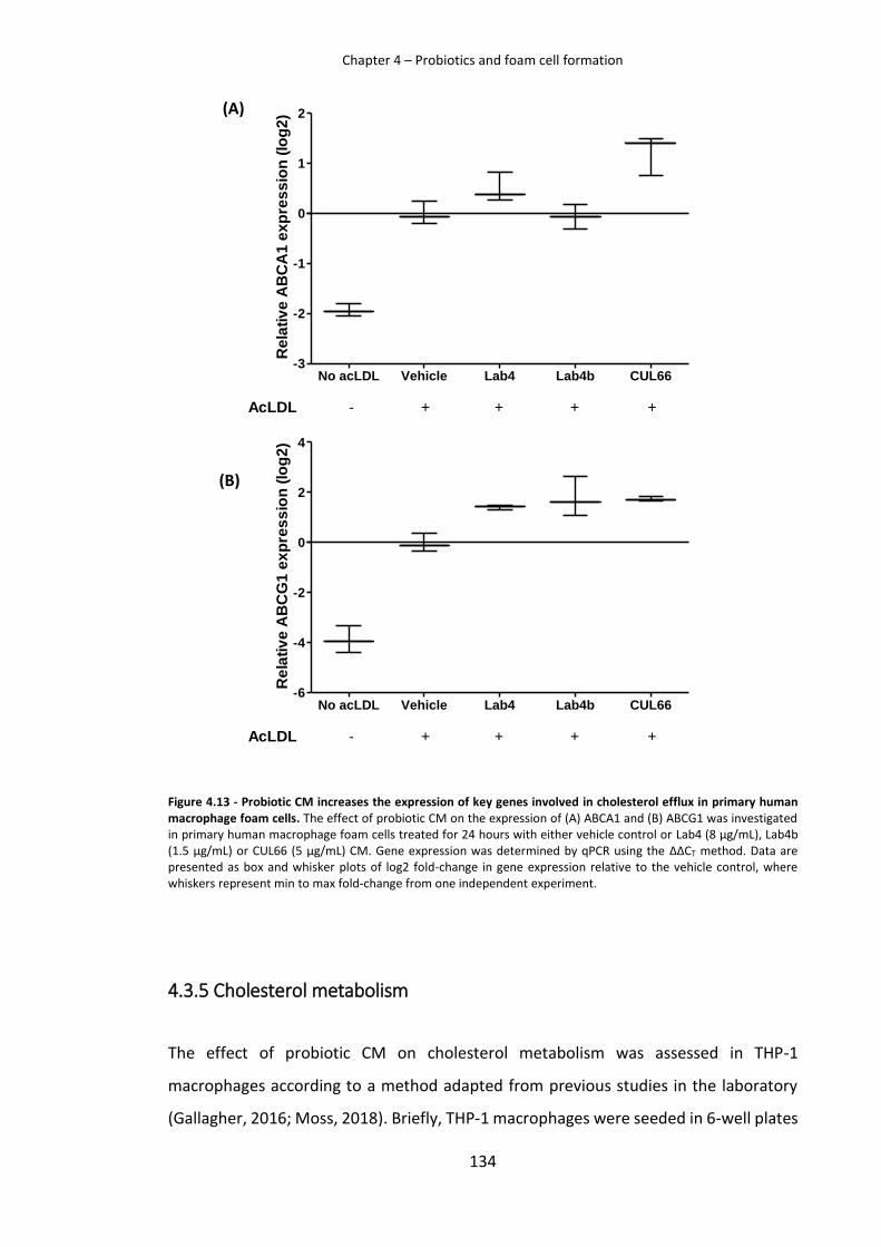
Chapter 4 – Probiotics and foam cell formation
134
Figure 4.13 - Probiotic CM increases the expression of key genes involved in cholesterol efflux in primary human macrophage foam cells. The effect of probiotic CM on the expression of (A) ABCA1 and (B) ABCG1 was investigated in primary human macrophage foam cells treated for 24 hours with either vehicle control or Lab4 (8 µg/mL), Lab4b (1.5 µg/mL) or CUL66 (5 µg/mL) CM. Gene expression was determined by qPCR using the ΔΔCT method. Data are presented as box and whisker plots of log2 fold-change in gene expression relative to the vehicle control, where whiskers represent min to max fold-change from one independent experiment.
4.3.5 Cholesterol metabolism
The effect of probiotic CM on cholesterol metabolism was assessed in THP-1
macrophages according to a method adapted from previous studies in the laboratory
(Gallagher, 2016; Moss, 2018). Briefly, THP-1 macrophages were seeded in 6-well plates
No acLDL Vehicle Lab4 Lab4b CUL66-3
-2
-1
0
1
2
AcLDL - + +++
Re
lati
ve
AB
CA
1 e
xp
res
sio
n (
log
2)
No acLDL Vehicle Lab4 Lab4b CUL66-6
-4
-2
0
2
4
AcLDL - + +++
Re
lati
ve
AB
CG
1 e
xp
res
sio
n (
log
2)
(A)
(B)

Chapter 4 – Probiotics and foam cell formation
135
at a concentration of 2 x 10⁶ cells per mL, and incubated for 24 hours with either vehicle
plus [14C] acetate (1 μCi/mL) (negative control), vehicle plus [14C] acetate (1 μCi/mL) and
25 µg/mL acLDL (vehicle control), or probiotic CM plus [14C] acetate (1 μCi/mL) and 25
µg/mL acLDL. Lipids were then extracted using Folch’s method as described in Section
2.7.4.1, and separated into major lipid classes by TLC (Section 2.7.4.2).The lipids on TLC
plates were scraped and the incorporation of [14C] acetate into individual fractions was
measured by liquid scintillation counting as described in Section 2.7.3. Total polar lipids
(PL), triglycerides (TG), free fatty acids (FFA) cholesterol esters (CE) and free cholesterol
(FC) were calculated as a percentage of total lipids. Results from one experiment are
shown in Figure 4.14. The total cellular content of PL and FC was reduced with probiotic
treatment, FFA were increased, with little change in TG components. Importantly,
treatment with CUL66 resulted in a large decrease in CE, while Lab4 and Lab4b produced
a smaller decrease in CE. However, these results are from one experiment, and
additional repeats are required before any firm conclusions can be made.
Figure 4.14 - The effect of probiotic CM on the incorporation of [14C] acetate into major lipid classes. THP-1 macrophages were treated with either vehicle plus [14C] acetate (Negative), vehicle plus [14C] acetate and acLDL (vehicle control; AcLDL) or Lab4 (8 µg/mL), Lab4b (1.5 µg/mL) or CUL66 (5 µg/mL) CM plus [14C] acetate and acLDL. Lipid classes were separated by TLC and the incorporation of [14C] acetate measured by scintillation counting. Data are presented as a percentage of the total lipids from one experiment. PL, polar lipids; FC, free cholesterol; FFA, free fatty acids; TG, triglycerides; CE, cholesterol esters.
PL FC FFA TAG CE0
20
40
60Negative
AcLDL
Lab4
Lab4b
CUL66
To
tal
lip
id (
%)
Chapter 4 – Probiotics and foam cell formation
136
4.3.6 Probiotic CM has strain-specific effects on the expression of genes
involved in intracellular cholesterol metabolism
To investigate the effect of probiotic CM on the expression of key genes involved in
cholesterol metabolism, THP-1 macrophages were treated with acLDL for 24 hours
followed by either vehicle control or probiotic CM for a further 24 hours. RNA was
extracted, reverse transcribed to cDNA and the expression of NCEH1 and ACAT1
determined by qPCR as described in Section 2.5. As shown in Figure 4.15, the expression
of NCEH1 was differentially regulated by probiotic CM with a significant 1.9-fold (p
=0.05) increase with Lab4, no significant change with Lab4b, and a significant 3.6-fold (p
<0.001) decrease with CUL66 CM. ACAT1 expression was significantly increased with
Lab4b CM by 1.7-fold (p <0.001), while treatment with Lab4 or CUL66 CM showed no
significant change in expression (Figure 4.15).

Chapter 4 – Probiotics and foam cell formation
137
Figure 4.15 – Probiotic CM has strain-specific effects on the expression of key genes involved in cholesterol metabolism. The effect of probiotic CM on the expression of key genes involved in cholesterol metabolism was investigated in THP-1 macrophage foam cells treated for 24 hours with either vehicle control or Lab4 (8 µg/mL), Lab4b (1.5 µg/mL) or CUL66 (5 µg/mL) CM. Gene expression was measured by qPCR and relative expression of (A) NCEH1 and (B) ACAT1 determined using the ΔΔCT method. Data are presented as box and whisker plots of log2 fold-change in gene expression relative to the vehicle control, where whiskers represent min to max fold-change from three independent experiments. Statistical analysis was performed using a One-way ANOVA with Dunnett (2-sided) post-hoc test where *p <0.05 and *** p <0.001.
No acLDL Vehicle Lab4 Lab4b CUL66-6
-4
-2
0
2
4
AcLDL - + +++
****
Re
lati
ve
NC
EH
1 e
xp
res
sio
n (
log
2)
No acLDL Vehicle Lab4 Lab4b CUL66-4
-2
0
2
4 ***
AcLDL - + +++
Re
lati
ve
AC
AT
1 e
xp
res
sio
n (
log
2)
(A)
(B)

Chapter 4 – Probiotics and foam cell formation
138
4.4 Discussion
Studies in the current chapter aimed to investigate the effect of probiotics on
macrophage foam cell formation, and subsequently elucidate possible mechanisms
underlying these effects. Initially, the effect of each probiotic CM on modified LDL
uptake, cholesterol efflux and intracellular cholesterol metabolism was investigated at
the cellular level. Studies then progressed to attempt to elucidate possible mechanisms
underlying these effects at the molecular level by analysis of any changes in the
expression of key genes involved in foam cell formation.
4.4.1 Modified LDL uptake
Firstly, the effect of probiotics on the uptake of modified LDL was investigated in THP-1
macrophages and HMDMs treated with Dil-oxLDL, and the uptake measured by flow
cytometry. Uptake of Dil-oxLDL was significantly attenuated by 50% or more in both
THP-1 macrophages (Figure 4.6) and HMDMs (Figure 4.7) treated with Lab4 or Lab4b
CM, while CUL66 CM treatment resulted in greater attenuation of more than 70% in
both cell model systems. Gene expression analysis in THP-1 macrophages showed that
the expression of scavenger receptors SR-A and CD36 was significantly attenuated by
each probiotic treatment, with Lab4b treatment resulting in notably greater attenuation
of SR-A and CD36 expression in addition to a significant reduction in LPL expression
(Figure 4.8). In the same series of experiments, Lab4 and CUL66 had no effect on the
expression of LPL, indicating possible strain-specific mechanisms underlying the
observed reduction in oxLDL uptake at the cellular level. In HMDMs, CD36 expression
was significantly attenuated by each probiotic CM, consistent with findings in THP-1
macrophages (Figure 4.9). However, the expression of SR-A was attenuated with Lab4
treatment only, again indicating strain-specific effects of the probiotic bacteria.
It was previously shown that each probiotic CM significantly attenuates
macropinocytosis in human macrophages (Figure 3.13). Studies presented in this
chapter demonstrated that each probiotic CM also attenuates receptor-mediated
uptake of oxLDL by macrophages, and that this effect is achieved at least in part by
attenuation of macrophage scavenger receptors at the gene expression level.

Chapter 4 – Probiotics and foam cell formation
139
Additionally, Lab4b specifically demonstrates an ability to attenuate the gene expression
of LPL in THP-1 macrophages (Figure 4.8). LPL can associate with lipoproteins, thereby
increasing the efficiency of their uptake (Serri et al., 2004). It also increases the
production of the proinflammatory cytokine TNF-α, increases monocyte adhesion to
endothelial cells and increases the proliferation of VSMCs (Serri et al., 2004). These
findings reinforce potential further beneficial effects of Lab4b treatment via attenuation
of LPL expression. Indeed, studies presented in Chapter 3 demonstrated that Lab4b
treatment attenuated both monocyte migration (Figure 3.7) and VSMC proliferation
(Figure 3.22). Additionally, it is notable that significant attenuation of CD36 expression
was consistently achieved in both THP-1 macrophages and HMDMs with all probiotic
CM treatments. This study is the first to investigate the effect of probiotic treatment on
oxLDL uptake in macrophages with no similar studies appearing in the literature to date.
However, a recent study investigating the effects of a specific strain of L. plantarum on
diet-induced obesity and insulin resistance demonstrated a reduction in hepatic
expression of CD36 in mice fed a high fat diet (Lee et al., 2018).
4.4.2 Cholesterol efflux
Overall, investigations of probiotic CM treatment on the uptake of oxLDL produced
interesting results and demonstrated beneficial effects on this key aspect of
macrophage foam cell formation. Next, the effect of probiotic treatment on the efflux
of cholesterol was investigated in THP-1 macrophages and HMDMs. In THP-1
macrophages, treatment with apoA1 resulted in a significant increase in cholesterol
efflux compared to no-apoA1 control, and treatment with all probiotic CM lead to a
further significant increase in efflux of approximately 50% (Figure 4.10). In HMDMs,
treatment with apoA1 alone did not produce a change in efflux of cholesterol; however,
Lab4 treatment resulted in a significant increase while Lab4b and CUL66 treatment
displayed a trend of increase in cholesterol efflux (Figure 4.11), which would likely reach
significance upon further experimental repeats. Further studies investigated the
possible mechanisms underlying this effect by analysis of ABCA1, ABCG1, APOE, LXRα
and LXRβ expression in THP-1 macrophage foam cells. Following treatment with Lab4 or
Lab4b CM, gene expression of ABCA1, ABCG1, LXRα and LXRβ were all significantly
increased (Figure 4.12), suggesting that the observed increase in cholesterol efflux at

Chapter 4 – Probiotics and foam cell formation
140
the cellular level may in part be due to upregulated LXR/ABC transporter pathways. Lab4
and Lab4b treatment had no effect on the expression of APOE (Figure 4.12). In contrast,
treatment with CUL66 had no effect on the expression of ABCA1, ABCG1 or LXRα, with
a decrease in the expression of LXRβ and APOE (Figure 4.12), highlighting strain-specific
mechanisms of action underlying the observed increase in cholesterol efflux at the
cellular level. The expression of ABCA1 and ABCG1 was also assessed in HMDMs, where
treatment with each CM appears to result in increased expression (Figure 4.13).
However, due to time constraints, results represent a single experimental repeat and
further experiments will be required before firm conclusions can be drawn.
Treatment with Lab4 and Lab4b CM resulted in a significant increase in cholesterol efflux
from both THP-1 macrophages and HMDMs (Figures 4.10 and 4.11), with a
corresponding increase in the expression of ABCA1, ABCG1, LXRα and LXRβ (Figure 4.12).
These results suggest that probiotic CM increases cholesterol efflux at least in part via
LXR and ABC transporter pathways. A recent study demonstrated inhibition of LXRα-
mediated ABCA1/ABCG1-dependent cholesterol efflux from THP-1 macrophages,
resulting in an increase in lesion area and plaque lipid accumulation in ApoE-/- mice (Jin
et al., 2018). In the same study, treatment with LXR activator T0901317 increased
ABCA1/ABCG1 expression both in vitro and in vivo, thereby reducing foam cell formation
and atherosclerotic lesion area in THP-1 macrophages and ApoE-/- mice respectively (Jin
et al., 2018). An earlier study showed that in cholesterol-loaded THP-1 macrophages,
ABCA1 was responsible for the efflux of cholesterol upon LXR activation, while silencing
of ABCG1 indicated that ABCG1 expression was not necessary to achieve this effect
(Larrede et al., 2009).
Therapeutic strategies aimed to stimulate RCT have targeted LXR owing to subsequent
effects in promoting cholesterol efflux from macrophages and HDL biosynthesis (Favari
et al., 2015). However, lipogenic toxicity associated with the systemic LXR agonists
T0901317 and GW3965 (Favari et al., 2015) is a major limitation of targeting LXRs.
Despite this, the intestine-specific LXR activator GW6340 has been shown to enhance
macrophage RCT while avoiding lipogenic toxicity (Yasuda et al., 2010). Increased
expression of ABC transporters A1/G1 as a result of LXR stimulation therefore represents
a possible mechanism by which probiotic treatment enhances cholesterol efflux from

Chapter 4 – Probiotics and foam cell formation
141
macrophages. Further research may aim to confirm these findings in a similar manner
to previous studies in which PPARγ activity was inhibited by siRNA knockdown,
abolishing the ABCA1 induced effect on cholesterol efflux (Sun et al., 2015). In addition
to the PPAR/LXR pathway, ABCA1 expression is known to be induced via the activation
of the Janus kinase 2 (JAK2)/signal transducer and activator of transcription 3 (STAT3)
pathway, in which activation of STAT3 increases ABCA1 expression and inhibition of JAK2
suppresses this effect (Fu et al., 2014). With a number of possible mechanisms resulting
in increased expression of ABC transporters, further investigation at the molecular and
cellular level is required to elucidate the exact pathways underlying this observed effect.
4.4.3 Intracellular cholesterol metabolism
The effect of probiotic CM on macrophage intracellular cholesterol metabolism was
investigated for the first time in human macrophages at the cellular and gene expression
levels. Formation of cholesterol esters and other major lipid classes in macrophage foam
cells was assessed by following the incorporation of [14C]acetate into major lipid classes;
a technique that has been utilised in studies of lipid metabolism for decades (Shireman
et al., 1988; Yamasaki et al., 2018). Acetate is metabolised within the macrophage and
incorporated into lipid classes, which can be separated by TLC and the radioactivity
contained within each fraction measured by scintillation counting. As in previous
studies, acLDL was used to induce foam cell formation and cholesterol ester
accumulation in THP-1 macrophages (Gallagher, 2016; Lada et al., 2003). Due to time
constraints, only one experimental repeat was carried out and additional repeats will be
required before firm conclusions can be made. Figure 4.14 shows that PL and FC
fractions were reduced with probiotic CM treatment whereas FFA content was
increased, with little change in TAG level. Notably, treatment with CUL66 resulted in a
large decrease in CE formation, while Lab4 and Lab4b produced a smaller decrease in
CE. In particular, the observed reduction in FC and CE fractions with probiotic CM
treatment is of interest, suggesting an overall reduction in the amount of cholesterol
stored within the cell. It should also be noted that the radioactivity contained within the
media was also measured from samples collected prior to processing of cells for lipid
extraction. It was noted that a higher level of [14C]acetate remained in the media when
the cells were treated with probiotic CM compared to those treated with vehicle alone

Chapter 4 – Probiotics and foam cell formation
142
(data not shown). This indicates that probiotic CM treated cells may have taken up less
acetate, coinciding with the ability of probiotic CM to attenuate lipid uptake in
macrophages. Additionally, in order to promote foam cell formation, the cells were
treated with acLDL at a concentration of 25 µg/mL which was selected based on previous
studies in the laboratory (Gallagher, 2016; Moss, 2018). One of these studies found that
treatment of THP-1 macrophages with the same concentration of acLDL failed to achieve
an accumulation of cholesterol esters, and so an alternative macrophage source was
used (murine RAW264.7; Gallagher 2016). In the literature however, THP-1
macrophages have been successfully utilised in similar studies upon treatment with
acLDL, with one study using a higher concentration of 100 µg/mL (Lada et al., 2003).
While THP-1 macrophages did accumulate cholesterol esters in this study, the level of
accumulation was low. It could therefore be suggested that this experiment may be
improved, either by using a higher concentration of acLDL or by utilising an alternative
macrophage source.
The effect of probiotic CM treatment on the expression of ACAT1 and NCEH1 was also
investigated in THP-1 macrophage foam cells and showed strain-specific results (Figure
4.15). Lab4 treatment resulted in a significant increase in the expression of NCEH1, but
had no effect on ACAT1 expression. In this case, an increase in the presence of free
cholesterol would be expected at the cellular level; however, in the current study a
decrease was observed (Figure 4.15) suggesting involvement of additional mechanisms.
Treatment with Lab4b resulted in no change in NCEH1 expression, but a significant
increase in the expression of ACAT1 (Figure 4.15). In this case an increase in the
accumulation of cholesterol esters might also be expected, contrary to the effect
observed at the cellular level (Figure 4.14), again suggesting the involvement of
additional mechanisms. With CUL66 treatment, the expression of NCEH1 was
significantly decreased with no change in ACAT1 expression (Figure 4.15), supporting an
increase in cholesterol accumulation which is again contradictory to the results obtained
at the cellular level, where CUL66 treatment resulted in a large decrease in the CE
fraction (Figure 4.14). Together, these results suggest that while the formation of
intracellular cholesterol esters may be reduced with probiotic treatment, it is unlikely
that this effect is achieved via modulation of ACAT1 and NCEH1 expression. However,

Chapter 4 – Probiotics and foam cell formation
143
the expression of ACAT1 and NCEH1 should be investigated at the protein level, possibly
by using standard Wester blotting methods, before firm conclusions can be made.
4.4.4 Summary and future work
In the previous chapter it was demonstrated that treatment with probiotic CM
attenuated macropinocytosis in human macrophages. Studies presented in the current
chapter showed that treatment of human macrophages with probiotic CM
demonstrated an attenuation of receptor-mediated uptake of modified LDL, with a
corresponding attenuation of scavenger receptors SR-A and CD36 at the gene
expression level. Furthermore, probiotic CM treatment resulted in a significant increase
in the efflux of cholesterol from the cell. However, a strain-specific mechanism was
observed at the gene expression level with Lab4 and Lab4b stimulating an increase in
the expression of ABCA1/G1 as well as LXRα and LXRβ, while CUL66 treatment resulted
in an increase in the expression of APOE. Although the investigation of intracellular
cholesterol metabolism was inconclusive, it’s likely that the observed attenuation of
modified LDL uptake together with increased efflux of cholesterol represents the
primary mechanism by which probiotic CM reduces macrophage foam cell formation.
Until recent years, monocyte-derived macrophages were thought to be the major
contributor to foam cell accumulation in the plaque. However, it has been demonstrated
that other cells types including endothelial cells and VSMCs are also able to form foam
cells and can contribute substantially to foam cell accumulation (Chistiakov et al., 2017).
In fact, it has recently been shown that VSMCs can constitute up to 50% or more of total
foam cell population in human coronary artery plaque (Dubland and Francis, 2016).
Further work may therefore investigate the effect of probiotics on foam cell formation
in additional cell types.
This is the first study to investigate the effect of Lab4, Lab4b and CUL66 on foam cell
formation in human macrophages. Given the marked implication of foam cell
accumulation in atherosclerosis, the ability of probiotics to reduce the formation of
foam cells is a highly desirable effect of an anti-atherosclerotic therapy. When
considered in conjunction with the beneficial effects observed in Chapter 3, it was
concluded that these probiotics represent a promising candidate for anti-atherosclerotic

Chapter 4 – Probiotics and foam cell formation
144
therapy. Research therefore progressed to in vivo investigation using atherosclerosis
mouse models, which are presented in the next chapter.

CHAPTER 5 –
The effects of Lab4 and CUL66 combination in vivo The effects of Lab4 and CUL66 combination on
atherosclerosis development in vivo

Chapter 5 – Probiotic effects in vivo
146
5.1 Introduction
In the studies presented in the previous chapters it was demonstrated that probiotic CM
treatment exhorted a number of beneficial effects in vitro, including the attenuation of
monocyte migration, inhibition of macrophage foam cell formation and reduced
proliferation of key cell types in atherosclerosis; highlighting their potential as an anti-
atherogenic agent. While in vitro study is very informative, it is also limited and
validation through in vivo study is essential for confirmation of findings in a more
relevant model system. In this chapter, the effect of probiotic dietary supplementation
on atherosclerotic plaque development, plaque composition and plasma lipids was
investigated in vivo using a widely employed atherosclerosis mouse model. The LDLr-/-
mutation induces hypercholesterolaemia and a plasma lipid profile resembling that of
dyslipidaemic humans (Getz and Reardon, 2016), making it a particularly suited model
for the study of effects on plasma lipids. While spontaneous plaque formation may be
observed in other atherosclerosis mouse models such as the ApoE-/- mouse, this is not a
characteristic of the LDLr-/- model where plaque and lesion development is diet induced
(Getz and Reardon, 2016). The LDLr-/- mouse was therefore selected as one of the most
extensively utilised atherosclerosis mouse models, and the most suitable model for use
in this study.
5.1.1 Study design
LDLr-/- mice (8-week old; male) were randomly assigned into two groups of 15 and
allocated to either HFD (control group) or HFD supplemented with probiotic (probiotic
group). A novel probiotic combination of Lab4 and CUL66 was used for the study as
decided by Cultech Ltd in line with company goals. Table 5.1 shows the bacteria strains
included in the Lab4/CUL66 supplement. The mice were fed HFD supplemented with
probiotic at a concentration of 1 x 10⁸ CFU/g, or 100 billion CFU/day equivalent human
dose, for a period of 12 weeks. Further details on animals and feeding are described in
Section 2.8.1. At study end point, mice were euthanised by increasing the levels of CO2
and death confirmed by absence of a pulse. Whole blood, subcutaneous and gonadal
fat, organs (heart, liver, spleen and thymus), descending aorta and rear legs were
collected and stored as described in Section 2.8.2. Fat and organ weights were recorded.

Chapter 5 – Probiotic effects in vivo
147
Although not immediately required, many of these tissues were collected and stored for
future experiments in order to minimise waste and satisfy the 3R’s of animal research
ethics.
Mouse body weights were analysed for any difference in final body weight or total
weight gained throughout the study. Organ weights were also analysed. To assess the
effect of probiotic on plaque size and lipid content, the aortic sinus was sectioned at 10
nm intervals. Sections were stained with H&E or Oil red O and the plaque area and lipid
content determined respectively. Further sections of the arterial sinus were used in the
immunohistochemical analysis of macrophage, T cell and SMC content using cell surface
marker antibodies. Blood samples were analysed and plasma cholesterol and TG content
quantified.
Table 5.1 - Bacteria species included in the Lab4/CUL66 probiotic supplement
Bacteria species
Lactobacillus acidophilus CUL21 (NCIMB 30156)
Lactobacillus acidophilus CUL60 (NCIMB 30157)
Bifidobacterium bifidum CUL20 (NCIMB 30153)
Bifidobacterium animalis subsp. lactis CUL34 (NCIMB 30172)
Lactobacillus plantarum CUL66
5.2 Experimental aim
The studies presented in this chapter aimed to assess the effect of probiotic
supplementation on atherosclerotic plaque development, plaque composition and
plasma lipid profile in LDLr-/- mice following 12 weeks feeding of HFD or HFD
supplemented with Lab4/CUL66. Detailed methods for this chapter are described in
Section 2.8, and an overview of the experimental strategy is presented in Figure 5.1.

Chapter 5 – Probiotic effects in vivo
148
Figure 5.1 - Experimental strategy used to assess the effect of probiotic treatment on plaque formation and lipid profile in vivo. HFD, high fat diet; TGs, triglycerides.

Chapter 5 – Probiotic effects in vivo
149
5.3 Results
5.3.1 Lab4/CUL66 supplementation had no effect on the body weight of LDLr-
/- mice fed a high fat diet
Mouse weights were recorded at study start, then every 2-3 days for 12 weeks with a
final measurement taken at study end point. Weights recorded at study start and end
were used to calculate total weight gained by each mouse throughout the study. No
difference in either final weight or total weight gain was observed between the control
and the probiotic groups (Figure 5.2).
Figure 5.2 - Probiotics have no effect on mouse body weights compared to the control group. Mice were fed either HFD or HFD supplemented with probiotic for 12 weeks and mouse weights recorded every 2-3 days. (A) Mouse weight measured at study end; (B) Mouse weight gain from study start to end point. Data are presented as mean ± SEM from 15 control and 15 probiotic mice. Statistical analysis was performed using a Student’s t-test.
Control Probiotic25
30
35
40
45
50
Mo
us
e w
eig
ht
(g)
Control Probiotic0
10
20
30
We
igh
t g
ain
(g
)
(A)
(B)

Chapter 5 – Probiotic effects in vivo
150
5.3.2 The effect of Lab4/CUL66 supplementation on mouse body fat and
organ weight
Subcutaneous and gonadal fat together with liver, spleen, heart and thymus were
removed and weighed at study end. Fat and organ weights are shown in Figure 5.3. No
significant difference between subcutaneous fat, gonadal fat, liver, heart or thymus
weight was observed between the control and the probiotic groups. However, spleen
weight was significantly increased in the probiotic group compared to the control (p
=0.034).
Figure 5.3 – Mouse fat and organ weight recorded at study end. Mice were fed either HFD (Control group) or HFD supplemented with probiotics (Probiotic group) for 12 weeks and fat and organ weights recorded at study end point. (A) Subcutaneous fat; (B) Gonadal fat; (C) Liver; (D) Spleen; (E) Heart; and (F) Thymus. Data are presented as mean ± SEM from 15 control and 15 probiotic mice. Statistical analysis was performed using a Student’s t-test where *p <0.05.
Control Probiotic0
1
2
3
4
Su
bc
uta
ne
ou
s f
at
(g)
Control Probiotic0
1
2
3
4
Go
na
da
l fa
t (g
)
Control Probiotic0.0
0.5
1.0
1.5
2.0
2.5
Liv
er
we
igh
t (g
)
Control Probiotic0.00
0.05
0.10
0.15
0.20*
Sp
lee
n w
eig
ht
(g)
Control Probiotic0.14
0.16
0.18
0.20
0.22
0.24
He
art
we
igh
t (g
)
Control Probiotic0.00
0.02
0.04
0.06
0.08
0.10
Th
ym
us
we
igh
t (g
)
(A) (B)
(C) (D)
(E) (F)

Chapter 5 – Probiotic effects in vivo
151
5.3.3 The effect of Lab4/CUL66 supplementation on plasma cholesterol levels
in mice fed a high fat diet
Total cholesterol, HDL, LDL and free cholesterol content of plasma was measured from
15 control and 15 probiotic mice as described in Section 2.8.3.1. Cholesterol ester level
was determined as the difference between total and free cholesterol fractions, and
ratios of LDL/HDL, TC/HDL and TC/LDL calculated. Results demonstrate a significant 32%
increase in plasma HDL cholesterol (p =0.041), in addition to a significant 19% (p =0.048)
decrease in LDL cholesterol in the probiotic group compared to the control.
Furthermore, calculation of the LDL/HDL ratio showed a significant 39% decrease (p
=0.002) in the probiotic group compared to the control (Figure 5.4). Probiotic treatment
had no significant effect on total cholesterol, free cholesterol or cholesterol esters
compared to the control. TC/HDL ratio was significantly decreased by 27% (p =0.031),
and TC/LDL ratio increased by 45% (p =0.044) in the probiotic group compared to the
control (Figure 5.5).

Chapter 5 – Probiotic effects in vivo
152
Figure 5.4 - Increased HDL and decreased LDL cholesterol levels in probiotic mice compared to the control. Plasma cholesterol levels were measured following 12 weeks feeding with either HFD (Control) or HFD supplemented with probiotic (Probiotic). (A) HDL cholesterol; (B) LDL cholesterol; and (C) LDL/HDL ratio. Data are presented as mean ± SEM from 15 control and 15 probiotic mice. Statistical analysis was performed using a Student’s t-test where *p <0.05 and **p <0.01.
Control Probiotic0
100
200
300
400 *
HD
L c
ho
les
tero
l (m
g/d
L)
Control Probiotic0
500
1000
1500*
LD
L c
ho
les
tero
l (m
g/d
L)
Control Probiotic0
5
10
15**
LD
L/H
DL
ra
tio
(A)
(B)
(C)

Chapter 5 – Probiotic effects in vivo
153
Figure 5.5 – Probiotic treatment has beneficial effects on cholesterol ratios compared to the control. Plasma cholesterol levels were measured following 12 weeks feeding with either HFD (Control) or HFD supplemented with probiotic (Probiotic). (A) Total cholesterol; (B) free cholesterol; and (C) cholesterol esters were measured and used in the calculation of (D) TC/HDL ratio and (E) TC/LDL ratio. Data are presented as mean ± SEM from 15 control and 15 probiotic mice. Statistical analysis was performed using a Student’s t-test where *p <0.05.
Control Probiotic1000
1500
2000
2500
3000
To
tal c
ho
les
tero
l (m
g/d
L)
Control Probiotic0
500
1000
1500
Fre
e c
ho
les
tero
l (m
g/d
L)
Control Probiotic0
500
1000
1500
2000
Ch
ole
ste
rol e
ste
rs (
mg
/dL
)
Control Probiotic0
5
10
15
20
25
*
TC
/HD
L r
ati
o
Control Probiotic0
1
2
3
4
5*
TC
/LD
L r
ati
o
(A) (B)
(C) (D)
(E)

Chapter 5 – Probiotic effects in vivo
154
5.3.4 Triglycerides were increased in the plasma of probiotic mice compared
to the control
Plasma TG levels were measured from 15 control and 15 probiotic treated mice as
described in section 2.8.3.2. TG levels were significantly increased in the plasma of the
probiotic group compared to the control (p =0.019) (Figure 5.6).
Figure 5.6 - Plasma triglyceride levels were increased in the probiotic compared to the control group. Plasma triglyceride levels were measured following 12 weeks feeding with either HFD (Control) or HFD supplemented with probiotic (Probiotic). Data are presented as mean ± SEM from 15 control and 15 probiotic mice. Statistical analysis was performed using a Student’s t-test where *p <0.05.
5.3.5 Plaque area and lipid content was reduced with Lab4/CUL66
supplementation
Sections of the aortic sinus were stained with H&E and Oil Red O as described in Sections
2.8.5.2 and 2.8.5.3 respectively. Figure 5.7 shows representative H&E and Oil red O
stained sections from control and probiotic groups. H&E and Oil red O stained sections
from 12 control and 11 probiotic mice were used in the measurement of plaque area
and lipid content respectively, using Image J software. Plaque area was significantly
reduced by 39% in the probiotic mice compared to the control (p =0.021) (Figure 5.8).
Lipid content determined as percentage staining of the plaque was significantly reduced
by 35% (p <0.001) compared to the control (Figure 5.9).
Control Probiotic0
200
400
600
800
*
Pla
sm
a t
rig
lyc
eri
de
s (
mg
/dL
)

Chapter 5 – Probiotic effects in vivo
155
Figure 5.7 - H&E and Oil Red O staining of the aortic sinus in the control and probiotic groups. Sections of the aortic sinus following 12 weeks feeding with HFD (Control; n=12) or HFD supplemented with probiotic (Probiotic; n=11). Images show H&E stained sections from (A) control and (B) probiotic mice and Oil red O stained sections from (C) control and (D) probiotic mice. Images taken at X40 magnification. Scale bar represents 50 µM.

Chapter 5 – Probiotic effects in vivo
156
Figure 5.8 - Plaque area was significantly reduced in probiotic mice compared to the control. Plaque area was measured in sections of the aortic sinus following 12 weeks feeding with either HFD (Control) or HFD supplemented with probiotic (Probiotic). Sections were stained with H&E and plaque area quantified using Image J software. Data are presented as mean ± SEM from 12 control and 11 probiotic mice. Statistical analysis was performed using a Student’s t-test where **p <0.01.
Figure 5.9 – Lipid content was significantly reduced in the aortic sinus of probiotic mice compared to the control. Following 12 weeks feeding with either HFD (Control) or HFD supplemented with probiotic (Probiotic), the aortic sinus were sectioned and lipid content assessed. Sections were stained with Oil red O and lipid content determined as percentage staining of the plaque using Image J software. Data are presented as mean ± SEM from 12 control and 11 probiotic mice. Statistical analysis was performed using a Student’s t-test where ***p <0.001.
Control Probiotic0
10
20
30
**
Pla
qu
e a
rea
(m
m2)
Control Probiotic0
10
20
30
40
***
Lip
id c
on
ten
t (%
)

Chapter 5 – Probiotic effects in vivo
157
5.3.6 The effects of Lab4/CUL66 supplementation on macrophage, SMC and T
cell content of plaques
Immunofluorescent staining was carried out using sections of the aortic sinus as
described in Section 2.8.5.4. Briefly, cell surface marker antibodies were used to detect
macrophages (anti-mouse MOMA-2), SMCs (anti-mouse αSMA) and T cells (anti-mouse
CD3). Images were captured at X40 magnification (X4/0.13 objective). DAPI, FITC and
AF-488 filters were applied for imaging of DAPI, AF-594 and AF-488 fluorophores
respectively. All images were captured using consistent exposure, intensity and contrast
settings for comparable downstream analyses. Intensity of staining within the plaque
was quantified using ImageJ software where raw intensity values were determined in
relation to maximum intensity values of segmented images. Representative images
obtained for control and probiotic groups are presented in Figures 5.10 – 5.12.
Macrophage, SMC and T cell content of sections obtained from control and probiotic
groups are presented as a percentage staining of plaque in Figure 5.13. Macrophage
content was significantly reduced by 39% in the probiotic group compared to the control
(p =0.024). T cell content was also reduced by 34% showing a non-significant trend of
decrease (p =0.096) in the probiotic group. Additionally, a significant 40% reduction (p
=0.024) in smooth muscle cell content was observed in the probiotic group. For each
section, the corresponding ISO control contained little to no staining (Figures 5.10 – 12),
showing that the staining was specific for the target site and not due to non-specific
background binding.

Chapter 5 – Probiotic effects in vivo
158
Figure 5.10 - Immunofluorescent staining of macrophages present in sections of the aortic sinus. Following 12 weeks feeding with either HFD (Control) or HFD supplemented with probiotic (Probiotic), the aortic sinus was sectioned and the macrophage content analysed by immunohistochemistry using cell surface marker antibodies. Sections were stained using the MOMA-2 cell surface antibody, mounted with DAPI and images captured by fluorescent microscopy. Representative images are presented for control and probiotic groups showing MOMA-2 staining (MOMA-2), MOMA-2 and DAPI staining (Composite) and IgG isotype control (ISO). Scale bars represent 50 µM. Red, MOMA-2 AF-488; Blue, DAPI.

Chapter 5 – Probiotic effects in vivo
159
Figure 5.11 - Immunofluorescent staining of T cells present in sections of the aortic sinus. Following 12 weeks feeding with either HFD (Control) or HFD supplemented with probiotic (Probiotic), the aortic sinus was sectioned and the T cell content analysed by immunohistochemistry using cell surface marker antibodies. Sections were stained using the CD3 cell surface antibody, mounted with DAPI, and the images visualised and captured by fluorescent microscopy. Representative images are presented for control and probiotic groups showing CD3 staining (CD3), CD3 and DAPI staining (Composite) and IgG isotype control (ISO). Scale bars represent 50 µM. Green, CD3 AF-594; Blue, DAPI.

Chapter 5 – Probiotic effects in vivo
160
Figure 5.12 - Immunofluorescent staining of SMCs present in sections of the aortic sinus. Following 12 weeks feeding with either HFD (Control) or HFD supplemented with probiotic (Probiotic), the aortic sinus was sectioned and the SMC content analysed by immunohistochemistry using cell surface marker antibodies. Sections were stained using the αSMA cell surface antibody, mounted with DAPI and images visualised and captured by fluorescent microscopy. Representative images are presented for control and probiotic groups showing αSMA staining (SMA), αSMA and DAPI staining (Composite) and IgG isotype control (ISO). Scale bars represent 50 µM. Green, αSMA AF-594; Blue, DAPI.

Chapter 5 – Probiotic effects in vivo
161
Figure 5.13 – Macrophage, SMC and T cell content was significantly reduced in the aortic sinus of probiotic mice compared to the control. Following 12 weeks feeding with either HFD (Control) or HFD supplemented with probiotic (Probiotic), the aortic sinus was sectioned and (A) macrophage, (B) T cell and (C) SMC content analysed by immunohistochemistry using cell surface marker antibodies. Images were captured using fluorescence microscopy. Regions of staining were quantified using ImageJ software and presented as percentage staining of plaque. Data are presented as mean ± SEM from either 4 (A), 5 (B) or 6 (C) control and 4 probiotic mice. Statistical analysis was performed using a Student’s t-test where *p <0.05.
Control Probiotic0
5
10
15
*
Ma
cro
ph
ag
es
(%
sta
inin
g)
Control Probiotic0
5
10
15
20p =0.097
T c
ell
s (
% s
tain
ing
)
Control Probiotic0
5
10
15
*
SM
Cs
(%
sta
inin
g)
(A)
(B)
(C)

Chapter 5 – Probiotic effects in vivo
162
5.4 Discussion
Following on from the promising results obtained from in vitro studies, the investigation
of the effect of probiotics on atherosclerosis was continued in vivo using atherosclerosis
mouse models. In this chapter, the effect of Lab4/CUL66 supplementation on plaque
development, plaque composition and plasma lipid profile was assessed in LDLr-/- mice
following 12 weeks feeding with either HFD or HFD supplemented with Lab4/CUL66 in
combination.
5.4.1 The effect of Lab4/CUL66 supplementation on mouse body and organ
weights
At study end, there was no significant difference in body weights in control versus
probiotic groups (Figure 5.2A). Mouse body weight at the study start was subtracted
from the weight at study end to determine total weight gain for individual mice over the
period of the study. Probiotic supplementation had no effect on the total weight gained
throughout the study (Figure 5.2B). Previous studies investigating the effects of
probiotic supplementation in a range of mouse models have shown strain-specific
effects with regards to weight gain. A previous study using ApoE-/- mice demonstrated
highly strain-specific effects on weight gain upon administration of different strains of L.
reuteri. A significant reduction in weight gain was observed with L. reuteri ATCC
treatment while conversely, treatment with L. reuteri L6798 resulted in a significant
increase in weight gain (Fåk and Bäckhed, 2012). Another recent study demonstrated
that treatment with different probiotic compositions resulted in strain-specific variation,
with L. casei IMV B-7280 (separately) and a composition of B. animalis VKL/B. animalis
VKB/L. casei IMV B-7280 having greater effect in decreasing body weight in high-calorie-
induced obesity (Bubnov et al., 2017). Recently, an acute study of Lab4/CUL66
supplementation in C57BL/6J mice reported suppression of diet-induced weight gain
after a period of 2-weeks (Michael et al., 2017). Further studies in C57BL/6 mice have
reported suppression of HFD-induced weight gain with L. acidophilus strain NS1 (Song
et al., 2015) or L. acidophilus AD031 (Li et al., 2016) supplementation. However, a
comparative meta-analysis of the effect of Lactobacillus species on weight gain reported

Chapter 5 – Probiotic effects in vivo
163
an overall significant weight gain in both humans and animal models administered L.
acidophilus in a large number of studies (Million et al., 2012). The same meta-analysis
reported inconsistent findings of both weight loss and gain with L. plantarum (Million et
al., 2012), supporting the notion that the effect of probiotic consumption on body
weight is not only species-specific but highly strain-specific.
In addition to body weight, organ weight was also recorded and analysed at study end.
No difference was observed in heart, liver or thymus weights; however, spleen weight
was significantly increased in the probiotic group compared to the control (Figure 5.3).
Increased spleen size has been associated with reduced atherosclerosis and plaque
development in LDLr-/- mice (Whitman et al., 2002). However, the mechanism of this
association is unclear. In ApoE-/- mice fed an atherogenic diet, splenectomy has been
shown to greatly accelerate atherosclerosis compared to a control group subjected to
sham operation (Caligiuri et al., 2002), implying an atheroprotective effect of the spleen
(Witztum, 2002). More recently, Grasset et al. (2015) investigated an athero-protective
role of the spleen via induction of a protective B cell response, highlighting the
importance of the spleen in atherosclerosis-associated immunity. The B cell response
involves the induction of atheroprotective antibodies against modified lipoproteins
including oxLDL by antigens presenting oxidation-specific epitopes, such as
phosphorylcholine (PC), leading to plaque reduction (Grasset et al., 2015). This
mechanism in which oxidation-specific epitopes give rise to an anti-PC response and
reduction of atherosclerosis has been demonstrated in a number of studies (Binder et
al., 2003; Ketelhuth and Hansson, 2016; Kimura et al., 2015). Together, these results
suggest that the observed increase in spleen weight in the probiotic group may be
associated with an anti-atherosclerotic response and may be a result of an enhanced B-
cell response and induced production of atheroprotective anti-PC antibodies.
5.4.2 Lab4/CUL66 supplementation has beneficial effects on plasma lipid
profile
Elevated blood cholesterol is an important atherosclerosis risk factor and plasma lipids
are routinely used as risk markers of atherosclerosis disease progression (Huang et al.,
2013). As discussed previously, among the beneficial effects attributed to probiotic

Chapter 5 – Probiotic effects in vivo
164
supplementation, their reported ability to lower plasma cholesterol levels is of particular
interest in the prevention and management of CVD. In order to assess the effect of
probiotic supplementation on plasma lipid profile, total cholesterol, HDL, LDL, free
cholesterol and triglyceride content of plasma was measured from 15 control and 15
probiotic mice following 12 weeks feeding with HFD or HFD supplemented with
Lab4/CUL66. Cholesterol ester level was determined as the difference between total
and free cholesterol fractions, and ratios of LDL/HDL, TC/HDL and TC/LDL calculated.
In this study, total cholesterol was largely increased across both groups, an effect
commonly associated with the LDLr-/- mouse model (Getz and Reardon, 2016). Probiotic
supplementation had no significant effect on total cholesterol levels compared to the
control (Figure 5.5A). Few similar studies have investigated the effect of probiotic
supplementation on plasma lipid profiles of LDLr-/- mice. However, there are studies
reporting varying effects in alternative animal models. Almost a decade ago it was
demonstrated that probiotic supplementation had the potential to positively affect
plasma lipid profiles in hypercholesterolemic rabbits, although no change in total
cholesterol was observed (Cavallini et al., 2009). A later study investigating the effect of
a number of Lactobacillus and Bifidobacterium strains in obese mice reported that L.
casei IMV B-7280 was particularly effective at lowering total plasma cholesterol, in
comparison to B. animalis VKB/B. animalis VKL treatment which showed no effect
(Bubnov et al., 2017). Moreover, a meta-analysis of randomised clinical trials
investigating the effect of probiotics on blood lipid concentrations revealed a highly
strain-specific effect on total cholesterol, with strains of L. acidophilus and E. faecium
showing a net reduction in total cholesterol levels while L. plantarum and L. helveticus
showed no significant net change (Cho and Kim, 2015). It is therefore not surprising that
the effects of probiotics on plasma lipids and total cholesterol level also displays strain-
specific variation. In the current study, probiotic supplementation had no effect on total
cholesterol; however, it is more relevant to consider HDL and LDL fractions individually
and in relation to each other as an HDL/LDL ratio.
Circulating levels of HDL cholesterol are inversely associated with atherosclerosis
disease risk and is classified as a negative risk factor for CVD (Bandeali and Farmer,
2012). In this study, supplementation with Lab4/CUL66 resulted in a significant 32%

Chapter 5 – Probiotic effects in vivo
165
increase in plasma HDL levels compared to the control (Figure 5.4A). Furthermore, the
ratio of total cholesterol to HDL cholesterol (TC/HDL) was significantly decreased in the
probiotic group compared to the control (Figure 5.5D) demonstrating that HDL levels
were increased in relation to the total cholesterol level. In a meta-analysis of
randomised controlled trials investigating the effect of probiotics on blood lipid
concentrations, Cho and Kim (2015) reported a significant net increase in HDL
cholesterol in studies utilising strains of L. acidophilus or a combination of L.
acidophilus/B. lactis. In contrast, they also reported a significant net decrease with L.
plantarum and L. helveticus, in addition to no net change in HDL levels with E. faecium
(Cho and Kim, 2015). Interestingly, Lab4 contains two different species of L. acidophilus,
suggesting that these bacteria may be responsible for the increase in HDL cholesterol
observed in this study. Although multifactorial, the anti-atherogenic association with
increased HDL levels is considered to be primarily due to its role in RCT where it is the
major cholesterol carrier (Bandeali and Farmer, 2012). As discussed previously in
Chapters 1 and 3, cholesterol is passed from lipid laden foam cells to HDL via a number
of mechanisms but is largely achieved via interaction with the membrane transporter
ABCA1. As demonstrated in Chapter 4, efflux of cholesterol from macrophage foam cells
is enhanced in the presence of Lab4 and CUL66 (Figure 4.10), which is accompanied by
an increase in the expression of ABCA1 at the mRNA level (Figure 4.12A). Together, these
results suggest that Lab4/CUL66 may exert anti-atherosclerotic effects by enhancing
ABCA1-associated efflux of cholesterol from lipid laden foam cells to increased levels of
circulating HDL cholesterol, thereby permitting enhanced transport of cholesterol to the
hepatic biliary excretion system.
Mutations in the LDLr gene disrupt the receptor-mediated clearance of LDL and is the
most common cause of the autosomal dominant disorder, FH (Linton et al., 2000). FH is
associated with highly elevated levels of LDL cholesterol and consequently premature
atherosclerotic disease. A recent consensus statement from the European
Atherosclerosis Society Consensus Panel reviewed evidence from epidemiological,
genetic and clinical studies, concluding that LDL burden is a central determining factor
in the initiation and progression of atherosclerosis (Ference et al., 2017). It was also
concluded that the lower the level of LDL cholesterol that could be achieved by an agent,
the greater the clinical benefit (Ference et al., 2017). Indeed, the association between

Chapter 5 – Probiotic effects in vivo
166
LDL cholesterol lowering and reduced atherosclerosis disease risk has long been
established with human studies showing significant attenuation of plaque progression
with lipid-lowering treatment (Pinkosky et al., 2016; Shimada and Cannon, 2015; Shin et
al., 2017). LDLr-/- mice used in this study naturally develop high levels of LDL cholesterol;
however, supplementation with Lab4/CUL66 resulted in a significant 19% reduction LDL
cholesterol levels (Figure 5.4B). Furthermore, the ratio of total cholesterol to LDL
cholesterol (TC/LDL) was significantly increased in the probiotic group compared to the
control (Figure 5.5D) demonstrating that LDL levels were increased in relation to the
total cholesterol levels. In a meta-analysis of the effect of probiotics on plasma lipids,
Cho and Kim (2015) reported a significant decrease in LDL cholesterol as demonstrated
in a total of 20 out of 23 randomised controlled trials (Cho and Kim, 2015), suggesting
that while strain-specific variation exists, the ability to lower LDL cholesterol is a
common feature across probiotic strains. Furthermore, many human studies have
demonstrated a reduction in LDL levels and generally improved lipid profile with
probiotic supplementation as outlined in a recent literature review (Sharma et al., 2018).
So far in this chapter, it has been shown that supplementation with Lab4/CUL66 can
positively modulate plasma lipids, by increasing levels of HDL and decreasing levels of
LDL cholesterol, both independently and in relation to total cholesterol levels. Although
this information is very useful when assessing cardiovascular and atherosclerosis disease
risk, even more relevant is the ratio of LDL to HDL cholesterol. In this study, the LDL/HDL
cholesterol ratio was calculated and found to be significantly reduced by 27% in the
probiotic group compared to the control (Figure 5.4C). This is a highly encouraging result
suggesting that Lab4/CUL66 supplementation may exert athero-protective effects via
the modulation of plasma lipid profile.
In addition to cholesterol levels, plasma TG was also analysed as a further important
indicator of atherosclerosis risk. Raised levels of circulating TG is a marker for several
atherogenic lipoproteins, including TG-rich lipoproteins, VLDL remnants and
chylomicron remnants, all of which are capable of enhancing atherosclerosis (Moss,
2018; Talayero and Sacks, 2011). In fact, while elevated LDL cholesterol is considered a
major risk factor for cardiovascular and atherosclerosis disease, hypertriglyceridemia
has been considered as an independent risk factor (Sarwar et al., 2007) so much that

Chapter 5 – Probiotic effects in vivo
167
when LDL cholesterol is maintained at goal levels, patients with hypertriglyceridemia
have been reported to remain at significant risk (Talayero and Sacks, 2011). In this study,
Lab4/CUL66 supplementation was unable to lower plasma TG, in fact TG levels were
significantly increased in the probiotic group compared to the control (Figure 5.6). Upon
review of the numerous animal and human studies carried out, it is clear that the effect
of probiotic supplementation on plasma TG level is also subject to species-specific
variations. While a number of in vivo studies have shown a significant decrease in TG
levels following probiotic supplementation (Fhoula et al., 2018; Huang et al., 2013;
Kumar et al., 2011; Wang et al., 2012), others have shown no effect (Greany et al., 2008;
Razmpoosh et al., 2019; Sharma et al., 2018). In a randomised double-blind controlled
study, patients with hypertriglyceridemia showed a significant reduction in TG following
12 weeks supplementation with L. curvatus HY7601 and L. plantarum KY1032 (Ahn et
al., 2015). Moreover, the effect was more pronounced in patients with higher fasting TG
at study start. These findings suggests the possibility that results are dependent not only
on the strain and the dose of the probiotic administered but also on the existing level of
hypertriglyceridemia in the subject.
5.4.3 Lab4/CUL66 supplementation reduces plaque size and lipid content in
LDLr-/- mice
This study aimed to elucidate the effect of Lab4/CUL66 supplementation on
atherosclerotic plaque formation. Sections of the aortic sinus were stained with H&E in
order to quantify plaque size and observe morphology, while staining with Oil red O was
carried out to determine plaque lipid content. Both plaque area and lipid content was
found to be significantly reduced by 39% and 35% respectively in the probiotic group
compared to the control (Figures 5.8 and 5.9). As can be seen in the representative
images shown in Figure 5.7, mice in the control group presented with large plaques and
distortion of the aorta. Although this specific probiotic mix has not previously been
investigated in LDLr-/- mice, these results do correlate with previous studies investigating
the effect of various probiotics on atherosclerotic plaque formation (Chan et al., 2016b).
As discussed previously, the VSL#3 probiotic consortium has been investigated for its
effects on plaque formation in ApoE-/- mice, demonstrating significant reduction in

Chapter 5 – Probiotic effects in vivo
168
atherosclerotic lesion development (Chan, 2012; Chan et al., 2016b). Similar to
Lab4/CUL66 combination, VSL#3 contains a mix of different Lactobacillus and
Bifidobacterium strains (Kühbacher et al., 2006) with both consortiums containing L.
plantarum and L. acidophilus. In contrast, supplementation with L. reuteri strains ATCC
4659, DSM or L6798 was unsuccessful in reducing lesion development in a similar model
using ApoE-/- mice. Although the number of studies investigating atherosclerotic plaque
development with probiotic supplementation are relatively few, it is not surprising that
again the effects of probiotic supplementation have been shown to be strain-specific.
5.4.4 The effect of Lab4/CUL66 supplementation on plaque composition
The presence of immune cells, particularly macrophages and T cells within plaques is
associated with plaque destabilisation and rupture (Shaikh et al., 2012). The vulnerable
plaque is characterised by a thin fibrous cap, along with large accumulation of T cells
and macrophages in relation to reduced numbers of SMCs, and a large collagen-poor,
lipid-rich necrotic core (Newby et al., 2009). In the current study, the effect of Lab4/CUL6
supplementation on macrophage, T cell and SMC composition of plaque lesions was
assessed by immunohistochemistry. Sections of the aortic sinus were stained with anti-
mouse cell surface marker primary antibodies, followed by fluorophore-conjugated
secondary antibodies. Antibodies raised against MOMA-2, αSMA and CD3 have been
used previously to successfully target macrophages, SMCs and T cells in the plaque
respectively (Dong et al., 2016; Gajda et al., 2008; Xiong et al., 2017). Details of
antibodies used are given in Section 2.8.5. To control for non-specific staining, an isotype
control was included for each section. The isotype is serum IgG correlating to the species
in which the primary antibodies were raised; any secondary antibody binding is
therefore non-specific. Figures 5.10 – 5.12 include representative ISO images which
were used during analysis to deduct any background staining for individual sections.
Images were captured by fluorescence microscopy using consistent exposure, intensity
and contrast settings for comparable downstream analyses. DAPI, FITC and AF-488
filters were applied for detection of DAPI (blue), AF-594 (green) and AF-488 (red)
fluorescence respectively. Macrophage, SMC and T cell content was quantified using
ImageJ software where images were first segmented, then percentage staining of
plaque was determined by raw intensity values in relation to maximum intensity values.

Chapter 5 – Probiotic effects in vivo
169
Intensity analysis was chosen in preference to simple pixel quantification as a more
accurate method of quantifying cellular presence in immunofluorescent images. Results
show that macrophage and SMC content of plaques was significantly reduced in the
probiotic group compared to the control, while T cell content also displays a trend of
reduction (Figure 5.10 – 5.12). For each section, the corresponding ISO control contained
little to no staining, showing that staining is specific for the target site and not due to
non-specific background binding.
MOMA-2 staining showed a large accumulation of macrophages in sections from the
control group, particularly within the area of plaque where staining is more intense
(Figure 5.10). Interestingly in Figure 5.10, the image representing the control group is
showing an absence of intense staining within one of the plaque lesions, suggesting that
a necrotic core may be present here. This result correlates with previous chapters in
which it was shown that probiotic CM attenuates monocyte recruitment (Figure 3.9) and
monocyte (Figure 3.20) and macrophage (Figure 3.21) proliferation in vitro. Macrophage
foam cells are the predominant source of extracellular proteases, particularly MMPs,
which directly degrade ECM components leading to plaque destabilisation (Newby et
al., 2009). Additionally, regions of dense accumulation of macrophages as seen in the
control group are prone to more frequent apoptosis and formation of a necrotic core
(De Meyer et al., 2012), further contributing to plaque vulnerability and rupture.
Prevention or depletion of plaque macrophage accumulation is considered athero-
protective; therefore, these results suggest that Lab4/CUL66 supplementation leads to
the attenuation of macrophage accumulation and consequently possible reduction of
plaque destabilisation.
After macrophages, T cells represent the second largest population of immune cells in
the atherosclerotic plaque (Shaikh et al., 2012). Although different classes of T cells can
exert different effects within an atherosclerotic lesion, in general T cells are considered
to exacerbate atherosclerosis, particularly in advanced lesions where T cells and B cells
are largely responsible for driving inflammation and plaque progression (Tse et al.,
2013). CD3, previously known as T3, is ubiquitously presented by all T cell classes. In this
study, staining of CD3 shows accumulation of T cells within the area of the plaque,
particularly visible in the control group compared to probiotic group where plaque area

Chapter 5 – Probiotic effects in vivo
170
is relatively small (Figure 5.11). Overall, reduced staining was observed in the probiotic
group (Figure 5.13B) and although this result did not reach significance, it did show a
trend of reduction with a p-value of 0.097. As mentioned previously, the CD3 protein is
presented by all T cells, therefore this result gives a general assessment of T cell content.
As the most abundant T cell subset present in the atherosclerotic plaque, it is likely that
the pro-inflammatory helper Th1 lymphocyte (Shaikh et al., 2012) represents the
majority of CD3 staining observed in this study. Future work may aim to elucidate the
exact contribution of individual T cell subtypes beginning with T helper (CD4+) and T
cytotoxic (CD8+) cells, thereby providing additional detail for more informed
conclusions.
VSMC proliferation and infiltration into an atherosclerotic lesion can be either pro- or
anti-atherogenic depending on the stage of disease progression. It is generally accepted
that VSMC accumulation confers stability to advanced atherosclerotic plaques via
formation of a protective fibrous cap (Zernecke, 2017), which consists primarily of
VSMC-derived collagen, elastin, proteoglycans and ECM. Apoptosis and senescence of
VSMCs results in loss of collagen and ECM proteins, promoting atherosclerosis
progression with degradation of the fibrous cap and subsequent plaque vulnerability
and rupture (Fernandez-Hernando et al., 2009; Wang et al., 2015a). In the current study,
VSMC presence in sections of the aortic sinus was quantified by immunohistochemistry
using antibodies targeting the SMC protein αSMA. Results show a significant 40%
reduction in VSMC in the probiotic group compared to the control (Figure 5.13C). In
Figure 5.12, differential accumulation of VSMCs can be seen; in the probiotic group, a
small amount of staining is visible within the plaque while more intense staining is visible
within the vessel wall; however, in the control group intense staining can be seen
particularly around the outside of the plaque. This pattern of staining suggests that
VSMCs may have formed a thick protective cap over the plaque, representative of a
more advanced but stable lesion. The amount of staining present in the probiotic group
was significantly reduced compared to the control group (Figure 5.13C). However, this
may simply be due to the presence of less advanced plaque in sections from the
probiotic group. For more conclusive results a larger sample size would be required.

Chapter 5 – Probiotic effects in vivo
171
5.4.5 Summary and future work
Results in this chapter have laid a strong foundation in the investigation of the effect of
Lab4/CUL66 supplementation in atherosclerosis development in vivo. Firstly, it was
shown that probiotic supplementation had no effect on mouse body or organ weight,
with the exception of spleen weight which was significantly increased in the probiotic
group compared to the control, possibly via an enhanced athero-protective B cell
response. Next, it was shown that Lab4/CUL66 supplementation had the ability to
beneficially modulate plasma lipid profile with mice in the probiotic group
demonstrating increased HDL levels, decreased LDL levels and beneficially altered
TC/HDL, TC/LDL and HDL/LDL cholesterol ratios. Drawing on results from previous
chapters, it is possible that Lab4/CUL66 may exert anti-atherosclerotic effects by
enhancing ABCA1-associated efflux of cholesterol from lipid laden foam cells to
increased levels of circulating HDL cholesterol, thereby permitting enhanced transport
of cholesterol to the hepatic biliary excretion system. This beneficial effect of
Lab4/CUL66 on plasma lipid profile is a highly encouraging result given the clinical
importance of plasma lipids in atherosclerosis risk assessment and disease monitoring.
Furthermore, it was demonstrated that Lab4/CUL66 supplementation led to a large
reduction in plaque area in the probiotic group compared to the control; in fact, sections
from the control group featured large plaques with extensive lipid accumulation and
distortion of the aorta. Distortion of the aorta was not observed in any of the sections
obtained from the probiotic group. Finally, investigation of plaque composition revealed
a significant reduction in macrophage accumulation, another exciting finding correlating
with reduced atherosclerosis disease progression in the probiotic group. In addition to
macrophages, accumulation of T cells also displayed a trend of reduction in the probiotic
group. Since the most likely abundance of T cells is the pro-inflammatory helper Th1
cells (Ilhan and Kalkanli, 2015), it can be suggested that this result shows anti-
atherogenic potential; however, more specific investigation of the contribution of
individual T cell subsets is required. While VSMC staining is reduced in the probiotic
group, the distribution of the staining is particularly notable, with the position of staining
in the control group being suggestive of the formation of a protective VSMC-dense
fibrous cap. While these results are encouraging, the availability of suitable sections for

Chapter 5 – Probiotic effects in vivo
172
immunohistochemical analysis of plaque composition was limited. For more conclusive
interpretation of the results obtained by immunofluorescent staining, a larger sample
size would be required.
Following on from the results presented in this chapter, investigation continued to
attempt to elucidate the mechanisms underlying these observed anti-atherosclerotic
effects. In the studies presented in the next chapter, stored tissues were utilised to
explore bone marrow stem cell populations and expression of atherosclerosis-
associated genes using qPCR microarrays.

CHAPTER 6 –
Mechanisms underlying the effect of Lab4/CUL66 supplementation in atherosclerosis
Mechanisms underlying the athero-protective effects of
Lab4/CUL66 supplementation in atherosclerosis

Chapter 6 – Further effects in vivo
174
6.1 Introduction
The effect of probiotics on atherosclerosis was investigated in LDLr-/- mice fed for 12
weeks with HFD or HFD supplemented with Lab4/CUL66 probiotic combination. Data
presented in Chapter 5 showed that Lab4/CUL66 supplementation attenuated
atherosclerotic plaque formation by 39% compared to the control, and beneficially
modulated plasma lipid profiles resulting in increased HDL with decreased LDL
cholesterol levels. Additionally, plaque composition was shown to be altered, notably
showing a reduction in plaque macrophage content as well reduced SMC and T cell
content. In light of these important results, the aims of the studies presented in this
chapter was to further investigate the effects of Lab4/CUL66 supplementation and to
elucidate the mechanisms underlying the results presented in the previous chapter.
In the studies presented in Chapter 3, the CM from either Lab4 or CUL66 was shown to
significantly affect gene expression, in particular genes involved in foam cell formation.
It’s possible that the reduced plaque formation observed in vivo is in part due to similar
alterations in gene expression. However, given the full range of the effects observed, it
is likely that there are multiple contributary mechanisms. Due to the high number of
genes involved in atherosclerosis, it was decided to screen a large number of genes
simultaneously using a qPCR microarray method. The liver plays a central role in
metabolism and the production of plasma proteins, all of which contribute to
atherogenesis, and is a common source of mRNA in the study of atherosclerosis gene
expression (Pan et al., 2018; Recinos et al., 2004; States et al., 2012). Liver gene
expression was therefore analysed following 12 weeks feeding with HFD (control group)
or HFD supplemented with Lab4/CUL66 (probiotic group), using Qiagen Atherosclerosis
RT2 Profiler PCR Array plates. The PCR microarray plates contained a large panel of 84
atherosclerosis-associated genes implicated in the control of inflammation, cell
adhesion, lipid metabolism, apoptosis and more. A table of the 84 genes included in the
qPCR microarray plate is shown in Appendix 1.
Alteration in bone marrow cell populations has been reported to play an important role
in hypercholesterolemia and atherosclerosis disease progression (Lang and Cimato,
2014; Ma and Feng, 2016). Therefore, in addition to the analysis of gene expression, the

Chapter 6 – Further effects in vivo
175
effect of Lab4/CUL66 supplementation on key bone marrow cell populations was
investigated in LDLr-/- mice following 12 weeks feeding with HFD (control) or HFD
supplemented with Lab4/CUL66 probiotic combination. Bone marrow populations of
stem cells, progenitor cells and lineage differentiated cells were analysed using a
method modified from one previously used in the laboratory (Moss, 2018). A simplified
representation of bone marrow cell population lineage is shown in Figure 6.1.
Haematopoietic stem/progenitor cells (HSPCs) are responsible for the generation of all
blood cells. Within the HSPC population, haematopoietic stem cells (HSCs) generate
multipotent progenitors (MPPs) which subsequently generate progenitor cells, lineage-
specific precursors and finally fully differentiated blood cells (Gao et al., 2014).
Figure 6.1 - Simplified representation of bone marrow cell populations. Abbreviations: HSC, haematopoietic stem cell; MPP, multipotent progenitor; HPC, haematopoietic progenitor cell; CMP, common myeloid progenitor; CLP, common lymphoid progenitor; MEP, megakaryocyte-erythroid progenitor; GMP, granulocyte-macrophage progenitor; MDSC, myeloid-derived suppressor cell.
Bone marrow contains heterogeneous populations of cells and so analysis of
populations of interest requires a method of discerning one cell type from another. In
the current study, this was achieved using a technique known as immunophenotyping
in which heterogenous cell populations are stained with antibodies targeting cell-
Chapter 6 – Further effects in vivo
176
specific surface antigens. Antibodies are typically conjugated to fluorophores and are
detected using flow cytometric analysis, allowing the identification of the presence and
proportion of cell populations of interest. (Sykora and Reschke, 2019) Staining of a
mixture of cell types with a cocktail of antibodies targeting one or more cell surface
antigens, known as a markers or identifiers, can therefore be used to discern one cell
type from another. In this study fluorochrome-conjugated antibodies targeting cell
specific surface markers were used to analyse key cell populations in the bone marrow
using flow cytometry. A list of all cell types with their corresponding identifier is given in
Table 2.8.
6.2 Experimental aim
The studies presented in this chapter aimed to assess the effect of Lab4/CUL66
supplementation on atherosclerosis-associated gene expression and bone marrow cell
populations in LDLr-/- mice following 12 weeks feeding of HFD or HFD supplemented with
Lab4/CUL66. Detailed methods for this chapter are described in Section 2.8.6 and 2.8.4
respectively. An overview of the experimental strategy is presented in Figure 6.2. Gating
strategy applied in the analysis of bone marrow cell populations is outlined in Figure 6.3.

Chapter 6 – Further effects in vivo
177
Figure 6.2 - Experimental strategy for the analysis of liver gene expression and bone marrow cell populations.

Chapter 6 – Further effects in vivo
178
Figure 6.3 – Representative flow plots outlining the gating strategy utilised in the analysis of haematopoietic cell populations by flow cytometry. In flow cytometry, forward scatter (FSC) provides a measurement of cell size by determining the amount of light which passes around it. Side scatter (SSC-A) provides a measurement of granularity by determining the amount of light which is reflected by particles within the cells. Application of filters and gating is used to isolate cell populations of interest. WBC, white blood cells; LIN, lineage; CLP, common lymphoid progenitor; HPC, haematopoietic progenitor cell; MPP, multipotent progenitor; HSC, haematopoietic stem cell; MDSC, myeloid-derived suppressor cells; MAC, macrophages; Sca1, stem cells antigen 1; FITC, Fluorescein isothiocyanate; PE, Phycoerythrin; APC, allophycocyanin.

Chapter 6 – Further effects in vivo
179
6.3 Results
6.3.1 The effects of Lab4/CUL66 supplementation on the expression of key
genes involved in atherosclerosis
To investigate the expression of key genes involved in atherosclerosis, samples of liver
which were snap frozen at study end point were selected at random to a total of 8 each
from control and probiotic groups. RNA was extracted from the liver tissue, reverse
transcribed and amplified by qPCR using Atherosclerosis RT2 Profiler PCR Array plates as
described in Section 2.8.6. For all test wells, CT values greater than 35 were not included
in downstream analysis due to the increased risk of non-specific amplification. In
addition to test wells, each microarray plate included the following controls:
• Genomic DNA control (GDC): A CT value of ≤35 was indicative of the presence of
genomic DNA contamination;
• Reverse transcription control (RTC): Included to assess the efficiency of the RT
reaction. Average CT values of the positive PCR control (PPC; see below) were
deducted from the RTC, and a value greater than 5 was indicative of impurities
and reduced RT reaction efficiency;
• PPC: Included to assess the efficiency of the PCR reaction, where average CT
values of approximately 20 ± 2 cycles was considered efficient. A large variation
in PPC values was considered indicative of the presence of PCR amplification
inhibitors, while CT values consistently greater than 22 indicated inefficient PCR
reaction conditions.
In addition to GDC, RTC and PPC controls, the CT values of housekeeping genes (Actb,
B2m, Gapdh, Gusb, Hsp90ab1) were checked for stability within 2 cycles. Melting curve
analysis was carried out on individual samples where a single peak representing
amplification of a single product was acceptable. Fold-change in gene expression was
calculated using the ΔΔCT method. A full list of expression changes for genes included in
the Atherosclerosis RT2 Profiler PCR Array is shown in Table 6.1. Genes for which changes
in expression reached significance are listed in Table 6.2 in addition to fold-change
increase/decrease and p values. Of the 84 genes tested, 63 achieved a minimum of 10%

Chapter 6 – Further effects in vivo
180
change in expression, while 19 achieved significant change and a further 51 showed a
trend in change in expression (p <0.100). A volcano plot showing global changes in gene
expression was produced using R software (Figure 6.4).

Chapter 6 – Further effects in vivo
181
Table 6.1 - Changes in the expression of each gene tested using the Atherosclerosis RT2 Profiler PCR Arrays
Gene GenBank ID Fold-change P value Change
Abca1 NM_013454 1.27 0.129 Increase
Ace NM_009598 1.19 0.403 Decrease
Apoa1 NM_009692 1.06 0.756 Increase
Apob NM_009693 1.18 0.048 Decrease
Apoe NM_009696 1.05 0.372 Increase
Bax NM_007527 1.41 0.073 Decrease
Bcl2 NM_009741 1.23 0.275 Increase
Bcl2a1a NM_009742 1.19 0.704 Decrease
Bcl2l1 NM_009743 1.47 0.074 Decrease
Bid NM_007544 1.18 0.427 Decrease
Birc3 NM_007464 1.03 0.572 Increase
Ccl2 NM_011333 2.08 0.111 Decrease
Ccl5 NM_013653 1.79 0.179 Decrease
Ccr1 NM_009912 1.70 0.003 Decrease
Ccr2 NM_009915 1.63 0.006 Decrease
Cd44 NM_009851 1.93 0.178 Decrease
Cdh5 NM_009868 1.08 0.980 Decrease
Cflar NM_009805 1.23 0.017 Decrease
Col3a1 NM_009930 2.38 0.253 Decrease
Csf2 NM_009969 1.79 0.317 Decrease
Ctgf NM_010217 1.25 0.814 Decrease
Cxcl1 NM_008176 3.09 0.087 Decrease
Eln NM_007925 1.07 0.684 Increase
Eng NM_007932 1.10 0.311 Increase
Fabp3 NM_010174 1.21 0.241 Increase
Fas NM_007987 1.19 0.315 Decrease
Fga NM_010196 1.12 0.542 Decrease
Fgb NM_181849 1.30 0.211 Increase

Chapter 6 – Further effects in vivo
182
Gene GenBank ID Fold-change P value Change
Fgf2 NM_008006 1.11 0.807 Decrease
Fn1 NM_010233 1.30 0.124 Increase
Hbegf NM_010415 1.13 0.024 Decrease
Icam1 NM_010493 1.34 0.025 Decrease
Ifng NM_008337 2.40 0.000 Decrease
Il1a NM_010554 1.47 0.110 Decrease
Il1b NM_008361 1.03 0.477 Increase
Il1r1 NM_008362 1.17 0.007 Decrease
Il1r2 NM_010555 1.30 0.815 Decrease
Il2 NM_008366 1.76 0.048 Decrease
Il3 NM_010556 1.12 0.154 Increase
Il4 NM_021283 1.21 0.183 Decrease
Il5 NM_010558 1.29 0.435 Decrease
Itga2 NM_008396 1.72 0.000 Decrease
Itga5 NM_010577 1.67 0.620 Decrease
Itgax NM_021334 1.23 0.246 Decrease
Itgb2 NM_008404 1.32 0.001 Decrease
Kdr NM_010612 1.05 0.462 Increase
Klf2 NM_008452 1.29 0.695 Increase
Lama1 NM_008480 1.28 0.171 Decrease
Ldlr NM_010700 1.00 0.713 Increase
Lif NM_008501 1.03 0.540 Increase
Lpl NM_008509 1.21 0.161 Decrease
Lypla1 NM_008866 2.52 0.014 Decrease
Mmp1a NM_032006 1.80 0.000 Decrease
Mmp3 NM_010809 1.50 0.018 Decrease
Msr1 NM_031195 1.40 0.351 Decrease
Nfkb1 NM_008689 1.09 0.310 Increase
Npy NM_023456 1.86 0.282 Decrease
Nr1h3 NM_013839 1.05 0.395 Increase

Chapter 6 – Further effects in vivo
183
Gene GenBank ID Fold-change P value Change
Pdgfa NM_008808 1.31 0.019 Increase
Pdgfb NM_011057 1.07 0.678 Decrease
Pdgfrb NM_008809 1.06 0.900 Increase
Plin2 NM_007408 1.12 0.187 Decrease
Ppara NM_011144 1.09 0.074 Increase
Ppard NM_011145 1.12 0.153 Increase
Pparg NM_011146 1.40 0.046 Decrease
Ptgs1 NM_008969 1.04 0.455 Increase
Rxra NM_011305 1.32 0.239 Increase
Sele NM_011345 1.56 0.005 Decrease
Sell NM_011346 1.42 0.414 Decrease
Selp NM_011347 1.17 0.171 Decrease
Selplg NM_009151 1.12 0.554 Decrease
Serpinb2 NM_011111 1.12 0.553 Decrease
Serpine1 NM_008871 2.27 0.001 Decrease
Sod1 NM_011434 1.02 0.639 Increase
Spp1 NM_009263 1.00 0.515 Increase
Tgfb1 NM_011577 1.09 0.266 Increase
Tgfb2 NM_009367 1.17 0.818 Decrease
Thbs4 NM_011582 1.09 0.385 Increase
Tnc NM_011607 1.16 0.349 Decrease
Tnf NM_013693 1.28 0.035 Decrease
Tnfaip3 NM_009397 2.26 0.098 Decrease
Vcam1 NM_011693 1.37 0.082 Decrease
Vegfa NM_009505 1.51 0.126 Decrease
Vwf NM_011708 1.13 0.336 Increase
Gene expression was assessed in the livers of mice following 12 weeks feeding with HFD or HFD
supplemented with Lab4/CUL66. Table shows gene, GenBank reference, fold-change in
expression, p value and increase/decrease in gene expression. A list of full gene names is
included in Appendix 1. Statistical analysis was performed using an unpaired Student’s t-test.

Chapter 6 – Further effects in vivo
184
Table 6.2 – Atherosclerosis-associated genes showing significant change, or trend of change in expression.
Gene GenBank ID Fold-change P value Change
Apob NM_009693 1.18 0.048 Decrease
Bax NM_007527 1.41 0.073* Decrease
Bcl2l1 NM_009743 1.47 0.074* Decrease
Ccr1 NM_009912 1.70 0.003 Decrease
Ccr2 NM_009915 1.63 0.006 Decrease
Cflar NM_009805 1.23 0.017 Decrease
Cxcl1 NM_008176 3.09 0.087* Decrease
Hbegf NM_010415 1.13 0.024 Decrease
Icam1 NM_010493 1.34 0.025 Decrease
Ifng NM_008337 2.40 0.000 Decrease
Il1r1 NM_008362 1.17 0.007 Decrease
Il2 NM_008366 1.76 0.048 Decrease
Itga2 NM_008396 1.72 0.000 Decrease
Itgb2 NM_008404 1.32 0.001 Decrease
Lypla1 NM_008866 2.52 0.014 Decrease
Mmp1a NM_032006 1.80 0.000 Decrease
Mmp3 NM_010809 1.50 0.018 Decrease
Pdgfa NM_008808 1.31 0.019 Increase
Ppara NM_011144 1.09 0.074* Increase
Pparg NM_011146 1.40 0.046 Decrease
Sele NM_011345 1.56 0.005 Decrease
Serpine1 NM_008871 2.27 0.001 Decrease
Tnf NM_013693 1.28 0.035 Decrease
Tnfaip3 NM_009397 2.26 0.098* Decrease
Vcam1 NM_011693 1.37 0.082* Decrease
Table shows gene, GenBank reference, fold-change in expression, p value and increase/decrease
in gene expression. Statistical analysis was performed using an unpaired Student’s t-test. * Trend
towards significance where p <0.100.

Chapter 6 – Further effects in vivo
185
Figure 6.4 - Volcano plot showing changes in gene expression with Lab4/CUL66 supplementation compared to the control. The effect of Lab4/CUL66 on atherosclerosis-associated gene expression was assessed in liver samples from 8 control and 8 probiotic mice following 12 weeks feeding with either HFD (control group) or HFD supplemented with Lab4/CUL66. Data are presented as mean fold-change in gene expression in the probiotic group compared to the control. The log2 fold-change in gene expression is represented on the x-axis. The y-axis shows the -log10 of the p value. A p value of 0.05 (dashed line) and fold change of ±10% (solid lines) are indicated. A black triangle indicates genes showing significant changes in expression, and a black square indicates genes showing a trend towards significance (p <0.100)

Chapter 6 – Further effects in vivo
186
The 84 atherosclerosis-associated genes included in the Atherosclerosis RT2 Profiler PCR
Array were arranged into groups of related genes based on their function. Cluster
analysis was performed and a heatmap produced for each group of related genes using
the online tool Morpheus, where colour and intensity gives visual representation of the
changes in gene expression for all genes (Figure 6.5).
Groups of related genes that show significant changes in expression are presented
graphically in Figure 6.6. Some genes are pleiotropic and are therefore included in more
than one related group. Fold-change increase/decrease together with the p-value for
each gene is given in Table 6.2. Of the genes involved in the inflammatory response
(Figure 6.6A) the expression of all of Ccr1, Ccr2, Infg, Il2, Itgb2, Pparg and Tnf was
significantly reduced in the probiotic group compared to the control. Similarly, the
expression of genes involved in lipid transport and metabolism (Abob, Lypla1 and Pparg)
was decreased (Figure 6.6B). Expression of a number of genes involved in cell adhesion
were also significantly reduced (Icam, Itga1, Itgb2, Sele) (Figure 6.6C). For genes
involved in cell growth and proliferation, the expression of Hbegf, Il2 and Ifng was
significantly decreased while that for Pdgfa was increased (Figure 6.6D). Expression of
genes involved in transcription regulation (Pparg, Ifng) and apoptosis (Cflar, Ifng) were
also significantly decreased (Figures 6.6E and 6.6F). Finally, the largest group of genes
that showed significant change in expression code for extracellular matrix molecules
(Figure 6.6G), where the expression of Serpine1, Mmp1a, Mmp3, Hbegf, Infg, Il1r1, Il2,
Itga2, Itgb2 and Sele decreased and that for Pdgfa increased.

Chapter 6 – Further effects in vivo
187
Figure 6.5 – Heatmaps showing a visual representation of fold-change in gene expression with Lab4/CUL66 supplementation. The effect of Lab4/CUL66 on atherosclerosis-associated gene expression was assessed in liver samples from 8 control and 8 probiotic treated mice following 12 weeks feeding with either HFD (control group) or HFD supplemented with Lab4/CUL66. Each heatmap consists of a family of related genes where (A) Cell growth and proliferation; (B) Apoptosis; (C) Stress responses; (D) Blood coagulation and circulation; (E) Cell adhesion molecules; (F) Extracellular matrix molecules; (G) Transcriptional regulation; and (H) Lipid transport and metabolism. Box colour and intensity represents fold-change in gene expression as depicted by the scale. Clustering displayed on the left of each heatmap is according to similarity of expression pattern between genes in each related group. Fold-change in gene expression was determined using the ΔΔCT method. Cluster analysis was performed and heatmaps produced using Morpheus software. A list of full gene names is included in Appendix 1.

Chapter 6 – Further effects in vivo
188
Figure 6.6 – Related genes show significant change in expression with Lab4/CUL66 supplementation compared to the control. The effect of Lab4/CUL66 on atherosclerosis-associated gene expression was assessed in liver samples from 8 control and 8 probiotic mice following 12 weeks feeding with either HFD (control group) or HFD supplemented with Lab4/CUL66 (probiotic group). Genes showing significant change in expression were sorted into related groups involved in (A) inflammatory responses, (B) lipid transport and metabolism, (C) cell adhesion, (D) cell growth and proliferation, (E) transcriptional regulation, (F) apoptosis and (G) extracellular matrix molecules. Data are presented as box and whisker plots of log2 fold-change in gene expression in the probiotic group relative to the control, where whiskers represent min to max fold-change. Statistical analysis was performed using an unpaired Student’s t-test where *p <0.05, **p <0.01, ***p <0.001.
Inflammatory response
Ccr1 Ccr2 Ifng Il2 Itgb2 Pparg Tnf-4
-2
0
2
**** * **** ***
Re
lati
ve
ge
ne
ex
pre
ss
ion
(lo
g2
)
Lipid transport & metabolism
Apob Lypla1 Pparg-4
-2
0
2
* * *
Re
lati
ve
ge
ne
ex
pre
ss
ion
(lo
g2
)
Cell adhesion molecules
Icam Itga2 Itgb2 Sele-3
-2
-1
0
1
2
* ***** **
Re
lati
ve
ge
ne
ex
pre
ss
ion
(lo
g2
)
Cell growth & proliferation
Hbegf Il2 Pdgfa Ifng-4
-2
0
2
* **** *
Re
lati
ve
ge
ne
ex
pre
ss
ion
(lo
g2
)
Transcriptional regulation
Pparg Ifng-3
-2
-1
0
1
2
* ***
Re
lati
ve
ge
ne
ex
pre
ss
ion
(lo
g2
)
Apoptosis
Cflar Ifng-3
-2
-1
0
1
* ***
Re
lati
ve
ge
ne
ex
pre
ss
ion
(lo
g2
)
Extracellular matrix molecules
Hbegf Ifng Il1r1 Il2 Itga2 Itgb2 Pdgfa Sele Serpine1 Mmp1a Mmp3-4
-2
0
2
* *** * * ********* ** *** *
Re
lati
ve
ge
ne
ex
pre
ss
ion
(lo
g2
)
(A)
(C)
(B)
(D)
(E) (F)
(G)

Chapter 6 – Further effects in vivo
189
6.3.2 The effect of Lab4/CUL66 supplementation on key bone marrow cell
populations
Following 12 weeks feeding with HFD (control) or HFD supplemented with Lab4/CUL66,
the bone marrow was extracted and cell populations analysed by immunophenotyping
as described in Section 2.8.4. Stem cell populations including HSCs, MPPs and HPC I and
II, were identified using antibodies targeting cell surface glycoproteins CD150 and CD48.
CD150 is also known as signalling lymphocyte activation molecule (SLAM) and hence
these populations are referred to as SLAM. For CLP, CD127 was used as an identifier. For
lineage positive differentiated cells, B cells were identified via the B220 marker, T cells
via the CD3 marker and macrophages, granulocytes and MDSCs via GR1 and Mac1
markers. The composition of antibody cocktails used in the preparation of SLAM,
progenitor and lineage positive cell populations is detailed in Table 2.7. Cell populations
of interest were identified using the gating strategy shown in Figure 6.3, and analysed
using FlowJo software. Total WBC counts were first analysed, and no significant change
in WBC count between the control and the probiotic groups was confirmed (Figure 6.7).
The proportion of each cell population was then determined as a frequency of the WBC
count for each individual sample.
Results for stem cell populations are shown in Figure 6.8. No significant change was seen
in the proportion of MPP or HPC I cell populations between the control and the probiotic
groups. In contrast, the proportions of HPC II (p <0.048) and HSC (p <0.034) populations
were significantly increased in the probiotic group compared to control. For progenitor
CLP, populations showed no significant change between the control and probiotic
groups (Figure 6.9). In the category of lineage positive differentiated cells, B cells showed
no significant change between control and probiotic groups (Figure 6.10). However, the
proportion of T cells was significantly reduced (p <0.005), and both MDSC (p <0.041) and
macrophage (p <0.046) populations were significantly reduced in the probiotic group
compared to control.

Chapter 6 – Further effects in vivo
190
Figure 6.7 – Treatment with Lab4/CUL66 had no significant effect on white blood cell counts in the bone marrow. Immunophenotyping of bone marrow stem cell populations from LDLr-/- mice was performed following 12 weeks feeding with either HFD (Control) or HFD supplemented with Lab4/CUL66 (Probiotic). Graph represents the total white blood cell counts from the Control and Probiotic groups. Data are presented as mean ± SEM from 15 control and 15 probiotic mice. Statistical analysis was performed using an unpaired Student’s t-test.
Control Probiotic0
2.0×10⁵
4.0×10⁵
6.0×10⁵
8.0×10⁵
1.0×10⁶
Wh
ite
blo
od
ce
ll c
ou
nt

Chapter 6 – Further effects in vivo
191
Figure 6.8 – The effect of Lab4/CUL66 supplementation on bone marrow stem cell populations. Immunophenotyping of bone marrow stem cell populations from LDLr-/- mice was performed following 12 weeks feeding with either HFD (Control) or HFD supplemented with Lab4/CUL66 (Probiotic). (A) Representative flow plots showing stem cell populations in control and probiotic groups. Gating shows position of populations representing HPC I (CD48+CD150-), HPC II (CD48+CD150+), MPP (CD48-CD150-) and HSC (CD48-CD150+) cells. Graphs represent total (B) HPC I; (C) HPC II; (D) MPP; and (E) HSC populations as a frequency of total white blood cells. Data are presented as mean ± SEM from 15 control and 15 probiotic mice. Statistical analysis was performed using an unpaired Student’s t-test where *p <0.05.
Control Probiotic0.0
0.5
1.0
1.5
HP
C I (
%)
Control Probiotic0.00
0.05
0.10
0.15
0.20
0.25 *
HP
C II (%
)
Control Probiotic0.00
0.05
0.10
0.15
MP
P (
%)
Control Probiotic0.00
0.05
0.10
0.15 *
HS
C (
%)
(A)
(B) (C)
(D) (E)

Chapter 6 – Further effects in vivo
192
Figure 6.9 - The effect of Lab4/CUL66 supplementation on bone marrow CLP progenitor cell populations. Immunophenotyping of bone marrow stem cell populations from LDLr-/- mice was performed following 12 weeks feeding with either HFD (Control) or HFD supplemented with Lab4/CUL66 (Probiotic). (A) Representative flow plots showing CLP progenitor cell populations in control and probiotic groups. (B) Graph represents total CLP populations as a frequency of total white blood cells. Data are presented as mean ± SEM from 15 control and 15 probiotic mice. Statistical analysis was performed using an unpaired Student’s t-test where *p <0.05, **p <0.01.
Control Probiotic0.00
0.05
0.10
0.15
CL
P (
%)
(A)
(B)

Chapter 6 – Further effects in vivo
193
Figure 6.10 - The effect of Lab4/CUL66 supplementation on bone marrow lineage differentiated cell populations. Immunophenotyping of bone marrow stem cell populations from LDLr-/- mice was performed following 12 weeks feeding with either HFD (control) or HFD supplemented with Lab4/CUL66 (probiotic). (A) Representative flow plots showing lineage positive cell populations in control and probiotic groups. Gating shows position of populations representing MDSC (GR-1+CD11b+), monocytes/macrophages (MAC; GR-1-CD11b+), (B) T Cells (CD3+B220-) and B Cells (CD3-B220+) cells. Graphs represent total (C) MDSC; (D) Monocyte/macrophage; (E) T cell; and (F) B cell populations as a frequency of total white blood cells. Data are presented as mean ± SEM from 15 control and 15 probiotic mice. Statistical analysis was performed using an unpaired Student’s t-test where *p <0.05, **p <0.01.
Control Probiotic0
5
10
15 *
MD
SC
(%
)
Control Probiotic0
1
2
3
4
5 *
Ma
cro
ph
ag
es
(%
)
Control Probiotic0.0
0.5
1.0
1.5
2.0 **
T c
ells
(%
)
Control Probiotic0
2
4
6
8
B c
ells
(%
)
(A)
(B)
(C) (D)
(E) (F)

Chapter 6 – Further effects in vivo
194
6.4 Discussion
The current study aimed to investigate the mechanisms underlying the findings from
initial in vivo investigations which were presented in Chapter 5. The hepatic gene
expression of 84 key genes involved in atherosclerosis was investigated following 12
weeks feeding with HFD (control) or HFD supplemented with Lab4/CUL66. Gene
expression was determined by qPCR using the Atherosclerosis RT2 Profiler PCR Array,
and fold-change in gene expression was calculated using the ΔΔCT method. Of the 84
genes tested, a total of 63 achieved a minimum of 10% change in expression, with 19 of
these genes achieving significance and a further 6 displaying a trend towards
significance. Genes showing a significant change in expression were further analysed
and grouped into related sets as shown in Figure 6.6.
6.4.1 Lab4/CUL66 supplementation significantly attenuated the expression of
key genes involved in cell adhesion
The expression of a number of genes involved in cell adhesion was significantly
decreased in the probiotic group compared to the control (Figure 6.6C). Icam and Sele
encode cell adhesion molecules ICAM1 and E-selectin respectively, both expressed at
high levels on the surface of activated endothelial cells in response to pro-inflammatory
cytokines, including TNF-α, and are involved in the induction of monocyte recruitment
and migration (Hoffman et al., 2018). Mice deficient in cell adhesion molecules have
been shown to display reduced atherosclerotic lesion size (Collins et al., 2000; Galkina
and Ley, 2007). Moreover, in LDLr-/- mice, the inhibition of the expression of cytokines
and adhesion molecules including ICAM1, has been shown to attenuate atherosclerosis
progression independent of changes in lipid profile (Srivastava et al., 2015). In the
studies presented in Chapter 3, it was shown that probiotic CM attenuated monocyte
migration in vitro (Figure 3.9). It could therefore be suggested that Lab4/CUL66
supplementation attenuates monocyte recruitment in vivo. Interestingly, in the current
study the expression of IFN-γ and TNF-α was also significantly decreased in the probiotic
group compared to the control (Figure 6.6A). IFN-γ potentiates the expression of
adhesion molecules induced by proinflammatory cytokines including TNF-α, thereby

Chapter 6 – Further effects in vivo
195
promoting monocyte recruitment (Voloshyna et al., 2014). It could therefore be
suggested that attenuation of the expression of pro-atherogenic cytokines including IFN-
γ and TNF-α, and subsequently adhesion molecules such as ICAM-1, represents one
possible mechanism underlying the probiotic-mediated attenuation of monocyte
recruitment and migration. As the TNF-α-induced expression of adhesion molecules
ICAM-1 and E-selectin requires the transcription factor NF-κB (Baker et al., 2011), it
would be useful to investigate the possible involvement of this pathway in future work.
Itga2 and Itgb2 encode integrin subunits α2 and β2 respectively. Integrins are
heterodimer transmembrane proteins consisting of two subunits, alpha and beta, each
of which binds to ECM molecules (Adorno-Cruz and Liu, 2019). Integrin subunit α2 forms
a heterodimer with the β1 subunit to form a collagen-binding integrin (Table 6.2), while
subunit β2 is part of the leukocyte-specific β2 integrin involved in the recognition of cell
adhesion molecules on the activated endothelium (Adorno-Cruz and Liu, 2019). β2
integrins recognise adhesion molecules ICAM1 and E-selectin, and following activation
by CC chemokine ligand (CCL) 2 binding, mediate firm adhesion of monocytes to the
endothelial layer (Liu et al., 2008). Previous studies have demonstrated the ability of β2
integrins to modulate the initiation and progression of atherosclerosis in LDLr-/- mice
(Merched et al., 2010).
Ccr1 and Ccr2 encode CC chemokine receptors 1 (CCR1) and 2 (CCR2). CCR1 and CCR2
are chemokine receptors for CCL1/CCL5 and CCL2 (MCP-1) respectively, and are involved
in the recruitment of leukocytes across the arterial wall (Boring et al., 1998). Studies
investigating the effect of CCR1 on atherosclerosis progression have shown conflicting
results though many concur that the receptor is athero-protective (Boring et al., 1998;
Braunersreuther et al., 2007; Wan and Murphy, 2013). However, this has not been the
case for CCR2 which is known to be pro-atherogenic and pro-inflammatory in nature
(Wan and Murphy, 2013). A number of studies in both ApoE-/- and LDLr-/- mice have
demonstrated that CCR2 deficiency leads to attenuation of atherosclerosis (Dawson et
al., 1999; Gu et al., 1998; Guo et al., 2003). In the current study, the expression of genes
encoding chemokine receptors CCR1 and CCR2 in addition to integrin subunits was
significantly decreased, along with genes encoding cell adhesion molecules ICAM1 and

Chapter 6 – Further effects in vivo
196
E-selectin. These findings further support the role of probiotic supplementation in the
attenuation of monocyte recruitment and migration.
6.4.2 The effect of Lab4/CUL66 supplementation on the expression of genes
involved in the regulation of the extracellular matrix
ECM is the major constituent of vascular walls. Genes associated with ECM molecules
represent the largest group of associated genes showing significant changes in
expression (Figure 6.6G). As discussed previously, the ECM is essential for plaque
stability and the formation of a fibrous cap; when components of the ECM are
compromised, destabilisation of the fibrous cap leads to plaque rupture and subsequent
clinical complications. Of the genes presented in Figure 6.6G, Serpine1, Mmp1a and
Mmp3 are discussed here in addition to the association of Ifng and Il2 with ECM
molecules. Itga2, Itgb2 and Sele encode cell adhesion molecules and have been
discussed previously in Section 6.4.1, while Hbegf, Il1r1 and Pdgfa are included in Table
6.3.
Serpine1 encodes plasminogen activator inhibitor 1 (PAI-1), a glycoprotein from the
serpin family and the primary inhibitor of plasminogen activators in vascular fibrinolysis
(Rouch et al., 2015). PAI-1 is known to be involved in the pathogenesis of many
conditions, including vascular thrombosis and atherosclerosis (Rouch et al., 2015); in
fact, and increased level of PAI-1 in the blood has been highlighted as an independent
risk factor for atherosclerosis progression and plaque rupture (Lijnen, 2005; Milenkovic
et al., 2017). Upregulation of PAI-1 expression has been demonstrated in human
atherosclerotic lesions (Milenkovic et al., 2017; Schneiderman et al., 1992), while studies
in ApoE-/- mice have reported an athero-protective effect of PAI-1 inhibition or
deficiency (Eitzman et al., 2000). Additionally, PAI-1 expression is induced by a range of
cytokines including TNF-α.
Mmp1a and Mmp3 encode matrix metalloprotease (MMP)-1a and MMP-3 respectively.
Metalloproteinases are a family of enzymes responsible for the degradation of ECM
components and are classified into several groups, including collagenases (MMP-1, -8,
and -13), gelatinases (MMP-2 and -9), and stromelysins (MMP-3, -10, and -11) (Ye,

Chapter 6 – Further effects in vivo
197
2015). Degradative action of MMP enzymes on ECM components breaks down the
fibrous cap leading to plaque instability and rupture. Macrophage-derived foam cells
accumulated within atherosclerotic lesions have been identified as a major source of
MMPs including MMP-3 (Magdalena et al., 2006). The correlation between MMP activity
and atherosclerosis is widely known; increased expression of MMPs is associated with
enhanced development of atherosclerotic lesions and increased SMC migration
(Magdalena et al., 2006; Prescott et al., 1999) and many studies have demonstrated
attenuation of plaque progression with deficiency of various MMPs, including MMP-10
(Purroy et al., 2018), MMP-9 (Jin et al., 2015) and MMP-3 (Beaudeux et al., 2003). In the
current study, the expression of MMP-1 and MMP-3 was downregulated in the livers of
probiotic supplemented mice compared to the control. These results are encouraging
and suggest that Lab4/CUL66 supplementation exerts positive effects via potential
downregulation of the expression of MMP enzymes. However, studies investigating the
effects of MMP -1 and -3 is limited while alternative MMPs dominate the literature. For
future studies, investigation of the expression of a wider range of MMPs at the gene and
protein level may be carried out at alternative sites including the aorta, providing a more
comprehensive view of the effect of probiotic supplementation on MMP activity.
Furthermore, the activity of MMP enzymes is counterbalanced by the presence of tissue
inhibitors of MMPs (TIMPs), which are also expressed in atherosclerotic lesions
(Heeneman et al., 2003). A better indication of potential levels of ECM degradation
would be provided if further studies aimed to assess the expression of TIMPs in addition
to MMPs.
Ifng and Il2 encode pro-inflammatory cytokines IFN-γ and IL-2 respectfully and have
been discussed previously in relation to the atherosclerotic inflammatory response in
Section 6.4.1. They are considered here in relation to the ECM. Both IFN-γ and IL-2
induce the release of MMPs, thereby promoting the degradation of the ECM (Wang et
al., 2015b) resulting in reduced plaque stability with eventual destabilisation and
rupture (Voloshyna et al., 2014; Weng et al., 2014). In the current study, the expression
of IFN-γ and IL-2, in addition to MMP-1 and -3, was significantly reduced with
Lab4/CUL66 supplementation compared to the control, suggesting a potential athero-
protective role of Lab4/CUL66 in maintaining plaque stability.
Chapter 6 – Further effects in vivo
198
6.4.3 Lipid transport and metabolism
Apob encodes the protein apoB, a key component of atherogenic lipoproteins as
discussed in Chapter 1. ApoB is an important predictor of atherosclerosis and a known
positive correlation exists between apoB levels and atherosclerotic conditions, with
decreased apoB expression reported to have a protective effect against
hypercholesterolemia (Dhingra and Bansal, 2005). In fact, it has been proposed that
apoB is a superior indicator of atherosclerosis risk (Harper and Jacobson, 2010), as
reported in a number of human trials where apoB was shown to be a more accurate
predictor of disease risk than the traditionally used LDL or non-HDL cholesterol levels
(Holme et al., 2008; McQueen et al., 2008; Walldius et al., 2001). This is particularly the
case for a subgroup of at-risk patients who exhibit low or normal levels of LDL
cholesterol despite high numbers of lipoprotein particles (Harper and Jacobson, 2010).
In the current study, the expression of Apob was attenuated by probiotic
supplementation at the gene expression level (Figure 6.6), correlating with results in
Chapter 5 where probiotic supplementation was shown to significantly reduce plasma
levels of LDL cholesterol (Figure 5.4). However, decreased expression of apoB at the
mRNA level cannot be directly associated with decreased levels of plasma LDL
cholesterol, and plasma apoB protein levels would need to be measured before firm
conclusions can be drawn. In future work it would be useful to analyse plasma levels of
apoB in the probiotic group compared to the control, and to consider this in relation to
the levels of non-HDL cholesterol.
In addition to Apob, expression of Lypla1 and Pparg was also significantly decreased in
the probiotic group compared to control. Lysophospholipase A1 (LYPLA1) is a cytosolic
serine hydrolase and is partially responsible for the metabolism of lysophospholipids
(Wepy et al., 2019). Under conditions of oxidative stress, up to half of the
phosphatidylcholine held within an LDL particle is converted to lysophosphatidylcholine
(LPC) (Wepy et al., 2019). LPC is a major bioactive phospholipid component of oxLDL
which is hydrolysed by lysophospholipases to form lysophosphatiditic acid (LPA) (Bao et
al., 2018). LPC levels in the circulation are associated with the development of
atherosclerotic plaques and endothelial cell dysfunction, and the LPC content of
circulating modified LDL has been shown to be increased in atherosclerosis patients (Bao

Chapter 6 – Further effects in vivo
199
et al., 2018; Zakiev et al., 2016). LPA is also pro-atherogenic and is abundant in the
necrotic core of atherosclerotic plaques. In the current study, Lypla1 expression was
significantly decreased in the probiotic group compared to control. This result indicates
the possibility of a corresponding reduction in the levels of LPC and LPA; however,
further investigations would be required to investigate this proposition.
Pparg, encoding PPAR-γ, has been discussed in Section 6.4.1 with regards to its role in
inflammatory responses. PPAR-γ is highly expressed in macrophage foam cells within
atherosclerotic lesions (Li et al., 2000) where activation has been reported to promote
monocyte/macrophage differentiation and induce the expression of CD36 (Tontonoz et
al., 1998). Endogenous PPAR-γ ligands have been identified within serum oxLDL, and it
has been suggested that atherogenic lipoproteins including oxLDL may induce their own
uptake via PPAR-γ-induced upregulation of CD36, thereby promoting foam cell
formation (Rosen and Spiegelman, 2000). Despite these findings, PPAR-γ agonists have
been shown to inhibit atherosclerosis development in animal (Li et al., 2000) and human
studies (Ivanova et al., 2017; Skochko and Kaidashev, 2017). PPAR-γ agonists are used
as therapeutic agents in a number of conditions associated with cardiovascular risk
including diabetes mellitus and hypertension (Ivanova et al., 2015, 2017); however, it is
possible that some of the anti-atherogenic effect of these agents are achieved via non-
PPAR-γ-related mechanisms (i.e. non-genomic effects). In the current study, gene
expression of PPAR-γ was significantly decreased in the probiotic group compared to
control. However, the effects of PPAR-γ are complex and further investigation is
required to ascertain the effect of attenuated PPAR-γ expression in atherogenesis.
6.4.4 Lab4/CUL66 supplementation attenuates gene expression of key pro-
inflammatory cytokines
Investigation of inflammatory gene expression in vitro displayed few notable effects in
THP-1 macrophages treated with probiotic CM compared to control (Figure 3.27). In
contrast, the expression of genes involved in the inflammatory response in vivo suggests
a beneficial effect of Lab4/CUL66 supplementation, with genes encoding key pro-
inflammatory molecules displaying decreased expression. Ifng, Tnf and Il2 encode key
pro-inflammatory and pro-atherogenic cytokines IFN-γ, TNF-α and IL-2 respectively,

Chapter 6 – Further effects in vivo
200
each of which is integral to atherosclerosis development and the expression of all was
significantly reduced with probiotic supplementation (Figure 6.6A).
As discussed in Chapter 1, IFN-γ is a T cell and macrophage-derived proinflammatory
cytokine known to be highly expressed in atherosclerotic lesions and deficiency of this
cytokine has been shown to attenuate atherosclerosis in both ApoE-/- and LDLr-/- mouse
models (Koga et al., 2007; Niwa et al., 2004). IFN-γ is a master regulator involved in all
stages of atherosclerosis initiation, development and progression, including monocyte
recruitment, foam cell formation, plaque formation and rupture, as well as the Th1-
associated immune responses (Voloshyna et al., 2014). Additionally, IFN-γ promotes
apoptotic cell death in human macrophages (Inagaki et al., 2002) and VSMCs (Geng et
al., 1996), thereby contributing to the formation of a necrotic core and decreased plaque
stability. An increase in IFN-γ has also been shown to significantly increase the T cell
content of atherosclerotic lesions (Voloshyna et al., 2014; Whitman et al., 2000). With
respect to the effect of IFN-γ on the T cell content of the plaque, this observed decrease
in Ifng expression correlates with results in Chapter 5 where it was shown that
Lab4/CUL66 supplementation led to a reduction in the T cell content of plaque in the
probiotic group compared to the control (Figure 5.13). The Janus kinase (JAK)-Signal
Transducers and Activators of Transcription (STAT) pathway represents the primary IFN-
γ signalling pathway (Moss and Ramji, 2015). As IFN-γ represents such a promising target
for anti-atherosclerotic therapeutics, future work should involve more in-depth
investigation of Lab4/CUL66 supplementation on IFN-γ-associated responses.
Activation of macrophages by IFN-γ leads to the production of further proinflammatory
cytokines, including TNF-α (Voloshyna et al., 2014), the gene expression of which was
also shown to be decreased with Lab4/CUL66 supplementation. Like IFN-γ, TNF-α is a
key cytokine implicated in many stages of atherosclerosis exhibiting strong pro-
atherogenic effects, and attenuation of TNF-α action has been shown to diminish
atherosclerosis development (Branen et al., 2004; Ohta et al., 2005). Additionally, Il2
encodes IL-2, a pro-atherogenic cytokine involved in cell-mediated immune responses
and is primarily produced by activated T cells. Suppression of IL-2 action has
demonstrated anti-atherogenic outcomes, with one study reporting enhanced
atherosclerosis with IL-2 injection in addition to a reduction in atherosclerosis following

Chapter 6 – Further effects in vivo
201
injection with anti-IL-2 antibodies (Ramji and Davies, 2015; Upadhya et al., 2004). In the
current study, IL-2 mRNA expression was significantly decreased, presenting a further
anti-atherogenic effect of Lab4/CUL66 supplementation.
Table 6.3 - Further genes showing significant changes in expression and their role in atherosclerosis
Gene Pathway Role in atherosclerosis
Cflar Apoptosis
CASP8 and FADD-like apoptosis regulator CFLAR. Overexpression attenuates metabolic disorders associated with cardiovascular risk (Wang et al., 2017b)
Hbegf Cell growth & proliferation
Heparin-binding EGF-like growth factor (HBEGF), a potent mitogen and chemoattractant for VSMCs (Nakata et al. 1996). HBEGF expression positively correlates with cardiovascular risk (Kim et al., 2017)
Il1r1 Extracellular matrix molecules
IL-1 receptor 1 (IL-1R1), involved in vascular remodelling. IL-1R1 deficiency reduces atherosclerosis in Il1r1-/-/ApoE-/- mice (Alexander et al., 2012)
Itga2 Cell adhesion molecules
Integrin subunit α2 forms collagen-binding integrin, involved in monocyte attachment to collagen IV (Wang et al., 2010)
Pdgfa Cell growth & proliferation
PDGF alpha, stimulates migration and proliferation of VSMCs. Promotes vascular disease and atherosclerosis progression (Brown et al., 2010)
6.4.5 The effect of Lab4/CUL66 supplementation on key cell populations in
the bone marrow of LDLr-/- mice
In addition to the analysis of gene expression, the effect of Lab4/CUL66
supplementation on key bone marrow cell populations was investigated using a method
of immunophenotyping. To identify cell populations of interest, bone marrow extract
was incubated with antibodies targeting specific cell surface markers and flow
cytometry was carried out. Cell populations were identified and the proportion of each
cell type was determined as a frequency of total white blood cells from absolute cell

Chapter 6 – Further effects in vivo
202
numbers. Results are presented in Figures 6.8 - 6.10. Bone marrow cell populations that
demonstrated significant changes are highlighted in Figure 6.11.
Figure 6.11 - Bone marrow cell populations showing significant changes with probiotic supplementation. Simplified representation of bone marrow cell populations. Red squares highlight cell populations which were significantly increased (upwards arrow) or decreased (downwards arrow) in the bone marrow of probiotic supplemented mice compared to the control. HSC, haematopoietic stem cell; MPP, multipotent progenitor; HPC, haematopoietic progenitor cell; CMP, common myeloid progenitor; CLP, common lymphoid progenitor; MEP, megakaryocyte-erythroid progenitor; GMP, granulocyte-macrophage progenitor; MDSC, myeloid-derived suppressor cell.
As discussed previously, LSK (Lin- Sca-1+ c-Kit+) populations give rise to HSC, MPP and
subsequently HPC populations. In the current study, bone marrow populations of HSCs
and HPCs were significantly increased following probiotic supplementation (Figure 6.8).
An increase in HSPCs is known to correlate with hypercholesterolaemia (Feng et al.,
2012; Lang and Cimato, 2014), where a reduction in cholesterol level has been shown to
attenuate hypercholesterolaemia-induced HSPC expansion (Ma and Feng, 2016). To the
contrary in the current study, probiotic supplementation resulted in an increase in
HSPCs (HSC and HPC), independent of a significant decrease in LDL cholesterol as
demonstrated in Chapter 5 (Figure 5.4). It is therefore likely that the observed increase
in bone marrow HSPC population is unrelated to plasma cholesterol levels. Additionally,
increased bone marrow HSC proliferation is associated with increased leukocyte

Chapter 6 – Further effects in vivo
203
production (Hanna and Hedrick, 2014). However, in the current study probiotic
supplementation had no effect on the CLP cell population, and resulted in a significant
decrease in the CD3+ T cell population (Figure 6.10). The production and maturation of
T cells is complex and their role in atherosclerosis is dependent upon many factors,
including the specific phenotypic subset. The CD3 marker is a defining feature of T cells
and is particularly useful in the identification of bone marrow T cells, where the
CD3+CD4-CD8- phenotype represents over a third of the total population (Sykes, 1990).
T cell numbers may be used as an indirect marker of inflammatory status, therefore the
observed reduction in bone marrow T cells indicates a possible anti-inflammatory and
anti-atherogenic effect. However the origin, composition and fate of bone marrow T cell
populations is highly complex and largely unclear, and further research is required
before any firm conclusions can be drawn. In addition to a reduction in bone marrow
CD3+ T cells, it was shown in Chapter 5 that probiotic supplementation also resulted in
a trend of reduction of T cell content in atherosclerotic plaque (Figure 5.11), suggesting
that Lab4/CUL66 may exert beneficial effects via the modulation of T cells. In future
studies, it would be informative to perform similar immunophenotyping of T cell
populations in the plasma and/or the aorta, to gain a more in-depth understanding of
the T cell-mediated effects of Lab4/CUL66. Furthermore, it would also be useful to
determine the frequency of T cell subsets in these tissues, such as the pro-atherogenic
Th1 cells, or athero-protective regulatory T cells (Getz and Reardon, 2018).
The LK (Lin- cKit+) cell population within the bone marrow gives rise to myeloid
progenitors and subsequently all myeloid cells. CMPs are responsible for the generation
of both MEPs and GMPs, where GMPs give rise to MDSCs and finally monocytes and
macrophages. In the current study, both MDSCs and macrophages were significantly
reduced (Figure 6.10). Few studies have investigated the role of MDSCs in
atherosclerosis disease and the relationship remains unclear. One study reported that
MDSCs in the bone marrow of LDLr-/- mice strongly suppress the proliferation of T-cells,
particularly pro-inflammatory Th1 and Th17 cells in atherosclerotic conditions, and this
effect is associated with increased monocyte MDSCs (Foks et al., 2016). In the current
study, probiotic supplementation led to an increase in MDSCs (Figure 6.10). However,
monocyte/macrophage MDSCs in addition to the T-cell content of atherosclerotic
plaques was significantly reduced. This result indicates that the observed increase in

Chapter 6 – Further effects in vivo
204
MDSC population did not significantly contribute to atherosclerosis disease progression
in the current study.
Finally, the bone marrow monocyte/macrophage population was significantly
decreased in the probiotic group compared to control. As shown in Figure 6.10A,
monocytes/macrophages are CD11b (MAC1)+GR1- and distinguishable from the MDSC
CD11b+GR1+ population. Monocytes and macrophages are integral to atherosclerosis
initiation, development and progression and a reduction in these cells could lead to
decreased recruitment of monocytes, reduced foam cell formation and reduced plaque
formation. Plaque macrophages are known to drive many processes in atherosclerosis
disease progression; however, the role of bone marrow macrophages is unknown. It was
demonstrated in Chapter 5 that the macrophage content of atherosclerotic lesions was
significantly reduced in the probiotic group compared to control (Figure 5.13), indicating
a reduction in infiltrating monocytes possibly due to a reduction of circulating
monocytes. Macrophage populations in the bone marrow are largely erythroblastic
island macrophages and HSC niche macrophages (Kaur et al., 2017), therefore it could
be suggested that it is the CD11b+GR1- monocyte population that has greater relevance
in relation to atherosclerosis. Future work should aim to further distinguish populations
identified by the CD11b+GR1- marker. It would also be useful to identify and assess the
proportion of these cell populations in the plasma using a similar immunophenotyping
method.
6.4.6 Summary
Studies in this chapter have revealed a number of probiotic-induced changes in
atherosclerosis-associated gene expression. A significant decrease in the expression of
genes involved in cell adhesion was observed (Figure 6.6C), which when considered
together with the reduction in monocyte migration observed during in vitro
investigations (Figure 3.10), supports the probiotic-induced reduction in monocyte
recruitment via downregulation of the expression of genes involved in cell adhesion. In
addition, downregulation of genes involved in lipid transport and metabolism (Figure
6.6B) supports the reduction in plasma LDL-C and increase in HDL-C demonstrated in
Chapter 5 (Figure 5.4), while the significant downregulation of genes encoding ECM

Chapter 6 – Further effects in vivo
205
molecules (Figure 6.6G) suggests a potential role of probiotic supplementation in fibrous
cap formation and the maintenance of plaque stability. Furthermore, the
downregulation of genes encoding key pro-inflammatory cytokines, including IFN-γ and
TNF-α, represents a possible anti-inflammatory effect of Lab4/CUL66 supplementation.
Since IFN-γ and TNF-α are implicated in every stage of atherosclerosis initiation and
progression, downregulation of expression of these cytokines at the molecular level may
also be a contributing factor in the probiotic-induced reduction of monocyte
recruitment, foam cell formation, plaque formation, and T cell content of plaque as
observed in the current study.
In addition to gene expression changes, bone marrow populations of MDSCs,
monocytes/macrophages and T cells were significantly decreased in the probiotic group
compared to control. These changes are potentially anti-atherogenic in nature and may
correlate with the observed reduction in plaque macrophage and T cell content. While
this initial assessment of cell populations is useful, further research is required to assess
the proportion of more specific subsets of cell populations both in the plaque and in the
bone marrow; particularly T cell subsets which are numerous and variable in their effects
in atherosclerosis.

CHAPTER 7 –
General Discussion General Discussion

Chapter 7 – General Discussion
207
7.1 Introduction
Despite advances in therapeutic strategies, CVD remains the leading cause of death
worldwide with an estimated one in three deaths attributed to the disease.
Furthermore, the prevalence of CVD presents a tremendous financial burden, costing
the UK economy an estimated 19 billion annually. Atherosclerosis is a chronic
inflammatory disorder of the medium and large arteries and is the primary cause of CVD-
related morbidity and mortality. Atherosclerosis develops over a life-time with a number
of risk factors known to accelerate disease progression, including cigarette smoking,
hypertension, hyperglycaemia, dyslipidaemia and a genetic predisposition to the
disease.
The most commonly prescribed first-line pharmaceutical agents are statins, a class of
lipid-lowering agents known for their ability to inhibit the HMG CoA reductase enzyme.
Statins have shown some success as lipid-lowering agents; however, statin therapy even
in combination with life-style changes achieves relatively limited success and
considerable cardiovascular risk remains (Cannon et al., 2015). Furthermore, subsets of
patients suffer intolerable statin-related side effects, are non-compliant, or fail to
achieve target LDL-C levels even at the highest dose (Stroes et al., 2015; Whayne, 2013).
Due to the limitations of statin therapy, the search continues for more alternative or
additional agents including co-therapy with non-statin agents, PCSK9 inhibitors,
monoclonal antibodies or nutraceuticals.
Probiotics have shown great potential as anti-atherogenic agents with studies
demonstrating beneficial effects of probiotic treatment on atherosclerotic lesion
formation, as well as a number of cardiovascular risk factors including dyslipidaemia,
hypertension, hypercholesterolaemia and chronic inflammation. The Lab4 and Lab4b
probiotic consortia developed by Cultech Ltd are composed of distinct populations of
Lactobacillus spp. and Bifidobacterium spp. Over the previous 15 years, clinical studies
have shown the benefit of both Lab4 and Lab4b in a variety of functions including
digestive health, immune health, mood and cognition in adults, children and babies
(Allen et al., 2014; Hepburn et al., 2013; Owen et al., 2014; Williams et al., 2009). In
addition, the probiotic L. plantarum CUL66 has demonstrated cholesterol-lowering

Chapter 7 – General Discussion
208
ability, and in a preliminary short-term feeding study Lab4/CUL66 supplementation was
shown to attenuate hypercholesterolaemia in C57BL/6J mice fed a HFD (Michael et al.,
2016, 2017). Results from these preliminary studies are promising, and when considered
in conjunction with previously demonstrated immunomodulatory effects, highlight the
potential benefits of Lab4, Lab4b or CUL66 supplementation as potential athero-
protective agents. While several clinical studies have demonstrated the beneficial
effects of Lab4 and Lab4b consortia in a variety of conditions, their role in
atherosclerosis has not previously been investigated.
The overall aim of this project was therefore to assess the anti-atherogenic effects of
Lab4, Lab4b and CUL66 on key processes in atherosclerosis development. Initial
investigations were performed in vitro using human monocytes, macrophages and
VSMCs, and key findings confirmed in primary human macrophages. The project then
progressed to determine anti-atherogenic effects of supplementation with Lab4
combined with CUL66 in vivo using an atherosclerosis mouse model.
7.2 Summary of key findings
Overall results from this project have highlighted Lab4, Lab4b, CUL66, and the
Lab4/CUL66 combination as potential anti-atherosclerotic agents capable of exerting
beneficial effects in a variety of atherogenic processes both in vitro and in vivo. The key
anti-atherosclerotic effects of these probiotic consortia, as identified from results
presented in this study, are illustrated in Figure 7.1. In this section, results from each
experimental chapter are summarised, and key findings from in vitro and in vivo studies
are presented in Tables 7.1 and 7.2 respectively.

Chapter 7 – General Discussion
209
Figure 7.1 - Key anti-atherosclerotic effects of Lab4, Lab4b, CUL66 and Lab4/CUL66.
Chapter 3. Prior to the start of experiments on atherosclerosis-associated parameters,
probiotic CM concentration was optimised to the maximum protein concentration at
which cells maintain viability. Initial investigations were then carried out in vitro using
THP-1 monocytes and PMA-differentiated THP-1 macrophages to determine the effect
of Lab4, Lab4b and CUL66 on a number of key processes in atherosclerosis development.
Key results from these initial investigations suggested that all of Lab4/Lab4b/CUL66 can
attenuate MCP-1-induced monocyte migration (Figure 3.9), dramatically attenuate
macropinocytosis by macrophages (Figure 3.15), increase macrophage phagocytic
activity (Figure 3.18), and reduce proliferation of monocytes, macrophages and VSMCs
(Figures 3.20-24). Upon investigation of the effect of probiotics on inflammatory gene
expression, it was found that each of Lab4/Lab4b/CUL66 was able to attenuate TNF-α-
induced expression of ICAM1 but not MCP-1 (Figure 3.27). Additionally, CUL66
demonstrated the ability to increase mitochondrial respiratory capacity in macrophages
while Lab4 and Lab4b had no effect (Figure 3.17). In addition, Lab4 and Lab4b
attenuated VSMC invasion while CUL66 had no effect (Figure 3.25). Overall, results from
this chapter demonstrated the anti-atherosclerotic potential of each probiotic across
key processes in atherosclerosis development.

Chapter 7 – General Discussion
210
Chapter 4. In the previous chapter, it was found that probiotic treatment had a profound
effect on macropinocytosis in macrophages, suggesting a possible role for probiotics in
the prevention of lipoprotein uptake. Given the strong association between probiotics
and lipid modulation, investigations continued in vitro with a more specific focus on the
uptake, metabolism and efflux of cholesterol and macrophage foam cell formation. All
of Lab4/Lab4b/CUL66 inhibited macrophage uptake of oxLDL by a minimum of 50%
(Figure 4.6), with a corresponding decrease in gene expression of SR-A, CD36 and LPL
(Figure 4.8). Furthermore, all of Lab4/Lab4b/CUL66 increased efflux of cholesterol from
macrophage foam cells (Figure 4.10) where Lab4 and Lab4b resulted in a corresponding
increase in ABCA1, ABCG1 and LXRs at the gene expression level (Figure 4.12).
Intracellular cholesterol metabolism was also investigated and initial results suggest that
probiotic treatment can reduce the accumulation of cholesterol esters (Figure 4.14);
however, further repeats are required before any firm conclusions can be drawn.
Together, results from this chapter strongly suggest that these probiotics can reduce or
even prevent the formation of macrophage foam cells, a critical process in
atherosclerosis development.
Chapter 5. Following on from the promising results demonstrated in in vitro
investigations, studies continued to assess the anti-atherosclerotic effects of
Lab4/CUL66 supplementation in vivo using an atherosclerosis mouse model. LDLr-/- mice
were fed HFD (control) or HFD supplemented with Lab4/CUL66 (probiotic) for a period
of 12 weeks. Mice were then sacrificed and tissues collected. Plaque area was
significantly reduced by 39% in the probiotic group compared to control, in addition to
a 35% reduction in lipid content (Figures 5.8-9). The composition of plaque was also
altered where the probiotic group had a reduced level of macrophages, SMCs and a
trend of reduction in the T cell content compared to the control group (Figure 5.13).
Analysis of plasma lipids revealed a remarkable reduction in LDL-C with increased HDL-
C despite no change in TC levels. Furthermore, the ratios of LDL/HDL, TC/HDL and TC/LDL
were all beneficially altered (Figures 5.4-6). Together, these results show that
Lab4/CUL66 supplementation has beneficial anti-atherosclerotic effects in vivo,
including a significant improvement of plasma lipid profile and a remarkable reduction
in atherosclerotic plaque formation. In studies presented in Chapter 6, further tissues

Chapter 7 – General Discussion
211
were analysed in an attempt to delineate the possible mechanisms underlying these
observed anti-atherosclerotic effects.
Chapter 6. To investigate the mechanisms underlying the anti-atherosclerotic effects
demonstrated in Chapter 5, hepatic gene expression and bone marrow cell populations
were analysed. Immunophenotyping of bone marrow cell populations was carried out
by flow cytometry and key cell types identified. Stem cell populations were found to be
overall increased in the probiotic group compared to control (Figure 6.8). In the
lymphocytic cell lineage, T-cell populations were significantly reduced (Figure 6.10). In
the myeloid lineage, populations of monocytes/macrophages were also significantly
reduced in the probiotic group compared to control (Figure 6.10). Gene expression was
determined by qPCR using the Atherosclerosis RT2 Profiler PCR Array, and the fold-
change in gene expression calculated using the ΔΔCT method. Of the 84 genes tested, a
total of 63 achieved a minimum of 10% change in expression, while 19 of these genes
achieved significance and a further 6 displayed a trend of significance (Table 6.2). Of the
genes showing significant changes in expression, many were found to encode a number
of key molecules involved in atherosclerotic processes, including inflammatory
responses (IFN-γ, TNF-α, IL-2), cell adhesion (ICAM1, CCR2, E-selectin, integrins), lipid
transport (apoB), and ECM molecules (MMPs, IL-1R1, PAI-1). Overall, results from this
chapter correlate with anti-atherogenic effects observed in previous chapters and
provides insight into the mechanisms by which Lab4/CUL66 supplementation may exert
these effects.

Chapter 7 – General Discussion
212
Table 7.1 - Summary of key findings from in vitro investigations of the effects of Lab4, Lab4b and CUL66 on key processes in atherosclerosis.
Table 7.2 - Summary of key findings from in vivo investigation of the effect of Lab4/CUL66 supplementation in LDLr-/- mice.
Parameter Effect of Lab4/CUL66 supplementation
Plaque formation Reduced plaque area
Plaque composition Reduced plaque lipid content; reduced
plaque macrophage, T cell and SMC content
Fat and organ weights Increased spleen weight
Plasma lipids Increased HDL-C; reduced LDL-C and
HDL/LDL ratio; no change in TC; reduced TC/HDL and increased TC/LDL ratios
Gene expression Significant changes in expression of 19
atherosclerosis-associated genes
Bone marrow cell populations Increased stem cell and progenitor
populations; reduced T cells and monocytes/macrophages
Investigation Lab4 Lab4b CUL66
Monocyte migration ↓ ↓ ↓
Macropinocytosis ↓ ↓ ↓
Receptor-mediated uptake of oxLDL
↓ ↓ ↓
Cholesterol efflux ↑ ↑ ↑
VSMC invasion ↓ ↓
Phagocytosis ↑ ↑ ↑
Gen
e ex
pre
ssio
n c
han
ges
SR-A ↓ ↓ ↓
CD36 ↓ ↓ ↓
LPL ↓
ABCA1 ↑ ↑
ABCG1 ↑ ↑
LXRα ↑ ↑
LXRβ ↑ ↑ ↓

Chapter 7 – General Discussion
213
7.3 Mechanisms of action
It is clear from in vivo studies that Lab4/CUL66 supplementation can reduce the
formation of atherosclerotic lesions in the aortic sinus of LDLr-/- mice fed a HFD.
Furthermore, Lab4/CUL66 supplementation demonstrated plasma LDL-C lowering
capability as well as the ability to increase HDL-C, suggesting that this probiotic
combination may be an effective lipid-modulating agent for patients with high LDL-C
and/or low HDL-C levels. Dyslipidaemia is a major risk factor for atherosclerosis
development and it is likely that these lipid-modulating effects of probiotic treatment is
in part responsible for the observed reduction in plaque formation. However, while lipid-
lowering agents are known to reduce cardiovascular risk, the beneficial effects of the
Lab4 consortia in combination with CUL66 extends beyond lipid-modulation to a
number of atherogenic processes. Figure 7.2 shows an overview of atherosclerosis
progression highlighting key processes in which Lab4, Lab4b, CUL66 or Lab4/CUL66
combination have demonstrated beneficial anti-atherosclerotic effects. The possible
mechanisms underlying these effects are discussed in the next section.
Figure 7.2 - Probiotics exert effects in a number of key stages in atherosclerosis development. 1. Plasma lipids. Lab4/CUL66 supplementation lowers LDL-C and increases HDL-C in vivo. 2. Monocyte migration was attenuated in vitro. In vivo expression of genes encoding ICAM1, selectins and integrins involved in monocyte recruitment, adhesion and transendothelial migration was also attenuated by Lab4/CUL66 supplementation. 3. Foam cell formation was attenuated by probiotic treatment in vitro with attenuated uptake and increased efflux of cholesterol and corresponding changes in expression of related genes. In vivo Lab4/CUL66 supplementation led to reduced plaque formation and reduced plaque lipid content. 4. Macrophage phagocytic activity was increased in vitro. 5. SMC accumulation in the plaque was reduced in vivo. 6. Lab4/CUL66 supplementation attenuated In vivo expression of genes encoding proteins involved in the degradation of the protective fibrotic cap.

Chapter 7 – General Discussion
214
7.3.1 Plasma lipid modulation
The ability of probiotics to modulate plasma lipids is well documented in the literature
with a high number of studies reporting significant beneficial lipid-modulating effects
(Fortes et al., 2018; Guo et al., 2011; Han et al., 2019; Mahboobi et al., 2018; Sharma et
al., 2018). In a preliminary short-term feeding study carried out by Cultech Ltd,
Lab4/CUL66 supplementation was shown to attenuate hypercholesterolaemia in
C57BL/6J mice fed a HFD (Michael et al., 2017). This study reported no change in plasma
TC, LDL-C, HDL-C or triglycerides which is contrary to the findings of this project, likely
due to differences in models used (C57BL/6J v/s LDLr-/-) and duration of HFD feeding (2
weeks v/s 12 weeks). However, it is notable that an increase in total and unconjugated
bile acids was reported in the faeces of probiotic-fed mice, implicating BSH activity as a
possible mechanism of cholesterol-lowering activity. Deconjugation of bile salts by
probiotic BSH activity, together with the ability of probiotics to bind and to assimilate
cholesterol in the intestine (Pereira and Gibson, 2002) are thought to largely contribute
to their cholesterol-lowering capability (Kumar et al., 2012). In the same study by
Michael et al. (2017), the ability of Lab4 to hydrolyse bile salts, assimilate cholesterol
and regulate cholesterol transport by polarised Caco-2 enterocytes was also
demonstrated. It can therefore be suggested that supplementation with a combination
of Lab4/CUL66 has the ability to lower plasma cholesterol levels and that this is achieved
at least in part via assimilation of cholesterol in the intestine, and by deconjugation of
bile acids due to probiotic BSH activity. The enterohepatic circulation (EHC) describes
the movement of bile acid molecules from the liver to the intestine and back to the liver.
The EHC functions to maintain a large pool of bile acids and under normal conditions,
approximately 95% of bile acids are reabsorbed from the intestine and transported back
to the liver, with only a small volume lost in the faeces (Cai and Chen, 2014). De novo
synthesis of bile acids from cholesterol molecules compensates for this faecal loss. The
BSH enzyme produced by many probiotic bacteria in the gut catalyses the deconjugation
of bile acids, increasing the volume lost in faeces and therefore increasing de novo
synthesis of bile acids from hepatic cholesterol in order to compensate (Tsai et al., 2014).
By this mechanism, probiotic BSH activity is thought to contribute to the cholesterol-
lowering capability of probiotics including Lab4/CUL66 combination.

Chapter 7 – General Discussion
215
In addition to cholesterol assimilation and BSH activity of Lab4/CUL66 demonstrated
previously, in the current study gene expression of apoB was significantly reduced in
mice supplemented with Lab4/CUL66 compared to control (Table 6.2). As discussed in
Chapter 1, apoB is an essential component of atherogenic cholesterol-rich lipoproteins
which are considered a key causal agent in atherosclerosis disease (Shapiro and Fazio,
2017). ApoB-containing lipoproteins represent the primary source of cholesterol in
atherosclerosis, particularly LDL, which is implicated as a key driver of the initiation and
progression of atherosclerotic plaques (Tabas et al., 2007). Since all atherogenic
particles (VLDL, IDL, LDL) contain one apoB molecule, the level of apoB is a useful
indicator of the number of atherogenic particles (Kumar et al., 2012). In fact the direct
link between plasma cholesterol, apoB-containing lipoproteins and atherosclerosis led
to the discovery and development of statins (Shapiro and Fazio, 2017). Few studies have
investigated the effect of probiotics on apoB levels; however, in one study BSH-active L.
reuteri effectively lowered plasma apoB-100 in addition to LDL-C and TC in
hypercholesterolaemic patients (Jones et al., 2012). A recent study similarly reported a
reduction in apoB, TC and LDL-C in low cardiovascular risk patients following 12 weeks
supplementation with B. longum and red yeast rice extract (Ruscica et al., 2019). In the
current study, Lab4/CUL66 supplementation led to a reduction in apoB at the gene
expression level. This result is indicative of an additional mechanism by which
Lab4/CUL66 modulates plasma lipid levels and correlates with the observed decrease in
apoB-containing lipoproteins in the plasma. However, for a firmer conclusion future
work may include measurement of serum apoB where a reduction at the protein level
would further support the cholesterol-lowering capability of this probiotic combination.
7.3.2 Foam cell formation
As discussed previously, disruption of normal homeostatic mechanisms governing
macrophage cholesterol metabolism can result in the excessive uptake of modified
lipoproteins, particularly oxLDL via macrophage scavenger receptors, together with
decreased efflux of cholesterol and accumulation of intracellular cholesterol esters
(Chistiakov et al., 2017). Cholesterol esters are stored within cytoplasmic lipid droplets,
which accumulate forming lipid-laden foam cells. Macrophage foam cells exhibit altered
physiology, including reduced mobility and phagocytic activity in addition to increased

Chapter 7 – General Discussion
216
apoptotic cell death. The formation, accumulation and apoptosis of foam cells is a key
driver of atherosclerosis progression and the discovery of novel agents capable of
interrupting this process may be highly beneficial in atherosclerosis prevention or even
regression. Despite the plethora of evidence supporting the lipid-modulating effect of
probiotics, no previous studies were found that investigated the effect of probiotic
supplementation on macrophage foam cell formation in atherosclerosis. Few studies
have however reported probiotic-induced decrease in the expression of CD36 (Lee et
al., 2018) and ABCA1/ABCG1 (Wang et al., 2010).
In the current study, in vitro investigations revealed that all of Lab4, Lab4b and CUL66
were capable of dramatically attenuating receptor-mediated uptake of oxLDL at the
cellular level by a minimum of 50%, with a corresponding decrease in scavenger
receptors SR-A and CD36 at the gene expression level (Figure 4.6-8). The correlation
between SR-A/CD36 expression and the uptake of oxLDL is well established (Crucet et
al., 2013; Sun et al., 2007), and the ability of Lab4/Lab4b/CUL66 to attenuate oxLDL
uptake via attenuation of scavenger receptor expression represents a further likely anti-
atherogenic mechanism. Lab4b also significantly reduced gene expression of LPL while
Lab4 and CUL66 had no effect (Figure 4.8C), suggesting the presence of an additional
strain specific mechanism related to the different Lactobacillus species present in the
Lab4b consortium compared to Lab4. Furthermore, in vitro investigations also revealed
a significant increase in cholesterol efflux from macrophage foam cells with Lab4, Lab4b
or CUL66 treatment (Figure 4.10). Again for Lab4 and Lab4b this result at the cellular
level was confounded by a correlating increase in the expression of efflux machinery,
ABCA1 and ABCG1, in addition to increased expression of LXRs at the gene expression
level (Figure 4.12). Interestingly, gene expression of these key molecules involved in
cholesterol efflux was unchanged or even reduced with CUL66 treatment (Figure 4.12),
suggesting that an alternative mechanism underlies the observed CUL66-induced
increase in cholesterol efflux at the cellular level. For Lab4 and Lab4b consortia, these
results suggest that their ability to increase the efflux of cholesterol from macrophage
foam cells may be at least in part due to an increase in ABCA1 and ABCG1 expression,
induced via the upregulation LXR-α and LXR-β in response to intracellular cholesterol
accumulation (Bobryshev et al., 2016). In addition to cholesterol efflux, preliminary
investigation of intracellular cholesterol metabolism suggested that all of Lab4, Lab4b

Chapter 7 – General Discussion
217
and particularly CUL66 had the ability to reduce cholesterol ester accumulation.
However due to time constraints, this result is from a single experiment and further
repeats are required before any firm conclusions can be made.
Although many studies were carried out in vitro, in vivo results were also supportive of
a decrease in foam cell accumulation. Lab4/CUL66 supplementation resulted in a
significantly lower plaque lipid content compared to control, as well as significantly
reduced macrophage content and corresponding reduction in monocyte/macrophage
populations within the bone marrow. Furthermore, analysis of gene expression showed
a non-significant increase in ABCA1 expression (Table 6.1). Other key genes involved in
the uptake, intracellular metabolism and efflux of cholesterol were not present in the
Atherosclerosis RT2 Profiler Arrays, therefore future work could include the investigation
of, CD36, ABCG1 and LXR expression not only in the liver but also in samples of aorta
obtained from in vivo studies.
Together, results from the current study highlight Lab4/Lab4b/CUL66 as potential foam
cell-targeting agents capable of reducing or preventing their formation. Investigation of
the underlying mechanisms have revealed probiotic-induced reduction of oxLDL uptake
via a decrease in the expression of SR-A and CD36 scavenger receptors, as well as
increased cholesterol efflux via LXR activation and subsequent increase in
ABCA1/ABCG1 expression.
7.3.3 Monocyte recruitment
As previously discussed, monocyte recruitment in response to endothelial cell activation
represents one of the earliest events in atherogenesis. A number of cytokines and cell
adhesion molecules are involved in monocyte recruitment (MCP-1, TNF-α, M-CSF),
adhesion to endothelial cells (E- and P-selectins, integrins, VCAM1 and ICAM1) and
finally transmigration into the arterial intima (Gerhardt and Ley, 2015). Overall, results
of the current study strongly indicate a role of probiotic supplementation in the
attenuation of monocyte recruitment and adherence. At the cellular level, In vitro
investigations demonstrated significant attenuation of MCP-1-induced monocyte
migration in the presence of Lab4, Lab4b or CUL66, while gene expression of the MCP-1

Chapter 7 – General Discussion
218
receptor CCR2 was shown to be significantly decreased in vivo (Table 6.2). MCP-1 is a
key chemoattractant involved in monocyte migration and studies in atherosclerosis
mouse models deficient in MCP-1 or its receptor have demonstrated decreased
subendothelial monocyte accumulation and protection against lesion development
(Combadiere et al., 2008; Mestas and Ley, 2008; Raghu et al., 2017). The ability of
Lab4/Lab4b/CUL66 to attenuate MCP-1-induced monocyte migration via
downregulation of CCR2 expression therefore indicates a further potential anti-
atherosclerotic mechanism of probiotic treatment.
Analysis of gene expression in LDLr-/- mice supplemented with Lab4/CUL66 revealed
significant changes in the expression of a number of genes encoding key proteins
involved in monocyte recruitment and adhesion (Figure 6.6C). Lab4/CUL66
supplementation led to a significant reduction in the expression of cell adhesion
molecules ICAM1 and E-selectin in addition to integrin subunit β2, and a non-significant
reduction in expression of VCAM1. All of ICAM1, VCAM1, selectins and integrins are
expressed on the surface of activated endothelial cells in response to pro-inflammatory
cytokines including TNF-α and IFN-γ (Gerhardt and Ley, 2015), expression of which was
also significantly reduced with Lab4/CUL66 supplementation. Given the positive
correlation between reduced expression of cell adhesion molecules and attenuation of
atherosclerosis (Collins et al., 2000; Galkina and Ley, 2007), these results further support
the role of Lab4/Lab4b/CUL66 in the reduction of monocyte recruitment and present a
possible mechanism by which this effect is achieved.
7.3.4 Additional mechanisms of action
Atherosclerosis is a chronic inflammatory disorder and as such inflammatory pathways
are involved at every stage of atherosclerosis initiation and progression. Inflammation
is therefore a popular target for atherosclerosis intervention and research into suitable
anti-inflammatory agents has increased in recent decades (Fava and Montagnana,
2018). Probiotics have previously been identified as having immunomodulatory effects
on inflammatory cytokine production (Djaldetti and Bessler, 2017), increased
production of anti-atherogenic regulatory T cells (Karimi et al., 2009) and suppression of
pro-atherogenic T helper cells (Ding et al., 2017). However, these effects are highly

Chapter 7 – General Discussion
219
strain-specific and some strains have been reported to promote the production of T
helper cells and pro-inflammatory cytokines and/or suppress the production of anti-
inflammatory cytokines (Ding et al., 2017). Initial investigation of the anti-inflammatory
potential of Lab4, Lab4b and CUL66 was carried out in vitro where probiotic treatment
either had no effect on, or resulted in an increase in IFN-γ or IL-1β-induced expression
of MCP-1 and ICAM1 (Figure 3.27). TNF-α-induced expression of ICAM1, however, was
reduced with all of Lab4, Lab4b and CUL66 treatment (Figure 3.27). Investigation of the
effects of Lab4/CUL66 in vivo presented a more promising outcome with regards to anti-
inflammatory actions. Gene expression analysis revealed a significant reduction in the
expression of pro-inflammatory and pro-atherogenic cytokines IFN-γ, TNF-α and IL-2 in
the probiotic group compared to control (Figure 6.6A). Additionally, Lab4/CUL66
supplementation led to a significant reduction in T cell and macrophage populations in
the bone marrow, as well as a significant decrease in macrophage content and a trend
of reduction of T cell content of aortic plaques (Figure 5.13). Together, these results
suggest that Lab4/Lab4b/CUL66 and combinations thereof possess immunomodulatory
effects; however, inflammatory processes in atherosclerosis are highly complex and
further research is required to determine more specific interactions.
A further mechanism by which probiotics may exert anti-atherogenic effects is by
enhancing macrophage phagocytic activity. In atherosclerosis, phagocytosis becomes
ineffective with an increasing burden of apoptotic cells and debris leading to the
formation of a lipid-rich necrotic core characteristic of progressive atherosclerotic
lesions (Tabas, 2009). Although previous studies have demonstrated an athero-
protective effect of increased phagocytic activity (Laguna-Fernandez et al., 2018), the
effect of probiotics on macrophage phagocytic activity in the context of atherosclerosis
has not been studied. In the current study, treatment with Lab4, Lab4b or CUL66 was
shown to significantly enhance phagocytic activity of macrophages in vitro (Figure 3.18),
presenting a possible mechanism by which Lab4/Lab4b/CUL66 treatment may enhance
the clearance of apoptotic cells and debris and thereby reduce or delay necrotic core
formation.
In advanced atherosclerosis, the balance between the generation and degradation of
ECM molecules maintains the presence of a thick fibrous protective cap. When

Chapter 7 – General Discussion
220
degradation exceeds the generation of ECM proteins, the fibrous cap is compromised
and the plaque becomes unstable and vulnerable to rupture, thrombus formation and
subsequent clinical complications. ECM-degrading proteins, namely MMPs and serine
proteases, are secreted primarily by macrophage foam cells and are strongly implicated
in fibrous cap thinning (Magdalena et al., 2006). In the current study, in vivo gene
expression analysis revealed a significant reduction of MMP-1a and MMP-3 expression
in the probiotic group compared to the control (Table 6.2). Previous studies have
demonstrated that MMP deficiency, including that of MMP-3, is associated with
increased plaque stability and attenuation of plaque progression (Beaudeux et al., 2003;
Brown et al., 2017). It is therefore possible that Lab4/CUL66 supplementation exerts
further anti-atherogenic effects via the modulation of the expression of ECM-degrading
proteins and hence contributes to the maintenance of plaque stabilisation and improved
prognosis.
7.4 Further work and future perspectives
There are a number of further experiments that may provide data to compliment results
from the current study, particularly with regards to additional analysis of stored tissues
and faeces obtained from the in vivo investigation of Lab4/CUL66 supplementation in
LDLr-/- mice. As Lab4 and CUL66 are known to exhibit BSH activity, it would useful to
assess the level of deconjugated bile acids in the faeces collected at study end. An
increase in the presence of bile acids would be indicative of increased BSH activity, and
further support the hypothesis that the observed cholesterol-reducing capability of
Lab4/CUL66 is at least in part due to probiotic-induced de novo synthesis of bile acids
from cholesterol. Furthermore, it would be very informative to obtain a measure of the
levels of specific cytokines and proteins present in mouse plasma collected at study end.
For example, measurement of plasma apoB levels may compliment the observed
reduction in gene expression of Apob, while plasma levels of pro-inflammatory cytokines
such as IFN-γ, TNF-α and IL-2, and anti-inflammatory cytokines such as IL-10 and TGF-β
will aid in further understanding of probiotic immunomodulatory effects. Additionally,
immunophenotyping of bone marrow cell populations as carried out in the current study
may be applied to plasma cell populations, providing further information on the effect
of Lab4/CUL66 on circulating populations such as specific T cell and monocyte subtypes.

Chapter 7 – General Discussion
221
Future work could therefore include appropriate collection and processing of plasma at
study end for the purpose immunophenotyping using flow cytometry.
A further protein of interest for future research is the transmembrane protein NPC1L1,
expressed on enterocytes. NPC1L1 is an essential requirement for intestinal cholesterol
absorption (Altmann et al., 2004) and is the molecular target of cholesterol-lowering
agent ezetimibe (Betters and Yu, 2010). It has been suggested that inhibition of both
intestinal and hepatic NPC1L1 expression can promote excretion of biliary cholesterol,
the final step in the RCT pathway (Betters and Yu, 2010). Furthermore, few studies have
reported the ability of probiotics to inhibit cholesterol absorption via downregulation of
NPC1L1 expression (Huang et al., 2010, 2014). Deficiency of NPC1L1 has been shown to
significantly reduce the absorption of cholesterol, promote resistance to diet-induced
hypercholesterolaemia, and prevent atherosclerotic aortic lesion formation in apoE-/-
mice (Davis et al., 2007). Therefore, for the purpose of delineating mechanisms
underlying the cholesterol-lowering effects of Lab4/CUL66, assessment of any changes
in the expression of NPC1L1 both at the gene expression and protein levels may provide
valuable insight.
The question of active components present in probiotic CM is an important one and
although it is beyond the scope of this study, isolation of these active components could
prove invaluable in future research and in disease intervention. During the production
of probiotic CM, bacteria are pelleted and removed, and the resulting CM is treated with
pen/strep and filter sterilised as described in Section 2.2. This limits possible
components remaining in the media; however, bacterial metabolites such as short chain
fatty acids (SCFAs), long chain fatty acids (LCFAs), biogenic amines, serpins and bacterial
DNA all represent potential candidates. A number of studies have considered bacterial
metabolites as active components (Engevik and Versalovic, 2017). For example,
histamine produced by L. reuteri has been shown to inhibit TNF-α expression via
modulation of the PKA/ERK pathway (Thomas et al., 2012). Additionally, γ-aminobutyric
acid (GABA) produced by certain probiotic bacteria, largely Lactobacillus strains,
functions as an inhibitory neurotransmitter and mediates a range of effects on the host
(Engevik and Versalovic, 2017). Many beneficial effects of GABA are related to the gut-
brain axis where GABA has been shown to exert neuroprotective effects (Cho et al.,

Chapter 7 – General Discussion
222
2007); however, GABA has also been reported to reduce blood pressure in both animal
and human studies (Engevik and Versalovic, 2017; Hayakawa et al., 2004) indicating a
possible cardio-protective role for this probiotic metabolite. Furthermore, cell-free
bacterial DNA is known to exert immunomodulatory effects on the host, likely owing to
the presence of 20-fold greater frequency of unmethylated CpG dinucleotides in
microbial DNA (Krieg, 2002). It has been shown that DNA from probiotic bacteria can
exert beneficial effects on the host where VSL#3 DNA has been shown to attenuate
systemic release of TNF-α in mice in response to Escherichia coli DNA injection (Jijon et
al., 2004). A further study also showed VSL#3 DNA-induced anti-inflammatory effects in
wild type mice and in addition demonstrated absence of such effects with DNase-
treated probiotics (Rachmilewitz et al., 2004). A more recent study investigated the
effect of L. plantarum genomic DNA on LPS-induced inflammatory markers in vitro,
demonstrating inhibition of LPS-induced TNF-α production accompanied by suppression
of TLR2, TLR4 and TLR9 expression (Hee Kim et al., 2012). The reported anti-
inflammatory potential of probiotic DNA is encouraging; however, further research is
required to gain understanding of these effects and strain-specific mechanisms of
action.
In the current study, results obtained from in vivo investigation of Lab4/CUL66
supplementation in LDLr-/- mice highlighted this probiotic combination as a promising
candidate for atherosclerosis intervention. Since Lab4, Lab4b and CUL66 are already
included in current probiotic products available on the market, a logical next step is
progression to clinical trials in the context of Lab4/CUL66 as an anti-atherosclerotic
agent. It would be useful in the clinical setting to assess the effect of Lab4/CUL66
combination on TC, LDL-C, HDL-C, triglycerides, in addition to other major cardiovascular
risk factors such chronic inflammation and hypertension. Extended clinical studies
would also provide insight into the effects of Lab4/CUL66 supplementation on
atherosclerosis risk over a longer period where incidence of clinical outcomes can be
monitored and reported. Furthermore, given the multifactorial nature of atherosclerosis
aetiology and pathology, it can also be considered that probiotics may be administered
in combination with additional nutraceuticals that exert anti-atherosclerotic effects via
alternative mechanisms, such as antioxidant activity. As discussed in Chapter 1,
hydroxytyrosol is associated with reduced cardiovascular risk owing in part due to its

Chapter 7 – General Discussion
223
high antioxidant capacity and subsequent ability to reduce oxidative stress (Colica et al.,
2017). This antioxidant activity in combination with the lipid and immunomodulatory
effects of Lab4/CUL66 represents an even stronger candidate for atherosclerosis
intervention. Furthermore, Lab4/CUL66 may be considered in combination with statin
therapy. Lab4/CUL66 has demonstrated the ability to reduce cholesterol levels through
decreased intestinal absorption, and by depletion of cholesterol stores as a result of
increased synthesis of bile acids due to BSH activity. Lab4/CUL66 supplementation has
also shown the ability to reduce apoB at the gene expression level (Figure 6.6). This
ability of Lab4/CUL66 to reduce cholesterol levels by alternative mechanisms may be of
particular benefit for the subset of high risk patients that are unable to achieve target
cholesterol levels with HMG CoA inhibitors alone; where the cholesterol-reducing ability
of Lab4/CUL66 in combination with inhibition of de novo cholesterol synthesis could
provide an exceptionally high level of cholesterol reduction, finally achieving target
levels in these patients. The effects of probiotic versus statin treatment have been
compared (Cavallini et al., 2009), but have not previously been investigated as a novel
combination for the management of cardiovascular risk.
7.5 Conclusions
The current study aimed to investigate the anti-atherosclerotic effects of Lab4, Lab4b
and CUL66, and to delineate possible mechanisms underlying these effects. In vivo
investigation of a Lab4/CUL66 combination demonstrated a significant 39% reduction in
plaque formation in the probiotic group compared to control. Analysis of plasma lipids
revealed a reduction in LDL-C with a corresponding increase in HDL-C compared to
control, demonstrating probiotic lipid-modulating capability. Results from previous in
vivo and in vitro investigations suggest that the observed cholesterol-lowering effects of
Lab4 and CUL66 may be attributed to probiotic BSH activity and reduced intestinal
absorption of cholesterol. Further to lipid-modulating capability, the current study
demonstrated the ability of Lab4, Lab4b, CUL66 and a Lab4/CUL66 combination to exert
beneficial effects on a number of key processes in atherosclerosis development
including inflammatory responses, monocyte migration and macrophage foam cell
formation, which was further supported by results from investigation of the underlying
mechanisms.

Chapter 7 – General Discussion
224
Together, results from the current study highlight Lab4, Lab4b, CUL66 or combinations
of these consortia as promising candidates for atherosclerosis intervention.
Furthermore, these probiotic consortia may also be considered in combination with
additional nutraceuticals or even statins for more effective atherosclerosis intervention
in a wider patient group. Additional clinical trials of Lab4/CUL66 are required in the
context of atherosclerosis; however, due to the lack of adverse side effects, in addition
to the relatively low cost compared to traditional pharmaceutical agents, this work
supports the potential for world-wide application of probiotics as part of ongoing
atherosclerotic CVD prevention and management strategies.

APPENDIX
225
APPENDIX
Appendix 1 - Full list of genes included in the RT2 Atherosclerosis Array
Symbol GenBank ID Description
Abca1 NM_013454 ATP-binding cassette, sub-family A (ABC1), member 1
Ace NM_009598 Angiotensin I converting enzyme (peptidyl-dipeptidase A) 1
Apoa1 NM_009692 Apolipoprotein A-I
Apob NM_009693 Apolipoprotein B
Apoe NM_009696 Apolipoprotein E
Bax NM_007527 Bcl2-associated X protein
Bcl2 NM_009741 B-cell leukemia/lymphoma 2
Bcl2a1a NM_009742 B-cell leukemia/lymphoma 2 related protein A1a
Bcl2l1 NM_009743 Bcl2-like 1
Bid NM_007544 BH3 interacting domain death agonist
Birc3 NM_007464 Baculoviral IAP repeat-containing 3
Ccl2 NM_011333 Chemokine (C-C motif) ligand 2
Ccl5 NM_013653 Chemokine (C-C motif) ligand 5
Ccr1 NM_009912 Chemokine (C-C motif) receptor 1
Ccr2 NM_009915 Chemokine (C-C motif) receptor 2
Cd44 NM_009851 CD44 antigen
Cdh5 NM_009868 Cadherin 5
Cflar NM_009805 CASP8 and FADD-like apoptosis regulator
Col3a1 NM_009930 Collagen, type III, alpha 1
Csf2 NM_009969 Colony stimulating factor 2 (granulocyte-macrophage)
Ctgf NM_010217 Connective tissue growth factor
Cxcl1 NM_008176 Chemokine (C-X-C motif) ligand 1
Eln NM_007925 Elastin
Eng NM_007932 Endoglin
Fabp3 NM_010174 Fatty acid binding protein 3, muscle and heart
Fas NM_007987 Fas (TNF receptor superfamily member 6)
Fga NM_010196 Fibrinogen alpha chain
Fgb NM_181849 Fibrinogen beta chain
Fgf2 NM_008006 Fibroblast growth factor 2
Fn1 NM_010233 Fibronectin 1
Hbegf NM_010415 Heparin-binding EGF-like growth factor
Icam1 NM_010493 Intercellular adhesion molecule 1
Ifng NM_008337 Interferon gamma
Il1a NM_010554 Interleukin 1 alpha
Il1b NM_008361 Interleukin 1 beta
Il1r1 NM_008362 Interleukin 1 receptor, type I
Il1r2 NM_010555 Interleukin 1 receptor, type II

APPENDIX
226
Symbol GenBank ID Description
Il2 NM_008366 Interleukin 2
Il3 NM_010556 Interleukin 3
Il4 NM_021283 Interleukin 4
Il5 NM_010558 Interleukin 5
Itga2 NM_008396 Integrin alpha 2
Itga5 NM_010577 Integrin alpha 5 (fibronectin receptor alpha)
Itgax NM_021334 Integrin alpha X
Itgb2 NM_008404 Integrin beta 2
Kdr NM_010612 Kinase insert domain protein receptor
Klf2 NM_008452 Kruppel-like factor 2 (lung)
Lama1 NM_008480 Laminin, alpha 1
Ldlr NM_010700 Low density lipoprotein receptor
Lif NM_008501 Leukemia inhibitory factor
Lpl NM_008509 Lipoprotein lipase
Lypla1 NM_008866 Lysophospholipase 1
Mmp1a NM_032006 Matrix metallopeptidase 1a (interstitial collagenase)
Mmp3 NM_010809 Matrix metallopeptidase 3
Msr1 NM_031195 Macrophage scavenger receptor 1
Nfkb1 NM_008689 Nuclear factor of kappa light polypeptide gene enhancer in B-cells 1, p105
Npy NM_023456 Neuropeptide Y
Nr1h3 NM_013839 Nuclear receptor subfamily 1, group H, member 3
Pdgfa NM_008808 Platelet derived growth factor, alpha
Pdgfb NM_011057 Platelet derived growth factor, B polypeptide
Pdgfrb NM_008809 Platelet derived growth factor receptor, beta polypeptide
Plin2 NM_007408 Perilipin 2
Ppara NM_011144 Peroxisome proliferator activated receptor alpha
Ppard NM_011145 Peroxisome proliferator activator receptor delta
Pparg NM_011146 Peroxisome proliferator activated receptor gamma
Ptgs1 NM_008969 Prostaglandin-endoperoxide synthase 1
Rxra NM_011305 Retinoid X receptor alpha
Sele NM_011345 Selectin, endothelial cell
Sell NM_011346 Selectin, lymphocyte
Selp NM_011347 Selectin, platelet
Selplg NM_009151 Selectin, platelet (p-selectin) ligand
Serpinb2 NM_011111 Serine (or cysteine) peptidase inhibitor, clade B, member 2
Serpine1 NM_008871 Serine (or cysteine) peptidase inhibitor, clade E, member 1
Sod1 NM_011434 Superoxide dismutase 1, soluble
Spp1 NM_009263 Secreted phosphoprotein 1
Tgfb1 NM_011577 Transforming growth factor, beta 1
Tgfb2 NM_009367 Transforming growth factor, beta 2

APPENDIX
227
Symbol GenBank ID Description
Thbs4 NM_011582 Thrombospondin 4
Tnc NM_011607 Tenascin C
Tnf NM_013693 Tumor necrosis factor
Tnfaip3 NM_009397 Tumor necrosis factor, alpha-induced protein 3
Vcam1 NM_011693 Vascular cell adhesion molecule 1
Vegfa NM_009505 Vascular endothelial growth factor A
Vwf NM_011708 Von Willebrand factor homolog
REFERENCES

REFERENCES
228
Adorni, M. P., Cipollari, E., Favari, E., Zanotti, I., Zimetti, F., Corsini, A., Ricci, C., Bernini, F. and
Ferri, N. (2017) Inhibitory effect of PCSK9 on Abca1 protein expression and cholesterol efflux in
macrophages. Atherosclerosis. 256, pp. 1–6.
Adorno-Cruz, V. and Liu, H. (2019) Regulation and functions of integrin α2 in cell adhesion and
disease. Genes and Diseases. 6 (1), pp. 16–24.
Agilent Technologies (2019) Seahorse XF Cell Mito Stress Test Kit User Guide. Available from:
https://www.agilent.com/cs/library/usermanuals/public/XF_Cell_Mito_Stress_Test_Kit_User_
Guide.pdf [Accessed 2 June 2019].
Ahn, H. Y., Kim, M., Chae, J. S., Ahn, Y.-T., Sim, J.-H., Choi, I.-D., Lee, S.-H. and Lee, J. H. (2015)
Supplementation with two probiotic strains, Lactobacillus curvatus HY7601 and Lactobacillus
plantarum KY1032, reduces fasting triglycerides and enhances apolipoprotein A-V levels in
non-diabetic subjects with hypertriglyceridemia. Atherosclerosis. 241 (2), pp. 649–56.
Aiello, R. J., Bourassa, P.-A. K., Lindsey, S., Weng, W., Natoli, E., Rollins, B. J. and Milos, P. M.
(1999) Monocyte chemoattractant protein-1 accelerates atherosclerosis in apolipoprotein e-
deficient mice. Arteriosclerosis, Thrombosis, and Vascular Biology. 19 (6), pp. 1518–1525.
Alexander, M. R., Moehle, C. W., Johnson, J. L., Yang, Z., Lee, J. K., Jackson, C. L. and Owens, G.
K. (2012) Genetic inactivation of IL-1 signaling enhances atherosclerotic plaque instability and
reduces outward vessel remodeling in advanced atherosclerosis in mice. Journal of Clinical
Investigation. 122 (1), pp. 70–79.
Allen, S. J., Jordan, S., Storey, M., Thornton, C. A., Gravenor, M. B., Garaiova, I., Plummer, S. F.,
Wang, D. and Morgan, G. (2014) Probiotics in the prevention of eczema: a randomised
controlled trial. Archives of Disease in Childhood. 99 (11), pp. 1014–1019.
Allen, S. J., Jordan, S., Storey, M., Thornton, C. A., Gravenor, M., Garaiova, I., Plummer, S. F.,
Wang, D. and Morgan, G. (2010) Dietary supplementation with Lactobacilli and Bifidobacteria
is well tolerated and not associated with adverse events during late pregnancy and early
infancy. The Journal of Nutrition. 140 (3), pp. 483–488.
Altmann, S. W., Davis, H. R., Zhu, L.-J., Yao, X., Hoos, L. M., Tetzloff, G., et al. (2004) Niemann-
pick C1 like 1 protein is critical for intestinal cholesterol absorption. Science. 303 (5661), pp.

REFERENCES
229
1201–1204.
Amarowicz, R. and Pegg, R. B. (2017) The potential protective effects of phenolic compounds
against low-density lipoprotein oxidation. Current Pharmaceutical Design. 23 (19), pp. 2754–
2766.
Antonopoulos, A. S., Margaritis, M., Lee, R., Channon, K. and Antoniades, C. (2012) Statins as
anti-inflammatory agents in atherogenesis: molecular mechanisms and lessons from the recent
clinical trials. Current Pharmaceutical Design. 18 (11), pp. 1519–30.
Aparna Sudhakaran, V., Panwar, H., Chauhan, R., Duary, R. K., Rathore, R. K., Batish, V. K. and
Grover, S. (2013) Modulation of anti-inflammatory response in lipopolysaccharide stimulated
human THP-1 cell line and mouse model at gene expression level with indigenous putative
probiotic lactobacilli. Genes and Nutrition. 8 (6), pp. 637–648.
Araya, M., Morelli, L., Reid, G., Sanders, M.E., Stanton, C., Pineiro, M. and Ben Embarek, P.
(2002). Guidelines for the evaluation of probiotics in food. Report of a Joint FAO/WHO working
group on drafting guidelines for the evaluation of probiotics in food, London, Ontario, Canada.
Averna, M., Cefalù, A. B., Casula, M., Noto, D., Arca, M., Bertolini, S., et al. (2017) Familial
hypercholesterolemia: The Italian Atherosclerosis Society Network (LIPIGEN). Atherosclerosis
Supplements. 29, pp. 11–16.
Babaev, V. R., Huang, J., Ding, L., Zhang, Y., May, J. M. and Linton, M. F. (2018) Loss of rictor in
monocyte/macrophages suppresses their proliferation and viability reducing atherosclerosis in
LDLR null mice. Frontiers in Immunology. 9, pp. 215.
Bai, L., Li, Z., Li, Q., Guan, H., Zhao, S., Liu, R., et al. (2017) Mediator 1 is atherosclerosis
protective by regulating macrophage polarization. Arteriosclerosis, Thrombosis, and Vascular
Biology. 37 (8), pp. 1470–1481.
Bajaj, J. S. and Hays, R. A. (2019) Manipulation of the gut-liver axis using microbiome
restoration therapy in primary sclerosing cholangitis. The American Journal of
Gastroenterology. (1).
Baker, R. G., Hayden, M. S. and Ghosh, S. (2011) NF-κB, inflammation, and metabolic disease.

REFERENCES
230
Cell Metabolism. 13 (1), pp. 11–22.
Bandeali, S. and Farmer, J. (2012) High-density lipoprotein and atherosclerosis: the role of
antioxidant activity. Current Atherosclerosis Reports. 14 (2), pp. 101–107.
Bao, L., Qi, J., Wang, Y., Xi, Q., Tserennadmid, T., Zhao, P., Qi, J. and Damirin, A. (2018) The
atherogenic actions of LPC on vascular smooth muscle cells and its LPA receptor mediated
mechanism. Biochemical and Biophysical Research Communications. 503 (3), pp. 1911–1918.
Bayat, A., Azizi-Soleiman, F., Heidari-Beni, M., Feizi, A., Iraj, B., Ghiasvand, R. and Askari, G.
(2016) Effect of cucurbita ficifolia and probiotic yogurt consumption on blood glucose, lipid
profile, and inflammatory marker in type 2 diabetes. International Journal of Preventive
Medicine. 7, pp. 30.
Beaudeux, J.-L., Giral, P., Bruckert, E., Bernard, M., Foglietti, M.-J. and Chapman, M. J. (2003)
Serum matrix metalloproteinase-3 and tissue inhibitor of metalloproteinases-1 as potential
markers of carotid atherosclerosis in infraclinical hyperlipidemia. Atherosclerosis. 169 (1), pp.
139–46.
Bennett, B. J., Vallim, T. Q. de A., Wang, Z., Shih, D. M., Meng, Y., Gregory, J., et al. (2013)
Trimethylamine-N-Oxide, a metabolite associated with atherosclerosis, exhibits complex
genetic and dietary regulation. Cell Metabolism. 17 (1), pp. 49–60.
Bernini, L. J., Simão, A. N. C., Alfieri, D. F., Lozovoy, M. A. B., Mari, N. L., de Souza, C. H. B.,
Dichi, I. and Costa, G. N. (2016) Beneficial effects of Bifidobacterium lactis on lipid profile and
cytokines in patients with metabolic syndrome: A randomized trial. Effects of probiotics on
metabolic syndrome. Nutrition. 32 (6), pp. 716–719.
Bertrand, M.-J. and Tardif, J.-C. (2017) Inflammation and beyond: new directions and emerging
drugs for treating atherosclerosis. Expert Opinion on Emerging Drugs. 22 (1), pp. 1–26.
Betters, J. L. and Yu, L. (2010) NPC1L1 and Cholesterol Transport. FEBS Letters. 584 (13), pp.
2740.
Bhaskar, V., Yin, J., Mirza, A. M., Phan, D., Vanegas, S., Issafras, H., Michelson, K., Hunter, J. J.
and Kantak, S. S. (2011) Monoclonal antibodies targeting IL-1 beta reduce biomarkers of

REFERENCES
231
atherosclerosis in vitro and inhibit atherosclerotic plaque formation in Apolipoprotein E-
deficient mice. Atherosclerosis. 216 (2), pp. 313–320.
Bhatt, D. L. (2019) REDUCE-IT: Residual cardiovascular risk in statin-treated patients with
elevated triglycerides: Now we can REDUCE-IT. European Heart Journal. 40 (15), pp. 1174–
1175.
BHF (2018) Heart statistics - Heart and Circulatory Diseases in the UK - BHF. Available from:
https://www.bhf.org.uk/what-we-do/our-research/heart-statistics [Accessed 19 April 2019].
Binder, C. J., Hörkkö, S., Dewan, A., Chang, M.-K., Kieu, E. P., Goodyear, C. S., et al. (2003)
Pneumococcal vaccination decreases atherosclerotic lesion formation: molecular mimicry
between Streptococcus pneumoniae and oxidized LDL. Nature Medicine. 9 (6), pp. 736–743.
Bjerg, A. T., Sørensen, M. B., Krych, L., Hansen, L. H., Astrup, A., Kristensen, M. and Nielsen, D.
S. (2015) The effect of Lactobacillus paracasei subsp. paracasei L. casei W8® on blood levels of
triacylglycerol is independent of colonisation. Beneficial Microbes. 6 (3), pp. 263–269.
Bobryshev, Y. V, Ivanova, E. A., Chistiakov, D. A., Nikiforov, N. G. and Orekhov, A. N. (2016)
Macrophages and their role in atherosclerosis: Pathophysiology and transcriptome analysis.
BioMed Research International. 2016, pp. 9582430.
Boehme, B., Schelski, N., Makridakis, M., Henze, L., Vlahou, A., Lang, F., Pieske, B., Alesutan, I.
and Voelkl, J. (2018) Role of Cytosolic Serine Hydroxymethyl Transferase 1 (SHMT1) in
phosphate-induced vascular smooth muscle cell calcification. Kidney and Blood Pressure
Research. 43 (4), pp. 1212–1221.
Bohula, E. A., Wiviott, S. D., Giugliano, R. P., Blazing, M. A., Park, J.-G., Murphy, S. A., et al.
(2017) Prevention of stroke with the addition of ezetimibe to statin therapy in patients with
acute coronary syndrome in IMPROVE-IT (Improved Reduction of Outcomes: Vytorin Efficacy
International Trial). Circulation. 136 (25), pp. 2440–2450.
Borges, N. A., Stenvinkel, P., Bergman, P., Qureshi, A. R., Lindholm, B., Moraes, C., Stockler-
Pinto, M. B. and Mafra, D. (2018) Effects of probiotic supplementation on trimethylamine-n-
oxide plasma levels in hemodialysis patients: a pilot study. Probiotics and Antimicrobial
Proteins. 11 (2), pp. 648–654.

REFERENCES
232
Boring, L., Gosling, J., Cleary, M. and Charo, I. F. (1998) Decreased lesion formation in CCR2−/−
mice reveals a role for chemokines in the initiation of atherosclerosis. Nature. 394 (6696), pp.
894–897.
Branen, L., Hovgaard, L., Nitulescu, M., Bengtsson, E., Nilsson, J. and Jovinge, S. (2004)
Inhibition of tumor necrosis factor-α reduces atherosclerosis in Apolipoprotein E knockout
mice. Arteriosclerosis, Thrombosis, and Vascular Biology. 24 (11), pp. 2137–2142.
Braunersreuther, V., Zernecke, A., Arnaud, C., Liehn, E. A., Steffens, S., Shagdarsuren, E., et al.
(2007) Ccr5 but not ccr1 deficiency reduces development of diet-induced atherosclerosis in
mice. Arteriosclerosis, Thrombosis, and Vascular Biology. 27 (2), pp. 373–379.
Brown, B. A., Williams, H. and George, S. J. (2017) Evidence for the involvement of Matrix-
Degrading Metalloproteinases (MMPs) in atherosclerosis, in: Progress in Molecular Biology and
Translational Science.147, pp. 197–237.
Brown, X. Q., Bartolak-Suki, E., Williams, C., Walker, M. L., Weaver, V. M. and Wong, J. Y.
(2010) Effect of substrate stiffness and PDGF on the behavior of vascular smooth muscle cells:
Implications for atherosclerosis. Journal of Cellular Physiology. 225 (1), pp. 115–122.
Bu, J. and Wang, Z. (2018) Cross-talk between gut microbiota and heart via the routes of
metabolite and immunity. Gastroenterology Research and Practice. 2018, pp. 1–8.
Bubnov, R. V., Babenko, L. P., Lazarenko, L. M., Mokrozub, V. V., Demchenko, O. A.,
Nechypurenko, O. V. and Spivak, M. Y. (2017) Comparative study of probiotic effects of
Lactobacillus and Bifidobacteria strains on cholesterol levels, liver morphology and the gut
microbiota in obese mice. EPMA Journal. 8 (4), pp. 357–376.
Cai, J.-S. and Chen, J.-H. (2014) The mechanism of enterohepatic circulation in the formation of
gallstone disease. The Journal of Membrane Biology. 247 (11), pp. 1067–82.
Caligiuri, G., Nicoletti, A., Poirier, B. and Hansson, G. K. (2002) Protective immunity against
atherosclerosis carried by B cells of hypercholesterolemic mice. The Journal of Clinical
Investigation. 109 (6), pp. 745–53.
Camaré, C., Pucelle, M., Nègre-Salvayre, A. and Salvayre, R. (2017) Angiogenesis in the

REFERENCES
233
atherosclerotic plaque. Redox Biology. 12, pp. 18–34.
Cambeiro-Pérez, N., Hidalgo-Cantabrana, C., Moro-García, M. A., Alonso-Arias, R., Simal-
Gándara, J., Sánchez, B. and Martínez-Carballo, E. (2018) A metabolomics approach reveals
immunomodulatory effects of proteinaceous molecules derived from gut bacteria over human
peripheral blood mononuclear cells. Frontiers in Microbiology. 9, pp. 2701.
Cannon, C. P., Blazing, M. A., Giugliano, R. P., McCagg, A., White, J. A., Theroux, P., et al. (2015)
Ezetimibe added to statin therapy after acute coronary syndromes. New England Journal of
Medicine. 372 (25), pp. 2387–2397.
Cavallini, D. C. U., Bedani, R., Bomdespacho, L. Q., Vendramini, R. C. and Rossi, E. a (2009)
Effects of probiotic bacteria, isoflavones and simvastatin on lipid profile and atherosclerosis in
cholesterol-fed rabbits: a randomized double-blind study. Lipids in Health and Disease. 8, pp. 1.
Chan, Y. K. (2012) Modulation of atherosclerosis by probiotic bacteria VSL#3 and LGG in ApoE-
/- mice. University of Hong Kong.
Chan, Y. K., Brar, M. S., Kirjavainen, P. V, Chen, Y., Peng, J., Li, D., Leung, F. C.-C. and El-Nezami,
H. (2016a) High fat diet induced atherosclerosis is accompanied with low colonic bacterial
diversity and altered abundances that correlates with plaque size, plasma A-FABP and
cholesterol: a pilot study of high fat diet and its intervention with Lactobacillus rhamno. BMC
Microbiology. 16 (1), pp. 264.
Chan, Y. K., El-Nezami, H., Chen, Y., Kinnunen, K. and Kirjavainen, P. V (2016b) Probiotic
mixture VSL#3 reduce high fat diet induced vascular inflammation and atherosclerosis in
ApoE(-/-) mice. AMB Express. 6 (1), pp. 61.
Chanput, W., Mes, J. J. and Wichers, H. J. (2014) THP-1 cell line: An in vitro cell model for
immune modulation approach. International Immunopharmacology. 23 (1), pp. 37–45.
Chen, C., Yan, Y. and Liu, X. (2018) microRNA-612 is downregulated by platelet-derived growth
factor-BB treatment and has inhibitory effects on vascular smooth muscle cell proliferation and
migration via directly targeting AKT2. Experimental and Therapeutic Medicine. 15 (1), pp. 159–
165.

REFERENCES
234
Chen, L., Liu, W., Li, Y., Luo, S., Liu, Q., Zhong, Y., Jian, Z. and Bao, M. (2013) Lactobacillus
acidophilus ATCC 4356 attenuates the atherosclerotic progression through modulation of
oxidative stress and inflammatory process. International Immunopharmacology. 17 (1), pp.
108–115.
Chen, M.-F., Weng, K.-F., Huang, S.-Y., Liu, Y.-C., Tseng, S.-N., Ojcius, D. M. and Shih, S.-R.
(2017) Pretreatment with a heat-killed probiotic modulates monocyte chemoattractant
protein-1 and reduces the pathogenicity of influenza and enterovirus 71 infections. Mucosal
Immunology. 10 (1), pp. 215–227.
Chistiakov, D. A., Bobryshev, Y. V and Orekhov, A. N. (2016) Macrophage-mediated cholesterol
handling in atherosclerosis. Journal of Cellular and Molecular Medicine. 20 (1), pp. 17–28.
Chistiakov, D. A., Melnichenko, A. A., Myasoedova, V. A., Grechko, A. V. and Orekhov, A. N.
(2017) Mechanisms of foam cell formation in atherosclerosis. Journal of Molecular Medicine.
95 (11), pp. 1153–1165.
Cho, Y. A. and Kim, J. (2015) Effect of probiotics on blood lipid concentrations: a meta-analysis
of randomized controlled trials. Medicine. 94 (43), pp. e1714.
Cho, Y. R., Chang, J. Y. and Chang, H. C. (2007) Production of gamma-aminobutyric acid (GABA)
by Lactobacillus buchneri isolated from kimchi and its neuroprotective effect on neuronal cells.
Journal of Microbiology and Biotechnology. 17 (1), pp. 104–9.
Choi, S.-H., Sviridov, D. and Miller, Y. I. (2017) Oxidized cholesteryl esters and inflammation.
Biochimica et Biophysica Acta (BBA) - Molecular and Cell Biology of Lipids. 1862 (4), pp. 393–
397.
Chou, M.-Y., Hartvigsen, K., Hansen, L. F., Fogelstrand, L., Shaw, P. X., Boullier, A., Binder, C. J.
and Witztum, J. L. (2008) Oxidation-specific epitopes are important targets of innate immunity.
Journal of Internal Medicine. 263 (5), pp. 479–88.
Colica, C., Di Renzo, L., Trombetta, D., Smeriglio, A., Bernardini, S., Cioccoloni, G., et al. (2017)
Antioxidant effects of a hydroxytyrosol-based pharmaceutical formulation on body
composition, metabolic state, and gene expression: a randomized double-blinded, placebo-
controlled crossover trial. Oxidative Medicine and Cellular Longevity. 2017, pp. 2473495.

REFERENCES
235
Colin, S., Chinetti-Gbaguidi, G. and Staels, B. (2014) Macrophage phenotypes in
atherosclerosis. Immunological Reviews. 262 (1), pp. 153–166.
Collins, R. G., Velji, R., Guevara, N. V, Hicks, M. J., Chan, L. and Beaudet, A. L. (2000) P-Selectin
or intercellular adhesion molecule (ICAM)-1 deficiency substantially protects against
atherosclerosis in apolipoprotein E-deficient mice. The Journal of Experimental Medicine. 191
(1), pp. 189–94.
Combadiere, C., Potteaux, S., Rodero, M., Simon, T., Pezard, A., Esposito, B., et al. (2008)
Combined inhibition of CCL2, CX3CR1, and CCR5 abrogates Ly6C hi and Ly6C lo monocytosis and
almost abolishes atherosclerosis in hypercholesterolemic mice. Circulation. 117 (13), pp. 1649–
1657.
Corthesy, B., Gaskins, H. R. and Mercenier, A. (2007) Cross-talk between probiotic bacteria and
the host immune system. The Journal of Nutrition. 137 (3), pp. 781S–790S.
Costabile, A., Buttarazzi, I., Kolida, S., Quercia, S., Baldini, J., Swann, J. R., Brigidi, P. and Gibson,
G. R. (2017) An in vivo assessment of the cholesterol-lowering efficacy of Lactobacillus
plantarum ECGC 13110402 in normal to mildly hypercholesterolaemic adults. PLoS One. 12
(12), pp. e0187964.
Crowther, M. A. (2005) Pathogenesis of atherosclerosis. Hematology. American Society of
Hematology. Education Program. 2005 (1), pp. 436–41.
Crucet, M., Wüst, S. J. A., Spielmann, P., Lüscher, T. F., Wenger, R. H. and Matter, C. M. (2013)
Hypoxia enhances lipid uptake in macrophages: Role of the scavenger receptors Lox1, SRA, and
CD36. Atherosclerosis. 229 (1), pp. 110–117.
Dang, A. T. and Marsland, B. J. (2019) Microbes, metabolites, and the gut–lung axis. Mucosal
Immunology.
Davies, T. S., Plummer, S. F., Jack, A. A., Allen, M. D. and Michael, D. R. (2018) Lactobacillus and
bifidobacterium promote antibacterial and antiviral immune response in human macrophages.
Journal of Probiotics & Health. 06 (01), pp. 1–7.
Davis, H. R., Hoos, L. M., Tetzloff, G., Maguire, M., Zhu, L., Graziano, M. P. and Altmann, S. W.

REFERENCES
236
(2007) Deficiency of Niemann-Pick C1 Like 1 prevents atherosclerosis in ApoE −/− mice.
Arteriosclerosis, Thrombosis, and Vascular Biology. 27 (4), pp. 841–849.
Dawson, T. C., Kuziel, W. A., Osahar, T. A. and Maeda, N. (1999) Absence of CC chemokine
receptor-2 reduces atherosclerosis in apolipoprotein E-deficient mice. Atherosclerosis. 143 (1),
pp. 205–11.
De Melo Pereira, G. V., de Oliveira Coelho, B., Magalhães Júnior, A. I., Thomaz-Soccol, V. and
Soccol, C. R. (2018) How to select a probiotic? A review and update of methods and criteria.
Biotechnology Advances. 36 (8), pp. 2060–2076.
De Meyer, I., Martinet, W. and De Meyer, G. R. Y. (2012) Therapeutic strategies to deplete
macrophages in atherosclerotic plaques. British Journal of Clinical Pharmacology. 74 (2), pp.
246–63.
Deng, H., Li, Z., Tan, Y., Guo, Z., Liu, Y., Wang, Y., et al. (2016) A novel strain of Bacteroides
fragilis enhances phagocytosis and polarises M1 macrophages. Scientific Reports. 6 (1), pp.
29401.
Denny, J. E., Powers, J. B., Castro, H. F., Zhang, J., Joshi-Barve, S., Campagna, S. R. and Schmidt,
N. W. (2019) Differential sensitivity to plasmodium yoelii infection in C57BL/6 mice impacts
gut-liver axis homeostasis. Scientific Reports. 9 (1), pp. 3472.
Deshmane, S. L., Kremlev, S., Amini, S. and Sawaya, B. E. (2009) Monocyte chemoattractant
protein-1 (MCP-1): an overview. Journal of Interferon and Cytokine Research. 29 (6), pp. 313–
26.
Dhingra, S. and Bansal, M. P. (2005) Hypercholesterolemia and apolipoprotein B expression:
Regulation by selenium status. Lipids in Health and Disease. 4 (1), pp. 28.
Di Pietro, N., Formoso, G. and Pandolfi, A. (2016) Physiology and pathophysiology of oxLDL
uptake by vascular wall cells in atherosclerosis. Vascular Pharmacology. 84, pp. 1–7.
Diez-Juan, A., Pérez, P., Aracil, M., Sancho, D., Bernad, A., Sánchez-Madrid, F. and Andrés, V.
(2004) Selective inactivation of p27Kip1 in hematopoietic progenitor cells increases neointimal
macrophage proliferation and accelerates atherosclerosis. Blood. 103 (1), pp. 158–161.

REFERENCES
237
Dinan, T. G. and Cryan, J. F. (2017) Brain–gut–microbiota axis — mood, metabolism and
behaviour. Nature Reviews Gastroenterology & Hepatology. 14 (2), pp. 69–70.
Ding, Y.-H., Qian, L.-Y., Pang, J., Lin, J.-Y., Xu, Q., Wang, L.-H., Huang, D.-S. and Zou, H. (2017)
The regulation of immune cells by Lactobacilli: a potential therapeutic target for anti-
atherosclerosis therapy. Oncotarget. 8 (35), pp. 59915–59928.
Djaldetti, M. and Bessler, H. (2017) Probiotic strains modulate cytokine production and the
immune interplay between human peripheral blood mononucear cells and colon cancer cells.
FEMS Microbiology Letters. 364 (3), pp. fnx014.
Dolgin, E. (2017) Anti-inflammatory drug cuts risk of heart disease — and cancer. Nature
Reviews Drug Discovery. 16 (10), pp. 665–667.
Dong, R., Li, F., Qin, S., Wang, Y., Si, Y., Xu, X., et al. (2016) Dataset on inflammatory proteins
expressions and sialic acid levels in apolipoprotein E-deficient mice with administration of N-
acetylneuraminic acid and/or quercetin. Data in Brief. 8, pp. 613–7.
Dorighello, G. G., Paim, B. A., Leite, A. C. R., Vercesi, A. E. and Oliveira, H. C. F. (2018)
Spontaneous experimental atherosclerosis in hypercholesterolemic mice advances with ageing
and correlates with mitochondrial reactive oxygen species. Experimental Gerontology. 109, pp.
47–50.
Dubland, J. A. and Francis, G. A. (2016) So much cholesterol. Current Opinion in Lipidology. 27
(2), pp. 155–161.
Dumas, A., Bernard, L., Poquet, Y., Lugo-Villarino, G. and Neyrolles, O. (2018) The role of the
lung microbiota and the gut-lung axis in respiratory infectious diseases. Cellular Microbiology.
20 (12), pp. e12966.
Eitzman, D. T., Westrick, R. J., Xu, Z., Tyson, J. and Ginsburg, D. (2000) Plasminogen activator
inhibitor-1 deficiency protects against atherosclerosis progression in the mouse carotid artery.
Blood. 96 (13), pp. 4212–5.
Ejtahed, H. S., Mohtadi-Nia, J., Homayouni-Rad, A., Niafar, M., Asghari-Jafarabadi, M., Mofid,
V. and Akbarian-Moghari, A. (2011) Effect of probiotic yogurt containing Lactobacillus

REFERENCES
238
acidophilus and Bifidobacterium lactis on lipid profile in individuals with type 2 diabetes
mellitus. Journal of Dairy Science. 94 (7), pp. 3288–3294.
Engevik, M. A. and Versalovic, J. (2017) Biochemical features of beneficial microbes:
foundations for therapeutic microbiology. Microbiology Spectrum. 5 (5).
Enns, J. E., Yeganeh, A., Zarychanski, R., Abou-Setta, A. M., Friesen, C., Zahradka, P. and Taylor,
C. G. (2014) The impact of omega-3 polyunsaturated fatty acid supplementation on the
incidence of cardiovascular events and complications in peripheral arterial disease: a
systematic review and meta-analysis. BMC Cardiovascular Disorders. 14, pp. 70.
Fåk, F. and Bäckhed, F. (2012) Lactobacillus reuteri prevents diet-induced obesity, but not
atherosclerosis, in a strain dependent fashion in Apoe−/− mice. PLoS One. 7 (10), pp. e46837.
Farrer, S. (2018) Beyond statins: emerging evidence for HDL-increasing therapies and diet in
treating cardiovascular disease. Advances in Preventive Medicine. 2018, pp. 1–9.
Fava, C. and Montagnana, M. (2018) Atherosclerosis is an inflammatory disease which lacks a
common anti-inflammatory therapy: how human genetics can help to this issue. a narrative
review. Frontiers in Pharmacology. 9, pp. 55.
Favari, E., Chroni, A., Tietge, U. J. F., Zanotti, I., Escolà-Gil, J. C. and Bernini, F. (2015)
Cholesterol efflux and reverse cholesterol transport, in: von Eckardstein, A. and Kardassis, D.
(eds.) High Density Lipoproteins. Handbook of Experimental Pharmacology, vol 224. Springer,
Cham, pp. 181–206.
Fazio, S., Major, A. S., Swift, L. L., Gleaves, L. A., Accad, M., Linton, M. F. and Farese, R. V.
(2001) Increased atherosclerosis in LDL receptor–null mice lacking ACAT1 in macrophages.
Journal of Clinical Investigation. 107 (2), pp. 163–171.
Febbraio, M., Guy, E. and Silverstein, R. L. (2004) Stem cell transplantation reveals that
absence of macrophage CD36 is protective against atherosclerosis. Arteriosclerosis,
Thrombosis, and Vascular Biology. 24 (12), pp. 2333–2338.
Feng, B., Yao, P. M., Li, Y., Devlin, C. M., Zhang, D., Harding, H. P., et al. (2003) The endoplasmic
reticulum is the site of cholesterol-induced cytotoxicity in macrophages. Nature Cell Biology. 5

REFERENCES
239
(9), pp. 781–792.
Feng, Q., Chen, W.-D. and Wang, Y.-D. (2018) Gut microbiota: an integral moderator in health
and disease. Frontiers in Microbiology. 9, pp. 151.
Feng, Y., Schouteden, S., Geenens, R., Van Duppen, V., Herijgers, P., Holvoet, P., Van
Veldhoven, P. P. and Verfaillie, C. M. (2012) Hematopoietic stem/progenitor cell proliferation
and differentiation is differentially regulated by high-density and low-density lipoproteins in
mice. PloS One. 7 (11), pp. e47286.
Ference, B. A., Ginsberg, H. N., Graham, I., Ray, K. K., Packard, C. J., Bruckert, E., et al. (2017)
Low-density lipoproteins cause atherosclerotic cardiovascular disease. 1. Evidence from
genetic, epidemiologic, and clinical studies. A consensus statement from the European
Atherosclerosis Society Consensus Panel. European Heart Journal. 38 (32), pp. 2459–2472.
Fernandez-Hernando, C., Jozsef, L., Jenkins, D., Di Lorenzo, A. and Sessa, W. C. (2009) Absence
of akt1 reduces vascular smooth muscle cell migration and survival and induces features of
plaque vulnerability and cardiac dysfunction during atherosclerosis. Arteriosclerosis,
Thrombosis, and Vascular Biology. 29 (12), pp. 2033–2040.
Fhoula, I., Rehaiem, A., Najjari, A., Usai, D., Boudabous, A., Sechi, L. A. and Hadda-Imene, O.
(2018) Functional probiotic assessment and in vivo cholesterol-lowering efficacy of weissella
sp. associated with arid lands living-hosts. BioMed Research International. 2018, pp. 1–11.
Firouzi, S., Majid, H. A., Ismail, A., Kamaruddin, N. A. and Barakatun-Nisak, M.-Y. (2017) Effect
of multi-strain probiotics (multi-strain microbial cell preparation) on glycemic control and
other diabetes-related outcomes in people with type 2 diabetes: a randomized controlled trial.
European Journal of Nutrition. 56 (4), pp. 1535–1550.
Fitó, M., Cladellas, M., de la Torre, R., Martí, J., Alcántara, M., Pujadas-Bastardes, M., et al.
(2005) Antioxidant effect of virgin olive oil in patients with stable coronary heart disease: a
randomized, crossover, controlled, clinical trial. Atherosclerosis. 181 (1), pp. 149–158.
Fitó, M., Cladellas, M., de la Torre, R., Martí, J., Muñoz, D., Schröder, H., et al. (2008) Anti-
inflammatory effect of virgin olive oil in stable coronary disease patients: a randomized,
crossover, controlled trial. European Journal of Clinical Nutrition. 62 (4), pp. 570–574.

REFERENCES
240
Foks, A. C., Van Puijvelde, G. H. M., Wolbert, J., Kröner, M. J., Frodermann, V., Van Der Heijden,
T., et al. (2016) CD11b + Gr-1 + myeloid-derived suppressor cells reduce atherosclerotic lesion
development in LDLr deficient mice. Cardiovascular Research. 111 (3), pp. 252–261.
Folch, J., Lees, M. and Sloane Stanley, G. H. (1957) A simple method for the isolation and
purification of total lipides from animal tissues. The Journal of Biological Chemistry. 226 (1), pp.
497–509.
Fortes, P. M., Marques, S. M., Viana, K. A., Costa, L. R., Naghettini, A. V. and Costa, P. S. (2018)
The use of probiotics for improving lipid profiles in dyslipidemic individuals: an overview
protocol. Systematic Reviews. 7 (1), pp. 165.
Fu, H., Tang, Y., Ouyang, X., Tang, S., Su, H., Li, X., et al. (2014) Interleukin-27 inhibits foam cell
formation by promoting macrophage ABCA1 expression through JAK2/STAT3 pathway.
Biochemical and Biophysical Research Communications. 452 (4), pp. 881–887.
Fu, Y., Mukhamedova, N., Ip, S., D’Souza, W., Henley, K. J., DiTommaso, T., et al. (2013)
ABCA12 regulates ABCA1-dependent cholesterol efflux from macrophages and the
development of atherosclerosis. Cell Metabolism. 18 (2), pp. 225–238.
Gajda, M., Jawień, J., Mateuszuk, L., Lis, G. J., Radziszewski, A., Chłopicki, S. and Litwin, J. A.
(2008) Triple immunofluorescence labeling of atherosclerotic plaque components in
apoE/LDLR -/- mice. Folia Histochemica et Cytobiologica. 46 (2), pp. 143–6.
Galkina, E. and Ley, K. (2007) Vascular adhesion molecules in atherosclerosis. Arteriosclerosis,
Thrombosis, and Vascular Biology. 27 (11), pp. 2292–2301.
Gallagher, H. (2016) Anti-atherogenic actions of dihomo-gamma-linolenic acid in macrophages.
Cardiff University.
Gao, M., Zhao, D., Schouteden, S., Sorci-Thomas, M. G., Van Veldhoven, P. P., Eggermont, K.,
Liu, G., Verfaillie, C. M. and Feng, Y. (2014) Regulation of high-density lipoprotein on
hematopoietic stem/progenitor cells in atherosclerosis requires scavenger receptor type BI
expression. Arteriosclerosis, Thrombosis, and Vascular Biology. 34 (9), pp. 1900–9.
Garaiova, I., Muchová, J., Nagyová, Z., Wang, D., Li, J. V, Országhová, Z., Michael, D. R.,

REFERENCES
241
Plummer, S. F. and Ďuračkova, Z. (2015) Probiotics and vitamin C for the prevention of
respiratory tract infections in children attending preschool: a randomised controlled pilot
study. European Journal of Clinical Nutrition. 69 (3), pp. 373–9.
Garbus, J., Deluca, H. F., Loomans, M. E. and Strong, F. M. (1963) The rapid incorporation of
phosphate into mitochondrial lipids. The Journal of Biological Chemistry. 238, pp. 59–63.
Garcia-Calvo, M., Lisnock, J., Bull, H. G., Hawes, B. E., Burnett, D. A., Braun, M. P., et al. (2005)
The target of ezetimibe is Niemann-Pick C1-Like 1 (NPC1L1). Proceedings of the National
Academy of Sciences. 102 (23), pp. 8132–8137.
Geng, Y. J., Wu, Q., Muszynski, M., Hansson, G. K. and Libby, P. (1996) Apoptosis of vascular
smooth muscle cells induced by in vitro stimulation with interferon-gamma, tumor necrosis
factor-alpha, and interleukin-1 beta. Arteriosclerosis, Thrombosis, and Vascular Biology. 16 (1),
pp. 19–27.
Gerhardt, T. and Ley, K. (2015) Monocyte trafficking across the vessel wall. Cardiovascular
Research. 107 (3), pp. 321–30.
Getz, G. and Reardon, C. A. (2018) T Cells in Atherosclerosis in Ldlr-/- and Apoe-/- Mice. Journal
of Immunological Sciences. 2 (3), pp. 69–76.
Getz, G. S. and Reardon, C. A. (2015) Use of mouse models in atherosclerosis research, in:
Andrés, V. and Dorado, B. (eds.) Methods in Mouse Atherosclerosis. Methods in Molecular
Biology, vol 1339. Humana Press, New York, NY, pp. 1–16.
Getz, G. S. and Reardon, C. A. (2016) Do the Apoe-/- and Ldlr-/- mice yield the same insight on
atherogenesis? Arteriosclerosis, Thrombosis, and Vascular Biology. 36 (9), pp. 1734–41.
Giacco, F. and Brownlee, M. (2010) Oxidative stress and diabetic complications. Circulation
Research. 107 (9), pp. 1058–1070.
Gijsbers, L., Ding, E. L., Malik, V. S., de Goede, J., Geleijnse, J. M. and Soedamah-Muthu, S. S.
(2016) Consumption of dairy foods and diabetes incidence: a dose-response meta-analysis of
observational studies. The American Journal of Clinical Nutrition. 103 (4), pp. 1111–1124.

REFERENCES
242
Gijsberts, C. M., Seneviratna, A., de Carvalho, L. P., den Ruijter, H. M., Vidanapthirana, P.,
Sorokin, V., et al. (2015) Ethnicity modifies associations between cardiovascular risk factors
and disease severity in parallel dutch and singapore coronary cohorts. PLoS One. 10 (7), pp.
e0132278.
Gimeno, E., de la Torre-Carbot, K., Lamuela-Raventós, R. M., Castellote, A. I., Fitó, M., de la
Torre, R., Covas, M.-I. and Carmen López-Sabater, M. (2007) Changes in the phenolic content
of low density lipoprotein after olive oil consumption in men. A randomized crossover
controlled trial. British Journal of Nutrition. 98 (6), pp. 1243–1250.
Goldstein, J. L., Ho, Y. K., Basu, S. K. and Brown, M. S. (1979) Binding site on macrophages that
mediates uptake and degradation of acetylated low density lipoprotein, producing massive
cholesterol deposition. Proceedings of the National Academy of Sciences of the United States
of America. 76 (1), pp. 333–7.
Goncharov, N. V, Avdonin, P. V, Nadeev, A. D., Zharkikh, I. L. and Jenkins, R. O. (2015) Reactive
oxygen species in pathogenesis of atherosclerosis. Current Pharmaceutical Design. 21 (9), pp.
1134–46.
Gosling, J., Slaymaker, S., Gu, L., Tseng, S., Zlot, C. H., Young, S. G., Rollins, B. J. and Charo, I. F.
(1999) MCP-1 deficiency reduces susceptibility to atherosclerosis in mice that overexpress
human apolipoprotein B. The Journal of Clinical Investigation. 103 (6), pp. 773–8.
Grasset, E. K., Duhlin, A., Agardh, H. E., Ovchinnikova, O., Hägglöf, T., Forsell, M. N., et al.
(2015) Sterile inflammation in the spleen during atherosclerosis provides oxidation-specific
epitopes that induce a protective B-cell response. Proceedings of the National Academy of
Sciences of the United States of America. 112 (16), pp. E2030-8.
Greany, K. A., Bonorden, M. J. L., Hamilton-Reeves, J. M., McMullen, M. H., Wangen, K. E.,
Phipps, W. R., Feirtag, J., Thomas, W. and Kurzer, M. S. (2008) Probiotic capsules do not lower
plasma lipids in young women and men. European Journal of Clinical Nutrition. 62 (2), pp. 232–
237.
Greenow, K., Pearce, N. J. and Ramji, D. P. (2005) The key role of apolipoprotein E in
atherosclerosis. Journal of Molecular Medicine. 83 (5), pp. 329–342.

REFERENCES
243
Groh, L., Keating, S. T., Joosten, L. A. B., Netea, M. G. and Riksen, N. P. (2018) Monocyte and
macrophage immunometabolism in atherosclerosis. Seminars in Immunopathology. 40 (2), pp.
203–214.
Gu, L., Okada, Y., Clinton, S. K., Gerard, C., Sukhova, G. K., Libby, P. and Rollins, B. J. (1998)
Absence of monocyte chemoattractant protein-1 reduces atherosclerosis in low density
lipoprotein receptor-deficient mice. Molecular Cell. 2 (2), pp. 275–81.
Guo, J., Van Eck, M., Twisk, J., Maeda, N., Benson, G. M., Groot, P. H. E. and Van Berkel, T. J. C.
(2003) Transplantation of monocyte CC-chemokine receptor 2–deficient bone marrow into
apoe3–leiden mice inhibits atherogenesis. Arteriosclerosis, Thrombosis, and Vascular Biology.
23 (3), pp. 447–453.
Guo, Z., Liu, X. M., Zhang, Q. X., Shen, Z., Tian, F. W., Zhang, H., Sun, Z. H., Zhang, H. P. and
Chen, W. (2011) Influence of consumption of probiotics on the plasma lipid profile: A meta-
analysis of randomised controlled trials. Nutrition, Metabolism and Cardiovascular Diseases. 21
(11), pp. 844–850.
Han, M.-M., Sun, J.-F., Su, X.-H., Peng, Y.-F., Goyal, H., Wu, C.-H., Zhu, X.-Y. and Li, L. (2019)
Probiotics improve glucose and lipid metabolism in pregnant women: a meta-analysis. Annals
of Translational Medicine. 7 (5), pp. 99–99.
Hanna, R. N. and Hedrick, C. C. (2014) Stressing out stem cells: linking stress and hematopoiesis
in cardiovascular disease. Nature Medicine. 20 (7), pp. 707–8.
Harper, C. R. and Jacobson, T. A. (2010) Using apolipoprotein B to manage dyslipidemic
patients: time for a change? Mayo Clinic Proceedings. 85 (5), pp. 440–5.
Hayakawa, K., Kimura, M., Kasaha, K., Matsumoto, K., Sansawa, H. and Yamori, Y. (2004) Effect
of a gamma-aminobutyric acid-enriched dairy product on the blood pressure of spontaneously
hypertensive and normotensive Wistar-Kyoto rats. The British Journal of Nutrition. 92 (3), pp.
411–7.
He, C., Medley, S. C., Hu, T., Hinsdale, M. E., Lupu, F., Virmani, R. and Olson, L. E. (2015)
PDGFRβ signalling regulates local inflammation and synergizes with hypercholesterolaemia to
promote atherosclerosis. Nature Communications. 6 (1), pp. 7770.

REFERENCES
244
He, J., Zhang, F. and Han, Y. (2017) Effect of probiotics on lipid profiles and blood pressure in
patients with type 2 diabetes. Medicine. 96 (51), pp. e9166.
Hee Kim, C., Geun Kim, H., Yun Kim, J., Ra Kim, N., Jun Jung, B., Hye Jeong, J. and Kyun Chung,
D. (2012) Probiotic genomic DNA reduces the production of pro-inflammatory cytokine tumor
necrosis factor-alpha. FEMS Microbiology Letters. 328 (1), pp. 13–19.
Heeneman, S., Cleutjens, J. P., Faber, B. C., Creemers, E. E., van Suylen, R.-J., Lutgens, E.,
Cleutjens, K. B. and Daemen, M. J. (2003) The dynamic extracellular matrix: intervention
strategies during heart failure and atherosclerosis. The Journal of Pathology. 200 (4), pp. 516–
525.
Hemarajata, P. and Versalovic, J. (2013) Effects of probiotics on gut microbiota: mechanisms of
intestinal immunomodulation and neuromodulation. Therapeutic Advances in
Gastroenterology. 6 (1), pp. 39–51.
Hendijani, F. and Akbari, V. (2018) Probiotic supplementation for management of
cardiovascular risk factors in adults with type II diabetes: A systematic review and meta-
analysis. Clinical Nutrition. 37 (2), pp. 532–541.
Heo, W., Lee, E. S., Cho, H. T., Kim, J. H., Lee, J. H., Yoon, S. M., Kwon, H. T., Yang, S. and Kim,
Y.-J. (2018) Lactobacillus plantarum LRCC 5273 isolated from Kimchi ameliorates diet-induced
hypercholesterolemia in C57BL/6 mice. Bioscience, Biotechnology, and Biochemistry. 82 (11),
pp. 1964–1972.
Hepburn, N. J., Garaiova, I., Williams, E. A., Michael, D. R. and Plummer, S. (2013) Probiotic
supplement consumption alters cytokine production from peripheral blood mononuclear cells:
a preliminary study using healthy individuals. Beneficial Microbes. 4 (4), pp. 313–317.
Hinton, W., McGovern, A., Coyle, R., Han, T. S., Sharma, P., Correa, A., Ferreira, F. and
Lusignan, S. de (2018) Incidence and prevalence of cardiovascular disease in English primary
care: a cross-sectional and follow-up study of the Royal College of General Practitioners (RCGP)
Research and Surveillance Centre (RSC). BMJ Open. 8 (8), pp. e020282.
Hoffman, M., Ipp, H., Phatlhane, D. V, Erasmus, R. T. and Zemlin, A. E. (2018) E-Selectin and
markers of HIV disease severity, inflammation and coagulation in HIV-infected treatment-naïve
REFERENCES
245
individuals. African Health Sciences. 18 (4), pp. 1066–1075.
Holme, I., Cater, N. B., Faergeman, O., Kastelein, J. J. P., Olsson, A. G., Tikkanen, M. J., et al.
(2008) Lipoprotein predictors of cardiovascular events in statin-treated patients with coronary
heart disease. Insights from the Incremental Decrease In End-points Through Aggressive Lipid-
lowering Trial (IDEAL). Annals of Medicine. 40 (6), pp. 456–64.
Hong, Y.-F., Kim, H., Kim, H. S., Park, W. J., Kim, J.-Y. and Chung, D. K. (2016) Lactobacillus
acidophilus K301 inhibits atherogenesis via induction of 24 (s), 25-epoxycholesterol-mediated
ABCA1 and ABCG1 production and cholesterol efflux in macrophages. PloS One. 11 (4), pp.
e0154302.
Hooper, L., Thompson, R. L., Harrison, R. A., Summerbell, C. D., Ness, A. R., Moore, H. J., et al.
(2006) Risks and benefits of omega 3 fats for mortality, cardiovascular disease, and cancer:
systematic review. BMJ (Clinical Research Ed.). 332 (7544), pp. 752–60.
Hove, K. D., Brøns, C., Færch, K., Lund, S. S., Rossing, P. and Vaag, A. (2015) Effects of 12 weeks
of treatment with fermented milk on blood pressure, glucose metabolism and markers of
cardiovascular risk in patients with type 2 diabetes: a randomised double-blind placebo-
controlled study. European Journal of Endocrinology. 172 (1), pp. 11–20.
Hovingh, G. K., Kuivenhoven, J. A., Bisoendial, R. J., Groen, A. K., Dam, M., Tol, A., et al. (2004)
HDL deficiency and atherosclerosis: lessons from Tangier disease. Journal of Internal Medicine.
255 (2), pp. 299–301.
Hsu, S.-P., Wu, M.-S., Yang, C.-C., Huang, K.-C., Liou, S.-Y., Hsu, S.-M. and Chien, C.-T. (2007)
Chronic green tea extract supplementation reduces hemodialysis-enhanced production of
hydrogen peroxide and hypochlorous acid, atherosclerotic factors, and proinflammatory
cytokines. The American Journal of Clinical Nutrition. 86 (5), pp. 1539–1547.
Huang, L.-H., Melton, E. M., Li, H., Sohn, P., Rogers, M. A., Mulligan-Kehoe, M. J., et al. (2016)
Myeloid Acyl-CoA:Cholesterol Acyltransferase 1 Deficiency reduces lesion macrophage content
and suppresses atherosclerosis progression. The Journal of Biological Chemistry. 291 (12), pp.
6232–44.
Huang, L., Chambliss, K. L., Gao, X., Yuhanna, I. S., Behling-Kelly, E., Bergaya, S., et al. (2019) SR-

REFERENCES
246
B1 drives endothelial cell LDL transcytosis via DOCK4 to promote atherosclerosis. Nature.pp. 1.
Huang, L., Fan, B., Ma, A., Shaul, P. W. and Zhu, H. (2015) Inhibition of ABCA1 protein
degradation promotes HDL cholesterol efflux capacity and RCT and reduces atherosclerosis in
mice. Journal of Lipid Research. 56 (5), pp. 986–997.
Huang, Y., Wang, J., Cheng, Y. and Zheng, Y. (2010) The hypocholesterolaemic effects of
Lactobacillus acidophilus American Type Culture Collection 4356 in rats are mediated by the
down-regulation of Niemann-Pick C1-Like 1. British Journal of Nutrition. 104 (06), pp. 807–812.
Huang, Y., Wang, J., Quan, G., Wang, X., Yang, L. and Zhong, L. (2014) Lactobacillus acidophilus
ATCC 4356 prevents atherosclerosis via inhibition of intestinal cholesterol absorption in
apolipoprotein E-knockout mice. Applied and Environmental Microbiology. 80 (24), pp. 7496–
504.
Huang, Y., Wang, X., Wang, J., Wu, F., Sui, Y., Yang, L. and Wang, Z. (2013) Lactobacillus
plantarum strains as potential probiotic cultures with cholesterol-lowering activity. Journal of
Dairy Science. 96 (5), pp. 2746–2753.
Huang, Z. H., Lin, C. Y., Oram, J. F. and Mazzone, T. (2001) Sterol efflux mediated by
endogenous macrophage ApoE expression is independent of ABCA1. Arteriosclerosis,
Thrombosis, and Vascular Biology. 21 (12), pp. 2019–25.
Hujber, Z., Horváth, G., Petővari, G., Krencz, I., Danko, T., Meszáros, K., et al. (2018) GABA,
glutamine, glutamate oxidation and succinic semialdehyde dehydrogenase expression in
human gliomas. Journal of Experimental and Clinical Cancer Research. 37 (1), pp. 271.
Hütt, P., Songisepp, E., Rätsep, M., Mahlapuu, R., Kilk, K. and Mikelsaar, M. (2015) Impact of
probiotic Lactobacillus plantarum TENSIA in different dairy products on anthropometric and
blood biochemical indices of healthy adults. Beneficial Microbes. 6 (3), pp. 233–243.
Ilhan, F. and Kalkanli, S. T. (2015) Atherosclerosis and the role of immune cells. World Journal
of Clinical Cases. 3 (4), pp. 345–52.
Inagaki, Y., Yamagishi, S., Amano, S., Okamoto, T., Koga, K. and Makita, Z. (2002) Interferon-
gamma-induced apoptosis and activation of THP-1 macrophages. Life Sciences. 71 (21), pp.

REFERENCES
247
2499–508.
Ishimwe, N., Daliri, E. B., Lee, B. H., Fang, F. and Du, G. (2015) The perspective on cholesterol-
lowering mechanisms of probiotics. Molecular Nutrition and Food Research. 59 (1), pp. 94–105.
Ivanova, E. A., Myasoedova, V. A., Melnichenko, A. A. and Orekhov, A. N. (2017) Peroxisome
Proliferator-Activated Receptor (PPAR) gamma agonists as therapeutic agents for
cardiovascular disorders: focus on atherosclerosis. Current Pharmaceutical Design. 23 (7), pp.
1119–1124.
Ivanova, E. A., Parolari, A., Myasoedova, V., Melnichenko, A. A., Bobryshev, Y. V. and Orekhov,
A. N. (2015) Peroxisome proliferator-activated receptor (PPAR) gamma in cardiovascular
disorders and cardiovascular surgery. Journal of Cardiology. 66 (4), pp. 271–278.
Jaffar, N., Okinaga, T., Nishihara, T. and Maeda, T. (2018) Enhanced phagocytosis of
Aggregatibacter actinomycetemcomitans cells by macrophages activated by a probiotic
Lactobacillus strain. Journal of Dairy Science. 101 (7), pp. 5789–5798.
Jaiswal, S., Natarajan, P., Silver, A. J., Gibson, C. J., Bick, A. G., Shvartz, E., et al. (2017) Clonal
hematopoiesis and risk of atherosclerotic cardiovascular disease. New England Journal of
Medicine. 377 (2), pp. 111–121.
Jiang, C., Zhang, H., Zhang, W., Kong, W., Zhu, Y., Zhang, H., Xu, Q., Li, Y. and Wang, X. (2009)
Homocysteine promotes vascular smooth muscle cell migration by induction of the adipokine
resistin. American Journal of Physiology-Cell Physiology. 297 (6), pp. C1466–C1476.
Jiang, T., Ren, K., Chen, Q., Li, H., Yao, R., Hu, H., Lv, Y.-C. and Zhao, G.-J. (2017) Leonurine
prevents atherosclerosis via promoting the expression of ABCA1 and ABCG1 in a pparγ/lxrα
signaling pathway-dependent manner. Cellular Physiology and Biochemistry. 43 (4), pp. 1703–
1717.
Jiang, Y., Jiang, L.-L. I., Maimaitirexiati, X.-M. Z. Y., Zhang, Y. and Wu, L. (2015) Irbesartan
attenuates TNF-α-induced ICAM-1, VCAM-1, and E-selectin expression through suppression of
NF-κB pathway in HUVECs. European Review for Medical and Pharmacological Sciences. 19
(17), pp. 3295–302.

REFERENCES
248
Jiang, Y., Wang, M., Huang, K., Zhang, Z., Shao, N., Zhang, Y., Wang, W. and Wang, S. (2012)
Oxidized low-density lipoprotein induces secretion of interleukin-1β by macrophages via
reactive oxygen species-dependent NLRP3 inflammasome activation. Biochemical and
Biophysical Research Communications. 425 (2), pp. 121–126.
Jijon, H., Backer, J., Diaz, H., Yeung, H., Thiel, D., McKaigney, C., De Simone, C. and Madsen, K.
(2004) DNA from probiotic bacteria modulates murine and human epithelial and immune
function. Gastroenterology. 126 (5), pp. 1358–73.
Jin, P., Bian, Y., Wang, K., Cong, G., Yan, R., Sha, Y., et al. (2018) Homocysteine accelerates
atherosclerosis via inhibiting LXRα–mediated ABCA1/ABCG1–dependent cholesterol efflux
from macrophages. Life Sciences. 214, pp. 41–50.
Jin, Z.-X., Xiong, Q., Jia, F., Sun, C.-L., Zhu, H.-T. and Ke, F.-S. (2015) Investigation of RNA
interference suppression of matrix metalloproteinase-9 in mouse model of atherosclerosis.
International Journal of Clinical and Experimental Medicine. 8 (4), pp. 5272–8.
Jo, E.-K., Kim, J. K., Shin, D.-M. and Sasakawa, C. (2016) Molecular mechanisms regulating
NLRP3 inflammasome activation. Cellular and Molecular Immunology. 13 (2), pp. 148–59.
Johnson, J. L. and Newby, A. C. (2009) Macrophage heterogeneity in atherosclerotic plaques.
Current Opinion in Lipidology. 20 (5), pp. 370–8.
Jones, M. L., Martoni, C. J., Parent, M. and Prakash, S. (2012) Cholesterol-lowering efficacy of a
microencapsulated bile salt hydrolase-active Lactobacillus reuteri NCIMB 30242 yoghurt
formulation in hypercholesterolaemic adults. British Journal of Nutrition. 107 (10), pp. 1505–
1513.
Kankaanpää, P., Sütas, Y., Salminen, S. and Isolauri, E. (2003) Homogenates derived from
probiotic bacteria provide down-regulatory signals for peripheral blood mononuclear cells.
Food Chemistry. 83 (2), pp. 269–277.
Karimi, K., Inman, M. D., Bienenstock, J. and Forsythe, P. (2009) Lactobacillus reuteri –induced
regulatory T cells protect against an allergic airway response in mice. American Journal of
Respiratory and Critical Care Medicine. 179 (3), pp. 186–193.

REFERENCES
249
Katsi, V., Zerdes, I., Manolakou, S., Makris, T., Nihoyannopoulos, P., Tousoulis, D. and
Kallikazaros, I. (2014) Anti-VEGF anticancer drugs: Mind the hypertension. Recent Advances in
Cardiovascular Drug Discovery. 9 (2), pp. 63–72.
Kattoor, A. J., Pothineni, N. V. K., Palagiri, D. and Mehta, J. L. (2017) Oxidative Stress in
Atherosclerosis. Current Atherosclerosis Reports. 19 (11), pp. 42.
Kaur, S., Raggatt, L. J., Batoon, L., Hume, D. A., Levesque, J.-P. and Pettit, A. R. (2017) Role of
bone marrow macrophages in controlling homeostasis and repair in bone and bone marrow
niches. Seminars in Cell and Developmental Biology. 61, pp. 12–21.
Kekkonen, R.-A., Lummela, N., Karjalainen, H., Latvala, S., Tynkkynen, S., Jarvenpaa, S., et al.
(2008) Probiotic intervention has strain-specific anti-inflammatory effects in healthy adults.
World Journal of Gastroenterology. 14 (13), pp. 2029–36.
Kennedy, P. J., Cryan, J. F., Dinan, T. G. and Clarke, G. (2017) Kynurenine pathway metabolism
and the microbiota-gut-brain axis. Neuropharmacology. 112 (Pt B), pp. 399–412.
Ketelhuth, D. F. J. and Hansson, G. K. (2016) Adaptive response of T and B cells in
atherosclerosis. Circulation Research. 118 (4), pp. 668–678.
Khalesi, S., Sun, J., Buys, N. and Jayasinghe, R. (2014) Effect of probiotics on blood pressure: a
systematic review and meta-analysis of randomized, controlled trials. Hypertension. 64 (4), pp.
897–903.
Khunti, K., Danese, M. D., Kutikova, L., Catterick, D., Sorio-Vilela, F., Gleeson, M., Kondapally
Seshasai, S. R., Brownrigg, J. and Ray, K. K. (2018) Association of a combined measure of
adherence and treatment intensity with cardiovascular outcomes in patients with
atherosclerosis or other cardiovascular risk factors treated with statins and/or ezetimibe.
JAMA Network Open. 1 (8), pp. e185554.
Kiessling, G., Schneider, J. and Jahreis, G. (2002) Long-term consumption of fermented dairy
products over 6 months increases HDL cholesterol. European Journal of Clinical Nutrition. 56
(9), pp. 843–9.
Kim, J., Jang, S.-W., Park, E., Oh, M., Park, S. and Ko, J. (2014) The role of heat shock protein 90

REFERENCES
250
in migration and proliferation of vascular smooth muscle cells in the development of
atherosclerosis. Journal of Molecular and Cellular Cardiology. 72, pp. 157–67.
Kim, S., Yang, L., Kim, S., Lee, R. G., Graham, M. J., Berliner, J. A., et al. (2017) Targeting hepatic
heparin-binding EGF-like growth factor (HB-EGF) induces anti-hyperlipidemia leading to
reduction of angiotensin II-induced aneurysm development. PLoS One. 12 (8), pp. e0182566.
Kimura, T., Tse, K., Sette, A. and Ley, K. (2015) Vaccination to modulate atherosclerosis.
Autoimmunity. 48 (3), pp. 152–160.
Klein, A., Friedrich, U., Vogelsang, H. and Jahreis, G. (2008) Lactobacillus acidophilus 74-2 and
Bifidobacterium animalis subsp lactis DGCC 420 modulate unspecific cellular immune response
in healthy adults. European Journal of Clinical Nutrition. 62 (5), pp. 584–593.
Koczor, C. A., Torres, R. A., Fields, E., Qin, Q., Park, J., Ludaway, T., Russ, R. and Lewis, W.
(2013) Transgenic mouse model with deficient mitochondrial polymerase exhibits reduced
state IV respiration and enhanced cardiac fibrosis. Laboratory Investigation. 93 (2), pp. 151–
158.
Koga, M., Kai, H., Yasukawa, H., Yamamoto, T., Kawai, Y., Kato, S., et al. (2007) Inhibition of
progression and stabilization of plaques by postnatal interferon-γ function blocking in apoe-
knockout mice. Circulation Research. 101 (4), pp. 348–356.
Kojima, Y., Volkmer, J.-P., McKenna, K., Civelek, M., Lusis, A. J., Miller, C. L., et al. (2016) CD47-
blocking antibodies restore phagocytosis and prevent atherosclerosis. Nature. (7614), pp. 1–
20.
Konstantinov, S. R., Smidt, H., de Vos, W. M., Bruijns, S. C. M., Singh, S. K., Valence, F., et al.
(2008) S layer protein A of Lactobacillus acidophilus NCFM regulates immature dendritic cell
and T cell functions. Proceedings of the National Academy of Sciences. 105 (49), pp. 19474–
19479.
Kort, R., Westerik, N., Mariela Serrano, L., Douillard, F. P., Gottstein, W., Mukisa, I. M., et al.
(2015) A novel consortium of Lactobacillus rhamnosus and Streptococcus thermophilus for
increased access to functional fermented foods. Microbial Cell Factories. 14 (1), pp. 195.

REFERENCES
251
Kouvari, M., Panagiotakos, D. B., Chrysohoou, C., Georgousopoulou, E., Notara, V., Tousoulis,
D., Pitsavos, C. and The Attica Greecs Studies Investigators (2018) Gender-specific, lifestyle-
related factors and 10-year cardiovascular disease risk; the ATTICA and GREECS cohort studies.
Current Vascular Pharmacology.
Krieg, A. M. (2002) CpG motifs in bacterial DNA and their immune effects. Annual Review of
Immunology. 20 (1), pp. 709–760.
Kruth, H. S., Huang, W., Ishii, I. and Zhang, W.-Y. (2002) Macrophage foam cell formation with
native low density lipoprotein. Journal of Biological Chemistry. 277 (37), pp. 34573–34580.
Kuchibhotla, S., Vanegas, D., Kennedy, D. J., Guy, E., Nimako, G., Morton, R. E. and Febbraio,
M. (2008) Absence of CD36 protects against atherosclerosis in ApoE knock-out mice with no
additional protection provided by absence of scavenger receptor A I/II. Cardiovascular
Research. 78 (1), pp. 185–196.
Kühbacher, T., Ott, S. J., Helwig, U., Mimura, T., Rizzello, F., Kleessen, B., et al. (2006) Bacterial
and fungal microbiota in relation to probiotic therapy (VSL#3) in pouchitis. Gut. 55 (6), pp.
833–41.
Kumar, M., Nagpal, R., Kumar, R., Hemalatha, R., Verma, V., Kumar, A., et al. (2012)
Cholesterol-lowering probiotics as potential biotherapeutics for metabolic diseases.
Experimental Diabetes Research. 2012, pp. 902917.
Kumar, R., Grover, S. and Batish, V. K. (2011) Hypocholesterolaemic effect of dietary inclusion
of two putative probiotic bile salt hydrolase-producing Lactobacillus plantarum strains in
Sprague-Dawley rats. The British Journal of Nutrition. 105 (4), pp. 561–73.
Kunjathoor, V. V., Febbraio, M., Podrez, E. A., Moore, K. J., Andersson, L., Koehn, S., et al.
(2002) Scavenger receptors class a-I/II and CD36 are the principal receptors responsible for the
uptake of modified low density lipoprotein leading to lipid loading in macrophages. Journal of
Biological Chemistry. 277 (51), pp. 49982–49988.
Kuo, C.-L., Murphy, A. J., Sayers, S., Li, R., Yvan-Charvet, L., Davis, J. Z., et al. (2011) Cdkn2a Is
an atherosclerosis modifier locus that regulates monocyte/macrophage proliferation.
Arteriosclerosis, Thrombosis, and Vascular Biology. 31 (11), pp. 2483–2492.

REFERENCES
252
Kzhyshkowska, J., Neyen, C. and Gordon, S. (2012) Role of macrophage scavenger receptors in
atherosclerosis. Immunobiology. 217 (5), pp. 492–502.
Lada, A. T., Rudel, L. L. and Clair, R. W. St. (2003) Effects of LDL enriched with different dietary
fatty acids on cholesteryl ester accumulation and turnover in THP-1 macrophages. Journal of
Lipid Research. 44 (4), pp. 770–779.
Laguna-Fernandez, A., Checa, A., Carracedo, M., Artiach, G., Petri, M. H., Baumgartner, R., et
al. (2018) ERV1/ChemR23 signaling protects against atherosclerosis by modifying oxidized low-
density lipoprotein uptake and phagocytosis in macrophages. Circulation. 138 (16), pp. 1693–
1705.
Lang, J. K. and Cimato, T. R. (2014) Cholesterol and hematopoietic stem cells: inflammatory
mediators of atherosclerosis. STEM CELLS Translational Medicine. 3 (5), pp. 549–552.
Langmann, T., Schumacher, C., Morham, S. G., Honer, C., Heimerl, S., Moehle, C. and Schmitz,
G. (2003) ZNF202 is inversely regulated with its target genes ABCA1 and apoE during
macrophage differentiation and foam cell formation. Journal of Lipid Research. 44 (5), pp. 968–
77.
Lappas, M. (2017) The IL-1β signalling pathway and its role in regulating pro-inflammatory and
pro-labour mediators in human primary myometrial cells. Reproductive Biology. 17 (4), pp.
333–340.
Larrede, S., Quinn, C. M., Jessup, W., Frisdal, E., Olivier, M., Hsieh, V., et al. (2009) Stimulation
of cholesterol efflux by LXR agonists in cholesterol-loaded human macrophages is ABCA1-
dependent but ABCG1-independent. Arteriosclerosis, Thrombosis, and Vascular Biology. 29
(11), pp. 1930–1936.
Lassila, M., Allen, T. J., Cao, Z., Thallas, V., Jandeleit-Dahm, K. A., Candido, R. and Cooper, M. E.
(2004) Imatinib attenuates diabetes-associated atherosclerosis. Arteriosclerosis, Thrombosis,
and Vascular Biology. 24 (5), pp. 935–942.
Lau, K., Srivatsav, V., Rizwan, A., Nashed, A., Liu, R., Shen, R. and Akhtar, M. (2017) Bridging the
gap between gut microbial dysbiosis and cardiovascular diseases. Nutrients. 9 (8), pp. E859.

REFERENCES
253
Lee, E., Jung, S.-R., Lee, S.-Y., Lee, N.-K., Paik, H.-D. and Lim, S.-I. (2018) Lactobacillus plantarum
strain Ln4 attenuates diet-induced obesity, insulin resistance, and changes in hepatic mRNA
levels associated with glucose and lipid metabolism. Nutrients. 10 (5), pp. E643.
Lee, J., Son, H. and Ryu, O.-H. (2017) Management status of cardiovascular disease risk factors
for dyslipidemia among korean adults. Yonsei Medical Journal. 58 (2), pp. 326.
Li, A. C., Brown, K. K., Silvestre, M. J., Willson, T. M., Palinski, W. and Glass, C. K. (2000)
Peroxisome proliferator–activated receptor γ ligands inhibit development of atherosclerosis in
LDL receptor–deficient mice. Journal of Clinical Investigation. 106 (4), pp. 523–531.
Li, D. Y. and Tang, W. H. W. (2017) Gut microbiota and atherosclerosis. Current Atherosclerosis
Reports. 19 (10), pp. 39.
Li, H., Xia, N. and Förstermann, U. (2012) Cardiovascular effects and molecular targets of
resveratrol. Nitric Oxide. 26 (2), pp. 102–110.
Li, M., Qian, M., Kyler, K. and Xu, J. (2018) Endothelial-vascular smooth muscle cells
interactions in atherosclerosis. Frontiers in Cardiovascular Medicine. 5, pp. 151.
Li, R., Yang, J., Saffari, A., Jacobs, J., Baek, K. I., Hough, G., et al. (2017) Ambient ultrafine
particle ingestion alters gut microbiota in association with increased atherogenic lipid
metabolites. Scientific Reports. 7 (1), pp. 42906.
Li, S.-C., Hsu, W.-F., Chang, J.-S. and Shih, C.-K. (2019) Combination of lactobacillus acidophilus
and bifidobacterium animalis subsp. lactis shows a stronger anti-inflammatory effect than
individual strains in HT-29 cells. Nutrients. 11 (5), pp. 969.
Li, Z., Jin, H., Oh, S. Y. and Ji, G. E. (2016) Anti-obese effects of two Lactobacilli and two
Bifidobacteria on ICR mice fed on a high fat diet. Biochemical and Biophysical Research
Communications. 480 (2), pp. 222–227.
Libby, P. and Ebert, B. L. (2018) CHIP (Clonal Hematopoiesis of Indeterminate Potential).
Circulation. 138 (7), pp. 666–668.
Lijnen, H. R. (2005) Pleiotropic functions of plasminogen activator inhibitor-1. Journal of

REFERENCES
254
Thrombosis and Haemostasis. 3 (1), pp. 35–45.
Linton, M. F., Yancey, P. G., Davies, S. S., Jerome, W. G., Linton, E. F., Song, W. L., Doran, A. C.
and Vickers, K. C. (2019) The Role of Lipids and Lipoproteins in Atherosclerosis. In: Feingold KR,
Anawalt B, Boyce A, et al., editors. Endotext [Internet]. South Dartmouth (MA): MDText.com,
Inc.; 2000-. Available from: https://www.ncbi.nlm.nih.gov/books/NBK343489/.
Litvinov, D. Y., Savushkin, E. V, Garaeva, E. A. and Dergunov, A. D. (2016) Cholesterol efflux and
reverse cholesterol transport: experimental approaches. Current Medicinal Chemistry. 23 (34),
pp. 3883–3908.
Liu, C., Yang, L., Han, Y., Ouyang, W., Yin, W. and Xu, F. (2019) Mast cells participate in
regulation of lung‐gut axis during Staphylococcus aureus pneumonia. Cell Proliferation. 52 (2),
pp. e12565.
Liu, D.-Q., Li, L.-M., Guo, Y.-L., Bai, R., Wang, C., Bian, Z., Zhang, C.-Y. and Zen, K. (2008) Signal
regulatory protein alpha negatively regulates beta2 integrin-mediated monocyte adhesion,
transendothelial migration and phagocytosis. PloS One. 3 (9), pp. e3291.
Lloyd-Jones, D. M. (2014) Niacin and HDL cholesterol — Time to face facts. New England
Journal of Medicine. 371 (3), pp. 271–273.
Louis, S. F. and Zahradka, P. (2010) Vascular smooth muscle cell motility: From migration to
invasion. Experimental and Clinical Cardiology. 15 (4), pp. e75-85.
Lubin, J. H., Couper, D., Lutsey, P. L. and Yatsuya, H. (2017) Synergistic and non-synergistic
associations for cigarette smoking and non-tobacco risk factors for cardiovascular disease
incidence in the atherosclerosis risk in communities (ARIC) study. Nicotine and Tobacco
Research. 19 (7), pp. 826–835.
Lubomski, M., Tan, A. H., Lim, S.-Y., Holmes, A. J., Davis, R. L. and Sue, C. M. (2019) Parkinson’s
disease and the gastrointestinal microbiome. Journal of Neurology.pp. 1–17.
Ma, X. and Feng, Y. (2016) Hypercholesterolemia tunes hematopoietic stem/progenitor cells
for inflammation and atherosclerosis. International Journal of Molecular Sciences. 17 (7), pp.
1162.

REFERENCES
255
Madden, J. A. J., Plummer, S. F., Tang, J., Garaiova, I., Plummer, N. T., Herbison, M., et al.
(2005) Effect of probiotics on preventing disruption of the intestinal microflora following
antibiotic therapy: A double-blind, placebo-controlled pilot study. International
Immunopharmacology. 5 (6), pp. 1091–1097.
Madjd, A., Taylor, M. A., Mousavi, N., Delavari, A., Malekzadeh, R., Macdonald, I. A. and
Farshchi, H. R. (2016) Comparison of the effect of daily consumption of probiotic compared
with low-fat conventional yogurt on weight loss in healthy obese women following an energy-
restricted diet: a randomized controlled trial. The American Journal of Clinical Nutrition. 103
(2), pp. 323–9.
Magdalena, K., Magdalena, K. and Grazyna, S. (2006) The role of matrix metalloproteinase-3 in
the development of atherosclerosis and cardiovascular events. Electronic Journal of IFCC. 17
(1), pp. 2–5.
Mahboobi, S., Rahimi, F. and Jafarnejad, S. (2018) Effects of prebiotic and synbiotic
supplementation on glycaemia and lipid profile in type 2 diabetes: a meta-analysis of
randomized controlled trials. Advanced Pharmaceutical Bulletin. 8 (4), pp. 565–574.
Mahdy Ali, K., Wonnerth, A., Huber, K. and Wojta, J. (2012) Cardiovascular disease risk
reduction by raising HDL cholesterol--current therapies and future opportunities. British
Journal of Pharmacology. 167 (6), pp. 1177–94.
Makinen, P. I., Lappalainen, J. P., Heinonen, S. E., Leppanen, P., Lahteenvuo, M. T., Aarnio, J. V.,
Heikkila, J., Turunen, M. P. and Yla-Herttuala, S. (2010) Silencing of either SR-A or CD36
reduces atherosclerosis in hyperlipidaemic mice and reveals reciprocal upregulation of these
receptors. Cardiovascular Research. 88 (3), pp. 530–538.
Martin, F.-P. J., Wang, Y., Sprenger, N., Yap, I. K. S., Lundstedt, T., Lek, P., et al. (2008) Probiotic
modulation of symbiotic gut microbial–host metabolic interactions in a humanized microbiome
mouse model. Molecular Systems Biology. 4, pp. 157.
Matsuyama, T., Tanaka, Y., Kamimaki, I., Nagao, T. and Tokimitsu, I. (2008) Catechin safely
improved higher levels of fatness, blood pressure, and cholesterol in children. Obesity. 16 (6),
pp. 1338–1348.

REFERENCES
256
McCarthy, M. (2014) Niacin fails to reduce vascular events in large randomised trial. BMJ
(Clinical Research Ed.). 349, pp. g4774.
McLaren, J. E., Michael, D. R., Ashlin, T. G. and Ramji, D. P. (2011a) Cytokines, macrophage lipid
metabolism and foam cells: implications for cardiovascular disease therapy. Progress in Lipid
Research. 50 (4), pp. 331–47.
McLaren, J. E., Michael, D. R., Guschina, I. A., Harwood, J. L. and Ramji, D. P. (2011b)
Eicosapentaenoic acid and docosahexaenoic acid regulate modified LDL uptake and
macropinocytosis in human macrophages. Lipids. 46 (11), pp. 1053–1061.
McLaren, J. E., Michael, D. R., Salter, R. C., Ashlin, T. G., Calder, C. J., Miller, A. M., Liew, F. Y.
and Ramji, D. P. (2010) IL-33 reduces macrophage foam cell formation. Journal of Immunology.
185 (2), pp. 1222–9.
McLaren, J. E. and Ramji, D. P. (2009) Interferon gamma: A master regulator of atherosclerosis.
Cytokine and Growth Factor Reviews. 20 (2), pp. 125–135.
McQueen, M. J., Hawken, S., Wang, X., Ounpuu, S., Sniderman, A., Probstfield, J., et al. (2008)
Lipids, lipoproteins, and apolipoproteins as risk markers of myocardial infarction in 52
countries (the INTERHEART study): a case-control study. The Lancet. 372 (9634), pp. 224–233.
Mehrian-Shai, R., Reichardt, J. K. V., Harris, C. C. and Toren, A. (2019) The gut–brain axis,
paving the way to brain cancer. Trends in Cancer. 5 (4), pp. 200–207.
Mennigen, R. and Bruewer, M. (2009) Effect of probiotics on intestinal barrier function. Annals
of the New York Academy of Sciences. 1165 (1), pp. 183–189.
Mercer, J. R., Yu, E., Figg, N., Cheng, K.-K., Prime, T. A., Griffin, J. L., et al. (2012) The
mitochondria-targeted antioxidant MitoQ decreases features of the metabolic syndrome in
ATM+/–/ApoE–/– mice. Free Radical Biology and Medicine. 52 (5), pp. 841–849.
Merched, A., Tollefson, K. and Chan, L. (2010) Beta2 integrins modulate the initiation and
progression of atherosclerosis in low-density lipoprotein receptor knockout mice.
Cardiovascular Research. 85 (4), pp. 853–63.

REFERENCES
257
Mestas, J. and Ley, K. (2008) Monocyte-endothelial cell interactions in the development of
atherosclerosis. Trends in Cardiovascular Medicine. 18 (6), pp. 228–32.
Meyer, A. L., Elmadfa, I., Herbacek, I. and Micksche, M. (2007) Probiotic, as well as
conventional yogurt, can enhance the stimulated production of proinflammatory cytokines.
Journal of Human Nutrition and Dietetics. 20 (6), pp. 590–8.
Miao, X.-Y., Liu, H.-Z., Jin, M.-M., Sun, B.-R., Tian, H., Li, J., Li, N. and Yan, S.-T. (2019) A
comparative meta-analysis of the efficacy of statin-ezetimibe co-therapy versus statin
monotherapy in reducing cardiovascular and cerebrovascular adverse events in patients with
type 2 diabetes mellitus. European Review for Medical and Pharmacological Sciences. 23 (5),
pp. 2302–2310.
Michael, D. R., Ashlin, T. G., Davies, C. S., Gallagher, H., Stoneman, T. W., Buckley, M. L. and
Ramji, D. P. (2013) Differential regulation of macropinocytosis in macrophages by cytokines:
implications for foam cell formation and atherosclerosis. Cytokine. 64 (1), pp. 357–61.
Michael, D. R., Davies, T. S., Loxley, K. E., Allen, M. D., Good, M. A., Hughes, T. R. and Plummer,
S. F. (2019) In vitro neuroprotective activities of two distinct probiotic consortia. Beneficial
Microbes. 10 (4), pp. 437–447.
Michael, D. R., Davies, T. S., Moss, J. W. E., Calvente, D. L., Ramji, D. P., Marchesi, J. R.,
Pechlivanis, A., Plummer, S. F. and Hughes, T. R. (2017) The anti-cholesterolaemic effect of a
consortium of probiotics: An acute study in C57BL/6J mice. Scientific Reports. 7 (1), pp. 2883.
Michael, D. R., Moss, J. W. E., Calvente, D. L., Garaiova, I., Plummer, S. F. and Ramji, D. P.
(2016) Lactobacillus plantarum CUL66 can impact cholesterol homeostasis in Caco-2
enterocytes. Beneficial Microbes. 7 (3), pp. 443–51.
Milenkovic, J., Milojkovic, M., Jevtovic Stoimenov, T., Djindjic, B. and Miljkovic, E. (2017)
Mechanisms of plasminogen activator inhibitor 1 action in stromal remodeling and related
diseases. Biomedical Papers. 161 (4), pp. 339–347.
Million, M., Angelakis, E., Paul, M., Armougom, F., Leibovici, L. and Raoult, D. (2012)
Comparative meta-analysis of the effect of Lactobacillus species on weight gain in humans and
animals. Microbial Pathogenesis. 53 (2), pp. 100–108.

REFERENCES
258
Mizoguchi, T., Kasahara, K., Yamashita, T., Sasaki, N., Yodoi, K., Matsumoto, T., et al. (2017)
Oral administration of the lactic acid bacterium Pediococcus acidilactici attenuates
atherosclerosis in mice by inducing tolerogenic dendritic cells. Heart and Vessels. 32 (6), pp.
768–776.
Moludi, J., Alizadeh, M., Lotfi Yagin, N., Pasdar, Y., Nachvak, S. M., Abdollahzad, H. and
Sadeghpour Tabaei, A. (2018) New insights on atherosclerosis: A cross-talk between
endocannabinoid systems with gut microbiota. Journal of Cardiovascular and Thoracic
Research. 10 (3), pp. 129–137.
Moore, K. J. and Tabas, I. (2011) Macrophages in the pathogenesis of atherosclerosis. Cell. 145
(3), pp. 341–55.
Moss, J. (2018) Anti-inflammatory actions of nutraceuticals : Novel emerging therapies for
atherosclerosis ? Cardiff University.
Moss, J. W. E., Davies, T. S., Garaiova, I., Plummer, S. F., Michael, D. R. and Ramji, D. P. (2016) A
unique combination of nutritionally active ingredients can prevent several key processes
associated with atherosclerosis in vitro. Plos One. 11 (3), pp. e0151057.
Moss, J. W. E. and Ramji, D. P. (2016) Nutraceutical therapies for atherosclerosis. Nature
Reviews. Cardiology. 13 (9), pp. 513–32.
Moss, J. W. and Ramji, D. P. (2015) Interferon-γ: Promising therapeutic target in
atherosclerosis. World Journal of Experimental Medicine. 5 (3), pp. 154–9.
Mullard, A. (2014) GSK’s darapladib failures dim hopes for anti-inflammatory heart drugs.
Nature Reviews Drug Discovery. 13 (7), pp. 481–482.
Mullard, A. (2018) Anti-inflammatory cardiovascular therapies take another hit. Nature
Reviews Drug Discovery. 17 (12), pp. 853–853.
Myung, S.-K., Myung, S.-K., Lee, Y. J., Seo, H. G. and Korean Meta-analysis Study Group (2012)
Efficacy of omega-3 fatty acid supplements (eicosapentaenoic acid and docosahexaenoic acid)
in the secondary prevention of cardiovascular disease. Archives of Internal Medicine. 172 (9),
pp. 686.

REFERENCES
259
Nagenborg, J., Goossens, P., Biessen, E. A. L. and Donners, M. M. P. C. (2017) Heterogeneity of
atherosclerotic plaque macrophage origin, phenotype and functions: Implications for
treatment. European Journal of Pharmacology. 816, pp. 14–24.
Nakagawa-Toyama, Y., Yamashita, S., Miyagawa, J., Nishida, M., Nozaki, S., Nagaretani, H., et
al. (2001) Localization of CD36 and scavenger receptor class A in human coronary arteries — a
possible difference in the contribution of both receptors to plaque formation. Atherosclerosis.
156 (2), pp. 297–305.
Nanayakkara, N., Ranasinha, S., Gadowski, A. M., Davis, W. A., Flack, J. R., Wischer, N.,
Andrikopoulos, S. and Zoungas, S. (2018) Age-related differences in glycaemic control,
cardiovascular disease risk factors and treatment in patients with type 2 diabetes: a cross-
sectional study from the Australian National Diabetes Audit. BMJ Open. 8 (8), pp. e020677.
Newby, A. C., George, S. J., Ismail, Y., Johnson, J. L., Sala-Newby, G. B. and Thomas, A. C. (2009)
Vulnerable atherosclerotic plaque metalloproteinases and foam cell phenotypes. Thrombosis
and Haemostasis. 101 (6), pp. 1006–11.
Niemarkt, H. J., De Meij, T. G., van Ganzewinkel, C., de Boer, N. K. H., Andriessen, P., Hütten,
M. C. and Kramer, B. W. (2019) Necrotizing enterocolitis, gut microbiota, and brain
development: role of the brain-gut axis. Neonatology. 115 (4), pp. 423–431.
Niwa, T., Wada, H., Ohashi, H., Iwamoto, N., Ohta, H., Kirii, H., Fujii, H., Saito, K. and Seishima,
M. (2004) Interferon-gamma; Produced by bone marrow-derived cells attenuates
atherosclerotic lesion formation in LDLR-deficient mice. Journal of Atherosclerosis and
Thrombosis. 11 (2), pp. 79–87.
Nozaki, S., Kashiwagi, H., Yamashita, S., Nakagawa, T., Kostner, B., Tomiyama, Y., et al. (1995)
Reduced uptake of oxidized low density lipoproteins in monocyte-derived macrophages from
CD36-deficient subjects. Journal of Clinical Investigation. 96 (4), pp. 1859–1865.
Oesterle, A., Laufs, U. and Liao, J. K. (2017) Pleiotropic effects of statins on the cardiovascular
system. Circulation Research. 120 (1), pp. 229–243.
Ohta, H., Wada, H., Niwa, T., Kirii, H., Iwamoto, N., Fujii, H., Saito, K., Sekikawa, K. and
Seishima, M. (2005) Disruption of tumor necrosis factor-α gene diminishes the development of

REFERENCES
260
atherosclerosis in ApoE-deficient mice. Atherosclerosis. 180 (1), pp. 11–17.
Ohtani, N. and Kawada, N. (2019) Role of the gut-liver axis in liver inflammation, fibrosis, and
cancer: a special focus on the gut microbiota relationship. Hepatology Communications. 3 (4),
pp. 456–470.
Okazaki, H., Igarashi, M., Nishi, M., Sekiya, M., Tajima, M., Takase, S., et al. (2008)
Identification of neutral cholesterol ester hydrolase, a key enzyme removing cholesterol from
macrophages. Journal of Biological Chemistry. 283 (48), pp. 33357–33364.
Oppi, S., Lüscher, T. F. and Stein, S. (2019) Mouse models for atherosclerosis research-which is
my line? Frontiers in Cardiovascular Medicine. 6, pp. 46.
Owen, L., Reinders, M., Narramore, R., Marsh, A. M. R., Gar Lui, F., Baron, R., Plummer, S. and
Corfe, B. M. (2014) A double blind, placebo controlled, randomised pilot trial examining the
effects of probiotic administration on mood and cognitive function. Proceedings of the
Nutrition Society. 73 (OCE1), pp. E29.
Ozaki, E., Campbell, M. and Doyle, S. L. (2015) Targeting the NLRP3 inflammasome in chronic
inflammatory diseases: current perspectives. Journal of Inflammation Research. 8, pp. 15–27.
Pan, L.-L., Wu, Y., Wang, R.-Q., Chen, J.-W., Chen, J., Zhang, Y., et al. (2018) (-)-Epigallocatechin-
3-gallate ameliorates atherosclerosis and modulates hepatic lipid metabolic gene expression in
apolipoprotein e knockout mice: involvement of TTC39B. Frontiers in Pharmacology. 9, pp.
195.
Pereira, D. I. A. and Gibson, G. R. (2002) Cholesterol assimilation by lactic acid bacteria and
bifidobacteria isolated from the human gut. Applied and Environmental Microbiology. 68 (9),
pp. 4689–93.
Petruski-Ivleva, N., Viera, A. J., Shimbo, D., Muntner, P., Avery, C. L., Schneider, A. L. C., Couper,
D. and Kucharska-Newton, A. (2016) Longitudinal patterns of change in systolic blood pressure
and incidence of cardiovascular disease. Hypertension. 67 (6), pp. 1150–1156.
Pinkosky, S. L., Newton, R. S., Day, E. A., Ford, R. J., Lhotak, S., Austin, R. C., et al. (2016) Liver-
specific ATP-citrate lyase inhibition by bempedoic acid decreases LDL-C and attenuates

REFERENCES
261
atherosclerosis. Nature Communications. 7 (1), pp. 13457.
Plüddemann, A., Neyen, C. and Gordon, S. (2007) Macrophage scavenger receptors and host-
derived ligands. Methods. 43 (3), pp. 207–217.
Plummer, S. F., Garaiova, I., Sarvotham, T., Cottrell, S. L., Le Scouiller, S., Weaver, M. A., Tang,
J., Dee, P. and Hunter, J. (2005) Effects of probiotics on the composition of the intestinal
microbiota following antibiotic therapy. International Journal of Antimicrobial Agents. 26 (1),
pp. 69–74.
Plummer, S., Madden, J. A. J., Garaiova, I., Sen, S., Dear, K. L. E., Tarry, S. A., Williams, C. F. and
Hunter, J. O. (2013) Effects of probiotics on the caecal and faecal microbiota of Irritable Bowel
Syndrome patients receiving antibiotics: a pilot study. Short Communication.pp. 183–6.
Plummer, S., Weaver, M. A., Harris, J. C., Dee, P. and Hunter, J. (2004) Clostridium difficile pilot
study: effects of probiotic supplementation on the incidence of C. difficile diarrhoea.
International Microbiology. 7 (1), pp. 59–62.
Preiss, D. and Baigent, C. (2017) Cardiovascular disease: PCSK9 inhibition: a new player in
cholesterol-lowering therapies? Nature Reviews Nephrology. 13 (8), pp. 450–451.
Prescott, M. F., Sawyer, W. K., Von Linden-Reed, J., Jeune, M., Chou, M., Caplan, S. L. and Jeng,
A. Y. (1999) Effect of matrix metalloproteinase inhibition on progression of atherosclerosis and
aneurysm in LDL receptor-deficient mice overexpressing MMP-3, MMP-12, and MMP-13 and
on restenosis in rats after balloon injury. Annals of the New York Academy of Sciences. 878, pp.
179–90.
Purroy, A., Roncal, C., Orbe, J., Meilhac, O., Belzunce, M., Zalba, G., et al. (2018) Matrix
metalloproteinase-10 deficiency delays atherosclerosis progression and plaque calcification.
Atherosclerosis. 278, pp. 124–134.
Qin, Z. (2012) The use of THP-1 cells as a model for mimicking the function and regulation of
monocytes and macrophages in the vasculature. Atherosclerosis. 221 (1), pp. 2–11.
Qiu, L., Tao, X., Xiong, H., Yu, J. and Wei, H. (2018) Lactobacillus plantarum ZDY04 exhibits a
strain-specific property of lowering TMAO via the modulation of gut microbiota in mice. Food

REFERENCES
262
and Function. 9 (8), pp. 4299–4309.
Qiu, L., Yang, D., Tao, X., Yu, J., Xiong, H. and Wei, H. (2017) Enterobacter aerogenes ZDY01
attenuates choline-induced trimethylamine n-oxide levels by remodeling gut microbiota in
mice. Journal of Microbiology and Biotechnology. 27 (8), pp. 1491–1499.
Rachmilewitz, D., Katakura, K., Karmeli, F., Hayashi, T., Reinus, C., Rudensky, B., et al. (2004)
Toll-like receptor 9 signaling mediates the anti-inflammatory effects of probiotics in murine
experimental colitis. Gastroenterology. 126 (2), pp. 520–8.
Rad, A. H., Abbasalizadeh, S., Vazifekhah, S., Abbasalizadeh, F., Hassanalilou, T., Bastani, P., et
al. (2017) The future of diabetes management by healthy probiotic microorganisms. Current
Diabetes Reviews. 13 (6), pp. 582–589.
Raghu, H., Lepus, C. M., Wang, Q., Wong, H. H., Lingampalli, N., Oliviero, F., et al. (2017)
CCL2/CCR2, but not CCL5/CCR5, mediates monocyte recruitment, inflammation and cartilage
destruction in osteoarthritis. Annals of the Rheumatic Diseases. 76 (5), pp. 914–922.
Rajkumar, H., Mahmood, N., Kumar, M., Varikuti, S. R., Challa, H. R. and Myakala, S. P. (2014)
Effect of probiotic (VSL#3) and omega-3 on lipid profile, insulin sensitivity, inflammatory
markers, and gut colonization in overweight adults: a randomized, controlled trial. Mediators
of Inflammation. 2014, pp. 348959.
Ramji, D. P. and Davies, T. S. (2015) Cytokines in atherosclerosis: Key players in all stages of
disease and promising therapeutic targets. Cytokine and Growth Factor Reviews. 26 (6), pp.
673–85.
Randolph, G. J. (2014) Mechanisms that regulate macrophage burden in atherosclerosis.
Circulation Research. 114 (11), pp. 1757–71.
Rao, R. K. and Samak, G. (2013) Protection and restitution of gut barrier by probiotics:
nutritional and clinical implications. Current Nutrition and Food Science. 9 (2), pp. 99–107.
Rao, X., Huang, X., Zhou, Z. and Lin, X. (2013) An improvement of the 2ˆ(-delta delta CT)
method for quantitative real-time polymerase chain reaction data analysis. Biostatistics,
Bioinformatics and Biomathematics. 3 (3), pp. 71–85.

REFERENCES
263
Razmpoosh, E., Javadi, A., Ejtahed, H. S., Mirmiran, P., Javadi, M. and Yousefinejad, A. (2019)
The effect of probiotic supplementation on glycemic control and lipid profile in patients with
type 2 diabetes: A randomized placebo controlled trial. Diabetes and Metabolic Syndrome:
Clinical Research & Reviews. 13 (1), pp. 175–182.
Recinos, A., Carr, B. K., Bartos, D. B., Boldogh, I., Carmical, J. R., Belalcazar, L. M. and Brasier, A.
R. (2004) Liver gene expression associated with diet and lesion development in
atherosclerosis-prone mice: induction of components of alternative complement pathway.
Physiological Genomics. 19 (1), pp. 131–142.
Rerksuppaphol, S. and Rerksuppaphol, L. (2015) A randomized double-blind controlled trial of
lactobacillus acidophilus plus bifidobacterium bifidum versus placebo in patients with
hypercholesterolemia. Journal of Clinical and Diagnostic Research : JCDR. 9 (3), pp. KC01-4.
Rezaei, M., Sanagoo, A., Jouybari, L., Behnampoo, N. and Kavosi, A. (2017) The effect of
probiotic yogurt on blood glucose and cardiovascular biomarkers in patients with type II
diabetes: a randomized controlled trial. Mashhad University of Medical Sciences. 6 (4), pp. 26–
35.
Ricci, C. and Ferri, N. (2015) Naturally occurring PDGF receptor inhibitors with potential anti-
atherosclerotic properties. Vascular Pharmacology. 70, pp. 1–7.
Ridker, P. M., Everett, B. M., Pradhan, A., MacFadyen, J. G., Solomon, D. H., Zaharris, E., et al.
(2019) Low-dose methotrexate for the prevention of atherosclerotic events. New England
Journal of Medicine. 380 (8), pp. 752–762.
Rizos, E. C., Ntzani, E. E., Bika, E., Kostapanos, M. S. and Elisaf, M. S. (2012) Association
between omega-3 fatty acid supplementation and risk of major cardiovascular disease events.
JAMA. 308 (10), pp. 1024.
Robbins, C. S., Hilgendorf, I., Weber, G. F., Theurl, I., Iwamoto, Y., Figueiredo, J.-L., et al. (2013)
Local proliferation dominates lesional macrophage accumulation in atherosclerosis. Nature
Medicine. 19 (9), pp. 1166–1172.
Roberts, J. D., Suckling, C. A., Peedle, G. Y., Murphy, J. A., Dawkins, T. G. and Roberts, M. G.
(2016) An exploratory investigation of endotoxin levels in novice long distance triathletes, and

REFERENCES
264
the effects of a multi-strain probiotic/prebiotic, antioxidant intervention. Nutrients. 8 (11), pp.
733.
Rohwedder, I., Montanez, E., Beckmann, K., Bengtsson, E., Dunér, P., Nilsson, J., Soehnlein, O.
and Fässler, R. (2012) Plasma fibronectin deficiency impedes atherosclerosis progression and
fibrous cap formation. EMBO Molecular Medicine. 4 (7), pp. 564–76.
Rosen, E. D. and Spiegelman, B. M. (2000) Peroxisome proliferator-activated receptor gamma
ligands and atherosclerosis: ending the heartache. The Journal of Clinical Investigation. 106 (5),
pp. 629–31.
Rouch, A., Vanucci-Bacqué, C., Bedos-Belval, F. and Baltas, M. (2015) Small molecules
inhibitors of plasminogen activator inhibitor-1 – An overview. European Journal of Medicinal
Chemistry. 92, pp. 619–636.
Ruscica, M., Pavanello, C., Gandini, S., Macchi, C., Botta, M., Dall’Orto, D., et al. (2019)
Nutraceutical approach for the management of cardiovascular risk – a combination containing
the probiotic Bifidobacterium longum BB536 and red yeast rice extract: results from a
randomized, double-blind, placebo-controlled study. Nutrition Journal. 18 (1), pp. 13.
Sabico, S., Al-Mashharawi, A., Al-Daghri, N. M., Yakout, S., Alnaami, A. M., Alokail, M. S. and
McTernan, P. G. (2017) Effects of a multi-strain probiotic supplement for 12 weeks in
circulating endotoxin levels and cardiometabolic profiles of medication naïve T2DM patients: a
randomized clinical trial. Journal of Translational Medicine. 15 (1), pp. 249.
Saborowski, M., Dölle, M., Manns, M. P., Leitolf, H. and Zender, S. (2018) Lipid-lowering
therapy with PCSK9-inhibitors in the management of cardiovascular high-risk patients:
Effectiveness, therapy adherence and safety in a real world cohort. Cardiology Journal. 25 (1),
pp. 32–41.
Saikia, D., Manhar, A. K., Deka, B., Roy, R., Gupta, K., Namsa, N. D., Chattopadhyay, P., Doley, R.
and Mandal, M. (2018) Hypocholesterolemic activity of indigenous probiotic isolate
Saccharomyces cerevisiae ARDMC1 in a rat model. Journal of Food and Drug Analysis. 26 (1),
pp. 154–162.
Sampson, U. K., Fazio, S. and Linton, M. F. (2012) Residual cardiovascular risk despite optimal

REFERENCES
265
LDL cholesterol reduction with statins: the evidence, etiology, and therapeutic challenges.
Current Atherosclerosis Reports. 14 (1), pp. 1–10.
Sano, H., Sudo, T., Yokode, M., Murayama, T., Kataoka, H., Takakura, N., Nishikawa, S.,
Nishikawa, S. I. and Kita, T. (2001) Functional blockade of platelet-derived growth factor
receptor-beta but not of receptor-alpha prevents vascular smooth muscle cell accumulation in
fibrous cap lesions in apolipoprotein E-deficient mice. Circulation. 103 (24), pp. 2955–60.
Sansone, R., Rodriguez-Mateos, A., Heuel, J., Falk, D., Schuler, D., Wagstaff, R., et al. (2015)
Cocoa flavanol intake improves endothelial function and Framingham Risk Score in healthy
men and women: a randomised, controlled, double-masked trial: the Flaviola Health Study.
The British Journal of Nutrition. 114 (8), pp. 1246–55.
Sarwar, N., Danesh, J., Eiriksdottir, G., Sigurdsson, G., Wareham, N., Bingham, S., Boekholdt, S.
M., Khaw, K.-T. and Gudnason, V. (2007) Triglycerides and the risk of coronary heart disease:
10,158 incident cases among 262,525 participants in 29 Western prospective studies.
Circulation. 115 (4), pp. 450–8.
Saso, L., Korkina, L. and Zarkovic, N. (2017) Modulation of oxidative stress: pharmaceutical and
pharmacological aspects 2017. Oxidative Medicine and Cellular Longevity. 2017, pp. 1–2.
Schenkel, A. R., Mamdouh, Z. and Muller, W. A. (2004) Locomotion of monocytes on
endothelium is a critical step during extravasation. Nature Immunology. 5 (4), pp. 393–400.
Schneiderman, J., Sawdey, M. S., Keeton, M. R., Bordin, G. M., Bernstein, E. F., Dilley, R. B. and
Loskutoff, D. J. (1992) Increased type 1 plasminogen activator inhibitor gene expression in
atherosclerotic human arteries. Proceedings of the National Academy of Sciences of the United
States of America. 89 (15), pp. 6998–7002.
Schrijvers, D., Demeyer, G., Herman, A. and Martinet, W. (2007) Phagocytosis in
atherosclerosis: Molecular mechanisms and implications for plaque progression and stability.
Cardiovascular Research. 73 (3), pp. 470–480.
Schrijvers, D. M. (2005) Phagocytosis of apoptotic cells by macrophages is impaired in
atherosclerosis. Arteriosclerosis, Thrombosis, and Vascular Biology. 25 (6), pp. 1256–1261.

REFERENCES
266
Sekiya, M., Osuga, J.-I., Nagashima, S., Ohshiro, T., Igarashi, M., Okazaki, H., et al. (2009)
Ablation of neutral cholesterol ester hydrolase 1 accelerates atherosclerosis. Cell Metabolism.
10 (3), pp. 219–28.
Sekiya, M., Osuga, J., Igarashi, M., Okazaki, H. and Ishibashi, S. (2011) The role of neutral
cholesterol ester hydrolysis in macrophage foam cells. Journal of Atherosclerosis and
Thrombosis. 18 (5), pp. 359–364.
Serri, O., Li, L., Maingrette, F., Jaffry, N. and Renier, G. (2004) Enhanced lipoprotein lipase
secretion and foam cell formation by macrophages of patients with growth hormone
deficiency: possible contribution to increased risk of atherogenesis? The Journal of Clinical
Endocrinology and Metabolism. 89 (2), pp. 979–985.
Shah, M. M., Saio, M., Yamashita, H., Tanaka, H., Takami, T., Ezaki, T. and Inagaki, N. (2012)
Lactobacillus acidophilus strain L-92 induces CD4(+)CD25(+)Foxp3(+) regulatory T cells and
suppresses allergic contact dermatitis. Biological & Pharmaceutical Bulletin. 35 (4), pp. 612–6.
Shaikh, S., Brittenden, J., Lahiri, R., Brown, P. A. J., Thies, F. and Wilson, H. M. (2012)
Macrophage subtypes in symptomatic carotid artery and femoral artery plaques. European
Journal of Vascular and Endovascular Surgery. 44 (5), pp. 491–497.
Shapiro, M. D. and Fazio, S. (2017) Apolipoprotein B-containing lipoproteins and
atherosclerotic cardiovascular disease. F1000Research. 6, pp. 134.
Sharma, S., Puri, S. and Kurpad, A. V (2018) Potential of probiotics in hypercholesterolemia: a
review of in vitro and in vivo findings. Alternative Therapies in Health and Medicine. 24 (2), pp.
36–43.
Shimada, Y. J. and Cannon, C. P. (2015) PCSK9 (Proprotein convertase subtilisin/kexin type 9)
inhibitors: past, present, and the future. European Heart Journal. 36 (36), pp. 2415–2424.
Shimizu, M., Hashiguchi, M., Shiga, T., Tamura, H. and Mochizuki, M. (2015) Meta-analysis:
effects of probiotic supplementation on lipid profiles in normal to mildly hypercholesterolemic
individuals. PLoS One. 10 (10), pp. e0139795.
Shin, S., Park, H.-B., Chang, H.-J., Arsanjani, R., Min, J. K., Kim, Y.-J., et al. (2017) Impact of

REFERENCES
267
intensive LDL cholesterol lowering on coronary artery atherosclerosis progression. JACC:
Cardiovascular Imaging. 10 (4), pp. 437–446.
Shireman, R. B., Muth, J. and Toth, J. P. (1988) [14C]Acetate incorporation by cultured normal,
familial hypercholesterolemia and Down’s syndrome fibroblasts. Lipids and Lipid Metabolism.
958 (3), pp. 352–360.
Singh, K. and Rohatgi, A. (2018) Examining the paradox of high high-density lipoprotein and
elevated cardiovascular risk. Journal of Thoracic Disease. 10 (1), pp. 109–112.
Singh, S., Bhatia, R., Singh, A., Singh, P., Kaur, R., Khare, P., et al. (2018) Probiotic attributes and
prevention of LPS-induced pro-inflammatory stress in RAW264.7 macrophages and human
intestinal epithelial cell line (Caco-2) by newly isolated Weissella cibaria strains. Food and
Function. 9 (2), pp. 1254–1264.
Skochko, O. V and Kaidashev, I. P. (2017) Effect of pioglitazone on insulin resistance,
progression of atherosclerosis and clinical course of coronary heart disease. Wiadomosci
Lekarskie. 70 (5), pp. 881–890.
Smelt, M. J., de Haan, B. J., Bron, P. A., van Swam, I., Meijerink, M., Wells, J. M., Faas, M. M.
and de Vos, P. (2013) Probiotics can generate FoxP3 T-cell responses in the small intestine and
simultaneously inducing CD4 and CD8 T cell activation in the large intestine. PLoS One. 8 (7),
pp. e68952.
Smits, H. H., Engering, A., van der Kleij, D., de Jong, E. C., Schipper, K., van Capel, T. M. M., et
al. (2005) Selective probiotic bacteria induce IL-10–producing regulatory T cells in vitro by
modulating dendritic cell function through dendritic cell–specific intercellular adhesion
molecule 3–grabbing nonintegrin. Journal of Allergy and Clinical Immunology. 115 (6), pp.
1260–1267.
Song, M., Park, S., Lee, H., Min, B., Jung, S., Park, S., Kim, E. and Oh, S. (2015) Effect of
Lactobacillus acidophilus NS1 on plasma cholesterol levels in diet-induced obese mice. Journal
of Dairy Science. 98 (3), pp. 1492–1501.
Srivastava, R. A. K., Mistry, S. and Sharma, S. (2015) A novel anti-inflammatory natural product
from Sphaeranthus indicus inhibits expression of VCAM1 and ICAM1, and slows atherosclerosis

REFERENCES
268
progression independent of lipid changes. Nutrition & Metabolism. 12 (1), pp. 20.
Stancu, C. S., Sanda, G. M., Deleanu, M. and Sima, A. V. (2014) Probiotics determine
hypolipidemic and antioxidant effects in hyperlipidemic hamsters. Molecular Nutrition and
Food Research. 58 (3), pp. 559–568.
States, J. C., Singh, A. V., Knudsen, T. B., Rouchka, E. C., Ngalame, N. O., Arteel, G. E., Piao, Y.
and Ko, M. S. H. (2012) Prenatal arsenic exposure alters gene expression in the adult liver to a
proinflammatory state contributing to accelerated atherosclerosis. PLoS One. 7 (6), pp.
e38713.
Steffen, Y., Vuillaume, G., Stolle, K., Roewer, K., Lietz, M., Schueller, J., Lebrun, S. and
Wallerath, T. (2012) Cigarette smoke and LDL cooperate in reducing nitric oxide bioavailability
in endothelial cells via effects on both eNOS and NADPH oxidase. Nitric Oxide. 27 (3), pp. 176–
184.
Štofilova, J., Langerholc, T., Botta, C., Treven, P., Gradišnik, L., Salaj, R., et al. (2017) Cytokine
production in vitro and in rat model of colitis in response to Lactobacillus plantarum LS/07.
Biomedicine and Pharmacotherapy. 94, pp. 1176–1185.
Stroes, E. S., Thompson, P. D., Corsini, A., Vladutiu, G. D., Raal, F. J., Ray, K. K., et al. (2015)
Statin-associated muscle symptoms: impact on statin therapy-European atherosclerosis society
consensus panel statement on assessment, aetiology and management. European Heart
Journal. 36 (17), pp. 1012–22.
Sukhovershin, R. A., Toledano Furman, N. E., Tasciotti, E. and Trachtenberg, B. H. (2016) Local
inhibition of macrophage and smooth muscle cell proliferation to suppress plaque progression.
Methodist DeBakey Cardiovascular Journal. 12 (3), pp. 141–145.
Sun, B., Boyanovsky, B. B., Connelly, M. A., Shridas, P., van der Westhuyzen, D. R. and Webb, N.
R. (2007) Distinct mechanisms for OxLDL uptake and cellular trafficking by class B scavenger
receptors CD36 and SR-BI. Journal of Lipid Research. 48 (12), pp. 2560–70.
Sun, H.-J., Zhao, M.-X., Liu, T.-Y., Ren, X.-S., Chen, Q., Li, Y.-H., Kang, Y.-M. and Zhu, G.-Q. (2016)
Salusin-β induces foam cell formation and monocyte adhesion in human vascular smooth
muscle cells via miR155/NOX2/NFκB pathway. Scientific Reports. 6, pp. 23596.

REFERENCES
269
Sun, J. and Buys, N. (2015) Effects of probiotics consumption on lowering lipids and CVD risk
factors: A systematic review and meta-analysis of randomized controlled trials. Annals of
Medicine. 47 (6), pp. 430–440.
Sun, L., Li, E., Wang, F., Wang, T., Qin, Z., Niu, S. and Qiu, C. (2015) Quercetin increases
macrophage cholesterol efflux to inhibit foam cell formation through activating PPARγ-ABCA1
pathway. International Journal of Clinical and Experimental Pathology. 8 (9), pp. 10854–60.
Swanson, J. A. (1989) Phorbol esters stimulate macropinocytosis and solute flow through
macrophages. Journal of Cell Science. 94 ( Pt 1), pp. 135–42.
Sykes, M. (1990) Unusual T cell populations in adult murine bone marrow. Prevalence of
CD3+CD4-CD8- and alpha beta TCR+NK1.1+ cells. Journal of Immunology. 145 (10), pp. 3209–
15.
Sykora, M. M. and Reschke, M. (2019) Immunophenotyping of tissue samples using multicolor
flow cytometry. Methods in Molecular Biology. 1953, pp. 253–268.
Tabas, I. (2009) Macrophage apoptosis in atherosclerosis: consequences on plaque progression
and the role of endoplasmic reticulum stress. Antioxidants and Redox Signaling. 11 (9), pp.
2333–9.
Tabas, I., Williams, K. J. and Borén, J. (2007) Subendothelial lipoprotein retention as the
initiating process in atherosclerosis: update and therapeutic implications. Circulation. 116 (16),
pp. 1832–44.
Talayero, B. G. and Sacks, F. M. (2011) The role of triglycerides in atherosclerosis. Current
Cardiology Reports. 13 (6), pp. 544–52.
Tall, A. R., Yvan-Charvet, L., Terasaka, N., Pagler, T. and Wang, N. (2008) HDL, ABC transporters,
and cholesterol efflux: implications for the treatment of atherosclerosis. Cell Metabolism. 7 (5),
pp. 365–375.
Tan, B., Xiao, H., Li, F., Zeng, L. and Yin, Y. (2015) The profiles of mitochondrial respiration and
glycolysis using extracellular flux analysis in porcine enterocyte IPEC-J2. Animal Nutrition. 1 (3),
pp. 239–243.

REFERENCES
270
Thomas, C. M., Hong, T., van Pijkeren, J. P., Hemarajata, P., Trinh, D. V., Hu, W., Britton, R. A.,
Kalkum, M. and Versalovic, J. (2012) Histamine derived from probiotic lactobacillus reuteri
suppresses TNF via modulation of PKA and ERK signaling. PLoS One. 7 (2), pp. e31951.
Tian, J., Hu, S., Sun, Y., Yu, H., Han, X., Cheng, W., et al. (2013) Vasa vasorum and plaque
progression, and responses to atorvastatin in a rabbit model of atherosclerosis: contrast-
enhanced ultrasound imaging and intravascular ultrasound study. Heart. 99 (1), pp. 48–54.
Tontonoz, P., Nagy, L., Alvarez, J. G., Thomazy, V. A. and Evans, R. M. (1998) PPARgamma
promotes monocyte/macrophage differentiation and uptake of oxidized LDL. Cell. 93 (2), pp.
241–52.
Tosheska Trajkovska, K. and Topuzovska, S. (2017) High-density lipoprotein metabolism and
reverse cholesterol transport: strategies for raising HDL cholesterol. Anatolian Journal of
Cardiology. 18 (2), pp. 149–154.
Tsai, C.-C., Lin, P.-P., Hsieh, Y.-M., Zhang, Z., Wu, H.-C. and Huang, C.-C. (2014) Cholesterol-
lowering potentials of lactic acid bacteria based on bile-salt hydrolase activity and effect of
potent strains on cholesterol metabolism in vitro and in vivo. The Scientific World Journal.
2014, pp. 690752.
Tse, K., Tse, H., Sidney, J., Sette, A. and Ley, K. (2013) T cells in atherosclerosis. International
Immunology. 25 (11), pp. 615–22.
Ufnal, M., Zadlo, A. and Ostaszewski, R. (2015) TMAO: A small molecule of great expectations.
Nutrition. 31 (11–12), pp. 1317–1323.
Ukibe, K., Miyoshi, M. and Kadooka, Y. (2015) Administration of Lactobacillus gasseri SBT2055
suppresses macrophage infiltration into adipose tissue in diet-induced obese mice. British
Journal of Nutrition. 114 (08), pp. 1180–1187.
Upadhya, S., Mooteri, S., Peckham, N. and Pai, R. G. (2004) Atherogenic effect of interleukin-2
and antiatherogenic effect of interleukin-2 antibody in apo-E-deficient mice. Angiology. 55 (3),
pp. 289–294.
Valanti, E.-K., Dalakoura-Karagkouni, K. and Sanoudou, D. (2018) Current and emerging

REFERENCES
271
reconstituted HDL-apoA-I and HDL-apoE approaches to treat atherosclerosis. Journal of
Personalized Medicine. 8 (4).
Valls, R.-M., Farràs, M., Suárez, M., Fernández-Castillejo, S., Fitó, M., Konstantinidou, V., et al.
(2015) Effects of functional olive oil enriched with its own phenolic compounds on endothelial
function in hypertensive patients. A randomised controlled trial. Food Chemistry. 167, pp. 30–
35.
Vassiliadis, E., Barascuk, N. and Karsdal, M. A. (2013) Atherofibrosis - a unique and common
process of the disease pathogenesis of atherosclerosis and fibrosis - lessons for biomarker
development. American Journal of Translational Research. 5 (1), pp. 1–14.
Voloshyna, I., Littlefield, M. J. and Reiss, A. B. (2014) Atherosclerosis and interferon-γ: New
insights and therapeutic targets. Trends in Cardiovascular Medicine. 24 (1), pp. 45–51.
Wakabayashi, T., Takahashi, M., Yamamuro, D., Karasawa, T., Takei, A., Takei, S., et al. (2018)
Inflammasome activation aggravates cutaneous xanthomatosis and atherosclerosis in ACAT1
(Acyl-CoA Cholesterol Acyltransferase 1) deficiency in bone marrow. Arteriosclerosis,
Thrombosis, and Vascular Biology. 38 (11), pp. 2576–2589.
Walldius, G., Jungner, I., Holme, I., Aastveit, A. H., Kolar, W. and Steiner, E. (2001) High
apolipoprotein B, low apolipoprotein A-I, and improvement in the prediction of fatal
myocardial infarction (AMORIS study): a prospective study. The Lancet. 358 (9298), pp. 2026–
2033.
Wan, W. and Murphy, P. M. (2013) Regulation of atherogenesis by chemokines and chemokine
receptors. Archivum Immunologiae et Therapiae Experimentalis. 61 (1), pp. 1.
Wang, B., He, P.-P., Zeng, G.-F., Zhang, T. and Ou Yang, X.-P. (2017a) miR-467b regulates the
cholesterol ester formation via targeting ACAT1 gene in RAW 264.7 macrophages. Biochimie.
132, pp. 38–44.
Wang, C., Zhang, Y., Yang, Q., Yang, Y., Gu, Y., Wang, M. and Wu, K. (2007) A novel cultured
tissue model of rat aorta: VSMC proliferation mechanism in relationship to atherosclerosis.
Experimental and Molecular Pathology. 83 (3), pp. 453–458.

REFERENCES
272
Wang, J., Uryga, A. K., Reinhold, J., Figg, N., Baker, L., Finigan, A., et al. (2015a) Vascular
smooth muscle cell senescence promotes atherosclerosis and features of plaque vulnerability.
Circulation. 132 (20), pp. 1909–1919.
Wang, J., Zhang, H., Chen, X., Chen, Y., Menghebilige and Bao, Q. (2012) Selection of potential
probiotic lactobacilli for cholesterol-lowering properties and their effect on cholesterol
metabolism in rats fed a high-lipid diet. Journal of Dairy Science. 95 (4), pp. 1645–54.
Wang, L., Guo, M.-J., Gao, Q., Yang, J.-F., Yang, L., Pang, X.-L. and Jiang, X.-J. (2018) The effects
of probiotics on total cholesterol: A meta-analysis of randomized controlled trials. Medicine. 97
(5), pp. e9679.
Wang, P.-X., Ji, Y.-X., Zhang, X.-J., Zhao, L.-P., Yan, Z.-Z., Zhang, P., et al. (2017b) Targeting
CASP8 and FADD-like apoptosis regulator ameliorates nonalcoholic steatohepatitis in mice and
nonhuman primates. Nature Medicine. 23 (4), pp. 439–449.
Wang, T.-M., Chen, K.-C., Hsu, P.-Y., Lin, H.-F., Wang, Y.-S., Chen, C.-Y., Liao, Y.-C. and Juo, S.-H.
H. (2017c) microRNA let-7g suppresses PDGF-induced conversion of vascular smooth muscle
cell into the synthetic phenotype. Journal of Cellular and Molecular Medicine. 21 (12), pp.
3592–3601.
Wang, Y., Fu, W., Xie, F., Wang, Y., Chu, X., Wang, H., et al. (2010) Common polymorphisms in
ITGA2, PON1 and THBS2 are associated with coronary atherosclerosis in a candidate gene
association study of the Chinese Han population. Journal of Human Genetics. 55 (8), pp. 490–4.
Wang, Y., Wang, G. Z., Rabinovitch, P. S. and Tabas, I. (2014) Macrophage mitochondrial
oxidative stress promotes atherosclerosis and nuclear factor-κB–mediated inflammation in
macrophages. Circulation Research. 114 (3), pp. 421–433.
Wang, Y., Wang, W., Wang, N., Tall, A. R. and Tabas, I. (2017d) Mitochondrial oxidative stress
promotes atherosclerosis and neutrophil extracellular traps in aged mice. Arteriosclerosis,
Thrombosis, and Vascular Biology. 37 (8), pp. e99–e107.
Wang, Z., Wang, G., Zhu, X., Geng, D. and Yang, H. (2015b) Interleukin-2 is upregulated in
patients with a prolapsed lumbar intervertebral disc and modulates cell proliferation,
apoptosis and extracellular matrix metabolism of human nucleus pulposus cells. Experimental

REFERENCES
273
and Therapeutic Medicine. 10 (6), pp. 2437.
Wara, A. K., Croce, K., Foo, S., Sun, X., Icli, B., Tesmenitsky, Y., Esen, F., Rosenzweig, A. and
Feinberg, M. W. (2011) Bone marrow-derived CMPs and GMPs represent highly functional
proangiogenic cells: implications for ischemic cardiovascular disease. Blood. 118 (24), pp.
6461–4.
Weber, C., Fraemohs, L. and Dejana, E. (2007) The role of junctional adhesion molecules in
vascular inflammation. Nature Reviews Immunology. 7 (6), pp. 467–477.
Weintraub, W. S. and Gidding, S. S. (2016) PCSK9 inhibitors: a technology worth paying for?
PharmacoEconomics. 34 (3), pp. 217–20.
Weng, X., Cheng, X., Wu, X., Xu, H., Fang, M. and Xu, Y. (2014) Sin3B mediates collagen type I
gene repression by interferon gamma in vascular smooth muscle cells. Biochemical and
Biophysical Research Communications. 447 (2), pp. 263–270.
Wepy, J. A., Galligan, J. J., Kingsley, P. J., Xu, S., Goodman, M. C., Tallman, K. A., Rouzer, C. A.
and Marnett, L. J. (2019) Lysophospholipases cooperate to mediate lipid homeostasis and
lysophospholipid signaling. Journal of Lipid Research. 60 (2), pp. 360–374.
Whayne, T. (2013) Problems and possible solutions for therapy with statins. International
Journal of Angiology. 22 (02), pp. 075–082.
Whitman, S. C., Rateri, D. L., Szilvassy, S. J., Cornicelli, J. A. and Daugherty, A. (2002)
Macrophage-specific expression of class A scavenger receptors in LDL receptor(-/-) mice
decreases atherosclerosis and changes spleen morphology. Journal of Lipid Research. 43 (8),
pp. 1201–8.
Whitman, S. C., Ravisankar, P., Elam, H. and Daugherty, A. (2000) Exogenous interferon-γ
enhances atherosclerosis in apolipoprotein E−/− mice. The American Journal of Pathology. 157
(6), pp. 1819–1824.
Williams, E. A., Stimpson, J., Wang, D., Plummer, S., Garaiova, I., Barker, M. E. and Corfe, B. M.
(2009) Clinical trial: a multistrain probiotic preparation significantly reduces symptoms of
irritable bowel syndrome in a double-blind placebo-controlled study. Alimentary

REFERENCES
274
Pharmacology and Therapeutics. 29 (1), pp. 97–103.
Witztum, J. L. (2002) Splenic immunity and atherosclerosis: a glimpse into a novel paradigm?
The Journal of Clinical Investigation. 109 (6), pp. 721–4.
Wong, N. D., Zhao, Y., Quek, R. G. W., Blumenthal, R. S., Budoff, M. J., Cushman, M., et al.
(2017) Residual atherosclerotic cardiovascular disease risk in statin-treated adults: The multi-
ethnic study of atherosclerosis. Journal of Clinical Lipidology. 11 (5), pp. 1223–1233.
World Health Organization (2018) Cardiovascular diseases (CVDs). Available from:
https://www.who.int/en/news-room/fact-sheets/detail/cardiovascular-diseases-(cvds)
[Accessed 19 April 2019].
Xiao, N., Yin, M., Zhang, L., Qu, X., Du, H., Sun, X., et al. (2009) Tumor necrosis factor-alpha
deficiency retards early fatty-streak lesion by influencing the expression of inflammatory
factors in apoE-null mice. Molecular Genetics and Metabolism. 96 (4), pp. 239–244.
Xiong, W., Wang, X., Dai, D., Zhang, B., Lu, L. and Tao, R. (2017) The anti-inflammatory
vasostatin-2 attenuates atherosclerosis in ApoE-/- mice and inhibits monocyte/macrophage
recruitment. Thrombosis and Haemostasis. 117 (02), pp. 401–414.
Yamada, S., Senokuchi, T., Matsumura, T., Morita, Y., Ishii, N., Fukuda, K., et al. (2018)
Inhibition of local macrophage growth ameliorates focal inflammation and suppresses
atherosclerosis. Arteriosclerosis, Thrombosis, and Vascular Biology. 38 (5), pp. 994–1006.
Yamasaki, K., Yamashita, A., Zhao, Y., Shimizu, Y., Nishii, R., Kawai, K., et al. (2018) In vitro
uptake and metabolism of [ 14 C]acetate in rabbit atherosclerotic arteries: biological basis for
atherosclerosis imaging with [ 11 C]acetate. Nuclear Medicine and Biology. 56, pp. 21–25.
Yamazaki, H., Takahashi, M., Wakabayashi, T., Sakai, K., Yamamuro, D., Takei, A., et al. (2018)
Loss of ACAT1 attenuates atherosclerosis aggravated by loss of NCEH1 in bone marrow-derived
cells. Journal of Atherosclerosis and Thrombosis.pp. 44040.
Yancey, P. G., Bortnick, A. E., Kellner-Weibel, G., de la Llera-Moya, M., Phillips, M. C. and
Rothblat, G. H. (2003) Importance of different pathways of cellular cholesterol efflux.
Arteriosclerosis, Thrombosis, and Vascular Biology. 23 (5), pp. 712–719.

REFERENCES
275
Yao, W., Li, K. and Liao, K. (2009) Macropinocytosis contributes to the macrophage foam cell
formation in RAW264.7 cells. Acta Biochimica et Biophysica Sinica. 41 (9), pp. 773–780.
Yasuda, T., Grillot, D., Billheimer, J. T., Briand, F., Delerive, P., Huet, S. and Rader, D. J. (2010)
Tissue-specific liver X receptor activation promotes macrophage reverse cholesterol transport
in vivo. Arteriosclerosis, Thrombosis, and Vascular Biology. 30 (4), pp. 781–6.
Ye, S. (2015) Putative targeting of matrix metalloproteinase-8 in atherosclerosis. Pharmacology
& Therapeutics. 147, pp. 111–122.
Yin, K., Liao, D. and Tang, C. (2010) ATP-binding membrane cassette transporter A1 (ABCA1): A
possible link between inflammation and reverse cholesterol transport. Molecular Medicine. 16
(9–10), pp. 438–449.
Yokoyama, M., Origasa, H., Matsuzaki, M., Matsuzawa, Y., Saito, Y., Ishikawa, Y., et al. (2007)
Effects of eicosapentaenoic acid on major coronary events in hypercholesterolaemic patients
(JELIS): a randomised open-label, blinded endpoint analysis. The Lancet. 369 (9567), pp. 1090–
1098.
Yoon, H., Ju, J., Lee, J., Park, H., Lee, J., Shin, H., Holzapfel, W., Park, K. and Do, M.-S. (2013) The
probiotic Lactobacillus rhamnosus BFE5264 and Lactobacillus plantarum NR74 promote
cholesterol efflux and suppress inflammation in THP-1 cells. Journal of the Science of Food and
Agriculture. 93 (4), pp. 781–787.
Zadelaar, S., Kleemann, R., Verschuren, L., de Vries-Van der Weij, J., van der Hoorn, J., Princen,
H. M. and Kooistra, T. (2007) Mouse models for atherosclerosis and pharmaceutical modifiers.
Arteriosclerosis, Thrombosis, and Vascular Biology. 27 (8), pp. 1706–1721.
Zakiev, E. R., Sukhorukov, V. N., Melnichenko, A. A., Sobenin, I. A., Ivanova, E. A. and Orekhov,
A. N. (2016) Lipid composition of circulating multiple-modified low density lipoprotein. Lipids in
Health and Disease. 15 (1), pp. 134.
Zernecke, A. (2017) CD98 promotes vascular smooth muscle cell accumulation in
atherosclerosis to confer plaque stability. Atherosclerosis. 256, pp. 128–130.
Zhang, C. (2009) MicroRNA-145 in vascular smooth muscle cell biology: A new therapeutic

REFERENCES
276
target for vascular disease. Cell Cycle. 8 (21), pp. 3469–3473.
Zhou, R., Tardivel, A., Thorens, B., Choi, I. and Tschopp, J. (2010) Thioredoxin-interacting
protein links oxidative stress to inflammasome activation. Nature Immunology. 11 (2), pp. 136–
140.
Zou, T.-B., Zhu, S.-S., Luo, F., Li, W.-Q., Sun, X.-R. and Wu, H.-F. (2017) Effects of astaxanthin on
reverse cholesterol transport and atherosclerosis in mice. BioMed Research International.
2017, pp. 1–6.
Copyright © 2022 FDOKUMEN