Isolation of lactobacilli with probiotic properties from the human stomach
Upload
khangminh22Category
view
2download
0
Differential Metabolism of Exopolysaccharides from ProbioticLactobacilli by the Human Gut Symbiont Bacteroides thetaiotaomicron
Alicia Lammerts van Bueren,a Aakanksha Saraf,a Eric C. Martens,b Lubbert Dijkhuizena
Microbial Physiology, Groningen Biomolecular Sciences and Biotechnology Institute (GBB), University of Groningen, Groningen, The Netherlandsa; Department ofMicrobiology and Immunology, University of Michigan Medical School, Ann Arbor, Michigan, USAb
Probiotic microorganisms are ingested as food or supplements and impart positive health benefits to consumers. Previous stud-ies have indicated that probiotics transiently reside in the gastrointestinal tract and, in addition to modulating commensal spe-cies diversity, increase the expression of genes for carbohydrate metabolism in resident commensal bacterial species. In thisstudy, it is demonstrated that the human gut commensal species Bacteroides thetaiotaomicron efficiently metabolizes fructanexopolysaccharide (EPS) synthesized by probiotic Lactobacillus reuteri strain 121 while only partially degrading reuteran andisomalto/malto-polysaccharide (IMMP) �-glucan EPS polymers. B. thetaiotaomicron metabolized these EPS molecules via theactivation of enzymes and transport systems encoded by dedicated polysaccharide utilization loci specific for �-fructans and�-glucans. Reduced metabolism of reuteran and IMMP �-glucan EPS molecules may be due to reduced substrate binding bycomponents of the starch utilization system (sus). This study reveals that microbial EPS substrates activate genes for carbohy-drate metabolism in B. thetaiotaomicron and suggests that microbially derived carbohydrates provide a carbohydrate-rich reser-voir for B. thetaiotaomicron nutrient acquisition in the gastrointestinal tract.
Trillions of bacteria inhabit the gut, imparting symbiotic effectsthat benefit the overall health and well-being of individuals
(1). While the human genome has approximately 30,000 genes,the human microbiome contributes an additional 3 to 9 milliongene products (termed the gut metagenome) that contribute tofunctionalities in human lifestyle (2). One prominent example isthe human utilization of carbohydrates. Carbohydrates provide asubstantial proportion of the daily energy intake of an individual.However, humans have the ability to metabolize only very fewdietary carbohydrate compounds, such as lactose, starch, and su-crose, by enzymes either present in saliva (amylases) or anchoredto the epithelial wall of the small intestine with glycosylphosphati-dylinositol (GPI) anchors (such as invertases and lactases). Allother carbohydrates that traverse the gastrointestinal (GI) tracthave the potential to be metabolized by commensal gut bacteria,where it is estimated that gut microbiota provide an additional30,000 enzymes which facilitate the breakdown of carbohydratesin the gut (3).
Approximately 30% of resident human gut microbiota arefrom the Bacteriodetes phylum. These Gram-negative, obligate an-aerobes are well armed with a repertoire of carbohydrate-degrad-ing enzymes for harvesting carbohydrate nutritional resources.One species, Bacteroides thetaiotaomicron, devotes approximately18% of its genome to carbohydrate foraging (4). Discrete polysac-charide utilization loci (PULs) within its genome are upregulatedin response to carbohydrates dependent on the source. Most car-bohydrate sources for B. thetaiotaomicron nutrient acquisitionoriginate from dietary sources (such as nondigestible plant poly-saccharides) or from the mucosal layer lining the gastrointestinaltract (mucins and mucopolysaccharides) (5). The ability of B.thetaiotaomicron to efficiently utilize a wide range of carbohy-drates makes this organism well suited to inhabit the dynamicenvironment of the gastrointestinal tract.
Probiotic bacteria can be ingested without harm to an individualand provide a positive health benefit to the consumer (6). Thesestrains most commonly are consumed by ingesting fermented milk
products (FMPs) or are found at therapeutic levels by ingesting driedbacterial strains in capsular form. Ingesting probiotic bacteria andFMPs is correlated with creating balanced gut homeostasis by alteringgut microbial composition, altering bacterial metabolic activity, andproducing metabolites that directly affect host intestinal epithelium(7). Therefore, consuming probiotics is one strategy used to treatmicrobiome-associated diseases (8, 9).
Lactic acid bacteria (LAB) are a diverse group of probiotic mi-croorganisms that reside in the upper GI tract and have immuno-modulatory properties (10). Additionally, they are among themost abundantly consumed bacteria, as they are commonly usedin food production for meats, beverages, pickling agents, andFMPs, producing flavor and textural features while reducing foodspoilage. LAB produce lactic acid from fermentation of carbohy-drates, which leads to acidification of the surrounding environ-ment, and synthesize extensive exopolysaccharides (EPS) thatpromote their adhesion and growth in the gastrointestinal tract(11) and offer protection from pathogenic bacteria (12). The mostwidely known example is EPS from Streptococcus mutans that syn-thesizes a sticky biofilm called mutan, which aids in its coloniza-tion of the oral cavity (13). S. mutans and the majority of other
Received 26 January 2015 Accepted 27 March 2015
Accepted manuscript posted online 3 April 2015
Citation Lammerts van Bueren A, Saraf A, Martens EC, Dijkhuizen L. 2015.Differential metabolism of exopolysaccharides from probiotic lactobacilli by thehuman gut symbiont Bacteroides thetaiotaomicron. Appl Environ Microbiol81:3973–3983. doi:10.1128/AEM.00149-15.
Editor: H. L. Drake
Address correspondence to Alicia Lammerts van Bueren,[email protected].
Supplemental material for this article may be found at http://dx.doi.org/10.1128/AEM.00149-15.
Copyright © 2015, American Society for Microbiology. All Rights Reserved.
doi:10.1128/AEM.00149-15
June 2015 Volume 81 Number 12 aem.asm.org 3973Applied and Environmental Microbiology
on April 20, 2020 at U
niversity of Michigan Library
http://aem.asm
.org/D
ownloaded from

LAB genera, such as Leuconostoc and Lactobacillus, synthesize�-fructan and �-glucan EPS polymers, which protect the bacteriafrom immune detection and aid in attachment of bacteria to themucosal lining of the intestine (14, 15). These homopolysaccha-ride EPS molecules are derived from dietary sucrose and are syn-thesized mainly in the oral cavity and small intestine, where thereis an abundance of dietary sucrose present. LAB employ cell sur-face-associated transglycosidases using sucrose as the donor sub-strate to synthesize these extensive �-fructan and �-glucan EPSpolymers (16, 17).
Several studies have investigated the effects of probiotics onaltering gut microbial composition and how this may contributeto the health of an individual (7, 18–21). One area of particularinterest is the observation that ingesting probiotic bacteria altersthe gene expression profile and metabolic output of commensalbacteria, and the changes in metabolic output correlate withhealth benefits in an individual (20). Research by McNulty et al.showed that monozygotic twins and gnotobiotic mice adminis-tered common probiotic strains from FMPs exhibited littlechange in bacterial composition over time; however, gene expres-sion profiles in commensal bacterial strains were significantly al-tered (20). Most significant increases were observed for genes in-volved in carbohydrate metabolism and transport, specificallystarch and sucrose metabolism pathways, including levanases (EC3.2.1.65), pectinesterases (EC 3.1.1.11), and cellobiose phosphor-ylase (EC 2.4.1.20). The researchers could not establish what ex-actly caused the effect of increased carbohydrate metabolism, butthey determined that the effect was rapid and consistent duringprobiotic supplementation.
As probiotic LAB strains transit the GI tract, commensal mi-crobiota will be exposed to LAB-synthesized carbohydrate-basedEPS molecules which would be available for fermentation. If EPSis a fermentable substrate, its presence would cause an upregula-tion of genes required for metabolism of these carbohydrate com-pounds. Therefore, the hypothesis in this study posits that humangut commensal bacteria have the ability to degrade LAB EPS byactivating and producing genome-encoded carbohydrate-activeenzymes (CAZymes) and transport systems with activity directedtoward �-fructan and �-glucan EPS substrates, specifically genesencoding levanases and starch-metabolizing CAZymes, leading tothe metabolism of LAB EPS molecules by gut commensal mi-crobes.
To test this hypothesis, different preparations of EPS derivedfrom Lactobacillus reuteri 121 (termed LrEPS and isomalto/malto-polysaccharide [IMMP]) were synthesized and purified. PurifiedEPS was provided as a sole carbon source to the human saccharo-lytic bacterial symbiont Bacteroides thetaiotaomicron to seewhether bacteria can metabolize LAB EPS products and, further-more, what molecular mechanisms are involved in EPS break-down. Results show that B. thetaiotaomicron completely metabo-lized LAB-derived �-fructan EPS, as would be predicted fromprevious work on selective levan metabolism by this species (22),and partially degraded �-glucan EPS. Furthermore, EPS degrada-tion was associated with the production of enzymes and transportproteins encoded by polysaccharide utilization loci (PULs) in theB. thetaiotaomicron genome, namely, PULs for fructan and starchcatabolism (22–24). Although L. reuteri �-glucan EPS triggeredthe production of starch and dextran PUL-associated compo-nents, B. thetaiotaomicron was unable to completely degrade�-glucan EPS polymers. To our knowledge, this is the first study to
demonstrate that B. thetaiotaomicron activates PULs in responseto LAB-derived EPS molecules. These results provide a plausibleexplanation for the observed upregulation of carbohydrate metab-olism genes in commensal gut bacterial species in the presence ofprobiotic bacteria. Furthermore, these findings suggest a mecha-nism for EPS metabolism by members of gut microbial speciesthat may transiently or persistently colonize the gut.
MATERIALS AND METHODSBacterial strains, media, and reagents. Bacteroides thetaiotaomicron VPI-5482 was purchased from the ATCC. A B. thetaiotaomicron starch deletionmutant (Bt�3702) and �-fructan deletion mutant (Bt�1760) were pro-vided by E. C. Martens. Lactobacillus reuteri strain 121 and L. reuteri strain35-5 were from TNO Quality of Life, Zeist, The Netherlands. All mediumcomponents were purchased from Sigma (Zwijndrecht, Netherlands) oras mentioned.
Purification of EPS components. L. reuteri EPS was purified as de-scribed in reference 25 using the following procedure. L. reuteri 121 andthe 35-5 mutant were grown on MRS agar containing either 10% sucrose(to obtain levan plus reuteran EPS) or 10% maltodextrin (to obtainIMMP) and incubated anaerobically in anaerobic jars (Oxoid) containinga GasPak (BD) for 48 h. After growth, plates were scraped of colonies andresuspended in deionized water. Tubes were stirred for 2 h at room tem-perature to release EPS into solution. After 2 h, cells were pelleted andsupernatants were collected, to which 3 volumes of ice-cold 100% ethanolwere added and stirred overnight at 4°C to allow precipitation of EPSpolysaccharides. The precipitated material was pelleted, dried, and dis-solved in deionized water. Ten percent trichloroacetic acid (TCA) wasadded to each preparation to precipitate out any contaminating proteins.After 1 h of incubation with 10% TCA at room temperature, precipitatedmaterial was pelleted and supernatants containing EPS were dialyzed intodeionized water for 3 days with 3,500-molecular-weight-cutoff (MWCO)tubing (Thermo Scientific). After dialysis, polysaccharides were freeze-dried and stored under desiccating conditions until further use.
NMR analysis. Nuclear magnetic resonance (NMR) was carried out asdescribed previously (25). Samples were exchanged twice with 200 �l of99.9 atom% D2O (Cambridge Isotope Laboratories Ltd., United King-dom) with intermediate lyophilization and finally dissolved in 650 �l of99.9 atom% D2O containing 25 ppm acetone as an internal standard (�1H2.225). One-dimensional 600-MHz 1H NMR spectra were recorded on aVarian Inova 600 spectrometer (NMR Department, University of Gro-ningen, The Netherlands), collecting 32 transients of 16,000 complex datapoints, using a 5,000-Hz spectral width. The residual HOD (chemical shiftof H2O) peak was suppressed using a WET1D suppression pulse. All spec-tra were processed using MestReNova 5.3 (Mestrelabs Research SL, San-tiago de Compostella, Spain), using Whittaker Smoother baseline correc-tion.
B. thetaiotaomicron growth experiments. B. thetaiotaomicrongrowth was carried out as described previously (26). Briefly, overnightcultures of B. thetaiotaomicron were grown at 37°C under anaerobic con-ditions. The following day, 1 ml of B. thetaiotaomicron overnight culturewas prepared in a carbon-limited minimally defined medium of 100 mMKH2PO4 (pH 7.2), 15 mM NaCl, 8.5 mM (NH4)2SO4, 4 mM L-cysteine,1.9 �M hematin, 200 �M L-histidine, 100 nM MgCl2, 1.4 nM FeSO4 · 7H2O, 50 �M CaCl2, 1 �g/ml vitamin K3, 5 ng/ml vitamin B12, and indi-vidual carbon sources (0.5%, wt/vol). Growth curves were obtained byincubating microtiter plates at 37°C, and the optical density at 600 nm(OD600) was recorded at 30-min intervals.
To obtain culture supernatants for analysis of carbohydrate activity, B.thetaiotaomicron cells were prepared in the same carbon-limited minimalmedia as that for the microtiter plates. One milliliter of bacterial cell sus-pension was added to 1 ml of 10 mg/ml carbohydrate solution (finalconcentration of 5 mg/ml) in a sterilized glass test tube with a metal cap,placed in an anaerobic jar (Oxoid) with a GasPak (BD), and placed in a37°C incubator for 48 h.
Lammerts van Bueren et al.
3974 aem.asm.org June 2015 Volume 81 Number 12Applied and Environmental Microbiology
on April 20, 2020 at U
niversity of Michigan Library
http://aem.asm
.org/D
ownloaded from
After growth, cultures of B. thetaiotaomicron in carbohydrate-definedmedia were centrifuged to obtain supernatants which subsequently werefiltered through a 0.2-�m-pore-size syringe filter (Millipore). One hun-dred �l of culture supernatant was added to 100 �l of a 10 mg/ml solutionof carbohydrate (35-5 EPS reuteran, wild-type LrEPS, or IMMP) in de-ionized water and incubated at 37°C overnight to allow the enzyme reac-tions to proceed.
qPCR of PUL genes. Isolation of RNA, synthesis of cDNA, and quan-titative PCR (qPCR) measurements were carried out as described previ-ously (27). Values reported are the fold difference compared to that of theglucose control RNA and were normalized against 16S gene expression.Primers used for quantification of PUL activation were directed againstsusC-like genes for each designated PUL and are listed in Table S1 in thesupplemental material.
TLC. Thin-layer chromatography (TLC) of carbohydrate productswas completed as described in the literature (28) Briefly, 2 �l of eachreaction mixture was spotted, dried, and then subsequently spotted againon a silica gel 60 plate (Millipore). The solvent system used was 3:1:1isopropanol, ethylacetate, and deionized water, and plates were run in aTLC jar for approximately 4 to 5 h. Plates were removed from the jar anddried, and spots were visualized by staining with 20% sulfuric acid plus5% orcinol in methanol and heated at 110°C for half an hour. Plates werescanned and figures prepared using Adobe Photoshop. For enzymaticdegradation of reuteran, 5 U of each enzyme was added to 100 �l of 1%reuteran solution in phosphate-buffered saline (PBS) and incubated for 1h at appropriate temperatures. Products were observed by TLC as men-tioned above.
HPAEC-PAD. The oligosaccharides produced by B. thetaiotaomicronculture supernatants were analyzed by high-pH anion-exchange chroma-tography on a Dionex DX500 work station equipped with an ED40 pulsedamperometric detection system (HPAEC-PAD). The oligosaccharideswere separated on a CarboPac PA-1 column (250 by 5 mm; Dionex) byusing a linear gradient of 10 to 240 mM sodium acetate in 100 mM NaOH(1 ml/min).
Proteomic analysis of culture supernatants. The proteins in the su-pernatants were precipitated using cold trichloroacetic acid and washed 3times with ice-cold acetone. Proteins were redissolved in 50 mM ammo-nium bicarbonate (pH 8), chemically reduced by adding 5 �l of 5 mMtris(2-carboxyethyl)phosphine, and alkylated by adding 5 �l of 100 mMiodoacetamide at room temperature for 30 min. To incorporate proteinsinto a gel directly in the Eppendorf vial, 18.5 �l of acrylamide-bisacryl-amide solution (40%, vol/vol, 29:1), 2.5 �l of 10% (wt/vol) ammoniumpersulfate, and 1 �l of 100% N,N,N=,N=-tetramethylethylenediamine(TEMED) was applied to the protein solution and polymerized in 30 min.The resulting gel was cut into small pieces and washed several times with1 ml of 50 mM ammonium bicarbonate containing 50% (vol/vol) aceto-nitrile. The gel samples were further dehydrated with 100% acetonitrile.Proteolytic digestion then was performed with trypsin in 50 mM ammo-nium bicarbonate with incubation overnight at 37°C. The peptide mix-ture was acidified by adding 5 �l formic acid and extracted. Samples wereanalyzed by nano-liquid chromatography-tandem mass spectrometry(nano-LC-MS/MS) on an Ultimate 3000 system (Dionex, Amsterdam,The Netherlands) interfaced on-line with an LTQ-Orbitrap-XL massspectrometer (Thermo Fisher Scientific, San Jose, CA). Peptide mixtureswere loaded onto a trapping microcolumn (inner diameter [i.d.], 5 mm by300 �m) packed with C18 PepMAP100 5-�m particles (Dionex) in 0.1%formic acid at a flow rate of 20 �l/min. After loading and washing for 5min, peptides were back-flush eluted onto a nanocolumn (15 cm by 75�m i.d.) packed with C18 PepMAP100 3-�m particles (Dionex). The fol-lowing mobile-phase gradient was delivered at a flow rate of 300 nl/min: 2to 50% solvent B for 60 min, 50 to 90% B for 7 min, 90% B for 10 min, andback to 2% B for 5 min. Solvent A was H2O-acetonitrile (100:0, vol/vol)with 0.1% formic acid, and solvent B was H2O-acetonitrile (0:100, vol/vol) with 0.1% formic acid.
Peptides were infused into the mass spectrometer via a dynamic nanos-
pray probe (Thermo Electron Corp.) with a stainless steel emitter (Proxeon,Odense, Denmark). The typical spray voltage was 1.8 kV with no sheath andauxiliary gas flow; the ion transfer tube temperature was 200°C. The massspectrometer was operated in data-dependent mode. The automated gaincontrol (AGC) was set to 5 � 105 charges and 1 � 104 charges for MS/MS onthe linear ion trap analyzer. The data-dependent acquisition (DDA) cycleconsisted of the survey scan within m/z 300 to 1,300 on the Orbitrap analyzerwith target mass resolution of 60,000 (full width at half maximum [FWHM]at m/z 400), followed by MS/MS fragmentation of the five most intense pre-cursor ions under the relative collision energy of 35% in the linear trap. Singlycharged ions were excluded from MS/MS experiments, and the m/z values offragmented precursor ions were dynamically excluded for a further 60 s. Theion selection threshold for triggering MS/MS experiments was set to 500counts.
The software PEAKS Studio (version 7) was applied to the spectragenerated by the LTQ Orbitrap XL to search against either the proteinsequence database UniProtKB/Trembl of the UniProt Knowledgebase(UniProtKB), limited to protein sequences of Bacteroides thetaiotaomi-cron, or the Swiss-Prot database. Searching for the fixed-modificationcarbamidomethylation of cysteine and the variable posttranslationalmodifications, oxidation of methionine was done with a maximum of 5posttranslational modifications per peptide at a parent mass error toler-ance of 10 ppm. The false discovery rate was set at 0.1%.
SusD binding activity assay. SusD protein was purified as describedpreviously (26). SusD was buffer exchanged into 100 mM sodium bicar-bonate buffer and labeled with fluorescein isothiocyanate (FITC). LabeledSusD was isolated and buffer exchanged into 20 mM Tris, pH 8.0, 150 mMNaCl, 5 mM CaCl2 (BB) buffer solution and concentrated with an AmiconCentricon device with a 10,000-MWCO membrane to a concentrationof �20 mg/ml. The control protein, Thermotoga maritima CBM41(TmCBM41), was prepared as previously described (29) and FITC la-beled. Membranes for carbohydrate macroarrays were prepared in-housebased on previously published procedures (29). Briefly, 1 �l of a 1% (wt/vol) carbohydrate solution was spotted onto a nitrocellulose membranegrid and allowed to dry for 1 h at room temperature. After macroarrayswere prepared, membranes were blocked in BB plus 0.5% Tween 20(BBT) and 1% bovine serum albumin (BSA) for 1 h at room temperature.After blocking, membranes were washed 3� and placed in BB plus 200 �gof corresponding protein at a final concentration of 40 �g/ml and incu-bated overnight at 4°C with continuous agitation. The following day,membranes were washed 3� with BBT and visualized on a Chemidocwith fluorescein filters (Thermo Scientific).
For testing binding to insoluble starch, 10 mg of insoluble wheat starchwas washed 3� with BBT and resuspended in 1 ml of BB. Two hundredmicrograms of FITC-labeled SusD or TmCBM41 was added to individualtubes and incubated at room temperature for 1 h with continuous agita-tion. After 1 h, Eppendorf tubes containing starch and bound proteinwere centrifuged to pellet the starch. The starch pellet was thoroughlywashed 3� with BBT. It was resuspended in a 100-�l final volume of BB,and 5 �l of the suspension was spotted on a nitrocellulose membrane,dried, and visualized.
SusG activity. Enzymatic reactions were carried out as described pre-viously (28). Reuteran, IMMP, amylopectin, and insoluble amylose wereprepared at a concentration of 10 mg/ml in 20 mM Tris, pH 8.0, 150 mMNaCl. Reactions were carried out in 20-�l volumes, where 2 �l (approx-imately 50 �g) of SusG was added to 18 �l of each carbohydrate solution.Reaction mixtures were incubated at 37°C for 4 h and analyzed by TLC asdescribed above.
RESULTSB. thetaiotaomicron metabolized L. reuteri �-fructan exopoly-saccharides. EPS was purified from the probiotic strain Lactoba-cillus reuteri 121, which synthesizes a thick EPS layer composed ofthe fructan polymer levan (�-2,6-fructan) and the �-glucan poly-mer reuteran from a sucrose donor substrate and IMMP
Mechanisms of Bacteroides Exopolysaccharide Metabolism
June 2015 Volume 81 Number 12 aem.asm.org 3975Applied and Environmental Microbiology
on April 20, 2020 at U
niversity of Michigan Library
http://aem.asm
.org/D
ownloaded from
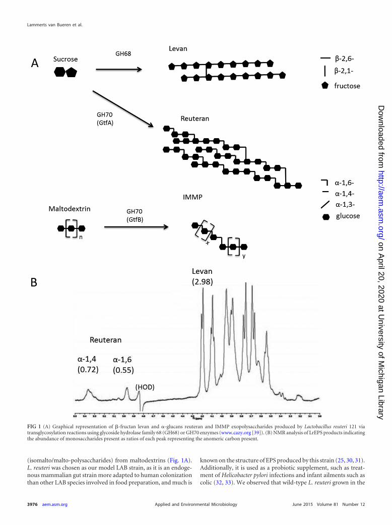
(isomalto/malto-polysaccharides) from maltodextrins (Fig. 1A).L. reuteri was chosen as our model LAB strain, as it is an endoge-nous mammalian gut strain more adapted to human colonizationthan other LAB species involved in food preparation, and much is
known on the structure of EPS produced by this strain (25, 30, 31).Additionally, it is used as a probiotic supplement, such as treat-ment of Helicobacter pylori infections and infant ailments such ascolic (32, 33). We observed that wild-type L. reuteri grown in the
FIG 1 (A) Graphical representation of �-fructan levan and �-glucans reuteran and IMMP exopolysaccharides produced by Lactobacillus reuteri 121 viatransglycosylation reactions using glycoside hydrolase family 68 (GH68) or GH70 enzymes (www.cazy.org [39]). (B) NMR analysis of LrEPS products indicatingthe abundance of monosaccharides present as ratios of each peak representing the anomeric carbon present.
Lammerts van Bueren et al.
3976 aem.asm.org June 2015 Volume 81 Number 12Applied and Environmental Microbiology
on April 20, 2020 at U
niversity of Michigan Library
http://aem.asm
.org/D
ownloaded from
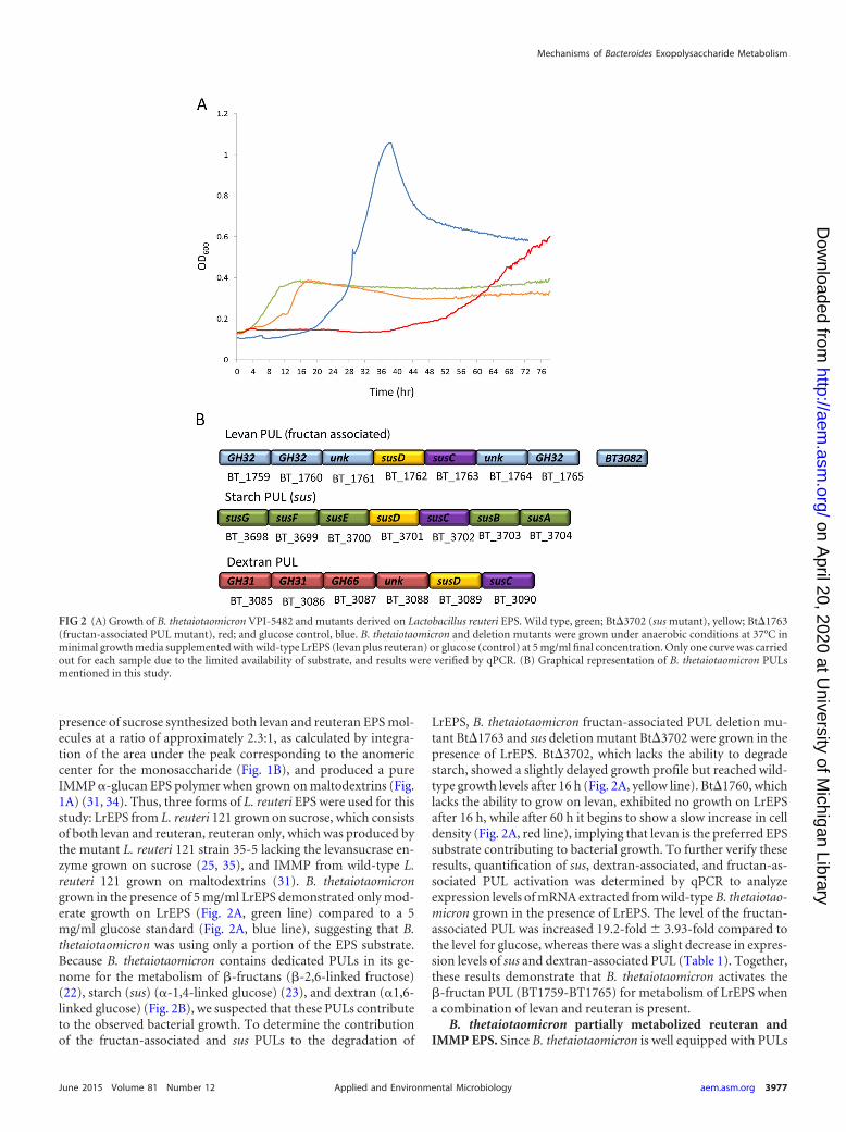
presence of sucrose synthesized both levan and reuteran EPS mol-ecules at a ratio of approximately 2.3:1, as calculated by integra-tion of the area under the peak corresponding to the anomericcenter for the monosaccharide (Fig. 1B), and produced a pureIMMP �-glucan EPS polymer when grown on maltodextrins (Fig.1A) (31, 34). Thus, three forms of L. reuteri EPS were used for thisstudy: LrEPS from L. reuteri 121 grown on sucrose, which consistsof both levan and reuteran, reuteran only, which was produced bythe mutant L. reuteri 121 strain 35-5 lacking the levansucrase en-zyme grown on sucrose (25, 35), and IMMP from wild-type L.reuteri 121 grown on maltodextrins (31). B. thetaiotaomicrongrown in the presence of 5 mg/ml LrEPS demonstrated only mod-erate growth on LrEPS (Fig. 2A, green line) compared to a 5mg/ml glucose standard (Fig. 2A, blue line), suggesting that B.thetaiotaomicron was using only a portion of the EPS substrate.Because B. thetaiotaomicron contains dedicated PULs in its ge-nome for the metabolism of �-fructans (�-2,6-linked fructose)(22), starch (sus) (�-1,4-linked glucose) (23), and dextran (�1,6-linked glucose) (Fig. 2B), we suspected that these PULs contributeto the observed bacterial growth. To determine the contributionof the fructan-associated and sus PULs to the degradation of
LrEPS, B. thetaiotaomicron fructan-associated PUL deletion mu-tant Bt�1763 and sus deletion mutant Bt�3702 were grown in thepresence of LrEPS. Bt�3702, which lacks the ability to degradestarch, showed a slightly delayed growth profile but reached wild-type growth levels after 16 h (Fig. 2A, yellow line). Bt�1760, whichlacks the ability to grow on levan, exhibited no growth on LrEPSafter 16 h, while after 60 h it begins to show a slow increase in celldensity (Fig. 2A, red line), implying that levan is the preferred EPSsubstrate contributing to bacterial growth. To further verify theseresults, quantification of sus, dextran-associated, and fructan-as-sociated PUL activation was determined by qPCR to analyzeexpression levels of mRNA extracted from wild-type B. thetaiotao-micron grown in the presence of LrEPS. The level of the fructan-associated PUL was increased 19.2-fold 3.93-fold compared tothe level for glucose, whereas there was a slight decrease in expres-sion levels of sus and dextran-associated PUL (Table 1). Together,these results demonstrate that B. thetaiotaomicron activates the�-fructan PUL (BT1759-BT1765) for metabolism of LrEPS whena combination of levan and reuteran is present.
B. thetaiotaomicron partially metabolized reuteran andIMMP EPS. Since B. thetaiotaomicron is well equipped with PULs
FIG 2 (A) Growth of B. thetaiotaomicron VPI-5482 and mutants derived on Lactobacillus reuteri EPS. Wild type, green; Bt�3702 (sus mutant), yellow; Bt�1763(fructan-associated PUL mutant), red; and glucose control, blue. B. thetaiotaomicron and deletion mutants were grown under anaerobic conditions at 37°C inminimal growth media supplemented with wild-type LrEPS (levan plus reuteran) or glucose (control) at 5 mg/ml final concentration. Only one curve was carriedout for each sample due to the limited availability of substrate, and results were verified by qPCR. (B) Graphical representation of B. thetaiotaomicron PULsmentioned in this study.
Mechanisms of Bacteroides Exopolysaccharide Metabolism
June 2015 Volume 81 Number 12 aem.asm.org 3977Applied and Environmental Microbiology
on April 20, 2020 at U
niversity of Michigan Library
http://aem.asm
.org/D
ownloaded from
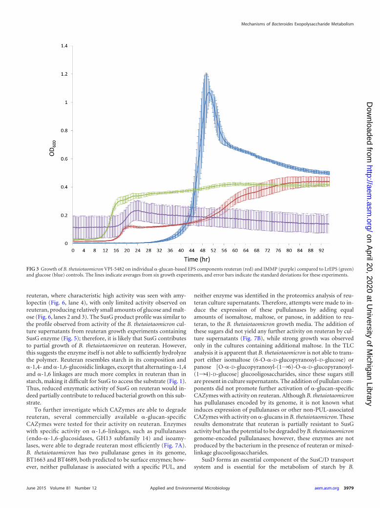
for the degradation of starch and dextran, it was surprising to seethat reuteran from the LrEPS mixture was not being metabolized.Therefore, independent B. thetaiotaomicron culture experimentswere performed to test for bacterial growth on the �-glucan EPSsubstrates reuteran and IMMP separately. Compared to B. theta-iotaomicron cultures of LrEPS (2.3:1 levan-reuteran) (Fig. 3, greenline), reuteran substrate showed a biphasic growth profile with aslight increase in growth after 16 h and an increase up to an OD600
of 0.4 after 52 h (Fig. 3, red line), while IMMP as a substrateshowed very little increase in growth after 72 h (change in OD600
of 0.12 0.09) (Fig. 3, purple line). Although B. thetaiotaomicroncan effectively degrade starch, a polymer of amylose (�-1,4-glu-can) and amylopectin (�-1,4-�-1,6-mixed glucan), it was difficultfor B. thetaiotaomicron to metabolize �-glucan EPS substratescontaining the same types of glucan linkages found in starch.
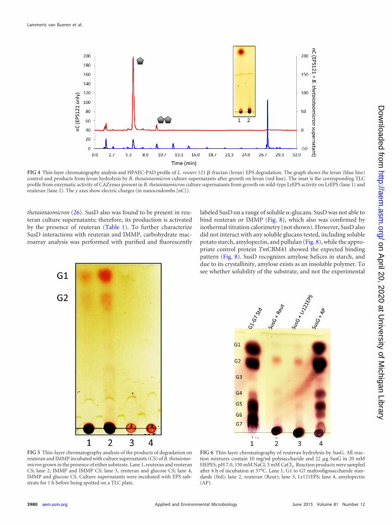
The identification and activity of B. thetaiotaomicronCAZymes induced by LrEPS, reuteran, and IMMP. Previous ob-servations from the seminal work of the Anderson and Salyerslaboratory on starch degradation by B. thetaiotaomicron showedthat PUL-activated CAZymes can be found in culture superna-tants (23). Therefore, to further characterize the activity ofCAZymes produced by B. thetaiotaomicron in the presence ofLrEPS, fresh B. thetaiotaomicron culture supernatants from wholeLrEPS, reuteran, and IMMP growth experiments were incubatedwith EPS molecules, and the products of EPS degradation byCAZymes present in culture supernatants were analyzed by thin-layer chromatography and HPAEC-PAD. Enzymes present in cul-ture supernatants from wild-type LrEPS cultures completely de-graded levan into its monosaccharide component, fructose, andsmall amounts of fructose disaccharide (Fig. 4 red line; also see theinset, lane 1) but showed no activity on reuteran (Fig. 4, inset, lane2). Culture supernatants then were analyzed for the presence ofPUL-encoded proteins using proteomics (see Materials andMethods). Results from proteomic analysis of LrEPS culture su-pernatants showed that almost all extracellular enzymatic compo-nents of the fructan-associated PUL were present (Table 1 and Fig.2B) (see File S1 in the supplemental material for a full listing of
proteins identified in culture supernatants), which accounts forthe observed degradation of levan in LrEPS.
Culture supernatants from B. thetaiotaomicron growth on reu-teran and IMMP also were tested for enzymatic activity on purereuteran or IMMP substrates. The activity of reuteran culture su-pernatants shows that small amounts of glucose and maltose wereproduced (Fig. 5, lane 1), which is not observed in the glucosecontrol supernatant (Fig. 5, lane 3), while IMMP culture superna-tant activity on IMMP showed the same general product profile(Fig. 5, lane 2). Proteomic analysis of reuteran culture superna-tants identified several components from the sus starch utilizationPUL (Table 1 and Fig. 2B), including the transporter componentSusD and extracellular amylase SusG, suggesting that the partialgrowth phenotype exhibited by B. thetaiotaomicron on reuteran isdue to sus PUL-associated enzymes. Culture supernatants fromIMMP growth revealed that enzymes from the sus operon werepresent, whereas additional enzymes from the dextran-associatedPUL BT3085-BT3090 also were present (Table 1 and Fig. 2B; alsosee File S1 in the supplemental material for a full listing). Addi-tional enzymes found in the IMMP supernatant include endodex-tranase BT3087 (GH66) and �-glucosidase II BT3086 (GH31)(Table 1). These results suggest that sus and dextran-associatedPUL-encoded �-glucan-specific CAZymes are produced by B.thetaiotaomicron in the presence of LAB �-glucan EPS, implyingthat other factors, such as substrate recognition or enzymatic ac-tivity, prevent the full degradation and metabolism of these sub-strates.
Role of sus-encoded transport component SusD and surface-associated �-amylase SusG on �-glucan EPS metabolism. Theextracellular �-amylase SusG and transport component SusDwere identified in B. thetaiotaomicron culture supernatant grownin reuteran only (Table 1). To further elucidate the effects of sussystem components on reuteran digestion by B. thetaiotaomicron,purified SusG and SusD were tested for their activity on reuteran.SusG is the surface-associated �-amylase from the sus system in-volved in the breakdown of starch into smaller glucooligosaccha-rides (28). SusG was tested for its degradation properties on
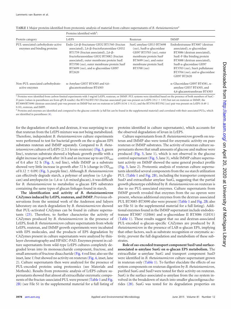
TABLE 1 Major proteins identified from proteomic analysis of material from culture supernatants of B. thetaiotaomicrona
Protein category
Proteins identified withb:
LrEPS Reuteran IMMP
PUL-associated carbohydrate-activeenzymes and binding proteins
Endo-2,6-�-fructanase GH32 BT1760 (fructanassociated), 2,6-�-fructofuranosidase GH32BT1759 (fructan associated), 2,6-�-fructofuranosidase GH32 BT3082 (fructanassociated), outer membrane protein SusEBT3700 (sus), outer membrane protein SusFBT3699 (sus), and �-glucosidase GH97BT2620
SusG amylase GH13 BT3698(sus), SusB �-glucosidaseGH97 BT3703 (sus), outermembrane protein SusFBT3699 (sus), and outermembrane protein SusEBT3700 (sus)
Endodextranase BT3087 (dextranassociated), �-glucosidaseBT3086 (dextran associated),SusE-F-like binding proteinBT3088 (dextran associated),SusB �-glucosidase GH97BT3703 (sus), SusA pullulanaseBT3704 (sus), and �-glucosidaseGH97 BT2620
Non-PUL-associated carbohydrate-active enzymes
�-Amylase GH57 BT4305 and 4,6-glucanotransferase BT4303
�-Glucosidase GH97 BT4581, �-amylase GH57 BT4305, and4,6-glucanotransferase BT4303
a Proteins were identified from carbon-limited experiments with 5 mg/ml LrEPS, reuteran, or IMMP. PUL systems were identified based on the presence of both members of SusC-D pairs (values in parentheses are from qPCR results): the BT1762/BT1763 (fructan-associated) pair was present on LrEPS (19.2 3.93) but not on reuteran or IMMP, theBT3089/BT3090 (dextran-associated) pair was present on IMMP but not on reuteran or LrEPS (0.54 0.12), and the BT3701/BT3702 (sus) pair was present on LrEPS (0.49 0.10), reuteran, and IMPP.b Proteins and enzymes are identified and compared to the glucose controls (a full list can be found in the supplemental material) and correlated with their associated PULs, whichare identified in parentheses (4).
Lammerts van Bueren et al.
3978 aem.asm.org June 2015 Volume 81 Number 12Applied and Environmental Microbiology
on April 20, 2020 at U
niversity of Michigan Library
http://aem.asm
.org/D
ownloaded from
reuteran, where characteristic high activity was seen with amy-lopectin (Fig. 6, lane 4), with only limited activity observed onreuteran, producing relatively small amounts of glucose and malt-ose (Fig. 6, lanes 2 and 3). The SusG product profile was similar tothe profile observed from activity of the B. thetaiotaomicron cul-ture supernatants from reuteran growth experiments containingSusG enzyme (Fig. 5); therefore, it is likely that SusG contributesto partial growth of B. thetaiotaomicron on reuteran. However,this suggests the enzyme itself is not able to sufficiently hydrolyzethe polymer. Reuteran resembles starch in its composition and�-1,4- and �-1,6-glucosidic linkages, except that alternating �-1,4and �-1,6 linkages are much more complex in reuteran than instarch, making it difficult for SusG to access the substrate (Fig. 1).Thus, reduced enzymatic activity of SusG on reuteran would in-deed partially contribute to reduced bacterial growth on this sub-strate.
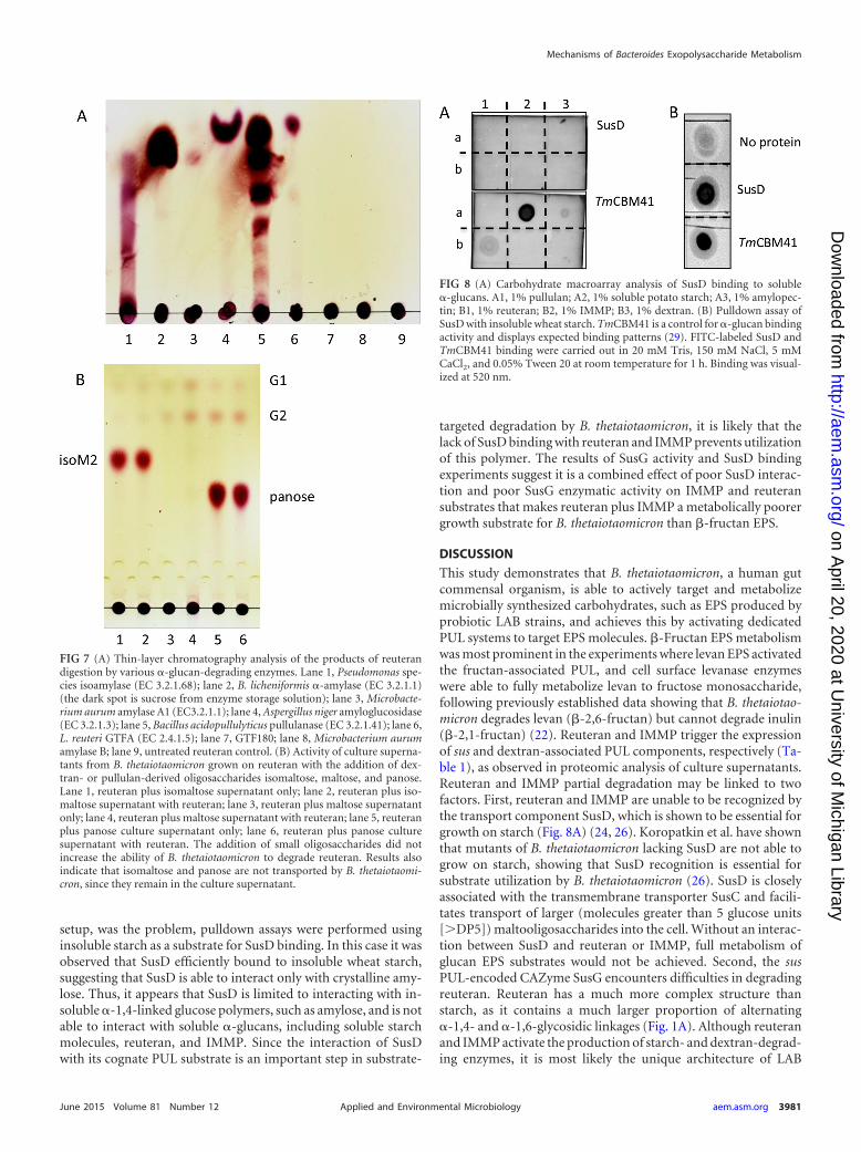
To further investigate which CAZymes are able to degradereuteran, several commercially available �-glucan-specificCAZymes were tested for their activity on reuteran. Enzymeswith specific activity on �-1,6-linkages, such as pullulanases(endo-�-1,6-glucosidases, GH13 subfamily 14) and isoamy-lases, were able to degrade reuteran most efficiently (Fig. 7A).B. thetaiotaomicron has two pullulanase genes in its genome,BT1663 and BT4689, both predicted to be surface enzymes; how-ever, neither pullulanase is associated with a specific PUL, and
neither enzyme was identified in the proteomics analysis of reu-teran culture supernatants. Therefore, attempts were made to in-duce the expression of these pullulanases by adding equalamounts of isomaltose, maltose, or panose, in addition to reu-teran, to the B. thetaiotaomicron growth media. The addition ofthese sugars did not yield any further activity on reuteran by cul-ture supernatants (Fig. 7B), while strong growth was observedonly in the cultures containing additional maltose. In the TLCanalysis it is apparent that B. thetaiotaomicron is not able to trans-port either isomaltose (6-O-�-D-glucopyranosyl–D-glucose) orpanose [O-�-D-glucopyranosyl-(1¡6)-O-�-D-glucopyranosyl-(1¡4)-D-glucose] glucooligosaccharides, since these sugars stillare present in culture supernatants. The addition of pullulan com-ponents did not promote further activation of �-glucan-specificCAZymes with activity on reuteran. Although B. thetaiotaomicronhas pullulanases encoded by its genome, it is not known whatinduces expression of pullulanases or other non-PUL-associatedCAZymes with activity on �-glucans in B. thetaiotaomicron. Theseresults demonstrate that reuteran is partially resistant to SusGactivity but has the potential to be degraded by B. thetaiotaomicrongenome-encoded pullulanases; however, these enzymes are notproduced by the bacterium in the presence of reuteran or mixed-linkage glucooligosaccharides.
SusD forms an essential component of the SusC/D transportsystem and is essential for the metabolism of starch by B.
FIG 3 Growth of B. thetaiotaomicron VPI-5482 on individual �-glucan-based EPS components reuteran (red) and IMMP (purple) compared to LrEPS (green)and glucose (blue) controls. The lines indicate averages from six growth experiments, and error bars indicate the standard deviations for these experiments.
Mechanisms of Bacteroides Exopolysaccharide Metabolism
June 2015 Volume 81 Number 12 aem.asm.org 3979Applied and Environmental Microbiology
on April 20, 2020 at U
niversity of Michigan Library
http://aem.asm
.org/D
ownloaded from
thetaiotaomicron (26). SusD also was found to be present in reu-teran culture supernatants; therefore, its production is activatedby the presence of reuteran (Table 1). To further characterizeSusD interactions with reuteran and IMMP, carbohydrate mac-roarray analysis was performed with purified and fluorescently
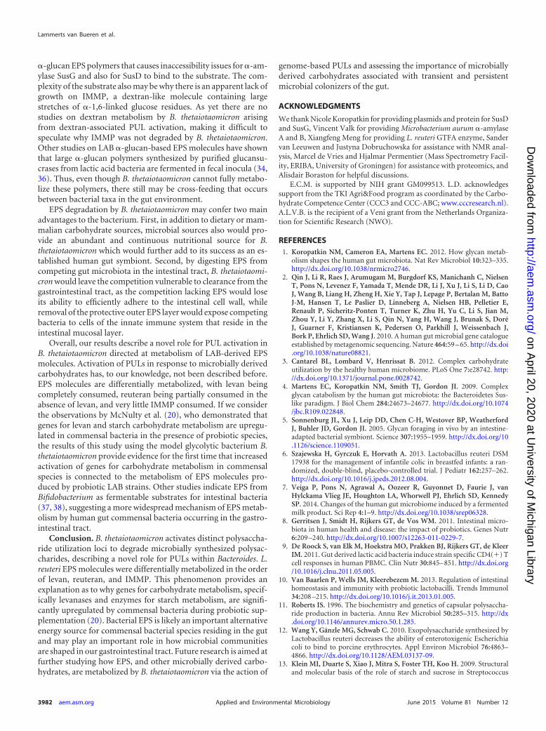
labeled SusD on a range of soluble �-glucans. SusD was not able tobind reuteran or IMMP (Fig. 8), which also was confirmed byisothermal titration calorimetry (not shown). However, SusD alsodid not interact with any soluble glucans tested, including solublepotato starch, amylopectin, and pullulan (Fig. 8), while the appro-priate control protein TmCBM41 showed the expected bindingpattern (Fig. 8). SusD recognizes amylose helices in starch, anddue to its crystallinity, amylose exists as an insoluble polymer. Tosee whether solubility of the substrate, and not the experimental
FIG 4 Thin-layer chromatography analysis and HPAEC-PAD profile of L. reuteri 121 �-fructan (levan) EPS degradation. The graph shows the levan (blue line)control and products from levan hydrolysis by B. thetaiotaomicron culture supernatants after growth on levan (red line). The inset is the corresponding TLCprofile from enzymatic activity of CAZymes present in B. thetaiotaomicron culture supernatants from growth on wild-type LrEPS activity on LrEPS (lane 1) andreuteran (lane 2). The y axes show electric charges (in nanocoulombs [nC]).
FIG 5 Thin-layer chromatography analysis of the products of degradation onreuteran and IMMP incubated with culture supernatants (CS) of B. thetaiotao-micron grown in the presence of either substrate. Lane 1, reuteran and reuteranCS; lane 2, IMMP and IMMP CS; lane 3, reuteran and glucose CS; lane 4,IMMP and glucose CS. Culture supernatants were incubated with EPS sub-strate for 1 h before being spotted on a TLC plate.
FIG 6 Thin-layer chromatography of reuteran hydrolysis by SusG. All reac-tion mixtures contain 10 mg/ml polysaccharide and 22 �g SusG in 20 mMHEPES, pH 7.0, 150 mM NaCl, 5 mM CaCl2. Reaction products were sampledafter 4 h of incubation at 37°C. Lane 1, G1 to G7 maltooligosaccharide stan-dards (Std); lane 2, reuteran (Reut); lane 3, Lr121EPS; lane 4, amylopectin(AP).
Lammerts van Bueren et al.
3980 aem.asm.org June 2015 Volume 81 Number 12Applied and Environmental Microbiology
on April 20, 2020 at U
niversity of Michigan Library
http://aem.asm
.org/D
ownloaded from
setup, was the problem, pulldown assays were performed usinginsoluble starch as a substrate for SusD binding. In this case it wasobserved that SusD efficiently bound to insoluble wheat starch,suggesting that SusD is able to interact only with crystalline amy-lose. Thus, it appears that SusD is limited to interacting with in-soluble �-1,4-linked glucose polymers, such as amylose, and is notable to interact with soluble �-glucans, including soluble starchmolecules, reuteran, and IMMP. Since the interaction of SusDwith its cognate PUL substrate is an important step in substrate-
targeted degradation by B. thetaiotaomicron, it is likely that thelack of SusD binding with reuteran and IMMP prevents utilizationof this polymer. The results of SusG activity and SusD bindingexperiments suggest it is a combined effect of poor SusD interac-tion and poor SusG enzymatic activity on IMMP and reuteransubstrates that makes reuteran plus IMMP a metabolically poorergrowth substrate for B. thetaiotaomicron than �-fructan EPS.
DISCUSSION
This study demonstrates that B. thetaiotaomicron, a human gutcommensal organism, is able to actively target and metabolizemicrobially synthesized carbohydrates, such as EPS produced byprobiotic LAB strains, and achieves this by activating dedicatedPUL systems to target EPS molecules. �-Fructan EPS metabolismwas most prominent in the experiments where levan EPS activatedthe fructan-associated PUL, and cell surface levanase enzymeswere able to fully metabolize levan to fructose monosaccharide,following previously established data showing that B. thetaiotao-micron degrades levan (�-2,6-fructan) but cannot degrade inulin(�-2,1-fructan) (22). Reuteran and IMMP trigger the expressionof sus and dextran-associated PUL components, respectively (Ta-ble 1), as observed in proteomic analysis of culture supernatants.Reuteran and IMMP partial degradation may be linked to twofactors. First, reuteran and IMMP are unable to be recognized bythe transport component SusD, which is shown to be essential forgrowth on starch (Fig. 8A) (24, 26). Koropatkin et al. have shownthat mutants of B. thetaiotaomicron lacking SusD are not able togrow on starch, showing that SusD recognition is essential forsubstrate utilization by B. thetaiotaomicron (26). SusD is closelyassociated with the transmembrane transporter SusC and facili-tates transport of larger (molecules greater than 5 glucose units[DP5]) maltooligosaccharides into the cell. Without an interac-tion between SusD and reuteran or IMMP, full metabolism ofglucan EPS substrates would not be achieved. Second, the susPUL-encoded CAZyme SusG encounters difficulties in degradingreuteran. Reuteran has a much more complex structure thanstarch, as it contains a much larger proportion of alternating�-1,4- and �-1,6-glycosidic linkages (Fig. 1A). Although reuteranand IMMP activate the production of starch- and dextran-degrad-ing enzymes, it is most likely the unique architecture of LAB
FIG 7 (A) Thin-layer chromatography analysis of the products of reuterandigestion by various �-glucan-degrading enzymes. Lane 1, Pseudomonas spe-cies isoamylase (EC 3.2.1.68); lane 2, B. licheniformis �-amylase (EC 3.2.1.1)(the dark spot is sucrose from enzyme storage solution); lane 3, Microbacte-rium aurum amylase A1 (EC3.2.1.1); lane 4, Aspergillus niger amyloglucosidase(EC 3.2.1.3); lane 5, Bacillus acidopullulyticus pullulanase (EC 3.2.1.41); lane 6,L. reuteri GTFA (EC 2.4.1.5); lane 7, GTF180; lane 8, Microbacterium aurumamylase B; lane 9, untreated reuteran control. (B) Activity of culture superna-tants from B. thetaiotaomicron grown on reuteran with the addition of dex-tran- or pullulan-derived oligosaccharides isomaltose, maltose, and panose.Lane 1, reuteran plus isomaltose supernatant only; lane 2, reuteran plus iso-maltose supernatant with reuteran; lane 3, reuteran plus maltose supernatantonly; lane 4, reuteran plus maltose supernatant with reuteran; lane 5, reuteranplus panose culture supernatant only; lane 6, reuteran plus panose culturesupernatant with reuteran. The addition of small oligosaccharides did notincrease the ability of B. thetaiotaomicron to degrade reuteran. Results alsoindicate that isomaltose and panose are not transported by B. thetaiotaomi-cron, since they remain in the culture supernatant.
FIG 8 (A) Carbohydrate macroarray analysis of SusD binding to soluble�-glucans. A1, 1% pullulan; A2, 1% soluble potato starch; A3, 1% amylopec-tin; B1, 1% reuteran; B2, 1% IMMP; B3, 1% dextran. (B) Pulldown assay ofSusD with insoluble wheat starch. TmCBM41 is a control for �-glucan bindingactivity and displays expected binding patterns (29). FITC-labeled SusD andTmCBM41 binding were carried out in 20 mM Tris, 150 mM NaCl, 5 mMCaCl2, and 0.05% Tween 20 at room temperature for 1 h. Binding was visual-ized at 520 nm.
Mechanisms of Bacteroides Exopolysaccharide Metabolism
June 2015 Volume 81 Number 12 aem.asm.org 3981Applied and Environmental Microbiology
on April 20, 2020 at U
niversity of Michigan Library
http://aem.asm
.org/D
ownloaded from
�-glucan EPS polymers that causes inaccessibility issues for �-am-ylase SusG and also for SusD to bind to the substrate. The com-plexity of the substrate also may be why there is an apparent lack ofgrowth on IMMP, a dextran-like molecule containing largestretches of �-1,6-linked glucose residues. As yet there are nostudies on dextran metabolism by B. thetaiotaomicron arisingfrom dextran-associated PUL activation, making it difficult tospeculate why IMMP was not degraded by B. thetaiotaomicron.Other studies on LAB �-glucan-based EPS molecules have shownthat large �-glucan polymers synthesized by purified glucansu-crases from lactic acid bacteria are fermented in fecal inocula (34,36). Thus, even though B. thetaiotaomicron cannot fully metabo-lize these polymers, there still may be cross-feeding that occursbetween bacterial taxa in the gut environment.
EPS degradation by B. thetaiotaomicron may confer two mainadvantages to the bacterium. First, in addition to dietary or mam-malian carbohydrate sources, microbial sources also would pro-vide an abundant and continuous nutritional source for B.thetaiotaomicron which would further add to its success as an es-tablished human gut symbiont. Second, by digesting EPS fromcompeting gut microbiota in the intestinal tract, B. thetaiotaomi-cron would leave the competition vulnerable to clearance from thegastrointestinal tract, as the competition lacking EPS would loseits ability to efficiently adhere to the intestinal cell wall, whileremoval of the protective outer EPS layer would expose competingbacteria to cells of the innate immune system that reside in theintestinal mucosal layer.
Overall, our results describe a novel role for PUL activation inB. thetaiotaomicron directed at metabolism of LAB-derived EPSmolecules. Activation of PULs in response to microbially derivedcarbohydrates has, to our knowledge, not been described before.EPS molecules are differentially metabolized, with levan beingcompletely consumed, reuteran being partially consumed in theabsence of levan, and very little IMMP consumed. If we considerthe observations by McNulty et al. (20), who demonstrated thatgenes for levan and starch carbohydrate metabolism are upregu-lated in commensal bacteria in the presence of probiotic species,the results of this study using the model glycolytic bacterium B.thetaiotaomicron provide evidence for the first time that increasedactivation of genes for carbohydrate metabolism in commensalspecies is connected to the metabolism of EPS molecules pro-duced by probiotic LAB strains. Other studies indicate EPS fromBifidobacterium as fermentable substrates for intestinal bacteria(37, 38), suggesting a more widespread mechanism of EPS metab-olism by human gut commensal bacteria occurring in the gastro-intestinal tract.
Conclusion. B. thetaiotaomicron activates distinct polysaccha-ride utilization loci to degrade microbially synthesized polysac-charides, describing a novel role for PULs within Bacteroides. L.reuteri EPS molecules were differentially metabolized in the orderof levan, reuteran, and IMMP. This phenomenon provides anexplanation as to why genes for carbohydrate metabolism, specif-ically levanases and enzymes for starch metabolism, are signifi-cantly upregulated by commensal bacteria during probiotic sup-plementation (20). Bacterial EPS is likely an important alternativeenergy source for commensal bacterial species residing in the gutand may play an important role in how microbial communitiesare shaped in our gastrointestinal tract. Future research is aimed atfurther studying how EPS, and other microbially derived carbo-hydrates, are metabolized by B. thetaiotaomicron via the action of
genome-based PULs and assessing the importance of microbiallyderived carbohydrates associated with transient and persistentmicrobial colonizers of the gut.
ACKNOWLEDGMENTS
We thank Nicole Koropatkin for providing plasmids and protein for SusDand SusG, Vincent Valk for providing Microbacterium aurum �-amylaseA and B, Xiangfeng Meng for providing L. reuteri GTFA enzyme, Sandervan Leeuwen and Justyna Dobruchowska for assistance with NMR anal-ysis, Marcel de Vries and Hjalmar Permentier (Mass Spectrometry Facil-ity, ERIBA, University of Groningen) for assistance with proteomics, andAlisdair Boraston for helpful discussions.
E.C.M. is supported by NIH grant GM099513. L.D. acknowledgessupport from the TKI Agri&Food program as coordinated by the Carbo-hydrate Competence Center (CCC3 and CCC-ABC; www.cccresearch.nl).A.L.V.B. is the recipient of a Veni grant from the Netherlands Organiza-tion for Scientific Research (NWO).
REFERENCES1. Koropatkin NM, Cameron EA, Martens EC. 2012. How glycan metab-
olism shapes the human gut microbiota. Nat Rev Microbiol 10:323–335.http://dx.doi.org/10.1038/nrmicro2746.
2. Qin J, Li R, Raes J, Arumugam M, Burgdorf KS, Manichanh C, NielsenT, Pons N, Levenez F, Yamada T, Mende DR, Li J, Xu J, Li S, Li D, CaoJ, Wang B, Liang H, Zheng H, Xie Y, Tap J, Lepage P, Bertalan M, BattoJ-M, Hansen T, Le Paslier D, Linneberg A, Nielsen HB, Pelletier E,Renault P, Sicheritz-Ponten T, Turner K, Zhu H, Yu C, Li S, Jian M,Zhou Y, Li Y, Zhang X, Li S, Qin N, Yang H, Wang J, Brunak S, DoréJ, Guarner F, Kristiansen K, Pedersen O, Parkhill J, Weissenbach J,Bork P, Ehrlich SD, Wang J. 2010. A human gut microbial gene catalogueestablished by metagenomic sequencing. Nature 464:59 – 65. http://dx.doi.org/10.1038/nature08821.
3. Cantarel BL, Lombard V, Henrissat B. 2012. Complex carbohydrateutilization by the healthy human microbiome. PLoS One 7:e28742. http://dx.doi.org/10.1371/journal.pone.0028742.
4. Martens EC, Koropatkin NM, Smith TJ, Gordon JI. 2009. Complexglycan catabolism by the human gut microbiota: the Bacteroidetes Sus-like paradigm. J Biol Chem 284:24673–24677. http://dx.doi.org/10.1074/jbc.R109.022848.
5. Sonnenburg JL, Xu J, Leip DD, Chen C-H, Westover BP, WeatherfordJ, Buhler JD, Gordon JI. 2005. Glycan foraging in vivo by an intestine-adapted bacterial symbiont. Science 307:1955–1959. http://dx.doi.org/10.1126/science.1109051.
6. Szajewska H, Gyrczuk E, Horvath A. 2013. Lactobacillus reuteri DSM17938 for the management of infantile colic in breastfed infants: a ran-domized, double-blind, placebo-controlled trial. J Pediatr 162:257–262.http://dx.doi.org/10.1016/j.jpeds.2012.08.004.
7. Veiga P, Pons N, Agrawal A, Oozeer R, Guyonnet D, Faurie J, vanHylckama Vlieg JE, Houghton LA, Whorwell PJ, Ehrlich SD, KennedySP. 2014. Changes of the human gut microbiome induced by a fermentedmilk product. Sci Rep 4:1–9. http://dx.doi.org/10.1038/srep06328.
8. Gerritsen J, Smidt H, Rijkers GT, de Vos WM. 2011. Intestinal micro-biota in human health and disease: the impact of probiotics. Genes Nutr6:209 –240. http://dx.doi.org/10.1007/s12263-011-0229-7.
9. De Roock S, van Elk M, Hoekstra MO, Prakken BJ, Rijkers GT, de KleerIM. 2011. Gut derived lactic acid bacteria induce strain specific CD4(�) Tcell responses in human PBMC. Clin Nutr 30:845– 851. http://dx.doi.org/10.1016/j.clnu.2011.05.005.
10. Van Baarlen P, Wells JM, Kleerebezem M. 2013. Regulation of intestinalhomeostasis and immunity with probiotic lactobacilli. Trends Immunol34:208 –215. http://dx.doi.org/10.1016/j.it.2013.01.005.
11. Roberts IS. 1996. The biochemistry and genetics of capsular polysaccha-ride production in bacteria. Annu Rev Microbiol 50:285–315. http://dx.doi.org/10.1146/annurev.micro.50.1.285.
12. Wang Y, Gänzle MG, Schwab C. 2010. Exopolysaccharide synthesized byLactobacillus reuteri decreases the ability of enterotoxigenic Escherichiacoli to bind to porcine erythrocytes. Appl Environ Microbiol 76:4863–4866. http://dx.doi.org/10.1128/AEM.03137-09.
13. Klein MI, Duarte S, Xiao J, Mitra S, Foster TH, Koo H. 2009. Structuraland molecular basis of the role of starch and sucrose in Streptococcus
Lammerts van Bueren et al.
3982 aem.asm.org June 2015 Volume 81 Number 12Applied and Environmental Microbiology
on April 20, 2020 at U
niversity of Michigan Library
http://aem.asm
.org/D
ownloaded from
mutans biofilm development. Appl Environ Microbiol 75:837– 841. http://dx.doi.org/10.1128/AEM.01299-08.
14. Leemhuis H, Pijning T, Dobruchowska JM, van Leeuwen SS, Kralj S,Dijkstra BW, Dijkhuizen L. 2013. Glucansucrases: three-dimensionalstructures, reactions, mechanism, �-glucan analysis and their implica-tions in biotechnology and food applications. J Biotechnol 163:250 –272.http://dx.doi.org/10.1016/j.jbiotec.2012.06.037.
15. Horn N, Wegmann U, Dertli E, Mulholland F, Collins SR, WaldronKW, Bongaerts RJ, Mayer MJ, Narbad A. 2013. Spontaneous mutationreveals influence of exopolysaccharide on Lactobacillus johnsonii surfacecharacteristics. PLoS One 8:e59957. http://dx.doi.org/10.1371/journal.pone.0059957.
16. Ozimek LK, Kralj S, van der Maarel MJEC, Dijkhuizen L. 2006. Thelevansucrase and inulosucrase enzymes of Lactobacillus reuteri 121 ca-talyse processive and non-processive transglycosylation reactions. Micro-biology 152:1187–1196. http://dx.doi.org/10.1099/mic.0.28484-0.
17. Van Hijum SAFT, Kralj S, Ozimek LK, Dijkhuizen L, van Geel-SchuttenIGH. 2006. Structure-function relationships of glucansucrase and fruc-tansucrase enzymes from lactic acid bacteria. Microbiol Mol Biol Rev70:157–176. http://dx.doi.org/10.1128/MMBR.70.1.157-176.2006.
18. Duncan SH, Flint HJ. 2013. Probiotics and prebiotics and health in ageingpopulations. Maturitas 75:44–50. http://dx.doi.org/10.1016/j.maturitas.2013.02.004.
19. Arthur JC, Gharaibeh RZ, Uronis JM, Perez-Chanona E, Sha W, Tom-kovich S, Mühlbauer M, Fodor A, Jobin C. 2013. VSL#3 probioticmodifies mucosal microbial composition but does not reduce colitis-associated colorectal cancer. Sci Rep 3:2868. http://dx.doi.org/10.1038/srep02868.
20. McNulty NP, Yatsunenko T, Hsiao A, Faith JJ, Muegge BD, GoodmanAL, Henrissat B, Oozeer R, Cools-Portier S, Gobert G, Chervaux C,Knights D, Lozupone CA, Knight R, Duncan AE, Bain JR, MuehlbauerMJ, Newgard CB, Heath AC, Gordon JI. 2011. The impact of a consor-tium of fermented milk strains on the gut microbiome of gnotobiotic miceand monozygotic twins. Sci Transl Med 3:106ra106. http://dx.doi.org/10.1126/scitranslmed.3002701.
21. Uyeno Y, Sekiguchi Y, Kamagata Y. 2008. Impact of consumption ofprobiotic lactobacilli-containing yogurt on microbial composition in hu-man feces. Int J Food Microbiol 122:16 –22. http://dx.doi.org/10.1016/j.ijfoodmicro.2007.11.042.
22. Sonnenburg ED, Zheng H, Joglekar P, Higginbottom SK, Firbank SJ,Bolam DN, Sonnenburg JL. 2010. Specificity of polysaccharide use inintestinal bacteroides species determines diet-induced microbiota altera-tions. Cell 141:1241–1252. http://dx.doi.org/10.1016/j.cell.2010.05.005.
23. Anderson KL, Salyers AA. 1989. Biochemical evidence that starch break-down by Bacteroides thetaiotaomicron involves outer membrane starch-binding sites and periplasmic starch-degrading enzymes. J Bacteriol 171:3192–3198.
24. Cameron EA, Kwiatkowski KJ, Lee B, Hamaker BR, Koropatkin NM,Martens C. 2014. Multifunctional nutrient-binding proteins adapt hu-man symbiotic bacteria for glycan competition in the gut by separatelypromoting enhanced sensing and catalysis. mBio 5:e01441-14. http://dx.doi.org/10.1128/mBio.01441-14.
25. Van Leeuwen SS, Kralj S, van Geel-Schutten IH, Gerwig GJ, DijkhuizenL, Kamerling JP. 2008. Structural analysis of the alpha-D-glucan(EPS35-5) produced by the Lactobacillus reuteri strain 35-5 glucansucraseGTFA enzyme. Carbohydr Res 343:1251–1265. http://dx.doi.org/10.1016/j.carres.2008.01.044.
26. Koropatkin NM, Martens EC, Gordon JI, Smith TJ. 2008. Starch catabolismby a prominent human gut symbiont is directed by the recognition of amylosehelices. Structure 16:1105–1115. http://dx.doi.org/10.1016/j.str.2008.03.017.
27. Rogers TE, Pudlo N, Koropatkin NM, Bell JSK, Moya Balasch M, JaskerK, Martens EC. 2013. Dynamic responses of Bacteroides thetaiotaomi-cron during growth on glycan mixtures. Mol Microbiol 88:876 – 890. http://dx.doi.org/10.1111/mmi.12228.
28. Koropatkin NM, Smith TJ. 2010. SusG: a unique cell-membrane-associated alpha-amylase from a prominent human gut symbiont tar-gets complex starch molecules. Structure 18:200 –215. http://dx.doi.org/10.1016/j.str.2009.12.010.
29. Lammerts van Bueren A, Finn R, Ausio J, Boraston AB. 2004. A-glucanrecognition by a new family of carbohydrate-binding modules found pri-marily in bacterial pathogens. Biochemistry 43:15633–15642. http://dx.doi.org/10.1021/bi048215z.
30. Van Hijum SAFT, Szalowska E, van der Maarel MJEC, Dijkhuizen L.2004. Biochemical and molecular characterization of a levansucrase fromLactobacillus reuteri. Microbiology 150:621– 630. http://dx.doi.org/10.1099/mic.0.26671-0.
31. Dobruchowska JM, Gerwig GJ, Kralj S, Grijpstra P, Leemhuis H,Dijkhuizen L, Kamerling JP. 2012. Structural characterization of linearisomalto-/malto-oligomer products synthesized by the novel GTFB 4,6-�-glucanotransferase enzyme from Lactobacillus reuteri 121. Glycobiol-ogy 22:517–528. http://dx.doi.org/10.1093/glycob/cwr167.
32. Savino F, Pelle E, Palumeri E, Oggero R, Miniero R. 2007. Lactobacillusreuteri (American Type Culture Collection strain 55730) versus simethi-cone in the treatment of infantile colic: a prospective randomized study.Pediatrics 119:e124 – e130. http://dx.doi.org/10.1542/peds.2006-1222.
33. Lionetti E, Miniello VL, Castellaneta SP, Magistá A, Mde Canio A, Mau-rogiovanni G, Ierardi E, Cavallo L, Francavilla R. 2006. Lactobacillus reuteritherapy to reduce side-effects during anti-Helicobacter pylori treatment inchildren: a randomized placebo controlled trial. Aliment Pharmacol Ther24:1461–1468. http://dx.doi.org/10.1111/j.1365-2036.2006.03145.x.
34. Leemhuis H, Dobruchowska JM, Ebbelaar M, Faber F, Buwalda PL, vander Maarel MJEC, Kamerling JP, Dijkhuizen L. 2014. Isomalto/malto-polysaccharide, a novel soluble dietary fiber made via enzymatic conver-sion of starch. J Agric Food Chem 62:12034 –12044. http://dx.doi.org/10.1021/jf503970a.
35. Van Geel-Schutten GH, Faber EJ, Smit E, Bonting K, Smith MR, BrinkBTEN, Kamerling JP, Vliegenthart JFG, Dijkhuizen L. 1999. Biochem-ical and structural characterization of the glucan and fructan exopolysac-charides synthesized by the Lactobacillus reuteri wild-type strain and bymutant strains. Appl Environ Microbiol 65:3008 –3014.
36. Hernandez-Hernandez O, Côté GL, Kolida S, Rastall R, Sanz ML. 2011.In vitro fermentation of alternansucrase raffinose-derived oligosaccha-rides by human gut bacteria. J Agric Food Chem 59:10901–10906. http://dx.doi.org/10.1021/jf202466s.
37. Salazar N, Gueimonde M, Hernández-Barranco AM, Ruas-Madiedo P,de los Reyes-Gavilán CG. 2008. Exopolysaccharides produced by intes-tinal Bifidobacterium strains act as fermentable substrates for human in-testinal bacteria. Appl Environ Microbiol 74:4737– 4745. http://dx.doi.org/10.1128/AEM.00325-08.
38. Rios-Covian D, Arboleya S, Hernandez-Barranco AM, Alvarez-BuyllaJR, Ruas-Madiedo P, Gueimonde M, de los Reyes-Gavilan CG. 2013.Interactions between Bifidobacterium and Bacteroides species in cofer-mentations are affected by carbon sources, including exopolysaccharidesproduced by bifidobacteria. Appl Environ Microbiol 79:7518 –7524. http://dx.doi.org/10.1128/AEM.02545-13.
39. Lombard V, Golaconda Ramulu H, Drula E, Coutinho PM, HenrissatB. 2014. The carbohydrate-active enzymes database (CAZy) in 2013. Nu-cleic Acids Res 42:490 – 495. http://dx.doi.org/10.1093/nar/gkt1178.
Mechanisms of Bacteroides Exopolysaccharide Metabolism
June 2015 Volume 81 Number 12 aem.asm.org 3983Applied and Environmental Microbiology
on April 20, 2020 at U
niversity of Michigan Library
http://aem.asm
.org/D
ownloaded from
Copyright © 2022 FDOKUMEN