Luminescent solar concentrators employing new Eu(TTA) 3 phen-containing parylene films
Upload
khangminh22Category
view
2download
0
Gadow, Fricke, Strasburger and Wood: Synthesis and evaluation of luminescent tracers 337
J. Clin. Chem. Clin. Biochem.Vol. 22, 1984, pp. 337-347
Synthesis and Evaluation of Luminescent Tracersand Hapten-Protein Conjugates for Use in Luminescence Immunoassayswith Immobilised Antibodies and AntigensA critical study of macro solid phases for use in immunoassay Systems, Part II1)
By A. Gadow, H. Fricke, C. J. Strasburger and W. G. Wood
Klinik für Innere Medizin (Direktor: Prof. Dr. P. C. Scriba)Medizinische Hochschule Lübeck
(Received June 7/December 16, 1983)
Summary: This article describes the Synthesis of labelfc and hapten-protein conjugates for use in bio- andchemiluminescent immunoassay Systems, together witn the problems encountered.The effects of maleimide upon acetate-, adenylate- and pyruvate kinase activity have been studied, äs well äsupon the luciferin-luciferase monitoring System. Maleimide inhibited both acetate and adenylate kinase butshowed no Inhibition of pyruvate kinase and the monitoring reagent.Fouf heterobifunctional reagents were tested for their capability in forming pyruvate kinase-donkey-anti-rab-bit IgG conjugates which retained enzyme and antibody activity. The best results were obtained with succin-imidyl-4-(N-maleimidomethyl)-cyclohexane-l-carboxylate and sucdnimidyl-6-(p-maleimidophenyl)-hexa-noate.The relationship between the amounts of succinimidyl-4-(N-maleimidomethyl)-cyclohexane-l-carboxylateand IgG was studied with respect to enzymic activity of the conjugate.The Michaelis-Menten constants for both conjugated and non-conjugated pyruvate kinase were calculated andcompared. It was found that the maximal velocity (Vmax) of the conjugated enzyme was lower than that of thenon-conjugated enzyme although the "apparent" Km value was the same for both conjugated and non-conju-gated pyruvate kinase.The pyruvate kinase-anti rabbit IgG conjugate was tested for its ability to bind to rabbit-IgG coated polysty-rene balls.In addition to bioluininescent labels, the synthesis of chemiluminescent markers was undertaken and opti-mised. The three substances used for labelling were diazoluminol, diazoisoluminol and N-(4-aminobutyl)-N-ethylisolumiiiol hemisuccinamide the latter being used äs an N-hydroxysuccinamide "active" ester.The-ratio of label to IgG was studied ior diazoluminol and N-(4-aminobutyl)-N-ethylisoluminol hemisuccin-amide active ester after it had been discovered that diazoisoluminol was not suitable for coupling to antibo-dies. The optimal molar ratios label: IgG were for diazoluminol 40:1 and for N-(4-aminobutyl)-N-ethylisolu-minol hemisuccinamide active ester 60:1. Increasing the Substitution rate led to a lessening of the dynamicränge, shown by an increase in the ratio between utispecific binding (noise) to maximal binding (signal) in anassay.The synthesis of hapten-protein conjugates for covalent coupling to polystyrene balls was undertaken äs thisformed part of the preparation for the assays described in Part III. The optimal production of gentamicin-bo-vine serum albumin and thyroxine-transferrin conjugates has been described in de tau.
0 Part 1: J. Clin. Chem. Clin. Biochem. 27, 789-797. Part III: J. Clin. Chem. Clin. Biochem. 22, 349-356.
J. Clin. Chem. Clin. Biocbeni. / Vol. 22, 1984 / No. 5

338 Gadow, Fricke, Strasburger and Wood: Synthcsis and evaluation of luminescent tracers
Synthese und Beurteilung von Lumineszenz-Markern und Haplen-Protein-Konjugaten für Lumineszenz-Im-munoassays mit immobilisierten Antikörpern und Antigenen.Eine kritische Untersuchung von Makro-Festphasen zum Gebrauch in Immunoassay* Systemen, Teil 7/1)
Zusammenfassung: Dieser Beitrag beschreibt die Herstellung von Markern für die Verwendung in Bib- undChemilumineszenz-Immunoassays und zeigt die dabei bestehenden Probleme auf. · \
Der Effekt von Maleinimid auf Acetat-, Pyruvat- und Adenylat(Myo)kinase zur Klärung ihrer Verwendbar-keit als Biolumineszenzmarker wurde untersucht. Lediglich bei der Pyrüvatkinase wurde keine Hemmungbeobachtet, das Luciferin-Luciferase-System (Indikator-Reagenz) blieb ebenfalls unbeeinflußt.
Vier heterobifunktionelle Reagenzien wurden auf ihre Eignung für die Synthese von Esel-anti-KaninchenIgG-Pyruvatkinase-Konjugaten untersucht. Die besten Konjugate wurden mit Succinimidyl^-iN-maleimi-domethyl)-cyclohexan-l-carboxylat und Succinimidyl-6-(p-maleimidophenyl)-hexanoat erzielt.
Das molare Verhältnis von Succinimidyl-4-(N-maleimidomethyl)-cyclohexan-l-carboxylat zu IgG wurde imHinblick auf die Beeinflussung der enzymatischen Aktivität ihres Konjügates mit Pyrüvatkinase untersucht.
Die Michaelis-Menten Konstanten für konjugierte sowie unkonjugierte Pyruvatkinase wurden bestimmt undgegenübergestellt. Dabei konnte eine Abnahme der maximalen Reaktionsgeschwindigkeit des an IgG gekop-pelten Enzyms nachgewiesen werden. Es ergab sich dabei ein „apparenter" /Cm-Wert, der dem ungebundenerPyruvatkinase entsprach.
Die Bindungsfähigkeit der Pyruvatkinase-Esel-anti-Kaninchen-IgG^Komplexe an mit Kaninchen-IgG be-schichtete Polystyrolkugeln wurde untersucht und dokumentiert.
Darüberhinaus wurde die Synthese von Chemilumineszenz-Konjugaten untersucht und optimiert. Die Eig-nung von Diazoluminol, Diazoisoluminol sowie N-(4-aminobutyl)-N-ethylisoluminol hemisuccinamid (akti-ver Ester) zur Kopplung an IgG wurde untersucht und die optimalen Verhältnisse zu IgG erarbeitet. Diesebetragen für Diazoluminol zu IgG etwa 40:1 und für N-(4-aminobutyl)-N-ethylisoluminol hemisuccinamid(aktiver Ester) zu IgG etwa 60:1. Diazoisoluminol war nicht zur Kopplung an IgG verwendbar. HöhereSubstitutionsraten führten für Diazoluminol zu einem Anstieg der unspezifischen Bindung bei gleichzeitigerErniedrigung der spezifischen Bindung. Im Falle von N-(4-aminobutyl)-N-ethylisoluminol hemisuccinamid(aktiver Ester) veränderte sich das Verhältnis von unspezifischer zu spezifischer Bindung trotz höherer Sub-stitution nur unwesentlich.
Die Synthese von Hapten-Protein-Konjugaten zur kovalenten Kopplung an Polystyrölkügeln wurde durchge-führt, da sie Teil der Vorbereitung für die in Teil III beschriebenen Assays ist. Die optimierte Herstellung vonGentamycin-Rinderserumalbumin- und Thyroxin-Transferrin-Konjugaten ist im Detail beschrieben.
Introduction
There are two distinct types of label used in lumines-cence immonoassays, which are usually referred toäs chemiluminescent and bioluminescent markersubstances. The difference äs far äs the assay tech-nique itself is concerned, is only in the way in whichthe light is produced at the end of all reaction Steps.
Labels commonly used in bioluminescence immu-noassays are kinases (l, 2, 3) and NAD(P)-depend-ent dehydrogenases (4, 5).
Difficulties arise in stabilising bioluminescent labelsin liquid form however, äs the enzymes present areinhibited by conventional antimicrobial agents suchäs azide or merthiolate.
Chemiluminescent labels include derivatives of iso-luminol (6, 7, 8), acridinium esters (9), azoluminol(10) and fluorescein isothiocyanate (11).
Useful labels examined in this study are pyruvate ki-nase (EC 2.7.1.40) äs bioluminescence-(1^3), dia-zpluminol (10) and N-(4-aminobutyl)-N-ethylisolu-minol hemisuccinamide active ester äs chemilumi-nescence labels.
The bioluminescent System is based ori" an ATP-geri-erating System coupled to a firefly lüciferin=4ucife-fase light generating reaction (1—3). A species-spe-cific antibody is labelled, which results in the forma-tion of a pyruvate kmase-immunbglobulin G conju-gate.
J. Clin. Chem. Clin. {Kochern* / Vol. 22, 1984 / No. 5

Gadow, Fricke, Strasburger and Wood: Synthesis and evaluation of luminescent tracers 339
Coupling reactions have been tested using differentheterobifunctional reagents (l, 12) having a maleim-ide and N-hydroxysuccinimide functional group forcoupling via thiol and aminogroups respectively.This type of reagent is far superior to alternative rea-gents such äs pentane-l,5-dial, carbodiimides ormixed anhydride reactions. The reaction is a two-step one, the first being the aminoacylation of theIgG followed by the formation of a thioether withthe pyruvate kinase.
The optimisation of Signal to noise ratio in terms ofthe maximum and unspecific binding has been stu-died in terms of reagent and reactant purity and de-gree of Substitution of the label.
The somewhat complicated synthesis of N-(4-ami-nobutyl)-N-ethylisoluminol hemisuccinamide activeester (8) described in the literature was replaced by asimpler method which could be performed in almostany laboratory.
Tab. l. Reactions catalysed by the kinases used in this study. All, reactions were "driven" in the direction of ATP forma-
tion.
1. Pyruvate kinase - EC 2.7. l.40 (from rabbit muscle)Phosphoenolpyruvate + ADP = Pyruvate + ATPCofactors Mg+2 and K+
pH Optimum — 7.0
2. Acetate kinase - EC 2.7.2.1 (from E. Coli)Acetyl phosphate -H ADP = Acetate + ATPCofactors probably Mg"1"2 or Mn+2
pH Optimum — 7.4
3. Adenylate kinase - EC 2.7.4.3 (from rabbit muscle)2 ADP = ATP + AMPCofactor Mg*2
pH Optimum — 6.7
Explanations:Adenylate kinase is also known äs myokinase.All reactions could be run in the reverse direction äs only an ex-change of high energy phosphate bonds took place.
Materials and MethodsMaterials
Unless otherwise stated, materials were obtained from the samesources äs in the first part of this publication (14). Pyruvate ki-nase, acetate kinase and adenylate kinase (myokinase) were pur-chased from Boehringer-Mannheim, Mannheim, D.
All heterobifunctional reagents were obtained from Sigma, Mu-nich, D, except for the n-hexanoyl derivative, which was a giftfrom Boehringer-Mannheim.
ATP-monitpring kits arid N-(4-aminobutyl)-N-ethyIisoluminolhemisuccinamide were bought from LKB, Munich, äs were thegels Ultrogel A4 and A6.
Glutathione (reduced form), 5,5/-dithiobis(2-nitrobenzoic acid),adenosine diphosphate (ADP) and phosphoenolpyruvate wereobtained from Sigma, Munich.
Buffer sübstances were purchased from Merck, Darmstadt, D.,and from EGA-Chemie, (Aldrich) Steinbach, D.
Methods
Effects of maleimide upon monitoring reagent and pyruvate-, ace-tate- and adenylate kinase activity
All enzyme reactions were run in the "reverse" direction, synthes-ising adenosine triphosphate (ATP) from the relevant compo-nents. The enzyme reactions are shown in Table 1.
First the effect of maleimide upon the ATP-monitoring reagentwas studied, to check for Inhibition of light production in the luci-ferin-luciferase System.
Increasing amounts of maleimide (covering the ränge 0.1—125mmol/tübe) were added to suitable dilutions öf the enzymes inincubatiön buffer (0.05 raol/1 Tns-HCl containing 2.5 g/l bovineserum albiimin and 0.1 mol/1 KC1 adjusted to pH 7.4), the effectson the rate of ATP synthesis being plotted against time. ATP pro-duction was measured kinetically using an ATP-monitoring kitand an LKB 1250 Luminometer with printer display (see section"Studies upon the effect . . ." for detailed Information).
Synthesis of the pyruvate kinase IgC conjugates
The -globulin fraction of a donkey anti-rabbit IgG serum (Well-come RD 17) was prepared using polyethylene glycol precipita-tion followed by DEAE-cellulose column chromatography, (2,15). The IgG concentration was around 15 g/l in the eluate.
To a solution of IgG (15 g/l) containing a total of 15 mg dissolvedin l ml buffer 2 (0.02 mol/1 potassium phosphate containing 0.15mol/ NaCl, adjusted to pH 7) was added l ml of the respectiveheterobifunctional reagent (for structure of these reagents seefig. 1.) representing l umol^-lO (see section "Studies uponthe e f f e c t . . . " below). The mixture was incubated for 45 min atambient temperature and then dialysed twice for l h against 11buffer 2. The degree of coupling was determined by the titrationof the free thiol groups on the IgG-heterobifunctional reagentcomplex with Ellman's reagent (5,5'-dithiobis-(2-nitrobenzoicacid)) in which 2-nitro-5-mercaptobenzoic acid is produced in thepresence of free —SH groups. The latter gives an intensive yellowcoloufed anion which can be measured photometrically at 412 nm(16,17). A Standard curve was set up using reduced glutathione äscalibratpr covering the ränge 0—50 nmol/tube.
In order to obtain a label with a higher molar ratio pyruvate ki-nase: IgG, the pyruvate kinase concentration was held at 4 timesthat of IgG.The reaction was allowed to proceed for 25 min at room tempera-ture and was stopped by addition of 100 nmol reduced glutathi-one. The mixture was then separated on an Ultrogel A6 column(100 x 1.8 cm) using buffer 2 äs eluent. The antibody bindingpotency and enzyme activity were tested in each fraction, the op-timal fractions showing both enzyme activity and specific antibodybinding pooled, portioned and lyophilised after addition of l mglactose to each portion. The pyruvate kinase activity was testedfor äs already described (1), the antibody binding potency bybinding studies using rabbit IgG covalently coupled to polystyreneballs (14).
Studies upon the effect of heterobifunctional reagent concentrationwith respect to enzyme activityThe effect upon enzyme activity after coupling to IgG was studiedusing three different concentrations of succinimidyl-4-(N-malc-
J. Clin. Chem. Clin. Biochcm. / Vol. 22,1984 / No. 5

340 Gadow, Fricke, Strasburger and Wood: Synthesis and evaluation of luminescent tracers
CH2-B
A-(CH2)5-
ΙΠ
Fig. 1. The structure of the four heterobifunctional reagents usedin this study:
I m-maleimidobenzoyl N-hydroxysuccinimide esterII succinimidyl-4-(N-maleimidomethyl)-cyclohexane-l-
carboxylateIII succinimidyl-6-(p-maleimidophenyl)-hexanoateIV suc nimidyI-4-(p-maleimidophenyI)-butyrateThe structures A (N-hydroxysuccinimide-ester group) andB (maleimide group) shown in the lower portion of thediagram are the two functional groups common to the rea-gents represented here.
The same System was used to calculate the Km value for phospho-enolpyruvate. Here the ADP concentration was kept constant at6 g/l, the concentrations for phosphoenolpyruvate were varied sdescribed above for ADP. The Km value for "free" pyruvate ki-nase was measured using a 10 μΐ aliquot, equivalent to l ng/tube.
Lineweaver-Burk plots were made in order to derive the Km-values for ADP and phosphoenolpyruvate/
Diaiotisation of luminol and isoluminol
On account of conflicting reports in the litefature (10) and per-sonal expef ience, the effects of diazotisation time, pH and Substi-tution rate into IgG have been studied. Binding studies have beencarried out using the relevant IgG against which the labelled anti-body is directed, coupled to polystyrene balls (14).
Luminol or isoluminol (l mmol) was suspended in 5 ml l mol/1HCI and cooled under constant stirring to 0 °C (ice bath). KNO^(1.5 mmol) was dissolved in l ml distilled water and cooled in ice.The cold KNO2 solution was added dropwise to the luminol/isolu-minol Suspension respectively, until the green-yellow slurry be-came a clear orange (luminol) or yellow-orange (isoluminol) solu-tion. The time taken for the addition of nitrite was 15—20 min.An excess of HNO? is usually undesirable if the diazotised reacrtant is to be used straight away because of further unwanted dia-zotisation of the second reactant, here IgG. In this case, s IgGmolecules contain no arylamino groups, no such side reactionsshould occur. However, the presence of excess nitrite ions weretested for using starch iodide paper, a blue^black coloration de-noting the presence of nitrite. This was removed by adding a fewdrops of urea solution (l mol/1) until the reaction with starch-io-dide paper was negative. The diazoluminol or diazoisoluminol was•used immediately for the preparation of labels. The diazo con>pounds were stable for periods up to 7 days at —20 °C, but evenduring this time, their effectiveness s coupling reagents deterio-rated. A quick test to see if the diazoluminol was still useable wasthat if, after addition of a drop of K2CO3 (l mol/1) the red c lora^tion was stable for more than 30 minutes it could still be used forcoupling. In the case of diazoisoluminol, the yellow orange solu-tion turned golden yellow and crystallised out inside 30 minutesafter beeing synthesised. The resulting Suspension was used forconjugation to IgG. ^ ?-· *
imidomethyl)-cyclohexane-l-carboxylate, with a molar ratio IgG:succinimidyl-4-(N-maleimidomethyl)-cyclohexane-1 -carboxylateof 1:1.2, 1:6 and and 1:10.
Conjugates were made s in section above, but with additionalpurification after the succinimidyl-4-(N-maleimidomethyl)-cyclo-hexane-1-carboxylate-IgG coupling using a Sephadex G-25 co-lumn (30 x 1.5 cm). Pyruvate kinase was allowed to react in afour-fold excess, and the optimal fractions (see above) used tocalculate the Michaelis-Menten constant. The necessary experi-ments were carried out s set out below:
100 μΐ of a 1:600 dilution of the conjugate (ca. 2.8 pg IgG) werereacted with a rabbit IgG-coated polystyrene ball (14) in 200 ulincubation buffer for 3 h at ambient temperature (23 °C). Thepolystyrene ball was then washed with incubation buffer (2 x 500ul) and 0.15 mol/1 NaCl (l x 500 ul).
The measurement of ATP production in order to calculate theKm-value for ADP was carried out using the following System:
200 μΐ Tris-HCl buffer (0.1 mol/1, pH 7.4)100 μΐ ATP monitoring reagent50 μΐ Phosphoenolpyruvate (2 g/l in 0.2 mol/1 KCl/MgSO4)50 μΐ ADP (end concentration/tube 4, 0.8, 0.4, 0.08, 0.04 and
0,008 μιηοΐ)
Preparation of diazoluminol· and diazoisoluminol·antibody con-jugates
These experiments were carried out to check the effect of pH,time and the ratio of coupling reagent to IgG oh the q ality of theproducts formed.
The diazoluminol or diazoisoluminol preparation was added slow-ly to l ml of the IgG solution (15 g/l) in buffef 2 under constantstirring in an ice bath to give final ratios of diazoluminol/diazoiscnluminohlgG of 20:1. The re ction times were 6, 24 and 72 h atpH 4 and pH 9 at 4°C. Potassium carbonate solution (l mol/1)was added dropwise to maintain the pH between 9.0 and 9.2. Thereactions at pH 4 were carried out maintaining the pH with 0.5mol/1 potassium phosphate buffer pH 7.
After the addition of diazotised (iso)Iuminol, the reaction was al-lowed to proceed at 4 °C during which time the reaction mixturechanged from red to yellow in case of pH 9 derivatives, those atpH 4 remaining pale yellow throughout the coupling procedure.
After the reaction all reation mixtures were purified over an Ul-trogel A4 column (30 x 2.5 cm) with buffer 2 s eluent, collecting30 drop fractions (1.5 ml). The coloured fractions were tested forspecific and unspecific binding to IgG and transferrin coated poly-styrene balls respectively (14).
To check the effect of changing the ratio lurninogen: IgG diazolu-minol was used at pH 9 with a 24 h reaction time at 4 °C s theseconditions were optimal.
J. Clin. Chem. Clin. Bipehem. / Vol. 22,1984 / No. 5

Gadow, Fricke, Strasburger and Wood: Synthesis and evaluation of luminescent tracers 341
The ratio of diazoluminol: IgG was varied between 8: l and 200: l.Separation of the reaction componenls was made with an UltrogelA4 (see above).
From the resulting data it was estimated that an effective (immu-noreactive) Substitution of 23-28 gentamicin per bovine serumalbumin had taken placc.
Optitnised preparation of N- (4-aminobutyl) -N-ethylisoluminol he-misuccinamide active ester
18.5 mg (50 μιηοΐ) N-(4-aminobutyl)-N-ethyIisoIuminol hcmisuc-cinamide wcre dissolved in 800 μΐ dry dimethylformamide underslight warming. N-Hydroxysuccinimide (5.75 mg = 50 μπιοί) wasadded at room temperature, the mixture being stirred. When theN-hydroxysuccinimide had been dissolved, 30.9 mg (150 μηηοΐ)dicyclohexylcarbodiimide dissolved in 200 μΐ dry dimethylfor-mamide were added. After 20 h at room temperature without stir-ring and under light exclusion (aluminium foil covered reactionvessel) the mixture was used for direct coupling to antibodies andantigens. During the reaction, crystals of dicyclohexyl urca precip-itated out.
A low temperature of about 4 °C slows down the formation of theactive ester and an increase in incubation time at room tempera-ture (>20 h) gave no increase in N-(4-aminobutyl)-N-ethylisolu-minol hemisuccinamide active ester formation.
Product formation was confirmed using thin layer Chromato-graphie techniques. The solvent used was ethyl acetate ethanolNH3 (25 g/l) 5 + 2 + l (by volume). The Rf-values obtained weres follows: N-(4-aminobutyl)-N-ethylisoluminol hemisuccinam-
ide - 0.074, N-hydroxysuccinimide - 0.14, N-(4-aminobutyl)-N-ethylisoluminol hemisuccinamide active ester — 0.4. Some otherspots were observed which demonstrated neither fluorescencewhen excitated at 366 nm nor luminescence when excited withalkaline microperoxidase and H2Oz.
Preparation of N-(4-aminobutyl)-N-ethylisoluminol hemisuccin-amide active ester-antibody conjugates
The γ-globulin fraction of a donkey anti-rabbit IgG serum wasprepared s described in Section Synthesis . . .". To 3 x 2 ml(30 mg protein) portions of this preparation different amounts ofthe N-(4-aminobutyl)-N-ethylisoIuminol hemisuccinamide activeester preparation were added to give final ratios N-(4-aminob -tyl)-N-ethylisoluminol hemisuccinamide active ester: IgG of 30:1,60:1 and 120:1 respectively. After the addition, a precipitate ofwater insoluble components formed immediately. The reactionwas allowed to continue for 24 h at 4 °C under light exclusion.After centrifugation (10 min at 4000 min""1) the supernatant wastransferred to an Ultrogel A4 (30 x 2.5 cm) column using 0.05mol/1 tris/HCl pH 8 s eluent, collecting fractions of 1.5ml.
The test System for specific and unspecific binding was identicalwith that described in the preparation of diazoluminol and diazo-isoluminol-IgG.
Synthesis of a gentamicin-bovine serum albumin conjugate for thegentamicin SPALT assay (see tab. 2)
The coupling reagefit used was l-cyclohexyl-3-(2-mofpholinyl-(4)-ietllyl)-carbodiimide methyl-p-toluene sulphonate (MCDI).The molar ratios ch sen were: bovine serum albumin: carbodiim-ide (MCDI): gentamicin — 1:60:80. Bovine serum albumin(68 mg) and 48 mg gentamicin were dissolved in 4 ml distilled wa-ter and the pH adjusted to 4.5 with dropwise addition of 0.1 mol/1HC1. Carbodiimide (MCDI, 28 mg) were dissolved in 500 μΐ dis-tilled water and added dropwise to the bovine serum albumin-gentamicin mixture. The pH of the resulting mixture was heldconstant at 4.5 with 0.1 mol/1 HC1 for the next hour after whichthe reaction was ailowed to proceed at 4°C overnight.
Seperation of complex and unreacted gentamicin was effected viaan Ultrogel A6 column (30 x 2.5 em) using buffer 2 s elutionbuffer. The column fractions were tested for protein conterit
) and gentamicin immunoreactivity (radioimmunoassay).
Synthesis of a thyroxine-transferrin conjugate for a 7VSPALTas-say
This conjugate was synthesized for the T4-SPALT assay in part IIIof this trilogy and is described here s it is part of the preparationfor an assay.
The coupling reagents ch sen were l-cyclohexyl-3-(2-morpholin-yl-(4)-ethyl)-carbodiimide methyl-p-toluene sulphonate (seeabove) or l-ethyl-3-(3-dimethyl aminopropyl)carbodiimide hy-drochloride (EDAC).
The molar ratios ch sen were:Transferrin:carbodiimide(EDAC):thyroxine - 1:50:200.
44 mg transferrin were dissolved in 500 μΐ distilled water and thepH adjusted to 6 using 0.1 mol/1 HC1. 20 mg thyroxine were sus-pended in l ml distilled water and brought into solution by drop-wise addition of 0.5 mol/1 NaOH. The carbodiimide solution wasadded dropwise to the transferrin solution keeping the pH at 6,the mixture bcing ailowed to stand for 5 minutes. The thyroxinesolution was given dropwise to the mixture over a period of 90—120 minutes, keeping the pH between 6 and 6.5. The mixture wasthen ailowed to stand overnight at 4 °C. The thyroxine did not allremain in solution with l-ethyl-3-(3-dimethyl aminopropyl)car-bodiimide hydrochloride s coupling reagent. The high molar ra-tio of thyroxine was meant to compensate for possible precipita-tion during the reaction, s it was known that it is almost impossi-ble to keep thyroxine in aqueous solution at pH values lying onthe acid side of neutrality.
The purification of the conjugate was via an Ultrogel A6 column(30 x 2.5 cm) using centrifugation step before the column run toremove particulate material.
The Substitution rate was determined spectrophotometricallyusing the molar lineic absorbance for thyroxine at 328 nm s56 m2 · mol'1.
The Substitution rate with l-cyclohexyl-3-(2-morpholinyl-(4)-ethyl)-carbodiimide methyl-p-toluene sulphonate was 16 thyrox-ine per transferrin and for l-ethyl-3-(3-dimethyl aminopropyl)-carbodiimide hydrochloride 24 thyroxine per transferrin.
Results
Bioluminescent conjugatesFigure 2 shows the effect of maleimide on acetate-and adenylate- (or myo-) kinase, figure 3 on theATP-monitoring reagent.
Table 2 shows the binding of the four pyruvate ki-nase-IgG conjugates made using the different hete-robifunctional reagents
m-maleimidobenzoyl N-hydroxysuccinimide ester,succinimidyl-4-()9-maleimidophenyl)-butyrate,succinimidyl-4-(N-maleimidomethyl)-cyclohexane-1-carboxylate andsuccinimidyl-6-(/7-maleimidophenyl)-hexanoate,
J. Clin. Chem. Clin. Biochfem. / Vol. 22, 1984 / No. 5

342 Gadow, Fricke, Strasburger and Wood: Synthesis and evaluation of luminescent tracers
3V)φcl 2
2 3t (min)
ΦOcφ
0 1 2 3 4 5t (min)
Fig. 2. The effect of maleimide on kinases in terms of concentra-tion and time. The values on the Ordinate show the re^sponse of the luminometer to ATP-production during theincubation time. A flatter slope corresponds to a lowerrate of ATP-production, i. e. a higher degree of Inhibitionby maleimide.a) adenylate kinase (myokinase)b) acetate kinase, using an excess of acetyl phosphate to
drive the reaction in the direction of ATP-production.Δ—Δ ηο maleimidex—x 30 mmol/l maleimideD—D 3 mmol/l maleimideO—O 0.3 mmol/l maleimideO—O 0.3 mmol/l maleimide, after 30 min
5
<υucωU *5
S-&— ^=3—^ 1̂3 —D—
2 -
1 -
1 2 3t imin]
Fig. 3. The effect of maleimide on the ATP-monitoring System(luciferin-luciferase) after injecting ATP (l · 10~6 mol/l -50 μΐ).Δ—Δ ηο maleimidex—x 30 mmol/l maleimide
—D 3 mmol/l maleimide
Tab. 2. Binding of IgG-pyruvate kinase conjugates prepared byusing the heterobifunctional reagents shown in figure 1.The B and B values are from a gentamicin SPALT as-say (3) using polystyrene balls s a support for a genta-micin-bovine serum albumin conjugate (see. Materialsand Methods section and I.e. (14)).Dilution of the pyruvate Jdnase-l£G conjugate 1:40 in in-cubation buffer. For unspecific binding ( B) bovine se-rum albumin replaced the gentamicin-bovine serum albu-min conjugate on the polystyrene balls (14).All measurements made on the LKB 1250 l minometer.
Conjugate of donkey anti-rabbitserum and pyruvate kinase with
m-Maleimidobenzoyl N^hydroxy- Isuccinimide ester
SuccinimidyI^4-(N-maleimido- IImethyl)-cyclohexane- 1 -carboxylate
Sucxinimidyl-6-(p-maleimidp- IIIphenyl)-hexanoateSuccinimidyl-4-(p^maleimido- IVphenyl)-butyrate
B(mV/s)
18.10
71.83
70.33
12.25
B(mV/s)
0.64
0.47
0.60
0,46
The assay conditions were identical to those already published,(3), replacing the microcrystalline cellulose by the antigen-coatedpolystyrene ball and omitting the centrifugation Steps.Results given s millivolt/second (mV/s) reflecting the rate ofATP production.
to rabbit IgG coated polystyrene balls. All s bse-quent tests were made with the donkey anti rabbitIgG-succinimidyl-4^(N-maleimiclomethyI)-cyclo-hexane-1-carboxylate-pyruvate kinase conjugate sthis performed best. Figure.^^shaws the Lineweaver*Burk plots to test the effect on the enzyme kineticsafter coupling pyruvate kinase to IgG. Figure 4a isfor ADP, figure 4b for phosphoenolpyruvate.
The results show that the enzyme kinetics are influ^enced by the coupling. The Michaelis-Menten con-stants for ADP and phosphoenolpyruvate in theantibody-pyruvate kinase conjugates s well s thosefor unconjugated pyruvate kinase were calculatedfrom figure 4a and 4b. It can be seen that a verticalline connecting the intersection of the antibody con-jugate concentrations with the abscissa, goes throughthe point where unconjugated pyruvate kinase inter>sects the abscissa. By increasing the amount ofsuccinimidyl-4-(Nr-m leimidomethyl)^cyclohexane-1-carboxylate to IgG, an increase in reaction ratecan be observed, however the maximal velocity(Vmax) of unconjugated pyruvate kinase is never at-tained.The Km value at 23 0C observed for ADP was 1,74 xΙΟ"4 mol/l (lit. 3.0 x 10~4 at 30 0C (13)) and forphosphoenolpyruvate 2.3 x IQ^4 mol/l (lit. 7.0 xΙΟ"5 mol/l at 30 PC (13)).
J: Olin. Chem. Clin. Biochem. / Vol. 22,1984 / No. 5

Gadow, Fricke,. Strasburger and Wood: Synthesis and evaluation of luminescent tracers 343
Fig. 4. The Lineweaver-Burk plot used to determine the Km vaiue for conjugated pyruvate kinase, compared with the Km value for thenoneonjugated enzyme (x—x). The arrow on the abscissa represents the Km value.a) /Cm-values for ADPb) /Cm-values for phosphoenol pyruvate (PEP). Molar ratios enzyme: coupling reagent succinimidyl-4-(N-maleimidomethyl)-
cyclohexane-1 -carboxy late:1:1.2 D—D 1:6 O—O 1:10 —
Chemiluminescent conjugatesFigure 5 shows the relative light intensity generatedby diazoluminol, diazoisoluminol and N-(4-amino-butyl)-N-ethylisoluminol hemisuccinamide underidentical conditions. The detection limit of diazoiso-luminol is around 3 decades higher than that for di-azoluminol. The detection limit of (N-(4-aminobu-tyl)-N-ethylisoluminol hemisuccinamide is sirhilar tothat for diazoluminol.The results from the preparation of antibody conju-gates using diazoluminol and diazoisoluminol agreedin parts with other studies (10), however differ inthat in this läböratory mainly diazoluminol, and notdiazoisoluminol, conjugates have been found to bemofe suitable. Diazoisoluminol gave no useable con-jugates with antibodies ät the pH arid incubationtimes tested here.Figüre 6 shows the effect of testing the binding of thecolumn eluates of diazoluminol and diazoisoluminoldonkey-anti-rabbit IgG conjugates to räbbit IgG co-ated polystyrene balls (= Bö or specific binding) andto transferriii-coated balls äs protein-coated ballsnot containing räbbit IgG (= ÜB or unspecific bind-ing) under identical conditions namely: 2 elüate,300 incubation buffer and a coated polystyreneball were incubated at ambient temperature for l h,
10000
t 1000cn
CM
c'(
<U
100
10
13x10 13x10'12
c(mol/tube]13x10"
Fig. 5. The light Output obtained from different concentrationsof diazoluminol (·—·), diazoisoluminol (El—B) andN-(4-aminobutyl)-N-ethylisoluminol hemisuccinamide(A—A). The concentrations (mol/tube) of the test sub-stances are shown on the abscissa. The values on the ordi-nate represent the area under the light-emission curve in-tegrated over 20 seconds.
J. Clin. Chem. Clin. Biochem, / Vol. 22,1984 / No. 5
344 Gndow, Fricke, Strasburger and Wood: Synthesis and evaluation of luminescent tracers
"5σ>£500
s'S 300
100
25 50 75Fraction No.
100
Fig. 6. Binding properties of diazoluminol-IgG and diazoisolu-minol-IgG in fractions from an Ultrogel A4 column. 5 μΐ/fraction were incubated with a rabbit IgG coated polysty-rene ball (B , closed symbols) and a human transferrin co-ated ball ( B, symbols open) to determine specific and
nspecific binding respectively.Circles: diazoluminol-IgGSquares: diazoisoluminol IgG
Tab. 3a. Comparison between the maximal (B ) and nspecificbinding ( B) of different diazoluminol IgG conjugatesrepresented by the fraction from the Ultrogel A4 co-lumn eluate with maximal binding.
Ratio IgG todiazoluminol
:8:20:40:100:200
B(mV · s)
8233310
1344098409715
B(mV · s)
5266
220465975
RatioUB/BO
0.0630.0190.0160.0470.100
All measurements performed on an LKB 1251 Luminometer.Assay conditions:For B - rabbit IgG coated polystyrene ballsFor B — human transferrin coated balls10 μΐ column eluate, 300 μΐ incubation buffer and l coated poly-styrene ball were incubated for 60 min at ambient temperature,washed with l x l ml incubation buffer containing 0.5 ml/l Tween20, followed by l x l ml 0.15 mol/1 NaCl.
Measuring conditions:Transfer ball to measuring cuvette.Light reaction initiated with 200 μΐ 0.8 mol/1 NaOH, 100 μΐ mi-croperoxidase - MP 11 (5 μηιοΐ/ΐ), 120 μΐ Η2Ο2 (0.3 ml/l).Integration time 20 seconds, results given s millivolt · seconds(mV - s).
washed with l x l ml incubation buffer and l X l ml0.15 mol/1 NaCl, then transferred to the luminome-ter where the light emission was initiated.
As can be seen, the diazoisoluminol conjugate showsa much lower specific binding than the diazoluminolconjugate, showing that isoluminol derivatives couldnot be used in that way in any assay Systems.
At pH 4 diazoluminol gave no useful conjugates.When using diazoluminol a reaction time of 24 h at4 °C gave optimal results. Shorter incubation timesgave a lower Substitution rate, whereas incubationtimes longer than 24 h did not give rise to better con-jugates.
Table 3a shows the effect of the Substitution ratio ofdiazoluminol: donkey anti rabbit IgG, the optimalSubstitution rate being around 40: l s far s the sig-nal to noise ratio (B0/UB) was concerned.
Figure 7 shows the effect of the molar ratio diazolu-minol: IgG on the binding characteristics of the Ul-trogel A4 column fractions. 10 μΐ per fraction weretested analogically to the conditions described abovefor figure 6.
It can be seen that a higher Substitution rate only ledto a dramatic increase in the nspecific binding scan be seen in the 1:100 curve.
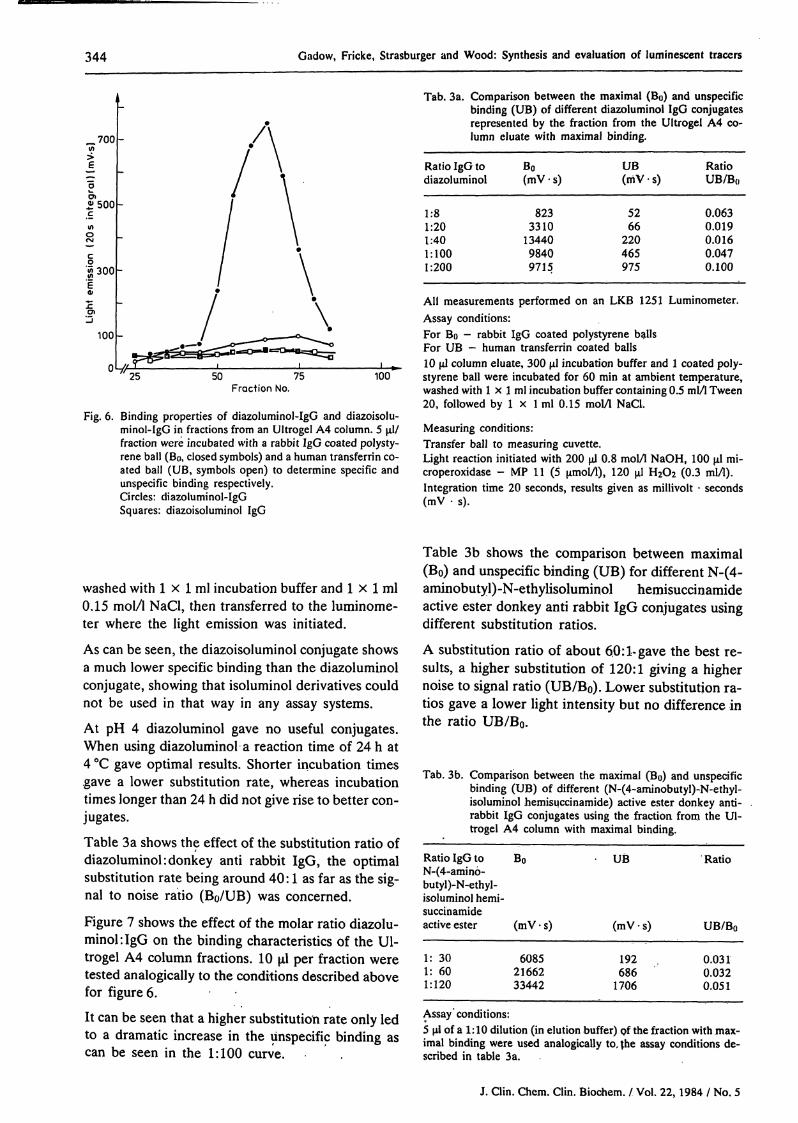
Table 3b shows the comparison between maximal(B ) and nspecific binding ( B) for different N-(4-aminobutyl)-N-ethylisoluminol hemisuccinamideactive ester donkey anti rabbit IgG conjugates usingdifferent Substitution ratios.
A Substitution ratio of about 60: l· gave the best re-sults, a higher Substitution of 120:1 giving a highernoise to signal ratio (UB/B0). Lower Substitution ra-tios gave a lower light intensity but no difference inthe ratio UB/B0.
Tab. 3b. Comparison between the maximal (B ) and nspecificbinding ( B) of different (N-(4-aminobutyl)-N-ethyl-isoluminol hemisuccinamide) active ester donkey anti-rabbit IgG conjugates using the fraction from the Ul-trogel A4 column with maximal binding.
Ratio IgG toN-(4-amino-butyl)-N-ethyl-isoluminol hemi-succinamideactive ester
1: 301: 601:120
B
(mV- s)
60852166233442
B
(mV · s)
192686
1706
Ratio
UB/BO
0.0310.0320.051
Assay'conditions:5 μΐ of a 1:10 dilution (in elution buffer) of the fraction with max-imal binding were used analogically to, {he assay conditions de-scribed in table 3a.
J. Clin. Chem. Clin. Biochem. / Vol. 22, 1984 / No. 5

Gadow, Fricke, Strasburger and Wood: Synthesis and evaluation of luminescent tracers 345
12
10
£ 8
cn
- 6c
g 4
Fraction No.
Fig. 7. Effect of different Substitution rates diazoluminol: IgG onbinding profile of column eluent. The labelled antibodywas äs in figure 6, i.e. an anti rabbit IgG, the coated bailsfor specific (Bö) and unspectfic binding (ÜB) again beingrabbit-IgG and human transferrin äs above. The incuba-tion time was 60 min, 5 of the column fraction beingtested. The ÜB represents the non-specific adsorption toor reaction with the transferrin coated polystyrene ball.
RatioIgG: diazoluminol
Bö O—O 1:100 O—O ÜBBö — 1: 40 — ÜBBö — 1: 20Bö — 1: 8
Discession
The pfoblems and possible Solutions for set up solidphase based luminescence immunoassays have beenlimited to the preparation of luminescence conju-gates and hapteii-pfotein conjugates in the secondpaft of this trilogy.
The bioluminescent labels here described show theproblems of working with kinases, that is the cou-pling of kinases to antigens or antibodies withoutloss of antigenic or enzymatic actjvity. Of all kinasestested only pyruvate kinase was robust enough to"survive" coupling using the heterobifunctional rea-gents tested here. The other kinases, although at-traetive on paper, for example adenylate kinase withits molecular weight of 21000, resistanee to acid pH,high turnover rate and single Substrate (ADP) wereunable to be used due to their sensitivity to maleim-ide. Other coupling reagents (carbodiimides, pen-tane-l,5-dial, bisfcpoxirane) were found to be un-suitable äs the enzyme activity disappeared duringcoupling. The results show that pyruvate kinase, des-
pite its high molecular weight (240000) was used be-eause of available SH-groups not present in the ac-tive centre.Whether the use of SH-group introducing reagentssuch äs (N-succinimidyl 3-(2-pyridyldithio) propion-ate) could make the other kinases useable, is a pointworth considering.The results from the Lineweaver-Burk plots con-firmed that the coupling reagents here chosen led tochanges in enzyme activity and structure, äs mea-sured by Substrate affinity. As previously stated (1),difficulties of finding a suitable antimicrobial reagentfor bioluminescent labels of this type presents a ma-jor drawback, which suggests that such labels shouldonly be used where insufficient sensitivity can be ob-tained with chemiluminescent labels. The latter donot suffer such problems, partly due to their hydra-zide structure in the case of diazoluminol, which initself appears to exert bacteriostatic properties.
Many of the chemiluminescent labels described inthe literatüre suffer from the drawback that theirsynthesis is somewhat complicated (6). The labelsdescribed here are simple to produce, and do not re-quire much expertise and equipment, äs well äs giv-ing out sufficient light for most purposes, althoüghN-(4-aminobutyl)-N-ethylisoluminol hemisuccin-amide gave the best results.Diazoluminol conjugates have in contrast to diazo-isoluminol conjugates a higher light Output (10). Thereaction of diazoisoluminol in the presence of pro-teins was far quicker than of diazoluminol, äs moni-tored by the decolorisation. This may be explainedby the active diazogroup, which in isoluminol is lo-cated on the less sterically hindered 6 (4)-position.Diazoluminol is not äs reactive due to the position ofthe oxo-group on the azine ring, and due to its abilityto build ä stable internal structure (see fig. 8). Thereaction of proteins with diazoluminol could be di-vided into two distinct phases, a rapid reaction withthe protein in alkaline solution (pH 9—10) in whichthe reaction mixture remained red (excited electronsin the diazo bridge are still present). Such conjugatesare stable enough to be separated on an agarose gelcolumn (Ultrogel A4 or A6), however they are un-stable enough to transfer the diazoluminol group toanother protein if the latter is present in excess. Atthis stage a transitional bonding is present in whichboth ionic and covalent components are present. Thesecond stage of the reaction is slower and is associat-ed with a disappearence of the red coloration, andthe appearance of a yellow-orange colour, in whichthe covalent bonding is stable. These conjugates canbe stored at —20 °C for over a year without appreci-able loss in immunological and luminogenic activity.
J. Clin. Chem. Clin. Biochem. / Vol. 22, 1984 / No. 5

346 Gadow, Fricke, Strasburgcr and Wood: Synthesis and evaluation of luminescent tracers
^ Internal stobilisation(preferred)
Oimer formation(not preferred)
Formation of diazotate under alkaline conditions
Structure of diazoisoluminol(dimer formation preferred)
Fig. 8. This set of reactions shows the possible reactions occur-ring during diazotisation under alkaline conditions, äs car-ried out here. Diazoluminol prefers an internal stabilisa-tion (above reaction sequence - centre picture) whereasdiazoisoluminol prefers dimer formation äs the position ofthe diazogroup is unfavorable for an internal stabilisation.This may account for the fact that diazoisoluminol isformed in solution, but precipitates out within 30 minutesäs a golden-yellow crystalline deposit. Diazoluminol atequimolar concentrations remains in solution. The middleset of reactions shows the formation of diazotate underalkaline conditions.
An important point to mention here, is that thematerials to be diazotised must be free from azide, äsdiazoluminol reacts with sodium azide to form com-pounds which do not react with the tyrosine moietiesof the protein. Here the decolorisation of the reac-tion mixture takes place within seconds, a yellöw so-lution being formed. If this solution is then adjustedto pH 7 with phosphate buffer, it darkens due toreaction with atmospheric oxygen, suggesting thatpoly^phenol derivatives may be formed. This wouldbe in accordance with a free radical reactiqn follow-ing the loss of nitrogen from the diazoluminol-azidecomplex. These unwanted effects can be avoided bytreatment öf the material to be diazotised withDEAE cellulose in the form of a short column (2,15), äs already described.
The comparison of the detection limits for diazolu-minol and N-(4-aminobutyl)-N-ethylisoluminol he-misuccinämide shows no significaiit difference. Onthe other hand N-(4-aminobutyl)-N-ethyüsplüminolhemisuccinamide active ester labelled IgG-mole-cules show much better light intensities than thoselabelled with diazoluminol. This could be explainedby the different reaction mechanism of these twocompounds. The reaction of diazoluminol with ty-rosyl-residues of IgG is of an oxidative nature andmay destroy antibödy stracture (compare chloram-ine-T iodination of certain proteins which lead to ac-tivity loss). The N-(4-aminobutyl)-N-ethylisolumin-ol hemisuccinamide active ester reacts with amino-moieties under non-oxidative conditions (compareBolton-Hunter iodination of proteins).
Finally, it can be stated that the choice of label used,i.e., whether chemi- or bioluminescent depends to alarge extent upon the sensitivity of the assay, and al-though the assays described to date mäinly use theformer (7, 8, 9), the latter may have to be used untilsufficient sensitivity can be achieved using chemi-luminescent markers.
References1. Fricke, H., Strasburger, C. J. & Wood, W. G. (1982) J. Clin.
Chem. Clin. Biochem. 20, 91-94.2. Strasburger, C. J., Fricke, H. & Wood, W. G. (1982) In: Ra-
dioimmunoassay and related procedures in medicine 1982,IAEA, Vienna, pp. 757-777.
3: Wood, W. G., Fricke, H., Strasburger, C. J. & Scriba, P. C.(1982) J. Clin. Chem. Clin. Biochem. 20, 825-831.
4. Hastings, J. W. & Nealson, K. H. (1977) Ann. Rev. Micro-biol. 31, 549-595.
5. Schräm, E. (1981) In: Monoclonal antibodies and develop-ments in immunoassay (Ekins, R. & Albertini, A., eds.) Ei-sevier North-Holland Biomedical Press, Amsterdam, pp.123-134.
6. Schroeder, H. R., Boguslaski, R. C., Carrico, R. J. & Buckler,R. T. (1978) Meth. Erizymol. 57, 424-445.
7. Pazzagli, M., Serio, M., Munson, P. & Rodbard, D. (1982)In: Radioimmunoässay and related techniques in medicine1982, IAEA, Vienna, pp. 747-755.
8. Schroeder, H. R., Hines, C. M., Osborn, D. D«% Moore, R. P.,Hurtle, R. L., Wogomänn, F. F., Rpgers, R. W. & Vogelhut,P. O. (1981) Clin, Chem. 27, 1378-1384.
9. Woodhead, J. S., Simpson, J. S. A., Weeks, I., Patel, A.,. Campbell, A. K., Hart, R., Richardson, A. & McCapra, F.
(1981) In: Monoclonal antibodies and.developments in im-munoassay (Ekins, R. & Albertini, A^eds.) Eisevier North-Holland Biomedical Press, Amsterdam, pp. 135—145.
J. Clin. Chem. Clin. Biochem. / Vol. 22,1984 / Nö. 5

Gadow, Fricke, Strasburger and Wood: Synthesis and evaluation of luminescent tracers 347
10. Hemmilä, I. & Lövgren, T. (1982) Wallac Report - Studieson luminescent immunoassay I — Preparation of luminescentconjugates, Wallac Oy, Turku, Finland, 27 Pages.
11. Bundesrepublik Deutschland - Deutsches Patentamt (1976)Offenlegungsschrift 2618511, (US Patent 28.4.75 No572008).
12. Kitagawa, T. & Aikawa, T. (1976) J. Biochem. 79,233-236.13. Barman, T. E. (1969) In: Enzyme Handbook Vol I, Springer
Verlag Berlin, pp. 394, 412-413, 428.
14. Wood, W. G. & Gadow, A. (1983) J. Clin. Chem. Clin. Bio-. ehem. 27, 789-797.15. Cooper, T. G, (1981) In: Biochemische Arbeitsmethoden,
De Gruyter, Berlin, New York, pp. 126—156.16. Ellman, G. L. (1958) Arch. Biochem. Biophys. 74, 443.17. Sedlak, J. & Lindsay, R. H. (1968) Anal. Biochem. 25,192-
205.
Priv.-Doz. Dr. W. G. WoodKlinische LaboratorienKlinik für Innere MedizinMedizinische Hochschule LübeckRatzeburger Allee 160D-2400 Lübeck l
J. Clin. Chem. Clin. Biochem. / Vol, 22, 1984 / No. 5

Copyright © 2022 FDOKUMEN