about the use and benefits of worms in aquaculture - AquaVIP
ZOOSYMPOSIAISSN 1178-9905 (print edition)
ISSN 1178-9913 (online edition)Copyright © 2009 · Magnolia Press
Zoosymposia 2: 379–389 (2009) www.mapress.com/zoosymposia/
On the morphology of elytra as luminescent organs in scale-worms (Polychaeta, Polynoidae)
MARIA PLYUSCHEVA1 & DANIEL MARTINCentre d'Estudis Avancats de Blanes (CEAB, CSIC), Carrer d’accés a la Cala Sant Francesc, 14, 17300 Blanes (Girona), Catalunya, Spain. E-mail: [email protected], [email protected] author
Abstract
Polynoid polychaetes are common marine invertebrates worldwide that are characterized by bearing series of paired elytra attached to dorsal prominences (the elytrophores) arising from the notopodia, and whose dorsal surface is usually ornamented with different papillae (usually thought to be sensory organs). Upon stimulation, some species of the sub-family Polynoinae are able to emit light flashes from the ventral epithelium of the elytra. This bioluminescence originates in a protein called polynoidin, and seems to be induced by the destruction of the electrochemical coupling between body and elytra when the latter are detached. However, the elytral structure, as well as the function of the papillae and tubercles in relation to the bioluminescence is poorly known. In this paper, we report on the elytral morphology of two “luminescent” and two “non-luminescent” (Nicol 1953) species from the White and Mediterranean Seas. In both polynoid types, the elytral tubercles are formed by a layer of hard, non-organized, autofluorescent tissue, apparently filled by expansions protruding from cells forming a distinct subjacent layer. Our study allowed us to suggest that the luminescent protein is located in the cells of the basal layer, while the tubercles may act as lenses helping in the light flash transfer towards the exterior. The reasons why the studied species are or are not bioluminescent are discussed.
Key words: polychaetes, scale-worms, bioluminescence, elytra, polynoidin, White Sea, NW Mediterranean
Introduction
Bioluminescence occurs in many different species in phylogenetically diverse groups (Chalfie & Kain 2005). The type of light and the emission methods, as well as the color, may be very different. The functions may differ among organisms but also a given organism may utilize luminescence in more than one way (Morin 1983; Hastings1983; Hastings & Morin 1991). Bioluminescence functions may be classified under three major categories: defensive (as a help to escape from predators), offensive (as a support for predation), and communication (for courtship or mating). Within each category a number of different specific strategies are recognized. For instance, luminescence may be used as a diverting decoy, as a frightening flash, or as a shape camouflage using ventral luminescent spots (Chalfie & Kain 2005).
The biochemistry of luminous systems is known in detail only for bacteria, dinoflagellates, cnidarians, and fireflies (Hastings & Morin 1991; Chalfie & Kain 2005), although some information exists for another half-dozen or so luminescent taxa (Chalfie & Kain 2005), the polychaetes among
Accepted by N. Maciolek: 20 Apr. 2009; published: 31 Aug. 2009 379
them (Nicol 1953; Fischer & Fischer 1995; Zörner & Fisher 2006). However, this phenomenon occurs sporadically in unrelated polychaete species having different modes of life and belonging to different families, such as the Aphroditidae, Tomopteridae, Syllidae, Alciopidae, Chaetopteridae, Cirratulidae, Terebellidae, and Polynoidae (Nicol 1953).
The polynoids are found worldwide from the tropics to the Antarctic (Hartman 1978) and Arctic (Britayev 1991) Seas. They occur from the intertidal (Hanley et al. 1990) to deep waters (Levenstein & Hutchings 1984, Pettibone 1989), where they have been reported from abyssal and hadal depths (Hartman & Fauchald 1971), and may be common on both soft and hard bottoms. Their dorsum is covered by ornamented scales or elytra, so that they also known as scale-worms. Elytra can be minute or large and overlapping, and can be smooth or covered with micro- or macrotubercles. In fact, elytra are dorsal cirri modified into a flat, discoid expansion composed from a single-layered epithelium covered with a cuticle (Bassot & Nicolas 1995). The epithelial cells send long pillars, which are anchored to the cells of the opposite face and account for the thickness of the elytrum (Pavans de Ceccatty et al. 1972).
When polynoids lose their elytra, the elytrophore remains as a stump, which is rapidly closed by the musculature. In some species, elytral dehiscence occurs across a distinct line and only during strong mechanical stimuli, while, in some others, the elytra may easily autotomize at the level of the insertion of its elytrophore (Storch & Alberti 1995). Afterwards, elytra can be regenerated in 10–15 days.
In some species of the scale-worm sub-family Polynoinae (Harmothoe Kinberg, 1856) and sub-family Acholoinae (Acholoe Claparède, 1870) an area of the epithelium of the lower surface of the elytra has been reported to emit light flashes upon stimulation. Some other polynoids seem not to be luminescent (e.g., Lepidonotus clava Montagu, 1808, L. squamatus Linnaeus, 1767, Halosydna gelatinosa Sars, 1835, Lepidasthenia argus Hodgson, 1900) (Nicol 1953, 1954, 1957a–c, 1958). In the light-producing species, the lower surface of the elytra has a layer of luminescent cells or photocytes (actually modified epidermal cells), which are lacking in the non-luminescent species (Nicol 1953). The behavioral or ecological function of this bioluminescence remains unknown, although it has been suggested that the signal could be either a warning or a distracting mechanism (Bassot & Nicolas 1995). The bioluminescence is actually originated in a membrane photoprotein that reacts specifically to the presence of superoxide anions (a reactive oxygen species, ROS), but no to other ROS such as hydrogen peroxide (Bassot & Nicolas 1995). This membrane photoprotein was later called polynoidin (Bassot & Nicolas 1987).
In this paper, we analyze the elytral morphology of two “luminescent” and two “non-luminescent” species of scale-worms from the White and Mediterranean Seas in order to assess the reasons why some polynoids are luminescent and some others are not.
Materials and methods
Harmothoe imbricata Linnaeus, 1767 and Lepidonotus squamatus were collected in Kandalaksha Bay (White Sea, 18–20 m deep) and kept alive in artificial sea water at 6–8°С. Harmothoe areolataGrube, 1860 and Lepidonotus clava were collected near Blanes (NW Mediterranean Sea, 3–30 m depth) and kept alive in native seawater at 17–18°C.
Luminescence was measured as a single quanta per second emitted from the dorsal or ventral surface of elytra on a microluminometer linked to a PC, either in seawater or in a 50 mM, Na-phosphate buffer with 150 mM NaCl and pH 7.4, both in the presence or absence (in special cases) of 1 μM Ca2+ and 1 mM EGTA. The intensity of luminescence was measured on the two elytral
PLYUSCHEVA & MARTIN380 · Zoosymposia 2 © 2009 Magnolia Press
surfaces (i.e., dorsal and ventral) using elytra showing different coloration intensity: blackish, reddish, and beige.
The protein was originally purified by homogenizing 10–15 elytra in a Potter glass-Teflon homogenizer for 2–5 min at 4°С in a phosphate buffer (see above), containing 0.05% of the nonionic detergent Triton X-100 (ratio 25:1 ml/g of tissue) and a cocktail of protease inhibitors. The homogenate was then centrifuged at 1000 g for 3 min on a Beckman high-speed centrifuge model J2-21, JA-21 rotor at 4ºС. Particle-free supernatant was loaded on a Sepharose CL6B Pharmacia pre-packed 30/60 column, equilibrated with the same phosphate buffer, and the protein was eluted in a volume corresponding to 60 kDa (according to a calibration curve with protein standards in the 14–200 kDa range). The presence of polynoidin was detected by its luminescence in a phosphate buffer supplemented by 0.2 mM hypoxanthine and 1 U/ml of xanthine oxidase under the microluminometer.
The purity of the protein and its molecular mass was confirmed by Laemmli SDS-PAGE electrophoresis. Molecular mass determination was repeated by rapid ultracentrifugation at 400,000 g on a Beckman analytical ultracentrifuge in 0.3 M sucrose medium. The polynoidin isoelectric point was determined by the O’Farrel method in a pH gradient from 3 to 10 in 6% PAGE on a Biometra capillary system for isoelectric focusing. The protein determination was done with bicinchoninic acid. The protein spectra and extinction coefficient was obtained on an Aminco DW2000 double beam spectrophotometer.
The histological studies were done with classical protocols for paraffin embedding (Valovaja & Kavtaradze 1993). Before fixation worms were relaxed with the help of 7.5 % MgCl2 solution and then fixed with 4% paraformaldehyde in phosphate buffer (PBS, pH 7.5). Material was embedded in paraffin, sectioned at 5 µm, and stained with Harris iron haematoxylin.
The spatial distribution of polynoidin was studied with original polyclonal antipolynoidin rabbit antibodies, according to the following immunolabeling protocol. Selected elytra of the four studied species were fixed with 4% paraformaldehyde in artificial seawater (pH 8.2) for 24 h at 4°C, then washed three times in 0.1 M phosphate buffered saline (PBS) for 1 h and stored in 1% Triton X-100 in 0.1 M PBS (PBT) at 4°C to permeabilize the tissue. The nonspecific binding sites were blocked by storing them in PBT with 5% goat serum (block-PBT) for 3 h at room temperature (RT). The next steps were all performed at 4°C. The elytra were incubated in a 1:100 dilution of the primary polyclonal antipolynoidin antibodies in block-PBT for 24 h. After washing them three times (20 min each) in PBS, the elytra were incubated in a 1:200 dilution of Oregon Green goat anti-rabbit (Molecular Probes) in block-PBT for 12 h, washed three times (20 min each) in PBS and immersed in glycerol. Negative controls were obtained by omitting the primary antibodies to show the nonspecific staining.
All observations were made with a Zeiss Axioplan microscope equipped with fluorescence. The image capturing was made with a Jenoptic digital camera and software.
Results
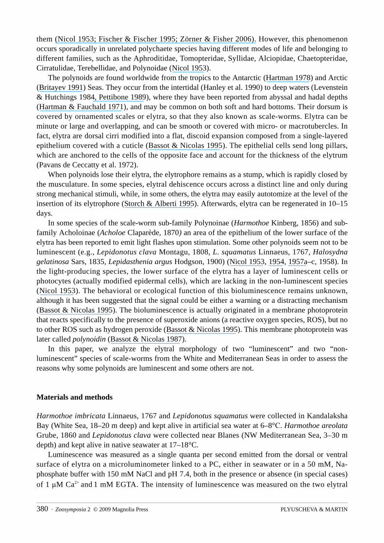
Luminescence measurementsThe measurements of elytral luminescence directly on the dorsal side of a single small Harmothoe imbricata (without stimulation) revealed light emissions of the same level as background seawater emissions. Bright luminescence was registered in two cases: (1) after electrically stimulating the whole worm by means of a single +10mV/20Hz impulse, and (2) by adding the hypoxanthine-xanthine oxidase system, which produces superoxide anion radicals (Fig. 1).
Zoosymposia 2 © 2009 Magnolia Press · 381BIOLUMINESCENCE IN POLYNOIDAE
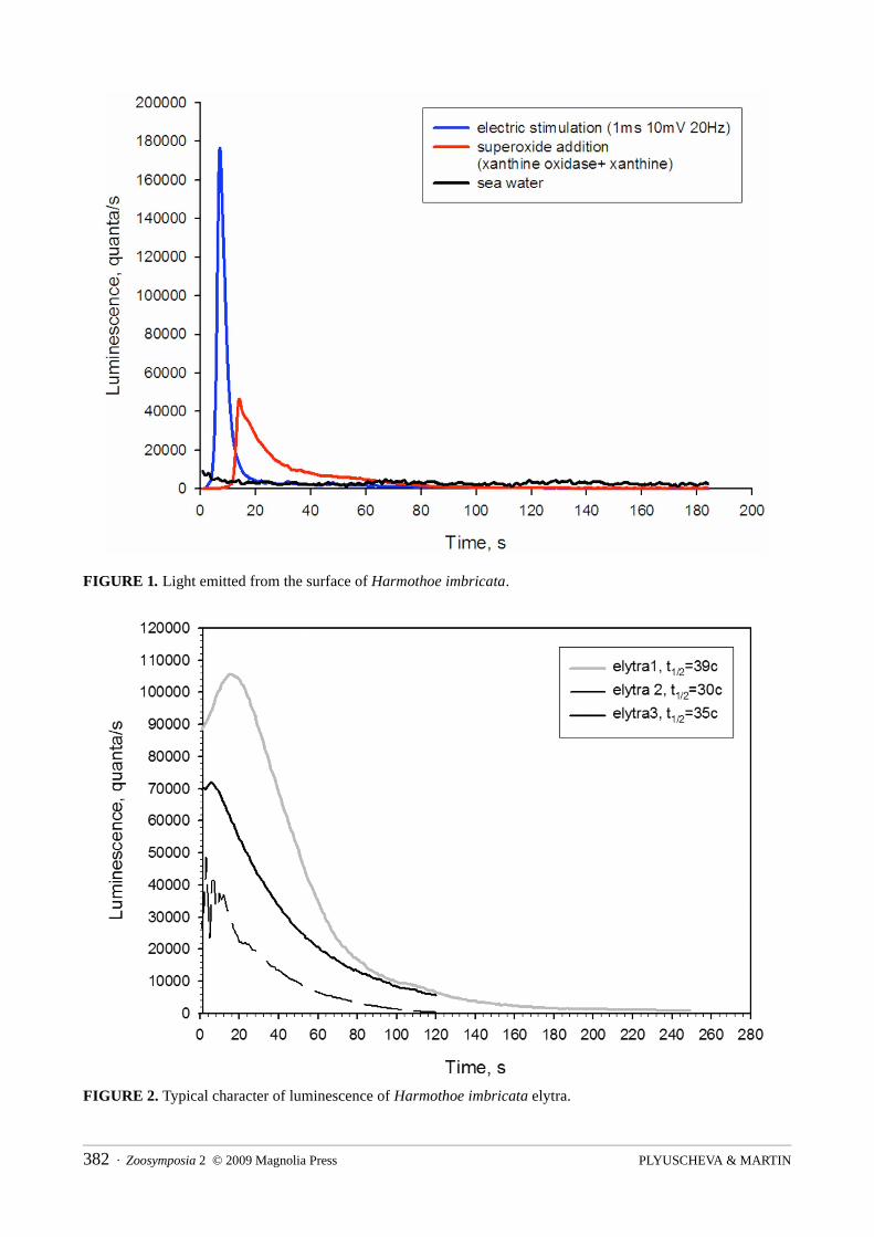
FIGURE 1. Light emitted from the surface of Harmothoe imbricata.
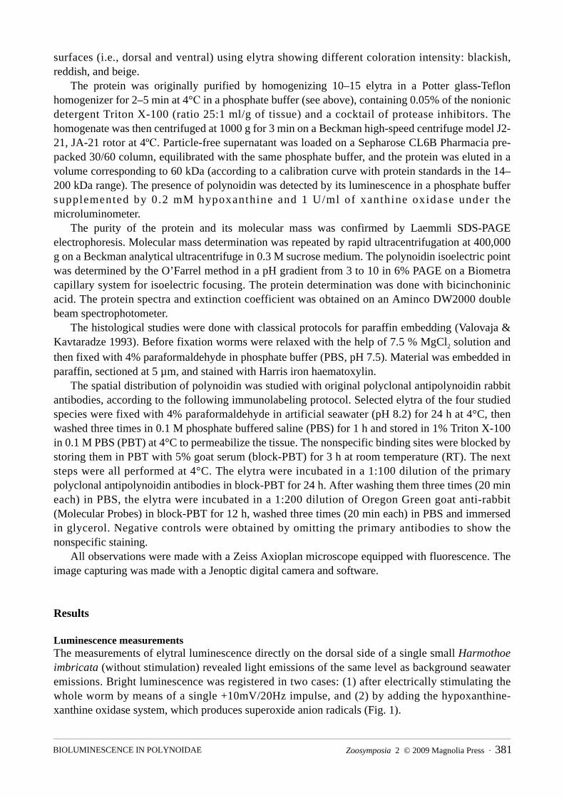
FIGURE 2. Typical character of luminescence of Harmothoe imbricata elytra.
PLYUSCHEVA & MARTIN382 · Zoosymposia 2 © 2009 Magnolia Press
Several assays also demonstrated that luminescence can be induced by detaching the elytra from worm’s body (Fig. 2), either by artificial means or by natural autotomization as a response to irritation. After autotomizing, the isolated elytra of luminescent polynoids continue to flash for some time. Independently of the elytral pigmentation (i.e., blackish, reddish or beige), the light emission was always more intense from the dorsal than from the ventral face (Fig. 3).
FIGURE 3. The luminescence intensity of Harmothoe imbricata elytra with different pigmentation. A, black; B, red; C, beige.
The elytra of Lepidonotus squamatus were non–bioluminescent, neither with nor without stimulation. Luminescence was neither registered after electrically stimulating the whole worm (by a single +10mV/20Hz impulse) nor in the presence of the superoxide anion radicals (produced by the hypoxanthine-xanthine oxidase system). Conversely, the experimentally homogenized elytra emitted an increasing luminescence in the presence of a superoxide donor system (Fig. 4).
Zoosymposia 2 © 2009 Magnolia Press · 383BIOLUMINESCENCE IN POLYNOIDAE
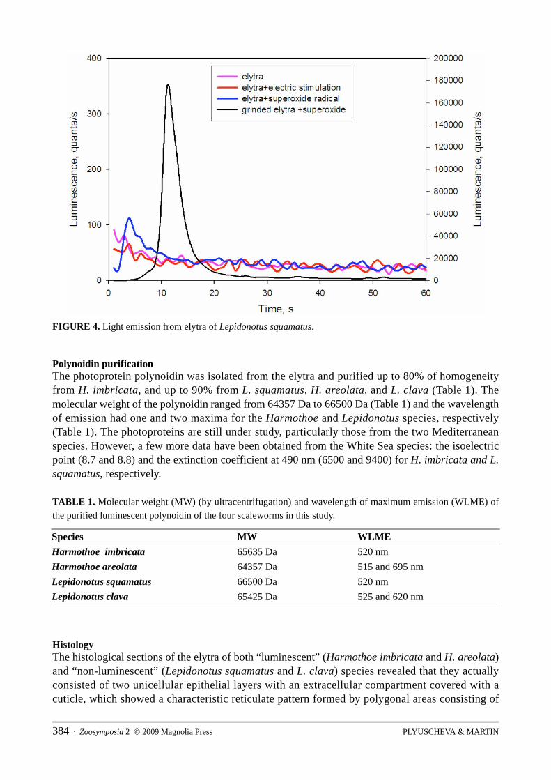
FIGURE 4. Light emission from elytra of Lepidonotus squamatus.
Polynoidin purificationThe photoprotein polynoidin was isolated from the elytra and purified up to 80% of homogeneity from H. imbricata, and up to 90% from L. squamatus, H. areolata, and L. clava (Table 1). The molecular weight of the polynoidin ranged from 64357 Da to 66500 Da (Table 1) and the wavelength of emission had one and two maxima for the Harmothoe and Lepidonotus species, respectively (Table 1). The photoproteins are still under study, particularly those from the two Mediterranean species. However, a few more data have been obtained from the White Sea species: the isoelectric point (8.7 and 8.8) and the extinction coefficient at 490 nm (6500 and 9400) for H. imbricata and L. squamatus, respectively.
TABLE 1. Molecular weight (MW) (by ultracentrifugation) and wavelength of maximum emission (WLME) of the purified luminescent polynoidin of the four scaleworms in this study.
HistologyThe histological sections of the elytra of both “luminescent” (Harmothoe imbricata and H. areolata) and “non-luminescent” (Lepidonotus squamatus and L. clava) species revealed that they actually consisted of two unicellular epithelial layers with an extracellular compartment covered with a cuticle, which showed a characteristic reticulate pattern formed by polygonal areas consisting of
Species MW WLME
Harmothoe imbricata 65635 Da 520 nm
Harmothoe areolata 64357 Da 515 and 695 nm
Lepidonotus squamatus 66500 Da 520 nm
Lepidonotus clava 65425 Da 525 and 620 nm
PLYUSCHEVA & MARTIN384 · Zoosymposia 2 © 2009 Magnolia Press
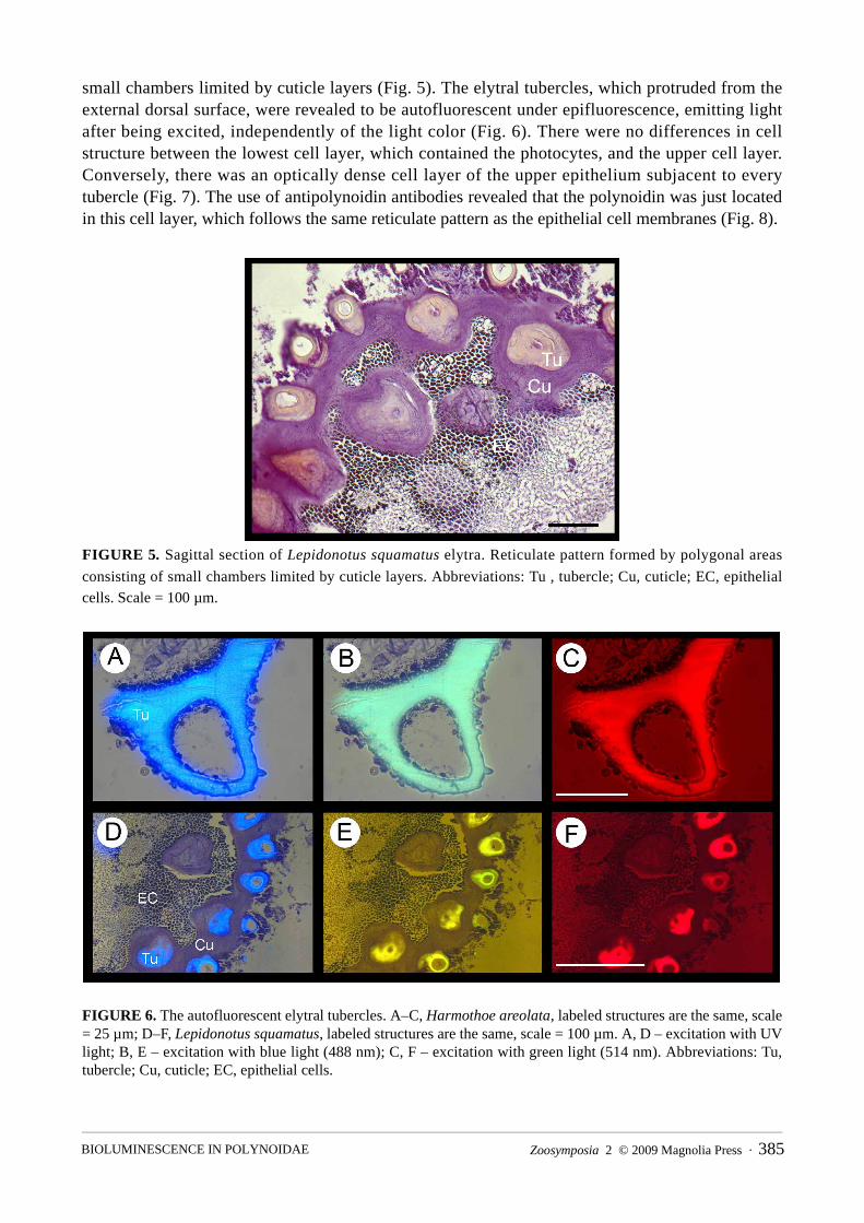
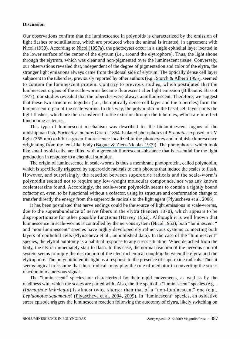
small chambers limited by cuticle layers (Fig. 5). The elytral tubercles, which protruded from the external dorsal surface, were revealed to be autofluorescent under epifluorescence, emitting light after being excited, independently of the light color (Fig. 6). There were no differences in cell structure between the lowest cell layer, which contained the photocytes, and the upper cell layer. Conversely, there was an optically dense cell layer of the upper epithelium subjacent to every tubercle (Fig. 7). The use of antipolynoidin antibodies revealed that the polynoidin was just located in this cell layer, which follows the same reticulate pattern as the epithelial cell membranes (Fig. 8).
FIGURE 5. Sagittal section of Lepidonotus squamatus elytra. Reticulate pattern formed by polygonal areas
consisting of small chambers limited by cuticle layers. Abbreviations: Tu , tubercle; Cu, cuticle; EC, epithelial cells. Scale = 100 µm.
FIGURE 6. The autofluorescent elytral tubercles. A–C, Harmothoe areolata, labeled structures are the same, scale = 25 µm; D–F, Lepidonotus squamatus, labeled structures are the same, scale = 100 µm. A, D – excitation with UV light; B, E – excitation with blue light (488 nm); C, F – excitation with green light (514 nm). Abbreviations: Tu, tubercle; Cu, cuticle; EC, epithelial cells.
Zoosymposia 2 © 2009 Magnolia Press · 385BIOLUMINESCENCE IN POLYNOIDAE
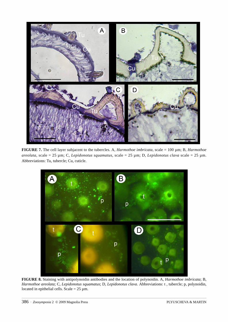
FIGURE 7. The cell layer subjacent to the tubercles. A, Harmothoe imbricata, scale = 100 µm; B, Harmothoe
areolata, scale = 25 µm; C, Lepidonotus squamatus, scale = 25 µm; D, Lepidonotus clava scale = 25 µm. Abbreviations: Tu, tubercle; Cu, cuticle.
FIGURE 8. Staining with antipolynoidin antibodies and the location of polynoidin. A, Harmothoe imbricata; B, Harmothoe areolata; C, Lepidonotus squamatus; D, Lepidonotus clava. Abbreviations: t , tubercle; p, polynoidin, located in epithelial cells. Scale = 25 µm.
PLYUSCHEVA & MARTIN386 · Zoosymposia 2 © 2009 Magnolia Press
Discussion
Our observations confirm that the luminescence in polynoids is characterized by the emission of light flashes or scintillations, which are produced when the animal is irritated, in agreement with Nicol (1953). According to Nicol (1957a), the photocytes occur in a single epithelial layer located in the lower surface of the center of the elytrum (i.e., around the elytrophore). Thus, the light shone through the elytrum, which was clear and non-pigmented over the luminescent tissue. Conversely, our observations revealed that, independent of the degree of pigmentation and color of the elytra, the stronger light emissions always came from the dorsal side of elytrum. The optically dense cell layer subjacent to the tubercles, previously reported by other authors (e.g., Storch & Alberti 1995), seemed to contain the luminescent protein. Contrary to previous studies, which postulated that the luminescent organs of the scale-worms became fluorescent after light emission (Bilbaut & Bassot 1977), our studies revealed that the tubercles were always autofluorescent. Therefore, we suggest that these two structures together (i.e., the optically dense cell layer and the tubercles) form the luminescent organ of the scale-worms. In this way, the polynoidin in the basal cell layer emits the light flashes, which are then transferred to the exterior through the tubercles, which are in effect functioning as lenses.
This type of luminescent mechanism was described for the bioluminescent organs of the midshipman fish, Porichthys notatus Girard, 1854. Isolated photophores of P. notatus exposed to UV light (365 nm) exhibit a green fluorescence localized in the photocytes and a bluish fluorescence originating from the lens-like body (Baguet & Zietz-Nicolas 1979). The photophores, which look like small ovoid cells, are filled with a greenish fluorescent substance that is essential for the light production in response to a chemical stimulus.
The origin of luminescence in scale-worms is thus a membrane photoprotein, called polynoidin, which is specifically triggered by superoxide radicals to emit photons that induce the scales to flash. However, and surprisingly, the reaction between superoxide radicals and the scale-worm’s polynoidin seemed not to require any low-weight molecular compounds, nor was any known coelenterazine found. Accordingly, the scale-worm polynoidin seems to contain a tightly bound cofactor or, even, to be functional without a cofactor, using its structure and conformation change to transfer directly the energy from the superoxide radicals to the light agent (Plyuscheva et al. 2006).
It has been postulated that nerve endings could be the source of light emissions in scale-worms, due to the superabundance of nerve fibers in the elytra (Panceri 1878), which appears to be disproportionate for other possible functions (Harvey 1952). Although it is well known that luminescence in scale-worms is controlled by the nervous system (Nicol 1953), both “luminescent “ and “non-luminescent” species have highly developed elytral nervous systems connecting both layers of epithelial cells (Plyuscheva et al., unpublished data). In the case of the “luminescent” species, the elytral autotomy is a habitual response to any stress situation. When detached from the body, the elytra immediately start to flash. In this case, the normal reaction of the nervous control system seems to imply the destruction of the electrochemical coupling between the elytra and the elytrophore. The polynoidin emits light as a response to the presence of superoxide radicals. Thus it seems logical to assume that these radicals may play the role of mediator in converting the stress reaction into a nervous signal.
The “luminescent” species are characterized by their rapid movements, as well as by the readiness with which the scales are parted with. Also, the life span of a “luminescent” species (e.g. , Harmothoe imbricata) is almost twice shorter than that of a “non-luminescent” one (e.g., Lepidonotus squamatus) (Plyuscheva et al. 2004, 2005). In “luminescent” species, an oxidative stress episode triggers the luminescent reaction following the autotomy of elytra, likely switching on
Zoosymposia 2 © 2009 Magnolia Press · 387BIOLUMINESCENCE IN POLYNOIDAE
a controlled cell–death cascade reaction. The luminescent reaction seems to be energetically expensive for the organism, with the apoptosis and the programmed cell–death reaction occurring in epithelial cells on one hand, and the further regeneration of autotomized elytra on the other. This could certainly be related with the relative shortness of their life span.
In turn, the presence of polynoidin in “non-luminescent” species allows us to suggest that the ability of being bioluminescent could be a primitive feature that has been lost and replaced by a mechanism able to catch the superoxide radicals, which could play a protective role as endogenous inhibitor of oxidative stress. Certainly, this would explain why “non-luminescent worms may have longer life span. Like this, they could also save the energy previously spent in being bioluminescent, to address it into other defensive strategies such as the more robust elytra and coil up behavior reported for Lepidonotus spp. (Plyuscheva et al. 2004), which can in turn be connected with their longer life span.
Acknowledgements
This paper has been possible thanks to a grant of the Spanish Agency for the International Cooperation (AECI) of the “Ministerio de Esuntos Extrangeros” to the first author and a grant of the Russian Foundation for Basic Research � 07-04-01545-a. The paper is a contribution to the project MARMOL (Ref Numb. CTM2007-66635-CO2-01) financed by the Spanish Commission of Science and Technology (CICYT). The authors would like to thank Mikhail Vyssokikh for his help with the polynoidin purification and analyses and antibody preparation. We would also like to thank the White Sea Biological Station team for the help during the collection of the scale-worms.
References
Baguet, F. & Zietz-Nicolas, A.M. (1979) Fluorescence and Luminescence of Isolated Photophores of Porichthys.
Journal of Experimental Biology, 78, 47–57.Bassot, J.M. & Nicolas, G. (1987) An optional dyadic junctional complex revealed by fast-freeze fixation in the
bioluminescent system of the scale worm. The Journal of Cell Biology, 105, 2245–2256.Bassot, J.M. & Nicolas, G. (1995) Bioluminescence in scale-worm photosomes: the photoprotein polynoidin is
specific for the detection of superoxide radicals. Histochemistry and Cell Biology, 104, 199–210.Bilbaut, A. & Bassot, J.M. (1977) Bioluminescence des e1ytres d’Acholoe. II. Donnees photometriques. Biology of
the Cell, 28, 145–154.Britayev, T.A. (1991) Life cycle of the symbiotic scale-worm Arctonoe vittata (Polychaeta: Polynoidae). In:
Petersen, M.E. & Kirkegaard, J.B. (Eds.) Systematics, Biology and Morphology of World Polychaeta. Proceedings of the Second International Polychaeta Conference. Ophelia Supplement 5, 305–312.
Chalfie, M. & Kain, S.R. (2005) Green fluorescent protein: Properties, Applications, and Protocols. 2nd edition. John Wiley and Sons, Inc., Hoboken, New Jersey, 443 pp.
Fischer, A. & Fischer, U. (1995) On the life-style and life-cycle of the luminescent polychaete Odontosyllis enopla
(Annelida, Polychaeta). Invertebrate Biology, 114, 236–247.Hanley, J.R., Burke, M., Wells, F.E., Walker, D.I., Kirkman, H. & Lethbridge, R. (1990) Scale-worms (Polychaeta:
Polynoidae) of Albany, Western Australia. In: Wells, F.E. (Ed), Proceedings of the Third International Marine
Biological Workshop: The Marine Fauna of Albany, Western Australia. Western Australia Museum, Perth, p 203–236.
Hartman, O. (1978) Polychaeta from the Weddell Sea quadrant, Antarctica. Antarctic Research Series, 26, 125–217.
PLYUSCHEVA & MARTIN388 · Zoosymposia 2 © 2009 Magnolia Press
Hartman, O. & Fauchald, K. (1971) Deep-water benthic polychaetous annelids off New England to Bermuda and other North Atlantic Areas. Part II. Allan Hancock Monographs in Marine Biology, 6, 1–327.
Harvey, E.N. (1952) Bioluminescence. Academic Press, New York, 649 pp.Hastings, J.V. (1983) Biological diversity, chemical mechanisms, and the evolutionary origins of bioluminescent
systems. Journal of Molecular Evolution, 19(5), 309–321.Hastings, J.W. & Morin, J.G. (1991) Bioluminescence. In: Prosser, C.L. (Ed.) Neural and Integrative Animal
Physiology, 4th edition. Wiley, New York, pp 131–170.Haswell, W.A. (1882) On the structure and functions of the elytra of the Aphroditean Annelids. Annals and
Magazine of Natural History, 10 (Ser. 5), 238–242.Levenstein, R.Ya. & Hutchings, P.A. (1984) On the ways of formation of deep-sea polychaeta fauna of the family
Polynoidae. In: Hutchings, P.A. (Ed.) Proceedings of the First International Polychaete Conference. The Linnean Society of New South Wales, Sydney, Australia, 72–85.
Morin, J.G. (1983) Coastal bioluminescence: patterns and functions. Bulletin of Marine Science, 33, 787–817.Nicol, J.A.C. (1953) Luminescence in polynoid worms. Journal of the Marine Biological Association of the United
Kingdom, 32, 65–84.Nicol, J.A.C. (1954) The nervous control of luminescent responses in polynoid worms. Journal of the Marine
Biological Association of the United Kingdom, 33, 225–255.Nicol, J.A.C. (1957a) Luminescence in polynoids. II Different modes of response in the elytra. Journal of the
Marine Biological Association of the United Kingdom, 36, 261–269.Nicol, J.A.C. (1957b). Luminescence in polynoids. III Propagation of excitation through the nerve cord. Journal of
the Marine Biological Association of the United Kingdom, 36, 271–273.Nicol, J.A.C. (1957c) Spectral composition of light of polynoid worms. Journal of the Marine Biological
Association of the United Kingdom, 36, 529–538.Nicol, J.A.C. (1958) Luminescence in polynoids. IV Measurement of light intensity. Journal of the Marine
Biological Association of the United Kingdom, 37, 33–41.Panceri, P. (1878) La luce e gli organi luminosi di alcuni annelidi. Rendiconto dell'Accademia delle Scienze Fisiche
e Matematiche (Sezione della Società Reale di Napoli), 14, 21–25.Pavans de Ceccatty, M., Bassot, J.M., Bilbaut, A. & Nicolas, M.T. (1972) Genese des paracristaux photogenes et de
leurs structures d’excitation dans les cellules de l’e1ytre d’Acholoe astericola. Les Comptes rendus de
l'Académie des sciences (Paris), 275, 2363–2366.Pettibone, M.H. (1989) New species of scale-worms (Polychaeta: Polynoidae) from the hydrothermal rift-area of
the Mariana Back-arc Basin in the Western Central Pacific. Proceedings of the Biological Society of
Washington, 102, 137–153.Plyuscheva, M.V., Martin, D. & Britayev, T.A. (2004) Population ecology of two simpatric polychaetes,
Lepidonotus squamatus and Harmothoe imbricata (Polychaeta, Polynoidae), in the White Sea. Invertebrate
Zoology, 1, 65 –73.Plyuscheva, M.V., Britayev, T.A. & Martin, D. (2005) Life cycle strategy and population ecology of polychaetes
polynoids Lepidonotus squamatus and Harmothoe imbricata in the White Sea. In: Tzetlin, A.B. (Ed.), Proceedings of the IXth Scientific Conference of White Sea Biological Station of Moscow State University. 125 – 130 [In Russian].
Plyuscheva, M., Manukhov, I. & Vissokikh, M. (2006) Peculiarities of luminescence of Harmothoe imbricata. In:
Tzetlin, A.B. (Ed.), Proceedings of the Xth Scientific Conference of White Sea Biological Station of Moscow
State University. 99–103. [In Russian].Storch, V. & Alberti, G. (1995) Elytra of Iphione muricata ((Savigny): a reinterpretation of its architecture based on
TEM. Mitteilungenaus dem Hamburgischen Zoologischen Museum und Institut, 92, 103–124.Valovaja, M.A. & Kavtaradze, D.N. (1993). Microtechnique. Tools. Methods. Craft. Experiment. Moscow State
University Press, 240 pp.Zörner, S.A. & Fischer, A. (2006) The spatial pattern of bioluminescent flashes in the polychaete Eusyllis
blomstrandi (Annelida). Helgoland Marine Research 61, 1, 55–66.
Zoosymposia 2 © 2009 Magnolia Press · 389BIOLUMINESCENCE IN POLYNOIDAE
Copyright © 2022 FDOKUMEN