Growth patterns in Onychophora (velvet worms): lack of a localised posterior proliferation zone
12
RESEARCH ARTICLE Open Access Growth patterns in Onychophora (velvet worms): lack of a localised posterior proliferation zone Georg Mayer 1* , Chiharu Kato 2 , Björn Quast 2 , Rebecca H Chisholm 3 , Kerry A Landman 3 , Leonie M Quinn 4 Abstract Background: During embryonic development of segmented animals, body segments are thought to arise from the so-called “posterior growth zone” and the occurrence of this “zone” has been used to support the homology of segmentation between arthropods, annelids, and vertebrates. However, the term “posterior growth zone” is used ambiguously in the literature, mostly referring to a region of increased proliferation at the posterior end of the embryo. To determine whether such a localised posterior proliferation zone is an ancestral feature of Panarthropoda (Onychophora + Tardigrada + Arthropoda), we examined cell division patterns in embryos of Onychophora. Results: Using in vivo incorporation of the DNA replication marker BrdU (5-bromo-2’-deoxyuridine) and anti- phospho-histone H3 immunolabelling, we found that a localised posterior region of proliferating cells does not occur at any developmental stage in onychophoran embryos. This contrasts with a localised pattern of cell divisions at the posterior end of annelid embryos, which we used as a positive control. Based on our data, we present a mathematical model, which challenges the paradigm that a localised posterior proliferation zone is necessary for segment patterning in short germ developing arthropods. Conclusions: Our findings suggest that a posterior proliferation zone was absent in the last common ancestor of Onychophora and Arthropoda. By comparing our data from Onychophora with those from annelids, arthropods, and chordates, we suggest that the occurrence of a “posterior growth zone” currently cannot be used to support the homology of segmentation between these three animal groups. Background The most obvious subdivision of the body into serially repeated units or segments occurs in annelids (ringed worms), panarthropods (onychophorans, tardigrades and arthropods), and chordates (including vertebrates, uro- chordates and cephalochordates). During embryonic development, segments are commonly believed to origi- nate from the so-called “posterior growth zone” (review [1]). However, this term has been applied very broadly in the past, which has resulted in ambiguity. For exam- ple, the occurrence of a “posterior growth zone” has been used to support the homology of segmentation either specifically in annelids and panarthropods [2-4] or in all three groups of segmented animals, suggesting that segmentation was present in their last common ancestor [1,5-8]. Traditionally, the term “posterior growth zone” has been used to describe a localised and highly proliferative terminal body region, which has been dubbed the “pro- liferating area” or “zone of proliferation” [9-11]. While it seems clear that such a localised proliferation zone is present in embryos, larvae, or juveniles of annelids, including clitellates and polychaetes [11-18], the situa- tion is less clear for chordates. In vertebrate embryos, a higher proliferative activity, as compared to the pre- somitic mesoderm region, consistent with the presence of stem cells has been observed in the tailbud [19-24]. In cephalochordate embryos, the pre-somitic mesoderm region is absent, but the tailbud shows a high number of proliferating cells during somitogenesis [25,26]. In contrast, a “posterior growth zone” is lacking completely from embryos of urochordates [1], as evidenced by var- ious cell lineage and cell proliferation studies [27-29]. * Correspondence: [email protected] 1 Institute of Biology II: Animal Evolution & Development, University of Leipzig, Talstrasse 33, D-04103 Leipzig, Germany Full list of author information is available at the end of the article Mayer et al. BMC Evolutionary Biology 2010, 10:339 http://www.biomedcentral.com/1471-2148/10/339 © 2010 Mayer et al; licensee BioMed Central Ltd. This is an Open Access article distributed under the terms of the Creative Commons Attribution License (http://creativecommons.org/licenses/by/2.0), which permits unrestricted use, distribution, and reproduction in any medium, provided the original work is properly cited.
-
Upload
independent -
Category
Documents
-
view
1 -
download
0
Transcript of Growth patterns in Onychophora (velvet worms): lack of a localised posterior proliferation zone

RESEARCH ARTICLE Open Access
Growth patterns in Onychophora (velvet worms):lack of a localised posterior proliferation zoneGeorg Mayer1*, Chiharu Kato2, Björn Quast2, Rebecca H Chisholm3, Kerry A Landman3, Leonie M Quinn4
Abstract
Background: During embryonic development of segmented animals, body segments are thought to arise fromthe so-called “posterior growth zone” and the occurrence of this “zone” has been used to support the homologyof segmentation between arthropods, annelids, and vertebrates. However, the term “posterior growth zone” is usedambiguously in the literature, mostly referring to a region of increased proliferation at the posterior end of theembryo. To determine whether such a localised posterior proliferation zone is an ancestral feature ofPanarthropoda (Onychophora + Tardigrada + Arthropoda), we examined cell division patterns in embryos ofOnychophora.
Results: Using in vivo incorporation of the DNA replication marker BrdU (5-bromo-2’-deoxyuridine) and anti-phospho-histone H3 immunolabelling, we found that a localised posterior region of proliferating cells does notoccur at any developmental stage in onychophoran embryos. This contrasts with a localised pattern of celldivisions at the posterior end of annelid embryos, which we used as a positive control. Based on our data, wepresent a mathematical model, which challenges the paradigm that a localised posterior proliferation zone isnecessary for segment patterning in short germ developing arthropods.
Conclusions: Our findings suggest that a posterior proliferation zone was absent in the last common ancestor ofOnychophora and Arthropoda. By comparing our data from Onychophora with those from annelids, arthropods,and chordates, we suggest that the occurrence of a “posterior growth zone” currently cannot be used to supportthe homology of segmentation between these three animal groups.
BackgroundThe most obvious subdivision of the body into seriallyrepeated units or segments occurs in annelids (ringedworms), panarthropods (onychophorans, tardigrades andarthropods), and chordates (including vertebrates, uro-chordates and cephalochordates). During embryonicdevelopment, segments are commonly believed to origi-nate from the so-called “posterior growth zone” (review[1]). However, this term has been applied very broadlyin the past, which has resulted in ambiguity. For exam-ple, the occurrence of a “posterior growth zone” hasbeen used to support the homology of segmentationeither specifically in annelids and panarthropods [2-4]or in all three groups of segmented animals, suggesting
that segmentation was present in their last commonancestor [1,5-8].Traditionally, the term “posterior growth zone” has
been used to describe a localised and highly proliferativeterminal body region, which has been dubbed the “pro-liferating area” or “zone of proliferation” [9-11]. While itseems clear that such a localised proliferation zone ispresent in embryos, larvae, or juveniles of annelids,including clitellates and polychaetes [11-18], the situa-tion is less clear for chordates. In vertebrate embryos, ahigher proliferative activity, as compared to the pre-somitic mesoderm region, consistent with the presenceof stem cells has been observed in the tailbud [19-24].In cephalochordate embryos, the pre-somitic mesodermregion is absent, but the tailbud shows a high numberof proliferating cells during somitogenesis [25,26]. Incontrast, a “posterior growth zone” is lacking completelyfrom embryos of urochordates [1], as evidenced by var-ious cell lineage and cell proliferation studies [27-29].
* Correspondence: [email protected] of Biology II: Animal Evolution & Development, University ofLeipzig, Talstrasse 33, D-04103 Leipzig, GermanyFull list of author information is available at the end of the article
Mayer et al. BMC Evolutionary Biology 2010, 10:339http://www.biomedcentral.com/1471-2148/10/339
© 2010 Mayer et al; licensee BioMed Central Ltd. This is an Open Access article distributed under the terms of the Creative CommonsAttribution License (http://creativecommons.org/licenses/by/2.0), which permits unrestricted use, distribution, and reproduction inany medium, provided the original work is properly cited.

Thus, the ancestral state for the chordates remainsunclear.Apart from annelids and vertebrates, a pool of prolif-
erating cells, or stem-like cells, at the posterior end havebeen proposed for the arthropod embryos [3,4,11,30].However, the existence of such a localised zone has onlybeen confirmed for embryos of malacostracan crusta-ceans [31-33]. Although the malacostracan stem-likecells are reminiscent of the clitellate teloblasts, theirhomology is questionable [4,31,32,34,35]. Leaving asidethe issue of the homology of crustacean and clitellateteloblasts, the existence of a posterior pool of proliferat-ing cells has been doubted for all remaining arthropodgroups [35-40]. Thus, the question arises of whether alocalised posterior proliferation zone is an ancestral fea-ture of (pan)arthropods. To clarify this question, an ana-lysis of the pattern of cell division in embryos of aclosely related outgroup, such as Onychophora or velvetworms, is required.So far, specific markers of dividing cells have not been
used to investigate the mode of axis elongation in ony-chophoran embryos, which instead has been deducedfrom classical histological methods and scanning elec-tron microscopy. Based on these studies, it is generallyassumed that there is a distinct posterior proliferationzone in onychophoran embryos [10,11,41,42]. However,the original illustrations [10,43-49] do not bear this out,and the ancestral mode of cell proliferation and axiselongation in Panarthropoda has remained obscure.Despite this, others have assumed all arthropods have arestricted posterior proliferation zone. Indeed, Jaegerand Goodwin [50,51] have developed mathematicalmodels based on the concept of a proliferation zone toinvestigate the dynamics of sequential addition of seg-ments during development in segmented animals,including the arthropods.To clarify whether a posterior proliferation zone exists
in Onychophora, we analysed the cell division patternsin embryos from the two major onychophoran groups:the Peripatidae and the Peripatopsidae. Our datademonstrate the absence of a posterior proliferationzone in the last common ancestor of Onychophora andArthropoda. We have therefore modified the mathema-tical segmentation model of Jaeger and Goodwin [50,51]by assuming distributed, rather than localised, cell pro-liferation during development.
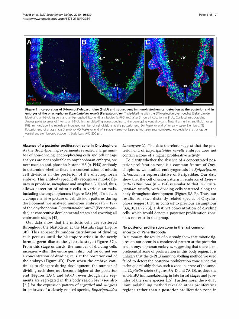
Results and discussionAnti-BrdU immunolabelling does not reveal a posteriorproliferation zone in OnychophoraIn vivo incorporation of the DNA replication markerBrdU, in conjunction with anti-BrdU immunolabelling,is a commonly used method for analysing embryoniccell division patterns [15,52-59]. Among annelids, anti-
BrdU immunolabelling revealed a distinct posterior pro-liferation zone in post-metamorphic stages of poly-chaetes, including the echiurans [15,16,54]. A similarlocalised region containing stem-like cells or teloblastsalso occurs in clitellate embryos [13,14,18]. To obtaincomparative data from Onychophora, we applied theanti-BrdU immunolabelling in elongating embryos ofthe velvet worm species Euperipatoides rowelli. In con-trast to annelids, we did not detect a higher number ofBrdU labelled cells at the posterior end of the onycho-phoran embryos than in the rest of the body (Figures1A-C). Thus, this method does not confirm the exis-tence of a posterior proliferation zone in Onychophora.
Anti-BrdU immunolabelling is not specific to dividingcellsIt was possible we were unable to detect the posteriorproliferation zone in onychophoran embryos as a conse-quence of non-specific incorporation of BrdU intoactively dividing cells. However, extensive work hasshown that BrdU will be incorporated into all cellsundergoing DNA synthesis, including endocycling cells[60-63]. The latter are specialised cells, which increasetheir biosynthetic activity by entering endocycles, i.e.,successive rounds of DNA replication without an inter-vening mitosis [63-65]. Due to ongoing DNA synthesisin these cells, BrdU is incorporated in their nuclei;although these cells can grow larger they do not divide.Our BrdU-labelling experiments on onychophoran
embryos revealed specific labelling patterns correspond-ing to some developing structures and organs (Figures2A-E). In particular, the so-called ventral organs and theanlagen of their derivatives, the hypocerebral organs,show a high number of BrdU-positive cells, with vir-tually every cell labelled in the superficial layer (Figures2A-E). The nuclei of the BrdU-positive cells show adivergent morphology compared to other cells sincethey are columnar in shape, larger in size, and containconspicuously condensed chromatin [66,67]. Notably,increased size and condensed chromatin is a feature ofother endoreplicating cells, e.g., the salivary gland cellsand nurse cells in Drosophila melanogaster [68,69].Furthermore, our data show that in the ventral organsBrdU is initially incorporated in a conspicuous punctatepattern (Figure 2F), which is typical of endocycling cells[62]. Due to this peculiar pattern of BrdU incorporationand modified cell morphology, we suggest that mostcells in the ventral organs (and in the hypocerebralorgan anlagen) are endocycling cells. Since we cannotexclude the possibility that other embryonic cells alsoenter the endocycle, we caution that anti-BrdU immu-nolabelling will not provide a definitive method fordetecting mitotic cell division patterns in onychophoranembryos.
Mayer et al. BMC Evolutionary Biology 2010, 10:339http://www.biomedcentral.com/1471-2148/10/339
Page 2 of 12
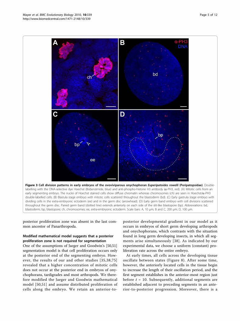
Absence of a posterior proliferation zone in OnychophoraAs the BrdU-labelling experiments revealed a large num-ber of non-dividing, endoreplicating cells and cell lineageanalyses are not applicable to onychophoran embryos, wenext used an anti-phospho-histone H3 (a-PH3) antibodyto determine whether there is a concentration of mitoticcell divisions in the posterior of the onychophoranembryo. This antibody specifically recognises mitotic fig-ures in prophase, metaphase and anaphase [70] and, thus,allows detection of mitotic cells in various animals,including the onychophorans (Figure 3A) [66]. To obtaina comprehensive picture of cell division patterns duringdevelopment, we analysed numerous embryos (n = 187)of the onychophoran Euperipatoides rowelli (Peripatopsi-dae) at consecutive developmental stages and covering allembryonic stages [42].Our data show that the mitotic cells are scattered
throughout the blastoderm at the blastula stage (Figure3B). This apparently random distribution of dividingcells persists until the blastopore arises in the newlyformed germ disc at the gastrula stage (Figure 3C).From this stage onwards, the number of dividing cellsincreases within the entire germ disc, but we do not seea concentration of dividing cells at the posterior end ofthe embryo (Figure 3D). Even when the embryo con-tinues to elongate during development, the number ofdividing cells does not become higher at the posteriorend (Figures 1A-C and 4A-D), even though new seg-ments are segregated in this body region [42] (see also[71] for the expression pattern of engrailed and winglessin embryos of a closely related species, Euperipatoides
kanangrensis). The data therefore suggest that the pos-terior end of Euperipatoides rowelli embryos does notcontain a zone of a higher proliferative activity.To clarify whether the absence of a concentrated pos-
terior proliferation zone is a common feature of Ony-chophora, we studied embryogenesis in Epiperipatusisthmicola, a representative of Peripatidae. Our datashow that the cell division pattern in embryos of Epiper-ipatus isthmicola (n = 124) is similar to that in Euperi-patoides rowelli, with dividing cells scattered along thebody throughout development (Figues 5A-E). Thus, ourresults from two distantly related species of Onycho-phora suggest that, in contrast to previous assumptions[3,4,10,11,72,73], a distinct concentration of dividingcells, which would denote a posterior proliferation zone,does not exist in this group.
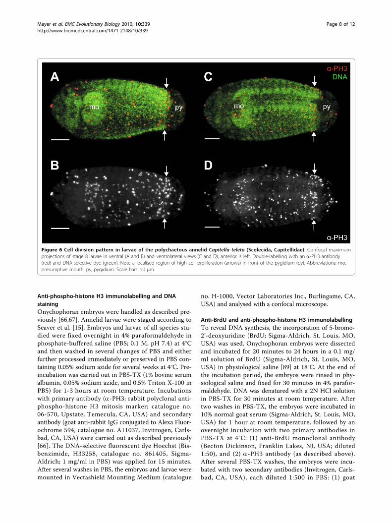
No posterior proliferation zone in the last commonancestor of PanarthropodaIn summary, the results of our study show that mitotic fig-ures do not occur in a condensed pattern at the posteriorend in onychophoran embryos, suggesting that there is nopreferential zone of proliferation in this body region. It isunlikely that the a-PH3 immunolabelling method we usedfailed to detect the posterior proliferation zone since thistechnique reliably shows such a zone in larvae of the anne-lid Capitella teleta (Figures 6A-D and 7A-D), as does theanti-BrdU immunolabelling in late larval stages and juve-niles of the same species [15]. Furthermore, the a-PH3immunolabelling method revealed other proliferatingregions rather than a posterior proliferation zone in
Figure 1 Incorporation of 5-bromo-2’-deoxyuridine (BrdU) and subsequent immunohistochemical detection at the posterior end inembryos of the onychophoran Euperipatoides rowelli (Peripatopsidae). Triple-labelling with the DNA-selective dye Hoechst (Bisbenzimide,blue), and anti-BrdU (green) and anti-phospho-histone H3 antibodies (a-PH3, red) after 3 hours incubation in BrdU. Confocal micrographs.Arrows point to areas of intense anti-BrdU immunolabelling corresponding to the developing ventral organs. Note that neither anti-BrdU nor a-PH3 immunolabelling reveals an increased number of cell divisions at the posterior end. (A) Posterior end of an early stage 3 embryo. (B)Posterior end of a late stage 3 embryo. (C) Posterior end of a stage 4 embryo. Leg-bearing segments numbered. Abbreviations: as, anus; ve,ventral extra-embryonic ectoderm. Scale bars: A-C, 200 μm.
Mayer et al. BMC Evolutionary Biology 2010, 10:339http://www.biomedcentral.com/1471-2148/10/339
Page 3 of 12

onychophoran embryos. For example, concentric rings ofproliferating cells, which correspond in timing and posi-tion with the anlagen of the hypocerebral organs [66], arefound in the antennal segment of the onychophoranembryos (Figures 4D and 5D).
The lack of evidence for a localised posterior prolifera-tion zone in Onychophora corresponds well with theapparent absence of such a zone in tardigrades [74,75]and most arthropods [35-40], excepting the malacostra-can crustaceans. We therefore suggest that a localised
Figure 2 Incorporation of 5-bromo-2’-deoxyuridine (BrdU) and subsequent immunocytochemical detection in embryos of theonychophoran Euperipatoides rowelli (Peripatopsidae). Confocal micrographs. Note the intense labelling in the ventral organs and anlagenof the hypocerebral organs, which are derivatives of the anterior-most pair of ventral organs. (A, B) Ventral and lateral views of a late stage 6embryo after 12 hours incubation in BrdU. Arrows point to the ventral organs. (C) Ventral view of the head of a late stage 6 embryo after 10hours incubation in BrdU. Double-labelling with the DNA-selective dye Hoechst (Bisbenzimide, blue) and anti-BrdU antibody (green). (D) Detail oftwo pairs of ventral organs from a stage 6 embryo after 12 hours incubation in BrdU. (E) Optical cross-section of an anlage of the hypocerebralorgan after 12 hours incubation in BrdU showing a superficial layer of anti-BrdU labelled cells. (F) Detail of ventral organ nuclei after 3 hoursincubation in BrdU (ventral view). Double-labelling with anti-BrdU (green) and anti-phospho-histone H3 antibodies (a-PH3, red). Arrowheadspoint to BrdU incorporation foci in each nucleus. Abbreviations: br, presumptive brain tissue; ho, anlagen of hypocerebral organs; mo,presumptive mouth opening; vo, ventral organs. Scale bars: A and B, 500 μm; C, 100 μm; D, 50 μm; E and F, 20 μm.
Mayer et al. BMC Evolutionary Biology 2010, 10:339http://www.biomedcentral.com/1471-2148/10/339
Page 4 of 12
posterior proliferation zone was absent in the last com-mon ancestor of Panarthropoda.
Modified mathematical model suggests that a posteriorproliferation zone is not required for segmentationOne of the assumptions of Jaeger and Goodwin’s [50,51]segmentation model is that cell proliferation occurs onlyat the posterior end of the segmenting embryo. How-ever, the results of our and other studies [35,38,75]revealed that a higher concentration of mitotic cellsdoes not occur at the posterior end in embryos of ony-chophorans, tardigrades and most arthropods. We there-fore modified the Jaeger and Goodwin mathematicalmodel [50,51] and assume distributed proliferation ofcells along the embryo. We retain an anterior-to-
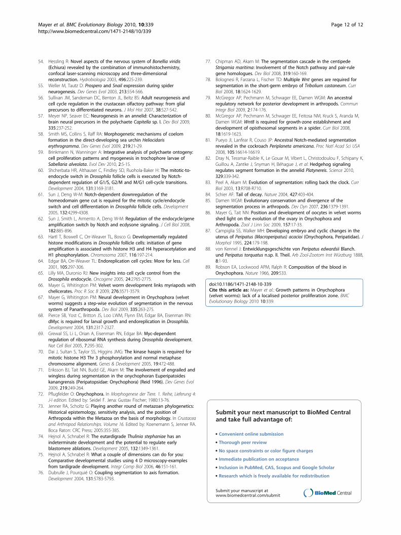
posterior developmental gradient in our model as itoccurs in embryos of short germ developing arthropodsand onychophorans, which contrasts with the situationfound in long germ developing insects, in which all seg-ments arise simultaneously [38]. As indicated by ourexperimental data, we choose a uniform (constant) pro-liferation rate across the entire embryo.At early times, all cells across the developing tissue
oscillate between states (Figure 8). After some time,however, the anteriorly located cells in the tissue beginto increase the length of their oscillation period, and thefirst segment establishes in the anterior-most region justbefore t = 10. Subsequently, additional segments areestablished adjacent to preceding segments in an ante-rior-to-posterior progression. Moreover, there is a
Figure 3 Cell division patterns in early embryos of the ovoviviparous onychophoran Euperipatoides rowelli (Peripatopsidae). Double-labelling with the DNA-selective dye Hoechst (Bisbenzimide, blue) and anti-phospho-histone H3 antibody (a-PH3, red). (A) Mitotic cells from anearly segmenting embryo. The nuclei of Hoechst stained cells show diffuse chromatin whereas chromosomes (ch) are seen in Hoechst/a-PH3double-labelled cells. (B) Blastula stage embryo with mitotic cells scattered throughout the blastoderm (bd). (C) Early gastrula stage embryo withdividing cells in the extra-embryonic ectoderm (ee) and in the germ disc (arrowhead). (D) Early germ band embryo with cell divisions scatteredthroughout the germ disc. Paired germ band (dotted line) extends anteriorly on each side of the slit-like blastopore (bp). Abbreviations: bd,blastoderm; bp, blastopore; ch, chromosomes; ee, extra-embryonic ectoderm. Scale bars: A, 10 μm; B and C, 200 μm; D, 100 μm.
Mayer et al. BMC Evolutionary Biology 2010, 10:339http://www.biomedcentral.com/1471-2148/10/339
Page 5 of 12

decrease in size of the newly established segments fromanterior to posterior end. Here, since the cells within asegment continue to proliferate, the established seg-ments also grow in width. This finding corresponds wellwith the observed external and internal anatomy of theembryos studied (Figures 4A, B and 5D).Taken together, the results of our mathematical model
show that segments can be patterned successfully with-out the involvement of a localised posterior proliferationzone in embryos of short germ developing arthropodsand onychophorans.
ConclusionsThe term “posterior growth zone” is commonly used todescribe the terminal body region, which gives rise tosegments in embryos of most panarthropods, annelids,and chordates [1,4,5,7,11,76]. However, according to ourresults, the “posterior growth zone” of panarthropods isnot a localised “zone” of proliferation but rather an area,in which segments are patterned, as evidenced by variousgene expression data available from various arthropods[35,77-81]. This contrasts with the “posterior growthzone” of annelids, in which both an increased number of
Figure 4 Cell division patterns in segmenting embryos of the ovoviviparous onychophoran Euperipatoides rowelli (Peripatopsidae). Thefull number of 15 leg-bearing segments has not been established yet. Double-labelling with the DNA-selective dye Hoechst (Bisbenzimide, blue)and anti-phospho-histone H3 antibody (a-PH3, red). (A) Flat preparation of an embryo with two leg-bearing segments. (B) Flat preparation of anembryo with four leg-bearing segments. (C) Ventrolateral view of a late stage 3 embryo with 11 leg-bearing segments. (D) Ventrolateral view ofan early stage 4 embryo with 13 leg-bearing segments. Note the concentric rings of proliferating cells in the antennal segment (arrowheads).Arrows (in B and C) point to remnants of the blastopore in front of the future anus. Leg-bearing segments numbered. Abbreviations: an,antennal segment; as, anus; at, presumptive antenna; bp, blastopore; de, dorsal extra-embryonic ectoderm; jw, jaw segment/presumptive jaw;mo, embryonic mouth; sp, slime papilla segment/presumptive slime papilla; ve, ventral extra-embryonic ectoderm. Scale bars: A-D, 200 μm.
Mayer et al. BMC Evolutionary Biology 2010, 10:339http://www.biomedcentral.com/1471-2148/10/339
Page 6 of 12

cell divisions and segment patterning occur [13-18,82].With respect to vertebrates, the term “posterior growthzone” is applied in different ways and refers either to thetailbud, which proliferates cells for somites, or to thepre-somitic mesoderm area, which establishes segmentalborders [1,22,24,83-85]. Since the terminal body regiondiffers considerably in composition and extent amongpanarthropods, annelids, and chordates, the term “poster-ior growth zone” is imprecise and therefore cannot beused to support the homology hypothesis [1] of segmen-tation between these three animal groups.
Materials and methodsSpecimens and embryosFemales of the onychophoran species Euperipatoidesrowelli Reid, 1996 and Epiperipatus isthmicola (Bouvier,
1905) were collected and maintained in the laboratory asdescribed previously [67,86]. Females were dissected atvarious times of the year to obtain a range of consecutivedevelopmental stages. Embryos were staged according toprevious descriptions of onychophoran embryogenesis[42,66,67,87,88]. For positive controls, Capitella teletaBlake, Grassle & Eckelbarger 2009 (”Capitella sp. I” sensu[15]) larvae and juveniles were obtained from a culture atthe Department of Evolutionary Biology (University ofBonn, Germany). The animals were reared in 20 × 20 cmplastic boxes containing 1 cm sieved mud (500 μm) cov-ered with 4 cm ultrafiltrated seawater from the northernWadden Sea at 18°C. Water and sediment were changedevery two weeks and the boxes aerated continuously. Toobtain developmental stages, brood tubes were takenfrom the sediment and opened with minute needles.
Figure 5 Cell division patterns in embryos of the placental viviparous onychophoran Epiperipatus isthmicola (Peripatidae). The fullnumber of leg-bearing segments (30-32 in females and 27-29 in males) has not been established yet. Double-labelling with the DNA-selectivedye Hoechst (Bisbenzimide, blue) and anti-phospho-histone H3 antibody (a-PH3, red). (A) Gastrula stage embryo. (B) Elongating germ bandembryo with separate mouth and anus openings. (C) Elongating embryo with 10 leg-bearing segments. (D) Elongating “coiled stage” embryowith 20 leg-bearing segments. Arrowheads indicate concentric rings of proliferating cells in the antennal segment. (E) A “coiled stage” embryowith 26 leg-bearing segments. Abbreviations: an, antennal segment; as, anus; at, presumptive antenna; bp, blastopore; jw, jaw segment/presumptive jaw; mo, embryonic mouth; sp, slime papilla segment/presumptive slime papilla. Scale bars: A, 25 μm; B, 50 μm; C and D, 100 μm, E,150 μm.
Mayer et al. BMC Evolutionary Biology 2010, 10:339http://www.biomedcentral.com/1471-2148/10/339
Page 7 of 12
Anti-phospho-histone H3 immunolabelling and DNAstainingOnychophoran embryos were handled as described pre-viously [66,67]. Annelid larvae were staged according toSeaver et al. [15]. Embryos and larvae of all species stu-died were fixed overnight in 4% paraformaldehyde inphosphate-buffered saline (PBS; 0.1 M, pH 7.4) at 4°Cand then washed in several changes of PBS and eitherfurther processed immediately or preserved in PBS con-taining 0.05% sodium azide for several weeks at 4°C. Pre-incubation was carried out in PBS-TX (1% bovine serumalbumin, 0.05% sodium azide, and 0.5% Triton X-100 inPBS) for 1-3 hours at room temperature. Incubationswith primary antibody (a-PH3; rabbit polyclonal anti-phospho-histone H3 mitosis marker; catalogue no.06-570, Upstate, Temecula, CA, USA) and secondaryantibody (goat anti-rabbit IgG conjugated to Alexa Fluor-ochrome 594, catalogue no. A11037, Invitrogen, Carls-bad, CA, USA) were carried out as described previously[66]. The DNA-selective fluorescent dye Hoechst (Bis-benzimide, H33258, catalogue no. 861405, Sigma-Aldrich; 1 mg/ml in PBS) was applied for 15 minutes.After several washes in PBS, the embryos and larvae weremounted in Vectashield Mounting Medium (catalogue
no. H-1000, Vector Laboratories Inc., Burlingame, CA,USA) and analysed with a confocal microscope.
Anti-BrdU and anti-phospho-histone H3 immunolabellingTo reveal DNA synthesis, the incorporation of 5-bromo-2’-deoxyuridine (BrdU; Sigma-Aldrich, St. Louis, MO,USA) was used. Onychophoran embryos were dissectedand incubated for 20 minutes to 24 hours in a 0.1 mg/ml solution of BrdU (Sigma-Aldrich, St. Louis, MO,USA) in physiological saline [89] at 18°C. At the end ofthe incubation period, the embryos were rinsed in phy-siological saline and fixed for 30 minutes in 4% parafor-maldehyde. DNA was denatured with a 2N HCl solutionin PBS-TX for 30 minutes at room temperature. Aftertwo washes in PBS-TX, the embryos were incubated in10% normal goat serum (Sigma-Aldrich, St. Louis, MO,USA) for 1 hour at room temperature, followed by anovernight incubation with two primary antibodies inPBS-TX at 4°C: (1) anti-BrdU monoclonal antibody(Becton Dickinson, Franklin Lakes, NJ, USA; diluted1:50), and (2) a-PH3 antibody (as described above).After several PBS-TX washes, the embryos were incu-bated with two secondary antibodies (Invitrogen, Carls-bad, CA, USA), each diluted 1:500 in PBS: (1) goat
Figure 6 Cell division pattern in larvae of the polychaetous annelid Capitella teleta (Scolecida, Capitellidae). Confocal maximumprojections of stage 8 larvae in ventral (A and B) and ventrolateral views (C and D); anterior is left. Double-labelling with an a-PH3 antibody(red) and DNA-selective dye (green). Note a localised region of high cell proliferation (arrows) in front of the pygidium (py). Abbreviations: mo,presumptive mouth; py, pygidium. Scale bars: 50 μm.
Mayer et al. BMC Evolutionary Biology 2010, 10:339http://www.biomedcentral.com/1471-2148/10/339
Page 8 of 12

Figure 7 Cell division pattern at the posterior end in larvae of the polychaetous annelid Capitella teleta (Scolecida, Capitellidae).Confocal (A, C) and light micrographs (B, D) of posterior ends of an early (A, B) and a late stage 8 larvae (C, D), labelled with an a-PH3 antibody.Note a localised region of high cell proliferation (arrows) in front of the telotroch (te). Scale bars: 50 μm.
Figure 8 Space time diagram of cell state as a function of position (horizontal axis) and time (vertical axis) in a uniformly proliferatingtissue with a linear growth rate. Due to proliferation throughout the tissue, the length increases with time t as L(t) = t + 2. The parameters inthe Jaeger and Goodwin model [50,51] are set as A = 0.5, B = 5, T0 = 1 for 0 <t < 50. The cell state ranges from -10 <z < 10 and this isrepresented on the graph as shading between black and white (corresponding to z = -10, z = 10 respectively). The region above the dottedyellow line has segmented. Areas outside the tissue are coloured in blue.
Mayer et al. BMC Evolutionary Biology 2010, 10:339http://www.biomedcentral.com/1471-2148/10/339
Page 9 of 12

anti-mouse IgG (H+L), conjugated to Alexa Fluoro-chrome 488 (catalogue no. A11017), and (2) goat anti-rabbit IgG conjugated to Alexa Fluorochrome 594 (cata-logue no. A11037). Hoechst staining was applied asdescribed above. After several washes in PBS, theembryos were mounted in Vectashield Mounting Med-ium and analysed with a confocal microscope. For con-trols, the embryos were treated in the same way, butwithout the addition of BrdU to the physiological saline.This resulted in a complete lack of anti-BrdU labellingin the nuclei. The specificity of the secondary antibodywas tested by abolishing the primary antibody from theexperimental procedures, which resulted in a completelack of a positive signal within the cells. The only struc-tures showing autofluorescence in the green and UVchannels were the sclerotised claws and jaws.
Microscopy and image processingEmbryos and larvae were analysed with the confocallaser-scanning microscopes LSM 510 META (Carl ZeissMicroImaging GmbH) and TCS SPE (Leica Microsys-tems Wetzlar). The image stacks were merged digitallyinto partial and maximum projections with the ZeissLSM Image Browser software (version 4.0.0.241) andImageJ (version 1.43q). Image intensity histograms wereadjusted by using Adobe (San Jose, Ca) Photoshop CS2.The adjustment was kept at a minimum to allow themicrographs of the same plate to have similar intensity.Final panels were designed with Adobe Illustrator CS2.
Mathematical modellingOur model adapts the one used by Jaeger and Goodwin[50,51] for animal segmentation and is based on cellularoscillators, where the phase determines the state of thecell and cells oscillate between two states. Jaeger andGoodwin [50,51] assume that there is a localised posteriorproliferation zone-they call it a progress zone. In the Jae-ger and Goodwin model, cells in the progress zone areoscillating in phase with each other. However, when theyleave the progress zone, their oscillations slow down withtheir physiological age. Accordingly, the cells towards theanterior oscillate slower since they have a higher physiolo-gical age than the posterior ones. Segmentation occurs,when the state of the cell no longer oscillates and remainsconstant. This mechanism results in a gradient of slowingcellular oscillations and sets up a “wave” of cell state stabi-lisation moving in an anterior-to-posterior direction,which leads to a spatially periodic pattern of cell state thatcan be interpreted as sequentially forming segments.In contrast to the Jaeger and Goodwin model [50,51],
we assume that all cells have the ability to proliferate atsome rate r(x, t), which can be a function of spatial posi-tion x, and time t. In our model, a system of discrete timeequations describes the phase and period of the
oscillators. It is convenient to convert the discrete timesystem of equations to a continuous time system, whichis solved using MATLAB software (MathWorks™). Wemodel the developing tissue in one spatial dimension, x,growing in time t. In our distributed growth model, cellscan proliferate anywhere in the developing tissue. Thus,the older cells are no longer located towards the anteriorof the tissues and the younger ones are no longer locatedtowards the posterior. Accordingly, the assumed mechan-ism that cell oscillations slow with age [50,51] cannotresult in the formation of segments in our model. Wetherefore modified the equations of the previous modeland choose the oscillation period to be correlated withdistance from the posterior end, rather than the cell age.Such positional information can be obtained from a gra-dient of signalling molecules in the cell’s local environ-ment. The modified cellular oscillator model gives rise toa gradient of oscillation period along the tissue length,with the faster oscillations at the posterior end.
AcknowledgementsThe staff of the Instituto Nacional de Biodiversidad (INBio, Heredia, CostaRica), the National System of Conservation Areas (SINAC, MINAE, Costa Rica)and State Forests NSW (New South Wales, Australia) are gratefullyacknowledged for providing permits. G.M. is thankful to Paul Whitington forproviding lab space, reading the first draft of the manuscript and criticaldiscussions, to Alvaro Herrera, Paul Sunnucks and Noel Tait for theirassistance with collection of specimens, and to Thomas Stach for sharing hisknowledge on urochordates and cephalochordates and pointing us to therelevant literature. We are grateful to Tobias Kaller for providing the annelidlarvae. Two anonymous reviewers provided numerous constructive criticisms,which helped improve the manuscript. This work was supported by grantsfrom the German Research Foundation to G.M. (Ma 4147/3-1) and theAustralian Research Council to K.A.L. (ARC: DP 0878200). K.A.L. is an ARCProfessorial Fellow. G.M. is a Research Group Leader supported by the EmmyNoether Programme of the DFG.
Author details1Institute of Biology II: Animal Evolution & Development, University ofLeipzig, Talstrasse 33, D-04103 Leipzig, Germany. 2Institut fürEvolutionsbiologie und Ökologie, Universität Bonn, An der Immenburg 1, D-53121 Bonn, Germany. 3Department of Mathematics and Statistics, Universityof Melbourne, Victoria 3010, Australia. 4Department of Anatomy and CellBiology, University of Melbourne, Victoria 3010, Australia.
Authors’ contributionsGM conceived, designed and performed the experiments on onychophoransand wrote the first draft of the manuscript. CK and BQ carried out theexperiments on the Capitella teleta larvae and juveniles. RHC and KALperformed the mathematical modelling. LMQ provided continuous inputand knowledge on cell proliferation and endoreplication. All authorsparticipated in the discussion of the results and the preparation of the finalmanuscript.
Competing interestsThe authors declare that they have no competing interests.
Received: 7 June 2010 Accepted: 4 November 2010Published: 4 November 2010
References1. Martin BL, Kimelman D: Wnt signaling and the evolution of embryonic
posterior development. Curr Biol 2009, 19:R215-R219.
Mayer et al. BMC Evolutionary Biology 2010, 10:339http://www.biomedcentral.com/1471-2148/10/339
Page 10 of 12

2. Ax P: Multicellular animals. The Phylogenetic System of the Metazoa Berlin:Springer; 2000.
3. Nielsen C: Animal Evolution: Interrelationships of the Living Phyla Oxford:Oxford University Press; 2001.
4. Scholtz G: The Articulata hypothesis-or what is a segment? Org DiversEvol 2002, 2:197-215.
5. Balavoine G, Adoutte A: The segmented Urbilateria: a testable scenario.Integr Comp Biol 2003, 43:137-147.
6. de Rosa R, Prud’homme B, Balavoine G: Caudal and even-skipped in theannelid Platynereis dumerilii and the ancestry of posterior growth. EvolDev 2005, 7:574-587.
7. De Robertis EM: The molecular ancestry of segmentation mechanisms.Proc Natl Acad Sci USA 2008, 105:16411-16412.
8. Saudemont A, Dray N, Hudry B, Le Gouar M, Vervoort M, Balavoine G:Complementary striped expression patterns of NK homeobox genesduring segment formation in the annelid Platynereis. Dev Biol 2008,317:430-443.
9. Snodgrass RE: Evolution of the Annelida, Onychophora and Arthropoda.Smithson Misc Coll 1938, 97:1-159.
10. Manton SM: Studies on the Onychophora VII. The early embryonic stagesof Peripatopsis, and some general considerations concerning themorphology and phylogeny of the Arthropoda. Phil Trans R Soc B 1949,233:483-580.
11. Anderson DT: Embryology and Phylogeny in Annelids and Arthropods Oxford:Pergamon Press; 1973.
12. Shankland M, Seaver EC: Evolution of the bilaterian body plan: What havewe learned from annelids? Proc Natl Acad Sci USA 2000, 97:4434-4437.
13. Matsuo K, Yoshida H, Shimizu T: Differential expression of caudal anddorsal genes in the teloblast lineages of the oligochaete annelid Tubifextubifex. Dev Genes Evol 2005, 215:238-247.
14. Rivera AS, Gonsalves FC, Song MH, Norris BJ, Weisblat DA: Characterizationof Notch-class gene expression in segmentation stem cells and segmentfounder cells in Helobdella robusta (Lophotrochozoa; Annelida; Clitellata;Hirudinida; Glossiphoniidae). Evol Dev 2005, 7:588-599.
15. Seaver EC, Thamm K, Hill SD: Growth patterns during segmentation inthe two polychaete annelids, Capitella sp. I and Hydroides elegans:comparison at distinct life history stages. Evol Dev 2005, 7:312-326.
16. Rebscher N, Zelada-González F, Banisch TU, Raible F, Arendt D: Vasa unveilsa common origin of germ cells and of somatic stem cells from theposterior growth zone in the polychaete Platynereis dumerilii. Dev Biol2007, 306:599-611.
17. Thamm K, Seaver EC: Notch signaling during larval and juveniledevelopment in the polychaete annelid Capitella sp. I. Dev Biol 2008,320:304-318.
18. Zhang SO, Kuo D-H, Weisblat DA: Grandparental stem cells in leechsegmentation: Differences in CDC42 expression are 2 correlated with analternating pattern of blast cell fates. Dev Biol 2009, 336:112-121.
19. Tam PPL, Beddington RSP: The metameric organization of the presomiticmesoderm and somite specification in the mouse embryo. In Somites inDeveloping Embryos. Edited by: Bellairs R, Ede DA, Lash J. New York: PlenumPub. Co; 1986:17-36.
20. Eloy-Trinquet S, Nicolas J-F: Cell coherence during production of thepresomitic mesoderm and somitogenesis in the mouse embryo.Development 2002, 129:3609-3619.
21. Nicolas JF, Mathis L, Bonnerot C: Evidence in the mouse for self-renewingstem cells in the formation of a segmented longitudinal structure, themyotome. Development 1996, 122:2933-2946.
22. Pourquié O: Vertebrate somitogenesis: a novel paradigm for animalsegmentation? Int J Dev Biol 2003, 47:597-603.
23. Thorpe CJ, Weidinger G, Moon RT: Wnt/β-catenin regulation of the Sp1-related transcription factor sp5l promotes tail development in zebrafish.Development 2005, 132:1763-1772.
24. Holley SA: The genetics and embryology of zebrafish metamerism. DevDyn 2007, 236:1422-1449.
25. Schubert M, Holland LZ, Stokes MD, Holland ND: Three amphioxus Wntgenes (AmphiWnt3, AmphiWnt5, and AmphiWnt6) associated with the tailbud: the evolution of somitogenesis in chordates. Dev Biol 2001,240:262-273.
26. Holland ND, Holland LZ: Stage-and tissue-specific patterns of cell divisionin embryonic and larval tissues of amphioxus during normaldevelopment. Evol Dev 2006, 8:142-149.
27. Nishida H: Cell lineage analysis in ascidian embryos by intracellularinjection of a tracer enzyme. III. Up to the tissue restricted stage. DevBiol 1987, 121:526-541.
28. Søviknes AM, Glover JC: Spatiotemporal patterns of neurogenesis in theappendicularian Oikopleura dioica. Dev Biol 2007, 311:264-275.
29. Stach T, Winter J, Bouquet J-M, Chourrout D, Schnabel R: Embryology of aplanktonic tunicate reveals traces of sessility. Proc Natl Acad Sci USA 2008,105:7229-7234.
30. Nielsen C: Proposing a solution to the Articulata-Ecdysozoa controversy.Zool Scr 2003, 32:475-482.
31. Dohle W, Scholtz G: Clonal analysis of the crustacean segment: thediscordance between genealogical and segmental borders. Development1988, 104:147-160.
32. Scholtz G: The formation, differentiation and segmentation of thepost-naupliar germ band of the amphipod Gammarus pulex L.(Crustacea, Malacostraca, Peracarida). Proc R Soc Lond B 1990,239:163-211.
33. Scholtz G: Teloblasts in decapod embryos: an embryonic characterreveals the monophyletic origin of freshwater crayfishes (Crustacea,Decapoda). Zool Anz 1993, 230:45-54.
34. Seaver EC: Segmentation: mono-or polyphyletic? Int J Dev Biol 2003,47:583-595.
35. Chipman AD: Parallel evolution of segmentation by co-option ofancestral gene regulatory networks. BioEssays 2010, 32:60-70.
36. Davis GK, Patel NH: Short, long, and beyond: Molecular andembryological approaches to insect segmentation. Annu Rev Entomol2002, 47:669-699.
37. Minelli A, Fusco G: Evo-devo perspectives on segmentation: modelorganisms, and beyond. Trends Ecol Evol 2004, 19:423-429.
38. Liu PZ, Kaufman TC: Short and long germ segmentation: unansweredquestions in the evolution of a developmental mode. Evol Dev 2005,7:629-646.
39. Minelli A: A morphologist’s perspective on terminal growth andsegmentation. Evol Dev 2005, 7:568-573.
40. Chipman AD: Thoughts and speculations on the ancestral arthropodsegmentation pathway. In Evolving Pathways: Key Themes in EvolutionaryDevelopmental Biology. Edited by: Minelli A, Fusco G. Cambridge: CambridgeUniversity Press; 2008:343-358.
41. Walker MH: Relatively recent evolution of an unusual pattern of earlyembryonic development (long germ band?) in a South Africanonychophoran, Opisthopatus cinctipes Purcell (Onychophora:Peripatopsidae). Zool J Linn Soc 1995, 114:61-75.
42. Walker MH, Tait NN: Studies on embryonic development and thereproductive cycle in ovoviviparous Australian Onychophora(Peripatopsidae). J Zool 2004, 264:333-354.
43. von Kennel J: Entwicklungsgeschichte von Peripatus edwardsii Blanch.und Peripatus torquatus n.sp. I. Theil. Arb Zool-Zootom Inst Würzburg 1885,7:95-229.
44. Sedgwick A: The development of the Cape species of Peripatus. Part II. QJ Microsc Sci 1886, 26:175-212.
45. Sheldon L: On the development of Peripatus novae-zealandiae. Q JMicrosc Sci 1887, 28:205-237.
46. Evans R: On the Malayan species of Onychophora. Part II.-Thedevelopment of Eoperipatus weldoni. Q J Microsc Sci 1901, 45:41-88.
47. Pflugfelder O: Entwicklung von Paraperipatus amboinensis n. sp. Zool JbAnat 1948, 69:443-492.
48. Pflugfelder O: Onychophora. In Lehrbuch der Entwicklungsgeschichte undEntwicklungsphysiologie der Tiere. Edited by: Pflugfelder O. Jena: GustavFischer; 1962:139-144.
49. Pflugfelder O: Onychophora. In Grosses Zoologisches Praktikum. 13a edition.Edited by: Czihak G. Stuttgart: Gustav Fischer; 1968:1-42.
50. Jaeger J, Goodwin BC: Cellular oscillators in animal segmentation. In SilicoBiology 2002, 2:111-123.
51. Jaeger J, Goodwin BC: A cellular oscillator model for periodic patternformation. J theor Biol 2001, 213:171-181.
52. Whitington PM, Meier T, King P: Segmentation, neurogenesis andformation of early axonal pathways in the centipede, Ethmostigmusrubripes (Brandt). Roux Arch Dev Biol 1991, 199:349-363.
53. Harzsch S: Neurogenesis in the crustacean ventral nerve cord: homologyof neuronal stem cells in Malacostraca and Brachiopoda? Evol Dev 2001,3:154-169.
Mayer et al. BMC Evolutionary Biology 2010, 10:339http://www.biomedcentral.com/1471-2148/10/339
Page 11 of 12
54. Hessling R: Novel aspects of the nervous system of Bonellia viridis(Echiura) revealed by the combination of immunohistochemistry,confocal laser-scanning microscopy and three-dimensionalreconstruction. Hydrobiologia 2003, 496:225-239.
55. Weller M, Tautz D: Prospero and Snail expression during spiderneurogenesis. Dev Genes Evol 2003, 213:554-566.
56. Sullivan JM, Sandeman DC, Benton JL, Beltz BS: Adult neurogenesis andcell cycle regulation in the crustacean olfactory pathway: from glialprecursors to differentiated neurons. J Mol Hist 2007, 38:527-542.
57. Meyer NP, Seaver EC: Neurogenesis in an annelid: Characterization ofbrain neural precursors in the polychaete Capitella sp. I. Dev Biol 2009,335:237-252.
58. Smith MS, Collins S, Raff RA: Morphogenetic mechanisms of coelomformation in the direct-developing sea urchin Heliocidariserythrogramma. Dev Genes Evol 2009, 219:21-29.
59. Brinkmann N, Wanninger A: Integrative analysis of polychaete ontogeny:cell proliferation patterns and myogenesis in trochophore larvae ofSabellaria alveolata. Evol Dev 2010, 2:5-15.
60. Shcherbata HR, Althauser C, Findley SD, Ruohola-Baker H: The mitotic-to-endocycle switch in Drosophila follicle cells is executed by Notch-dependent regulation of G1/S, G2/M and M/G1 cell-cycle transitions.Development 2004, 131:3169-3181.
61. Sun J, Deng W-M: Notch-dependent downregulation of thehomeodomain gene cut is required for the mitotic cycle/endocycleswitch and cell differentiation in Drosophila follicle cells. Development2005, 132:4299-4308.
62. Sun J, Smith L, Armento A, Deng W-M: Regulation of the endocycle/geneamplification switch by Notch and ecdysone signaling. J Cell Biol 2008,182:885-896.
63. Hartl T, Boswell C, Orr-Weaver TL, Bosco G: Developmentally regulatedhistone modifications in Drosophila follicle cells: initiation of geneamplification is associated with histone H3 and H4 hyperacetylation andH1 phosphorylation. Chromosoma 2007, 116:197-214.
64. Edgar BA, Orr-Weaver TL: Endoreplication cell cycles: More for less. Cell2001, 105:297-306.
65. Lilly MA, Duronio RJ: New insights into cell cycle control from theDrosophila endocycle. Oncogene 2005, 24:2765-2775.
66. Mayer G, Whitington PM: Velvet worm development links myriapods withchelicerates. Proc R Soc B 2009, 276:3571-3579.
67. Mayer G, Whitington PM: Neural development in Onychophora (velvetworms) suggests a step-wise evolution of segmentation in the nervoussystem of Panarthropoda. Dev Biol 2009, 335:263-275.
68. Pierce SB, Yost C, Britton JS, Loo LWM, Flynn EM, Edgar BA, Eisenman RN:dMyc is required for larval growth and endoreplication in Drosophila.Development 2004, 131:2317-2327.
69. Grewal SS, Li L, Orian A, Eisenman RN, Edgar BA: Myc-dependentregulation of ribosomal RNA synthesis during Drosophila development.Nat Cell Biol 2005, 7:295-302.
70. Dai J, Sultan S, Taylor SS, Higgins JMG: The kinase haspin is required formitotic histone H3 Thr 3 phosphorylation and normal metaphasechromosome alignment. Genes & Development 2005, 19:472-488.
71. Eriksson BJ, Tait NN, Budd GE, Akam M: The involvement of engrailed andwingless during segmentation in the onychophoran Euperipatoideskanangrensis (Peripatopsidae: Onychophora) (Reid 1996). Dev Genes Evol2009, 219:249-264.
72. Pflugfelder O: Onychophora. In Morphogenese der Tiere. 1. Reihe, Lieferung 4:J-I edition. Edited by: Seidel F. Jena: Gustav Fischer; 1980:13-76.
73. Jenner RA, Scholtz G: Playing another round of metazoan phylogenetics:Historical epistemology, sensitivity analysis, and the position ofArthropoda within the Metazoa on the basis of morphology. In Crustaceaand Arthropod Relationships. Volume 16. Edited by: Koenemann S, Jenner RA.Boca Raton: CRC Press; 2005:355-385.
74. Hejnol A, Schnabel R: The eutardigrade Thulinia stephaniae has anindeterminate development and the potential to regulate earlyblastomere ablations. Development 2005, 132:1349-1361.
75. Hejnol A, Schnabel R: What a couple of dimensions can do for you:Comparative developmental studies using 4 D microscopy-examplesfrom tardigrade development. Integr Comp Biol 2006, 46:151-161.
76. Dubrulle J, Pourquié O: Coupling segmentation to axis formation.Development 2004, 131:5783-5793.
77. Chipman AD, Akam M: The segmentation cascade in the centipedeStrigamia maritima: Involvement of the Notch pathway and pair-rulegene homologues. Dev Biol 2008, 319:160-169.
78. Bolognesi R, Farzana L, Fischer TD: Multiple Wnt genes are required forsegmentation in the short-germ embryo of Tribolium castaneum. CurrBiol 2008, 18:1624-1629.
79. McGregor AP, Pechmann M, Schwager EE, Damen WGM: An ancestralregulatory network for posterior development in arthropods. CommunIntegr Biol 2009, 2:174-176.
80. McGregor AP, Pechmann M, Schwager EE, Feitosa NM, Kruck S, Aranda M,Damen WGM: Wnt8 is required for growth-zone establishment anddevelopment of opisthosomal segments in a spider. Curr Biol 2008,18:1619-1623.
81. Pueyo JI, Lanfear R, Couso JP: Ancestral Notch-mediated segmentationrevealed in the cockroach Periplaneta americana. Proc Natl Acad Sci USA2008, 105:16614-16619.
82. Dray N, Tessmar-Raible K, Le Gouar M, Vibert L, Christodoulou F, Schipany K,Guillou A, Zantke J, Snyman H, Béhague J, et al: Hedgehog signalingregulates segment formation in the annelid Platynereis. Science 2010,329:339-342.
83. Peel A, Akam M: Evolution of segmentation: rolling back the clock. CurrBiol 2003, 13:R708-R710.
84. Schier AF: Tail of decay. Nature 2004, 427:403-404.85. Damen WGM: Evolutionary conservation and divergence of the
segmentation process in arthropods. Dev Dyn 2007, 236:1379-1391.86. Mayer G, Tait NN: Position and development of oocytes in velvet worms
shed light on the evolution of the ovary in Onychophora andArthropoda. Zool J Linn Soc 2009, 157:17-33.
87. Campiglia SS, Walker MH: Developing embryo and cyclic changes in theuterus of Peripatus (Macroperipatus) acacioi (Onychophora, Peripatidae). JMorphol 1995, 224:179-198.
88. von Kennel J: Entwicklungsgeschichte von Peripatus edwardsii Blanch.und Peripatus torquatus n.sp. II. Theil. Arb Zool-Zootom Inst Würzburg 1888,8:1-93.
89. Robson EA, Lockwood APM, Ralph R: Composition of the blood inOnychophora. Nature 1966, 209:533.
doi:10.1186/1471-2148-10-339Cite this article as: Mayer et al.: Growth patterns in Onychophora(velvet worms): lack of a localised posterior proliferation zone. BMCEvolutionary Biology 2010 10:339.
Submit your next manuscript to BioMed Centraland take full advantage of:
• Convenient online submission
• Thorough peer review
• No space constraints or color figure charges
• Immediate publication on acceptance
• Inclusion in PubMed, CAS, Scopus and Google Scholar
• Research which is freely available for redistribution
Submit your manuscript at www.biomedcentral.com/submit
Mayer et al. BMC Evolutionary Biology 2010, 10:339http://www.biomedcentral.com/1471-2148/10/339
Page 12 of 12