A revision of brain composition in Onychophora (velvet worms) suggests that the tritocerebrum...
9
RESEARCH ARTICLE Open Access A revision of brain composition in Onychophora (velvet worms) suggests that the tritocerebrum evolved in arthropods Georg Mayer 1* , Paul M Whitington 2 , Paul Sunnucks 3 , Hans-Joachim Pflüger 4 Abstract Background: The composition of the arthropod head is one of the most contentious issues in animal evolution. In particular, controversy surrounds the homology and innervation of segmental cephalic appendages by the brain. Onychophora (velvet worms) play a crucial role in understanding the evolution of the arthropod brain, because they are close relatives of arthropods and have apparently changed little since the Early Cambrian. However, the segmental origins of their brain neuropils and the number of cephalic appendages innervated by the brain - key issues in clarifying brain composition in the last common ancestor of Onychophora and Arthropoda - remain unclear. Results: Using immunolabelling and neuronal tracing techniques in the developing and adult onychophoran brain, we found that the major brain neuropils arise from only the anterior-most body segment, and that two pairs of segmental appendages are innervated by the brain. The region of the central nervous system corresponding to the arthropod tritocerebrum is not differentiated as part of the onychophoran brain but instead belongs to the ventral nerve cords. Conclusions: Our results contradict the assumptions of a tripartite (three-segmented) brain in Onychophora and instead confirm the hypothesis of bipartite (two-segmented) brain composition. They suggest that the last common ancestor of Onychophora and Arthropoda possessed a brain consisting of protocerebrum and deutocerebrum whereas the tritocerebrum evolved in arthropods. Background The head of arthropods is a specialised anterior body region, which is distinguished by fused segments and several pairs of modified appendages [1,2]. These appen- dages serve for swimming, feeding, defence, or sensory perception, and their movements are coordinated by a complex brain situated within the head. Despite over a century of intense research in this area, the ancestral composition of the arthropod head remains obscure and is one of the most controversial topics in zoology [2-8]. Fossils have contributed much to our knowledge [1,4,8], but their limited preservation constrains definitive con- clusions about the degree of cephalisation in the last common ancestor of Panarthropoda (Onychophora + Tardigrada + Arthropoda). The extant Onychophora are a key group when consid- ering this issue, since they are close relatives of arthropods and resemble Cambrian lobopodians [9-13], while their internal anatomy and embryology are accessible for detailed examination. As in various lobopodians, the ony- chophoran “head” is not clearly delineated from the trunk, but shows three pairs of modified appendages: sensory antennae, jaws situated within the mouth cavity, and slime papillae, which are used for defence and capturing prey organisms (Figure 1A). These modified appendages have been assigned to each body segment by studying embryo- genesis, which revealed that the antennae belong to the first (ocular) body segment, the jaws to the second, and the slime papillae to the third segment [14-20]. Most importantly, these studies have provided no evidence of any additional vestigial cephalic segments [21-24] in * Correspondence: [email protected] 1 Institute of Biology II: Animal Evolution & Development, University of Leipzig, Talstrasse 33, D-04103 Leipzig, Germany Full list of author information is available at the end of the article Mayer et al. BMC Evolutionary Biology 2010, 10:255 http://www.biomedcentral.com/1471-2148/10/255 © 2010 Mayer et al; licensee BioMed Central Ltd. This is an Open Access article distributed under the terms of the Creative Commons Attribution License (http://creativecommons.org/licenses/by/2.0), which permits unrestricted use, distribution, and reproduction in any medium, provided the original work is properly cited.
Transcript of A revision of brain composition in Onychophora (velvet worms) suggests that the tritocerebrum...

RESEARCH ARTICLE Open Access
A revision of brain composition in Onychophora(velvet worms) suggests that the tritocerebrumevolved in arthropodsGeorg Mayer1*, Paul M Whitington2, Paul Sunnucks3, Hans-Joachim Pflüger4
Abstract
Background: The composition of the arthropod head is one of the most contentious issues in animal evolution. Inparticular, controversy surrounds the homology and innervation of segmental cephalic appendages by the brain.Onychophora (velvet worms) play a crucial role in understanding the evolution of the arthropod brain, becausethey are close relatives of arthropods and have apparently changed little since the Early Cambrian. However, thesegmental origins of their brain neuropils and the number of cephalic appendages innervated by the brain - keyissues in clarifying brain composition in the last common ancestor of Onychophora and Arthropoda - remainunclear.
Results: Using immunolabelling and neuronal tracing techniques in the developing and adult onychophoran brain,we found that the major brain neuropils arise from only the anterior-most body segment, and that two pairs ofsegmental appendages are innervated by the brain. The region of the central nervous system corresponding to thearthropod tritocerebrum is not differentiated as part of the onychophoran brain but instead belongs to the ventralnerve cords.
Conclusions: Our results contradict the assumptions of a tripartite (three-segmented) brain in Onychophora andinstead confirm the hypothesis of bipartite (two-segmented) brain composition. They suggest that the lastcommon ancestor of Onychophora and Arthropoda possessed a brain consisting of protocerebrum anddeutocerebrum whereas the tritocerebrum evolved in arthropods.
BackgroundThe head of arthropods is a specialised anterior bodyregion, which is distinguished by fused segments andseveral pairs of modified appendages [1,2]. These appen-dages serve for swimming, feeding, defence, or sensoryperception, and their movements are coordinated by acomplex brain situated within the head. Despite over acentury of intense research in this area, the ancestralcomposition of the arthropod head remains obscure andis one of the most controversial topics in zoology [2-8].Fossils have contributed much to our knowledge [1,4,8],but their limited preservation constrains definitive con-clusions about the degree of cephalisation in the last
common ancestor of Panarthropoda (Onychophora +Tardigrada + Arthropoda).The extant Onychophora are a key group when consid-
ering this issue, since they are close relatives of arthropodsand resemble Cambrian lobopodians [9-13], while theirinternal anatomy and embryology are accessible fordetailed examination. As in various lobopodians, the ony-chophoran “head” is not clearly delineated from the trunk,but shows three pairs of modified appendages: sensoryantennae, jaws situated within the mouth cavity, and slimepapillae, which are used for defence and capturing preyorganisms (Figure 1A). These modified appendages havebeen assigned to each body segment by studying embryo-genesis, which revealed that the antennae belong to thefirst (ocular) body segment, the jaws to the second, andthe slime papillae to the third segment [14-20]. Mostimportantly, these studies have provided no evidence ofany additional vestigial cephalic segments [21-24] in
* Correspondence: [email protected] of Biology II: Animal Evolution & Development, University ofLeipzig, Talstrasse 33, D-04103 Leipzig, GermanyFull list of author information is available at the end of the article
Mayer et al. BMC Evolutionary Biology 2010, 10:255http://www.biomedcentral.com/1471-2148/10/255
© 2010 Mayer et al; licensee BioMed Central Ltd. This is an Open Access article distributed under the terms of the Creative CommonsAttribution License (http://creativecommons.org/licenses/by/2.0), which permits unrestricted use, distribution, and reproduction inany medium, provided the original work is properly cited.

Onychophora. This is supported by the expression data ofsegment polarity genes in onychophoran embryos [25],which show only three domains anterior to the leg-bearingsegments, corresponding to the three cephalic segments(Figure 1B).Based on various studies of embryology [14-20],
including the expression data of the anterior Hox genes
labial, proboscipedia, Hox3 and Deformed [26], the ony-chophoran “head” appendages can therefore be alignedwith the corresponding appendages of arthropods(Figure 1C). According to this alignment, the onycho-phoran antennae are either serial homologues of thearthropod labrum or, alternatively, the correspondingpair of appendages may have been lost in arthropods - an
Figure 1 Head composition and homology of cephalic appendages in Onychophora and Arthropoda. (A) Ventral view of onychophoran“head” showing three pairs of modified appendages: antennae (at), jaws (jw), and slime papillae (sp). Scanning electron micrograph. Scale bar:500 μm. (B) Diagram of expression pattern of segment polarity gene engrailed in an onychophoran embryo in lateral view (based on fig. 1a from[25]). Scale bar: 200 μm. Note that there are only three anterior expression domains corresponding to posterior borders of antennal (as), jaw (js),and slime papilla segments (ss), in addition to eight trunk segments (numbered). (C) Alignment and serial homology of anterior appendages inOnychophora and the four major arthropod groups [after [2]]. Note that the onychophoran eyes (black filled circle) may be homologous to themedian ocelli [62] rather than to the compound eyes of arthropods (checked ovals), although all these ocular structures belong to the same,anterior-most body segment. Abbreviations: as, antennal segment; at, antenna; at1, first antenna; at2, second antenna; ch, chelicera; jw, jaw; js,jaw segment; le, leg; md, mandible; mx, maxilla; pp, pedipalp; sp, slime papilla; ss, slime papilla segment.
Mayer et al. BMC Evolutionary Biology 2010, 10:255http://www.biomedcentral.com/1471-2148/10/255
Page 2 of 9

issue that is still controversial [5,27-29]. (It has alsobeen argued that the arthropod labrum is a modifiedappendage of the third body segment [30]. However,the Hox gene expression data referred to above,together with the common expression of the anteriormarker six3 in the insect labrum and onychophoranantenna [26], speak against this possibility.) Since theonychophoran antennae belong to the anterior-mostbody segment bearing the eyes [19,20], they cannot behomologised with the chelicerae of chelicerates or the(first) antennae of crustaceans, insects, and myriapods,which belong to the second body segment [2,3,31]. Thechelicerae and the (first) antennae of arthropods areinstead serially homologous to the onychophoran jaws(Figure 1C). The onychophoran slime papillae are, inturn, serially homologous to the pedipalps of chelice-rates and to the second antennae of crustaceanswhereas the corresponding pair of appendages was lostin hexapods and myriapods [review [2]].This alignment of head segments is reflected in the
organisation of the central nervous system. Three majorbrain regions are generally recognised in arthropods(Figure 2A): the protocerebrum (forebrain), the deuto-cerebrum (midbrain), and the tritocerebrum (hindbrain),corresponding to the three anterior-most body segments[2,27,31-35]. Such an organisation has also been sug-gested for the Onychophora, based on studies of adultbrain anatomy and its neuropilar structure [21,22,31].
However, an alternative view [36,37] suggests that theonychophoran brain or “cerebral ganglion” [38,39] isbipartite and does not include the region homologous tothe arthropod tritocerebrum.One feature that has previously been used to deter-
mine the segmental organisation of the brain in Ony-chophora is the position and number of transverseneuropils in the adult [31]. Three major neuropils havebeen identified, leading to the conclusion that the ony-chophoran brain is tripartite. However, this rests on theassumption that each neuropil arises from a separatesegment during development - an issue, which has notbeen clarified thus far. An additional feature that couldbe used to identify the degree of segmentation of theonychophoran brain is the position of neuronal cellbodies innervating the head appendages. If the cellbodies of neurons innervating the tritocerebrum werefound to lie within the brain (Figure 2B), the hypothesisof tripartite organisation [31] would be supported. Incontrast, a position of these neuronal cell bodies foundoutside the brain (Figure 2C) would speak against theexistence of the tritocerebrum in Onychophora.To clarify the segmental composition of the onycho-
phoran brain, we combined two approaches. First, westudied brain development to determine the embryonicorigin of transverse neuropils. Second, we analysed theposition of neuronal cell bodies innervating the cephalicappendages. Our results show that the major transverse
Figure 2 Subdivision of arthropod brain and alternative possibilities for the position of neuronal cell bodies innervating the third pairof cephalic appendages in Onychophora. (A) Position of protocerebral (yellow), deutocerebral (red) and tritocerebral structures (green) in thebrain of the fruitfly Drosophila melanogaster [modified from [32]]. (B) A position within the brain of neuronal cell bodies innervating theonychophoran slime papillae, as shown in this diagram, would support the existence of a region of the onychophoran brain equivalent to thearthropod tritocerebrum. (C) A position outside the brain of neuronal cell bodies innervating the onychophoran slime papillae, as shown in thisdiagram, would speak against the existence of a tritocerebrum in the onychophoran brain. Abbreviations: al, antennal lobe; an, antennal nerve;br, cerebral ganglion or brain; cc, central complex; jn, jaw nerve; lb, labral nerve; mb, mushroom body; nc, nerve cord; oc, ocellar nerve; ol, opticlobe; sn, slime papilla nerves.
Mayer et al. BMC Evolutionary Biology 2010, 10:255http://www.biomedcentral.com/1471-2148/10/255
Page 3 of 9
neuropils of the onychophoran brain arise from onlyone (the anterior-most) body segment, and that only theantennae and jaws are innervated by the brain. Thesefindings suggest that the onychophorans show a lowerdegree of cephalisation in relation to their brain organi-sation than the arthropods and that the tritocerebrumwas not integrated into the brain in the last commonancestor of Onychophora and Arthropoda.
Results and DiscussionThe formation of onychophoran brain neuropils involvesonly one segmentDespite two recent and extensive studies of brain devel-opment in Onychophora [19,40], the embryonic originand segmental identities of transverse brain neuropils,other than the first ("antennal”) commissure, remainunclear. Strausfeld et al. [31] subdivided the adult ony-chophoran brain into protocerebrum, deutocerebrumand tritocerebrum by analysing series of histological and
silver- and osmium-stained sections and assessing thenumber and spatial separation of brain neuropils. Toclarify whether these brain neuropils have independentorigins from different segments, we examined braindevelopment in onychophoran embryos using an anti-body raised against acetylated a-tubulin. This antibodylabels mainly nerve tracts and neuropils in the develop-ing nervous system [19,40-42].At an early stage, we detected only one transverse com-
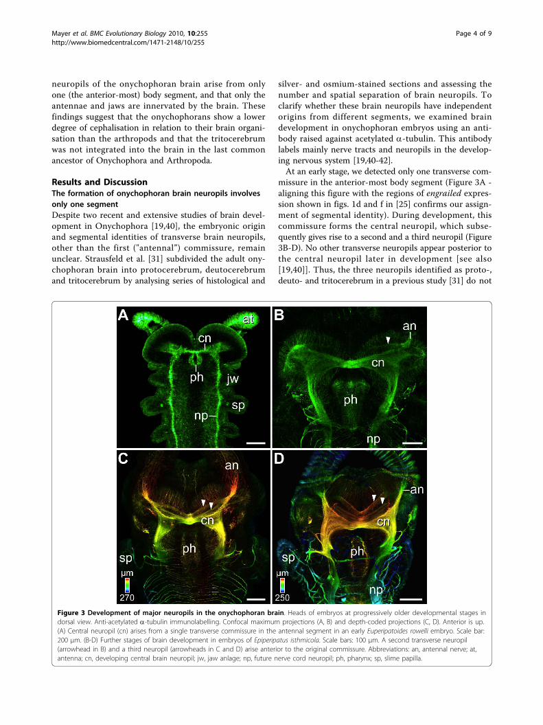
missure in the anterior-most body segment (Figure 3A -aligning this figure with the regions of engrailed expres-sion shown in figs. 1d and f in [25] confirms our assign-ment of segmental identity). During development, thiscommissure forms the central neuropil, which subse-quently gives rise to a second and a third neuropil (Figure3B-D). No other transverse neuropils appear posterior tothe central neuropil later in development [see also[19,40]]. Thus, the three neuropils identified as proto-,deuto- and tritocerebrum in a previous study [31] do not
Figure 3 Development of major neuropils in the onychophoran brain. Heads of embryos at progressively older developmental stages indorsal view. Anti-acetylated a-tubulin immunolabelling. Confocal maximum projections (A, B) and depth-coded projections (C, D). Anterior is up.(A) Central neuropil (cn) arises from a single transverse commissure in the antennal segment in an early Euperipatoides rowelli embryo. Scale bar:200 μm. (B-D) Further stages of brain development in embryos of Epiperipatus isthmicola. Scale bars: 100 μm. A second transverse neuropil(arrowhead in B) and a third neuropil (arrowheads in C and D) arise anterior to the original commissure. Abbreviations: an, antennal nerve; at,antenna; cn, developing central brain neuropil; jw, jaw anlage; np, future nerve cord neuropil; ph, pharynx; sp, slime papilla.
Mayer et al. BMC Evolutionary Biology 2010, 10:255http://www.biomedcentral.com/1471-2148/10/255
Page 4 of 9
arise from three different segments. We suggest there-fore that the position and physical separation of neuro-pils in the adult brain alone [22,31] is an unreliablecriterion for identifying its segmental organisation.Thus, our immunolabelling experiments do not resolvethe controversy of bipartite [36,37] versus tripartite[21,22,31] brain composition in Onychophora. Alterna-tive approaches are required to decide between thesetwo hypotheses.
Retrograde axonal tracing reveals that the tritocerebrumis absent from the onychophoran brainThe position of neurons that project out the segmentalnerves within the onychophoran head might be a keyfeature for determining the segmental identity of differ-ent brain regions. We therefore performed retrogradeaxonal tracing studies (backfills) of segmental cephalicnerves in adult onychophorans, using dextran coupledto different fluorochromes as a tracer [43].
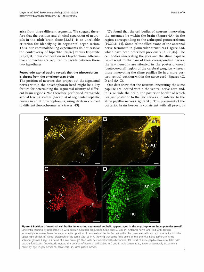
We found that the cell bodies of neurons innervatingthe antennae lie within the brain (Figure 4A), in theregion corresponding to the arthropod protocerebrum[19,20,31,44]. Some of the filled axons of the antennalnerve terminate in glomerular structures (Figure 4B),which have been described previously [31,38,44]. Thecell bodies innervating the jaws and the slime papillaelie adjacent to the base of their corresponding nerves:the jaw neurons are situated in the posterior-most(deutocerebral) region of the cerebral ganglion whereasthose innervating the slime papillae lie in a more pos-tero-ventral position within the nerve cord (Figures 4C,D and 5A-C).Our data show that the neurons innervating the slime
papillae are located within the ventral nerve cord and,thus, outside the brain, the posterior border of whichlies just posterior to the jaw nerves and anterior to theslime papillae nerves (Figure 5C). This placement of theposterior brain border is consistent with all previous
Figure 4 Position of neuronal cell bodies innervating segmental cephalic appendages in the onychophoran Euperipatoides rowelli.Differential staining by retrograde fills with dextran. Confocal projections. Scale bars: 50 μm. (A) Antennal nerve (an) filled with dextran-tetramethylrhodamine. Note the antero-median position of neuronal cell bodies (arrow) within the protocerebral brain region. Anterior is in theupper right corner. (B) Partial projection of the same stack as in A showing that some filled axons of the antennal nerve terminate in theantennal glomeruli (ag). (C) Detail of a jaw nerve (jn) filled with dextran-tetramethylrhodamine. (D) Detail of slime papilla nerves (sn) filled withdextran-fluorescein. Arrowheads indicate the position of neuronal cell bodies in C and D. Abbreviations: ag, antennal glomeruli; an, antennalnerve; ey, eye; jn, jaw nerve; nc, nerve cord; sn, slime papilla nerves.
Mayer et al. BMC Evolutionary Biology 2010, 10:255http://www.biomedcentral.com/1471-2148/10/255
Page 5 of 9

studies of the adult onychophoran brain anatomy [e.g.[21,22,31,36-39,44,45] and references therein]. The neu-rons innervating the slime papillae cannot be consideredpart of the brain as the corresponding region of the cen-tral nervous system does not show any particular con-densation of neurons or other morphologicalcharacteristics that would distinguish it from the medul-lary nerve cords.Our backfill data reveal that only the cell bodies of
neurons supplying the antennae and jaws lie within the
brain whereas the region corresponding to the arthro-pod tritocerebrum belongs to the nerve cord (Figure5A-C). This finding contradicts previous assumptions ofa tripartite (three-segmented) brain in Onychophora[2,21,22,24,31]. The absence of the tritocerebrum fromthe onychophoran brain implies that the bipartite braincomposition is an ancestral feature of Onychophora. Analternative scenario proposing that the tritocerebrummight have become separated from the onychophoranbrain secondarily is unlikely since one would have to
Figure 5 Position of neuronal cell bodies innervating segmental cephalic appendages in Onychophora and posterior border of theonychophoran brain. (A) Overview of the differential staining of segmental cephalic nerves in Euperipatoides rowelli by retrograde fills withdextran (confocal projection). Jaw nerves (jn) and slime papilla nerves (sn) from both sides of the body were filled with dextran-tetramethylrhodamine (red) and dextran-fluorescein (green). Anterior is up. Scale bar: 200 μm. (B) Diagram summarising the location of neuronalcell bodies innervating the antennae (yellow), jaws (red), and slime papillae (green). Note that the cell bodies of neurons innervating the slimepapillae lie outside the brain. (C) Anterior portion of the onychophoran nervous system (reconstruction based on confocal images of animmunolabelled embryo; see Additional file 1, Figure S1). The position of neurons innervating the segmental cephalic appendages (colourcoding as in B) is mapped on the reconstructed nervous system. Blue dashed line indicates the posterior brain border behind the cerebralaccumulation of neurons. Abbreviations: an, antennal nerve; br, cerebral ganglion or brain; cn, central neuropil; dc, deutocerebrum; ho,hypocerebral organ; jn, jaw nerve; ln, leg nerves; nc, nerve cord; pc, protocerebrum; sn, slime papilla nerves.
Mayer et al. BMC Evolutionary Biology 2010, 10:255http://www.biomedcentral.com/1471-2148/10/255
Page 6 of 9

assume opposite relocation events during the evolutionof the onychophoran head: while the slime papillae havebeen incorporated into the head by moving anteriorly,the corresponding brain region would have becomeseparated from the cerebral ganglion by a postero-ven-tral relocation. Moreover, this region would have lost itsganglionic organisation and reverted back to a portionof the medullary nerve cord. Studies of early neuraldevelopment in the onychophoran embryo [19,41,42]have revealed no evidence for an origin in the presump-tive brain of the neural precursors that give rise to neu-rons innervating the slime papillae. We therefore regardthis scenario as unlikely and suggest that the tritocereb-rum was not present in the last common ancestor ofOnychophora and Arthropoda but rather evolved inarthropods (Figure 6).
ConclusionsIn summary, our findings suggest an increase in thenumber of segmental brain regions in the (pan)arthro-pod lineage, from two in the last common ancestor ofOnychophora and Arthropoda, to at least three in var-ious arthropods [e.g. [2,27,31,32]]. This evolutionary
sequence may help clarify the phylogenetic position ofTardigrada (water bears), which is still controversial.Currently, tardigrades are regarded as either the sistergroup of arthropods, of onychophorans, of onychophor-ans plus arthropods, or of one of the cycloneuralian taxa(nematodes, kinorhynchs, and allies) [10,11,41,46-54].Our findings suggest that the number of segments inthe tardigrade brain, which remains unclear [48,55-58],will be a key feature in elucidating the position of thisanimal group within the Ecdysozoa.Furthermore, our suggestion of a two-segmented brain
in the last common ancestor of Onychophora andArthropoda challenges the hypothesis that a tripartitebrain existed in the last common ancestor of the bilater-ally symmetrical animals, the so-called “urbilaterian”[59,60]. Such a brain is absent in all protostomes apartfrom arthropods. Moreover, the closest relatives of chor-dates, including hemichordates and echinoderms [48],lack a centralised brain. We therefore suggest that simi-lar gene expression patterns in the anterior body regionof arthropods and vertebrates [59,60] are not related tobrain segmentation but rather to a general patterning ofthe antero-posterior body axis in these animals.
Figure 6 Implications of the current findings for the evolution of the arthropod brain. The brain of the last common ancestor ofOnychophora and Arthropoda was composed of the protocerebrum and deutocerebrum whereas the tritocerebrum, as part of the brain,evolved in arthropods (chelicerates, myriapods, crustaceans, and hexapods). The phylogenetic position of Myriapoda is unresolved [42,63].Double lines for Crustacea indicate that this group might not be monophyletic [64].
Mayer et al. BMC Evolutionary Biology 2010, 10:255http://www.biomedcentral.com/1471-2148/10/255
Page 7 of 9

MethodsSpecimens of Euperipatoides rowelli Reid, 1996 andEpiperipatus isthmicola (Bouvier, 1902) were collectedand handled and the embryos staged and labelled withan antibody raised against acetylated a-tubulin asdescribed previously [41,42]. For neuronal tracing, adultbrain nerves were dissected in physiological saline basedon onychophoran blood composition [61]. Retrogradefills of the antennal nerves (n = 3), jaw nerves (n = 9),and slime papillae nerves (n = 7) were carried out withdextran (MW 3000) coupled to either tetramethylrhoda-mine or fluorescein according to standard proceduresused for arthropods [43]. Scanning electron microscopyand immunohistochemistry were performed as describedpreviously [42]. Stained specimens were dehydratedthrough a methanol series and mounted between twocover slips in a 2:1 mixture of benzyl benzoate andbenzyl alcohol. Confocal laser-scanning microscopy andimage processing were carried out as describedpreviously [41,42].
Additional material
Additional file 1: Figure S1. Anterior nervous system in an almost fullydeveloped embryo of the onychophoran Epiperipatus isthmicola. Confocalmaximum projection. Dorso-lateral view (anterior is left, dorsal is up).Anti-acetylated a-tubulin immunolabelling. Abbreviations: an, antennalnerves; br, brain; cn, developing central brain neuropil; jn, jaw nerve; ln,paired leg nerves; mo, mouth position; nc, ventrolateral nerve cords; sn,slime papilla nerves. Scale bar: 200 μm.
AcknowledgementsThis work was supported by a grant from the German Research Foundation(DFG) to GM (Ma 4147/3-1). GM is a Research Group Leader supported bythe Emmy Noether Programme of the DFG.
Author details1Institute of Biology II: Animal Evolution & Development, University ofLeipzig, Talstrasse 33, D-04103 Leipzig, Germany. 2Department of Anatomyand Cell Biology, University of Melbourne, Victoria 3010, Australia. 3School ofBiological Sciences and Australian Centre for Biodiversity, Monash University,Melbourne, Victoria 3800, Australia. 4Institut für Biologie, Neurobiologie, FreieUniversität Berlin, Königin-Luise-Str. 28-30, D-14195 Berlin, Germany.
Authors’ contributionsGM conceived, designed and performed the experiments and wrote the firstdraft of the manuscript. PS and PMW helped with specimen collection. PMWand H-JP contributed laboratory space, reagents, materials and analysis tools.All authors participated in the discussion of the results and the preparationof the final manuscript.
Received: 13 May 2010 Accepted: 21 August 2010Published: 21 August 2010
References1. Budd GE: A palaeontological solution to the arthropod head problem.
Nature 2002, 417:271-275.2. Scholtz G, Edgecombe GD: The evolution of arthropod heads: reconciling
morphological, developmental and palaeontological evidence. Dev GenesEvol 2006, 216:395-415.
3. Scholtz G, Edgecombe GD: Heads, Hox and the phylogenetic position oftrilobites. In Crustacea and Arthropod Phylogeny. Edited by: Koenemann S,Jenner R, Vonk R. Boca Raton: CRC Press; 2005:16:139-165.
4. Chen J, Waloszek D, Maas A: A new “great appendage” arthropod fromthe Lower Cambrian of China and homology of chelicerate cheliceraeand raptorial antero-ventral appendages. Lethaia 2004, 37:3-20.
5. Budd GE, Telford MJ: The origin and evolution of arthropods. Nature 2009,457:812-817.
6. Jager M, Murienne J, Clabaut C, Deutsch J, Le Guyander H, Manuel M:Homology of arthropod anterior appendages revealed by Hox geneexpression in a sea spider. Nature 2006, 441:506-508.
7. Maxmen A, Browne WE, Martindale MQ, Giribet G: Neuroanatomy of seaspiders implies an appendicular origin of the protocerebral segment.Nature 2005, 437:1144-1148.
8. Waloszek D, Maas A, Chen J, Stein M: Evolution of cephalic feedingstructures and the phylogeny of Arthropoda. Palaeogeography,Palaeoclimatology, Palaeoecology 2007, 254:273-287.
9. Kusche K, Ruhberg H, Burmester T: A hemocyanin from the Onychophoraand the emergence of respiratory proteins. Proc Natl Acad Sci USA 2002,99:10545-10548.
10. Mallatt J, Giribet G: Further use of nearly complete 28 S and 18 S rRNAgenes to classify Ecdysozoa: 37 more arthropods and a kinorhynch. MolBiol Evol 2006, 40:772-794.
11. Dunn CW, Hejnol A, Matus DQ, Pang K, Browne WE, Smith SA, Seaver E,Rouse GW, Obst M, Edgecombe GD, et al: Broad phylogenomic samplingimproves resolution of the animal tree of life. Nature 2008, 452:745-749.
12. Budd GE: Why are arthropods segmented? Evol Dev 2001, 3:332-342.13. Maas A, Mayer G, Kristensen RM, Waloszek D: A Cambrian micro-
lobopodian and the evolution of arthropod locomotion andreproduction. Chin Sci Bull 2007, 52:3385-3392.
14. von Kennel J: Entwicklungsgeschichte von Peripatus edwardsii Blanch.und Peripatus torquatus n.sp. I. Theil. Arb Zool -Zootom Inst Würzburg1885, 7:95-229.
15. von Kennel J: Entwicklungsgeschichte von Peripatus edwardsii Blanch.und Peripatus torquatus n.sp. II. Theil. Arb Zool -Zootom Inst Würzburg1888, 8:1-93.
16. Sedgwick A: The development of the Cape species of Peripatus. Part III.On the changes from stage A to stage F. Q J Microsc Sci 1887, 27:467-550.
17. Evans R: On the Malayan species of Onychophora. Part II. - Thedevelopment of Eoperipatus weldoni. Q J Microsc Sci 1901, 45:41-88.
18. Walker M, Campiglia S: Some aspects of segment formation and post-placental development in Peripatus acacioi Marcus and Marcus(Onychophora). J Morphol 1988, 195:123-140.
19. Eriksson BJ, Tait NN, Budd GE: Head development in the onychophoranEuperipatoides kanangrensis. With particular reference to the centralnervous system. J Morphol 2003, 255:1-23.
20. Mayer G, Koch M: Ultrastructure and fate of the nephridial anlagen in theantennal segment of Epiperipatus biolleyi (Onychophora, Peripatidae) -evidence for the onychophoran antennae being modified legs.Arthropod Struct Dev 2005, 34:471-480.
21. Henry LM: The nervous system and the segmentation of the head in theAnnulata. Microentomology 1948, 13:27-48.
22. Fedorow B: Zur Anatomie des Nervensystems von Peripatus. II. DasNervensystem des vorderen Körperendes und seine Metamerie. Zool JbAnat 1929, 50:279-332.
23. Pflugfelder O: Entwicklung von Paraperipatus amboinensis n. sp. Zool JbAnat 1948, 69:443-492.
24. Pflugfelder O: Onychophora. In Grosses Zoologisches Praktikum. Edited by:Czihak G. Stuttgart: Gustav Fischer; , 13a 1968:1-42.
25. Eriksson BJ, Tait NN, Budd GE, Akam M: The involvement of engrailed andwingless during segmentation in the onychophoran Euperipatoideskanangrensis (Peripatopsidae: Onychophora) (Reid 1996). Dev Genes Evol2009, 219:249-264.
26. Eriksson BJ, Tait NN, Budd GE, Janssen R, Akam M: Head patterning andHox gene expression in an onychophoran and its implications for thearthropod head problem. Dev Genes Evol 2010.
27. Harzsch S: Neurophylogeny: Architecture of the nervous system and afresh view on arthropod phyologeny. Integr Comp Biol 2006, 46:162-194.
28. Liu Y, Maas A, Waloszek D: Early development of the anterior bodyregion of the grey widow spider Latrodectus geometricus Koch, 1841(Theridiidae, Araneae). Arthropod Struct Dev 2009, 38:401-416.
Mayer et al. BMC Evolutionary Biology 2010, 10:255http://www.biomedcentral.com/1471-2148/10/255
Page 8 of 9

29. Posnien N, Bashasab F, Bucher G: The insect upper lip (labrum) is anonsegmental appendage-like structure. Evol Dev 2009, 11:480-488.
30. Boyan GS, Williams JLD, Posser S, Bräunig P: Morphological and moleculardata argue for the labrum being non-apical, articulated, and theappendage of the intercalary segment in the locust. Arthropod Struct Dev2002, 31:65-76.
31. Strausfeld NJ, Strausfeld C, Stowe S, Rowell D, Loesel R: The organizationand evolutionary implications of neuropils and their neurons in thebrain of the onychophoran Euperipatoides rowelli. Arthropod Struct Dev2006, 35:169-196.
32. Urbach R, Technau GM: Early steps in building the insect brain:neuroblast formation and segmental patterning in the developing brainof different insect species. Arthropod Struct Dev 2003, 32:103-123.
33. Harzsch S: The tritocerebrum of Euarthropoda: a “non-drosophilocentric”perspective. Evol Dev 2004, 6:303-309.
34. Boyan GS, Williams JLD, Hirth F: Commissural organization and brainsegmentation in insects. In Theories, Development, Invertebrates. Edited by:Striedter GF, Rubenstein JLR. Oxford: Academic Press; 2007:1:349-359.
35. Urbach R, Technau GM: Segmental organization of cephalic ganglia inarthropods. In Theories, Development, Invertebrates. Edited by: Striedter GF.Rubenstein JLR. Oxford: Academic Press; 2007:1:337-348.
36. Holmgren NF: Zur vergleichenden Anatomie des Gehirns vonPolychaeten, Onychophoren, Xiphosuren, Arachniden, Crustaceen,Myriapoden, und Insekten. Vorstudien zu einer Phylogenie derArthropoden. K Svenska Vet Handl [Ser 2] 1916, 56:1-303.
37. Hanström B: Onychophora. Vergleichende Anatomie des Nervensystems derWirbellosen Tiere unter Berücksichtigung seiner Funktion Berlin, Germany: J.Springer 1928, 341-351.
38. Schürmann FW: Histology and ultrastructure of the onychophoran brain.In Arthropod Brain, its Evolution, Development, Structure, and Functions.Edited by: Gupta AP. New York: John Wiley 1987:159-180.
39. Lane NJ, Campiglia SS: The lack of a structured blood-brain barrier in theonychophoran Peripatus acacioi. J Neurocytol 1987, 16:93-104.
40. Eriksson BJ, Budd GE: Onychophoran cephalic nerves and their bearingon our understanding of head segmentation and stem-group evolutionof Arthropoda. Arthropod Struct Dev 2000, 29:197-209.
41. Mayer G, Whitington PM: Neural development in Onychophora (velvetworms) suggests a step-wise evolution of segmentation in the nervoussystem of Panarthropoda. Dev Biol 2009, 335:263-275.
42. Mayer G, Whitington PM: Velvet worm development links myriapods withchelicerates. Proc R Soc B 2009, 276:3571-3579.
43. Pflüger H-J, Field LH: A locust chordotonal organ coding forproprioceptive and acoustic stimuli. J Comp Physiol A 1999, 184:169-183.
44. Strausfeld NJ, Strausfeld CM, Loesel R, Rowell D, Stowe S: Arthropodphylogeny: onychophoran brain organization suggests an archaicrelationship with a chelicerate stem lineage. Proc R Soc B 2006,273:1857-1866.
45. Hanström B: Bemerkungen über das Gehirn und die Sinnesorgane derOnychophoren. Lunds Univ Årsskrift NF 1935, 31:1-37.
46. Budd GE: Tardigrades as ‘stem-group arthropods’: The evidence from theCambrian fauna. Zool Anz 2001, 240:265-279.
47. Maas A, Waloszek D: Cambrian derivatives of the early arthropod stemlineage, Pentastomids, Tardigrades and Lobopodians - an “Orsten”perspective. Zool Anz 2001, 240:451-459.
48. Nielsen C: Animal Evolution: Interrelationships of the Living Phyla Oxford:Oxford University Press 2001.
49. Telford MJ, Bourlat SJ, Economou A, Papillon D, Rota-Stabelli O: Theevolution of the Ecdysozoa. Phil Trans R Soc B 2008, 363:1529-1537.
50. Jenner RA, Scholtz G: Playing another round of metazoan phylogenetics:Historical epistemology, sensitivity analysis, and the position ofArthropoda within the Metazoa on the basis of morphology. In Crustaceaand Arthropod Relationships. Edited by: Koenemann S, Jenner RA. BocaRaton: CRC Press; 2005:16:355-385.
51. Park J-K, Rho HS, Kristensen RM, Kim W, Giribet G: First molecular data onthe phylum Loricifera - an investigation into the phylogeny ofEcdysozoa with emphasis on the positions of Loricifera and Priapulida.Zool Sci 2006, 23:943-954.
52. Lartillot N, Philippe H: Improvement of molecular phylogenetic inferenceand the phylogeny of Bilateria. Phil Trans R Soc B 2008, 363:1463-1472.
53. Hejnol A, Obst M, Stamatakis A, Ott M, Rouse GW, Edgecombe GD,Martinez P, Baguñà J, Bailly X, Jondelius U, et al: Assessing the root of
bilaterian animals with scalable phylogenomic methods. Proc R Soc B2009, 276:4261-4270.
54. Rota-Stabelli O, Kayal E, Gleeson D, Daub J, Boore J, Telford M, Pisani D,Blaxter M, Lavrov D: Ecdysozoan mitogenomics: Evidence for a commonorigin of the legged invertebrates, the Panarthropoda. Genome Biologyand Evolution 2010.
55. Kristensen RM, Higgins RP: Revision of Styraconyx (Tardigrada:Halechiniscidae) with descriptions of two new species from Disko Bay,West Greenland. Smithson Contrib Zool 1984, 391:1-40.
56. Dewel RA, Dewel WC: The brain of Echiniscus viridissimus Peterfi, 1956(Heterotardigrada): a key to understanding the phylogenetic position oftardigrades and the evolution of the arthropod head. Zool J Linn Soc1996, 116:35-49.
57. Hejnol A, Schnabel R: What a couple of dimensions can do for you:Comparative developmental studies using 4 D microscopy - examplesfrom tardigrade development. Integr Comp Biol 2006, 46:151-161.
58. Zantke J, Wolff C, Scholtz G: Three-dimensional reconstruction of thecentral nervous system of Macrobiotus hufelandi (Eutardigrada,Parachela): implications for the phylogenetic position of Tardigrada.Zoomorphology 2008, 127:21-36.
59. Hirth F, Kammermeier L, Frei E, Walldorf U, Noll M, Reichert H: Anurbilaterian origin of the tripartite brain: developmental genetic insightsfrom Drosophila. Development 2003, 130:2365-2373.
60. Urbach R: A procephalic territory in Drosophila exhibiting similarities anddissimilarities compared to the vertebrate midbrain/hindbrain boundaryregion. Neural Dev 2007, 2:23.
61. Robson EA, Lockwood APM, Ralph R: Composition of the blood inOnychophora. Nature 1966, 209:533.
62. Mayer G: Structure and development of onychophoran eyes - what isthe ancestral visual organ in arthropods? Arthropod Struct Dev 2006,35:231-245.
63. Edgecombe GD: Arthropod phylogeny: An overview from theperspectives of morphology, molecular data and the fossil record.Arthropod Struct Dev 2010, 39:74-87.
64. Regier JC, Shultz JW, Zwick A, Hussey A, Ball B, Wetzer R, Martin JW,Cunningham CW: Arthropod relationships revealed by phylogenomicanalysis of nuclear protein-coding sequences. Nature 2010,463:1079-1083.
doi:10.1186/1471-2148-10-255Cite this article as: Mayer et al.: A revision of brain composition inOnychophora (velvet worms) suggests that the tritocerebrum evolvedin arthropods. BMC Evolutionary Biology 2010 10:255.
Submit your next manuscript to BioMed Centraland take full advantage of:
• Convenient online submission
• Thorough peer review
• No space constraints or color figure charges
• Immediate publication on acceptance
• Inclusion in PubMed, CAS, Scopus and Google Scholar
• Research which is freely available for redistribution
Submit your manuscript at www.biomedcentral.com/submit
Mayer et al. BMC Evolutionary Biology 2010, 10:255http://www.biomedcentral.com/1471-2148/10/255
Page 9 of 9