Platelet-Activating Factor Receptor Deficiency Delays Elimination of Adult Worms but Reduces...
9
10.1128/IAI.72.2.1135-1142.2004. 2004, 72(2):1135. DOI: Infect. Immun. Teixeira Michele M. Barsante, Adriano L. S. Souza and Mauro M. Deborah Negrão-Corrêa, Danielle G. Souza, Vanessa Pinho, -Infected Mice Strongyloides venezuelensis Worms but Reduces Fecundity in Deficiency Delays Elimination of Adult Platelet-Activating Factor Receptor http://iai.asm.org/content/72/2/1135 Updated information and services can be found at: These include: REFERENCES http://iai.asm.org/content/72/2/1135#ref-list-1 at: This article cites 55 articles, 15 of which can be accessed free CONTENT ALERTS more» articles cite this article), Receive: RSS Feeds, eTOCs, free email alerts (when new http://journals.asm.org/site/misc/reprints.xhtml Information about commercial reprint orders: http://journals.asm.org/site/subscriptions/ To subscribe to to another ASM Journal go to: on February 19, 2014 by guest http://iai.asm.org/ Downloaded from on February 19, 2014 by guest http://iai.asm.org/ Downloaded from
-
Upload
independent -
Category
Documents
-
view
1 -
download
0
Transcript of Platelet-Activating Factor Receptor Deficiency Delays Elimination of Adult Worms but Reduces...
10.1128/IAI.72.2.1135-1142.2004.
2004, 72(2):1135. DOI:Infect. Immun. TeixeiraMichele M. Barsante, Adriano L. S. Souza and Mauro M. Deborah Negrão-Corrêa, Danielle G. Souza, Vanessa Pinho,
-Infected MiceStrongyloides venezuelensisWorms but Reduces Fecundity in Deficiency Delays Elimination of Adult Platelet-Activating Factor Receptor
http://iai.asm.org/content/72/2/1135Updated information and services can be found at:
These include:
REFERENCEShttp://iai.asm.org/content/72/2/1135#ref-list-1at:
This article cites 55 articles, 15 of which can be accessed free
CONTENT ALERTS more»articles cite this article),
Receive: RSS Feeds, eTOCs, free email alerts (when new
http://journals.asm.org/site/misc/reprints.xhtmlInformation about commercial reprint orders: http://journals.asm.org/site/subscriptions/To subscribe to to another ASM Journal go to:
on February 19, 2014 by guest
http://iai.asm.org/
Dow
nloaded from
on February 19, 2014 by guest
http://iai.asm.org/
Dow
nloaded from

INFECTION AND IMMUNITY, Feb. 2004, p. 1135–1142 Vol. 72, No. 20019-9567/04/$08.00�0 DOI: 10.1128/IAI.72.2.1135–1142.2004Copyright © 2004, American Society for Microbiology. All Rights Reserved.
Platelet-Activating Factor Receptor Deficiency Delays Elimination ofAdult Worms but Reduces Fecundity in Strongyloides
venezuelensis-Infected MiceDeborah Negrao-Correa,1* Danielle G. Souza,2 Vanessa Pinho,2 Michele M. Barsante,2
Adriano L. S. Souza,2 and Mauro M. Teixeira2,3
Departamento de Parasitologia1 and Departamento de Bioquımica e Imunologia,2 Instituto de Ciencias Biologicas, UniversidadeFederal de Minas Gerais, and Centro de Pesquisas Rene Rachou-FIOCRUZ,3 Belo Horizonte, MG, Brazil
Received 16 July 2003/Returned for modification 16 August 2003/Accepted 8 October 2003
We describe the parasitological kinetics and histopathological and immunological alterations in platelet-activating factor receptor-deficient (PAFR�/�) and wild-type mice after a single Strongyloides venezuelensisinfection (subcutaneous inoculation of 500 L3 larvae). There was no difference in the numbers of worms thatreached and became established in the small intestines of PAFR�/� and wild-type mice. However, at 12 daysafter infection, significantly more worms were recovered from PAFR�/� mice. Although PAFR�/� infected miceshowed a delay in elimination of adult worms, worms established in the small intestine of these mice produceda significantly lower number of eggs due to a reduction in worm fecundity. There were also significantreductions in the number of circulating and tissue eosinophils and tumor necrosis factor levels in the smallintestines of PAFR�/� mice infected for 7 days compared to the number and level in wild-type mice. Histo-logical analysis confirmed the reduced inflammatory process and revealed that the PAFR�/� mice had asmaller number of goblet cells. The concentrations of the type 2 cytokines interleukin-4 (IL-4), IL-5, and IL-10were lower in small intestine homogenates and in supernatants of antigen-stimulated lymphocytes fromspleens or mesenteric lymph nodes of PAFR�/� mice than in the corresponding preparations from wild-typemice. Thus, in S. venezuelensis-infected PAFR�/� mice, decreased intestinal inflammation is associated withenhanced worm survival but decreased fecundity. We suggest that although a Th2-predominant inflammatoryresponse decreases worm survival, the worm may use factors produced during this response to facilitate eggoutput and reproduction. PAFR-mediated responses appear to modulate these host-derived signals that areimportant for worm fecundity.
Gastrointestinal nematode species which have pulmonarymigration in their life cycles, such as Necator americanus, An-cylostoma duodenalis, Strongyloides stercoralis, and Ascaris lum-bricoides, are the most prevalent parasites in humans, infectingover one-quarter of the world’s population (9, 11). Despite thehigh prevalence and chronic morbidity produced by these nem-atodes, immunopathologic and/or immunoprotective mecha-nisms involved in the response against these parasites are notcompletely understood. Experimental infections with gastroin-testinal nematodes in rodents induce predominantly a Th2 typeof immune response that has been associated with host pro-tection. Nevertheless, the precise mechanisms of protectionare still not clear and appear to be considerably different fordifferent helminths (17, 18, 36).
One experimental model that has been used to study theaspects of immunoprotection and immunoregulation duringgastrointestinal infection is infection of rodents with Strongy-loides venezuelensis (29, 30, 40, 50), a nematode that naturallyinfects wild rats. In experimental infections, S. venezuelensislarvae have an obligatory migration through the lungs of thehost before they become established in the duodenal mucosa(52), similar to the migration of S. stercoralis in humans, andthe adult worms are spontaneously eliminated after 5 weeks in
rats (55) and after 10 to 14 days of infection in mice (46). Themigration of parasite larvae (37, 50) or establishment of theworm in the gut (24, 26) results in eosinophilic inflammation.Moreover, arrival of the parasite in the intestine is accompa-nied by intestinal eosinophilia and mastocytosis, which may beassociated with the process of worm elimination (10, 24, 26).
The lipid mediator platelet-activating factor (1-O-alkyl-2-acetyl-sn-glyceryl-3-phosphorocholine) (PAF) is produced by alarge number of inflammatory cells, including macrophages,neutrophils, basophils, eosinophils, platelets, and endothelialcells (7, 33). PAF is minimally expressed under normal physi-ological conditions; however, PAF has been implicated in anumber of inflammatory conditions, including allergic inflam-mation (7, 21, 25, 33). Once released, PAF activates PAFreceptors (PAFR), which results in diverse biological activitiesassociated with inflammation (21), including macrophage andeosinophil activation and chemotaxis, alterations in vascularpermeability, platelet activation, and induction of mucus pro-duction by epithelial cells, all of which may contribute to thecourse of infection with helminths. Using PAFR�/� mice (22),we analyzed the role of PAFR during the course of infectionwith the intestinal parasite S. venezuelensis.
MATERIALS AND METHODSAnimals. C57BL/6 mice lacking the PAFR (PAFR�/� mice) were generated as
previously described by Ishii et al. (22). These PAFR-deficient mice were pro-vided by Takao Shimizu (Department of Biochemistry and Molecular Biology,University of Tokyo, Tokyo, Japan) and have been bred and maintained at thebioscience unit of the Institute Goncalo Muniz (Fundacao Oswaldo Cruz, Sal-
* Corresponding author. Mailing address: Departamento de Parasi-tologia, ICB, UFMG, Av. Antonio Carlos 6627, Campus Pampulha,Belo Horizonte, MG, Brazil CEP-31270-901. Phone: 55 31 3499-2840.Fax: 55 31 3499-2979. E-mail: [email protected].
1135
on February 19, 2014 by guest
http://iai.asm.org/
Dow
nloaded from

vador, Brazil). For the experiments described here, C57BL/6 PAFR�/� andwild-type female mice that were 8 to 10 weeks old were used. All animals werekept at the Department of Biochemistry and Immunology (Federal University ofMinas Gerais State, Belo Horizonte, Brazil) vivarium for infected animals duringthe experimental procedure, fed laboratory chow (Nuvilab; Colombo, Parana,Brazil), and given tap water ad libitum. For experimental procedures we receivedprior approval from the local animal ethics committee.
Parasite and parasitological techniques. S. venezuelensis, the intestinal nem-atode used in all the experiments, was isolated from Rattus norvegicus (8) and hasbeen maintained in the Department of Parasitology (Federal University of MinasGerais State, Belo Horizonte, Brazil) by serial passage in Wistar rats. S. venezue-lensis infective filiform larvae (L3) were obtained from charcoal cultures of infected-rat feces. The cultures were kept for 48 to 72 h at 28°C, and the infective larvae werecollected and concentrated by using a Baermann apparatus. Subsequently, the larvaerecovered were washed several times in phosphate-buffered saline (PBS) andcounted, and the concentration was adjusted to 5,000 L3 per ml of PBS for infection.PAFR�/� and wild-type mice were individually inoculated via subcutaneous in-jection of 100 �l of PBS containing 500 infective larvae in the abdominal region.
Infectivity rates were determined by assessing fecal egg counts and numbers ofworms recovered from the small intestine at 5, 7, and 12 days after infection. Forrecovery of worms from the small intestine, the upper half of the small intestinefrom each infected mouse was removed after sacrifice, washed, cut open longi-tudinally, and incubated in PBS at 37°C for 4 h. Worms that emerged from theintestinal tissue were quantified by stereomicroscopy. The remaining intestinaltissue was placed in PBS and incubated overnight at 4°C, the worms werequantified again, and the total number of worms was determined. After theupper half of the small intestine was separated for worm counting, the first 2 cmof the lower half of each small intestine was processed for histological analysis,and the remaining piece was frozen at �70°C for quantification of cytokines andfor the eosinophil peroxidase (EPO) assay. To determine the number of nema-tode eggs eliminated in the feces, well-formed fecal pellets were obtained fromthe rectum of each infected mouse, weighed, and homogenized in a knownvolume of PBS. For each infected mouse, two fecal samples (100 �l) wereexamined by light microscopy, and the total number of parasite eggs was deter-mined. The number of S. venezuelensis eggs was expressed per gram of mousefeces. Since S. venezuelensis-infected hosts have only female worms in the smallintestine, worm fecundity was estimated by dividing the number of eggs elimi-nated in feces by the number of worms recovered from the intestine of eachmouse at each time point.
To obtain L3 soluble antigen, a large number of infective larvae were exten-sively washed with PBS, resuspended in PBS containing protease inhibitor cocktail(one tablet in 25 ml of PBS; Boehringer Mannheim, Indianapolis, Ind.), and dis-rupted by vortexing with glass beads (five cycles, 1 min each) and by sonication witha cell sonic disrupter (PGC Scientific, Gaithersburg, Md.) by using 10 1-min cyclesat the highest power allowed for the standard microtip (48). After the insolubleparticles were removed by centrifugation of the larval homogenate at 400 � g for 30min, the supernatant was removed, and the protein concentration was deter-mined before division into aliquots and storage at �20°C. The antigen prepara-tion was used to stimulate cells obtained from mesenteric lymph nodes (MLN)and spleens.
Leukocytes in blood and BAL fluid of infected mice. At 7 and 12 days after S.venezuelensis infection, PAFR�/� and wild-type mice were anesthetized and bledvia the tail vein, and the blood samples were used to estimate the circulating cellcompositions. Bronchoalveolar lavage (BAL) was performed 5 and 7 days afterinfection by intratracheal instillation of three aliquots of 1 ml of PBS containing0.3% bovine serum albumin (Sigma) and protease inhibitor cocktail (one tabletin 50 ml of PBS; Boehringer Mannheim). The lavage fluid was centrifuged at 200� g for 7 min, and the cell pellet from the BAL fluid was resuspended in 1 ml ofPBS containing 0.3% bovine serum albumin.
The total numbers of leukocytes in blood and BAL fluid were estimated byusing a Neubauer chamber. Cytospin slides prepared from BAL fluid and bloodsmears were stained with May-Grunwald-Giemsa stain. Cells were differentiatedinto mononuclear cells, mature eosinophils, and mature neutrophils by usingstandard morphological criteria, and at least 200 cells were counted per slide bylight microscopy.
Histopathology. As mentioned above, a section of the intestine collected fromthe jejunal portion was fixed in 10% buffered formalin and embedded in paraffin,and 5-�m sections were prepared for histology. Sections were stained with hema-toxylin and eosin for assessment of the overall inflammatory response. Goblet celland mucus production was analyzed by using periodic acid-Schiff-stained slides.
EPO assay. The EPO assay, performed as described by Silveira et al. (50), wasused to estimate the numbers of eosinophils in the lungs and intestine.
Cell culture. Cytokine production was measured in supernatants from spleensand MLN cells restimulated with L3 total antigen or concanavalin A (ConA). Forthese experiments, uninfected PAFR�/� and wild-type mice and PAFR�/� andwild-type mice that were infected with S. venezuelensis for 7 and 12 days wereused. Briefly, the spleen and MLN were aseptically removed, a single-cell sus-pension was prepared, and red blood cells were lysed. Individual spleens andMLN from four or five mice per group were used at each time. The cells obtainedfrom these organs were centrifuged at 200 � g for 5 min, counted, plated in96-well, flat-bottom microplates (NUNC, Naperville, Ill.) at a concentration of1 � 106 cells/well in RPMI 1640 containing 25 mM HEPES and sodium bicar-bonate (Sigma, St. Louis, Mo.) and supplemented with 10% fetal calf serum, 100U of penicillin per ml, 100 �g of streptomycin per ml, 10 mM sodium pyruvate,and 2 mM L-glutamine (Sigma), and cultured at 37°C in the presence of 5% CO2
for 72 h. Cells were cultured in duplicate in 200 �l either alone or in the presenceof 2 �g of ConA (Sigma) per ml or 100 �g of parasite antigen per ml. Super-natants were collected after 72 h, divided into aliquots, and kept at �20°C forquantification of interleukin-4 (IL-4), IL-5, IL-10, and gamma interferon (IFN-�).
Measurement of cytokines in intestinal homogenates and spleen and MLNculture supernatants. Cytokines were quantified in cell culture supernatants andin homogenates prepared from the small intestine. The latter samples wereobtained by homogenizing 100 mg of the lower half of the small intestine fromPAFR�/� or wild-type mice obtained 7 and 12 days after infection with S.venezuelensis. The intestinal samples were homogenized in 1 ml of PBS contain-ing protease inhibitors (0.1 mM phenylmethylsulfonyl fluoride, 0.1 mM benze-thonium chloride, 10 mM EDTA, 20 KI aprotinin A, and 0.05% Tween 20). Eachsample was then centrifuged for 10 min at 3,000 � g, and the supernatant wasused for an enzyme-linked immunosorbent assay (ELISA).
Concentrations of IL-4, IL-5, IL-10, IFN-�, and tumor necrosis factor alpha(TNF-�) were determined by an ELISA technique with commercially availableantibody pairs used according to the instructions supplied by the manufacturer(R&D Systems). Known concentrations of the recombinant proteins were usedto generate a standard curve to convert optical density values of samples topicograms per milliliter. The sensitivity of the assays was 16 pg/ml.
Statistical analysis. Data are reported below as means � standard errors ofthe means and were analyzed by using Student’s t test (two groups) or one-wayanalysis of variance. In the latter analysis, P values were assigned by using Tukeypost hoc analysis. Differences were considered significant at a P value of �0.05.
RESULTS
PAFR-deficient mice have delayed elimination of S. venezue-lensis from the small intestine. The kinetics of S. venezuelensisinfection in mice (Fig. 1A) revealed that there was no statisti-cal difference in the numbers of worms that arrived and be-came established in the small intestines of PAFR�/� and wild-type mice at 7 days after infection (201 � 12 worms inPAFR�/� mice and 189 � 10 worms in wild-type mice). How-ever, at 12 days after infection, there were 76 � 7 worms/mouse in PAFR�/� infected mice, while only 30 � 8 worms/mouse were recovered from the wild-type controls (P 0.05).
Interestingly, the number of S. venezuelensis eggs eliminatedin the feces of PAFR�/� infected mice did not reflect thekinetics of adult worm presence in the intestine (Fig. 1B). At 7days after infection, a time when the numbers of worms recov-ered from the small intestine were similar for the two groups,PAFR�/� infected mice eliminated significantly fewer S. vene-zuelensis eggs in the feces than infected controls eliminated(25,000 � 3,800 eggs for PAFR�/� mice compared to 44,760 �5,000 eggs for wild-type controls). At 12 days after infection,PAFR�/� mice and infected wild-type controls eliminated sim-ilar numbers of eggs, even though the former group of animalshad more worms in the small intestine. The smaller number ofparasite eggs eliminated in the feces of PAFR�/� infectedmice was a consequence of the lower fecundity of the wormsestablished in the small intestine of these animals (Fig. 1C).Indeed, the fecundity of worms from PAFR�/� mice was lower
1136 NEGRAO-CORREA ET AL. INFECT. IMMUN.
on February 19, 2014 by guest
http://iai.asm.org/
Dow
nloaded from
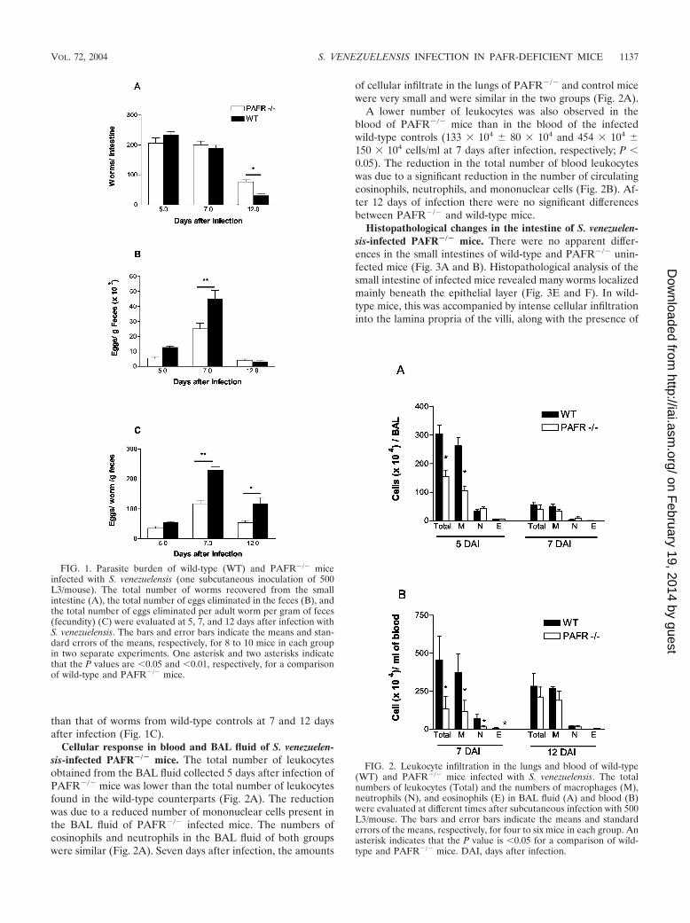
than that of worms from wild-type controls at 7 and 12 daysafter infection (Fig. 1C).
Cellular response in blood and BAL fluid of S. venezuelen-sis-infected PAFR�/� mice. The total number of leukocytesobtained from the BAL fluid collected 5 days after infection ofPAFR�/� mice was lower than the total number of leukocytesfound in the wild-type counterparts (Fig. 2A). The reductionwas due to a reduced number of mononuclear cells present inthe BAL fluid of PAFR�/� infected mice. The numbers ofeosinophils and neutrophils in the BAL fluid of both groupswere similar (Fig. 2A). Seven days after infection, the amounts
of cellular infiltrate in the lungs of PAFR�/� and control micewere very small and were similar in the two groups (Fig. 2A).
A lower number of leukocytes was also observed in theblood of PAFR�/� mice than in the blood of the infectedwild-type controls (133 � 104 � 80 � 104 and 454 � 104 �150 � 104 cells/ml at 7 days after infection, respectively; P 0.05). The reduction in the total number of blood leukocyteswas due to a significant reduction in the number of circulatingeosinophils, neutrophils, and mononuclear cells (Fig. 2B). Af-ter 12 days of infection there were no significant differencesbetween PAFR�/� and wild-type mice.
Histopathological changes in the intestine of S. venezuelen-sis-infected PAFR�/� mice. There were no apparent differ-ences in the small intestines of wild-type and PAFR�/� unin-fected mice (Fig. 3A and B). Histopathological analysis of thesmall intestine of infected mice revealed many worms localizedmainly beneath the epithelial layer (Fig. 3E and F). In wild-type mice, this was accompanied by intense cellular infiltrationinto the lamina propria of the villi, along with the presence of
FIG. 1. Parasite burden of wild-type (WT) and PAFR�/� miceinfected with S. venezuelensis (one subcutaneous inoculation of 500L3/mouse). The total number of worms recovered from the smallintestine (A), the total number of eggs eliminated in the feces (B), andthe total number of eggs eliminated per adult worm per gram of feces(fecundity) (C) were evaluated at 5, 7, and 12 days after infection withS. venezuelensis. The bars and error bars indicate the means and stan-dard errors of the means, respectively, for 8 to 10 mice in each groupin two separate experiments. One asterisk and two asterisks indicatethat the P values are 0.05 and 0.01, respectively, for a comparisonof wild-type and PAFR�/� mice.
FIG. 2. Leukocyte infiltration in the lungs and blood of wild-type(WT) and PAFR�/� mice infected with S. venezuelensis. The totalnumbers of leukocytes (Total) and the numbers of macrophages (M),neutrophils (N), and eosinophils (E) in BAL fluid (A) and blood (B)were evaluated at different times after subcutaneous infection with 500L3/mouse. The bars and error bars indicate the means and standarderrors of the means, respectively, for four to six mice in each group. Anasterisk indicates that the P value is 0.05 for a comparison of wild-type and PAFR�/� mice. DAI, days after infection.
VOL. 72, 2004 S. VENEZUELENSIS INFECTION IN PAFR-DEFICIENT MICE 1137
on February 19, 2014 by guest
http://iai.asm.org/
Dow
nloaded from

eosinophils (Fig. 3G). Eosinophil-rich infiltrates were not evi-dent in the small intestine of PAFR�/� infected mice (Fig.3H). At 12 days after infection, adult worms were still presentin the small intestine of PAFR�/� infected mice, confirmingthe delay in elimination of worms (Fig. 3D). Moreover, at 12days after infection, there were more goblet cells filled withmucus in the epithelial layer of wild-type infected mice (Fig. 3C)than in the epithelial layer of PAFR�/� infected mice (Fig. 3D).
With the intent of quantifying the inflammatory response inthe small intestine, we assessed eosinophil infiltration indi-rectly by measuring the activity of EPO (Fig. 4A) and theTNF-� concentrations by ELISA in tissue homogenates (Fig.4B). As shown in Fig. 4, the levels of EPO and TNF-� weresignificantly lower in PAFR�/� infected mice than in infectedwild-type controls at 7 days after infection. Interestingly, bothvalues were greater for PAFR�/� infected mice than for wild-type controls at 12 days after infection (Fig. 4).
S. venezuelensis-infected PAFR�/� mice expressed lower lev-els of Th2 cytokines. Cytokine analysis of homogenates of thesmall intestine showed that there was a discrete increase in theconcentration of IFN-� 7 days after infection, but the concen-tration returned to the normal level 12 days after S. venezue-lensis infection in both PAFR�/� and wild-type mice (Fig. 5A).The concentrations of IL-10 exhibited similar kinetics; i.e.,there was a threefold increase above the levels found in non-infected animals at day 7, and background concentrations wereobserved at day 12 after infection (Fig. 5B). The concentra-tions of the Th2 cytokines IL-4 and IL-5 exhibited the greatestincreases above the values found in noninfected animals.Moreover, elevated concentrations of both IL-4 and IL-5 werefound even after 12 days of infection (Fig. 5C and D). At day7 after infection, the concentrations of IL-4 and IL-5 weresignificantly lower in PAFR�/� mice than in the small intes-tines of the wild-type controls (Fig. 5C and D).
Cytokine concentrations were also measured in supernatantsof spleen and MLN cells collected from infected mice andrestimulated in vitro with L3 antigen. Similar to the concen-trations of cytokines in the intestine, there were lower concen-trations of cytokines in the supernatants of spleen and MLNcell cultures from PAFR�/� mice infected for 7 days than inthe supernatants of spleen and MLN cultures from the wild-type controls (Table 1). Specifically, the concentrations of IL-5and IFN-� in supernatants of spleen cell cultures and theconcentrations of IL-4, IL-5, and IFN-� in supernatants ofMLN cell cultures were significantly lower for PAFR�/� micethan for wild-type mice (Table 1). Cytokine levels were re-duced after 12 days of infection in both PAFR�/� and wild-type mice (data not shown). Stimulation with ConA resulted ingreater and more persistent production of IL-4 and IL-5 in
FIG. 3. Small intestines of wild-type (WT) and PAFR�/� miceinfected with S. venezuelensis. (A, C, E, and G) Sections from wild-typemice; (B, D, F, and H) sections from PAFR�/� mice. Uninfected micewere evaluated (A and B), and mice infected with S. venezuelensis wereevaluated 12 days (C and D) or 7 days (E to H) after infection. Note
the greater number of goblet cells (C) and the eosinophilic tissueinfiltration (G) in wild-type mice. In panel D, the arrows indicatesections of worms in the small intestine of a PAFR�/� mouse after 12days of S. venezuelensis infection. In panel G, the arrowheads indicatethe presence of eosinophils. The tissue was fixed with buffered forma-lin and embedded in paraffin, and 5-�m sections were stained withperiodic acid-Schiff stain (A to D) or hematoxylin and eosin (E to G).(A to F) Magnification, �200; (G and H) magnification, �400. DAI,days after infection.
1138 NEGRAO-CORREA ET AL. INFECT. IMMUN.
on February 19, 2014 by guest
http://iai.asm.org/
Dow
nloaded from

both spleen and MLN cells collected from infected mice.ConA-induced IFN-� production by spleen and MLN cellsfrom normal animals was not different than ConA-inducedIFN-� production by spleen and MLN cells from infected an-imals (data not shown). Moreover, there were no significantdifferences between the ConA-stimulated cytokine levels inPAFR�/� and wild-type mice (data not shown).
DISCUSSION
PAF is a potent phospholipid mediator that has been shownto mediate a broad spectrum of biological activities, includingleukocyte recruitment and activation, airway constriction, andvascular hyperpermeability, through its G-protein-coupled re-ceptor (21). In addition, several experiments have linked PAFto allergic processes (25; reviewed in reference 21). It has beendemonstrated previously that S. venezuelensis infection induceseosinophilic lung inflammation that is accompanied by airwayhyperreactivity in rats (37, 50). A similar inflammatory reactionis observed in the small intestine of Strongyloides-infected mice(15, 24, 32). The participation of PAF or the PAFR duringgastrointestinal nematode infection has been poorly docu-mented; Nippostrongylus brasiliensis-infected rats showed ele-vated levels of PAF, systemically and in the intestinal lumen,only during the process of elimination of worms (34). More-over, the adult worms secreted an acetylhydrolase that func-tionally inhibited PAF activity (6, 19). PAF has also beenassociated with the development of dilated cardiomyopathyobserved in chronic Trichinella spiralis-infected rats (42).
In the present work, it was demonstrated that S. venezuelen-sis larval migration and intestinal establishment were not af-fected by the absence of the PAFR, at least during the primaryparasite infection. However, PAFR�/� infected mice exhibiteddelayed elimination of adult worms, and there was decreasedworm fecundity compared to that in the infected wild-typecontrols. The delay in elimination of adult worms coincidedwith lower leukocyte recruitment and activation, as detected bythe decreased number of eosinophils and goblet cells in tissueand diminished cytokine production, respectively. Specifically,there was a reduction in the production of Th2 cytokines (IL-4,IL-5, and IL-10) in the small intestine tissue and by specificantigen-stimulated spleen or MLN cells. Interestingly, the IL-4level was not reduced significantly in MLN cells, suggestingthat IL-4 could be regulated independent of IL-5 and IL-10 inthis compartment.
The induction of a predominant Th2 immune response,characterized by production of the IL-4, IL-5, IL-9, IL-10, andIL-13 cytokines (1), is associated with protective immunityagainst gastrointestinal nematodes in experimental models ofinfection by Trichuris muris, Heligmosomoides polygyrus, N. bra-siliensis, and T. spiralis (5, 16, 17, 18, 28). In Strongyloides-infected mice the relevance of IL-4-mediated responses forelimination of worms has not been directly confirmed (54).
FIG. 4. Levels of EPO (A) and TNF-� (B) in small intestine ho-mogenates 7 and 12 days after S. venezuelensis infection of wild-type(WT) and PAFR�/� mice. EPO activity was measured by a colorimet-ric assay, and the TNF-� level was estimated by an ELISA, as de-scribed in Materials and Methods. The dashed lines indicate the back-ground levels of EPO and TNF-� in wild-type mice. The backgroundlevels in PAFR�/� mice were not significantly different from the back-ground levels in wild-type mice and are not shown for clarity. The barsand error bars indicate the means and standard errors of the means,respectively, for four mice. Two asterisks indicate that the P value is0.01 for a comparison of wild-type and PAFR�/� mice. KO,PAFR�/� mice.
TABLE 1. Concentrations of cytokines in supernatants of spleenand MLN cells stimulated with S. venezuelensis L3 antigen
Cytokine
Concn (pg/ml of supernatant) ina:
Spleen cells MLN cells
Wild-typemice
PAFR�/�
miceWild-type
micePAFR�/�
mice
IL-4 22.9 � 3.2 6.7 � 6.6b 26.0 � 9.5 23.8 � 19.4IL-5 372 � 50 173 � 89b 285 � 97 99 � 57b
IL-10 5,074 � 740 4,876 � 232 966 � 268 71 � 66b
IFN-� 1,880 � 356 509 � 226b 1,291 � 393 1,087 � 476
a Spleens and MLN were obtained 7 days after infection with S. venezuelensis.The values are means � standard errors of the means for five mice in each group.The experiment was repeated twice, and similar results were obtained.
b Value is significantly different (P 0.05) from the value for wild-type mice.
VOL. 72, 2004 S. VENEZUELENSIS INFECTION IN PAFR-DEFICIENT MICE 1139
on February 19, 2014 by guest
http://iai.asm.org/
Dow
nloaded from

However, there is evidence in humans infected by S. stercoralis(38), as well as in experimental models of this infection (24, 27,29, 30), that a predominant Th2 type of immune response isrelated to host protection. Increased levels of IL-5 and theconsequent eosinophilia have been associated with protectionin Strongyloides-infected hosts (10, 15, 29, 41). However, itappeared that activated eosinophils were relevant for the de-struction of migrating larvae rather than for elimination ofworms (10, 15), suggesting that the observed effects on IL-5and eosinophils are unlikely to account for the delay in elim-ination of worms in our system. In contrast, Strongyloides ratti-infected IL-5-deficient mice showed higher fecundity (41).Therefore, although IL-5 and eosinophils may not contributeto elimination of worms, further studies are needed to evaluatetheir contribution to changes in worm fecundity.
Another aspect of the Th2 immune response induced byStrongyloides infection is the intestinal mastocytosis and gobletcell hyperplasia. It has been demonstrated that IL-4 acting insynergy with IL-13 regulates intestinal mastocytosis (27, 54),and IL-13 has been associated with the control of goblet cell
hyperplasia (31) observed during elimination of worms. More-over, experiments with mast cell-deficient W/WV and nudemice (2, 24, 27, 35) have suggested that there is an associationbetween intestinal mastocytosis and the ability of mice to expelS. venezuelensis and S. ratti adult worms.
Recently, Maruyama et al. (30) suggested that sulfated car-bohydrates, including mast cell glycosaminoglycans, inhibit at-tachment of adult worms and subsequent invasion of S. vene-zuelensis worms in the intestinal epithelium. Similarly, heavilysulfated mucins from goblet cells of S. venezuelensis-infectedhamsters or rats were associated with elimination of worms(23, 48). These studies suggested that production of mucus,especially sulfated mucins, favors elimination of intestinal par-asites. PAF, through the activation of its receptor, may func-tion as a mucus secretagogue on airway epithelium and ap-pears to be an important activator of goblet cell secretionduring airway inflammation (13, 44). In at least one study theworkers described an effect of PAF on intestinal epitheliummucin secretion (51). In our study, goblet cell hyperplasia wasprevented in S. venezuelensis-infected PAFR�/� mice, suggest-
FIG. 5. Levels of IFN-� (A), IL-10 (B), IL-4 (C), and IL-5 (D) in small intestine homogenates 7 and 12 days after S. venezuelensis infection ofwild-type (WT) and PAFR�/� mice. Cytokine levels were measured by ELISA, as described in Materials and Methods. The dashed lines indicatethe background levels of cytokines in wild-type mice. The background levels in PAFR�/� mice were not significantly different from the backgroundlevels in wild-type mice and are not shown for clarity. The bars and error bars indicate the means and standard errors of the means, respectively,for four mice. One asterisk and two asterisks indicate that the P values are 0.05 and 0.01, respectively, for a comparison of wild-type andPAFR�/� mice. KO, PAFR�/� mice.
1140 NEGRAO-CORREA ET AL. INFECT. IMMUN.
on February 19, 2014 by guest
http://iai.asm.org/
Dow
nloaded from

ing that PAFR plays a role in mediating mucus productionfollowing intestinal helminth infection. Based on the discussionabove, it is possible that the diminished goblet cell hyperplasiaobserved in S. venezuelensis-infected PAFR�/� mice also con-tributes to the delay in elimination of adult worms.
S. venezuelensis-infected PAFR�/� mice had reduced tissuelevels of IL-10 and TNF-�. Interestingly, T. muris-infectedIL-10-deficient mice failed to expel the parasite after 20days of infection and displayed increased morbidity and mor-tality. This increased susceptibility to the parasite was corre-lated with increased production of type 1 cytokines (IFN-� andTNF-�), increased inflammation at the intestinal site, de-creased serum eosinophilia, and the absence of goblet cellhyperplasia and mucus production (47). Similarly, T. spiralis-infected TNF-� receptor-deficient mice showed increased en-teropathology, but no difference was observed in the kinetics ofelimination of worms, suggesting that TNF-� was not associ-ated with the mechanism of nematode expulsion (28).
Altogether, the studies described above suggest that Th2cytokines and effector mechanisms (mastocytosis and eosino-philia) play a role in mediating resistance against Strongyloidesinfection in rodent models. PAFR�/� mice infected with S.venezuelensis exhibited decreased production of Th2 cytokinesin tissues and after stimulation of immune cells with specificantigen. Although the reduction in cytokine levels in tissue wasnot marked (around 20 to 30% inhibition), this reduction wassufficient to affect the recruitment of cells and the productionof proinflammatory cytokines, such as TNF-�. Thus, it is pos-sible that the observed reduction in the production of Th2cytokines and the ensuing inflammation accounted for the ob-served delay in parasite expulsion but did not prevent parasiteexpulsion. It is not clear at present why a Th2 immune re-sponse would be defective or partially suppressed in PAFR�/�
mice, as there is no direct evidence which suggests that PAFRactivation plays a role in determining Th2 cytokine production.One possible explanation stems from the important role ofPAFR in the phagocytosis of particles (4, 53). In the absence ofPAFR or when there is a blockade of PAFR, phagocytosis and,consequently, antigen presentation could be impaired. One al-ternative possibility stems from the work of Shinkai and col-leagues (49), who showed that parasites are capable of induc-ing innate cells, such as eosinophils, to produce Th2 cytokines,such as IL-4. As eosinophil migration is defective early in thecourse of S. venezuelensis infection, it is also possible that a de-fective Th2 response is secondary to reduced eosinophil influx inPAFR�/� mice. These possibilities clearly deserve further study.
In PAFR�/� mice infected for 7 days, in which the numberof adult worms in the small intestine was similar to the numberin wild-type mice infected for 7 days, the number of eggseliminated and worm fecundity were significantly lower. Lowerworm fecundity was accompanied by less intestinal inflamma-tion, as assessed histologically and by TNF-� and EPO levels.Thus, whereas parasite survival was enhanced in PAFR�/�
mice, worm fecundity was greatly suppressed. These appar-ently contradictory results suggest that the effector mecha-nisms that lead to elimination of adult worms may be differentfrom the mechanisms that are involved in an antifecundityimmune response. Indeed, parasites are able to use host-de-rived signals for growth and developmental control (45). Thereis evidence which suggests that T cells might be a source of
some of these host signals, as severe combined immunodefi-ciency (SCID), nude, RAG�/�, or T-cell-depleted mice showedreduced Schistosoma mansoni and Schistosoma japonicumgrowth and fecundity (3, 14, 20, 43). Although the findings arecontroversial (12), Amiri et al. (3) have shown that TNF-� canenhance the fertility of female schistosome worms. In this way,treatment of S. mansoni-infected SCID mice with TNF-� re-stored the reproductive capacity of the schistosome. Moreover,it has been shown that inhibition of intestinal mucosal mast cellhyperplasia with anti-stem cell factor was accompanied by areduction in fecal egg output in N. brasiliensis-infected rats(39). A situation may arise in which decreased intestinal in-flammation (as observed in S. venezuelensis-infected PAFR�/�
mice or in S. mansoni-infected SCID mice) is associated withenhanced survival of worms but decreased fecundity. On theother hand, the greater host inflammatory response against theworms (as observed in wild-type mice) may result in reducedsurvival but significantly greater worm fecundity. This is aninteresting example of an evolutionary trade-off used by theparasite to enhance its survival ability; i.e., although a predom-inant Th2 inflammatory response decreases survival of the worms,the worms may use factors produced during this response tofacilitate egg output and reproduction (for example, S. mansonimay use TNF-� and N. brasiliensis may use stem cell factor). Thefactors that might modulate the fecundity of S. venezuelensisremain to be determined.
Overall, our results indicate that the PAFR participates inthe cascade of events leading to elimination of S. venezuelensisfrom the small intestine. Moreover, PAFR-mediated responsesappear to modulate host-derived signals that are important forworm fecundity. Host-derived signals necessary for worm fe-cundity may be good targets for strategies aimed at control ofhelminth transmission.
ACKNOWLEDGMENTS
We are grateful to Satoshi Ishii and Takao Shimizu of the Depart-ment of Biochemistry and Molecular Biology, University of Tokyo,Tokyo, Japan, for providing the PAFR�/� mice used in the experi-ments. We also thank Jeferson do Carmo Bernardes for technicalsupport in all the experiments reported here.
This work was supported by the Fundacao de Amparo a Pesquisa doEstado de Minas Gerais (Minas Gerais State Foundation for ScientificResearch), by the Conselho Nacional de Desenvolvimento Cientıfico eTecnologico (National Council for Scientific and Technological De-velopment), and by the Programa de Apoio ao Desenvolvimento Ci-entıfico e Tecnologico (Program for the Support of Scientific andTechnological Development).
REFERENCES
1. Abbas, A. K., K. M. Murphy, and A. Sher. 1996. Functional diversity ofhelper T lymphocytes. Nature (London) 383:787–793.
2. Abe, T., and Y. Nawa. 1988. Worm expulsion and mucosal mast cell responseinduced by repetitive IL-3 administration in Strongyloides ratti-infected nudemice. Immunology 63:181–185.
3. Amiri, P., R. M. Locksley, T. G. Parslow, M. Sadick, E. Rector, D. Ritter, andJ. H. McKerrow. 1992. Tumour necrosis factor alpha restores granulomasand induces parasite egg-laying in schistosome-infected SCID mice. Nature356:604–607.
4. Au, B. T., M. M. Teixeira, P. D. Collins, and T. J. Williams. 2001. Blockadeof PAF receptors controls interleukin-8 production by regulating the activa-tion of neutrophil CD11/CD18. Eur. J. Pharmacol. 425:65–71.
5. Bancroft, A. J., A. N. McKenzie, and R. K. Grencis. 1998. A critical role forIL-13 in resistance to intestinal nematode infection. J. Immunol. 160:3453–3461.
6. Blackburn., C. C., and M. E. Selkirk. 1992. Inactivation of platelet-activatingfactor by a putative acetylhydrolase from the gastrointestinal nematodeparasite Nippostrongylus brasiliensis. Immunology 75:41–46.
VOL. 72, 2004 S. VENEZUELENSIS INFECTION IN PAFR-DEFICIENT MICE 1141
on February 19, 2014 by guest
http://iai.asm.org/
Dow
nloaded from

7. Braquet, P., L. Touqui, T. Y. Shen, and B. B. Vargaftig. 1987. Perspectives inplatelet-activating factor research. Pharmacol. Rev. 39:97–145.
8. Brener, Z., and G. Chaia. 1960. Isolamento e manutencao do Strongyloidesratti (Sandground, 1925) em condicoes de laboratorio. Rev. Bras. Biol. 20:447–451.
9. Bundi, D. A. 1994. Immunoepidemiology of intestinal helminthic infections.1. The global burden of intestinal nematode disease. Trans. R. Soc. Trop.Med. Hyg. 88:259–261.
10. Cara, D. C., D. Negrao-Correa, and M. M Teixeira. 2000. Mechanismsunderlying eosinophil trafficking and their relevance in vivo. Histol. His-topathol. 15:899–920.
11. Chan, M.-S. 1997. The global burden of intestinal nematode infections. Fiftyyears on. Parasitol. Today 13:438–443.
12. Cheever, A. W., R. W. Poindexter, and T. A. Wynn. 1999. Egg laying isdelayed but worm fecundity is normal in SCID mice infected with Schisto-soma japonicum and S. mansoni with or without recombinant tumor necrosisfactor alpha treatment. Infect. Immun. 67:2201–2208.
13. Christie, P. E., and W. R. Henderson, Jr. 2002. Lipid inflammatory media-tors: leukotrienes, prostaglandins, platelet-activating factors. Clin. AllergyImmunol. 16:233–254.
14. Davies, S. J., J. L. Grogan, R. B. Blank, K. C. Lim, R. M. Locksley, and J. H.McKerrow. 2001. Modulation of blood fluke development in the liver byhepatic CD4� lymphocytes. Science 294:1358–1361.
15. El-Malky, M., H. Maruyama, Y. Hirabayashi, S. Shimada, A. Yoshida, T.Amano, A. Tominaga, K. Takatsu, and N. Ohta. 2003. Intraepithelial infil-tration of eosinophils and their contribution to the elimination of adult in-testinal nematode Strongyloides venezuelensis in mice. Parasitol. Int. 52:71–79.
16. Else, K. J., F. D. Finkelman, C. R. Maliszewski, and R. K. Grencis. 1994.Cytokine-mediated regulation of chronic intestinal helminth infection. J.Exp. Med. 179:347–351.
17. Finkelman, F. D., T. Shea-Donohue, J. Goldhill, C. A. Sullivan, S. C. Morris,K. B. Madden, W. C. Gause, and J. F. Urban, Jr. 1997. Cytokine regulationof host defense against parasitic gastrointestinal nematodes: lessons fromstudies with rodent models. Annu. Rev. Immunol. 15:505–533.
18. Gause, W. C., J. F. Urban, and M. J. Stadecker. 2003. The immune response toparasitic helminths: insights from murine models. Trends Immunol. 24:269–277.
19. Grigg, M. E., K. Gounaris, and M. E. Selkirk. 1996. Characterization of aplatelet-activating factor acetylhydrolase secreted by the nematode parasiteNippostrongylus brasiliensis. Biochem. J. 317:541–547.
20. Harrison, R. A., and M. J. Doenhoff. 1983. Retarded development of Schis-tosoma mansoni in immunosuppressed mice. Parasitology 86:429–438.
21. Ishii, S., and T. Shimizu. 2000. Platelet-activating factor (PAF) receptor andgenetically engineered PAF receptor mutant mice. Prog. Lipid Res. 39:41–82.
22. Ishii, S., T. Kuwaki, T. Nagase, K. Maki, F. Tashiro, S. Sunaga, W. H. Cao,K. Kume, Y. Fukuchi, K. Ikuta, J. Miyazaki, M. Kumada, and T. Shimizu.1998. Impaired anaphylactic responses with intact sensitivity to endotoxin inmice lacking a platelet-activating factor receptor. J. Exp. Med. 187:1779–1788.
23. Ishikawa, N., B. B. Shi, A. I. Khan, and Y. Nawa. 1995. Reserpine-inducedsulphomucin production by goblet cells in the jejunum of rats and its significancein the establishment of intestinal helminths. Parasite Immunol. 17:581–586.
24. Khan, A. I., Y. Horii, Y. Tiuria, Y. Sato, and Y. Nawa. 1993. Mucosal mastcells and the expulsive mechanisms of mice against Strongyloides venezuelen-sis. Int. J. Parasitol. 23:551–555.
25. Klein, A., V. Pinho, A. L. Alessandrini, T. Shimizu, S. Ishii, and M. M.Teixeira. 2002. Platelet-activating factor drives eotaxin production in anallergic pleurisy in mice. Br. J. Pharmacol. 135:1213–1218.
26. Korenaga, M., Y. Hitoshi, N. Yamaguchi, Y. Sato, K. Takatsu, and I. Tada.1991. The role of interleukin-5 in protective immunity to Strongyloides vene-zuelensis infection in mice. Immunology 72:502–507.
27. Lantz, C. S., J. Boesiger, C. H. Song, N. Mach, T. Kobayashi, R. C. Mulligan,Y. Nawa, G. Dranoff, and S. J. Galli. 1998. Role for interleukin-3 in mast-celland basophil development and in immunity to parasites. Nature 392:90–93.
28. Lawrence, C. E., J. C. M. Paterson, L. M. Higgins, T. T. MacDonald, M. W.Kennedy, and P. Garside. 1998. IL-4-regulated enteropathy in an intestinalnematode infection. Eur. J. Immunol. 28:2672–2684.
29. Maruyama, H., Y. Osada, A. Yoshida, M. Futakuchi, H. Kawaguchi, R.Zhang, J. Fu, T. Shirai, S. Kojima, and N. Ohta. 2000. Protective mecha-nisms against the intestinal nematode Strongyloides venezuelensis in Schisto-soma japonicum-infected mice. Parasite Immunol. 22:279–286.
30. Maruyama, H., Y. Yabu, A. Yoshida, Y. Nawa, and N. Ohta. 2000. A role ofmast cell glycosaminoglycans for the immunological expulsion of intestinalnematode, Strongyloides venezuelensis. J. Immunol. 164:3749–3754.
31. McKenzie, G. J., A. Bancroft, R. K. Grencis, and A. N. McKenzie. 1998. Adistinct role for interleukin-13 in Th2-cell-mediated immune responses.Curr. Biol. 8:339–342.
32. Mimori, T., Y. Nawa, M. Korenaga, and I. Tada. 1982. Strongyloides ratti:mast cell and goblet cell responses in the small intestine of infected rats. Exp.Parasitol. 54:366–370.
33. Montrucchio, G., G. Alloatti, and G. Camussi. 2000. Role of platelet-acti-vating factor in vascular cardiovascular pathophysiology. Physiol. Rev. 80:1669–1699.
34. Moqbel, R., A. J. MacDonald, A. B. Kay, and H. R. P. Miller. 1989. Plateletactivating factor (PAF) release during intestinal anaphylaxis in rats. FASEBJ. 3:A1337.
35. Nawa, Y., M. Kiyota, M. Korenaga, and M. Kotani. 1985. Defective protec-tive capacity of W/Wv mice against Strongyloides ratti infection and its re-constitution with bone marrow cells. Parasite Immunol. 7:429–438.
36. Negrao-Correa, D. 2001. Importance of immunoglobulin E (IgE) in theprotective mechanism against gastrointestinal nematode infection: looking atthe intestinal mucosae. Rev. Inst. Med. Trop. Sao Paulo 43:291–299.
37. Negrao-Correa, D., M. R. Silveira, C. M. Borges, D. G. Souza, and M. M.Teixeira. 2003. Changes in pulmonary function and parasite burden in ratsinfected with Strongyloides venezuelensis concomitant with induction of aller-gic airway inflammation. Infect. Immun. 71:2607–2614.
38. Neva, F. A., J. Oliveira Filho, A. A. Gam, R. Thompson, V. Freitas, A. Melo,and E. M. Carvalho. 1998. Interferon-� and interleukin-4 responses in rela-tion to serum IgE levels in persons infected with human T lymphotropic virustype I and Strongyloides stercoralis. J. Infect. Dis. 178:1856–1859.
39. Newlands, G. F. J., H. R. P. Miller, A. Mackeller, and S. J. Galli. 1995. Stemcell factor contributes to intestinal mucosal mast cell hyperplasia in ratsinfected with Nippostrongylus brasiliensis or Trichinella spiralis, but anti-stemcell factor treatment decreases parasite egg production during N. brasiliensisinfection. Blood 86:1968–1976.
40. Onah, D. N., F. Uchiyama, Y. Nagakui, M. Ono, T. Takai, and Y. Nawa. 2000.Mucosal defense against gastrointestinal nematodes: responses of mucosalmast cells and mouse mast cell protease 1 during primary Strongyloidesvenezuelensis infection in FcR�-knockout mice. Infect. Immun. 68:4968–4971.
41. Ovington, K. S., K. McKie, K. I. Matthaei, I. G. Young, and C. A. Behm.1998. Regulation of primary Strongyloides ratti infections in mice: a role forinterleukin-5. Immunology 95:488–493.
42. Paolocci, N., M. Sironi, M. Bettini, G. Batoli, S. Michalak, C. Bandi, F.Magni, and F. Bruschi. 1998. Immunopathological mechanisms underlyingthe time-course of Trichinella spiralis cardiomyopathy in rats. Virchows Arch.432:261–266.
43. Ridi, R. E., T. Ozaki, T. Inabi, M. Ito, and H. Kamiya. 1997. Schistosomamansoni oviposition in vitro reflects worm fecundity in vivo: individual-,parasite age- and host-dependent variations. Int. J. Parasitol. 27:381–387.
44. Rieves, R. D., J. Goff, T. Wu, P. Larivee, C. Logun, and J. H. Shelhamer.1992. Airway epithelial cell mucin release: immunologic quantitation and re-sponse to platelet-activating factor. Am. J. Respir. Cell. Mol. Biol. 6:158–167.
45. Salzet, M., A. Capron, and G. B. Stefano. 2000. Molecular crosstalk inhost-parasite relationships: schistosome- and leech-host interactions. Para-sitol. Today 16:536–540.
46. Sato, Y., and H. Toma. 1990. Strongyloides venezuelensis infections in mice.Int. J. Parasitol. 20:57–62.
47. Schopf, L. R., K. F. Hoffmann, A. W. Cheever, J. F. Urban, Jr., and T. A.Wynn. 2002. IL-10 is critical for host resistance and survival during gastro-intestinal helminth infection. J. Immunol. 168:2383–2892.
48. Shi, B. B., N. Ishikawa, H. Itoh, H. Ide, K. Tsuchiya, Y. Horii, F. Uchiyama,and Y. Nawa. 1994. Goblet cell mucins of four genera of the subfamilyCricetinae with reference to the protective activity against Strongyloides vene-zuelensis. Parasite Immunol. 16:553–559.
49. Shinkai, K., M. Mohrs, and R. M. Locksley. 2002. Helper T cells regulatetype-2 innate immunity in vivo. Nature 420:825–829.
50. Silveira, M. R., K. P. Nunes, D. C. Cara, D. G. Souza, A. Correa, Jr., M. M.Teixeira, and D. Negrao-Correa. 2002. Infection with Strongyloides venezue-lensis induces transient airway eosinophilic inflammation, an increase inimmunoglobulin E, and hyperresponsiveness in rats. Infect. Immun. 70:6263–6272.
51. Sperber, K., J. Shim, M. Mehra, A. Lin, I. George, S. Ogata, L. Mayer, andS. Itzkowitz. 1998. Mucin secretion in inflammatory bowel disease: compar-ison of a macrophage-derived secretagogue (MMS-68) to conventionalsecretagogues. Inflamm. Bowel Dis. 4:12–17.
52. Takamure, A. 1995. Migration route of Strongyloides venezuelensis in rodents.Int. J. Parasitol. 25:907–911.
53. Talvani, A., F. S. Machado, G. C. Santana, A. Klein, L. Barcelos, J. S. Silva,and M. M. Teixeira. 2002. Leukotriene B(4) induces nitric oxide synthesis inTrypanosoma cruzi-infected murine macrophages and mediates resistance toinfection. Infect. Immun. 70:4247–4253.
54. Watanabe, K., S. Hamano, S. Yada, K. Noda, K. Kishihara, K. Nomoto, andI. Tada. 2001. The effect of interleukin-4 on the induction of intestinal mastcells and chronological cytokine profiles during intestinal nematode Strongy-loides ratti infection. Parasitol. Res. 87:149–154.
55. Wertheim, G. 1970. Growth and development of Strongyloides venezuelensisBrumpt, 1934 in the albino rat. Parasitology 61:381–388.
Editor: W. A. Petri, Jr.
1142 NEGRAO-CORREA ET AL. INFECT. IMMUN.
on February 19, 2014 by guest
http://iai.asm.org/
Dow
nloaded from