Priapulid worms from the Cambrian of China shed light on ...
10
HAL Id: hal-03451733 https://hal.archives-ouvertes.fr/hal-03451733 Submitted on 26 Nov 2021 HAL is a multi-disciplinary open access archive for the deposit and dissemination of sci- entific research documents, whether they are pub- lished or not. The documents may come from teaching and research institutions in France or abroad, or from public or private research centers. L’archive ouverte pluridisciplinaire HAL, est destinée au dépôt et à la diffusion de documents scientifiques de niveau recherche, publiés ou non, émanant des établissements d’enseignement et de recherche français ou étrangers, des laboratoires publics ou privés. Priapulid worms from the Cambrian of China shed light on reproduction in early animals Xiao-yu yang, Jean Vannier, Jie yang, Deng Wang, Xi-Guang Zhang To cite this version: Xiao-yu yang, Jean Vannier, Jie yang, Deng Wang, Xi-Guang Zhang. Priapulid worms from the Cambrian of China shed light on reproduction in early animals. Geoscience Frontiers, Elsevier, 2021, 12 (6), 10.1016/j.gsf.2021.101234. hal-03451733
-
Upload
khangminh22 -
Category
Documents
-
view
0 -
download
0
Transcript of Priapulid worms from the Cambrian of China shed light on ...
HAL Id: hal-03451733https://hal.archives-ouvertes.fr/hal-03451733
Submitted on 26 Nov 2021
HAL is a multi-disciplinary open accessarchive for the deposit and dissemination of sci-entific research documents, whether they are pub-lished or not. The documents may come fromteaching and research institutions in France orabroad, or from public or private research centers.
L’archive ouverte pluridisciplinaire HAL, estdestinée au dépôt et à la diffusion de documentsscientifiques de niveau recherche, publiés ou non,émanant des établissements d’enseignement et derecherche français ou étrangers, des laboratoirespublics ou privés.
Priapulid worms from the Cambrian of China shed lighton reproduction in early animals
Xiao-yu yang, Jean Vannier, Jie yang, Deng Wang, Xi-Guang Zhang
To cite this version:Xiao-yu yang, Jean Vannier, Jie yang, Deng Wang, Xi-Guang Zhang. Priapulid worms from theCambrian of China shed light on reproduction in early animals. Geoscience Frontiers, Elsevier, 2021,12 (6), �10.1016/j.gsf.2021.101234�. �hal-03451733�
Geoscience Frontiers 12 (2021) 101234
Contents lists available at ScienceDirect
Geoscience Frontiers
journal homepage: www.elsevier .com/locate /gsf
Research Paper
Priapulid worms from the Cambrian of China shed light on reproductionin early animals
https://doi.org/10.1016/j.gsf.2021.1012341674-9871/� 2021 China University of Geosciences (Beijing) and Peking University. Production and hosting by Elsevier B.V.This is an open access article under the CC BY-NC-ND license (http://creativecommons.org/licenses/by-nc-nd/4.0/).
⇑ Corresponding author.E-mail address: [email protected] (X.-g. Zhang).
Xiao-yu Yang a, Jean Vannier b, Jie Yang a, Deng Wang b,c, Xi-guang Zhang a,⇑aKey Laboratory for Paleobiology and MEC International Joint Laboratory for Paleoenvironment, Yunnan University, Kunming 650091, ChinabUniv Lyon, Univ Lyon 1, ENSL, CNRS, LGL-TPE, F-69622 Villeurbanne, Francec Shaanxi Key Laboratory of Early Life and Environments, State Key Laboratory of Continental Dynamics, Department of Geology, Northwest University, Xi’an 710069, China
a r t i c l e i n f o
Article history:Received 18 January 2021Revised 5 May 2021Accepted 19 May 2021Available online 21 May 2021Handling Editor: M. Santosh
Keywords:PriapulidaReproductive systemPalaeoecologyEvolutionBurgess Shale-type preservationChina
a b s t r a c t
In the recent years, exceptional fossil sites have revealed astonishing details on the anatomy, lifestylesand behaviour of Cambrian animals but surprisingly, very little is known about one of their most vitalfeatures, reproduction. We describe here in situ eggs (clusters of 3 to 30 oocytes) in the tube-dwellingpriapulid worm Paraselkirkia sinica from the Cambrian Stage 3 Xiaoshiba Lagerstätte (ca. 514 Ma,South China). These oocytes were accommodated within paired tubular ovaries located in the posteriorhalf of the primary body cavity as in modern meiobenthic priapulid worms, thus indicating that the gen-eral organization of female tubular gonads in priapulid worms has remained virtually unchanged for halfa billion years. Our findings provide for the first time, key information on the reproductive organs andstrategies of early ecdysozoans, a huge animal clade that dominated Cambrian marine ecosystems andaccounts for a large part of today’s biodiversity (e.g. arthropods). Moreover, we also emphasize the crit-ical role of ecology on the reproductive strategies and lifestyles of both modern and Cambrian worms.
� 2021 China University of Geosciences (Beijing) and Peking University. Production and hosting byElsevier B.V. This is an open access article under the CC BY-NC-ND license (http://creativecommons.org/
licenses/by-nc-nd/4.0/).
1. Introduction
Despite significant advances on vital aspects of the biology ofearly animals, many issues concerning their reproduction anddevelopmental modes are still unresolved. Exceptionally preservedfossils from Lagerstätten such as that of the Burgess Shale andChengjiang reveal exquisite details about the functional anatomy,development, lifestyles and behavior of early animals but amaz-ingly do not shed light on their reproductive organs. Fossilized eggsfrom the Paleozoic Era are rare and limited to a few cases of iso-lated strands (Middle Pennsylvanian from Illinois) (Godfrey,1992) or clusters laid by undetermined animals (Middle CambrianKaili Lagerstätte from China; e.g. Lin et al., 2006) and to clutchescarried or brooded by Cambrian arthropods (Duan et al., 2014;Caron and Vannier, 2016; Ou et al., 2020). Secondarily phospha-tized embryos found in Cambrian Orsten-type deposits from Chinaand Siberia (Zhang and Pratt, 1994; Bengtson and Yue, 1997;Conway Morris, 1998; Dong et al., 2004; Donoghue et al., 2006)
provide detailed information about the embryonic developmentof early animals but tell us nothing about the younger steps of theirreproduction cycle (gonads, eggs, fertilization).
Ecdysozoans represent a huge proportion of the present-daybiodiversity and share a unique feature, that of growing via succes-sive moulting stages. They include arthropods (insects, chelicer-ates, crustaceans, and myriapods) and diverse worm groups suchas nematoids and scalidophorans (including priapulids). As shownby their rich fossil record, ecdysozoan worms were abundant anddiverse throughout the Cambrian and are likely to have played asignificant role in early benthic ecosystems, as bioturbators(Vannier et al., 2010; Kesidis et al., 2019), predators and recyclers(Vannier, 2012). Scalidophorans in particular are well representedin several Cambrian Lagerstätten such as the Burgess Shale (Briggset al., 1994), Chengjiang (Hou et al., 2017) and Qingjiang (Fu et al.,2019).
Recent studies of in situ brooded eggs found in bivalved arthro-pods from the Chengjiang and Burgess Shale Lagerstätten (ca.518 Ma and 508 Ma, respectively) showed that extended invest-ment in offspring survivorship and possible evolutionary trade-offs related to brood care (Caron and Vannier, 2016; Ou et al.,
Xiao-yu Yang, J. Vannier, J. Yang et al. Geoscience Frontiers 12 (2021) 101234
2020), developed soon after the Cambrian emergence of animals.Exceptionally well-preserved embryos from the early Cambrianto the Early Ordovician have provided key information on theembryonic development of ecdysozoan worms. More precisely,synchrotron-radiation X-Ray-tomographic microscopy (SRXTM)has revealed three-dimensional aspects of the development ofMarkuelia (Donoghue et al., 2006), a worm comparable to livingscalidophorans, but the reproductive organs and oogenesis of thisanimal still remain unknown.
Selkirkiid worms share important morphological features withextant (e.g. priapulids; Schmidt-Rhaesa, 2013) and fossil scali-dophoran worms, such as an eversible introvert lined with scalidrows and a circum-oral pharyngeal structure bearing teeth. Itsmost distinctive, possibly derived character, is the presence of aconical tube, which with numerous, evenly spaced annuli, is openat both ends, encases the whole trunk, and presumably representsa cuticular structure secreted and renewed by underlying epider-mal tissues (see Wang et al. 2019 for ecdysis in early Cambrianscalidophoran worms). Selkirkiids are represented by three Cam-brian genera, namely Selkirkia from North America (ConwayMorris, 1977), Paraselkirkia from South China (Hou et al., 2017)and Sullulika from North Greenland (Peel and Willman, 2018).However, Paraselkirkia is likely a junior synonym of Selkirkia(Maas et al., 2007). Selkirkiids are often regarded as having closeaffinities with Priapulida and may belong to its stem group(Budd and Jensen, 2000; Smith et al., 2015).
We describe in situ-preserved oocytes in Paraselkirkia sinica(Luo et al., 1999), a stem-group priapulid from the Cambrian Stage3 Xiaoshiba Lagerstätte (ca. 514 Ma). This finding fills an importantgap of knowledge concerning the reproduction of early ecdysozoanworms. Detailed comparison with the reproductive system of mod-ern meiobenthic priapulids leads to considering possible interac-tions between ecology and reproduction in the early evolution ofscalidophorans.
2. Materials and methods
All studied fossils are from the early Cambrian Stage 3 (localCanglangpuan) Hongjingshao Formation at the Xiaoshiba section(ca. 3.7 km SE of Ala village, Kunming, eastern Yunnan) (Yanget al., 2018; Hou et al., 2019). Fossil specimens were preparedman-ually with fine needles under a Nikon SMZ800 or 1500 stereomi-croscope, and then photographed using a Leica DFC 500 digitalcamera mounted to a Leica M205-C stereomicroscope underbright-field illumination, and a Leica DFC7000 T monochrome dig-ital camera attached to a Leica M205 FA fluorescence stereomicro-scope. Back Scattered Electron (BSE) image capture and EDXanalysis were performed by using a FEI Quanta 650 scanning elec-tron microscope (SEM) under low vacuum and high acceleratingvoltage (30 kV). Priapulus caudatus was collected from the Gull-marsfjord near the Kristineberg Marine Station, SE Sweden (low-oxygenated muds; ca. 40 m deep), and along the White Sea of Rus-sia near the Nikolai Pertsov White Sea Biological Station (intertidalenvironments), then fixed with glutaraldehyde or formaldehyde,dried in a Leica critical point dryer and observed under a Zeiss Mer-lin Compact SEM after being coated with Au-Pd. Meiobenthic spec-imens (Maccabeus tentaculatus) collected from Cyprus (nearLimassol and Paphos) and preserved in 70% ethanol were obtainedfrom the zoological collections of the Hebrew University of Jerusa-lem, imaged under a Leica MZ125 binocular microscope and pro-cessed for SEM. Digital photographs were processed with AdobePhotoshop CS6 and CorelDRAW X8, diagrams with Adobe Illustra-tor CS6. Selected specimens were scanned using a micro-CT (Zeiss
2
Xradia 520) to show potential internal structures, and the datawere processed with Drishti (Version 2.6.4) software, resulting ina suite of 3D-projection images representing virtual sectionsthrough examined specimens. All the fossil specimens in this paperare deposited in the Key Laboratory for Palaeobiology, YunnanUniversity (YKLP). Correspondence and requests for materialsshould be addressed to X.G.Z. ([email protected]).
3. Results
Paraselkirkia sinica is a typical element of the early CambrianChengjiang and Xiaoshiba biotas from China, occurring in manylocalities and frequently found in dense aggregations (Lan et al.,2015).
Our study is based on about 200 specimens of P. sinica, all col-lected from the early Cambrian Stage 3 Xiaoshiba Lagerstätte (Kun-ming, eastern Yunnan Province, China). Like most fossils from thislocality (Yang et al., 2013, 2018) and those from the Chengjiangand Qingjiang Lagerstätten (Hou et al., 2017; Fu et al., 2019), P.sinica occurs as two-dimensional compressions, except its pellet-like gut contents (Fig. 1a, 2c, j; Supplementary Data, Fig. S1g)which retain some relief (see Micro-CT analysis; SupplementaryData, Movie M1). Both internal (e.g. gut tract) and cuticular (e.g.introvert, pharynx, tube) features are underlined by brownish, red-dish or yellowish iron oxides and locally by remains of organicmatter. These oxides resulted from the weathering of both primaryand secondary pyrite deposited on organic tissues through theaction of sulfate-reducing bacteria under anaerobic conditions.This taphonomic scenario is common to other Lagerstätten suchas that of Chengjiang (Cambrian Stage 3; Gabbott et al., 2004;Zhu et al., 2005), and Fezouata (Ordovician, Tremadocian) fromMorocco (e.g. Kouraiss et al., 2018).
Among the ca. 200 specimens studied here, many are carcassesshowing remains of soft parts (e.g. introvert, digestive tract); therest are empty tubes. In particular, eleven bore clusters of up to30 ovoid elements that invariably occur below the transverse mid-line of the trunk, have a relatively sharp rounded outline and oftenappear as conspicuous dark spots (Figs. 1, Fig. 2g, h; Supplemen-tary Data, Fig. S1). Their diameter ranges between 300 mm and450 mm. Some of them clearly lie on top of the intestine and areoverprinted by the annulated pattern of the tube (Fig. 1g), indicat-ing that they were located within the interspace between thedigestive tract and the inner body wall (Fig. 1a, b, f, g; Supplemen-tary Data, Fig. S1a, b) (primary body cavity as in extant priapulids;see Figs. 3 and 4, and Schmidt-Rhaesa 2007). They do not seem tobe scattered randomly within the primary body cavity but instead,form relatively coherent clusters (Fig. 1c–g). In a few specimens,they seem to spread as elongated, possibly paired clusters on eitherside of the gut (Fig. 1h, i; Supplementary Data, Fig. S1f, g). In others,clusters occur on a single side (Supplementary Data, Fig. S1a, b, e, f,i, j). Their consistent location, shape and size all suggest that theserounded objects are eggs sensu lato (i.e. fertilized or not) carried byfemale individuals. As revealed by elemental mapping (EDS), theseeggs mainly contain C, Al and Si, with small amounts of Fe and P(Supplementary Data, Fig. S2). Micro-CT analysis revealed exqui-site details of scalids and digestive tract (Fig. 2j; SupplementaryData, Movie M1) but failed to provide insight into the detailedmorphology of eggs (Fig. 2g–i). Fluorescence imaging of one eggcluster (Fig. 1e) allowed to distinguish a possible external envelope(weak fluorescence) with sharp external margins from a brighterinner core which appears to be detached from the peripheralstructure.
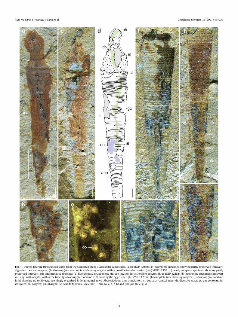
Fig. 1. Oocyte-bearing Paraselkirkia sinica from the Cambrian Stage 3 Xiaoshiba Lagerstätte. (a, b) YKLP 12089: (a) incomplete specimen showing partly preserved introvert,digestive tract and oocytes; (b) close-up (see location in a) showing oocytes within possible tubular ovaries. (c–e) YKLP 12350: (c) nearly complete specimen showing partlypreserved introvert; (d) interpretative drawing; (e) fluorescence image (close-up, see location in c) showing oocytes. (f, g) YKLP 12351: (f) incomplete specimen (introvertmissing) with oocytes within the tube; (g) close-up (see location in f) showing the egg cluster. (h, i) YKLP 12352: (h) complete tube showing oocytes; (i) close-up (see locationin h) showing up to 30 eggs seemingly organized in longitudinal rows. Abbreviations: ann, annulation; ct, cuticular conical tube; dt, digestive tract; gc, gut contents; in,introvert; oo, oocytes; ph, pharynx; sc, scalid; tr, trunk. Scale bar: 1 mm (a, c, d, f, h) and 500 lm (b, e, g, i).
Xiao-yu Yang, J. Vannier, J. Yang et al. Geoscience Frontiers 12 (2021) 101234
3
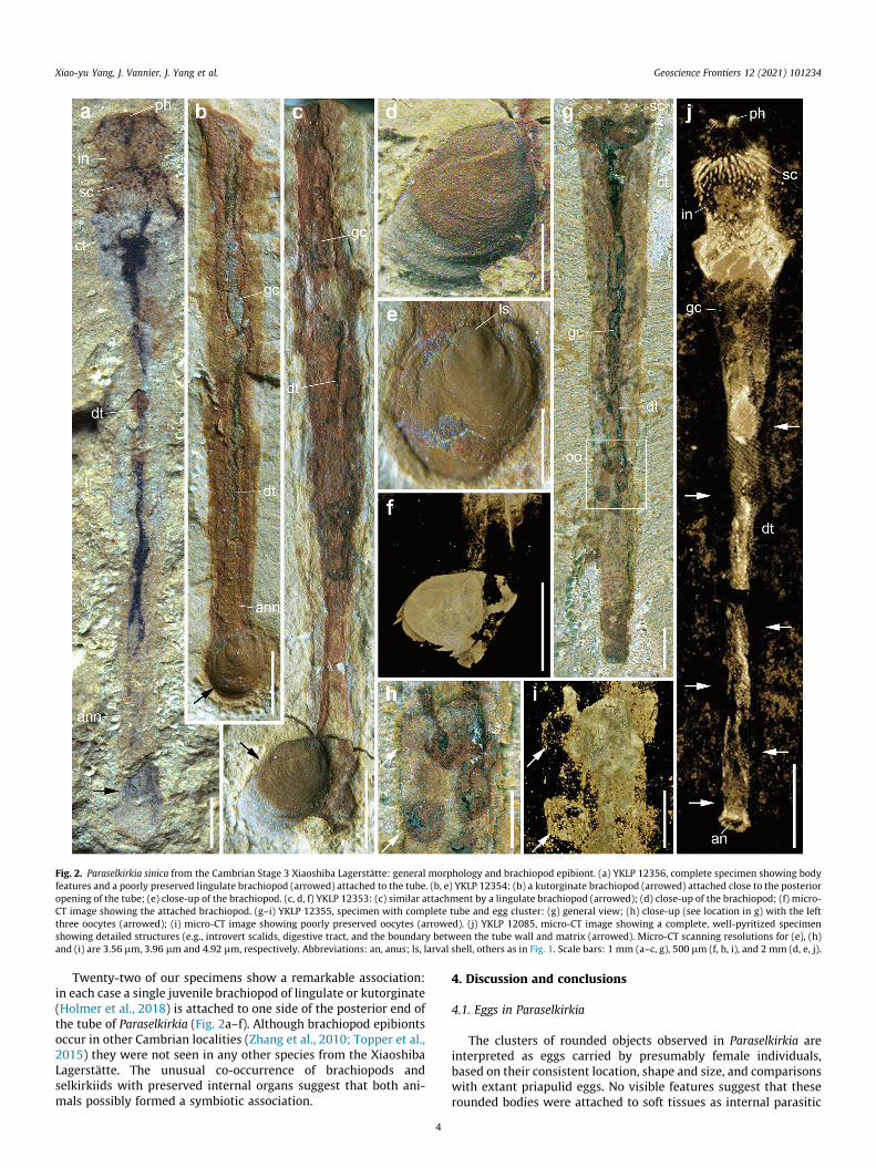
Fig. 2. Paraselkirkia sinica from the Cambrian Stage 3 Xiaoshiba Lagerstätte: general morphology and brachiopod epibiont. (a) YKLP 12356, complete specimen showing bodyfeatures and a poorly preserved lingulate brachiopod (arrowed) attached to the tube. (b, e) YKLP 12354: (b) a kutorginate brachiopod (arrowed) attached close to the posterioropening of the tube; (e) close-up of the brachiopod. (c, d, f) YKLP 12353: (c) similar attachment by a lingulate brachiopod (arrowed); (d) close-up of the brachiopod; (f) micro-CT image showing the attached brachiopod. (g–i) YKLP 12355, specimen with complete tube and egg cluster: (g) general view; (h) close-up (see location in g) with the leftthree oocytes (arrowed); (i) micro-CT image showing poorly preserved oocytes (arrowed). (j) YKLP 12085, micro-CT image showing a complete, well-pyritized specimenshowing detailed structures (e.g., introvert scalids, digestive tract, and the boundary between the tube wall and matrix (arrowed). Micro-CT scanning resolutions for (e), (h)and (i) are 3.56 lm, 3.96 lm and 4.92 lm, respectively. Abbreviations: an, anus; ls, larval shell, others as in Fig. 1. Scale bars: 1 mm (a–c, g), 500 lm (f, h, i), and 2 mm (d, e, j).
Xiao-yu Yang, J. Vannier, J. Yang et al. Geoscience Frontiers 12 (2021) 101234
Twenty-two of our specimens show a remarkable association:in each case a single juvenile brachiopod of lingulate or kutorginate(Holmer et al., 2018) is attached to one side of the posterior end ofthe tube of Paraselkirkia (Fig. 2a–f). Although brachiopod epibiontsoccur in other Cambrian localities (Zhang et al., 2010; Topper et al.,2015) they were not seen in any other species from the XiaoshibaLagerstätte. The unusual co-occurrence of brachiopods andselkirkiids with preserved internal organs suggest that both ani-mals possibly formed a symbiotic association.
4
4. Discussion and conclusions
4.1. Eggs in Paraselkirkia
The clusters of rounded objects observed in Paraselkirkia areinterpreted as eggs carried by presumably female individuals,based on their consistent location, shape and size, and comparisonswith extant priapulid eggs. No visible features suggest that theserounded bodies were attached to soft tissues as internal parasitic
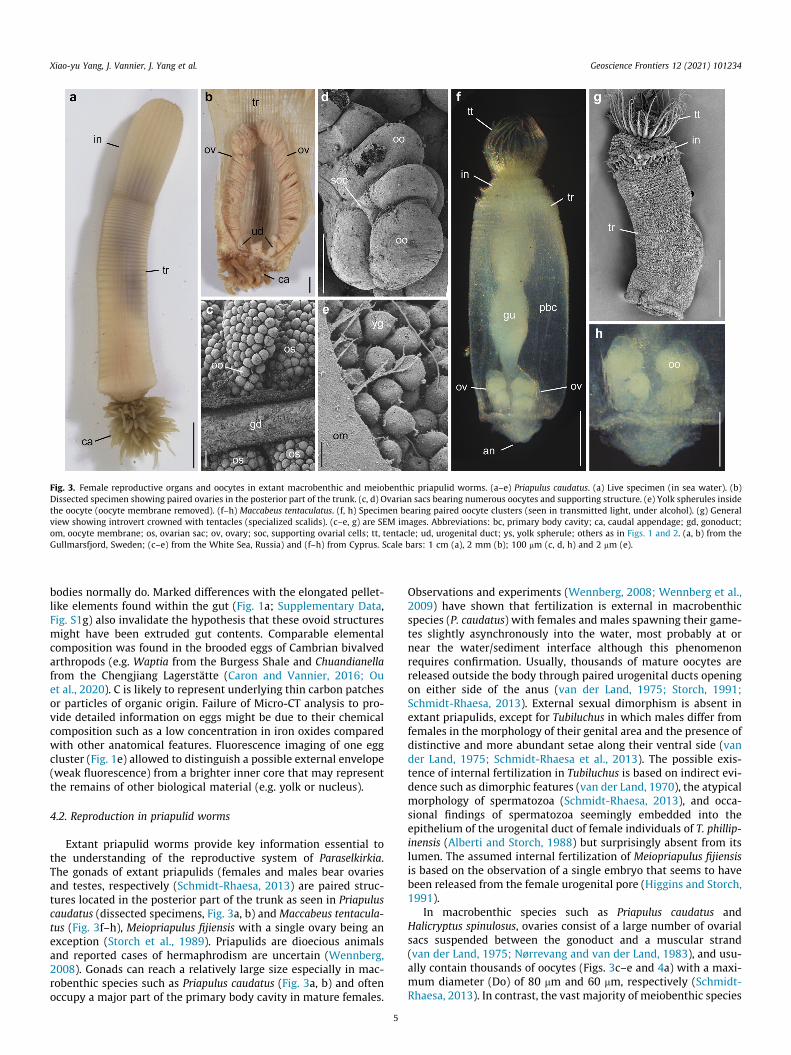
Fig. 3. Female reproductive organs and oocytes in extant macrobenthic and meiobenthic priapulid worms. (a–e) Priapulus caudatus. (a) Live specimen (in sea water). (b)Dissected specimen showing paired ovaries in the posterior part of the trunk. (c, d) Ovarian sacs bearing numerous oocytes and supporting structure. (e) Yolk spherules insidethe oocyte (oocyte membrane removed). (f–h) Maccabeus tentaculatus. (f, h) Specimen bearing paired oocyte clusters (seen in transmitted light, under alcohol). (g) Generalview showing introvert crowned with tentacles (specialized scalids). (c–e, g) are SEM images. Abbreviations: bc, primary body cavity; ca, caudal appendage; gd, gonoduct;om, oocyte membrane; os, ovarian sac; ov, ovary; soc, supporting ovarial cells; tt, tentacle; ud, urogenital duct; ys, yolk spherule; others as in Figs. 1 and 2. (a, b) from theGullmarsfjord, Sweden; (c–e) from the White Sea, Russia) and (f–h) from Cyprus. Scale bars: 1 cm (a), 2 mm (b); 100 mm (c, d, h) and 2 mm (e).
Xiao-yu Yang, J. Vannier, J. Yang et al. Geoscience Frontiers 12 (2021) 101234
bodies normally do. Marked differences with the elongated pellet-like elements found within the gut (Fig. 1a; Supplementary Data,Fig. S1g) also invalidate the hypothesis that these ovoid structuresmight have been extruded gut contents. Comparable elementalcomposition was found in the brooded eggs of Cambrian bivalvedarthropods (e.g. Waptia from the Burgess Shale and Chuandianellafrom the Chengjiang Lagerstätte (Caron and Vannier, 2016; Ouet al., 2020). C is likely to represent underlying thin carbon patchesor particles of organic origin. Failure of Micro-CT analysis to pro-vide detailed information on eggs might be due to their chemicalcomposition such as a low concentration in iron oxides comparedwith other anatomical features. Fluorescence imaging of one eggcluster (Fig. 1e) allowed to distinguish a possible external envelope(weak fluorescence) from a brighter inner core that may representthe remains of other biological material (e.g. yolk or nucleus).
4.2. Reproduction in priapulid worms
Extant priapulid worms provide key information essential tothe understanding of the reproductive system of Paraselkirkia.The gonads of extant priapulids (females and males bear ovariesand testes, respectively (Schmidt-Rhaesa, 2013) are paired struc-tures located in the posterior part of the trunk as seen in Priapuluscaudatus (dissected specimens, Fig. 3a, b) and Maccabeus tentacula-tus (Fig. 3f–h), Meiopriapulus fijiensis with a single ovary being anexception (Storch et al., 1989). Priapulids are dioecious animalsand reported cases of hermaphrodism are uncertain (Wennberg,2008). Gonads can reach a relatively large size especially in mac-robenthic species such as Priapulus caudatus (Fig. 3a, b) and oftenoccupy a major part of the primary body cavity in mature females.
5
Observations and experiments (Wennberg, 2008; Wennberg et al.,2009) have shown that fertilization is external in macrobenthicspecies (P. caudatus) with females and males spawning their game-tes slightly asynchronously into the water, most probably at ornear the water/sediment interface although this phenomenonrequires confirmation. Usually, thousands of mature oocytes arereleased outside the body through paired urogenital ducts openingon either side of the anus (van der Land, 1975; Storch, 1991;Schmidt-Rhaesa, 2013). External sexual dimorphism is absent inextant priapulids, except for Tubiluchus in which males differ fromfemales in the morphology of their genital area and the presence ofdistinctive and more abundant setae along their ventral side (vander Land, 1975; Schmidt-Rhaesa et al., 2013). The possible exis-tence of internal fertilization in Tubiluchus is based on indirect evi-dence such as dimorphic features (van der Land, 1970), the atypicalmorphology of spermatozoa (Schmidt-Rhaesa, 2013), and occa-sional findings of spermatozoa seemingly embedded into theepithelium of the urogenital duct of female individuals of T. phillip-inensis (Alberti and Storch, 1988) but surprisingly absent from itslumen. The assumed internal fertilization of Meiopriapulus fijiensisis based on the observation of a single embryo that seems to havebeen released from the female urogenital pore (Higgins and Storch,1991).
In macrobenthic species such as Priapulus caudatus andHalicryptus spinulosus, ovaries consist of a large number of ovarialsacs suspended between the gonoduct and a muscular strand(van der Land, 1975; Nørrevang and van der Land, 1983), and usu-ally contain thousands of oocytes (Figs. 3c–e and 4a) with a maxi-mum diameter (Do) of 80 mm and 60 mm, respectively (Schmidt-Rhaesa, 2013). In contrast, the vast majority of meiobenthic species
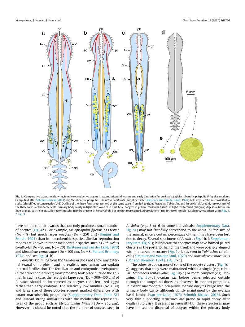
Fig. 4. Comparative diagrams showing female reproductive organs in extant priapulid worms and early Cambrian Paraselkirkia. (a) Macrobenthic priapulid Priapulus caudatus(simplified after Schmidt-Rhaesa, 2013). (b) Meiobenthic priapulid Tubiluchus corallicola (simplified after Kirsteuer and van der Land, 1970). (c) Early Cambrian Paraselkirkiasinica (simplified reconstruction). (d) Outline of the three forms represented at the same scale (from left to right: Priapulus, Tubiluchus and Paraselkirkia). (e) Mature oocytes ofthe three forms at the same scale. Primary body cavity in light blue, ovaries in dark blue, oocytes in yellow, muscular tissues in light red (around pharynx), digestive tissues inlight orange, cuticle in gray. Retractor muscles may be present in Paraselkirkia but are not represented. Abbreviations: rm, retractor muscle; s, solenocytes; others as in Figs. 1,2 and 3.
Xiao-yu Yang, J. Vannier, J. Yang et al. Geoscience Frontiers 12 (2021) 101234
have simple tubular ovaries that can only produce a small numberof oocytes (Fig. 4b). For example, Meiopriapulus fijiensis has fewer(No = 8) but much larger oocytes (Do = 250 mm) (Higgins andStorch, 1991) than in macrobenthic species. Similar reproductionmodes are known in other meiobenthic species such as Tubiluchuscorallicola (Do = 80 mm; No = 20) (Kirsteuer and van der Land, 1970)andMaccabeus tentaculatus (Do = 100 mm; No = 8; Por and Bromley,1974; and see Fig. 3f–h).
Paraselkirkia sinica from the Cambrian does not show any exter-nal sexual dimorphism and no realistic mechanism can explaininternal fertilization. The fertilization and embryonic development(either direct or indirect) most probably took place outside the ani-mal. In such a case, the relatively large eggs (Do = 300–450 mm) ofP. sinica should be interpreted as oocytes (non-fertilized eggs)rather than early embryos. The relatively low number (No < 30)and large size of these oocytes suggest marked differences withextant macrobenthic priapulids (Supplementary Data, Table S1)and instead strong similarities with the meiobenthic representa-tives of the group such as Meiopriapulus fijiensis (Do = 250 mm).However, it should be noted that the number of oocytes seen in
6
P. sinica (e.g., 3 or 6 in some individuals; Supplementary Data,Fig. S1) may not faithfully correspond to the actual clutch size ofthe animal, since a certain percentage of them may have been lostdue to decay. Several specimens of P. sinica (Fig. 1h, I; Supplemen-tary Data, Fig. S1g, h) indicate that oocytes may have formed pairedclusters in the posterior half of the trunk and were possibly alignedwithin a tubular structure (Fig. 1a, b) as seen in Tubiluchus coralli-cola (Kirsteuer and van der Land, 1970) and Maccabeus tentaculatus(Por and Bromley, 1974) (Fig. 3f–h).
The cohesive appearance of some of the oocyte clusters (Fig. 1c–g) suggests that they were maintained within a single (e.g., tubu-lar; Maccabeus tentaculatus, Fig. 3g–h) or more complex (e.g. Pria-pulus, Fig. 3b–d) ovarian sac before being released outsidethrough the urogenital ducts, as observed in modern priapulids.In extant macrobenthic priapulids mature oocytes bulge into theprimary body cavity although tightly maintained by the ovarianbasal lamina (van der Land, 1975; Schmidt-Rhaesa, 2013). Suchvery thin supporting structures are prone to rapid decay afterdeath (autolysis). If present in Paraselkirkia, these structures mayhave limited the dispersal of oocytes within the primary body
Xiao-yu Yang, J. Vannier, J. Yang et al. Geoscience Frontiers 12 (2021) 101234
cavity as observed in our fossil specimens (Fig. 1c–g). The relativelylarge internal structures observed within the oocytes of Paraselkir-kia (fluorescence images, Fig. 1e) are comparable in size with thenucleus of oocytes in Priapulus caudatus (ca. 40% of oocyte diame-ter; Nørrevang and van der Land, 1983) and may therefore beinterpreted as such.
4.3. Conservatism in priapulids
Our study suggests that the general organization of the femalereproductive system of priapulid worms with paired tubular ovar-ies has remained virtually unchanged since the early Cambrian, i.e.,over a period of more than 500 million years. The intriguingabsence of overlap between Cambrian and post-Cambrian pria-pulids as seen in the character morphospace obtained by Willset al. (2012) should not disguise the fact that numerous morpho-functional features of the priapulid body plan do appear to behighly conserved (introvert structure and symmetry, pharynx,muscle system, etc.). Although very little is known about evolu-tionary mechanisms that led to such extremely conservative bodyplans, some authors (e.g. Estes and Arnold 2007) stressed theimportance of the lack of adaptation into various significantly dif-ferent ecological niches. The persistence of endobenthic and bur-rowing lifestyles might have favoured evolutionary conservatismin priapulids through time.
4.4. Selkirkiid lifestyle and brachiopod epibionts
Selkirkiids have been interpreted as burrowers that possiblylived vertically embedded in sediment (Conway Morris, 1977;Hou et al., 2017), or alternatively as epibenthic tubicolous worms(Maas et al., 2007). They have no exact counterpart among extantpriapulid worms that are overwhelmingly active infaunal burrow-ers (e.g., macrobenthic species such as Priapulus). Maccabeus ten-taculatus (maximum length of ca. 3 mm) is the only one exampleof an extant tubicolous priapulid worm (Por and Bromley, 1974).Its cylindrical tube described as a flimsy structure formed by agglu-tinated plant fragments (e.g., Posidonia) is fundamentally differentfrom the rigid, annulated, cuticular tube of Paraselkirkia.Maccabeus
Fig. 5. Assumed lifestyle and reproductive mode of Paraselkirkia. (a) Living at the water/stube via a short pedicle. (b) Moving slightly below the water/sediment interface for feedinspermatozoids presumably emitted within sediment. (d) In-situ development of larvae.sediment interface; others as in Fig. 1.
7
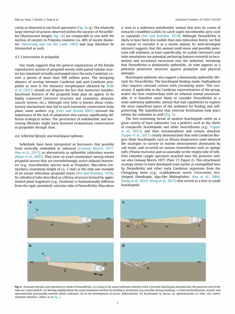
is seen as a sedentary meiobenthic animal that uses its crown oftentacles (modified scalids) to catch vagile microbenthic prey suchas copepods (Por and Bromley, 1974). Although Paraselkirkia islikely to have been less mobile than non-tubicolous forms, we findno reason to consider it as a sessile animal. Its well-developedintrovert suggests that this animal could move and possibly pene-trate soft sediment, at least superficially. Its scalids (introvert) andtube annulations are potential anchoring features essential to loco-motion and occasional excursions into the sediment. Assumingthat Paraselkirkia is dominantly epibenthic, its tube appears as apotential protective structure against predation and physicaldamages.
Brachiopod epibionts also support a dominantly epibenthic life-style for Paraselkirkia. The brachiopod feeding mode (lophophoralcilia) requires constant contact with circulating water. This con-straint, if applicable to the Cambrian representatives of the group,makes the host relationships with an infaunal animal unsustain-able. It is therefore more likely to consider Paraselkirkia as asemi-sedentary epibenthic animal that had capabilities to explorethe most superficial layers of the sediment for feeding and self-protecting. We hypothesize that external fertilization took placewithin the sediment as well (Fig. 5).
The free-swimming larvae of modern brachiopods settle on agreat variety of hard substrates (via a pedicle) such as the shellsof conspecific brachiopods and other invertebrates (e.g., Topperet al., 2015), and then metamorphose and remain attached.Topper et al. (2015) clearly demonstrated that mid-Cambrian Bur-gess Shale brachiopods such as Nisusia burgessensis used identicallife strategies to survive in marine environments dominated bysoft muds, and occurred on various invertebrates such as spongetufts (Pirania muricata) and occasionally on the empty tube of Selk-irkia columbia (single specimen attached near the posterior end;see also Conway Morris 1977, Plate 17, Figure 2). This attachmentstrategy seems to have developed even earlier as exemplified hereby Paraselkirkia and other early Cambrian organisms from theChengjiang biota (e.g., scalidophoran worm Cricocosmia, bra-chiopod Diandongia, alga-like Malongitubus; Han et al., 2004;Zhang et al., 2010; Wang et al., 2012) that served as a host to smallbrachiopods.
ediment interface with a juvenile brachiopod attached near the posterior end of theg or protection (e.g. possibly during moulting). (c) External fertilization; oocytes andAbbreviations: br, brachiopod; la, larvae; sp, spermatozoids; tu, tube; wsi, water/
Xiao-yu Yang, J. Vannier, J. Yang et al. Geoscience Frontiers 12 (2021) 101234
Two possible options are proposed to explain the consistentsmall size (ca. 1 mm) of the twenty-two brachiopod epibiontsstudied here: they represent either subadult stages of an unusuallysmall-sized species or juveniles of a larger-sized species. We tenta-tively favour the second option on the basis of the very small sizeof the brachiopods, being aware that it raises the question of theintriguing absence of more mature stages. One might hypothesizethat brachiopod larvae could easily settle on the tube of Paraselkir-kia but had a relatively low probability to reach adulthood for thesimple reason that their host moved and interacted with sediment(e.g., in contrast with more stable shelly substrates), possible caus-ing a significant proportion of post-juvenile brachiopods to detach.
Equally uncertain, is the nature of the Paraselkirkia-brachiopodassociation that may have been beneficial to both parties (mutual-ism) or not (e.g., commensalism). Particularly enigmatic is the con-sistent settlement of the brachiopod close to posterior opening ofthe tube. We tentatively suggest that this preferential locationmay have favored access to possible food particles emitted throughthe anus of the worm.
4.5. Reproductive strategies and ecology
The reproduction mode of Paraselkirkia differs from that ofextant macrobenthic priapulids (e.g., Priapulus) and instead recallsthat of meiobenthic forms such as Meiopriapulus fijiensis (Higginsand Storch, 1991), Tubiluchus corallicola (Kirsteuer and van derLand, 1970) and Maccabeus tentaculatus (Por and Bromley, 1974)that inhabit interstitial benthic environments and typically pro-duce a small number of large oocytes (Fig. 1f, g and 3f–h). It isworth noting that these resemblances strictly concern the repro-ductive mode. Paraselkirkia does not belong to the category ofmeiobenthic animals that, by definition, rarely exceeds 1 mm. Thisraises the question of the possible relation between ecology andreproductive mode in extant and early priapulids. In meiobenthicpriapulids, optimal offspring survivorship seems to be achievedthrough a small number of yolk-rich embryos that presumablydevelop within a protective interstitial environment, suggestingthat energy investment is oriented towards quality rather thanquantity. Each fertilized embryo has a relatively high probabilityof surviving to adulthood (e.g., as in K-selected species). By releas-ing thousands of gametes presumably outside the substrate, mac-robenthic species (e.g., Priapulus) seem to rely on a differentreproductive strategy. Their embryos are likely to develop in a lessprotective environment, comparatively more exposed to predationand physical damage than in interstitial species, in which theimportant loss of offspring is offset by high fecundity (e.g., as inr-selected species). The reason why the reproductive strategy ofselkirkiids resembles that of modern meiobenthic priapulidsremains an open question but might result from shared ecologicalfeatures such as a relatively small size, limited mobility and eggdeposition within the sediments (Fig. 5).
In summary, we suggest that the basic organization of thereproductive system (e.g., paired ovaries and oviducts) of pria-pulids was established very early in the evolution of the group.This organ system created conditions for various reproductivemodes and strategies to develop, in possible response to environ-mental factors, biological pressure within ecosystem and specificecologies (e.g., tubicolous lifestyle, low mobility).
Interestingly, embryos and post-embryonic stages of scali-dophoran worms from the basal Cambrian (Fortunian; seeDonoghue et al., 2006; Liu et al., 2014) have a relatively large sizethat seems to match that of oocytes found in P. sinica. This wouldsuggest that reproductive strategies involving a small number oflarge eggs may have prevailed in the early evolution of the group.However, we cannot establish with certainty that this particular
8
reproductive mode represents the ancestral condition for scali-dophorans or priapulids.
4.6. Key steps in the early evolution of reproductive systems
Present-day metazoans display a huge variety of asexual andsexual reproduction modes that include aspects related to the for-mation of gametes and gonads, fertilization processes and embry-onic development. Gonads are either tubular organs or morecomplex sac-like structures often arranged in symmetrical pairs.The development mode of gonads is diverse and suggests that theyprobably evolved independently within various metazoan groups(Schmidt-Rhaesa, 2007). Fertilization occurs externally or inter-nally via copulation or other methods such as the deposition ofspermatophores. Diverse brood care behaviors accompany theembryonic development of numerous extant species (e.g., crus-taceans). The reproductive behaviors of early animals haveremained largely enigmatic because of the lack or scarcity of infor-mation on their reproductive organs and strategies. However,exceptional fossils from lower Palaeozoic Lagerstätten providekey elements that shed light on the evolution of reproductive sys-tems of a major group of early animals, the ecdysozoans whichstand as a dominant element of early animal life (SupplementaryData, Fig. S3). (i) Paired ovaries and external fertilization seem tocharacterize basal scalidophorans exemplified by Paraselkirkia(present study); (ii) brood care is known from two groups of lowerPalaeozoic euarthropods, the Cambrian waptiids (Caron andVannier, 2016; Ou et al., 2020) and Ordovician ostracods (Siveteret al., 2014). In waptiids, egg clusters are carried symmetricallyon either side the female’s body suggesting that they were releasedfrom paired gonads as in Paraselkirkia; (iii) Copulatory organs werepresent in early Silurian myodocope ostracods from the Herefor-shire Lagerstätte (Siveter et al., 2003) (ca. 425 Ma) indicating thatcopulation and internal fertilization have a very ancient originamong euarthropods. Although fragmentary these few examplessuggest that at least four of the most essential features of bilaterianreproduction (paired gonads, external and internal fertilization,copulation) had already evolved in the early Palaeozoic throughEcdysozoa.
Declaration of Competing Interest
The authors declare that they have no known competing finan-cial interests or personal relationships that could have appearedto influence the work reported in this paper.
Acknowledgements
This study was supported by the National Natural Science Founda-tion of China (Grant No. 41730318) to X.G.Z. and J.Y., the Innova-tive Research Fund for Graduate Students of Yunnan University(Grant No. 2019228) to X.Y.Y., and the ASSEMBLE, PRC (CNRS,France and NSFC, China) and PAI (Univ. Lyon 1, Région AuvergneRhône Alpes) grants to J.V. We thank T. Lan, J.-B. Hou, K.-S. Du, J.-F. He and K.-R. Li for assistance with field work, Q. Ou for help withliterature search, H.-J. Mai for assistance with micro-CT analysis,the staff of the Kristineberg Marine Station (Göteborg University,Sweden) and the Nikolai Pertsov White Sea Biological Station(Moscow University, Russia) for assistance in collecting extant pri-apulids, A. Chipman (Hebrew University of Jerusalem, Israel) forthe loan of Maccabeus specimens from Cyprus, the Centre Tech-nologique des Microstructures (CTm, Univ. Lyon 1) and C. Aria forlinguistic corrections and insightful remarks, and S.-H. Xiao andtwo anonymous reviewers for insightful criticism.
Xiao-yu Yang, J. Vannier, J. Yang et al. Geoscience Frontiers 12 (2021) 101234
Author contributions
J.Y. collected the material and provided the geological informa-tion; X.Y.Y., X.G.Z. and J.V. designed the study; X.Y.Y., J.V., J.Y. and X.G.Z. performed the investigation; X.Y.Y., J.V. and X.G.Z. wrote themanuscript with input from other authors; J.Y. prepared all fossilsfor photography; X.Y.Y. and X.G.Z. produced the fossil figures, D.W.and J.V. studied extant priapulids and produced the relevant fig-ures and drawings, and X.Y.Y. designed and operated the elementalmap analysis.
Supplementary data
Supplementary data to this article can be found online athttps://doi.org/10.1016/j.gsf.2021.101234.
References
Alberti, G., Storch, V., 1988. Internal fertilization in a meiobenthic priapulid worm:Tubiluchus philippinensis (Tubiluchidae, Priapulida). Protoplasma 143, 193–196.
Bengtson, S., Yue, Z., 1997. Fossilized metazoan embryos from the earliestCambrian. Science 277, 1645–1648.
Briggs, D.E.G., Erwin, D.H., Collier, F.J., 1994. The Fossils of the Burgess Shale.Smithsonian Institution Press, Washington and London, pp. 1–238.
Budd, G.E., Jensen, S., 2000. A critical reappraisal of the fossil record of the bilaterianphyla. Biol. Rev. 75, 253–295.
Caron, J.-B., Vannier, J., 2016. Waptia and the diversification of brood care in earlyarthropods. Curr. Biol. 26, 69–74.
Conway Morris, S., 1977. Fossil priapulid worms. Spec. Pap. in Palaeontol. 20, 1–95.Conway Morris, S., 1998. Eggs and embryos from the Cambrian. Bioessays 20, 676–
682.Dong, X.-P., Donoghue, P.C.J., Cheng, H., Liu, J.-B., 2004. Fossil embryos from the
Middle and Late Cambrian period of Hunan, south China. Nature 427, 237–240.Donoghue, P.C.J., Bengtson, S., Dong, X.-P., Gostling, N.J., Huldtgren, T., Cunningham,
J.A., Yin, C.-Y., Yue, Z., 2006. Synchrotron X-ray tomographic microscopy of fossilembryos. Nature 442, 680–683.
Duan, Y.-H., Han, J., Fu, D.-J., Zhang, X.-L., Yang, X.-G., Tsuyoshi, K., Shu, D.-G., 2014.Reproductive strategy of the bradoriid arthropod Kunmingella douvillei from theLower Cambrian Chengjiang Lagerstätte, South China. Gondwana Res. 25, 983–990.
Estes, S., Arnold, S.J., 2007. Resolving the paradox of stasis: models with stabilizingselection explain evolutionary divergence on all timescales. Am. Nat. 169, 227–244.
Fu, D.-J., Tong, G.-H., Dai, T., Liu, W., Yang, Y.-N., Zheng, Y., Cui, L.-H., Li, L.-Y., Yun, H.,Wu, Y., Sun, A., Liu, C., Pei, W.-R., Gains, R.R., Zhang, X.-L., 2019. The Qingjiangbiota–A Burgess Shale-type fossil Lagerstätte from the early Cambrian of SouthChina. Science 363, 1338–1342.
Gabbott, S.E., Hou, X.-G., Norry, M.J., Siveter, D.J., 2004. Preservation of EarlyCambrian animals of the Chengjiang biota. Geology 32, 901–904.
Godfrey, S.J., 1992. Fossilized eggs from Illinois. Nature 356, 21.Han, J., Zhang, Z.-F., Liu, J.-N., 2004. Taphonomy and ecology of the introverts from
the Chengjiang fauna. J. Northwest Univ. (Nat. Sci. Ed.) 34, 207–212 (in Chinesewith English abstract).
Higgins, R.P., Storch, V., 1991. Evidence for direct development in Meiopriapulusfijiensis (Priapulida). Trans. Am. Microsc. Soc. 110, 37–46.
Holmer, L.E., Zhang, Z.-F., Topper, T.P., Popov, L., Claybourn, T.M., 2018. Theattachment strategies of Cambrian kutorginate brachiopods: the curious case oftwo pedicle openings and their phylogenetic significance. J. Paleontol. 92, 33–39.
Hou, X.-G., Siveter, D.J., Siveter, D.J., Aldridge, R.J., Cong, P.-Y., Gabbott, S.E., Ma, X.-Y.,2017. The Cambrian Fossils of Chengjiang, China: The Flowering of Early AnimalLife. Wiley Blackwell, Chichester, p. 316.
Hou, J.-B., Yang, J., Zhang, X.-G., Hughes, N.C., Lan, T., 2019. Trilobite-basedbiostratigraphy of the Xiaoshiba Lagerstätte. Fossils Strata 64, 173–191.
Kesidis, G., Slater, B.J., Jensen, S., Budd, G.E., 2019. Caught in the act: priapulidburrowers in early Cambrian substrates. Proc. Royal Soc. B 286, 20182505.
Kirsteuer, E., van der Land, J., 1970. Some notes on Tubiluchus corallicola (Priapulida)from Barbados, West Indies. Mar. Biol. 7, 230–238.
Kouraiss, K., El Hariri, K., El Albani, A., Azizi, A., Mazurier, A., Vannier, J., 2018. X-raymicrotomography applied to fossils preserved in compression: Palaeoscolescidworms from the Lower Ordovician Fezouata Shale. Palaeogeogr.,Palaeoclimatol., Palaeoecol. 508, 48–58.
Lan, T., Yang, J., Hou, J.-B., Zhang, X.-G., 2015. The feeding behaviour of the Cambriantubiculous priapulid Selkirkia. Lethaia 48, 125–132.
9
Lin, J.-P., Scott, A.C., Li, C.-W., Wu, H.-Y., Hwu, Y.-K., 2006. Silicified egg clusters froma Middle Cambrian Burgess Shale–type deposit, Guizhou, south China. Geology34, 1037–1040.
Liu, Y.-H., Xiao, S.-H., Shao, T.-Q., Broce, J., Zhang, H.-Q., 2014. The oldest knownpriapulid-like scalidophoran animal and its implications for the early evolutionof cycloneuralians and ecdysozoans. Evol. Dev. 163, 155–165.
Luo, H.-L., Hu, S.-X., Chen, L.-Z., Zhang, S.-S., Tao, Y.-H., 1999. Early CambrianChengjiang Fauna from Kunming Region, China. Yunnan Science TechnologyPress, Kunming, 129 pp. (in Chinese with English abstract).
Maas, A., Huang, D.-Y., Chen, J.-Y., Waloszek, D., Braun, A., 2007. Maotianshan-Shalenemathelminthes—morphology, biology, and the phylogeny ofNemathelminthes. Palaeogeogr., Palaeoclimatol., Palaeoecol. 254, 288–306.
Nørrevang, A., van der Land, J., 1983. Priapulida. In: Adiyodi, K.G., Adiyodi, R.G.(Eds.), Reproductive Biology of Invertebrates. Oogenesis, Oviposition, andOosorption. John Wiley & Sons, Chichester, pp. 269–282.
Ou, Q., Vannier, J., Yang, X.-F., Chen, A.-L., Mayer, G., 2020. Evolutionary trade-off inreproduction of Cambrian arthropods. Sci. Adv. 6, 3376–3405.
Peel, J.S., Willman, S., 2018. The Buen Formation (Cambrian Series 2) biota of NorthGreenland. Pap. Palaeontol. 4, 381–432.
Por, F.D., Bromley, H.J., 1974. Morphology and anatomy of Maccabeus tentaculatus(Priapulida: Seticoronaria). J. Zool. 173, 173–197.
Schmidt-Rhaesa, A., 2007. The Evolution of Organ Systems. Oxford University Press,Oxford and New York, pp. 1–368.
Schmidt-Rhaesa, A., Rothe, B.H., Martínez, A.G., 2013. Tubiluchus lemburgi, a newspecies of meiobenthic Priapulida. Zool. Anz. 253, 158–163.
Schmidt-Rhaesa, A., 2013. Priapulida. In: Schmidt-Rhaesa, A. (Ed.), Handbook ofZoology. Gastrotricha, Cycloneuralia and Gnathifera. Berlin-Boston: De Gruyter,pp. 147–180.
Siveter, D.J., Sutton, M.D., Briggs, D.E.G., Siveter, D.J., 2003. An ostracode crustaceanwith soft parts from the Lower Silurian. Science 302, 1749–1751.
Siveter, D.J., Tanaka, G., Farrell, Ú.C., Martin, M.J., Siveter, D.J., Briggs, D.E.G., 2014.Exceptionally preserved 450-million-year-old Ordovician ostracods with broodcare. Curr. Biol. 24, 801–806.
Smith, M.R., Harvey, T.H.P., Butterfield, N.J., 2015. The macro- and microfossil recordof the Cambrian priapulid Ottoia. Palaeontology 58, 705–721.
Storch, V., 1991. Priapulida. In: Harrison, F.W., Ruppert, E.E. (Eds.), MicroscopicAnatomy of Invertebrates. Aschelminthes, Wiley-Liss, New York, pp. 333–350.
Storch, V., Higgins, R.P., Morse, M.P., 1989. Internal anatomy of Meiopriapulusfijiensis (Priapulida). Trans. Am. Microsc. Soc. 108, 245–261.
Topper, T.P., Strotz, L.C., Holmer, L.E., Caron, J.-B., 2015. Survival on a soft seafloor:life strategies of brachiopods from the Cambrian Burgess Shale. Earth-Sci. Rev.151, 266–287.
van der Land, J., 1970. Systematics, zoogeography, and ecology of the Priapulida.Zool. Verh. 112, 1–118.
van der Land, J., 1975. Priapulida. In: Giese, A., Pearse, J.S. (Eds.), Reproduction ofMarine Invertebrates. Entoprocts and Lesser Protostomes. Academic Press, NewYork, pp. 55–65.
Vannier, J., 2012. Gut contents as direct indicators for trophic relationships in theCambrian marine ecosystem. PLoS ONE 7, e52200.
Vannier, J., Calandra, I., Gaillard, C., _Zylinska, A., 2010. Priapulid worms: Pioneerhorizontal burrowers at the Precambrian-Cambrian boundary. Geology 38,711–714.
Wang, D., Vannier, J., Schumann, I., Wang, X., Yang, X.-G., Komiya, T., Uesugi, K., Sun,J., Han, J., 2019. Origin of ecdysis: fossil evidence from 535-million-year-oldscalidophoran worms. Proc. of Royal Soc. B 286, 20190791.
Wang, H., Zhang, Z.-F., Holmer, L.E., Hu, S.-X., Wang, X.-R., Li, G.-X., 2012. Peduncularattached secondary tiering acrotretoid brachiopods from the Chengjiang fauna:Implications for the ecological expansion of brachiopods during the Cambrianexplosion. Palaeogeogr., Palaeoclimatol., Palaeoecol. 323–325, 60–67.
Wennberg, S.A., 2008. Aspects of Priapulid Development Ph.D. thesis,. UppsalaUniversity, Department of Earth Sciences.
Wennberg, S.A., Janssen, R., Budd, G.E., 2009. Hatching and earliest larval stages ofthe priapulid worm Priapulus caudatus. Invertebr. Biol. 128, 157–171.
Wills, M.A., Gerber, S., Ruta, M., Hughes, M., 2012. The disparity of priapulid,archaeopriapulid and palaeoscolecid worms in the light of new data. J. Evol.Biol. 25, 2056–2076.
Yang, J., Ortega-Hernández, J., Butterfield, N.J., Zhang, X.-G., 2013. Specializedappendages in fuxianhuiids and the head organization of early euarthropods.Nature 494, 468–471.
Yang, J., Ortega-Hernández, J., Legg, D.A., Lan, T., Hou, J.-B., Zhang, X.-G., 2018. EarlyCambrian fuxianhuiids from China reveal origin of the gnathobasic protopoditein euarthropods. Nat. Commun. 9, 470.
Zhang, Z.-F., Han, J., Wang, Y., Emig, C.C., Shu, D.-G., 2010. Epibionts on the lingulatebrachiopod Diandongia from the Early Cambrian Chengjiang Lagerstätte, SouthChina. Proc. of the Royal Soc. B 277, 175–181.
Zhang, X.-G., Pratt, B.R., 1994. Middle Cambrian arthropod embryos withblastomeres. Science 266, 637–639.
Zhu, M.-Y., Babcock, L.E., Steiner, M., 2005. Fossilization modes in the ChengjiangLagerstätte (Cambrian of China): testing the role of organic preservation anddiagenetic alteration in exceptional preservation. Palaeogeogr., Palaeoclimatol.,Palaeoecol. 220, 31–46.