Faunas and Cambrian Volcanism on the Avalonian Marginal Platform, Southern New Brunswick
Upload
independentCategory
view
0download
0
CAMBRIAN TRILOBITES FROM THE PARAHIO AND ZANSKARVALLEYS, INDIAN HIMALAYA
SHANCHI PENG,1 NIGEL C. HUGHES,2 NOEL A. HEIM,2,5 BRYAN K. SELL,2,6 XUEJIAN ZHU,1 PAULM. MYROW,3
AND SURAJ K. PARCHA4
1State Key Laboratory on Paleobiology and Stratigraphy, Nanjing Institute of Geology and Paleontology, Nanjing 210008, China, ,[email protected].; 2Department of Earth Sciences, University of California, Riverside, 92521, USA, ,[email protected].; 3Department of Geology,Colorado College, Colorado Springs, 80903, USA; 4Wadia Institute of Himalayan Geology, Dehra Dun, Uttranchal 248001, India; 5Present address:Department of Geology and Geophysics, University of Wisconsin-Madison, Madison 53706, USA; and 6Present address: Department of Earth Sciences,
University of Geneva, rue des Maraıchers 13, 1205 Geneva, Switzerland
ABSTRACT—New collections of trilobites from the type section of the Parahio Formation in the Parahio Valley, Spiti, and from theParahio, Karsha, and Kurgiakh formations in the Zanskar Valley, permit biozonation based on material precisely located withinmeasured stratigraphic sections. Specimens preserved in limestone with mild tectonic deformation clarify the features of severalHimalayan taxa known previously only from severely deformed specimens preserved in shale. A total of 75 trilobite taxa from theCambrian of Spiti and Zanskar can be referred, questionably at least, at the generic level or below, and 61 of these are present in our newcollections. This new material is assigned with confidence to 29 existing species, and to 12 new species. Three new genera, Haydenaspis,Bhargavia, and Himalisania, are established; new species include Haydenaspis parvatya, Prozacanthoides lahiri, Probowmania bhatti,Xingrenaspis parthiva, X. shyamalae, Bhargavia prakritika, Kaotaia prachina, Gunnia smithi, Sudanamonocarina sinindica, Proasaphiscussimoni, Koldinia odelli, and Torifera jelli. Ten additional Himalayan forms are assigned at the generic level only, and another 11 arequestionably assigned to genera or species. The zonation proposed includes 6 zones and 3 levels, including the Haydenaspis parvatya level,the Oryctocephalus indicus level, the Kaotaia prachina Zone, the Paramecephalus defossus Zone, the Oryctocephalus salteri Zone, theIranoleesia butes level, the Sudanomocarina sinindica Zone, the Lejopyge acantha Zone, and the Proagnostus bulbus Zone. The sectionsspan from the upper part of the informal Stage 4, Series 2 of the Cambrian System, about 511 Ma old, to the Proagnostus bulbus zone ofthe Guzhangian Stage near the top of Series 3, dated at about 501 Ma. This time interval is represented by about 2000 m of section, whichis thick compared to similar intervals elsewhere and is consistent with high rates of sedimentation along the Himalayan margin at thetime. The fauna resembles others from equatorial peri-Gondwanaland, with closest similarity to that of South China. It also bears strongaffinity to the North China fauna. Juvenile trilobites are described for the first time from India. A new Chinese species, Monanocephalusliquani, is also described.
INTRODUCTION
ALTHOUGH CAMBRIAN trilobites have been known fromthe Himalaya for over a hundred years, the biostratig-
raphy of the Himalayan Cambrian remains poorly resolved.The great majority of these trilobites have been tectonicallydeformed (see Jell and Hughes, 1997), and the stratigraphiccontext of many previous collections is not well understood.Consequently, global assessments of Cambrian stratigraphyand paleogeography generally overlook the Himalayanregion. A steep ridge along the north side of the Parahiovalley of the Spiti and Lahaul district of Himachal Pradesh,India (Fig. 1), first studied by Sir Henry Hayden in the 1890’s(Hayden, 1904; Reed, 1910) provides a continuous strati-graphic section (Myrow et al., 2006a) from which we havecollected Cambrian trilobites at successive horizons. We havealso collected several sections in the Zanskar Valley of theLadakh region of Jammu and Kashmir, India (Myrow et al.,2006b), a region some 150 km northwest of the ParahioValley (Fig. 1) and also part of the Tethyan Himalaya. Muchof this new material is preserved in limestone and, while notentirely free from tectonic deformation, is of sufficient qualityto permit direct comparison with well-preserved specimensfrom elsewhere.
This study presents systematic descriptions of our collec-tions and clarifies taxonomic concepts based on these well-preserved specimens. Correlations between the Parahio andZanskar Valleys are considered, and our new material isintegrated with previous collections to construct a biostrati-graphic scheme for this region for parts of the second andthird series of the Cambrian System. We also consider theimplications of our study for dating and correlation with otherparts of the Himalaya and beyond.
GEOLOGICAL SETTING
The central part of the Himalaya, located between its easternand western syntaxes, consists of four major lithotectonic zonesto the south of the Indus-Tsangpo suture zone, the boundarywith the Lhasa block of Tibet (Fig. 1.1). The collectionsdescribed herein come from the northernmost of the four zones,the ‘‘Tethyan’’ or ‘‘Tibetan’’ Himalaya, and all belong to theupper part of the Haimanta Group (Myrow et al., 2006a). TheSpiti and Zanskar valleys are adjacent drainage basins(Fig. 1.2), and the Parahio Formation has been recognized inboth regions (Myrow et al., 2006a,b). It consists of siliclasticdeltaic deposits with subordinate carbonate beds.
In Spiti, the Parahio Formation is unconformably overlainby the conglomeratic basal part of the Ordovician ThangoFormation. Collections from the Parahio Valley of Spiti weremade from (1) a section on a ridge on the northern side of theParahio River (Fig. 2), which is where Hayden (1904)measured his original section, (2) from an outcrop on thebanks of the south side of the Parahio River near theconfluence with the Sumna River and, (3) the slopes on theeast side of the Sumna River (see Appendix 1 for details ofcollection localities). Aspects of the lithostratigraphy andpaleoenvironments of these sections, and the history of theirinterpretation, have been reviewed by Myrow et al. (2006a).Trilobites, bradorid arthropods, phosphatic brachiopods, andsponge spicules are found in carbonate grainstone beds thatare interpreted to represent intermittent decreases in thesupply of terrigenous siliciclastic sediment. The grainstonetexture suggests reworking by ocean waves and currents.Although there is some evidence of silicification in thesecarbonate beds (Myrow et al., 2006a), acidic dissolution of thelimestone did not yield silicified fossils other than sponge
Copyright ’ 2009, The Paleontological Society
1

spicules. However, it did liberate phosphatic brachiopodsfrom each of the carbonate horizons that we have sampled.
Trilobites and brachiopods are also present in dark shalethat appears to have accumulated in a low energy marineenvironment, below fair-weather wave base. Although theentire section of the Parahio Formation at this locality exceeds1350 m in thickness, carbonate and dark shale facies typicallyform a small proportion of the total thickness of successive
shoaling cycles, particularly in the lower parts of the section.This explains the limited number of beds bearing macrofossils(Fig. 3). Episodic delta-lobe switching led to nonsystematicstratigraphic changes in cycle and facies thickness (Myrow etal., 2006a). Thick carbonate beds near the top of the sectionare dolomitized and do not contain abundant trilobites,although a poorly preserved agnostoid shield was observed inone dolomite bed. In a few cases dolomite beds bear laminae
FIGURE 1—Location of the Spiti and Zanskar valleys within the Himalaya. 1, showing position of Spiti/Zanskar region as part of the TethyanHimalaya, the most northerly of four lithotectonic zones of the Himalaya. 2, position of sections within the Zanskar and Spiti valleys, and other areas inthe western and central Himalaya from which Cambrian trilobites are known.
2 PENG ET AL.

rich in phosphatic brachiopods, and fossils described asconodonts have also been recovered from these beds (Bhattand Kumar, 1980). Various trace fossils have been found athorizons scattered throughout the section.
In Zanskar (Fig. 5), the Parahio Formation is lithologicallysimilar to its exposure in Spiti, although Zanskar may havebeen located in a slightly more distal setting (Myrow et al.,2006b). There the Parahio Formation is succeeded by theKarsha Formation, a thick red-weathering dolostone thatrepresents a clear-water carbonate-platform setting andcontains trilobites in its upper member (Myrow et al.,2006b) (Fig. 6). The Karsha Formation is conformablyoverlain by the Kurgiakh Formation (Fig. 6), which containssimilar lithofacies to the Parahio Formation, and also bearsCambrian trilobites. The Kurgiakh Formation is unconform-ably overlain by the Ordovician molasse deposits of theThaple Formation (Fig. 6), the lithological equivalent of theThango Formation of the Parahio Valley. These relationships,along with the constraints provided by the trilobite biostra-tigraphy, suggest that in this sector of the Himalaya theOrdovician unconformity cut to deeper stratigraphic levelstoward the southeast (Fig. 4).
We did not recover trilobites from carbonate beds in theParahio Formation of Zanskar because these beds were almostentirely dolomitized (phosphatic brachiopods were collectedfrom one of these beds, Fig. 6). We recovered trilobites fromshale in the upper part of the Parahio Formation in the Purni
section (Fig. 5), shortly beneath the contact with the KarshaFormation, and from a shale bed near the top of the ParahioFormation on the high slopes east of Kurgiakh village.Trilobites and phosphatic brachiopods were also recoveredfrom several thin carbonate grainstone beds in the TetaMember of the Karsha Formation, and from shales in theoverlying Surichun Member of the Kurgiakh Formation, bothin the section opposite Kuru and in the section withinKurgiakh nulla (Figs. 5, 6).
PRIOR SYSTEMATIC AND BIOSTRATIGRAPHIC WORK
Previous systematic studies of Cambrian trilobites fromSpiti and Zanskar, which include over ten publications, havebeen reviewed elsewhere (Jell and Hughes, 1997) with theexception of the following papers. Dungrakoti et al. (1977, pl.30, fig. 1) figured a poorly preserved exoskeleton from theKurgiakh nulla section that they attributed to the familyOlenellidae. No specimen number or repository informationwas given for that fossil, and we have not been able to examineit. In the light of our results we consider their attributionunlikely to be correct, but we cannot recognize any diagnosticfeatures in the published figure. Shah et al. (1996) and Kumar(1998) described and figured agnostoid and polymeridmaterial from the Kurgiakh region of the Zanskar Valley,apparently from the north bank of the Tsarap Lingti River, afew kilometers east of Kurgiakh village. Our searches in thatarea did not relocate these levels, but we discuss this material
FIGURE 2—The position of the measured sections and other collecting sites in the Parahio River Valley, Spiti. Hayden’s (1904) original section is thelong section marked to the north of the Parahio and Khemangar Rivers. The collecting site of the trace fossils illustrated by Bhargava et al. (1982) andParcha et al. (2005) is shown by the open star. KR indicates Khemangar River, DKR indicates Debsa Khad River, and SR indicates Sumna River.
HIMALAYAN CAMBRIAN TRILOBITES 3

FIGURE 3—Composite stratigraphic section of the Cambrian rocks of the Parahio Formation in the Parahio Valley based on sections shown in Fig. 2,with trilobite occurrence, local ranges shown, and biozonation. Sedimentological and other details of the section are provided in Myrow et al. (2006a).The chart includes all trilobite species that we consider to be valid from this section, including those not recovered by us. * indicates species only knownfrom shales. 4 and 5 refer to the new, informal stages of the Cambrian System. Here we represent the boundary between them at the first occurrence of O.indicus. Details of height in section and collection lithologies provided in Appendix 1 and 2. Note that the type specimens of Agnostus spitiensis Reed,1910 are missing, and hence we are unable to verify the validity of this form. Open star indicates dolomite containing abundant acrotretid brachiopods.sh indicates shale, st indicates silt, vfs and fs indicates very fine sandstone and fine sandstone respectively, and carb indicates carbonate.
4 PENG ET AL.

in the taxonomic discussions below, and estimate its strati-graphic position in our summary of the Cambrian biostratig-raphy of this region (Fig. 6).
Refining the Cambrian biostratigraphy of the Himalayarequires the precise location of collections within continuous,measured stratigraphic sections. The most comprehensiveattempt to date is that of Hayden (1904; Reed, 1910).Hayden’s (1904, p. 14–15) section for the Parahio Formationat the type section accords with our own in terms of theidentity and sequence of trilobite-bearing horizons and relativethicknesses between these horizons. However, Hayden’s (1904)estimate of the total thickness of the Parahio Formation(362 m) is less than 25% of our own estimate (1360 m) for thesame section. Hayden (1904, p. 13) argued that foldingincreased the apparent thickness of the section, but our workdoes not support that interpretation. We saw no evidence forstructural complexity in the Parahio Formation other thanminor faulting, and the trilobite collections occurred in astratigraphic order consistent with that seen in other areas ofAsia that have suffered little or no tectonism. Hayden (1904)recovered trilobites from five stratigraphic levels within hissection: his levels 2, 4, 6, 9, and 13. We have recovered trilobitespecies from beds that represent each of these levels or theirclose equivalents with the exception of his level 2. We havealso discovered two additional trilobite-bearing units locatedstratigraphically adjacent to those of Hayden’s level 2 (Fig. 3).
Jell and Hughes (1997, fig. 4) presented a scheme for thecorrelation between the Parahio Valley and Zanskar regions,in addition to other parts of the Himalaya, but did notpropose a formal biostratigraphic zonation for the region. Ourwork is consistent with the scheme that they proposed, andpermits the recognition of a series of faunal levels and zoneswithin the region.
The Hayden/Reed stratigraphy and our own differ from thatpresented in several other papers on the Cambrian trilobitebiostratigraphy of the Parahio Valley (e.g., Shah and Sat Paul,1987; Shah et al., 1988; Shah and Raina, 1990; Parcha, 1996,1998). Those papers suggested that 12 fossiliferous horizonsspanned parts of the traditional early, middle and lateCambrian across less than 400 m of Parahio Formation sectionand also mention an occurrence of Redlichia within the ParahioValley section. Revisions by Jell and Hughes (1997) and theresults of the present study indicate that the strata are nearly1400 m thick and span much less of the Cambrian System. In
addition, we did not recover any specimens of Redlichia orknow of anyRedlichia specimens previously recovered from thisvalley. Much of the material used to construct the alternativescheme has since been reassigned to different taxa (Jell andHughes, 1997). However, these differences in the stratigraphicviews extend beyond matters of taxonomic assignment. Ourwork is based on a stratigraphic section that was measured frombase to top as we collected: the alternative scheme was made byestimating the relative stratigraphic positions of new findingswith respect to older findings. Our inferred position ofHayden’s level 2, which contains the important taxonOryctocephalus indicus, is a projection, but it is based on ourcorrected estimates of the thickness of Hayden’s section.We areexplicit about the basis of this estimate and are reassured thatthis projected position is consistent with the recorded positionof O. indicus in other sections worldwide.
FOSSIL PRESERVATION
Almost all of the Cambrian trilobites previously describedfrom Spiti and Zanskar have suffered significant flatteningand tectonic shearing due to their preservation in deformedfine-grained siliciclastic rocks (such as is evident in Figs. 35,56, 57). Such deformation can also induce novel features thathave mistakenly been considered of taxonomic significance insome cases (see Hughes and Jell, 1992). On the other hand,trilobite material preserved in fine-grained siliciclastic rocks ismore commonly articulated than that preserved in limestoneor silica (e.g., Fig. 35) (Hughes, 1995). Out of the 75Himalayan taxa that are recognized in this monograph, 31are known exclusively from fine-grained siliciclastic strata, 28exclusively from carbonate, and 16 are known from bothfacies (Appendix 2). Thus, 44 taxa are now known fromcarbonate material, and for four species we have been able toassociate isolated sclerites preserved in carbonate witharticulated specimens preserved in siliciclastic strata, allowinga more nearly complete characterization of these taxa. All butone of the 14 previously described species that we failed torecover in our new collections are from siliciclastic strata.
Although our carbonate collections are not free from theeffects of compaction and tectonic shearing, the degree towhich these factors have influenced the fossils is generallymodest compared to that in associated shales. In certain casesin which specimens have experienced simple shear, we haveemployed digital retrodeformation in an attempt to restore theoriginal form in order to facilitate comparison with unde-formed material known from elsewhere (Hughes and Jell,1992). The degree to which tectonism influenced specimenspreserved in carbonate varied within beds, with somespecimens showing deformation and others apparently freefrom it, and our figures illustrate both conditions.
DIVERSITY AND BIOFACIES
The time spent at different collecting levels ranged from 2 to6 person-days per level. With the exception of the unusuallydiverse limestone beds at the top of the Teta Member of theKarsha Formation, the diversity of polymerid trilobitesamong the levels varied between three and ten species, andmost collections appear to be dominated by a relatively smallnumber of taxa. There is a remarkable abundance ofagnostoid species in the upper parts of the Teta Member ofthe Karsha Formation and in the Surichun Member of theKurgiakh Formation. This sharp increase in agnostoiddiversity is consistent with the pattern seen elsewhere, anddocumented in particular detail in South China (Peng andRobison, 2000), and does not require a local explanation.
Carbonate and siliciclastic beds bearing species of similarstratigraphic age occurred at several levels, and these provide
FIGURE 4—Lithostratigraphic correlation of the units considered in thiswork between the Zanskar and Spiti regions, based on Myrow et al.(2006b, fig. 2), and supported by the biostratigraphic zonation document-ed herein. Note that the contact between the Sanugba and Haimantagroups is an erosional unconformity in both sections, but that the entiretyof the Karsha and Kurgiakh formations is missing in Spiti.
HIMALAYAN CAMBRIAN TRILOBITES 5

some opportunity to assess the lithofacies preferences of species.The relatively long ranged ptychopariid Kunmingaspis stracheyiwas abundant in shale beds from several horizons, but we did notrecord it in any of the carbonate beds that occurred within itsstratigraphic range. On the other hand, we foundmany species inboth carbonate and siliciclastic rocks. Biofacies differencesbetween carbonate and siliciclastic facies are common amongCambrian trilobites (e.g. Ludvigsen and Westrop, 1983) and theoccurrence of specific Himalayan taxa in both lithologies maysuggest that the transitions between these facies in this setting didnot reflect major shifts in paleoenvironment.
Kaotaia prachina n. sp. and Bhargavia prakritika n. gen. andsp. from the interval from 433.44 m to 439.67 m in theParahio Valley section bore unusually thick exoskeletons.Thicker exoskeletons may suggest a need for robust shields inshallow water conditions (Fortey and Wilmott, 1991).However, the exoskeleton of the co-occurrent Xingrenaspisparthiva was apparently of normal thickness.
BIOSTRATIGRAPHY AND GLOBAL CORRELATION
The biostratigraphic zonation proposed here for theTethyan Himalaya of northern India is based on the local
occurrence of well-characterized taxa. Where age diagnostictaxa are restricted to single bed the term ‘‘level’’ is applied withthe epithet of a species characteristic of that level. We applythe term ‘‘zone’’ when the eponymous species occurred inmore than a single bed beneath the first appearance datum(FAD) of the next level or zone. Intervals of ‘‘no zonation’’ areindicated in thick, apparently barren, strata above levels orzones where further work is required to clarify the biostratig-raphy. The base of each unit is defined by the FAD of theeponymous species and, where thick barren strata are absent,the base of the next overlying zone defines the top of eachzone.
Our biostratigraphic study of Hayden’s section in the SpitiValley and three sections in the Zanskar Valley enable us todevelop a formal biostratigraphic succession with six trilobitezones, three levels, and six intervals of no zonation for theCambrian System of the Spiti and Zanskar Valleys (Figs. 3, 6).It spans an interval from the upper part of the as yet-unnamedglobal Stage 4 through the lower half of the Guzhangian Stageof the Cambrian System (Figs. 7–8). In ascending order thesedivisions are: the Haydenaspis parvatya level, interval 1 of nozonation, the Oryctocephalus indicus level, interval 2 of nozonation, the Kaotaia prachina Zone, interval 3 of no
FIGURE 5—The position of the measured sections and collecting sites in the Zanskar Valley. KH2 and KH3 collecting sites near Kurgiakh: KU1 –Kuru 1 section, KU2 – Kuru 2 section, PU1 – Purni 1 section, PU2 – Purni 2 section, and PU3 – Purni 3 section.
6 PENG ET AL.

zonation, the Paramecephalus defossus Zone, the Oryctoce-phalus salteri Zone, interval 4 of no zonation, the Iranoleesiabutes level, interval 5 of no zonation, the Sudanomocarinasinindica Zone, interval 6 of no zonation, the Lejopyge acanthaZone, and the Proagnostus bulbus Zone.
The relatively dense sampling of the beds of the TetaMember of the Karsha Formation and the overlying SurichunMember of the Kurgiakh Formation, coupled with thestriking abundance of taxa, permits the recognition of astratigraphically lower fauna characterized by the occurrence
of Lejopyge acantha that is succeeded by a zone bearingProagnostus bulbus (Fig. 6). A similar transition is knownfrom multiple sections worldwide and provides grounds forconstraining the age of these beds rather precisely (Fig. 7).
A striking characteristic of the biostratigraphic distributionof trilobite taxa from the Cambrian of Zanskar and Spiti isthat trilobite-bearing beds are restricted to narrow strati-graphic intervals. Individual taxa were commonly collectedfrom multiple horizons, sometimes of different lithology,within intervals around 10 m thick, but such horizons were
FIGURE 6—Composite stratigraphic section of the Cambrian rocks of the Parahio, Karsha and Kurgiakh Formations in the Zanskar Valley based onsections shown in Fig. 5, with trilobite occurrence, local ranges shown, and biozonation. Sedimentological and other details of the section are providedin Myrow et al. (2006b). * indicates species only known from shales. Measured sections containing material described herein are indicated. PU is Purni,KU is opposite Kuru. 5 refers to the new informal fifth stage of the Cambrian System. Open star indicates dolomite containing abundant acrotretidbrachiopods. sh indicates shale, ss indicates sandstone, and carb indicates carbonate.
HIMALAYAN CAMBRIAN TRILOBITES 7

often separated by more than 100 m of unfossiliferous rock(Figs. 3, 6). Taxa that ranged over thick (,100 m or more)intervals include the agnostid Ptychagnostus aculeatus andprobably also Peronopsis acadica, and polymerids includingKunmingaspis stracheyi and Parablackwelderia sheridanorum.Specimens of Paramecephalus defossus and Shantungaspishimalaica were collected from an interval that spanned about80 m of the Parahio Formation in the Parahio Valley section.
We interpret the restriction of most taxa to narrowstratigraphic intervals to reflect the paleoenvironment andaccumulation history of these deposits: collections arelocalized near the boundaries of major shoaling cycles. Thestratigraphic thickness between our oldest and youngestcollections in the Parahio Valley, PO3 and PO10, isapproximately 900 m. The Kaili Formation in GuizhouProvince of the South China block represents a significantlylonger temporal interval and is 326 m thick at its type section(Zhang et al., 1979). Similarly, while the stratigraphic intervalbetween the base of the range of Sudananomocarina sinindicaand Proagnostus bulbus is about 270 m in the Paibian typesection at Paibi in northwestern Hunan (Peng and Robison,2000), the same interval is apparently about double thatthickness in Zanskar, where it is represented by the carbonate-rich Karsha Formation. Hence, the Indian sections arerelatively thick, consistent with the interpretation of theirbeing largely the products of deltaic deposition.
The average sediment accumulation rate calculated for themeasured thickness of the Parahio Formation in the ParahioValley section was 30.4 cm/1000 years, a high rate that issimilar to time averaged accumulation rates in deltaic settings(Myrow et al., 2006a). Much of the time represented by thesection was likely accommodated at disconformity surfacesand within stratigraphically condensed horizons associatedwith the boundaries between depositional cycles. For example,the transition between the top of the range of P. defossus andthe first appearance of the fauna of the subsequent Orycto-cephalus salteri Zone, occurs within a 3 m interval (Fig. 3). Ifthe degree of faunal transition was related to temporalduration, more Cambrian time may be represented withinthat 3 meter interval than in the 80 m preceding it, which allbears the fauna of the defossus Zone.
FIGURE 7—Correlation between the Cambrian biostratigraphy of Zanskar and Spiti with that of other regions. Note that, for the purpose ofinterregional comparison, the Himalayan levels and zones are here shown to encompass the interval from their first occurrence datum (FAD) until theFAD of the next zone or level. Lv. signifies level.
FIGURE 8—The Cambrian sections of the Spiti/Zanskar region in thecontext of global Cambrian stratigraphy. The Himalayan sections (shadedportion) represent a relatively small proportion of the Cambrian System.
8 PENG ET AL.

The age of the interval studied herein is well constrained bymany of the polymerid trilobites that are endemic to easternGondwana and that provide correlation with well establishedsuccessions in China and Australia, and by the richassemblages of agnostoids in the Lejopyge acantha andProagnostus bulbus zones (Figs. 3, 6). The later enable aprecise global correlation for those Tethyan Himalayan zones(Figs. 7, 8). The Lejopyge acantha zone correlates globallywith the Lejopyge laevigata Zone and, as defined, the FAD ofLejopyge laevigata coincides with the base of the globalGuzhangian Stage (Peng and Babcock, 2008; Peng et al.,2009). In addition to the agnostoids, the age of the intervalstudied is constrained by the occurrence of Oryctocephalusindicus in the lower part of the Parahio Formation in theParahio Valley section. As one of the key internationalstratigraphic marker species for this time interval, the FAD ofOryctocephalus indicus is one of the criteria proposed fordefining the base of the as-yet-unnamed global Stage 5 (Pengand Babcock, 2005; Babcock et al., 2005).
Haydenaspis parvatya level.—The oldest fauna in Hayden’ssection is recorded at 78.07 m above the base of the ParahioFormation, Parahio Valley, Spiti (Fig. 3), where the epony-mous species, Haydenaspis parvatya n. gen. and sp., occurs inassociation with Prozacanthoides lahiri n. sp. and some basalptychoparioids including Probowmania bhatti n. sp. andMufushania nankingensis? The latter species is closely compa-rable to M. nankingensis, which occurs in the basal Mao-chuangian Stage and equivalent formations in China (Yuan etal., 2002), and suggests that this level is equivalent to the baseof the Maochuangian as used in North China, or within thetop part of the Duyunian Stage as used in South China.Globally, this level lies within the upper part of the as-yet-unnamed Stage 4 of the Cambrian System, and thus to theuppermost part of the second Series of the Cambrian System(Figs. 7, 8).
Oryctocephalus indicus level.—Unfortunately, we recoveredno O. indicus (Reed, 1910) in our collections, but Hayden(1904) recovered the eponymous species in association withKunmingaspis pervulgata (Reed, 1910), Pagetia significans(Etheridge, 1902) and an undetermined ptychoparioid speciesfrom his level 2, in the lower part of the Parahio Formation inthe same section (Fig. 3). An Oryctocephalus indicus Zone hasbeen recognized in South China and in the Great Basin (Yuanet al., 2002; Sundberg and McCollum, 2003). Although neitherthe stratigraphical range nor FAD of Oryctocephalus indicus isprecisely known in the Parahio Valley section, where ourcollections show species in common with his, the relativethicknesses of the stratigraphic intervals between Hayden’scollecting horizons correlate well with our own, despiteHayden’s (1904) significant underestimate of the totalstratigraphic thickness of the entire section. Hence we canproject the stratigraphic height of Hayden’s O. indicus levelwithin our section. We estimate the O. indicus level to be atabout 200 m above the base of the measured section (Fig. 3),which also is the level at which a thick sandstone unit issucceeded by a thick shale, probably equivalent to theboundary between a large-scale shoaling cycle. Globally, theFAD of O. indicus will perhaps be used for defining the base ofthe as-yet-unnamed Stage 5 of the Cambrian System (Pengand Babcock, 2005; Babcock et al., 2005) (Figs. 7, 8). TheTethyan Himalayan O. indicus level in Spiti appears tocorrelate with the base of the Taijiangian Stage in SouthChina and with a level within the basal part of DelamaranStage, which is close to the boundary between the Delamaranand the underlying Dyeran stage in Laurentia. In SouthChina, the base of the informal global Stage 5 lies at about thelevel of the top of the lower third of the Kaili Formation (Pengat al., 2004c; Yuan et al., 2002; Zhao et al., 2004) (Figs. 7, 8).
Kaotaia prachina Zone.—Three new species, Kaotaia pra-china n. sp., Bhargavia prakritika n. gen. and n. sp., andXingrenaspis parthiva n. sp., characterize this zone (Fig. 3).The lowest occurrence of the eponymous species is at 433.4 mabove the base of the Parahio Formation in the Parahio Valleysection, and the zone extends at least up to 439.67 m. Thepotential for correlation of the zone is limited because each ofthese species is new, but based on its inferred stratigraphicposition above the Oryctocephalus indicus level and below thatof Paramecephalus defossus, it must lie within the lower part ofthe informal global Stage 5. Both the occurrence of Kaotaia(sensu stricto), which ranges through the middle and upperparts of the Kaili Formation in South China (O. indicus Zonethrough Oryctocephalus orientalis Zone) (Zhang et al., 1980a;Yuan et al., 2002), and the occurrence of Bhargavia in theGreat Basin, which occurs in the Albertella Zone of theCarrara Formation (Palmer and Halley, 1979), support thiscorrelation.
At 580.2 m in the Parahio Valley section we collectedGunnia sp. 1 (Fig. 3). We estimate this to be equivalent to theposition of Hayden’s (1904) level 4, from which he collectedmaterial that we here attribute to Mufushania sp.
Paramecephalus defossus Zone.—The middle part of theParahio Formation in Hayden’s section contains a faunaincluding Kunmingaspis stracheyi (Reed, 1910), Gunnia smithin. sp., Paramecephalus defossus (Reed, 1910), Douposiellahimalaica (Reed, 1910), and Changqingia sp. Monanocephalusurceolatus (Reed, 1910) also belongs to the fauna (Hayden,1904; Reed, 1910; Jell and Hughes, 1997) (Fig. 3). Para-mecephalus defossus initially appears in shale 765.14 m abovethe base of the Parahio Formation and we infer this to be thesite of Hayden’s (1904) level 6. All species are confined to thiszone except for K. stracheyi, which ranges upward into theOryctocephalus salteri Zone. The interval occupied by thiszone and the subsequent O. salteri Zone is probably correlatedto the middle of the Taijiangian Stage of South China basedon their faunal assemblage, and this correlation is discussedfurther below.
Oryctocephalus salteri Zone.—This zone contains therichest and most diverse trilobite assemblage within theParahio Valley section (Fig. 3). It occurs within a 0.75 minterval on the north side of the Parahio Valley, where its baseis at 835.66 m, and within an equivalent stratigraphic intervalof no more than 10 m thickness at the northwestern end of theSumna Valley, Spiti (Fig. 2). The lowest occurrence of theeponymous species is associated with Ziboaspis hostilis (Reed,1910). Other trilobites belonging to this zone are from slightlyhigher levels from the Sumna Valley and include Opsidiscushaimantensis? (Reed, 1910), Monanocephalus maopoensis(Reed, 1910), Hundwarella memor (Reed, 1910), Solenopariatalingensis (Dames, 1883), Solenoparia sp. cf. S. shanxiensis(Zhang and Wang, 1985), Altiocculus sp. cf. A. striatus (Nanand Chang, 1980), Xingrenaspis shyamalae n. sp., Poriagraulossp. indet., and Gunnia sp. 2. All these species are confined tothis zone, which also contains a shale at 836.41 m above thebase of the section on the north side of the Parahio Valley,which is the location of Hayden’s (1904) level 9.
With the exceptions of Altiocculus, Hundwarella, andOpsidiscus, all of the genera in this and the underlyingParamecephalus defossus Zone occur in South China exclu-sively in formations belonging to the Taijiangian Stage (i.e.,the Doupossu, Kaotai and the middle and upper Kailiformations), and in North China within formations belongingto the upper Maochuangian and lower Hsuchuangian stages.Solenoparia talingensis provides specific correlation with theupper Hsuchuang Formation in central Shandong Province,North China. In Australia, Gunnia is known from formationsof the Ordian-early Templetonian stages, and Kruse (1990)
HIMALAYAN CAMBRIAN TRILOBITES 9

10 PENG ET AL.

reported a post-Redlichia age for G. lutea in the Daly Basin,North Territory. Opsidiscus is a fairly widespread form,known from the middle Cambrian of Australia, the AltaiMountains, the Siberian Platform, Greenland, and Sweden.Taking these stratigraphic positions into account, the TethyanHimalayan Paramecephalus defossus and Oryctocephalus salt-eri zones are best correlated with the middle Taijiangian Stageof South China, and with the upper Early or early LateTempletonian stages of Australia (Fig. 7).
Iranoleesia butes level.—Only the eponymous species occursin this level in our collection, but a previously describedspecimen of the species Xingrenaspis dardapurensis (Reed,1934) from Hayden’s (1904) level 13 has also been reported tooccur at this level, which is at about 1050 m above the base ofthe section (Fig. 3). Jell and Hughes (1997) suggested a latestHsuchuangian age for this level, which corresponds to the lateTaijiangian of South China (probably in the Ptychagnostusgibbus Zone). Although we did not recognize any level or zonethat could be directly correlated between the Parahio Valleyand Zanskar Valley sections using trilobites, the lowesttrilobite-bearing beds that we recovered from the ParahioFormation in Zanskar (the Sudanomocarina sinindica Zone)may be only slightly younger than the uppermost trilobite bedswe recognized in the Parahio Valley section (the Iranoleesiabutes level), as the species Iranoleesia butes is common to bothhorizons.
Sudanomocarina sinindica Zone.—This zone occupies thetopmost part of the Parahio Formation in the Purni 1 sectionin the Zanskar Valley (Fig. 6). Sudanomocarina sinindica n. sp.is the most common taxon, and has been collected from 6beds, with its lowest observed occurrence at 500.32 m, where itis associated with Peronopsis acadica and Eosoptychoparia sp.Its highest observed occurrence is at 510.04 m in the section.Other species that also characterize the zone include Proasa-phiscus simoni n. sp. and Hundwarella memor?, both of whichoccur within the range of the eponymous species. Sudanomo-carina is known from the upper Ptychagnostus gibbus Zonethrough the basal Lejopyge laevigata Zone in South China,which is equivalent to the uppermost Taijiangian throughbasal Guzhangian stages, the Peronopsis [5Euagnostus]opimus Zone in Queensland, which corresponds to the upperpart of the P. atavus Zone of South China, and to theCrepicephalus Zone in North China, which roughly corre-sponds to the P. gibbus and P. atavus zones of South China.This suggests a correlation of the Sudanomocarina sinindicaZone with the uppermost Taijiangian plus the overlyingWangcunian stages of South China or the Floran andUndillian stages of Australia (Shergold et al., 1985) (Fig. 7).The occurrence of Proasaphiscus in the equivalent interval inboth South and North China supports this correlation.Peronopsis acadica has its upper boundary within this interval(Robison, 1995).
Lejopyge acantha Zone.—The Teta Member of the KarshaFormation in Kuru 1 section (Fig. 5) includes the most diversetrilobite assemblage yet known in the Himalaya, and isdominated by the presence of agnostoid taxa (Fig. 6). The
agnostoid fauna collected includes Ammagnostus sp. cf. A.laiwuensis (Lorenz, 1906), Clavagnostus trispinus Zhou andYang in Zhou et al., 1977, Diplagnostus planicauda (Angelin,1851), Diplagnostus sp., Hypagnostus brevifrons (Angelin,1851), Lejopyge acantha Robison, 1984, ?Pseudophalacromaovale Yang, 1982, Ptychagnostus aculeatus (Angelin, 1851),Tomagnostella exsculpta (Angelin, 1851), Utagnostus neglectusJago, 1976a, and Valenagnostus imitans? The associatedpolymerid trilobites in our new collections include Chatiania?sp., Fuchouia bulba Peng et al. 2004a, Koldinia odelli n. sp.,Himalisania sudani (Jell and Hughes, 1997), Neoanomocarellaasiatica Hsiang in Egorova et al., 1963, Torifera jelli n. sp.,Parablackwelderia jimaensis (Yang in Lu et al., 1974a), P.sheridanorum (Jell and Hughes, 1997), and P. yangi? Thisfauna is clearly age equivalent to the Lejopyge laevigata Zoneof South China (Peng and Robison, 2000; Peng et al.,2004a,b), and all these Himalayan agnostoid taxa are alsocommon in the Lejopyge laevigata Zone in other parts of theworld. This interval of the Teta Member of the upper KarshaFormation is referred to the Lejopyge acantha Zone. As notedby Peng and Robison (2000), the L. laevigata Zone of SouthChina is correlatable with the Solenopleura brachymetopaZone of Sweden (Westergard, 1946), and to the lower part ofthe revised L. laevigata Zone of Sweden, into which theSwedish S. brachymetopa Zone has recently been merged(Axheimer et al., 2006). Globally, the Himalayan Lejopygeacantha Zone can be correlated with the basal part ofGuzhangian Stage as the base of the stage coincides with theFAD of Lejopyge laevigata (Peng et al., 2009) (Figs. 7, 8).
Proagnostus bulbus Zone.—The Surichun Member of theKurgiakh Formation constitutes the Proagnostus bulbus Zone(Fig. 6). The KH3 collection from the Surichun La nullasection, north of Kurgiakh, Zanskar Valley, yields a diversefauna with Baltagnostus rakuroensis (Kobayashi, 1935),Clavagnostus calensis Rusconi, 1950a, Diplagnostus planicauda(Angelin, 1851), Fuchouia oratolimba Yang in Zhou et al.,1997, Goniagnostus spiniger (Westergard, 1931)(see Jell andHughes, 1997, Peng and Robison, 2000), Goniagnostus sp.indet., Himalisania sudani (Jell and Hughes, 1997), Hypagnos-tus parvifrons (Linnarsson, 1869), Lejopyge acantha Robison,1984, Lejopyge armata (Linnarsson, 1869), Lejopyge calva?,Lejopyge sp., Parablackwelderia sheridanorum (Jell andHughes, 1997), Schmalenseeia amphionura Moberg, 1903,Torifera jelli n. sp., Hypagnostus parvifrons (Linnarsson,1869), Linguagnostus kjerulfi (Brogger, 1878), and Proagnostusbulbus bulbus Butts, 1926.
Proagnostus bulbus bulbus also occurs 97.44 m above thebase of the Kuru 2 section (in collection KU2) on the northside of the Tsarap Lingti Chu, Zanskar Valley.
The eponym suggests a direct correlation to the P. bulbusZone of South China. The presence of Fuchouia oratolimba inthe Proagnostus bulbus Zone of both countries supports thiscorrelation. The Proagnostus bulbus Zone of South Chinacorresponds to the traditional Lejopyge laevigata Zone ofSweden (Peng and Robison, 2000), which corresponds only to
r
FIGURE 9—Clavagnostidae from the Zanskar valley. Specimens darkened with India ink and coated with magnesium oxide prior to photography. 1,2,Clavagnostus calensis Rusconi, 1950a, cephalon from shale collected at KH3 locality in Surichun La nulla, Surichun Member, Kurgiakh Formation,Zanskar, WIHGF635, 315. 3–5, Clavagnostus trispinus Zhou and Yang in Zhou, Liu, Mong, and Sun, 1977 from limestone at 36.55 m (KU4 collection)above base of the Kuru 1 section opposite Kuru, Teta Member, Karsha Formation, on the north side of the Tsarap Lingti Chu. 3, cephalon,WIHGF701, 313. 4, 5, pygidium, WIHGF697A and -697B, 311.5: 4, WIHGF679A; 5, latex of counterpart, WIHGF679B. 6–18a, Utagnostus neglectusJago, 1976 from limestone collected at 36.05 m above base (KU6 collection) above base of the Kuru 1 section opposite Kuru, Teta Member, KarshaFormation, on the north side of the Tsarap Lingti Chu. 6–11, 13, 14, 17, cephala: 6, WIHGF609.10, 332; 7, WIHGF609.8, 337.5; 8, WIHGF609.2,336.25; 9, WIHGF609.12, 343.5; 10, WIHGF609.13, 329.5; 11, WIHGF609.4, 337; 12, pygidium, WIHGF609.6, 334.25; 13, WIHGF609.7, 333.5;14, WIHGF1007, 322; 15, two pygidia, WIHGF609.6, 342; 16, pygidium WIHGF1027.2, 339; 17, WIHGF609.3, 331.75; 18, pygidium (labeled ‘a’),WIHGF609.5, with cephalon of Hypagnostus brevifrons WIHGF609.14, (labeled ‘b’), 337.5.
HIMALAYAN CAMBRIAN TRILOBITES 11

the upper half of the expanded Lejopyge laevigata Zone(Axheimer et al., 2006) (Figs. 7, 8).
Significant additional material from these sections.—Therecord of Paranomocarella conjunctiva (Reed, 1910) within a‘‘limestone conglomerate’’ near ‘‘Changnu EncampingGround’’ near the confluence of the ‘‘two main branches ofthe Parahio River’’ (Hayden, 1904, p. 17; Reed, 1910, p. 46) isproblematical. We take this location to be where the ParahioRiver meets the Sumna River. Our collections PO11–PO14 werefrom a limestone outcrop on the south bank of the ParahioRiver near this point, but no limestone conglomerate wasrecorded there or anywhere in our stratigraphic analysis of theParahio Formation. Furthermore, specimens collected from thislevel are clearly older than rocks elsewhere that bear Para-nomocarella or Szeaspis. The only conglomerate that we haveseen in the Parahio section is the Thango Formation itself,which is made up of locally derived clasts (Myrow et al. 2006a).The most parsimonious explanation is that the three specimensof Paranomocarella conjunctiva were derived from a clast withinthe Thango conglomerate, which accounts for the anomalouslyyoung age of these specimens compared to those known fromHayden’s measured section. This is also consistent with ourestimate of the age of the top of the Parahio Formation inZanskar, which is younger than the top of the ParahioFormation in the Parahio Valley but older than the base ofthe known stratigraphic range of Paranomocarella and Szeaspis.
We were unable to relocate the site of Kumar’s (1998)collection from the Kurgiahk region of the Zanskar Valleybut, on the basis of inspection of the published figures, weconsider it to contain Peronopsis acadica, Eosoptychoparia sp.,and undetermined proasaphiscid and kooteniid trilobites.From this assemblage, and the location reported, we considerit to be stratigraphically situated within the interval spanningthe Iranoleesia butes level to the Sudanomocarina sinindicaZone. From Kumar’s (1998) description his locality may havebeen close to the site of our KH2 locality.
REGIONAL CORRELATION
Our refined biostratigraphic scheme accords with the generalscheme presented by Jell andHughes (1997), Hughes (1997) andHughes and Jell (1999), and an updated, integrated review ofthe Cambrian biostratigraphy of the Himalaya will bepublished after we complete further systematic work onmaterial from elsewhere in the Himalaya. Here we commenton the relationship between the Cambrian fauna of the ParahioValley and that of the Pohru Valley of Kashmir, as this hasrecently been debated (Parcha, 2005a,b, 2006; Hughes, 2006).
With regard to the age of the youngest Cambrian Kashmirifaunas, regional correlations suggest that all Kashmiri taxa,including the damesellid material (Shah and Sudan, 1983,1987a) and that described by Jell (1986) pre-date the level atwhich Linguagnostus reconditus first appears. As the FAD ofL. reconditus defines the base of the traditional ‘‘lateCambrian’’, no ‘‘late Cambrian’’ trilobites are yet knownfrom the Tethyan Himalaya of India. Hence, this conclusionapplies not only to the Parahio Formation in the ParahioValley, which has persistently been considered to range up intothe late Cambrian (e.g. Reed, 1910; Parcha, 2008) but to allrocks in the Indian Tethyan Himalaya. However, Furongiantrilobites are known from TH-equivalent rocks in Bhutan(Hughes et al., in review).
The biostratigraphy of well-sampled sections suggests thatmorphologically distinctive, eurytopic, and geographically wide-spread trilobites from the Nutunus Formation in Kashmir suchas Tonkinella breviceps and Bailiella lantenoisi are younger thanthe Paramecephalus defossus Zone of the Parahio Formation,and likely correlate with the Oryctocephalus salteri Zone or
Iranoleesia butes level in the upper part of the ParahioFormation. This conclusion, first proposed by Jell and Hughes(1997), is supported by recent detailed work on the Cambrianstratigraphy of South China (Yuan et al., 2002) and other areas(Sundberg, 1994; Gozalo et al., 2003). Hence we reject the recentreassertion (Parcha, 2005a, 2006) of the view that trilobites fromthe lower part of the Nutunus Formation indicate temporalequivalence to the lower part of the Parahio Formation.
The occurrence of trilobites in Spiti and Zanskar isrestricted to relatively narrow stratigraphic intervals withinthe unusually thick successions that resulted from deltaicdeposition. Sampling intervals within such successions areexpected to be restricted to narrow horizons of locally variableage. Hence, temporally sporadic preservation of trilobite-bearing facies likely contributes to the apparent ‘‘patchiness’’of the local biostratigraphic record. In this context the absenceof T. breviceps and B. lantenoisi in the Parahio Valley section isunremarkable, and may not require marked biofacies differ-entiation between the Kashmiri and Parahio Valley sectionsthat has been invoked by some authors (e.g. Shah, 1993).
FAUNAL PROVINCIALITY
Tabulation of genera and species from Zanskar and Spiti inthe context of global occurrence (Table 1) confirms differencesin the geographic ranges of the agnostoid and polymeridcomponents of the fauna (Jell and Hughes, 1997). Himalayanagnostoid genera were largely cosmopolitan in their distribu-tion: the diverse agnostoid fauna of the Karsha and Kurgiakhformations is Guzhangian in age, a time interval particularlyknown for cosmopolitan agnostoid species (Brock et al.,2000). Although agnostoid species-level similarity is highestwith faunas from South China, many of the species arerecognized around the globe. The almost complete absence ofagnostoids from Vietnam and Iran probably reflects the poorknowledge of the Cambrian faunas from these regions.Among the eodiscinids, Opsidicus had a sporadic butcosmopolitan distribution, whereas Pagetia was restricted toequatorial Gondwanaland.
Most Himalayan polymerid taxa have close relativesrecovered from other regions, and these span a widegeographic range including Europe, Siberia, Kazakhstan,and North America. Cosmopolitan polymerid species includeOryctocephalus indicus and Schmalenseeia amphionura, andgenera such as Redlichia, Prozacanthoides and Torifera, all ofwhich occur on several Cambrian paleocontinents. A partic-ularly notable distribution is that of Koldinia, of which wehave a new species. Other Koldinia species are known onlyfrom Europe and Siberia. Likewise, the new genus Bhargaviais known from only the Himalaya and North America.
The great majority of Himalayan polymerid taxa havecongeners within southern Asia, and the occurrence ofcommon redlichiid, oryctocephalid, ptychopariid, soleno-pleurid, damesellid, and lisaniid taxa all evince equatorialperi-Gondwanan affinity. Among those Himalayan polymer-ids confidently assigned to either genera or species, sharedoccurrence is highest with South China. Ten out of 37 (27%)Himalayan species are common to both regions, and 24 out ofthe 31 (78%) Himalayan genera are also common to both.This supports a strong faunal link between these areas in thesecond and third series of the Cambrian System. Although thishigh degree of similarity may reflect particular proximitybetween these regions, documentation of faunas of this agefrom the Yangtze block is particularly comprehensive and thismight inflate apparent similarity. Many Himalayan taxa arealso common to North China [3 species (8%) and 17 genera(53%)] and to Australia [11 genera (34%)]. Thirteen Himala-yan taxa (either genera or species) found in North China are
12 PENG ET AL.

TABLE 1—Distribution of Himalayan Cambrian trilobite genera and species from Zanskar and Spiti. Line signifies boundary between agnostoid and othertrilobites. Italics indicate presence in region of specimens definitively assigned to genera or species, even thoughHimalayan assignments are questionable.
South China North China Australia Kazakhstan Siberia Europe North America Vietnam Iran
Proagnostus bulbus bulbus S G S S S S SClavagnostus calensis S G G G G S GBaltagnostus rakuroensis G S S G*Hypagnostus parvifrons S S S S S S SLejopyge armata S S S S S S SLejopyge acantha S S SLejopyge calva? SLejopyge sp. indet.Linguagnostus kjerulfi S S S S S S GGoniagnostus spiniger S S S S GGoniagnostus sp. indet.Utagnostus neglectus S S G* SDiplagnostus planicauda S G S S S S SDiplagnostus sp. indet.Hypagnostus brevifrons S G S S S S S?Pseudophalocroma ovale S G G G GPtychagnostus aculeatus S G S S* S S S G*Tomagnostella exsculpta S G G S S S SAmmagnostus sp. cf.
laiwuensis G G G G G G GValenagnostus imitans? S S G* GClavagnostus trispinus S G G G G S SPeronopsis acadica S G S S S S SNeoanomocarella asiatica S SHimalisania sudani GKoldinia odelli G GTorifera jelli G G G G G G GChatiania? sp. indet. GFuchouia bulba S G G G GFuchouia oratolimba SParablackwelderia jimaensis S G G SParablackwelderia
sheridanorumParablackwelderia yangi?Parablackwelderia? sp. indet.Schmalenseeia amphionura G G S SOlenus? sp. indet. G G G G G GSudanamonocarina sinindica S G GProasaphiscus simoni G G G* G GEosoptychoparia sp. indet. GProbowmaniella? sp. indet. GPoriagraulos sp. indet. G?Iranoleesia butes G S S GXingrenaspis dardapurensis G G GXingrenaspis hoboi S SXingrenaspis shyamalaeXingrenaspis parthivaParanomocarella conjunctiva G G*Solenoparia talingensis G SSolenoparia sp. cf. shanxiensisGunnia smithi G GGunnia sp. 1Gunnia sp. 2Altiocculus sp. cf. striatus G GMonanocephalus maopoensis G GMonanocephalus urceolataHundwarella memor S S G G GZiboaspis hostilis GOpsidiscus haimantensis? G G G GChanqingia sp. indet. G GParamecephalus defossus GDouposiella himalaica G GKunmingaspis stracheyi G GKunmingaspis pervulgata SKaotaia prachina G GKaotaia sp. cf. gedongensisBhargavia prakritika GOryctocephalus indicus S G S G S GOryctocephalus salteri SPagetia significans G G SHaydenaspis parvatyaProzacanthoides lahiri G G GMufushania civica GMufushania nankingensis SProbowmania bhatti G GInouyia sp. indet. G G*Redlichia noetlingi S G G G G G S
* indicates assignments for which the original illustrations have not been seen by us. G 5 same genus. S 5 same species.
HIMALAYAN CAMBRIAN TRILOBITES 13

also recorded in South China, 6 Himalayan taxa are found inboth North China and Australia and, of these, 5 taxa occur inthese areas and South China. Our results are consistent withrecent arguments (Peng et al. 2004a, b) that the Cambrianfaunas of North and South China are not as distinctive as waspreviously supposed (e.g. Shergold, 1988) and also suggestcloser affinity between the Himalaya and both South andNorth China than with Australia. The apparent absence fromthe Himalaya of xystridurids, nepeids, and mapaniids, whichare common in Australia at this time, is notable and consistentwith a higher degree of biogeographic separation betweenthese regions. However, such cosmopolitan polymerids asCentropleura are also unknown in the Himalaya, and thuslimited collecting and highly episodic preservation of theHimalayan fauna remains a plausible explanation for specifictaxon absences. The small number of Himalayan taxaoccurring in Vietnam and in Iran must relate partially tolimited description of faunas from those regions.
The distribution of Himalayan trilobite taxa presentedherein supports the paleogeographical conclusions of Jelland Hughes (1997), is consistent with recent summaries ofregional paleobiogeography (Chang, 1998; Brock et al.,2000), and is in broad agreement with most currentreconstructions of the paleogeography of Gondwanaland(e.g., Choi and Kim, 2006; Veevers et al., 2007; Torsvik andCocks, 2008). The faunal similarities between South Chinaand the Himalaya mirror the faunal and stratigraphicsimilarities in the first and second series of the CambrianSystem, reviewed by Hughes et al. (2005), and we suggest acloser association between South China and the Himalayanmargin than that advocated by Torsvik and Cocks (2008) forthe fourth series of the Cambrian. Our results are consistentwith the generally accepted view that Australia lay to thenorth and east of the Himalayan margin as part of coreGondwanaland and that South China and North China were‘‘outboard’’ terranes, with closer connections to the Himala-yan region.
SYSTEMATIC PALEONTOLOGY
The taxonomic section of this work is by Shanchi Peng andNigel Hughes and new taxon names should be attributed tothose authors. Terminology follows that of Whittington andKelly (1997) with additional terminology for agnostidsfollowing Peng and Robison (2000). Pre-occipital glabellarlength is specified as a comparative measure rather thanglabellar length (sensu Whittington and Kelly, 1997) in thosecases in which the occiptal spine is confluent with the posteriormargin of the occipital ring.
Systematic approach.—Many of the species described in thismonograph are basal libristomate trilobites assigned to theorder ‘‘Ptychopariida’’, a group widely acknowledged to beparaphyletic at best, and likely polyphyletic. Trilobites of thisorder are common in the three trilobite-bearing series of theCambrian System, and the relationships among these trilobiteshave proved difficult to resolve. This is commonly referred toas the ‘‘ptychopariid problem’’ (Rasetti, 1948; Rushton andHughes, 1996) and relates to the fact these trilobitescommonly differ subtly in the relative proportions or degreeof expression of a rather modest set of characters. This patternof variation, combined with the abundance of members of thegroup and elevated levels of intraspecific variation amongCambrian trilobites (Webster, 2007), has led to the recognitionof a vast number of ptychopariid genera and species, many ofwhich are of questionable value.
Resolving the ptychopariid problem presents several chal-lenges. While we anticipate that considerable synonymy amongCambrian ptychopariid species and genera will ultimately be
justified, in most cases the extent of information available is solimited that a more conservative approach is appropriate.Where possible we have referred our new specimens to welldescribed species in which the nature of intraspecific variation,ontogenetic or otherwise, can be evaluated to some degree.Where we have recognized new ptychopariid taxa, each is basedon multiple specimens that are preserved with original relief,that span a range of sizes and, for all but two of the 12 newHimalayan species (whether ptychopariid or not), are based onmore than a single sclerite type.
Repositories.—Those listed below include the following:Cincinnati Museum Center, Cincinnati, Ohio, USA (CMCP);Geological Survey of India, Kolkata, India (GSI); NanjingInstitute of Geology and Palaeontology, Chinese Academy ofSciences, Nanjing, China (NIGP), and the GeologicalMuseum, Wadia Institute of Himalayan Geology, DehraDun, India (WIHGF).
Class TRILOBITA Walch, 1771Order AGNOSTIDA Salter, 1864
Family AMMAGNOSTIDAE Opik, 1967Genus AMMAGNOSTUS Opik, 1967
Type species.—Ammagnostus psammius Opik, 1967, p. 139;from the Glyptagnostus stolidotus Zone, O’Hara Shale,Queensland, Australia, by original designation.
Diagnosis.—See Peng and Robison (2000, p. 25).Discussion.—Peng and Robison (2000) placed Agnosto-
glossa Opik, 1967, Glyptagnostus (Lispagnostus) Opik, 1967,and Tentagnostus Sun, 1989 in synonymy with AmmagnostusOpik, 1967 and provided a revised diagnosis for the genus.Their generic concept is followed herein.
AMMAGNOSTUS SP. cf. A. LAIWUENSIS (Lorenz, 1906)Figure 15.4–15.5
cf. Agnostus fallax var. laiwuensis LORENZ, 1906, p. 82–84, pl.4, figs. 7, 8.
cf.Ammagnostus laiwuensis (Lorenz); PENGAND ROBISON, 2000,p. 27, fig. 20. (see for synonymy up to 2000); JAGO AND
BROWN, 2001, p. 8, pl. 2, figs. G, K, L; JAGO, BAO AND
BAILLIE, 2004, p. 25, figs. 3B–F; LIEBERMAN, 2004, p. 9–10,fig. 7.
Material.—A single pygidium and its external mouldpreserved in limestone (WIHGF825B.1).
Discussion.—A single pygidium from Zanskar with an axisthat occupies the full length of the acrolobe and with an ogivalposterior that is closely similar to that of Peronopsis gulliniJago (1976a, pl. 21 figs. 1–9), which was placed in synonymywith Ammagnostus laiwuensis (Lorenz, 1906) by Peng andRobison (2000). The Indian pygidium is left in opennomenclature as it differs in having more anteriorly placedposterolateral spines and a more deeply bowed posteriormargin between the spines.
Occurrence.—In collection KU5 at 36.05 m above base ofthe Kuru 1 section opposite Kuru on the north side of theTsarap Lingti Chu, Zanskar valley, Karsha Formation (TetaMember); Guzhangian Stage, Lejopyge acantha Zone.
Genus PROAGNOSTUS Butts, 1926
Type species.—Proagnostus bulbus Butts, 1926, p. 76 (inpart); from the Middle Cambrian of Alabama, by originaldesignation.
PROAGNOSTUS BULBUS BULBUS Butts, 1926Figure 15.6, 15.7
Proagnostus bulbus BUTTS, 1926, p. 76 (in part), pl. 9, fig. 12,not 11; ROBISON, 1988, p. 41, figs. 8.9–8.15 (see for
14 PENG ET AL.

additional synonymy); PRATT, 1992, p. 30, figs. 8.9–8.15 (seefor additional synonymy); PEGEL, 2000 p. 1015, 1016, fig.12.2, fig. 13.1, 13.2; JAGO, BAO AND BAILLIE, 2004, p. 28,figs. 3G–Q.
Homagnostus fussus ROMANENKO, 1988; GOGIN AND PEGEL,1997, p. 109–110, pl. 23, figs. 5, 7, 11.
Agnostid indet., JELL AND HUGHES, 1997, p. 98, pl. 30, figs.14, 15.
Proagnostus bulbus bulbus BUTTS, 1926; PENG AND ROBISON,2000, fig. 25.14–25.21.
not Proagnostus bulbus sinensis (PENG, 1987); PENG AND
ROBISON, 2000, fig. 25.1–25.20 (see for additional synony-my).
New material.—One dorsal exoskeleton (WIHGF666)preserved in shale.
Discussion.—A single exoskeleton from opposite Kuru,Zanskar lacks most of the left side and has been deformedby transverse compression. The cephalic lateral border and thepygidial borders are also partially broken. This specimen has apreglabellar median furrow, a constricted acrolobe, a parallel-sided posteroglabella that is angular posteriorly and bearsa broad and inward-impressed F2, and a long pygidial axiswith the posteroaxis slightly expanded at the sides with F1and F2 furrows that are largely effaced. The transglabellarfurrow (F3) appears to be curved forward, but this, and theangularity of the posteroglabella, is likely the result ofdeformation. The observed features warrant assignment toProagnostus bulbus.
Additional material including an exoskeleton and a pygidiumdescribed previously as Agnostid indet. from low in theKurgiakhFormation at Kurgiakh, Zanskar (Jell and Hughes, 1997, pl. 30,figs. 14, 15). The exoskeleton has a transverse glabellar F3 that isdiagnostic of the subspecies Proagnostus bulbus bulbus.
Occurrence.—In shales from collection KU3, at an unspec-ified height in the interval between 97.44 m (KU2 collection)and 111.2 m (KU1 collection) above the base of the Kuru 2section opposite Kuru in the Zanskar Valley, and at theSurichun La nulla site near Kurgiakh (KH3), near the base ofthe Kurgiakh Formation (Surichun Member); GuzhangianStage, Proagnostus bulbus Zone. Proagnostus bulbus (s. l.) hasa global distribution in Australia, Canada, China, England,Kazakhstan, Greenland, Russia and the USA; in South ChinaProagnostus bulbus is known in the eponymous zone andranges up into the lower part of Linguagnostus reconditusZone, but the subspecies Proagnostus bulbus bulbus isrestricted almost exclusively to the Proagnostus bulbus Zone;middle Guzhangian (lower Kushanian) Stage.
Family CLAVAGNOSTIDAE Howell, 1937
Discussion.—To date this family includes only three validgenera, Clavagnostus Howell, 1937, Aspidagnostus White-house, 1936, and Utagnostus Robison, 1964, each of which isregarded as a subfamily of the Clavagnostidae. Some genera,such as Triadaspis and Nahannagnostus, were previously alsoassigned to Clavagnostidae but have been transferred to otherfamilies or are regarded as doubtfully assigned. Based onClavagnostus and Aspidagnostus respectively, Opik (1967)erected subfamilies Clavagonostinae and Aspidagnostinae.This viewpoint was followed by Shergold et al. (1990) andShergold and Laurie (1997), but was rejected by Pratt (1992)and Peng and Robison (2000), who favored the familyundivided. In reviving Clavagnostus (Paraclavagnostus) Yangand Liu in Yang et al., 1991 as a valid taxon and elevating it toa full genus, Jago et al. (2004) established a third subfamily,the Utagnostinae. Clavagnostus (Paraclavagnostus) was pre-viously regarded as a junior subjective synonym of Clavag-nostus (Peng and Robison, 2000) but is herewith reconsidered
to be synonymous with Utagnostus (see detailed discussionbelow under genus Utagnostus). In following Pratt (1992) andPeng and Robison (2000), we maintain Clavagnostidae at thefamilial rank and do not recognize subfamilies within it.
Genus CLAVAGNOSTUS Howell, 1937
Type species.—Agnostus repandus Westergard in Holm andWestergard, 1930, from the upper middle Cambrian ofSweden, by original designation.
Diagnosis.—See Peng and Robison, 2000, p. 38.
CLAVAGNOSTUS CALENSIS Rusconi, 1950aFigure 9.1–9.2
Clavagnostus calensis RUSCONI, 1950a, p. 73, fig. 3; PENG AND
ROBISON, 2000, p. 39, fig. 27 (see for complete synonymy).Clavagnostus cf. repandus (WESTERGARD in Holm andWestergard, 1930); JELL AND HUGHES, 1997 (in part),p. 96, figs. 30.2, 30.3 not 30.1, 30.4–30.7 [5Clavagnostustrispinus ZHOU AND YANG in Zhou, Liu, Mong and Sun,1977].
Clavagnostus sp., JELL AND HUGHES, 1997, p. 96, fig. 30.3.
New material.—One cephalic external mold (WIHGF635)preserved in shale.
Discussion.—A single newly recovered cephalon that isstrongly scrobiculate is referred to Clavagnostus calensis. Thecephalon has a pair of slender posterolateral border spines,and bears a firmly incised preglabellar median furrow. Theglabella is lanceolate, accuminate in front, and approximatelythree-fifths of the cephalic length with a centrally-placedelongate node. The basal lobes are clearly defined, andsubtriangular in shape. The glabellar posterior is not wellpreserved in this specimen but appears to be angular. Exceptfor stronger scrobiculation and the slightly shorter propor-tions of the glabella, the features of this specimen agree wellwith those of C. calensis. Such differences are minor and areregarded as intraspecific variations.
The present specimen is closely similar to C. spinosus inglabellar shape and proportion but differs in the presence ofscrobiculation and in the defined basal lobes. The glabella inboth C. repandus and C. trispinus is more rounded than that ofC. calensis and differs from the condition seen in this specimen.
The specimen illustrated from the same site in the SurichunLa nulla (the site at which the new KH3 collection was made),Zanskar as Clavagnostus sp. by Jell and Hughes (1997) ishereby assigned to C. calensis. The accuminate glabella, theposterolateral spine, the pattern of scrobiculation, and thelength of the glabella justify this assignment.
A cephalon from the Surichun La nulla site, Zanskar butattributed to Clavagnostus cf. repandus (Jell and Hughes, 1997,fig. 30.2) seems also to be conspecific. This specimen is similarlystrongly scrobiculate, bears a clearly defined preglabellarmedian furrow, and has a glabella of similar length. As showedby the illustration, the glabella in this specimen looks lessacuminate in front and less angular at the rear than our newmaterial. However, we consider these differences to be the resultof deformation associated with preservation in shale.
Occurrence.—The new collection has a single cranidium ofClavagnostus calensis in collection KH3 from the Surichun Lanulla section north of Kurgiakh, Zanskar, Kurgiakh Forma-tion (Surichun Member); Guzhangian Stage, Proagnostusbulbus Zone. In South China, this species occurs in theLejopyge laevigata Zone and possibly also in the Proagnostusbulbus Zone (Peng and Robison, 2000).
CLAVAGNOSTUS TRISPINUS Zhou and Yang in Zhou, Liu,Mong and Sun, 1977
Figure 9.3–9.5
HIMALAYAN CAMBRIAN TRILOBITES 15

Clavagnostus trispinus ZHOU AND YANG in ZHOU, LIU, MONG
AND SUN, 1977, p. 108, pl. 36, fig. 4; PENG AND ROBISON,2000, p. 41, fig. 29 (see for complete synonymy).
Clavagnostus cf. repandus WESTERGARD in Holm and Wester-gard, 1930); JELL AND HUGHES 1997 (in part), p. 96, figs.30.4, 30.5, 20.7, ?30.1, 30.6 not 30.2 [5Clavagnostus calensisRUSCONI, 1950a.]
New material.—One cephalon (WIHGF701) and onepygidium (part WIHGF697A and counterpart WIHGF697B)preserved in limestone.
Discussion.—Peng and Robison (2000) rediagnosed anddiscussed the species. The significant features that suggest anassignment to Clavagnostus trispinus for the new material fromthe Teta Member of the Karsha Formation, opposite Kuru,Zanskar, include the absence of a preglabellar median furrowand the presence of a median border spine on the pygidium.The glabellar features, including an entire anterior lobe, aparallel-sided posteroglabella, a rounded anterior outline, andan angular posterior end, also support this assignment. Someof the specimens from the Kurgiakh Formation at SurichunLa nulla compared with Clavagnostus repandus by Jell and
Hughes (1997, fig. 30.4–30.7) may also be referable to C.trispinus. These specimens are rather poorly preserved, but thepresence of a median border spine on the pygidium (Jell andHughes, 1997, fig. 30.5 and possibly also fig. 30.7) prohibitsassignment to C. repandus, as the absence of this featuredistinguishes that species from C. trispinus. Clavagnostusrepandus may differ further in having a less constrictedanteroglabella and a more clearly defined and more anteriorlyplaced glabellar node.
Occurrence.—Disarticulated cephala and pygidia of Clavag-nostus trispinus are present in limestones from collection KU4,at 36.55 m above the base of the Kuru 1 section opposite Kuru,Zanskar, Karsha Formation (Teta Member); GuzhangianStage, Lejopyge acantha Zone. In Zanskar, specimens nowassigned to this species have previously been recorded (Jell andHughes, 1997) from shales of the basal Kurgiakh Formation atSurichun La nulla (at the same site as the present KH3collection) from the Proagnostus bulbus Zone. Clavagnostustrispinus is known from South China andGreenland, and foundthrough most of the Lejopyge laevigata and Proagnostus bulbuszones (Peng and Robison, 2000). The present report extends itsoccurrence to the Tethyan Himalaya.
FIGURE 10—Diplagnostus from limestone collected at 36.05 m above base (KU6 collection) of the Kuru 1 section opposite Kuru, Teta Member,Karsha Formation, on the north side of the Tsarap Lingti Chu, Zanskar valley. Specimens darkened with India ink and coated with magnesium oxideprior to photography. All likely holaspid. 1–10, Diplagnostus planicauda (Angelin, 1851): 1–5, cephala. 1, WIHGF1007, 329.25; 2, WIHGF602.3, 331;3, WIHGF602.1, 322; 4, WIHGF1018.2, 323.25; 5; WIHGF722.1, 314.75; 6–11, pygidia; 6, WIHGF717, 312.75; 7, WIHGF1018.1 314.5; 8,WIHGF817.2, 317.25; 9, WIHGF1035, 315.75; 10, WIHGF859, 3 15; 11, Diplagnostus sp. indet., WIHGF1017, 323.5.
16 PENG ET AL.

Genus UTAGNOSTUS Robison, 1964
Utagnostus ROBISON, 1964, p. 532–533; PENG AND ROBISON,2000, p. 45 (see for synonymy up to 2000); SLOAN AND
LAURIE, 2004, p. 197.Clavagnostus (Paraclavagnostus) YANG AND LIU in Yang etal., 1991, p. 102; 1993, p. 132.
Paraclavagnostus YANG AND LIU in Yang et al., 1991; JAGO,BAO, AND BAILLIE., 2004, p. 31.
Type Species.—Utagnostus trispinulus Robison, 1964 fromthe Marjum Formation, upper part of middle Cambrian ofwestern Utah, by original designation.
Diagnosis.—See Peng and Robison, 2000, p. 45.Discussion.—Clavagnostus (Paraclavagnostus) Yang and
Liu in Yang et al., 1991, also Jago et al. (2004), is hereregarded to be synonymous with Utagnostus. Peng andRobison (2000, p. 37) considered the subgenus to be a juniorsynonym of Clavagnostus, but Jago et al. (2004) pointed outthat Clavagnostus (Paraclavagnostus) is more closely relatedto Utagnostus. The later authors elevated the subgenus togeneric status, and erected a new subfamily Utagnostinae Jagoet al. (2004) to accommodate Utagnostus and Paraclavagnos-tus. We agree with Jago et al. (2004) that Clavagnostus(Paraclavagnostus) is not a synonym of Clavagnostus and isclosely allied to Utagnostus but consider it as a junior synonymof Utagnostus. In their discussion, Jago et al. (2004, p. 31)emphasized that Paraclavagnostus can be differentiated fromUtagnostus by two features: Utagnostus trispinulus, the typespecies of Utagnostus, has ‘‘a distinct transverse glabellarfurrow and a longer, more expanding pygidial axis.’’ However,as these features are highly variable within Utagnostus we donot consider them of value in generic definition. Theontogenies illustrated from Utah (Robison, 1964) and SouthChina (Peng and Robison, 2000) demonstrate that suchcharacters vary even within single species. Additional materialfrom Utah shows that Utagnostus songae, which is character-ized by a distinct transverse glabellar furrow, has an evennarrower ogival postaxial region than Clavagnostus (Para-clavagnostus) variatus, the type species of Paraclavagnostus.The new material of Utagnostus neglectus, described belowfrom Zanskar, provides further support for the suppression ofParaclavagnostus because there is variation in the presence orabsence of the transglabellar furrow, suggesting that one of thefeatures considered diagnostic for Clavagnostus (Paraclavag-nostus) is an intraspecific variation in this case. All otherdiagnostic features given by Yang et al. (1991) for their genusseem of no more than specific significance at most. The lack ofa transverse depression on the posteroaxis and the presence ofa median border spine on the pygidial border are also sharedby Utagnostus.
The concept of Utagnostus outlined by Peng and Robison(2000) is followed herein. Utagnostus neglectus, which wasincluded in Utagnostus but transferred recently to Paracla-vagnostus (Jago et al., 2004), is here reassigned back toUtagnostus. Peng and Robison (2000, p. 45–46) regarded thatthe marked reduction of the anteroglabella justified theassignment of Utagnostus to Clavagnostidae. The variableoccurrence of the transverse glabellar furrow in our newmaterial of U. neglectus from Zanskar provides additionalsupport for this assignment. As mentioned above, we do notrecognize subfamilies within Clavagnostidae and, given thesuppression of Clavagnostus (Paraclavagnostus) as a juniorsynonym of Utagnostus, we further regard the differencesbetween Clavagnostus and Utagnostus as insufficient to justifyanother subfamily, and thus reject Utagnostinae.
Occurrence.—In addition to the occurrence of Utagnostus inUtah, Australia (Tasmania and New South Wales), and theCanadian Rocky Mountains (Robison, 1964; Jago, 1976a, b;
Westrop et al., 1996), the genus has also been reported fromSouth China (Peng and Robison, 2000). This record of U.neglectus in Zanskar extends its distribution to the TethyanHimalaya. The genus ranges from the Ptychagnostus punctu-osus Zone through the Proagnostus bulbus Zone (Peng andRobison, 2000, p. 45).
UTAGNOSTUS NEGLECTUS Jago, 1976aFigure 9.6–9.18a
Utagnostus neglectus JAGO, 1976a, p. 67, pl. 23, fig. 13;SHERGOLD AND LAURIE, 1997, p. 373, fig. 235.3c; PENG
AND ROBISON, 2000, p. 45.Utagnostus? nevel JAGO, 1976b, p. 5, pl. 2, figs. 9–10.Paraclavagnostus neglectus YANG AND LIU in Yang et al.,1991; JAGO, BAO AND BAILLIE, 2004, p. 31.
Material.—More than 10 disarticulated cephala and pygidiaon one piece of rock (WIHGF609.1–WIHGF609.13), cepha-lon (WIHGF1007) and pygidium (WIHGF1027.2), all pre-served in limestone.
Discussion.—The disarticulated cephala and pygidia showthe distinctive features that are identical in almost everyrespect to those of the complete exoskeleton named Utagnos-tus neglectus (Jago, 1976a, pl. 23, fig. 13, pl. 23, fig. 3) fromTasmania. The Tasmanian exoskeleton was recently trans-ferred to Paraclavagnostus by Jago et al., (2004), but the newmaterial from opposite Kuru, Zanskar justifies retaining itsoriginal assignment. According to Jago et al. (2004) the reasonfor excluding U. neglectus from Utagnostus is the undividedglabella that lacks a transverse glabellar furrow and anarrower pygidium than that of U. trispinulus, the type speciesof Utagnostus. However, the large sample from India showsthat the transverse glabellar furrow is variably present. Onsome specimens the furrow is lacking, but on most specimensit is obscurely, weakly, or even clearly evident (Fig. 9.6, 9.9,9.11, 9.13, 9.14) The pygidial axis of U. neglectus does notextend posteriorly as far as in full-grown pygidia of U.trispinulus but agrees well with that of meraspid or earlyholaspid specimens of U. trispinulus (Robison, 1964, pl. 2, figs.27, 28; Peng and Robison, 2000, fig. 10). The morphologyshown by the new material resembles the type species closelyand warrants an assignment to Utagnostus.
Utagnostus neglectus and Clavagnostus (Paraclavagnostus)variatus Yang and Liu in Yang et al. (1991, pl. 1, figs. 4–7) arehere considered to be synonymous. Previously, only a singlespecimen or few specimens have been available for eachspecies, and these suggested a taxonomically significantdifference in glabellar shape. The former has a lanceolateglabella that is sharply pointed in front, whereas the glabellaof the latter bears a rounded front and is markedly constrictedat the anteroaxis. However, the morphology of the glabellae ofboth species falls within the variation range evident among thenew material from India, suggesting that C. (P.) variatusshould be suppressed as a junior synonym. We follow Pengand Robison (2000) in placing Utagnostus (?) nevel and U.neglectus in synonymy. Although Jago et al. (2004) recentlyargued for the validity of Utagnostus (?) nevel on the basis of anarrow and more tapered pygidial axis, the large sample ofIndian material suggests that this variation occurred intraspe-cifically.
Utagnostus neglectus characterized the Lejopyge laevigata Ito III zones in Tasmania, Australia (Jago, 1976a, b),suggesting correlation of those zones with the KU6 collection.
Occurrence.—Disarticulated cephala and pygidia of Utag-nostus occur in collection KU6 from 36.05 m above base of theKuru 1 section opposite Kuru, Zanskar, Karsha Formation(Teta Member); Guzhangian Stage, Lejopyge acantha Zone.This species occurs in Australia, Canada, South China and the
HIMALAYAN CAMBRIAN TRILOBITES 17

United States, ranging from the base of Goniagnostus nathorstiZone through the Lejopyge laevigata Zone (Peng and Robison,2000).
Family DIPLAGNOSTIDAE Whitehouse, 1939Genus DIPLAGNOSTUS Jaekel, 1909
Type species.—Agnostus planicauda Angelin, 1851; from themiddle Cambrian of Sweden, by original designation. Wester-gard (1946) and some others have attributed the concept ofthis species to Tullberg (1880), but we follow Jaekel (1909),Shergold et al., (1990), and Jell and Adrain (2003) inattributing the concept to Angelin (1851).
Diagnosis.—See Peng and Robison, 2000, p. 48.Discussion.—Jell and Hughes (1997, p. 94) pointed out that
Enetagnostus humilis Whitehouse, 1936, the type species ofEnetagnostus, and Diplagnostus planicauda, the type species ofDiplagnostus are synonymous. Peng and Robison (2000), whodiscussed both the concepts of Diplagnostidae and Diplagnos-tus in some length, agreed with Jell and Hughes (1997) inplacing Enetagnostus in synonymy of Diplagnostus and furthersuppressed Tasagnostus Jago, 1976a as a junior synonym ofDiplagnostus. We follow the generic concept of Peng andRobison, although Jago and Brown (2001, p. 9) and Jago andBentley (2007, p. 288) argued for the validity of Tasagnostusand regarded the genus as a possible ancestor of Oidalagnos-tus. Ontogenetic series of Oidalagnostus (Peng and Robison,2000, fig. 42) shows that both genera differ in axial features,both in cephalon and pygidium, and in border zonation. Thecephalic morphology of the Diplagnostidae is conservativeand there is little difference among genera and species. Thepygidia, however, are distinctive. Jago and Bentley (2007) alsoconsidered Tasagnostus to differ in lacking a median sulcus onthe anterior lobe of the glabella, and in having a pair oftransversely elongated pits that merge into a well incisedfurrow on the posteroaxis. However, the absence of a mediansulcus does not justify generic differentiation, because thischaracter varies within diplagnostid genera. In Linguagnostus,for example, the sulcus is notably developed in L. kjerulfi butabsent in L. paibiensis (Peng and Robison, 2000, figs 38, 39).The paired pits that form the transverse depression varysimilarly in Ptychagnostus: the type species P. puctuosus bearsthe transverse depression, but other species lack it (Peng andRobison, 2000, figs 49–55). Accordingly, we deem neithercharacter to be of generic significance.
Diplagnostus is characterized by having a zonate pygidiumthat bears well developed furrows and a pair of posterolateralspines. The pygidial axis is constricted slightly at M2 and has alarge ridge-like median node on M2 or on the anteroaxis. Theposteroaxis, which is ogival in shape, is longer than theanteroaxis.
DIPLAGNOSTUS PLANICAUDA (Angelin, 1851)Figure 10.1–10.10
Agnostus planicauda ANGELIN, 1851, p. 7, pl. 6, fig. 9.Enetagnostus humilis WHITEHOUSE, 1936, p. 91, pl. 8, figs. 17–19.
Diplagnostus cf. humilis (Whitehouse); WHITTINGTON, 1986,p. 174, pl. 20, figs. 4–5.
Diplagnostus planicauda (Angelin); JELL AND HUGHES, 1997,p. 92, pl. 30, figs. 8–13; PENGAND ROBISON, 2000, p. 49, fig.35 (see for complete synonymy by 2000); AXHEIMER AND
AHLBERG, 2003, p. 144, figs.4A–D.
New material.—Ten disarticulated cephala (WIHGF602.1,WIHGF602.3, WIHGF772.1, WIHGF1007, WIHGF1018.2)and pygidia (WIHGF717, WIHGF817.2, WIHGF859,WIHGF1018.1, WIHGF1035) from limestone.
Discussion.—Peng and Robison (2000) discussed intraspe-cific variation within this species. The diagnostic characters ofDiplagnostus planicauda specimens from Zanskar are: themedian preglabellar furrow firmly incised, transglabellarfurrow transversely sinuous or curved anteriorly, mediannode elongate, M2 slightly expanded, F2 weakly impressed;pygidial F1 firmly impressed, F2 moderately impressed, axialnode elongate to cylindrical, terminates just posterior of F2;anterior border ridge wide (tr.), gently curved, distinct fromposterior border ridge; posterolateral spines weakly developedor absent.
Diplagnostus planicauda displays variation in the length ofthe cephalic border furrow. The material illustrated fromZanskar by Jell and Hughes (1997) has a relatively long (sag.)border furrow, but there is considerable variation in thislength among the Hunan material illustrated by Peng andRobison (2000). The cephala from opposite Kuru, illustratedhere, have a short cephalic border furrow. In contrast to theChinese material, the Zanskar specimens have a moreanteriorly rounded glabella and show more variation in thelength of the auxiliary furrow. The anterior of the glabella ofthe specimens illustrated by Yang et al. (1993) and Peng andRobison (2000) is quadrate or subquadrate, while the Zanskarmaterial reported here and by Jell and Hughes (1997) has amore anteriorly rounded glabella.
Occurrence.—New material includes disarticulated cephalaand pygidia of Diplagnostus planicauda Angelin, 1851 from
FIGURE 11—Baltagnostus rakuroensis (Kobayashi, 1935) from shalescollected at the KH2 and KH3 localities of Zanskar. 1–3, from theSurichun Member, Kurgiakh Formation, Surichun La nulla, Zanskarvalley: 1, 2, WIHGF641, KH3,316: 1, retrodeformation showing inferredstrain ellipse; 2, original dorsal exoskeleton; 3, cephalon, WIHGF637,KH3, retrodeformation showing inferred strain ellipse, 315.25; 4,pygidium, WIHGF633B, KH2, from the uppermost Parahio Formation,east of Kurgiakh village, 314.75.
18 PENG ET AL.

36.05 m above base (KU6 collection) of the Kuru 1 sectionopposite Kuru, Zanskar, Karsha Formation (Teta Member);Guzhangian Stage, Lejopyge acantha Zone. In Zanskar thisspecies has previously been recorded (Jell and Hughes, 1997)from shales of the basal Kurgiakh Formation at Surichun Lanulla (at the same site as to the present KH3 collection), whichis the base of the Proagnostus bulbus Zone. This species has aglobal distribution, and in South China ranges from thePtychagnostus atavus Zone to the Lejopyge laevigata Zone(Peng and Robison, 2000).
DIPLAGNOSTUS sp. indet.Figure 10.11
Diplagnostus sp., PENG AND ROBISON, 2000, p. 50, fig. 36.
Material.—One pygidium (WIHGF1017) from limestone.Discussion.—A single pygidium, also from the KU6
collection, resembles the pygidia assigned to Diplagnostus sp.from South China in having a sinuous anterior ridge on thezonate posterior border and a proportionally short axis thatbears a tiny secondary node on the terminal postaxis. A minordifference is that the Zanskar pygidium has a slender and moreevenly tapered axis. This specimen is possibly an aberrantform of D. planicauda.
Occurrence.—In collection KU6 from 36.05 m above baseof the Kuru 1 section opposite Kuru, Zanskar, KarshaFormation (Teta Member); Guzhangian Stage, Lejopygeacantha Zone.
BALTAGNOSTUS Lochman in Lochman and Duncan, 1944
Type species.—Proagnostus? centerensis Resser, 1938b,p. 48, pl. 10, figs. 18, from the middle Cambrian ConasaugaGroup of Alabama, by original designation.
Diagnosis.—See Peng and Robison, 2000, p. 50.Discussion.—We follow Shergold and Laurie (1997) in
recognizing Baltagnostus and Pseudoperonopsis as distinctgenera. Pseudoperonopsis differs in lacking a zonal posteriorborder of the pygidium, which is a synapomorphy ofDiplagnostidae. Robison (1964, in Jell and Robison, 1978)and Peng and Robison (2000) discussed the generic concept ofBaltagnostus, which is followed herein. The genus is charac-terized by a variably expressed median preglabellar furrow, anunsegmented or weakly segmented pygidial axis with acrescentic pygidial posterior border that is either commonlyzonate with a collar or simplimarginate, and has a pair ofposterolateral spines.
BALTAGNOSTUS RAKUROENSIS (Kobayashi, 1935)Figure 11
Agnostus rakuroensisKOBAYASHI, 1935, p. 103, pl. 14, figs. 17,18; pl. 21, figs. 1, 2.
Pseudoperonopsis syrma OPIK, 1979, p. 44, pl. 11, fig. 7.Pseudoperonopsis ancisa OPIK (in part), 1979, p. 45, pl. 11, fig.5 only.
Peronopsis liaotungensis (RESSER AND ENDO, 1937); NAN,1980, p. 485, pl. 200, figs. 9, 11.
Baltagnostus rakuroensis (Kobayashi); ZHANG AND JELL,1987, p. 39–40, pl. 1, figs. 1–15; pl. 2, figs. 1–6, 8; pl. 3,fig. 10 (see for synonymy).
Pseudoperonopsis rakuroensis (Kobayashi); SUN, 1989, p. 88,pl. 6, fig. 16; pl. 7, figs. 1–7; GUO, ZAN AND LUO, 1996,p. 45, pl. 1, figs. –16; pl. 8, figs. 7–12.
Baltagnostus cf. rakuroensis (Kobayashi); JELL AND HUGHES
(in part), 1997, p. 57, pl. 16, figs. 2–5, non fig. 1 [5Per-onopsis acadica (HARTT in Dawson, 1868)].
New material.—One exoskeleton (WIHGF641), one cepha-lon (WIHGF637) and one pygidium (WIHGF633B), allstrongly deformed.
Discussion.—Our material bears scrobicles, whereas somematerial assigned to Baltagnostus rakuroensis does not (e.g.Zhang and Jell, 1987, pl. 1, figs 1, 4, 5). However, intraspecificvariation in the presence of scrobicles is evident withinmaterial from North China (see Zhang and Jell, 1987, pl. 1,fig. 2), as also occurs in other agnostoid species such asPtychagnostus atavus (see Robison, 1984, fig. 11). Two pairs oflateral furrows are present on the pygidial axis in the Zanskarimaterial, as in some specimens from North China (Zhang andJell, pl. 2, fig. 8). Specimens assigned by Jell and Hughes(1997) to Baltagnostus cf. rakuroensis from Kashmir maybelong to different species. The single cephalon that wascompared by Kobayashi (1934, pl. 1, fig. 1) with KoreanAgnostus rakuroensis Kobayashi, 1935 is here consideredreferable to Peronopsis scutalis (Salter inHicks, 1872), whereasthe specimens from the Nutunus Formation near Nutunus,Pohru Valley, Kashmir are probably not conspecific withKobayashi’s cephalon but are assignable to Baltagnostusrakuroensis because these specimens compare well in boththe cephalic and pygidial characters with the type material ofB. rakuroensis from Korea, as well as the specimens fromNorth and Northeast China (Zhang and Jell, 1987). Thesefeatures include the presence of an incomplete preglabellarmedian furrow, the transverse glabellar F1, the absence of F1and F2 on the pygidial axis, and the shape and proportions ofthe axis. The new material from Kuru, Zanskar is alsoassigned to B. rakuroensis. Although the specimens aredeformed, their observed features seem identical with thosefrom Nutunus, Kashmir.
Occurrence.—In collections KH2 and KH3 from nearKurgiakh, Zanskar Valley, Kurgiakh Formation. KH2 isquite high upon the south-facing slopes, east of the villagealong the Zanskar valley, and is from the upper part of theParahio Formation. KH3 is from the Surichun La nullasection north, of Kurgiakh, Zanskar; Guzhangian Stage,Proagnostus bulbus Zone. Another Indian occurrence is atabout 1.5 km west of Nutunus, Pohru Valley, Kashmir. Thisspecies is also known from the middle Cambrian of North andNortheast China, North Korea, and Australia.
Family PERONOPSIDAE Westergard, 1936Genus PERONOPSIS Hawle and Corda, 1847
Peronopsis HAWLE AND CORDA, 1847, p. 115.Mesagnostus JAEKEL, 1909, p. 397.Acadagnostus KOBAYASHI, 1939, p. 113.Axagnostus LAURIE, 1990, p. 318.
Type species.—Battus integer Beyrich, 1845 from theParadoxides gracilis Zone of Czech Republic (Bohemia); bymonotypy.
Diagnosis.—See Robison, 1994, p. 42; 1995, p. 302.Discussion.—Robison (1994, 1995) discussed the concept of
Peronopsis and concluded that it is preferable to adopt a broadconcept for the genus that can accommodate a variety ofagnostoids with phylogenetically basal characters. A numberof genera including Acadagnostus were suppressed as syno-nyms of Peronopsis (Robison, 1964, 1994, 1995). Severalspecies of Acadagnostus were described from Kashmir andZanskar in Ladakh (Shah and Sudan, 1987a; Shah et al., 1996)and have been synonymized by Jell and Hughes (1997) withBaltagnostus cf. rakuroensis. In following Robison’s conceptmost of these species are now reassigned to Peronopsis.
PERONOPSIS ACADICA (Hartt in Dawson, 1868)Figure 12.1–12.12; Figure 15.12, 15.13
HIMALAYAN CAMBRIAN TRILOBITES 19

20 PENG ET AL.

Agnostus acadicus HARTT in Dawson (in part), 1868, p. 655,pl. 5, fig. 229 (cephalon only).
Peronopsis cf. tramitis OPIK, 1979; SHAH AND SUDAN, 1987a,p. 49, pl. 1, figs. a, c, d, p; text-fig. 1b, d.
Peronopsis amplaxis ROBISON, 1982; SHAH AND SUDAN,1987a, p. 52, pl. 1, figs. b, h, r; text-fig. 1f, 1k.
Peronopsis sp. A; SHAH AND SUDAN, 1987a, p. 55, pl. 1, fig. g;text-fig. 1i.
Peronopsis sp. B; SHAH AND SUDAN, 1987a, p. 56, pl. 1, figs. f,j; text-fig. 1a, h.
Acadagnostus acadicus (Hartt); SHAH AND SUDAN, 1987a,p. 57, pl. 1, fig. m, text-fig. 1l.
? Acadagnostus scutalis (SALTER in Hicks, 1872); SHAH AND
SUDAN, 1987a, p. 58, pl. 1, figs. e, n; text-fig. 1c.? Acadagnostus sp., SHAH AND SUDAN, 1987a, p. 58, pl. 1, fig.l; text-fig. 1j.
Pentagnostus cf. anabarensis LERMONTOVA, 1940; SHAH AND
SUDAN, 1987a, p. 59, pl. 1, fig. i; text-fig. 1g.Peronopsis acadica (Hartt); ROBISON, 1995, p. 302–305, figs.1.1–1.3 (see for synonymy).
Diplagnostus sp., SHAH, PARCHA AND RAINA, 1995, p. 221, pl.1, figs. g, i, j. k, ?h; pl. 2, figs. b, f. h; text-fig. 1d, e, f.
? Acadagnostus scutalis (Salter), SHAH, KUMAR AND SUDAN,1996; p. 952, figs. 2.1, 2.2, 2.5.
Peronopsis tramitis OPIK, 1979; SHAH, KUMAR AND SUDAN,1996, p. 952, figs. 2.8–2.10, 2.21.
Peronopsis cf. longinqua OPIK, 1979; SHAH, KUMAR AND
SUDAN, 1996, p. 952, figs. 2.7, 2.11, 2.17.Peronopsis (Itagnostus) cf. elkedraensis (ETHERIDGE JR.,1902); SHAH, KUMAR AND SUDAN, 1996, p. 952, figs. 2.14,2.16, 2.19.
Peronopsis sp., SHAH, KUMAR AND SUDAN, 1996, p. 952, figs.2.22, 2.24, 2.25.
Peronopsis amplaxa ROBISON, 1982; SHAH, KUMAR AND
SUDAN, 1996, p. 952, figs. 2.3, 2.4, 2.6, 2.13.Diplagnostus floralis OPIK 1979; SHAH, KUMAR AND SUDAN,1996, p. 952, figs. 2.18, 2.23.
Doryagnostus magister WHITEHOUSE, 1936; SHAH, KUMAR
AND SUDAN, 1996, p. 952, figs. 2.12, 2.15, 2.20.Baltagnostus cf. rakuroensis (Kobayashi); JELL AND HUGHES
(in part), 1997, p. 57–58, pl. 16, fig. 1 only.
New material.—One exoskeleton (WIHGF870.3), and morethan 10 disarticulated cephala (WIHGF134.2, WIHGF581,WIHGF587.1, WIHGF870.4, WIHGF873.2, WIHGF874.3,WIHGF1013, WIHGF1014.2) and pygidia (WIHGF139,WIHGF606, WIHGF628, WIHGF874.1), all from siltstones,all deformed.
Discussion.—When examining the type material of Agnostusacadicus Hartt, which includes a cephalon and a pygidium,Robison (1995) regarded each syntype to represent a separatespecies. The cephalon was chosen as representative ofAgnostus acadica, and the syntype pygidium was reassignedto Hypagnostus parvifrons. As rediagnosed, Agnostus acadicais characterized by having a relatively long and wide pygidialaxis that bears an acutely angular end. Species including
Peronopsis fallax are regarded as junior synonyms. The newmaterial from India is poorly preserved, but the observedfeatures warrant an assignment to Peronopsis acadica. Thesefeatures include a subquadrate cephalic outline, a glabella withthe anterior lobe rounded anteriorly, a straight or slightlyrearward-bowed transglabellar furrow (T3), a subcylindricalposteroglabella with a rounded posterior end, simple basallobes, a wide pygidial axis that is pointed posteriorly andslightly constricted at M2, with a short posteroaxial medianfurrow, and a relatively broad border furrow in both cephalonand pygidium.
Peronopsis-like agnostoids from Zanskar and Kashmir wereassigned to a diverse array of species, some of which werereferred to derived genera (Shah and Sudan, 1987a; Shah etal., 1996). We consider that these agnostoids are from theequivalent interval to the beds yielding our new material, andthat most of them bear close similarity with the new materialreferred here to Peronopsis acadica. Accordingly, they aretransferred to P. acadica above.
Peronopsis acadica is morphologically similar to Baltagnos-tus rakuroensis Kobayashi, which is also known from Zanskarand Kashmir, in its cephalic shape and other features, but itlacks a median preglabellar furrow on the cephalon and theasymmetrical collar on the pygidium. The latter differs furtherin having an angular rather than rounded posterior end of theglabella. Given the lack of a preglabellar median furrow,collar, and the nature of the glabella, we place these Indianspecimens in P. acadica. For a more comprehensive discussionof P. acadica see Robison (1994, 1995).
Occurrence.—New, disarticulated specimens of Peronopsisacadica (Hartt, 1872) are from siltstones and shales from500.32 m (PI14 collection), 501.72 m (PI15 collection),504.04 m (PI20 and PI21 collections) and 510.04 m (PI17collection) above the base of Purni Valley section 3, ParahioFormation, on east side of the Tsarap River, south of PhuktalGompa, Zanskar valley, Sudanomocarina sinindica Zone,uppermost part of the informal global Stage 5 of theCambrian System (or upper Taijiangian Stage as used inSouth China). This species has a global distribution in themiddle Cambrian with a composite range from the Ptychag-nostus praecurrens Zone to the Ptychagnostus punctuosus Zone(Robison, 1994).
Genus HYPAGNOSTUS Jaekel, 1909
Type species.—Agnostus parvifrons Linnarsson, 1869 fromthe middle Cambrian of Sweden, by original designation.
Diagnosis.—See Peng and Robison, 2000, p. 60.Discussion.—The genus Hypagnostus is characterized by the
effacement of the anteroglabella, the lack of inter-ring furrowsand spines on the pygidium and the presence of a weak mediannode on the pygidial axis. Species of Hypagnostus are variableand many characters have been used to define them. It hasbeen suggested by Peng and Robison (2000) that variabilitywithin the genus has led to the group being taxonomicallyover-split.
r
FIGURE 12—Peronopsidae from the Zanskar valley. Specimens darkened with India ink and coated with magnesium oxide prior to photography. 1–12, Peronopsis acadica (Hartt in Dawson, 1868) from shales collected at 500.32 m (PI14 collection), 501.72 m (PI15 collection), 502.04 m (PI16collection), 504.04 m (PI21 collection) and 510.04 m (PI17 collection) above base of Purni Valley section 3, Parahio Formation, on east side of TsarapRiver, south of Phuktal Gompa, Zanskar valley. All holaspid. 1–6, cephala: 1, WIHGF1013, PI15, 320; 2, WIHGF587.1, PI14, 39.5; 3, WIHGF873.2,PI21, 315; 4, WIHGF873.4, PI21, 318; 5, WIHGF870.4, PI21, 311.5; 6, WIHGF874.3, PI21, 327.5; 7, 8, 11, 12 pygidia: 7, WIHGF628, PI17, 38; 8,WIHGF139, PI15, 39.25; 9,10, dorsal shield, WIHGF870.3, PI21, 326: 9, original; 10, retrodeformation with inferred strain ellipse. 11, WIHGF874.1,PI21, 314. 12, WIHGF606, PI15, 312.5. 13–22, Hypagnostus brevifrons (Angelin, 1851) from limestone collected at 36.05 m above base (KU6collection) of the Kuru 1 section opposite Kuru, Teta Member, Karsha Formation, on the north side of the Tsarap Lingti Chu, Zanskar valley: 13, 14,16–18, cephala; 13, WIHGF612.1, 317.5; 14, WIHGF612.3, 39.5; 15, 19–22, pygidia; 15, WIHGF719, 311.75; 16, WIHGF726.1, 311; 17,WIHGF1019.3, 314; 18, WIHGF1027.1, 312; 19, WIHGF728, 310.75. 20, WIHGF732, 38.25; 21, WIHGF726.2, 311.75; 22, WIHGF724, 39.75.
HIMALAYAN CAMBRIAN TRILOBITES 21

HYPAGNOSTUS BREVIFRONS (Angelin, 1851)Figure 9.18b; Figure 12.13–12.22, Figure 15.10
Agnostus brevifrons ANGELIN, 1851, p. 6, pl. 6, fig. 4.Hypagnostus brevifrons (Angelin); PENG AND ROBISON, 2000,p. 63 (see for synonymy up to 2000); JAGO AND BROWN,2001, p. 8, pl. 2, H.
Material.—Over 10 disarticulated cephala (WIHGF609.14,WIHGF612.2, WIHGF612.3, WIHGF726.1, WIHGF1019.1,WIHGF1027.1) and pygidia (WIHGF602.2, WIHGF719,WIHGF724, WIHGF726.2, WIHGF728, WIHGF732).
Discussion.—The characteristic features of the Indianspecimens referable to Hypagnostus brevifrons are: cephalicF3 uniformly curved; pygidial axis parallel-sided or slightlyconstricted at M2 with a median axial node and a bluntlyrounded posterior end, axis of variable length; medianpostaxial furrow present, variably impressed.
Hypagnostus brevifrons is similar to H. correctus, alsoreported from the Himalaya (Jell and Hughes, 1997), in allcephalic features. Following Peng and Robison (2000, p. 62),we consider H. correctus to be a junior synonym of thevariable species H. parvifrons. The pygidium of H. brevifrons isdistinguished from type specimens of H. correctus by having amore posteriorly rounded axis. In the original speciesdescription of H. correctus Opik (1967) used the presence ofa median postaxial furrow to distinguish H. correctus from H.brevifrons. However, Peng and Robison (2000) observed thatin rare specimens of H. brevifrons a median postaxial furrow ispresent. The new specimens from opposite Kuru are placed inthat species because of the rounded shape of the pygidial axisalong with the median postaxial furrow. More generally,Hypagnostus parvifrons is distinguished from H. brevifrons inseveral cephalic and pygidial characters: the cephalic F3 in H.parvifrons forms a low-angle chevron shape, while in H.brevifrons F3 is uniformly curved. The pygidial axis in H.parvifrons is acuminate but H. brevifrons has a blunt pygidialaxis. Of the specimens assigned by Jell and Hughes (1997) toHypagnostus correctus we consider that only the specimensillustrated in their plate 29 figs. 1 and 6 belong to this species.The other specimens are better referred to species of Lejopyge.
Occurrence.—All available specimens of Hypagnostus brevi-frons Angelin, 1851 are from limestones in the KU6 collectionfrom 36.05 m above base of the Kuru 1 section opposite Kuru,Zanskar, Karsha Formation (Teta Member); GuzhangianStage, Lejopyge acantha Zone. This species has a longstratigraphic distribution, ranging from the Ptychagnostuspunctuosus Zone to the Glyptagnostus stolidotus Zone inHunan, South China (Peng and Robison, 2000).
Family PTYCHAGNOSTIDAE Kobayashi, 1939Genus PTYCHAGNOSTUS Jaekel, 1909
Type species.—Agnostus punctuosus Angelin, 1851,p. 8, fromthe middle Cambrian of Andrarum, Scania, southern Sweden,Ptychagnostus punctuosus Zone, by original designation.
Discussion.—This genus is characterized by a smooth orscrobiculate cephalon with a median preglabellar furrow and amedian node on the glabellar M2 or M3. The glabella isconical in shape and is unequally divided by F3. The pygidialaxis has a slight constriction at M2 and is shorter than theacrolobe. F1 and F2 are of subequal depths and spines may ormay not be present. We follow Robison, 1984; Westrop et al.,1996, and Peng and Robison, 2000 in regarding TriplagnostusHowell, 1935; Solenagnostus Whitehouse, 1936; PentagnostusLermontova, 1940; Huarpagnostus Rusconi, 1950b; Canotag-nostus Rusconi, 1951; Acidusus Opik, 1979; Aotagnostus Opik,1979; Aristarius Opik, 1979; Zeteagnostus Opik, 1979; andprobably Myrmecomimus Opik, 1979 as junior synonyms ofPtychagnostus.
FIGURE 13—Ptychagnostus aculeatus (Angelin, 1851) from limestonecollected at 36.05 m above base (KU6 collection) of the Kuru 1 sectionopposite Kuru, Teta Member, Karsha Formation, on the north side of theTsarap Lingti Chu, Zanskar valley. Specimens darkened with India inkand coated with magnesium oxide prior to photography. All holaspidexcept for 1. 1–4,7, cephala; 5, 6, 8, 9, pygidia. 1, meraspid,WIHGF1017.2, 327; 2, WIHGF1037.2, 38.5; 3, WIHGF696.2, 39.75;4, WIHGF602.4, 310.25; 5, WIHGF727.1, 310.25; 6, WIHGF1010.1,314.5; 7,WIHGF1008.2,311; 8,WIHGF1033,38.0; 9,WIHGF817,38.
22 PENG ET AL.

PTYCHAGNOSTUS ACULEATUS (Angelin, 1851)Figure 13, Figure 15.8–15.9
Agnostus aculeatus ANGELIN, 1851, p. 8, pl. 6, fig. 12.Goniagnostus (Alloboduchus) aculeatus (Angelin); JELL AND
HUGHES, 1997, p. 88, pl. 28, figs. 3–4.Ptychagnostus aculeatus (Angelin); PENG AND ROBISON, 2000,p. 68 (see for synonymy).
New material.—More than 12 disarticulated cephala(WIHGF602.4, WIHGF669.2, WIHGF696.2, WIHGF1008.2,WIHGF1017.2, WIHGF1037.2) and pygidia (WIHGF 671B,WIHGF727.1, WIHGF731, WIHGF817, WIHGF1010.1,WIHGF1033).
Discussion.—Peng and Robison (2000, p. 68) discussed thegeneric assignment of this species at some length. Thediagnostic features are: pustulate dorsal surface; scrobiculategenae; triangular, acuminate glabella, weakly tripartite longi-tudinally; glabellar furrows firmly impressed; basal lobe firmlydelineated, posterolateral cephalic spines present; acuminatepygidial axis, widest at M1; posterolateral spines absent.
Ptychagnostus aculeatus was previously reported fromZanskar by Jell and Hughes (1997), who referred the speciesto Goniagnostus. Peng and Robison (2000) discussed theclassification problem of aculeatus and concluded that it ismore closely allied to Ptychagnostus than to Goniagnostus.These authors support the suggestions by Westergard (1946)that aculeatusmay be part of a phylogenetic lineage containingPtychagnostus atavus and P. punctuosus. A meraspid cephalonin the new material of aculeatus (Fig. 13.1) shows nodifference from Ptychagnostus atavus except for surfacegranulation, and this provides more evidence to supportWestergard’s (1946) suggestion. Accordingly, aculeatus is hereclassified within Ptychagnostus.
Occurrence.—Disarticulated cephala and pygidia of Pty-chagnostus aculeatus Angelin, 1851 are in collection KU6 from36.05 m above base of the Kuru 1 section opposite Kuru,Zanskar, Karsha Formation (Teta Member), and in the KU2collection, 97.44 m above the base of the Kuru 2 section. Theyhave also previously been recorded from the KH3 collectionfrom the Surichun La nulla section north of Kurgiakh,Zanskar, Kurgiakh Formation (Surichun Member) (Jell andHughes, 1997, pl. 28, figs. 3, 4); Guzhangian Stage, Lejopygeacantha Zone and Proagnostus bulbus Zone. This species isfound in the Lejopyge laevigata Zone in South China (Pengand Robison, 2000), establishing a correlation of collectionKU6 within that zone in South China.
Genus GONIAGNOSTUS Howell, 1935
Type species.—Agnostus nathorsti Brøgger, 1878, p. 68;from the middle Cambrian Lejopyge laevigata Zone ofSweden, by original designation.
Discussion.—Opik (1979) subdivided Goniagnostus intothree subgenera, whereas Westrop et al. (1996) considered itas a subgenus of Ptychagnostus. In following Opik’s sub-generic classification, Jell and Hughes (1997) assignedspecimens of Goniagnostus collected from Zanskar to thesubgenus level. Peng and Robison (2000) rejected both Opik’sand Westrop et al.’s classifications of Goniagnostus andmaintained it at generic rank. Jago and Brown (2001) agreed.
GONIAGNOSTUS sp. indet.Figure 15.14
Material.—One dorsal exoskeleton (WIHGF844).Discussion.—A single, very poorly preserved exoskeleton is
referred to this genus. Several characters allow confidentgeneric assignment, but preclude identification at the specificlevel. Features indicating Goniagnostus include the long,
subtriangular anteroglabella with a longitudinally tripartiteposteroglabella, the strongly convex posterior half of themedian part of the posteroglabella, the sinuous transverseglabellar furrow, the strongly scrobiculate acrolobe on thecephalon, the longitudinally tripartite division of the pygidialanteroaxis, the posteroaxis bearing a depression, and thefirmly defined median postaxial furrow. The lack of coarsepustules or reticulate prosopon formed by the coalescence offine pustules on the pygidial pleural field excludes assignmentto G. fumicola or G. spiniger. Affinity is more likely with G.nathorsti or G. scarabaeus.
Occurrence.—In collection KH3 from the Surichun La nullasection north of Kurgiakh, Zanskar, Kurgiakh Formation(Surichun Member); Guzhangian Stage, Proagnostus bulbusZone.
Genus LEJOPYGE Hawle and Corda, 1847
Type species.—Battus laevigatusDalman, 1828, p. 136, fromthe middle Cambrian Lejopyge laevigata Zone of Sweden; byoriginal designation.
Discussion.—The genus Lejopyge varies in the degree ofeffacement of the dorsal furrows. Early species have welldeveloped furrows, while younger species are almost entirelyeffaced (Robison, 1984; Robison, 1994; Peng and Robison,2000). The emended diagnosis of Robison (1994) is followedhere.
Kobayashi (1939) erected the subfamily Ptychagnostinaebut placed Lejopyge in Lejopyginae Harrington in Kobayashi,1939. Lejopyginae is commonly regarded as a junior synonymof Ptychagnostinae (Opik, 1967). In following Opik (1967,1979) Jell and Hughes (1997) assigned Indian specimens ofLejopyge and Goniagnostus to the family Agnostidae, subfam-ily Ptychagnostinae, the later having been elevated to family-level by Robison (1984). Robison’s (1984) diagnosis for thefamily Ptychagnostidae is followed here and thus Lejopyge isplaced in that family.
LEJOPYGE ACANTHA Robison, 1984Figure 14.5–14.11; Figure 15.11
Lejopyge acantha ROBISON, 1984, p. 38, fig. 21.? Lejopyge laevigata (DALMAN, 1828); WHITTINGTON (inpart), 1986, p. 174, pl. 19, figs. 1, 3; pl. 20, fig. 1; not pl. 19,fig. 2 [?5L. calva].
New material.—Four cephala (WIHGF650, WIHG F726.3,WIHGF811, WIHGF1019.2) and two pygidia (WIHGF602.5, WIHGF651).
Diagnosis.—Lejopyge with posterolateral spines on thecephalon and on the posterior thoracic segment, and withoutposterolateral spines on the pygidium; cephalic axial furrowsrelatively effaced compared to other species; cephalic borderfurrows effaced to moderately defined; pygidium withmoderately wide border.
Discussion.—This species was well illustrated and describedwhen erected, although Robison (1984) omitted mention ofthe cephalic lateral border furrows and borders. Well-preserved specimens (Robison, 1984, figs. 21.2a, b, 21.7a, b)show clearly that the borders are thin and well defined. Thenew material from Zanskar is almost identical to the typespecimens of Lejopyge acantha from Utah and Nevada inevery respect, including the presence of the posterolateralcephalic spines evident on the right side of one specimen(Fig. 14.6) and from the bases of the broken spines in anotherspecimen (Fig. 14.8), the comparatively less effaced axialfurrows, the similarly rounded cephalic outline, the presenceof thin lateral borders, and the absence of pygidial borderspines. A minor difference, which is regarded as intraspecific
HIMALAYAN CAMBRIAN TRILOBITES 23

24 PENG ET AL.

variation, is that the pygidial borders of the Zanskari pygidianarrow forward less acutely than in the type material.
Occurrence.—In limestones of the KU6 collection from36.05 m above base of the Kuru 1 section opposite Kuru,Zanskar, Karsha Formation (Teta Member); GuzhangianStage, Lejopyge acantha Zone. This is the first record of thisspecies outside of Laurentia. Other Indian material includessome of the specimens assigned by Whittington (1986) to L.laevigata from the site of our KH3 collection, and indicatesthat in Zanskar L. acantha ranges into the Proagnostus bulbusZone. In Nevada (Robison, 1984), it is from the lowerLejopyge laevigata Zone in association with Ptychagnostusaculeatus in collection 155 (the base of member B, EmigrantSpring Formation).
LEJOPYGE ARMATA (Linnarsson, 1869)Figure 14.1–14.4
Agnostus laevigata var. armata LINNARSSON, 1869, p. 82, pl. 2,figs. 58–59.
Lejopyge laevigata armata (DALMAN, 1828); LU AND CHIEN,1964, p. 30, pl. 19, figs. 1–2.
Lejopyge armata (Linnarsson, 1869); JELL AND HUGHES, 1997,p. 90, pl. 28, figs. 5–13; pl. 29, 7–15; PENG AND ROBISON,2000, p. 77, fig. 61 (see for synonymy up to 2000).
New material.—Two dorsal exoskeletons (WIHGF652,WIHGF669.1), one cephalon (WIHGF642), and one pygidi-um (WIHGF634).
Discussion.—Lejopyge armata differs from L. laevigata(Dalman, 1828) and L. acantha Robison, 1984 in havingposterolateral spines on both cephalon and pygidium.Lejopyge laevigata bears no posterolateral spines on thecephalon and, except for having extremely tiny ones in rarecases, no such spines on the pygidium. Lejopyge acantha hascephalic posterolateral spines, but can be differentiated byhaving comparatively more effaced axial furrows, and bylacking pygidial posterolateral spines.
Jell and Hughes (1997, pl. 28, figs. 5–13, pl. 29, figs. 7–15)reported the occurrence of Lejopyge armata in Zanskar on thebasis of tectonically deformed specimens from shales. The newmaterial from Zanskar is similarly deformed tectonically butshows clearly posterolateral spines on the cephalon (Fig. 14.1)or the pygidium (Fig. 14.2, 14.4). Weak axial furrows and faintpygidial border furrows are observed on exoskeletons in the newmaterial (Fig. 14.1, 14.2), but the associated cephalon(Fig. 14.3) exhibits characters of L. armata with firmly definedaxial furrows that define the posterior half of the posteroglabellaand an acutely rounded glabellar rear. The rather effacedappearance of the exoskeletons is interpreted as the result ofpoor preservation rather than being of specific significance.
Occurrence.—Specimens assigned to Lejopyge armata are inshales from the KH3 collection in the Surichun La nullasection north of Kurgiakh, Zanskar, Kurgiakh Formation
(Surichun Member) (also see Jell and Hughes, 1997), and at97.44 m (KU2 collection) in the Kuru 2 section opposite Kuruin the Zanskar Valley, Kurgiakh Formation (SurichunMember); Guzhangian Stage, Proagnostus bulbus Zone. Innorthwestern Hunan, South China this species ranges from theLejopyge laevigata Zone to the Proagnostus bulbus Zone (Pengand Robison, 2000).
LEJOPYGE sp. indet.Figure 14.12–14.14
Material.—Three dorsal exoskeletons (WIHGF643,WIHGF647, WIHGF650) and two cephala.
Discussion.—Several exoskeletons assigned to Lejopyge arepoorly preserved and deformed. No axial furrows arepreserved, but pygidial border furrows are indicated. Thesmallest exoskeleton (Fig. 14.12) resembles that describedrecently from southwestern Tasmania that was assigned to L.laevigata (Jago and Brown, 2001, pl. 1, fig. P), and is closelycomparable in both morphology and size. Other exoskeletonsfrom Zanskar are also closely similar to the specimens of L.laevigata from Tasmania (Jago and Brown, 2001, pl. 1, figs. L,M) in the degree of effacement. However, other specimens ofL. laevigata in Jago and Brown’s collection seem less effacedthan the Zanskari material.
Occurrence.—In shales from the KH3 collection in theSurichun La nulla section north of Kurgiakh, Zanskar,Kurgiakh Formation (Surichun Member); Guzhangian Stage,Proagnostus bulbus Zone.
Genus PSEUDOPHALACROMA Pokrovskaya, 1958
Type species.—Pseudophalacroma crebrum Pokrovskaya,1958, p. 79 [5Pseudophalacroma dubium (Whitehouse, 1939)];from middle Cambrian Prohedinia–Forchhammeria-Anomocar-ioides limbataeformis Zone of Siberia, by original designation.
?PSEUDOPHALACROMA OVALE Yang, 1982Figure 14.15–14.16
? Pseudophalacroma ovale YANG, 1982, p. 302, pl. 2, fig. 1;PENGAND ROBISON, 2000, p. 82, fig. 67. (see for synonymy).
Material.—A possibly enrolled exoskeleton (WIHGF703),and one pygidium (WIHGF609.1) preserved in limestone.
Discussion.—Both specimens are partially broken. Theshield has an ovate outline and relatively wide lateral bordersthat markedly widen rearward. These features are almostidentical to those of Pseudophalacroma ovale Yang from SouthChina (Yang, 1982; Lu and Lin, 1989; Peng and Robison,2000). The Zanskari specimens differ from the typicalspecimens of South China in having incomplete, deeplyincised axial furrows at the anterior of the axis and, probably,a pair of posterolateral spines, as a wide spine base is apparentin one specimen (Fig 14.16). Both of these features arecharacteristic of Lejopyge, questioning both the generic and
r
FIGURE 14—Lejopyge and Pseudophalacroma from the Zanskar valley. Specimens darkened with India ink and coated with magnesium oxide prior tophotography. 1–4, Lejopyge armata (Linnarson, 1869) from shales of the Surichun Member, Kurgiakh Formation. Collected at 97.44 m. (KU2collection) in Kuru 2 section opposite Kuru on the north side of the Tsarap Lingti Chu, and at the KH3 locality in Surichun La nulla, Zanskar: 1, dorsalshield, WIHGF669.1, KU2, 324; 2, dorsal shield, WIHGF652, KH3, 323.5; 3, cephalon, WIHGF642, KH3, 311; 4, pygidium, WIHGF634, KH3,321. 5–11, Lejopyge acantha Robison, 1984 from limestone collected at 36.05 m above base (KU6 collection) of the Kuru 1 section opposite Kuru, TetaMember, Karsha Formation, on the north side of the Tsarap Lingti Chu, and from the Surichun Member, Kurgiakh Formation at the KH3 locality inSurichun La nulla: 5, 6, cephalon, WIHGF811, KU6, 312: 5, oblique right lateral view; 6, dorsal view. 7, pygidium, WIHGF602.5, KU6, 318.75. 8, 9,cephalon, WIHGF726.3, KU6, 39.75: 8, dorsal view; 9, oblique left lateral view. 10, cephalon, WIHGF650, KH3, 315.25. 11, pygidium, WIHGF651,KH3,37. 12–14, Lejopyge sp. indet., dorsal exoskeleton, from the Surichun Member, Kurgiakh Formation at the KH3 locality in Surichun La nulla: 12,WIHGF647, 330; 13, WIHGF643, 312; 14, WIHGF650, 327. 15, 16, ?Pseudophalacroma ovale Yang, 1982, from limestone collected at 36.05 m abovebase (KU6 collection) of the Kuru 1 section opposite Kuru, Teta Member, Karsha Formation, on the north side of the Tsarap Lingti Chu, pygidia. 15,possibly enrolled dorsal exoskeleton, showing pygidium with cephalic margin evident at the pygidial posterior, WIHGF703, 310.75. 16, partialpygidium, WIHGF609.1, 326.25.
HIMALAYAN CAMBRIAN TRILOBITES 25

FIGURE 15—Other Agnostida from the Zanskar valley. Specimens darkened with India ink and coated with magnesium oxide prior to photography.1–3, Valenagnostus imitans (Opik, 1961)?, cephalon, WIHGF756, from limestone collected at 36.05 m above base (KU7 collection) of the Kuru 1 sectionopposite Kuru, Teta Member, Karsha Formation, on the north side of the Tsarap Lingti Chu, 38: 1, dorsal view; 2, left anterior oblique lateral view; 3,left posterior oblique lateral view. 4, 5, Ammagnostus sp. cf. A. laiwuensis (Lorenz, 1906), pygidium from limestone collected at 36.05 m above base (KU5collection) of the Kuru 1 section opposite Kuru, Teta Member, Karsha Formation, on the north side of the Tsarap Lingti Chu: 4, WIHGF825B.1,316.25; 5, latex of WIHGF825B.1,316.25. 6, 7, Proagnostus bulbus bulbus Butts, 1926, dorsal shield, WIHGF666, at about 100 meters (KU3 collection)above the base of the Kuru 2 section, Surichun Member, Kurgiakh Formation on the north side of the Tsarap Lingti Chu, 314.75: 6, retrodeformationof 7 with inferred strain ellipse; 7, original. 8, 9, Ptychagnostus aculeatus (Angelin, 1851) collected at 97.44 m. (KU2 collection) in Kuru 2 sectionopposite Kuru on the north side of the Tsarap Lingti Chu. Surichun Member, Kurgiakh Formation: 8, latex of external mold of cephalon,WIHGF669.2, 38; 9, latex of external mold of pygidium, WIHGF671B, 314. 10,11, from 36.05 m above base (KU6 collection) above base of the Kuru1 section opposite Kuru, Teta Member, Karsha Formation, on the north side of the Tsarap Lingti Chu: 10, Hypagnostus brevifrons (Angelin, 1851),pygidium, WIHGF602.2, 315; 11, Lejopyge acantha Robison 1984, cephalon, WIHGF1019.2, 310.5; 12,13, Peronopsis acadica (Hartt, 1868), cephalafrom shales collected at 500.32 m (PI14 collection), and 504.04 m (PI21 collection) above base of Purni Valley section 3, Parahio Formation, on east sideof Tsarap River, south of Phuktal Gompa, Zanskar valley: 12, latex of external mold, WIHGF1014.2, PI14, 314; 13, WIHGF134.2, PI21, 312; 14,Goniagnostus sp. indet., dorsal exoskeleton, WIHGF844, KH3 locality in Surichun La nulla, Surichun Member, Kurgiakh Formation, 37.25.
26 PENG ET AL.

specific assignment, and therefore the Zanskar specimens areleft in open nomenclature.
Occurrence.—Specimens assigned to ?Pseudophalacromaovale Yang, 1982 are from limestone of the KU6 collection at36.05 m above base of the Kuru 1 section opposite Kuru,Zanskar, Karsha Formation (Teta Member); GuzhangianStage, Lejopyge acantha Zone.
Genus TOMAGNOSTELLA Kobayashi, 1939
Type species.—Agnostus exsculptus Angelin, 1851, p. 7;from the Andrarum limestone, middle Cambrian of Sweden,by original designation.
Discussion.—Although Tomagnostella is similar to Hypag-nostus in many aspects of the cephalic morphology, inTomagnostella there is a posteromedial node on M3, andM2 is expanded. The pygidia of the two genera aredistinguishable by the axial furrows. Tomagnostella has afirmly-incised F1 and variably developed F2, but there are nopygidial axial furrows in Hypagnostus.
TOMAGNOSTELLA EXSCULPTA (Angelin, 1851)Figure 16
Agnostus exsculptus ANGELIN (in part), 1851, p. 7, pl. 6, fig. 8(cephalon only).
Tomagnostella exsculpta (Angelin); PENG AND ROBISON, 2000,p. 84, fig. 69 (see for synonymy up to 2000); PENG ET AL.,
FIGURE 16—Tomagnostella exsculpta (Angelin, 1851) from limestonescollected at 36.05 m (KU6 collection) and at 36.55 m (KU4 collection)above base of the Kuru 1 section opposite Kuru, Teta Member, Karsha
FIGURE 17—Opsidiscus haimantensis (Reed, 1910)? from shale at836.41 m above the base (PO26 collection) of the Parahio Valley section,Spiti region, Parahio Formation. Specimens darkened with India ink andcoated with magnesium oxide prior to photography. 1–2, cranidia: 1,WIHGF909.2, 319.25; 2, WIHGF906.3, external mold, WIHGF906.3,323.25. 3, external mold of pygidium, WIHGF906.4, 316.5.
r
Formation, on the north side of the Tsarap Lingti Chu, Zanskar valley.Specimens darkened with India ink and coated with magnesium oxide priorto photography. 1–6, cephala. 1, WIHGF727.2, KU6, 317.5; 2,WIHGF1027.3, KU6,315; 3,WIHGF720, KU6,310.75; 4,WIHGF1038,KU6,312.75; 5,WIHGF1010.2, KU6,312.75; 6, cephalon,WIHGF735.1,KU6, 311.5. 7–12, pygidia. 7, 10, WIHGF701, KU6, 312: 7, dorsal view;10, right lateral view; 8, cephalon, WIHGF735.1 and pygidium,WIHGF735.2, KU6, 310. 9, 11, 12, pygidia: 9, WIHGF1019.1, KU6,312.25; 11, WIHGF612.2, 39; 12, WIHGF1039, 38.25.
HIMALAYAN CAMBRIAN TRILOBITES 27

2001b, p. 159, fig. 10.3–10.4; PENG ET AL., 2005, p. 130, fig.13.3–13.4.
Material.—Six cephala (WIHGF720, WIHGF727.2,WIHGF735.1, WIHGF1010.2, WIHGF1027.3, WIHGF1038)and five pygidia (WIHGF612.2, WIHGF701, WIHGF735.2,WIHGF1019.1, WIHGF1039).
Discussion.—The diagnostic features of these specimens are:long, narrow, pygidial axis that is nearly the length of theacrolobe; F1 firmly impressed, anteriorly arcuate; M2 withlarge median node.
Tomagnostella exsculpta differs from T. sulcifera in the lengthof the pygidial axis and in the development of cephalicscrobiculae. T. exsculpta has an axis that is almost the lengthof the acrolobe and poorly developed to imperceptiblescrobiculae. Tomagnostella sulcifera has moderately developedscrobiculae and a pygidial axis that is shorter than the acrolobe.
The cephala and pygidia from Zanskar conform in allmorphological respects to those of specimens from SouthChina (Peng and Robison, 2000), permitting them to beassigned to this species.
Occurrence.—From limestones 36.05 m (KU6 collection) and36.55 m (KU4 collection) above the base of the Kuru 1 sectionopposite Kuru, Zanskar, Karsha Formation (Teta Member);Guzhangian Stage, Lejopyge acantha Zone. This species rangesfrom the Ptychagnostus punctuosus Zone to the Glyptagnostusstolidotus Zone in South China (Peng and Robison, 2000).
AGNOSTIDS OF UNCERTAIN FAMILIAL AFFINITY
Genus VALENAGNOSTUS Jago, 1976a
Type species.—Agnostus nudus var. marginata Brøgger,1878, from the middle Cambrian, Krekling, Norway, byoriginal designation.
VALENAGNOSTUS IMITANS (Opik, 1961)?Figure 15.1–15.3
? Grandagnostus imitans OPIK, 1961, p. 65, pl. 23, figs. 12–15;pl. 24, figures 5–7.
? Valenagnostus imitans (Opik); JAGO, 1976a, p. 146; PENG
AND ROBISON, 2000, p. 96, fig. 80 (see for synonymy); JAGO,BAO AND BAILLIE., 2004, figs. 5B–G, N.
Material.—Cephalon (WIHGF756).Discussion.—A single, almost completely effaced cephalon
is characterized by a rounded outline, moderate convexity, aposteriorly placed glabellar node, and very narrow lateralborders defined by shallow border furrows. The cephalon isidentical in all observed respects to the cephala assigned toValenagnostus imitans from Queensland, Australia (Opik,1961; Jago, 1976a; Jago et al., 2004) and northwestern Hunan,
South China (Peng and Robison, 2000), but lack of anassociated pygidium prevents a confident assignment.
Occurrence.—From limestones of collection KU7, 36.05 mabove the base of the Kuru 1 section opposite Kuru, Zanskar,Karsha Formation (Teta Member); Guzhangian Stage,Lejopyge acantha Zone. In South China, Valenagnostusimitans occurs from the Ptychagnostus punctuosus Zone tothe Linguagnostus reconditus Zone (Peng and Robison, 2000).
Suborder EODISCINA Kobayashi, 1939Superfamily EODISCOIDEA Raymond, 1913
Family EODISCIDAE Raymond, 1913Genus OPSIDISCUS Westergard, 1949
Type species.—Aulacodiscus bilobatus Westergard, 1946,from the middle Cambrian (Lejopyge laevigata Zone) ofVastergotland, Sweden, by original designation.
OPSIDISCUS HAIMANTENSIS (Reed, 1910)?Figure 17
? Microdiscus haimantensis REED 1910, p. 7, pl. 1, fig. 12–13.? Opsidiscus haimantensis (Reed); JELL AND HUGHES, 1997,p. 31–32, pl. 5, figs. 6, 12–14.
New material.—Two flattened and distorted cranidia(WIHGF906.3, WIHGF909.2), and one pygidium(WIHGF906.4).
Discussion.—Jell and Hughes (1997) restudied the specimensfrom the Parahio Valley, Spiti, described originally by Reed(1910) as Microdiscus haimantensis Reed, and transferred thespecies to Opsidiscus. The new material from the same localityis poorly preserved, but overall characters suggest a tentativeassignment to that species.
Occurrence.—In shale from 836.41 m above the base of thesection (PO26 collection) on the north side of Parahio Valley,Spiti; Parahio Formation, Oryctocephalus salteri Zone,informal global Stage 5 of the Cambrian System.
Order CORYNEXOCHIDA Kobayashi, 1935Family DOLICHOMETOPIDAE Walcott, 1916
Genus CHATIANIA Yang in Zhou, Liu, Mong and Sun, 1977
Type species.—Chatiania chatianensis Yang in Zhou, Liu,Mong and Sun (1977, p. 136, pl. 43, figs. 14, 15) from theLiostracina-Chatiania Zone of the Huaqiao Formation,Chatian, Fenghuang, northwestern Hunan, China, by originaldesignation.
CHATIANIA? sp. indet.Figure 18
Material.—Two cranidia, including those of a meraspid(WIHGF833.1) and a holaspid (WIHGF833.2).
Discussion.—Except for having a very short preglabellarfield and an effaced occipital furrow in the holaspid, thesecranidia are closely comparable to those assigned to Chatiania[5Parachatiania] expansa by Yuan and Yin (1998, pl.3, figs. 1,4) and Peng et al., (2004a, pl. 4, figs. 1–14). The sharedfeatures include an effaced, forward expanded glabella with arounded front, a narrow palpebral field, a gently curved,moderately sized palpebral lobe lying opposite the mid-pointof the cranidium, and a strongly divergent posterior branch ofthe facial suture that encloses a short, blade-shaped postero-lateral projection. However, the presence of a preglabellarfield prevents confident generic assignment of these Zanskarispecimens, and until more material is available, they are left inopen nomenclature.
Occurrence.—In limestones from collection KU5 at 36.05 mabove base of the Kuru 1 section opposite Kuru on the northside of the Tsarap Lingti Chu, Zanskar valley, Karsha
FIGURE 18—Chatiania? sp. indet. from limestone collected at 36.05 mabove the base (KU5 collection) of the Kuru 1 section opposite Kuru,Teta Member, Karsha Formation, on the north side of the Tsarap LingtiChu, Zanskar valley. Specimens darkened with India ink and coated withmagnesium oxide prior to photography. 1, ?late meraspid cranidium,WIHGF833.1, 322.75; 2, cranidium, WIHGF833.2, 3 6.75.
28 PENG ET AL.

Formation (Teta Member), Lejopyge acantha Zone, Guzhan-gian Stage.
Genus FUCHOUIA Resser and Endo in Kobayashi, 1935
Synonymy: see Peng et al., 2004a, p. 60.
Type species.—Bathyuriscus manchuriensis Walcott, 1911,from the Changhia Formation, Changxingdao Island, south-ern Liaoning, China, by original designation.
Discussion.—Most authors have assigned Fuchouia genericrank, but Resser and Endo (1937) reduced Fuchouia to
FIGURE 19—Fuchouia from the Zanskar valley. Specimens darkened with India ink and coated with magnesium oxide prior to photography. 1–6,Fuchouia oratolimba Yang in Zhou, Liu, Mong and Sun, 1977: 1–3,5,6, collected at 111.2 m. (KU1 collection) in Kuru 2 section opposite Kuru on thenorth side of the Tsarap Lingti Chu, Surichun Member, Kurgiakh Formation: 1, cranidium, WIHGF663, 36.25; 2, latex cast of external mold ofpygidium, WIHGF1009.2, 34; 3, latex cast of external mold incomplete dorsal exoskeleton, WIHGF664B, 34.75; 4, latex cast of external moldincomplete dorsal exoskeleton, WIHGF644, KH3 locality in Surichun La nulla, Surichun Member, Kurgiakh Formation, 33; 5, cranidium,WIHGF1009.1,37; 6, incomplete dorsal exoskeleton, WIHGF665A,39.25. 7–13, Fuchouia bulba Peng et al., 2004a from limestone collected at 36.05 mabove base (KU6 collection) above base of the Kuru 1 section opposite Kuru, Teta Member, Karsha Formation, on the north side of the Tsarap LingtiChu: 7, partial pygidia, smaller specimen WIHGF1023.1, larger specimen WIHGF1023.2, 33.75; 8, partial cranidium, WIHGF737, 36; 9, latex ofcounterpart of left librigena, WIHGF1018.3, 33.75; 10, partial cranidium, WIHGF1008.1, 35.25; 11, partial pygidium, WIHGF1023.2, 35; 12, leftanterior glabella, WIHGF290, 34; 13, hypostome, WIHGF1024A, 35.
HIMALAYAN CAMBRIAN TRILOBITES 29

subgeneric status within Amphoton. Peng et al. (2004a, p. 60)discussed the generic concept of Fuchouia at length, plac-ing Fuchouia (Parafuchouia) Lu and Chien in Lu et al.,1974a, Borovikovia Kraskov 1977, and Fuchouia (Pseudofu-chouia) Zhang, 1981 in synonymy with Fuchouia andtransferring some species originally assigned to Amphoton(i.e., A. bensoni Opik, 1961; A. limbatum Zhou in Lu et al.,1974a. and A. promptum Zhou in Lu et al., 1974a) to Fuchouia.The concept of Peng et al. (2004a) is followed herein. Fuchouiais similar to Amphoton in many aspects of the cranidialmorphology, but the latter differs by having a long and morearcuate palpebral lobe, sub-parallel rather than divergentanterior branches of the facial suture, a much shorterposterolateral projection, and lacks pygidial interpleuralfurrows. Although there may be some regional variation,
holaspid Amphoton apparently had seven thoracic segmentsand Fuchouia had eight.
FUCHOUIA ORATOLIMBA Yang in Zhou, Liu, Mong, and Sun,1977
Figure 19.1–19.6
Fuchouia oratolimba YANG in Zhou, Liu, Mong, and Sun,1977 (in part), p. 135, pl. 43, fig. 8 only; PENG, BABCOCK
AND LIN, 2004a, p. 65, pl. 10, figs. 1–11; pl. 11, figs. 1–5 (seefor additional synonymy).
Asaphus sp., DUNGRAKOTI, SINHA AND DAS, 1974, p. 114, pl.1, fig. 1.
Fuchouia sp. indet.; WHITTINGTON, 1986, p. 177, pl. 19, fig. 5;pl. 20, figs. 2, 3.
FIGURE 20—Oryctocephalus salteri Reed, 1910 from limestone collected at 835.66 m (PO32 collection) and shale at 836.41 m. (PO26 collection) abovethe base of the Parahio Valley section, Spiti region, Parahio Formation. Also from limestone on south side of Parahio River on slopes just west ofconfluence of the Sumna River with Parahio River (PO11 and PO12 collections), estimated to be at about the 836 m level in the section on the north side.Specimens darkened with India ink and coated with magnesium oxide prior to photography. 1–14, cranidia. 1, WIHGF900.1, PO26, 316; 2,WIHGF355.2, PO11, 312.5; 3, WIHGF323.1, PO11, 311.5; 4, WIHGF359, PO11, 37.5; 5,9: WIHGF330, PO11, 310; 5, dorsal view; 9, anterior rightlateral oblique view; 6,12, WIHGF323.3, PO11, 317.5: 6, dorsal view; 12, anterior view; 7, WIHGF901.1, PO26, 310; 8, latex cast of external mold ofWIHGF630, PO32, 37.5; 10, 11, WIHGF374, PO12, 37.75: 10, dorsal view; 11, left lateral view. 13, 14, WIHGF324.1, PO11, 310: 13, dorsal view; 14,anterior right lateral view; 15–17, pygidia: 15, 16, WIHGF323.3, PO11, 311.5; 15, dorsal view; 16, posterior view. 17, WIHGF901.3, PO26, 328.5.
30 PENG ET AL.

Fuchouia sp. indet.; JELL AND HUGHES, 1997, p. 98, pl. 31,figs. 4, 5; ?pl. 32, fig. 8.
Newmaterial.—Two cranidia (WIHGF663, WIHGF1009.1),three thoracopygidia (WIHGF644,WIHGF664,WIHGF665A),one pygidium (WIHGF1009.2).
Discussion.—As discussed by Peng et al. (2004a), Fuchouiaoratolimba is easily distinguished among Fuchouia due to itslong, parallel-sided or laterally constricted glabella with firmlyincised S1 and S2 furrows, the absence of an occipital spinebut presence of an occipital node, the presence of apreglabellar field, the relatively short palpebral lobe that isplaced anterior to the glabellar midlength, a relatively narrowthoracic axis with a centrally placed node on each ring, aproportionally large pygidium with a long and thin pygidialaxis that bears more than 6 rings, and a distinctly furrowedpleural region with broad pleural furrows and ridge-like
posterior pleural bands. Although poorly preserved, the newmaterial, in addition to that described previously fromZanskar (Whittington, 1986; Jell and Hughes, 1997), isconsistent with the known features of Fuchouia oratolimba.
Occurrence.—In shales from 111.2 m (KU1 collection)above the base of the Kuru 2 section opposite Kuru in theZanskar Valley, and in shales from the KH3 collection fromthe Surichun La nulla section north of Kurgiakh, Zanskar,Kurgiakh Formation (Surichun Member); GuzhangianStage, Proagnostus bulbus Zone. This species has previouslybeen collected from the site of our KH3 collection (Jell andHughes, 1997; Whittington, 1986). In South China, thisspecies ranges from the upper Pianaspis sinensis Zone to theWanshania wanshanensis Zone (equivalent to middle Lejopygelaevigata Zone through Proagnostus bulbus Zone) (Peng et al.,2004a).
FIGURE 21—Prozacanthoides lahiri n. sp. from limestone collected at 78.07 m above the base (PO3 collection) of the Parahio Valley section on northside of Parahio River, Parahio Formation, Spiti region. Specimens darkened with India ink and coated with magnesium oxide prior to photography. Allholaspid except perhaps 1, all cranidia except 8. 1, WIHGF205B.3, 317; 2, WIHGF128, 310.5; 3, WIHGF155, 38; 4 ,5, WIHGF131, 35.75: 4, dorsalview; 5, right anterior lateral view; 6, 7, 11,WIHGF110,35: 6, left anterior lateral view; 7, dorsal view; 11, anterior view; 8, right librigena, WIHGF240,312; 9, 10, holotype, WIHGF136, 35.25: 9, dorsal view; 10, right anterior lateral view.
HIMALAYAN CAMBRIAN TRILOBITES 31

FUCHOUIA BULBA Peng, Babcock and Lin, 2004a(Figure 19.7–19.13)
Fuchouia bulba PENG, BABCOCK AND LIN, 2004a, p. 67, pl. 11,figs. 6–14; pl. 12, figs. 1–12; pl. 3, figs. 1–13 (see forsynonymy).
Material.—Three fragmented cranidia (WIHGF290,WIHGF737, WIHGF1008.1), two fragmented pygidia(WIHGF1023.1, WIHGF1023.2), one fragmented librigena(WIHGF1018.3), and one hypostome (WIHGF1024A).
Discussion.—Although the available specimens are allfragmentary except for the hypostome, almost all the importantcephalic, pygidial and hypostomal features render themindistinguishable from Fuchouia bulba of northwestern Hunan,South China, which was recently illustrated (Peng et al., 2004a,pl. 11, figs. 6–14; pl. 12, figs. 1–12; pl. 3, figs. 1–13). The keycharacters that confirm this assignment are: the parallel sidedposterior half of the glabella, the forward expanded anteriorhalf of the glabella, with a rounded anterior; evenly spacedlateral glabellar furrows, with S1 moderately incised anddiagonally directed, S2 gently oblique and weakly impressed,and S3 short and moderately impressed; a prominent posteri-orly placed node on the occipital ring; an occipital furrow that isweak medially and deep laterally; a palpebral lobe with itsposterior end opposite themid-length of S2; a posterior cephalicborder that is slightly widened abaxially and lacks an intergenalspine distally; a pygidial axis with rings moderately defined; anda pleural region with the anterior three pleurae defined weaklyby short (sag.) interpleural furrows. The hypostome also showsno differences from that of Fuchouia bulba.
Occurrence.—In limestone (KU 6 collection) from 36.05 mabove base of the Kuru 1 section opposite Kuru on the northside of the Tsarap Lingti Chu, Zanskar valley, KarshaFormation (Teta Member), Lejopyge acantha Zone, Guzhan-gian Stage. In South China, Fuchouia bulbus occurs within thePianaspis sinensis Zone (equivalent to the Goniagnostusnathorsti Zone and lower part of the Lejopyge laevigata Zone)(Peng et al., 2004a).
Family ORYCTOCEPHALIDAE Beecher, 1897Genus ORYCTOCEPHALUS Walcott, 1886
Oryctocephalus WALCOTT, 1886, P. 210; YUAN, ZHAO, LI AND
HUANG, 2002, p. 99 (the following should be added to thissynonymy: BEECHER, 1897, p. 192; MATTHEW, 1899, p. 63;EGOROVA ET AL. in Kalfina, 1960, p. 198; SUVOROVA AND
POKROVSKAYA in Chernysheva, 1960, p. 82; SUVOROVA,1964, p. 234; FRITZ, 1972, p. 37; YIN AND LI, 1978, p. 438;PALMER AND HALLEY, 1979, p. 84; BLAKER AND PEEL,1997, p. 115; LAURIE, 2004, p. 252).
Oryctocephalus (Vinakainella) RUSCONI, 1952, p. 97.Oryctocephalus (Eoryctocephalus) ZHAO AND YUAN in Yuanet al., 2001, p. 147 (nom. nud.); YUAN, ZHAO, LI AND
HUANG, 2002, p. 103–104, 238; YUAN, LI AND ZHAO, 2008,p. 84.
Type species.—Oryctocephalus primus Walcott, 1886, fromthe middle Cambrian Pioche Shale of Nevada, by originaldesignation.
Discussion.—The distinction between Oryctocephalus andOryctocephalites has been discussed by Shergold (1969), Palmerand Halley (1979), Whittington (1995), Jell and Hughes (1997),Sundberg and McCollum (1997), and most recently by Yuan etal. (2002). Divergence in generic assignment reflects the differentemphasis placed on such characters as the numbers of pygidial orthoracic segments, the numbers of connected lateral glabellarpits, and the shape of the glabella. Shergold (1969, p. 16–17)differentiated Oryctocephalites from Oryctocephalus by theformer possessing five pygidial pleurae, whereas Sundberg and
McCollum (1997, p. 1073) did so based on the former having lessthan 12 thoracic segments. Whittington (1995, p. 546) andPalmer and Halley (1979, p. 83) rejected differentiation based onthe number of pygidial segments, and Yuan et al. (2002, p. 99,107) questioned the utility of the number of thoracic segments asa criterion for differentiation. Palmer and Halley (1979)proposed the connection between glabellar pits as a criterionfor generic separation, but Whittington (1995, p. 546) found thisdifficulty to apply to flattened specimens, and preferred to isolatethe nameOryctocephalites until more material is available for thegenus. Sundberg and McCollum (1997) and Yuan et al. (2002)accepted Oryctocephalites as a separate genus but adopteddifferent concepts of it. On the basis of cladistic analysis,Sundberg and McCollum (1997) restricted Oryctocephalus tothose species that bear a parallel-sided or conical glabella andtransferred a large number of species originally assigned toOryctocephalus into Oryctocephalites, including Oryctocephaluswalcotti Resser, 1938a; O. burgessensis Resser, 1938c; O. salteriReed, 1910; O. reynoldsi Reed, 1899; O. reynoldsiformisLermontova, 1940; O. opiki Shergold, 1969; and O. runcinatusShergold, 1969. Of these species, O. salteri was originallydescribed from Spiti, India and, like all the other species, ischaracterized by having a forwardly expanded glabella. Yuan etal. (2002) did not accept Sundberg and McCollum’s (1997)reassignment and preferred to retain these species in Oryctoce-phalus. According to the latter authors (Yuan et al., 2002,p. 107), the straight axial furrows at the sides of the glabella andthe well defined eye ridge prohibit transfer of those species toOryctocephalites. Yuan et al. (2002) restrictedOryctocephalites tothose forms that are characterized by an ovate glabella, andtherefore with curved axial furrows, only one pair of linkedglabellar pits (S1), a weak or short eye ridge, a more archedanterior cranidial margin, and a thorax with 7–10 segments.These criteria are unsatisfactory because all these features areknown in species that Yuan et al. (2002) retained withinOryctocephalus. For example, curved axial furrows are presentin the holotype of O. opiki (Shergold, 1969, pl. 1, fig. 1).Similarly, Yuan et al.’s (2002) Oryctocephalus species all bear 7thoracic segments in the holaspid phase, andmost, if not all, haveconjoined glabellar pits on S1 only, and a strongly curvedanterior cranidial margin, as is developed in O. salteri (Jell andHughes, 1997, pl. 6, fig. 6). Oryctocephalites palmeri (Sundbergand McCollum, 1997, figs. 12.2, 12.2, 12.7, 12.10, 12.12) has astrong eye ridge that is comparable to that of typicalOryctocephalus. Parachangaspis, which is regarded as a subgenusof Oryctocephalites by Yuan et al. (2002), has an anteriorcranidial margin similar to that of Oryctocephalus. For all thesereasons, we prefer to follow Whittington (1995) and Jell andHughes (1997) in using a broad concept of Oryctocephalus untilthe concept of Oryctocephalites is satisfactorily clarified.
Yuan et al. (2002) subdivided both Oryctocephalus andOryctocephalites into subgenera based on differences that, inour opinion, are of no more than specific significance. Hencewe do not adopt the subgeneric classifications for both generaadvocated by Yuan et al. (2002).
Occurrence.—Argentina (Mendoza), Australia (New SouthWales, Queensland, Northern Territory), Canada (BritishColumbia), China (Guizhou), India (Spiti), NW Korea,Russia (Siberian Platform, Sayan-Altay), USA (California,Idaho, Nevada, Utah), Vietnam; informal global Stage 5 ofthe Cambrian System.
ORYCTOCEPHALUS SALTERI Reed, 1910Figure 20.1–20.17
Oryctocephalus salteri REED, 1910, p. 11, pl. 1, figs. 16–21;KOBAYASHI, 1935, p. 146; KOBAYASHI, 1967, p. 486, text-fig. 7.16a, b; SHERGOLD, 1969, p. 17; SHAH AND SAT PAUL,
32 PENG ET AL.

1987, p. 191, pl. 1, figs. f–h, o; JELL AND HUGHES, 1997,p. 35, pl. 6, figs. 1–8; YUAN, ZHAO, LI AND HUANG, 2002,p. 107.
Oryctocephalus opiki SHERGOLD, 1969; SHAH AND SAT PAUL,1987, p. 188, pl. 1, figs. a–e, i (cranidium only), j–n.
Oryctocephalus cf. sulcatus SHERGOLD, 1969; SHAH AND SAT
PAUL, 1987, p. 192, pl. 1, fig. i (pygidium).Oryctocephalites salteri (Reed); SUNDBERG AND MCCOLLUM,1997, p. 1073.
Lectotype.—Exoskeleton (Reed, 1910, pl. 1, fig. 20) fromHayden’s (1904) level 9, Parahio Valley; selected and refiguredby Jell and Hughes (1997, pl. 6, figs. 5, 6).
New material.—One cranidium preserved in shale with bothexternal and internalmolds (WIHGF900.1AandWIHGF900.1B),one other shale cranidium (WIHGF900.1), seven cranidia pre-served in limestone (WIHGF323.1, WIHGF323.3, WIHGF324.1,330, WIHGF359, WIHGF374, WIHGF630), and one shalepygidium (WIHGF 901.3), another preserved in limestone(WIHGF323.2).
Discussion.—As discussed above, although this species wastransferred to Oryctocephalites (Sundberg and McCollum,1997), we follow Yuan et al. (2002) in maintaining it withinOryctocephalus. The new material from Spiti includes speci-mens preserved in both limestone and shale, and differs in thetwo lithologies. For example, some of the specimens preservedin limestone show sinuous lateral glabellar margins (e.g.Fig. 20.3, 20.5, 20.6), but others do not (Fig. 20.1, 20.8) orare intermediate in form (Fig. 20.2). The greater degrees offlattening and tectonic deformation in specimens preserved inshale render the status of this character obscure in that lithology(e.g. Fig. 20.1, 20.7). Although the specimens preserved inlimestone apparently retain original relief (e.g. Fig. 20.9, 20.11,20.12, 20.16) they have also experienced some tectonicdeformation. The specimens in Fig. 20.10 and 20.13 resembleOryctocephalites runcinatus Shergold 1969 in outline but theirdifferences from the other specimens in the same bed may havebeen the result of tectonic compression subparallel to thesagittal axis. For this reason at present we prefer to retain all theParahio valley oryctocephalid material from this level withinO.salteri. With provision made for differences in preservationalmode and degree of deformation, the new specimens areconsistent with those from Spiti previously assigned by Reed(1910) and Jell and Hughes (1997) to O. salteri. This species ischaracterized by a forward expanding glabella with S1 pitsconnected by a transverse furrow, a holaspid thorax of sevensegments, and a pygidium with a relatively short axis andpleural spines of uniform length. We concur with Jell andHughes (1997) in regarding all specimens attributed by Shahand Sat Paul (1987) to three species of Oryctocephalus from theParahio Valley (near Thango) as conspecific with O. salteri.
Occurrence.—From limestone collected at 835.66 m (PO32collection) and shale at 836.41 m (PO26 collection) in thesection on the north side of the Parahio River, Spiti region,Parahio Formation. Also from limestone on south side ofParahio River on slopes just west of confluence of the SumnaRiver with Parahio River (PO11 and PO12 collections),estimated to be at about the 836 m level in the section onthe north side, Parahio Formation, Oryctocephalus salteriZone, informal global Stage 5 of the Cambrian System. Innorthern India, O. salteri occurs stratigraphically higher thanO. indicus, which is known from the eponymous zone of theTaijiangian Stage as used in South China.
Family ZACANTHOIDIDAE Swinnerton, 1915Genus PROZACANTHOIDES Resser, 1937
Prozacanthoides RESSER, 1937, p. 23–24; RESSER, 1938b,p. 96; RESSER, 1939, p. 26; SHIMER AND SHROCK, 1944,
p. 617; RASETTI in Harrington et al., 1959, p. 228; SUVOR-
OVA, 1964, p. 26; EGOROVA AND SAVITSKY, 1969, p. 163–165; GUO AND ZAN, 1991, p. 7; GUO, ZAN AND LUO, 1996,p. 57; DUAN ET AL., 2005, p. 102; LIN, 2008, p. 76.
Type species.—Olenoides stissingensis Dwight, 1889, fromthe Stissing Formation, Dutchess County, New York, USA,by original designation.
Other species.—Bonnia tokunagai Saito, 1934 (p. 228–229,pl. 37, figs. 1–7; text-fig. 6) from the upper Redlichia shales,near Pyongyang, northwestern Korea; Prozacanthoides soli-tarius Egorova, 1967 (p. 73, pl. 9, fig. 15), Bonnia fengjiatu-nensis Nan and Chang 1982a (p. 8, pl. 1, figs. 7–9) from theShiqiao Formation, southern Fuxian, Liaoning; and Proza-canthoides lahiri sp. nov.
Discussion.—By restudying the type material of Proza-canthoides stissingensis, the type species, Palmer and Halley(1979, p. 97) concluded that Resser’s concept of Proza-canthoides was not based on the type species, and that noneof the 14 species referred to the genus up to that point, exceptfor the type species itself, were properly attributed. Proza-canthoides is distinguished from Zacanthoides, as defined byPalmer (1954), by its relatively short palpebral lobe, itssubparallel rather than strongly divergent preocular facialsutures, its subtriangular posterolateral projection of thefixigena, and by its pygidium with 3 strong pairs of lateralspines, of which the last pair is the longest. No features of theglabella and librigena were clarified by Palmer and Halley(1979) for the type species, but according to the descriptionand the illustration of the type material (Dwight, 1889, p. 147,pl. 6, figs. 9–15), the species appears to be characterized by aglabella that expands forward slightly and a very narrowlibrigena with a wide-based genal spine. Neither Shimer andShrock (1944) nor Rasetti (in Harrington et al., 1959)illustrated the type species for Prozacanthoides.
Guo and Zan (1991) reassigned the only cranidium ofEndo’s type specimens for a new species of Bonnia, B.tokunagai Saito, 1934, to Bonnia fengjiatunensis Nan andChang, 1982a, and considered the other specimens asrepresentatives of the former species. This reassignment isrejected here because no clear morphological grounds forseparation are evident, and all Saito’s specimens of B.tokunagai came from a single collection at his locality e7 inHwamhai-do, North Korea. On the other hand, Guo and Zan(1991) and subsequent authors (Guo et al., 1996; Duan et al.,2005) transferred B. fengjiatunensis Nan and Chang (1982a,p. 8, pl. 1, figs. 7–9) to Prozacanthoides, which is supportedhere. For the same reason, we consider B. tokunagai shouldalso be transferred to Prozacanthoides. As synonymized byGuo and Zan (1991), Prozacanthoides tokunagai is probably asenior synonym of B. fengjiatunensis, but until more is knownabout both species, we prefer to leave their relationshipunresolved, and regard both as valid species.
Prozacanthoides is previously known from the traditionallatest lower Cambrian to the earlier middle Cambrian ofLaurentia, China, Siberia, and North Korea; the discovery inSpiti extends its geological distribution to India.
Occurrence.—USA (New York), Russia (Basin of KotuiRiver, Siberian Platform), China (Liaoning), India (Spiti),North Korea; informal global Stage 4 of the CambrianSystem.
PROZACANTHOIDES LAHIRI new speciesFigure 21
Etymology.—In honor of Sri Tapesh Lahiri, former DeputyDirector General (Northeastern region) of the GeologicalSurvey of India.
HIMALAYAN CAMBRIAN TRILOBITES 33

FIGURE 22—Haydenaspis parvatya n. gen. and sp. from limestone collected at 78.07 m above the base of the Parahio Valley section (PO3 collection)on north side of Parahio River, Parahio Formation, Spiti region. Specimens darkened with India ink and coated with magnesium oxide prior tophotography. All holaspid, expect, perhaps 1–3. 1–19, cranidia: 1, WIHGF237, 313.5; 2, WIHGF205.1, 315.5; 3, WIHGF200, 311.75; 4,
34 PENG ET AL.

Types.—Holotype, partially exfoliated cranidium,WIHGF136 (Fig. 21.9–21.10), five paratype cranidia(WIHGF110, WIHGF128, WIHGF131, WIHGF 155,WIHGF205B.3) and one paratype librigena (WIHGF240).
Diagnosis.—Glabella gently convex with glabellar furrowshighly effaced and occipital furrow weakly incised; palpebrallobe about two-thirds of pre-occipital glabellar length;anterior border flat to slightly convex; anterior branches offacial suture subparallel. Glabellar posterolateral projectionnarrow and short.
Description.—Cranidium subquadratic in outline. Glabellasubrectangular, gently convex, effaced, slightly expanded
forward, broadly rounded anteriorly; axial furrows moderate-ly incised; occipital ring subtriangular with sides convergingevenly rearward, defined by faint occipital furrow, broadbased occipital spine becoming slender rapidly; lateralglabellar furrows nearly completely effaced; sub-parallel.Preglabellar field absent; anterior border flat to slightlyconvex, occupying 0.16 to 0.18 of pre-occipital glabellarlength, defined by faint anterior border furrow that isconfluent with preglabellar furrow. Palpebral lobe 0.67 to0.5 of pre-occipital glabellar length, faintly defined; palpebralfield width (tr.) 0.67 to 0.48 of glabellar width betweenpalpebral lobes; eye ridge oriented diagonally. Anterior
r
WIHGF175.2, 310.5; 5, WIHGF180, 311; 6, WIHGF205B.1, 39; 7, WIHGF173, 38.5; 8, WIHGF135B, 310.25; 9, WIHGF206, 312.5; 10,WIHGF175.1, 311.75; 11, WIHGF219, 310.75; 12, WIHGF220B, 39; 13, WIHGF205B.2, 36.75; 14, WIHGF214, 36.5; 15, WIHGF109, 35.5; 16,WIHGF187A, 37.25; 17, 18, 19, WIHGF115, 35: 17, lateral view; 18, dorsal view; 19, right anterior view; 20, right librigena, WIGHF197, 36.5; 21–24,pygidia: 21, WIHGF221, 3 12; 22, WIHGF117.2, 38.5; 23, WIHGF217, 36; 24, WIHGF196, 36.
FIGURE 23—Damesellidae from the Teta Member, Karsha Formation, from limestone collected at 36.05 m above the base (KU6 and KU7collections) of the Kuru 1 section opposite Kuru, on the north side of the Tsarap Lingti Chu. Specimens darkened with India ink and coated withmagnesium oxide prior to photography. 1–6, Parablackwelderia jimaensis (Yang in Lu et al., 1974b): 1, cranidium, WIHGF770, KU7,312; 2–7, pygidia:2–4, WIHGF1037.1, KU6, 36.75; 2, dorsal view; 3, anterior oblique view; 4, right lateral oblique view; 5, WIHGF774, KU7, 33.25; 6, WIHGF1036,KU6, 35.25; 7, Parablackwelderia yangi Peng et al., 2004a: WIHGF775B, KU7, 35.25.
HIMALAYAN CAMBRIAN TRILOBITES 35

branch of facial suture running subparallel forward, defining arounded anterolateral cranidial corner; posterior branch short,gently divergent rearward, enclosing a short (tr., exs.) stronglydownward sloping posterolateral projection; posterior borderfurrow broad and moderately incised.
Librigenal genal field narrow, gently convex, moderatelyelevated, defined by weak lateral border furrow; lateralborders nearly as wide as genal field; posterior border narrow(tr.); genal spine broadly-based, long and moderately stout.
Discussion.—The new species is most similar to bothProzacanthoides tokunagai (Saito 1934, pl. 27, figs. 1–7) fromNorth Korea and Prozacanthoides fengjiatunensis from NEChina (Nan and Chang, 1982a, pl. 1, figs. 7–8; Guo et al.,1996, pl. 15, figs. 8–11; Duan et al., 2005, pl.3, figs. 7–10) inoverall cranidial morphology. Prozacanthoides tokunagai has aglabella that is proportionally longer and less roundedanteriorly and a less curved anterior cranidial border furrow,whereas P. fengjiatunensis has a glabella that is more convexand also proportionally longer. Prozacanthoides fengjiatunen-sis is further differentiated by its clearly defined occipitalfurrow and more convex anterior cranidial border. Proza-canthoides liaoningensis Nan and Chang (1982a, pl. 1, figs. 3–6), also from NE China, differs in having a more expandedglabella that bears clearly defined lateral furrows.
Occurrence.—From limestone collected at 78.07 m above thebase of the Parahio Valley section (PO3 collection) on north sideof Parahio River, Spiti region, Parahio Formation, Haydenaspisparvatya Zone, informal global Stage 4 of the Cambrian System(late Duyunian Stage as used in South China).
Family DINESIDAE Lermontova, 1940Genus HAYDENASPIS new genus
Type species.—Haydenaspis parvatya; designated herein.Etymology.—In honor of Sir Henry H. Hayden, pioneering
researcher on Himalayan stratigraphy; Latin, -aspis, shield orcarapace.
Diagnosis.—Dinesid trilobite with moderately long frontalarea, medial part of preglabellar field gently convex anddefined laterally by shallow furrows extending from antero-lateral margin of glabella to anterolateral margin of crani-dium; cylindrical glabella adaxially effaced but with strongobliquely directed S1; occipital ring bearing long axial spine;short palpebral lobes that are wider than eye ridge; anteriorand posterior branches of facial suture strongly divergent;posterior area of fixigena wide; librigena with wide genal fieldand advanced genal spine; transverse pygidium with a wideaxis occupying nearly all pygidial length, and shallow furrowson pleural field.
Discussion.—The Indian material represents a new genusassigned to the Family Dinesidae because of 1) thecylindrical glabella, 2) the presence of paired furrowsconnecting the anterolateral corners of glabella and crani-dium; 3) the relatively short palpebral lobe, 4) the wideposterior area of the fixigena, 5) the relatively wide (tr.) S1that is straight, abaxial only, and strongly oblique rearward,6) the eye ridge that is notably narrower than the palpebrallobe. Haydenaspis is distinguished from all other dinesids inlacking a deeply incised S1 furrow pair that is continuousacross the glabella.
FIGURE 24—Damesellidae from the Teta and Surichun Members, Zanskar Valley. Specimens darkened with India ink and coated with magnesiumoxide prior to photography. 1–4, Parablackwelderia sheridanorum Jell and Hughes 1997, external mold of cranidium, WIHGF645, KH3 locality inSurichun La nulla,37.5: 3, partial cranidium, WIHGF722.2, from limestone collected at 36.05 m above base (KU6 collection) above base of the Kuru 1section opposite Kuru, Teta Member, Karsha Formation,33.5; 4, partial cranidium, WIHGF684, from limestone collected at 36.55 m (KU4 collection)above base of the Kuru 1 section opposite Kuru, Teta Member, Karsha Formation,32; 5,6, Parablackwelderia? sp. WIHGF788, from shales collected at501.39 m (PI19 collection) above base of Purni Valley section 3, Parahio Formation, on east side of Tsarap River, south of Phuktal Gompa, Zanskarvalley, 35.
36 PENG ET AL.

Among the genera that Jell and Adrain (2003) assigned tothe family, the new genus resembles Proerbia Lermontova,1940, a genus that is known from Siberia, Artai Kazakhstan,and northeastmost China (Lermontova, 1940, 1951; Pokrovs-kaya, 1959; Ivshin, 1978; Repina and Romanenko, 1978,Duan and An, 2001) and is characterized by having asubrectangular glabella with firmly incised S1 furrows, apreglabellar field bearing three oval swellings that are definedby two pairs of furrows with the abaxial pair connecting theglabellar and cranidial anterolateral corners, a subparalleladaxial pair connecting the preglabellar furrow to the anteriorborder furrow, and an occipital ring with an axial spine. Thenew Indian genus is similar to Proerbia in overall cranidialmorphology, particularly in having a moderately longpreglabellar field that is medially inflated with a pair of
furrows connecting the anterolateral corners of the glabellaand the cranidium. However, it differs in lacking the threedistinct preglabellar swellings and in having strongly divergentrather than subparallel anterior branches of the facial suture, aweakly furrowed glabella, and a wider posterior area of thefixigena, and thus a wider librigena.
An alternative assignment to Protolenidae including Es-taingiinae Opik, 1975, a senior synonym of Ichangiinae Zhu inZhang, 1980a, has been rejected, although the new genusresembles Protolenus Matthew, 1892, and Ichangia Chang inLu et al., 1965 (see Jell and Adrain, 2003, p. 334 for the correctdate of its erection) and some other genera within that family,e.g.HupeiaKobayashi, 1944 and Shiqihepsis Chien and Yao inLu et al., 1974a. This group of genera are characterized by aglabellar shape that is anteriorly acutely rounded or obtusely
FIGURE 25—Koldinia odelli n. sp. from limestone collected at 36.05 m above base (KU5 collection) of the Kuru 1 section opposite Kuru, TetaMember, Karsha Formation, on the north side of the Tsarap Lingti Chu, Zanskar valley. Specimens darkened with India ink and coated withmagnesium oxide prior to photography. 1,4–6 holotype cranidium, WIHGF825A:1a, with pygidium of Neoanomocarella asiatica Hsiang 1963 (1b); 2,and cranidium of N. asiatica (1c): 4, right anterior lateral view; 5, anterior view; 6, left lateral view, KU5, 32; 2, pygidium, WIHGF819.1, and rightlibrigena, WIHGF819.2, KU5, 31.5: 3, right librigena, WIHGF819.2, KU5, 32; 7–10, pygidia: 7, WIHGF831, KU5, 32.5; 8, latex cast of counterpartof small pygidium, WIHGF825B.2, KU5, 311.25; 9, WIHGF820.5, KU5, 313.5; 10, WIHGF820.3, KU5, 39.
HIMALAYAN CAMBRIAN TRILOBITES 37

angled, a long and wide palpebral lobe that is separated fromthe eye ridge by a small furrow, the shorter posterior branch ofthe facial suture that encloses a short and narrow posterolat-eral projection, and a subcircular pygidium. These featuresprohibit assignment of the new genus to that family.
The ellipsocephalid Ornamentaspis Geyer, 1990 with its typespecies Ornamentaspis frequens Geyer (1990, pl. 24, figs. 1–13,text-fig. 42) from Morocco is also comparable. The Moroccangenus is differentiated by its anteriorly angulate glabella,shorter preglabellar area with a strongly curved anteriormargin, well defined palpebral furrow, and much narrower(tr.) librigena with a shorter genal spine. However, somespecimens assigned to one species of the genus, O. angustigena,(Geyer, 1990, pl. 27, figs. 6, 9, 11; pl. 28, figs. 3–10) are moresimilar to the new Indian material. These Moroccan specimensare all immature cranidia that are differentiated by having alarge palpebral lobe defined by a broad and deep palpebralfurrow, and an obscure plectrum placed sagittally at theposterior margin of the anterior border. The mature O.angustigena are unlike the new species, differing in almostcomplete elimination of the anterior cranidial border furrow,and a larger palpebral lobe defined by a broad palpebralfurrow.
Occurrence.—Parahio Valley, Indian Himalaya, ParahioFormation; informal global Stage 4 of the Cambrian System.
HAYDENASPIS PARVATYA new speciesFigure 22
Etymology.—From the Sanskrit parvatya meaning ‘‘of themountains’’, where Sir Henry worked as a geologist and methis demise in a climbing accident.
Types.—A nearly complete cranidium (WIHGF115) is holo-type (Fig. 22.17–22.19). Paratypes include cranidia of various sizes(WIHGF109, WIHGF135B, WIHGF173, WIHGF 175.1,WIHGF175.2, WIHGF187A, WIHGF200, WIHGF205.1,WIHGF205B.1, WIHGF205B.2, WIHGF206, WIHGF214,WIHGF219, WIHGF220B, WIHGF237), a librigena(WIHGF197), and four pygidia (WIHGF117.2, WIHGF196,WIHGF217, WIHGF221).
Diagnosis.—As for the genus.Description.—Cranidium moderately convex, length (ex-
cluding occipital spine) about equal to the width acrosspalpebral area of fixigena, anterior margin broadly rounded.Largest specimen has a maximum cranidial length (sag.) ofabout 9 mm. Glabella gently convex, subcylindrical in outline,about half as wide as long (including occipital ring butexcluding occipital spine), parallel sided or slightly expandedforward with variably rounded anterior margin that rangesfrom broadly rounded to gently convex forward, occupyingabout 0.75 to 0.78 of cranidial length; four pairs of lateralglabellar furrows indicated commonly as faint impressions butmore rarely as short furrows with S1 straight and directedinward and rearward, S2 shorter than S1, inward directed; S3and S4 even shorter and more weakly incised, directed inwardand forward; occipital furrow gently concave forward, shallowmedially, deepened laterally; occipital ring short, clearlydefined, transverse subtriangular in outline, extending rear-ward to form a long, slender axial spine. Anterior bordershort, gently convex, uniform in length, clearly defined byshallow anterior border furrow; preglabellar field longer (sag.)than anterior border, with a pair of shallow furrows runningfrom anterolateral corner of glabella to anterolateral cor-ner of cranidial border furrow and defining a convex area infront of glabella; preocular area of fixigena strongly slopingdownward; eye ridge weak, running inward and somewhatforward from anterior end of palpebral lobe into the axialfurrow at level of S4; palpebral lobe narrow (tr.), well defined,
length slightly shorter than half glabellar length, posteriorlyplaced with anterior end opposite L4 and posterior endopposite L1; palpebral field gently convex (tr., exs.), abouthalf as wide as glabella; posterior area of fixigena moderatelyconvex with posterolateral projection strongly sloping down-ward; posterior border furrow clearly defined, wideningabaxially. Anterior branches of facial sutures straight,diverging forward at an angle of 20–30 degrees to sagittalaxis; posterior branches running outward and slightlyrearward, crossing posterior border furrow, then turningsmoothly rearward to meet posterior margin at a distance toaxial furrow wider than the basal glabellar width (tr.).
Librigena with wide, slightly convex genal field, shallowborder furrows and a moderately wide lateral borderextending into a stout, advanced, genal spine.
Pygidium transverse, more than twice as wide as long, withanterior margin bowed forward, gently tapering rearward; axisprominent and strongly convex, occupying one-third or moreof maximum pygidial width and 0.80–0.95 pygidial length,consisting of two rings separated by broadly and clearlydefined ring furrows, and a semicircular terminal piece.Pleural field subtriangular, slightly convex, bearing twopleurae defined by thin, weakly incised interpleural furrows;first pleural furrow broad, slightly shallower than ringfurrows; second pleural furrow broad, weakly impressed.Lateral and posterior borders narrow, defined by obscureborder furrows in some specimens.
Cephalic and pygidial surfaces smooth.Discussion.—Cranidia assigned to this species show variable
morphology. Part of this variation is likely related topreservation, and particularly to the effects of tectonicdeformation. For example, the holotype appears generallybroader (Fig. 22.18) than a more elongated specimen ofcomparable size (Fig. 22.14), and this may reflect differentorientations of the specimens with respect to tectonicshortening. Other variations may relate to ontogeny. Smallcranidia have a forward expanding glabellar with a roundedanterior margin, whereas larger ones have a more quadrateglabella. Given the pervasive, if subtle, effects of deformation,we consider it preferable to adopt a relatively broad speciesconcept for this material.
Occurrence.—From limestone collected at 78.07 m abovethe base of the Parahio Valley section (PO3 collection) onnorth side of Parahio River, Spiti region, Parahio Formation,Haydenaspis parvatya Zone, informal global Stage 4 of theCambrian System (late Duyunian Stage as used in SouthChina).
Order LICHIDA Moore, 1959Superfamily DAMESELLOIDEA Kobayashi, 1935
Family DAMESELLIDAE Kobayashi, 1935Subfamily DAMESELLINAE Kobayashi, 1935
Genus PARABLACKWELDERIA Kobayashi, 1942
Synonymy.—See Peng et al., 2004a, p. 101.Hwangjuella Kim, 1987, p. 40–41, likely needs to be added
to this list.Type species.—Blackwelderia spectabilis Resser and Endo in
Endo and Resser, 1937, from the Kushan Formation,Changxingdao Island, Liaoning, by original designation.
Discussion.—Peng et al. (2004a) suppressed Damesops Chu,1959, Meringaspis Opik, 1967, Paradamesops Yang in Lu et al,1974b, and questionably Guancenshania Zhang and Wang,1985 as junior synonyms of Parablackwelderia. In addition tothis list, the North Korean genus Hwangjuella Kim, 1980 isprobably also a junior synonym of Parablackwelderia.Unfortunately the paper of Kim (1980) has been inaccessibleto us to date. Subsequently, Kim (1987, p. 40, pl. 22, 1, 3, 4, 9–11) published the genus Hwangjuella again with a single
38 PENG ET AL.
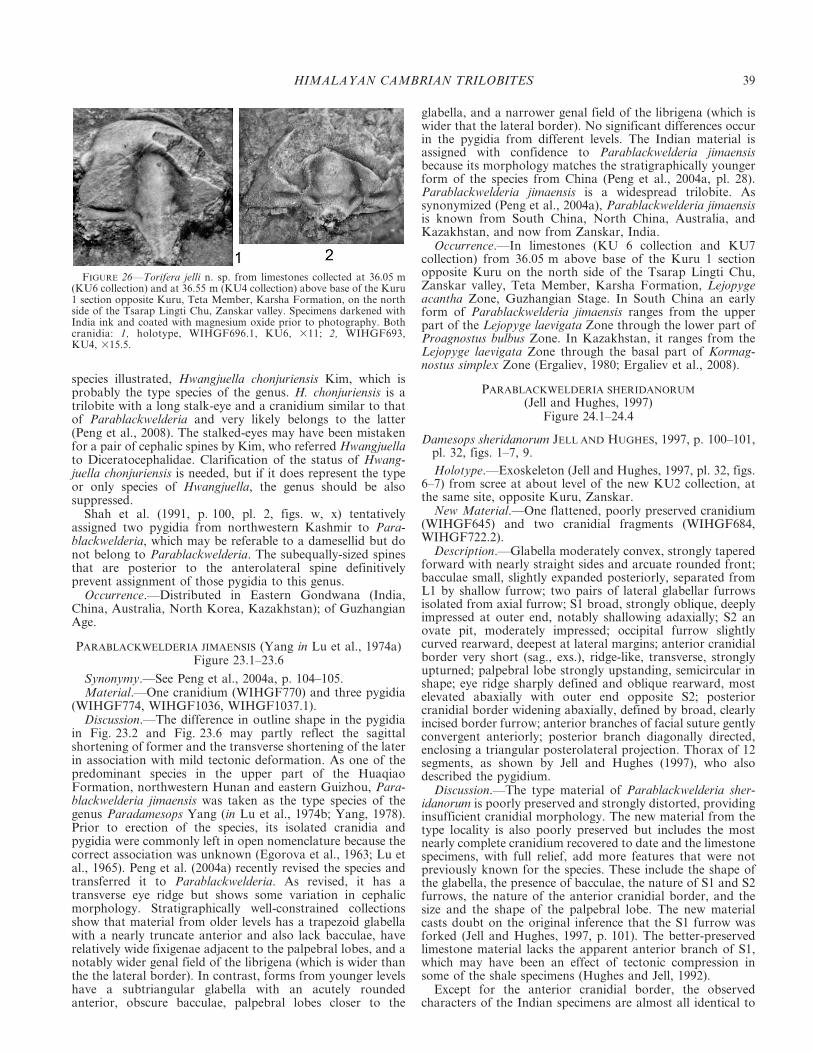
species illustrated, Hwangjuella chonjuriensis Kim, which isprobably the type species of the genus. H. chonjuriensis is atrilobite with a long stalk-eye and a cranidium similar to thatof Parablackwelderia and very likely belongs to the latter(Peng et al., 2008). The stalked-eyes may have been mistakenfor a pair of cephalic spines by Kim, who referred Hwangjuellato Diceratocephalidae. Clarification of the status of Hwang-juella chonjuriensis is needed, but if it does represent the typeor only species of Hwangjuella, the genus should be alsosuppressed.
Shah et al. (1991, p. 100, pl. 2, figs. w, x) tentativelyassigned two pygidia from northwestern Kashmir to Para-blackwelderia, which may be referable to a damesellid but donot belong to Parablackwelderia. The subequally-sized spinesthat are posterior to the anterolateral spine definitivelyprevent assignment of those pygidia to this genus.
Occurrence.—Distributed in Eastern Gondwana (India,China, Australia, North Korea, Kazakhstan); of GuzhangianAge.
PARABLACKWELDERIA JIMAENSIS (Yang in Lu et al., 1974a)Figure 23.1–23.6
Synonymy.—See Peng et al., 2004a, p. 104–105.Material.—One cranidium (WIHGF770) and three pygidia
(WIHGF774, WIHGF1036, WIHGF1037.1).Discussion.—The difference in outline shape in the pygidia
in Fig. 23.2 and Fig. 23.6 may partly reflect the sagittalshortening of former and the transverse shortening of the laterin association with mild tectonic deformation. As one of thepredominant species in the upper part of the HuaqiaoFormation, northwestern Hunan and eastern Guizhou, Para-blackwelderia jimaensis was taken as the type species of thegenus Paradamesops Yang (in Lu et al., 1974b; Yang, 1978).Prior to erection of the species, its isolated cranidia andpygidia were commonly left in open nomenclature because thecorrect association was unknown (Egorova et al., 1963; Lu etal., 1965). Peng et al. (2004a) recently revised the species andtransferred it to Parablackwelderia. As revised, it has atransverse eye ridge but shows some variation in cephalicmorphology. Stratigraphically well-constrained collectionsshow that material from older levels has a trapezoid glabellawith a nearly truncate anterior and also lack bacculae, haverelatively wide fixigenae adjacent to the palpebral lobes, and anotably wider genal field of the librigena (which is wider thanthe the lateral border). In contrast, forms from younger levelshave a subtriangular glabella with an acutely roundedanterior, obscure bacculae, palpebral lobes closer to the
glabella, and a narrower genal field of the librigena (which iswider that the lateral border). No significant differences occurin the pygidia from different levels. The Indian material isassigned with confidence to Parablackwelderia jimaensisbecause its morphology matches the stratigraphically youngerform of the species from China (Peng et al., 2004a, pl. 28).Parablackwelderia jimaensis is a widespread trilobite. Assynonymized (Peng et al., 2004a), Parablackwelderia jimaensisis known from South China, North China, Australia, andKazakhstan, and now from Zanskar, India.
Occurrence.—In limestones (KU 6 collection and KU7collection) from 36.05 m above base of the Kuru 1 sectionopposite Kuru on the north side of the Tsarap Lingti Chu,Zanskar valley, Teta Member, Karsha Formation, Lejopygeacantha Zone, Guzhangian Stage. In South China an earlyform of Parablackwelderia jimaensis ranges from the upperpart of the Lejopyge laevigata Zone through the lower part ofProagnostus bulbus Zone. In Kazakhstan, it ranges from theLejopyge laevigata Zone through the basal part of Kormag-nostus simplex Zone (Ergaliev, 1980; Ergaliev et al., 2008).
PARABLACKWELDERIA SHERIDANORUM
(Jell and Hughes, 1997)Figure 24.1–24.4
Damesops sheridanorum JELL AND HUGHES, 1997, p. 100–101,pl. 32, figs. 1–7, 9.
Holotype.—Exoskeleton (Jell and Hughes, 1997, pl. 32, figs.6–7) from scree at about level of the new KU2 collection, atthe same site, opposite Kuru, Zanskar.
New Material.—One flattened, poorly preserved cranidium(WIHGF645) and two cranidial fragments (WIHGF684,WIHGF722.2).
Description.—Glabella moderately convex, strongly taperedforward with nearly straight sides and arcuate rounded front;bacculae small, slightly expanded posteriorly, separated fromL1 by shallow furrow; two pairs of lateral glabellar furrowsisolated from axial furrow; S1 broad, strongly oblique, deeplyimpressed at outer end, notably shallowing adaxially; S2 anovate pit, moderately impressed; occipital furrow slightlycurved rearward, deepest at lateral margins; anterior cranidialborder very short (sag., exs.), ridge-like, transverse, stronglyupturned; palpebral lobe strongly upstanding, semicircular inshape; eye ridge sharply defined and oblique rearward, mostelevated abaxially with outer end opposite S2; posteriorcranidial border widening abaxially, defined by broad, clearlyincised border furrow; anterior branches of facial suture gentlyconvergent anteriorly; posterior branch diagonally directed,enclosing a triangular posterolateral projection. Thorax of 12segments, as shown by Jell and Hughes (1997), who alsodescribed the pygidium.
Discussion.—The type material of Parablackwelderia sher-idanorum is poorly preserved and strongly distorted, providinginsufficient cranidial morphology. The new material from thetype locality is also poorly preserved but includes the mostnearly complete cranidium recovered to date and the limestonespecimens, with full relief, add more features that were notpreviously known for the species. These include the shape ofthe glabella, the presence of bacculae, the nature of S1 and S2furrows, the nature of the anterior cranidial border, and thesize and the shape of the palpebral lobe. The new materialcasts doubt on the original inference that the S1 furrow wasforked (Jell and Hughes, 1997, p. 101). The better-preservedlimestone material lacks the apparent anterior branch of S1,which may have been an effect of tectonic compression insome of the shale specimens (Hughes and Jell, 1992).
Except for the anterior cranidial border, the observedcharacters of the Indian specimens are almost all identical to
FIGURE 26—Torifera jelli n. sp. from limestones collected at 36.05 m(KU6 collection) and at 36.55 m (KU4 collection) above base of the Kuru1 section opposite Kuru, Teta Member, Karsha Formation, on the northside of the Tsarap Lingti Chu, Zanskar valley. Specimens darkened withIndia ink and coated with magnesium oxide prior to photography. Bothcranidia: 1, holotype, WIHGF696.1, KU6, 311; 2, WIHGF693,KU4, 315.5.
HIMALAYAN CAMBRIAN TRILOBITES 39

those of Blackwelderia youshuica Peng et al. (2004a, p. 99–100,pl. 28, figs. 1–7) from the Huaqiao Formation (Lejopygelaevigata Zone), northeastern Hunan, South China. In B.youshuica, the anterior cranidial border is much longer but lessupturned than in Parablackwelderia sheridanorum. On theother hand, the complete specimen of P. sheridanorum, with a‘‘normal’’ rather than a stalked palpebral lobe and a uniquespinose pygidium may suggest the Chinese species is betterreferred to Parablackwelderia. Peng et al. (2004a) noted that agreat similarity exists between Blackwelderia youshuica andsome species of Parablackwelderia but failed to assign thisspecies to Parablackwelderia because they considered that the‘‘normal palpebral lobe’’ prevents assignment to Parablack-welderia. Peng et al. (2004a, p. 100) also noted that Black-welderia youshuica (or Parablackwelderia youshuica) occursstratigraphically slightly beneath Parablackwelderia and sug-gested that this or a closely related species may have given riseto Parablackwelderia. The close similarity between P. sher-idanorum and B. youshuica suggests that the former may bebasalmost within the Parablackwelderia lineage.
Occurrence.—From limestone at 36.05 m above the base(KU6 collection) and at 36.55 m (KU4 collection) of the Kuru1 section opposite Kuru, Zanskar, Teta Member, KarshaFormation. Guzhangian Stage, Lejopyge acantha Zone. Fromshale in the Surichun La nulla section (KH3 collection) northof Kurgiakh, Zanskar; Kurgiakh Formation (SurichunMember); Guzhangian Stage, Proagnostus bulbus Zone. Alsofrom a collection made by N.C.H. in 1992 at level of KU2collection in the section opposite Kuru (Jell and Hughes,1997). In South China Blackwelderia youshuica occurs in theLejopyge laevigata Zone.
PARABLACKWELDERIA YANGI Peng, Babcock and Lin, 2004a?Figure 23.7
? Paradamesops jimaensis Yang in LU ET AL. 1974b (in part),p. 86, 87, pl. 1, fig. 16 (only).
? Parablackwelderia yangi PENG, BABCOCK and LIN, 2004a,text-figure 12E, F (see for synonymy).
Material.—One fragmentary pygidium (WIHGF775B).Discussion.—This single pygidium, although incompletely
preserved, is characterized by the presence of fine granules onthe surface and by the lack of any additional projections orspines on the posterior border between the distinctiveposterolateral spines. Of species with a complete pygidialposterior border, only Parablackwelderia yangi has a granulateprosopon visible with the naked eye on the pygidial surface(Peng et al., 2004a, text-fig. 12E, F). The Indian granulosespecimen is tentatively assigned to that species because in it thegranules are denser and more closely spaced.
Occurrence.—From limestone at 36.05 m above the base(KU7 collection) of the Kuru 1 section opposite Kuru,Zanskar, Teta Member, Karsha Formation; GuzhangianStage, Lejopyge acantha Zone.
PARABLACKWELDERIA? sp. indet.Figure 24.5–24.6
Material.—Part and counterpart of one distorted cranidium(WIHGF788A and WIHGF788B).
Discussion.—This single cranidium is characterized by itstrapezoid glabella with a gently curved anterior margin andposterolaterally directed S1 and S2 furrows, a transverse eyeridge, with a relatively wide palpebral field and a large,triangular postocular field of the fixigena. It is uncertain if ithad a stalked palpebral lobe. In overall cranidial morphology,this specimen is closely comparable with the early form ofParablackwelderia jimaensis from the Huaqiao Formation,northwestern Hunan (Peng et al., 2004a, pl. 29, figs. 2–7),
which has a similarly shaped glabella, a similarly directed eyeridge and also lacks bacculae.
Occurrence.—In shales from the interval 500.32 m abovethe base of Purni Valley section 3 (collection PI14), ParahioFormation, on east side of Tsarap River, south of PhuktalGompa, Zanskar valley, Sudanomocarina sinindica Zone,informal global Stage 5 (or middle Taijiangian Stage as usedin South China). In South China an early form ofParablackwelderia jimaensis ranges from the upper part ofthe Lejopyge laevigata Zone through the lower part ofProagnostus bulbus Zone. The Zanskar specimen appears tobe a stratigraphically early record of this genus.
Order ASAPHIDA Salter, 1864Superfamily ANOMOCAROIDEA Poulsen, 1927
?Family ANOMOCARIDAE Poulsen, 1927Genus KOLDINIA Walcott and Resser, 1924
Koldinia WALCOTT AND RESSER, 1924, p. 5; KOBAYASHI,1943, p. 313; LOCHMAN in Harrington et al., 1959, p. 519;SUVOROVA in Chernysheva, 1960, p. 85; ROZOVA, 1964,p. 87; ROZOVA 1977, p. 60; NAZAROV, 1973, p. 63–73; JELLAND ADRAIN, 2003, p. 393.
Koldinia (Koldinia) WALCOTT AND RESSER, 1924; GOGIN AND
PEGEL, 1997, p. 115–116.
Type species.—Koldinia typa Walcott and Resser, 1924 fromtheOzarkian Sandstone,NovayaZemlya, by original designation.
Other species.—Koldinia micropthalma Kobayashi, 1943,p. 313–314, pl. 3, figs. 9–15 from the early Late CambrianKoldinia Zone, in the upper part of Giranda River, Siberia;Koldinia minor Kobayashi, 1943, p. 314–315, pl. 2, figs. 13, 14;pl. 3, figs. 7, 8 from the same locality as K. micropthalma; andKoldinia deflexa Nazarov, 1973, p. 60, pl. 8, figs. 3–5, fromMayan Stage, lower reaches of Maya River, Mayan Stage,southeastern Siberia.
Lazarenko (in Lazarenko and Nikiforov 1968) assigned fourspecies from the upper Cambrian of the northwestern SiberianPlatform to Koldinia, of which Koldinia infima Lazarenko(1968, p. 29, pl. 2, figs. 14–18) and Koldinia trita Lazarenko(1968, p. 29–30, pl. 6, figs. 13–18) have been transferred toParakoldinia by Rozova (1977), whereas Koldinia distinctaLazarenko (1968, p. 30–31, pl. 6, figs. 7–12) and Koldiniapusilla Lazarenko (in Lazarenko and Nikiforov 1968, p. 31–32, pl. 8, figs. 1–6) have been transferred to Parakoldinia byPeng (1992); Koldinia acris Pegel in Pegel and Gogin (1995,p. 68, pl. 5, figs. 1, 2) has a relatively wide anterior border andan evenly convex, continuously sloping anterior field on thecranidia and should also be transferred to Parakoldinia.
Discussion.—Gogin and Pegel (1997) regarded the differ-ences between Koldinia and Parakoldinia of no more thansubgeneric significance and assigned them both as subgeneraof Koldinia. Their classification is rejected here, as these generamay belong to different phylogenetic lineages. Both Koldiniaand Parakoldinia are highly convex and effaced, and thereforepresent few potentially defining features. When Rozova (1960,p. 51) established her new genus Parakoldinia from theSiberian upper Cambrian, she differentiated it from Koldiniaby the nature of the anterior cranidial border that is wire-like.Subsequently, Peng (1992) described some species of Para-koldinia, one of which clearly shows a preoccipital mediantubercle on the internal mold (Peng, 1992, fig. 36F, G; text-fig.58C), a feature that led to placing the genus within the familyNileidae. The familial assignment for Koldinia remains muchdebated. It is variously classified with the families Leiostegii-dae (Kobayashi, 1943), Nileidae (Rozova, 1964), Illaenuridea(Nazarov, 1973; Rosova, 1977), and Plethopeltidae (Suvorovain Chernysheva, 1960, Gogin and Pegel, 1997; Jell and Adrain,2003) or left as family uncertain (Lochman in Harrington et
40 PENG ET AL.

al., 1959). The Indian material of Koldinia is reasonably wellpreserved and adds a species with a cranidium of relatively lowconvexity to the genus and also provides information onlibrigenal morphology. The overall cranidial form is reminis-cent of some of anomocarids such as Afghanocare Wolfart,1974 and Paracoosia Kobayashi, 1936 except for a propor-tionally wider glabella and the relatively small and widelyspaced palpebral lobes. Accordingly, here we tentatively assignthis genus to Anomocaridae.
Occurrence.—Siberian Platform and India, Drumian andGuzhangian ages.
KOLDINIA ODELLI new speciesFigure 25.1a, 25.2–25.10
Etymology.—In honor of Prof. Noel E. Odell, geologist andclimber who, while standing on the deformed equivalent of theParahio Formation, last saw George Mallory and AndrewIrvine on the north face of Chumolungma (Mt. Everest)ascending rocks of equivalent depositional age to thoseyielding this fossil (Myrow et al., 2009).
Types.—Holotype, an incomplete, partially exfoliated cra-nidium (WIHGF825.1)(Fig. 25.1, 25.4–25.6). Paratype libri-genae (WIHGF819.2), and five paratype pygidia (WIHGF819.1,WIHGF820.3, WIHGF820.5, WIHGF825B.2, WIHGF831).
Diagnosis.—Koldinia with gently convex cranidium (tr. andsag.). Glabella half as long as cranidium, posterior half offrontal area continues the slope of the glabellar anterior,anterior half of frontal area gently and evenly upturned.Palpebral lobe small, about 0.25 glabellar length (sag.),midpoint opposite midpoint of glabella. Librigena withbroad-based, short genal spine. Pygidial axis two-thirds ofpygidial length; pleural field gently elevated; border furrowsabsent.
Description.—Cranidium subrectangular, length (sag.)about half posterior marginal width (tr.), largely effaced withanterior margin obtusely curved, gently convex, with preo-cular area sloping downward gently. Glabella subquadrate,tapered gently forward, obtusely rounded anteriorly, axialfurrow weakly incised; lateral furrows effaced except forfaintly impressed SO that defines a short (sag.) occipital ringthat narrows abaxially. Frontal area gently concave withposterior half continuing the slope of the glabella and anteriorhalf slightly upturned; eye ridge weak, parallel to anteriorcranidial border; palpebral lobe small, about 0.25 glabellarlength, midpoint situated posterior to cranidial mid-length,opposite glabellar midlength; palpebral field narrow (tr.), flat,about 0.3 of glabellar width at base; anterior branch of facialsuture runs forward and gently outward at an angle of about15 degrees to sagittal axis with rounded anterolateral corner;posterior branch markedly divergent rearward, enclosingtriangular posterolateral projection; posterior border furrowshallow and broad, somewhat widening abaxially. Posteriorborder gently convex, lengthening abaxially.
Librigena smooth, genal area weakly defined, genal cornerbearing broad-based, short, subtriangular genal spine.
Pygidium semicircular, pleural field moderately convex,bearing large, triangular facet, sloped at high angle abaxiallyfrom fulcrum; weakly incised pleural furrow on anteriormostsegment, other lateral and posterior border furrows wide,obscure or absent. Paradoublural line weakly incised at edgeof pleural platform. Axis narrow, gently convex, two-thirds ofpygidial length; ring furrows obscure, posterior axial endacutely rounded, separated from pleural field by slope change;anterior border narrow and shallow.
Discussion.—Variation in the outline of the pygidia(compare Fig. 25.7 and 25.10) may reflect differences in thepattern of tectonic compression. This Indian species is most
similar to Koldinia typa, the type species, but the latter has aproportionally wider glabella and wider pygidial axis and isconsiderably more convex. Koldinia micropthalma is easilydifferentiated from the new species by its shorter anteriorcranidial area, whereas Koldinia minor and K. deflexa differ bytheir proportionally narrower (tr.) cranidia, narrower (tr.)palpebral areas, and larger glabellae. This is the first species ofthis genus known beyond the Siberian Platform.
Occurrence.—In limestone from collection KU5 made at36.05 m above the base of the Kuru 1 section opposite Kuruon the north side of the Tsarap Lingti Chu, Zanskar valley,Teta Member, Karsha Formation, Lejopyge acantha Zone,Guzhangian Stage.
Family ALSATASPIDIDAE Turner, 1940Subfamily HAPALOPLEURINAE Harrington and Leanza, 1957
Genus TORIFERA Wolfart, 1974
ToriferaWOLFART, 1974, p. 96, 97; PENG, BABCOCKAND LIN,2004a, p. 174–175 (see for synonymy); YUAN AND LI, 2008,p. 127.
Xiangia PENG, 1987, p. 112, 113.
Type species.—Torifera triangularis Wolfart, 1974, from theupper Cambrian, near Surkh Bum, central Afghanistan, byoriginal designation.
Other species.—Ajrikina mera Romanenko in Goncharova etal., 1972;Cyclolorenzella tumaYang inZhou et al., 1977;Toriferayouyangensis Zhu in Zhang et al., 1980a; Xiangia taoyuanensisPeng, 1987; Torifera cf. T. triangularis Wolfart, 1974 (Fortey,1994); Torifera abrupta Peng et al., 2004a; and Torifera jelli n. sp.
Species questionably assigned to Torifera include Cyclolor-enzella paraconvexa Yang in Yin and Li, 1978; Agraulostongkinensis Mansuy, 1915 and Cyclolorenzella yantaiensis(Chu) sensu Zhou et al., 1977.
Discussion.—We follow previous authors (Fortey, 1994;Peng et al., 2004a) in using the original generic concept ofWolfart (1974). Based on well preserved material foundrecently from the Huaqiao Formation, northwestern Hunan,South China, Peng et al. (2004a) suppressed Xingia Peng 1987as a junior synonym of Torifera and transferred, orquestionably transferred, a number of species assignedpreviously to other genera to the genus while adding a newspecies. Those species are list above. Zhang and Jell (1987)synonymized Aulacodigmatidae Opik, 1976 and Diceratoce-phalidae Lu, 1954, and assigned Torifera to the latter. Theassignment of Torifera is not accepted by Peng et al. (2004a),who classified the genus within Alsataspididae Turner, 1940.The latter assignment is followed here.
As noted by Jell and Hughes (1997), the thorax of Toriferais known from the Himalayan species described below asTorifera jelli and contains 8 segments. Torifera mera(Romenenko, in Goncharova et al., 1972) from Gorlyi Altaialso has a holaspid thorax of 8 segments.
Occurrence.—Occurring in Asia (Afghanistan, India, GorlyiAltai, Oman, western Siberia, South China, Vietnam) andAustralia. The stratigraphical range of Torifera is wellconstrained in South China, where it occurs from the latestLejopyge laevigata Zone through the Linguagnostus reconditusZone, indicating a Guzhangian Age.
TORIFERA JELLI new speciesFigure 26
Cyclolorenzella? sp. indet., WHITTINGTON, 1986, p. 175, pl. 18,figs. 2–3.
? Cyclolorenzella? sp., SHAH, PARCHA AND RAINA , 1991 (inpart), p. 90–91, pl. 1, fig. c, non figs, f, i.
HIMALAYAN CAMBRIAN TRILOBITES 41

FIGURE 27—Bhargavia prakritika n. gen. and sp. from limestones collected at 433.44 m (PO15 and PO19 collections) and 439.57 m (PO18 collection)above base of Parahio Valley section on north side of Parahio River, Parahio Formation, Spiti region. Specimens darkened with India ink and coatedwith magnesium oxide prior to photography. All cranidia, all likely holaspid. Note the thick exoskeleton and the difference between the external and
42 PENG ET AL.

Torifera sp. nov., JELL AND HUGHES, 1997, p. 102, pl. 31, figs.7, 8, ?fig. 6.
Etymology.—In honor of Dr. Peter A. Jell, paleontologist ofthe Himalayan Cambrian.
Types.—Holotype, a cranidium preserved in limestone withthe anterolateral portion somewhat distorted (WIHGF696.1)(Fig. 26.1). One paratype damaged cranidium(WIHGF693).
Diagnosis.—Torifera with wide (tr.), rim-like anteriorcranidial border separated by short (sag., exs.), deeply incisedborder furrow; palpebral and posterior areas of fixigenaconvex; bacculae absent.
Description.—Cephalon subrectangular, wider that long.Glabella tapering gently forward, width 0.7 length, obtuselyrounded anteriorly, defined by broad and deep axial furrows,lacking bacculae; three pairs of lateral furrows: S1 furrowfirmly incised, strongly oblique rearward, defining a sub-triangular L1; S2 short, deeply notched, S3 faintly impressed;occipital furrow faint medially, notably deepened at ends;occipital ring large, subtriangularly-shaped, wider (tr.) thanL1, extending rearward into stout, wide-based occipital spinebearing a sagittal carina. Anterior cranidial area highlyconvex, with anterior portion strongly sloping downwardforward, occupying about 0.3 of cranidial length (excludingoccipital spine); anterior border rim-like, wider than glabellarlength, defined by deeply incised border furrow; palpebral lobesmall, 0.25 pre-occipital glabellar length, semicircular, mark-edly upstanding, fixigena moderately convex; preglabellar bossweak; eye ridge faint, running from anterior end of palpebrallobe inward and slightly rearward to axial furrow, thenrunning downward along the slope formed by the descendinginner part of palpebral area; palpebral area slightly wider thatbasal glabellar width, gently convex; anterior branch of facialsuture converging forward, meeting anterior cranidial borderat distance to sagittal line about the length of preglabellarfield; posterior branch gently divergent rearward, enclosing anarrow (tr.) triangular posterolateral projection that slopesstrongly downward; posterior border furrow deep, narrower(exs.) than posterior border, becoming obsolete distally.
Discussion.—Jell and Hughes (1997) realized that theirmaterial of Torifera from the Kurgiakh Formation near Kuru,Zanskar represented a new species but failed to name it becauseof insufficient material. The new material, also from Kuru, isagain a small collection but, in combination with the materialdescribed previously, can be assigned as a new species withconfidence because the combination of features seen is unknownin all species of Torifera described to date. The features thatcharacterize the new species include the absence of bacculae, theabsence of a large triangular depression on the palpebral andposterior areas of the fixigena, a relatively less strongly taperedglabella, the continuous slope of the anterior cranidial fieldwithout a median boss, a relatively wide (tr.) anterior cranidialborder, and the absence of surficial granulation.
The new species is closely similar to Torifera tuma Yang (inZhou et al., 1977, pl. 50, figs. 17, 18) from South China andTorifera mera Romenenko (in Goncharova et al., 1972, pl. 53,figs. 3–5) from western Siberia in lacking bacculae and havinga wide (tr.) anterior border. However, Torifera tuma has astrongly tapered glabella and a cranidium ornamented withgranules and Torifera mera has a notable boss on the
preglabellar area. Torifera cf. triangularis from Oman (Fortey,1994, fig. 6B–F) has similar anterior cranidial area and alsolacks bacculae, but again differs in having a conical glabella,and in a shorter (tr.) anterior cranidial border. Toriferatriangularis, the type species, and T. taoyuanensis (Peng, 1987,pl. 13, figs. 1–4, 6, non 5) are differentiated by having adepressed palpebral and posterior areas, and glabellarbacculae. Torifera abrupta (Peng et al., 2004a, pl. 60, figs. 1–10) differs by its folded anterior cranidial field with a clearlydefined boss medially and by the presence of bacculae.Torifera mera is easily distinguished by having a preglabellarboss, narrower palpebral area, and a more depressed fixigena.
Occurrence.—Preserved in limestone at 36.05 m (KU6collection) and at 36.55 m (KU4 collection) above the baseof the Kuru 1 section opposite Kuru, Zanskar, Teta Member,Karsha Formation; Guzhangian Stage, Lejopyge acanthaZone. Also known from shale in the Surichun La nullasection (equivalent to the present KH3 collection) north ofKurgiakh, Zanskar, Surichun Member, Kurgiakh Formation;Guzhangian Stage, Proagnostus bulbus Zone.
Order PTYCHOPARIIDA Swinnerton, 1915Superfamily ELLIPSOCEPHALIODEA Matthew, 1887
Family ELLIPSOCEPHALIDAE Matthew, 1887Genus BHARGAVIA new genus
Etymology.—In honor of Dr. Om N. Bhargava, for hisimportant and lifelong contributions to the Cambrian geologyof the Himalaya.
Type species.—Bhargavia prakritika n. sp.; designatedherein.
FIGURE 28—Bhargavia prakritika n. gen. and sp. from limestonescollected at 433.44 m (PO15 collection) and 439.57 m (PO18 collection)above the base of the Parahio Valley section on north side of ParahioRiver, Parahio Formation, Spiti region. Specimens darkened with Indiaink and coated with magnesium oxide prior to photography. All likelyholaspid. Note the thick exoskeleton, and the difference between theexternal and the internal surfaces. 1, 2, cranidium, WIHGF456, PO18,36.5: 1, dorsal view; 2, anterior left lateral view; 3, pygidium, WIHGF406,PO15, 314.75; 4, cranidium, WIHGF1025, PO18, 37.
r
internal surfaces: 1, WIHGF443B.5, PO18, 39; 2, WIHGF440A.3, PO18, 3 10.75; 3, WIHFG455.3, PO18, 39; 4, WIHGF440B, PO18, 311; 5,WIHGF440A.1, PO18, 3 9.75; 6, WIHGF455.5, PO18, 39.5; 7, WIHGF455.4, PO18, 310; 8, WIHGF412, PO15, 39.5; 9, WIHGF455.2, PO18, 310;10, WIHGF450B, PO18, 39.75; 11, WIHGF406, PO15, 310; 12, WIHGF443A.1, PO15, 38.5; 13, WIHGF1026, PO18, 38.5; 14, WIHGF401B, PO15,3 8.75. 15, 16, 17, holotype, WIHGF466B.1, PO19, 38.75: 15, anterior view; 16, anterior left lateral view; 17, dorsal view of external surface; 18,WIHGF466B.2, PO19, 310.25.
HIMALAYAN CAMBRIAN TRILOBITES 43

FIGURE 29—Douposiella himalaica (Reed, 1910) from limestones collected at 765.14 m (PO21 collection), 772.51 m (PO23 collection), and 775.41 m(PO24 collection) above the base of the Parahio Valley section on north side of Parahio River, Spiti region, Parahio Formation. Specimens darkenedwith India ink and coated with magnesium oxide prior to photography. All cranidia except 14, 18, 19: 1, WIHGF967, PO21, 38.5; 2, WIHGF605.2,PO23, 38; 3,6, WIGHF938.1, PO23, 36.25: 3, retrodeformation of 6 with inferred strain ellipse; 4, WIHGF948.1, PO23, 36.5; 5,9, WIHGF495, PO24,36.5: 5, anterior view; 9, dorsal view; 7,WIHGF965.2, PO21,3 5.5; 8,WIHGF934, PO23,36; 10,WIHGF942, PO24,36; 11, 12,WIHGF941.1, PO24,36: 11, dorsal view; 12, left lateral view; 13, WIHGF944, PO24, 36.75; 14, pygidium, WIHGF934.2, PO23, 3 10.25; 15–17, WIHGF943, PO24, 34.25:15, dorsal view; 16, anterior view; 17, right lateral view; 18, external mold of left librigena, WIHGF610, PO24,38.5; 19, external mold of right librigena,WIHGF935.1, PO23, 310; 20, WIHGF937.2, PO23, 34.25; 21, WIHGF936, PO23, 34.
44 PENG ET AL.

Other species.—Syspacephalus obscurus Palmer and Halley,1979 (p. 116, pl. 7, figs. 6–13, 15) from the Albertella Zone,California, and Nevada, southern Great Basin, USA.
Diagnosis.—A highly effaced ellipsocephalinean trilobitewith a thick exoskeleton. Cranidium with a weak sagittal keel;palpebral and pre-occipital glabellar furrows obscure; poste-rior border furrow confluent with posterior margin ofoccipital ring. Parietal morphology on internal mold indicatesa trapezoid glabella with four pairs of unevenly spaced lateralfurrows including an adaxially bifurcate S1, straight or slightlyconcave sides, and rounded anterolateral corners; frontal areaclearly divided into anterior border and preglabellar field; eyeridge parallels cranidial anterior margin, with inner endmerging into axial furrow; posterior end connecting topalpebral lobe posterior to the anterior end of palpebral lobe.Anterior branch of facial suture convergent; posterior branchrunning rearward and somewhat outward. Pygidium smalland transverse; axis extending rearward to posterior margin;pleura with bands of subequal length (exs.), posterior bandslonger.
Discussion.—The new genus is here classified with Ellipso-cephalinae because it is morphologically comparable toKingaspis Kobayashi, 1935 and Kingaspidoides Hupe, 1953,in the proportional length and width of the glabella, theconcave sides of the glabella, the size of palpebral lobe, thedirection of the eye ridge, and in being highly effaced.However, some features of the new genus are strikinglydifferent from those of above mentioned genera. The detailedreview and emendation of Kingaspis by Geyer (1990) andGeyer and Landing (2001) and the restudy of the type materialof the type species, Kingaspis campbelli (King, 1923) byRushton and Powell (1998) has greatly clarified the genericconcept of the genus. An exfoliated cranidium has beenselected as lectotype for the type species by the latter authors(Rushton and Powell, 1998, fig. 21). With regard to theexternal surface, Kingaspis differs from the new genus inhaving the palpebral lobe situated posterior to the mid-lengthof the cranidium, a divergent anterior branch of the facialsuture, and a cranidium of lower convexity. On the basis ofparietal morphology, Kingaspis is easily distinguished inhaving a subparallel-sided glabella with an expanded ratherthan evenly rounded anterior glabellar corner, a simple ratherthan bifurcated S1, an eye ridge with its anterior endconnecting with glabellar anterolateral corner, and anobscurely subdivided anterior glabellar area. Kingaspidoidesis closely related to Kingaspis, and recently Linan et al. (2003)regarded both as subgenera under the name Kingaspis. Thefeatures distinguishing Kingaspis and Bhargavia n. gen. listedabove also serve to differentiate the new genus from King-aspidoides. As recently emended (Geyer, 1990), Kingaspidoidesis characterized by the uneven convexity of its cranidium intransverse profile, in which it differs much from Bhargavia.
Syspacephalus obscurus is considered to be referable toBhargavia. It is similar to Bhargavia prakritika in both externaland internal morphology. As noted by Palmer and Halley(1979, p. 116), the obscure axial and border furrows differen-tiate it from all other species of Syspacephalus. The two speciesare distinguished by the longer preglabellar field and anteriorborder in B. obscura, the anterior branches of the facial sutureof which run forward parallel the sagittal axis, rather thanconverging forward as in Bhargavia prakritika, by the granularornamentation in B. obscura, and by the smaller size of theAmerican species.
Bhargavia is known definitely from India and the southernGreat Basin, but some species from Antarctica assignedquestionably as Glabella? pitans Palmer and Gatehouse(1972, p. 72, pl. 1, figs. 9, 10, 13, 14), Kingaspis? convexaPalmer and Rowell (1995, pl. 12, fig. 10.1–10.3, 10.5, 10.6,
10.9, 10.10), and Kingaspis? sp. 1–sp. 3 (Palmer and Rowell,1995, fig. 10.4, 10.7, 10.8, 10.11–10.18) are probably conge-neric. It is interesting that librigena found associated with twoAntarctic species are characterized by the absence of alibrigenal spine, whereas species of Kingaspis and King-aspidoides have genal spines on their librigena. Hence recoveryof librigenae may clarify relationships between Bhargaviaprakritika and the Antarctic species.
Occurrence.—In Spiti, India, in the lower part of informalglobal Stage 5 of Cambrian. Bhargavia obscura occurs in thelower part of the Albertella Zone (Palmer and Halley, 1979, pl.18) in California, at an apparently correlative stratigraphiclevel (Sundberg and McCollum, 2003), assuming that ourassessment of the level of O. indicus in the Parahio section iscorrect.
BHARGAVIA PRAKRITIKA new speciesFigures 27, 28
Etymology.—Adjective of the Sanskrit noun prakritik,meaning ‘‘of nature’’, to reflect Dr. Bhargava’s affection fornature.
FIGURE 30—Eosoptychoparia sp. indet. from shales collected at500.32 m (PI14 collection), 501.39 m (PI19 collection), 504.04 m (PI21collection), and 510.04 m (PI17 collection) above base of Purni Valleysection 3, Parahio Formation, on east side of Tsarap River, south ofPhuktal Gompa, Zanskar valley. Specimens darkened with India ink andcoated with magnesium oxide prior to photography. All holaspid cranidia.1, WIHGF626.1, PI17, 3 3.25; 2, latex of counterpart, WIHGF852B,PI19, 36.5; 3, WIHGF849, PI19, 35.5; 4, WIHGF587, PI14, 34.75; 5,WIHGF570, PI14, 36.25.
HIMALAYAN CAMBRIAN TRILOBITES 45

FIGURE 31—Gunnia smithi n. sp. from limestones collected at 765.14 m (PO21 collection) and 772.51 m (PO23 collection) above the base of theParahio Valley section on the north side of Parahio River, Spiti region, Parahio Formation. Specimens darkened with India ink and coated withmagnesium oxide prior to photography. All PO21 except 9: 1, meraspid cranidium, WIHGF956, 320.75; 2, cranidium, WIHGF951.3, 3 7.75; 3,cranidium, WIHGF578.2, 37; 4, cranidium, WIHGF955, 35.5; 5, cranidium and left librigena, WIHGF578.6, 35.75; 6, left librigena, WIHGF954.5,36.75; 7, 8, cranidium, WIHGF584.2, 3 8: 7, dorsal view; 8, left lateral view; 9, cranidium, WIHGF948.2, PO23, 34.5; 10, 11, cranidium,WIHGF951.4, 3 4.5: 10, anterior view; 11, dorsal view; 12, 13, holotype cranidium, WIHGF954.1, 34: 12, left lateral view; 13, dorsal view; 14, 15, 16,pygidium, WIHGF954.4, 34.75: 14, left oblique lateral view; 15, posterior view; 16, dorsal view.
46 PENG ET AL.

Material.—A complete, testaceous cranidium (WIHGF466B.1) is the holotype (Fig. 27.15–27.17). Paratypes include 18cranidia, all likely holaspid, (WIHGF401B, WIHGF406,WIHGF410, WIHGF412, WIHGF440A.1, WIHGF440A.3,WIHGF440B, WIHGF443B.5, WIHGF450B, WIHGF455.2,WIHGF455.3, WIHGF455.4, WIHGF455.5, WIHGF456,WIHGF466B.2, WIHGF1025, WIHGF1026), and one pygidi-um (WIHGF406).
Diagnosis.—As for the genus.Description.—Cranidium highly effaced, subtriangular in
outline, moderately convex with a weak sagittal keel. Anteriormargin broadly rounded, sagittal profile a continuous curveand transverse profile obtusely angled. Axial furrow weaklydefined via a change slope between glabella and fixigena;preglabellar furrow effaced. Occipital furrow short (sag.)distinct but weakly incised, continuous across axis, isolatedaxially, palpebral lobe small and narrow, opposite cranidialmid-length anterior to occipital furrow. Posterior borderfurrow short (sag.), firmly incised, confluent with posteriormargin of occipital ring, forming a smooth continuous arc;posterior border widening abaxially; anterior branches offacial suture gently convergent forward at angle of about 10 to15 degrees to sagittal axis; posterior branches directedrearward and gently outward, enclosing a narrow triangularposterolateral projection. Marked differences between crani-dial external and internal molds. On internal mold, all furrowsare firmly incised, glabella trapezoid in outline, with sidesstraight or slightly concave outward in anterior part, obtuselyrounded anteriorly, occupying about 0.72 to 0.82 cranidiallength; four pairs of lateral glabellar furrow isolated fromaxial furrows, unevenly spaced with S1 and S2 widely spacedand S3 and S4 closely spaced; S1 longest, bifurcate adaxiially,posteriorly directed; S2 transverse, rather long, S3 and S4short, subparallel, gently oblique forward; occipital furrowlong (sag.), transverse or slightly curved rearward, anteriormargin steeply sloped, confluent with axial furrows. Occipital
ring slightly shorter (sag.) than L1, narrowing at axial furrowswith a small axial node lying slightly posterior to mid-point.Frontal area about 0.25 glabellar length (sag.), anterior borderslightly convex, somewhat flatter than fixigenal convexity;preglabellar field uniformly convex (tr.), occupying 0.5–0.7 offrontal area length (sag.); anterior border furrow shallow butclearly defined. Eye ridge evenly curved, directed inward andforward to parallel anterior cranidial margin, with anteriorend merging into axial furrow opposite S4 and lying near theanterolateral corner of glabella, posterior end extending to apoint slightly posterior to anterior end of palpebral lobe.Palpebral area more than half maximum glabellar width (tr.),continuous convexity with glabella; palpebral lobe raisedslightly above palpebral area, about 0.3 glabellar length (sag.),well defined by moderately incised palpebral furrow withanterior end opposite S3, and posterior portion opposite L2.Posterior border furrow broad (exs.), moderately deep;posterior border gently convex, narrower (exs.) than posteriorborder furrow.
Occurrence.—From limestones collected at 433.44 m (PO15and PO19 collections), 439.57 m (PO18 collection), and439.67 m (PO17 collection) above the base of the ParahioValley section on the north side of the Parahio River, Spitiregion, Parahio Formation, Kaotaia prachina Zone, informalglobal Stage 5 of the Cambrian.
Superfamily PTYCHOPARIOIDAE Matthew, 1887Family PTYCHOPARIIDAE Matthew, 1887
Genus DOUPOSIELLA Lu and Chang in Lu et al., 1974a
Douposiella LU AND CHANG in Lu et al., 1974a, p. 104; YIN
AND LI, 1978, p. 463–464; ZHANG ET AL., 1980a, p. 353;LUO, JIANG AND TANG, 1994, p. 145; JELL AND HUGHES,1997, p. 52–54; YUAN, ZHAO, LI AND HUANG, 2002, p. 180–181; YUAN, LI, AND ZHAO, 2008, p. 102.
FIGURE 32—Gunnia sp. from Parahio Valley, Spiti. 1–4, Gunnia sp. 1 from limestone collected at 580.52 m above base of Parahio Valley section (PO20collection) on north side of Parahio River, Parahio Formation, Spiti region. Specimens darkened with India ink and coated with magnesium oxide priorto photography. Both likely holaspid cranidia. 1–3, WIHGF467, 312.75: 1, dorsal view; 2, anterior right lateral view; 3, right lateral view; 4,5,WIHGF988, 37.75: 4, left lateral view; 5, dorsal view; 6–9, Gunnia sp. 2 from limestone collected at 828.72 m above the base of the Parahio Valleysection (PO30 collection) on the north side of Parahio River, Parahio Formation, Spiti region: 6, 7, cranidium, WIHGF1029, 35: 6, right lateral view;7,dorsal view; 8, cranidium, WIHGF662A, 34.5; 9, right librigena, WIHGF682A, 34.75.
HIMALAYAN CAMBRIAN TRILOBITES 47

Danzhaiaspis (Nangaocephalus) YUAN in Zhang et al., 1980a,p. 358.
Type species.—Douposiella douposiensis Lu and Chang inLu et al., 1974a from the Doupossu Formation, Yiliang,Yunnan, China, by original designation.
Discussion.—The holotype of type species Douposielladouposiensis is apparently an immature cranidium with a lengthof only about 2.5 mm (Lu et al., 1974a, pl. 41, fig. 1; Zhang etal., 1980a, pl. 120, fig. 4; Yuan et al., 2002, pl. 45, fig. 8). Thelarge cranidia assigned subsequently to the species (Zhang et al.,1980a, pl. 120, fig. 3; Yuan et al., 1980, pl. 45, fig. 9; pl. 68, figs.1, 8) have a less tapered glabella, relatively shorter palpebrallobes, and more distinct lateral glabellar furrows.
As discussed below, Shantungaspis himalaica (Reed, 1910) ishere reassigned to Douposiella. Both Shantungaspis andDouposiella have long palpebral lobes, a moderately longglabella that is slightly longer than wide, relatively narrowintraocular fixigenae, and a relatively long preglabellar field,but the former differs in having an occipital spine and a nearlyparallel-sided rather than notably tapered glabella. Intransferring himalaica to Shantungaspis, Jell and Hughes(1997) suggested that Zhang and Jell’s (1987) claim of anoccipital spine in Shantungaspis was unfounded. In fact,Chang’s (1957) line drawing of the type species Shantungaspisaclis (Walcott, 1905), which shows only a median node on theoccipital ring, rather than a spine, is inaccurate. Largecollections of Shantungaspis aclis reveal that the type specieshas a well-developed occipital spine that is upward and gentlyrearward directed (Zhou et al., 1982, pl. 60, fig. 10; Guo andZan, 1991, pl. 2, fig. 14; An et al., 1996, pl. 1, fig. 1–3; Yuanand Li, 1999, pl. 6, figs. 1–3; Duan et al., 2005, pl., 7, figs. 2–15, text-figs. 7-1.3, 7-2) and this is regarded here as a criticalfeature for differentiating Shantungaspis from Douposiella.
Occurrence.—China (Guizhou, Yunnan) and India (Spiti);informal global Stage 5 of the Cambrian System.
DOUPOSIELLA HIMALAICA (Reed, 1910)Figure 29
Ptychoparia? himalaica REED, 1910, p. 36, pl. 4, fig. 27; pl. 5,figs. 1–4.
Megagraulos? himalaica (Reed); KOBAYASHI, 1967, p. 486,text-fig. 7.1.
Shantungaspis himalaica (Reed); JELL AND HUGHES, 1997,p. 46–48, pl. 11, figs. 1–5.
New material.—Thirteen cranidia (WIHGF495, WIHGF605.2, WIHGF934, WIHGF936, WIHGF937.2, WIHGF938,WIHGF941.1, WIHGF942, WIHGF943, WIHGF944,WIHGF948.1, WIHGF965.2 and WIHGF967), two librigena(WIHGF610, WIHGF935.1), and one pygidium (WIHGF934.2).
Discussion.—Specimens assigned to Douposiella himalaicafrom the Parahio Valley section are characterized by theirgently tapered glabella with three to four pairs of lateralfurrows and rounded front, long palpebral lobes that aremedially to posteriorly centered, a short and convex anteriorborder, and a slightly convex preglabellar field that is up totwice the length of the anterior border, although there isvariation in these proportions among the specimens. Most ofspecimens in the new material are collected from limestoneand maintain their original shape and convexity. They areidentical in all observed details to the shale specimens fromHayden’s Horizon 6 in the same section, which were named asPtychoparia? himalaica by Reed (1910) and renamed Shantun-gaspis himalaica by Jell and Hughes (1997). This species is hereclassified as Douposiella for reasons given above and becausethe associated pygidium conforms to that of Douposiella(Zhang et al., 1980a, pl. 120, fig. 7; Yuan et al., 2002, pl. 68,
fig. 1, 8), but differs from that of Shantungaspis (Walcott,1913, pl. 12, fig. 8a; Zhang and Jell, 1987, pl. 27, fig. 5).
Douposiella himalaica is closely similar to the type speciesDouposiella douposiensis but can be distinguished by its lesstapered glabella that is proportionally shorter.
Douposiella hostilis (Reed, 1910) of Jell and Hughes (1997)from Hayden’s Horizon 9, Parahio Valley, Spiti is herereassigned to Ziboaspis on the basis of new material inlimestone from the type locality.
Occurrence.—From limestones collected at 765.14 m (PO21collection), 772.51 m (PO23 collection), and 775.41 m (PO24collection) above the base of the Parahio Valley section onnorth side of Parahio River, Parahio Formation, Spiti region;Parahio Formation, Paramecephalus defossus Zone, informalglobal Stage 5 of the Cambrian System.
Genus EOSOPTYCHOPARIA Chang, 1963
Eosoptychoparia CHANG, 1963, p. 463–464, 478; LU ET AL.,1965, p. 133–134; ZHANG ET AL., 1980a, 321; ZHANG AND
WANG, 1985, p. 348; ZHANG AND JELL, 1987, p. 77; GUO,ZAN AND LUO, 1996, p. 67.
not Eosoptychoparia CHANG, 1963; NAN AND CHANG, 1982a,p. 9; 1982b, p. 29.
Eosoptychoparia (Eosoptychoparia) CHANG, 1963; YUAN,ZHAI, LI AND HUANG 2002, p. 171.
not Eosoptychoparia (Danzhaina) YUAN in Zhang et al., 1980a;YUAN, ZHAO, LI AND HUANG, 2002, p. 171–172; YUAN, LI,AND ZHAO, 2008, p. 102.
Type species.—Ptychoparia kochibei Walcott, 1911, fromthe Changhia Formation, Jinjiachengzi, Changxing Island,Liaoning, by original designation.
Discussion.—Eosoptychoparia was erected by Zhang(Chang, 1963, p. 463–464; 478) using Ptychoparia kochibeiWalcott, 1911 as the type species and selecting a lectotype.Subsequently, Zhang and Jell (1987, p. 77) emphasized thatonly the broad fixigena, the long preglabellar field, and thesmall pygidium characterize the genus. Zhang’s originalconcept, with Zhang and Jell’s modifications, is followedhere. Yuan et al. (2002) broadened the generic concept byincluding Pachyaspis (Danzhaina) Yuan in Zhang, 1980a withits junior synonyms Jiumenia Yuan in Zhang 1980a, andJimaoshania Qiu in Qiu et al., 1983, and regarded this group oftrilobites as a subgenus of Eosoptychoparia. This classificationis rejected here as Yuan et al. (2002) did not explain why thisgroup of trilobites is the phylogenetic sister taxon of otherEosoptychoparia. Pending thorough revision of the ptycho-pariids, we prefer to recognize them as a separate genus thatdiffers from Eosoptychoparia by its narrower fixigena,proportionally shorter preglabellar field and longer glabella.The generic name Danzhaina, as used by Yuan et al. (1997,p. 511, 522), is thus revived for these trilobites. Zhang and Jell(1987, p. 72) suppressed Pachyaspis (Danzhaina) lilia Yuan inZhang et al., 1980a (p. 323, pl. 107, figs. 1–7, text-fig. 90), thetype species of P. (Danzhaina) (and now Danzhaina), as ajunior synonym of Xingrenaspis hoboi (Resser and Endo inEndo and Resser, 1937), and thus automatically suppressedDanzhaina within Xingrenaspis. This suppression was rejectedby Yuan et al. (2002, p. 172), who considered them to beseparate species differentiated by cranidial outline, the size andposition of the palpebral lobe, the course of the facial sutures,and features of the anterior border including the distinction ofthe anterior border furrow. We follow Yuan et al. (2002) inrejecting the transfer made by Zhang and Jell (1987).
EOSOPTYCHOPARIA sp. indet.FIGURE 30
48 PENG ET AL.

Ptychoparia sp.; KUMAR, 1998, p. 674, pl. 1, fig. 12.Lyriaspis spitiensis REED, 1910; KUMAR, 1998, p. 674, pl. 1,figs. 11. ?13.
Elrathia cf. chuwansis KOBAYASHI, 1935; KUMAR, 1998,p. 677, pl. 2, figs. 7, 8, 14.
Material.—Five cranidia (WIHGF570,WIHGF587,WIHGF626.1, WIHGF849, WIHGF852B).
Discussion.—The cranidia collected from the uppermostpart of the Parahio Formation near Phuktal Gompa, Zanskarare characterized by a long preglabellar field, a short glabellawith S1 firmly incised and running strongly obliquelyrearward toward SO such that L1 is almost isolated, and arelatively wide fixigena. The eye ridge runs slightly obliquelyor transversely and the anterior branch of the facial suturediverges forward slightly. These features are closely compara-ble to those of the lectotype of type species Eosoptychopariakochibei (Walcott, 1911, pl. 12, fig. 5c; see also Chang, 1963,pl. 1, fig. 6), suggesting placement within Eosoptychoparia.However, no specific assignment is attempted because of thesmall number of specimens and their poor state of preserva-tion.
Occurrence.—From shales collected at 500.32 m (PI14collection), 501.39 m (PI19 collection), 504.04 m (PI21 collec-tion), and 510.04 m (PI17 collection) above base of the PurniValley section 3, on the east side of the Tsarap River, south ofPhuktal Gompa, Zanskar, Parahio Formation, Sudanomocar-ina sinindica Zone, informal global Stage 5 of the CambrianSystem.
Genus GUNNIA Gatehouse, 1968
Gunnia GATEHOUSE, 1968, p. 50; KRUSE, 1990, p. 15.Ellotia GATEHOUSE, 1968, p. 54.Xiangshanaspis LIN in Qiu et al., 1983, p. 148.Yiliangaspis LUO in Luo, Jiang and Tang, 1994, p. 141.Probowmania (Gunnia) GATEHOUSE, 1968; YUAN ET AL.,1997, p. 510; YUAN AND ZHAO, 1999, p. 121, 130; YUAN,ZHAO, LI, AND HUANG, 2002, p. 157–158; LIN, 2008, p. 69;YUAN, LI, AND ZHAO, 2008, p. 103.
Type species.—Gunnia lutea Gatehouse, 1968, from theTingall Limestone, Litchfield, Northern Territory, Australia,by original designation.
Other species.—Yiliangaspis yiliangensis Luo in Luo et al.,1994 (p. 141–142, pl. 32, figs. 8, 9) [5Antagmidae gen. et sp.indet. 1 (Zhang et al., 1980a, pl. 102, figs. 9], from theWulungqing Member, Tsanglangpu Formation, near Yiliang,Yunnan, China; Probowmaniella jixianensis Zhang in Zhang etal., 1995 (p. 35–36, pl. 10, figs. 4–10; pl. 11, figs. 1, 2) from theMaochuang Formation, Jixian, Henan, China; and Gunniasmithi n. sp., Gunnia sp. 1, and Gunnia sp. 2 described herein.
Yuan et al. (2002) transferred several species, sometentatively, to their subgenus P. (Gunnia), but we consideredthese assignments to be unacceptable. Transferred taxainclude Kunmingaspis huitingshanensis Sun, 1982 (p. 305, pl.2, figs. 1–3), Pachyaspis (Danzhaina) jingshanensis Sun, 1982(p. 305–306, pl. 2, figs. 8–11; pl. 3, fig. 9), Pachyaspis(Danzhaina) dahongshanensis Sun, 1982 (p. 306, pl. 2, figs.12–14) and tentatively, Ptychoparia? himalaica Reed, 1910(p. 36, pl. 7, fig. 27; pl. 5, figs. 1–4), Xingrenaspis quadrataYuan and Zhou in Zhang et al., 1980a (p. 331, pl. 109, figs.14–16), and Xiangshanaspis tongshanensis Lin in Qiu et al.,1983, (p. 148, pl. 49, fig. 6). We do not consider these taxa toform a monophyletic entity because we do not see putativesynapomorphies linking these forms.
Kruse (1990, p. 18–19, pl. 6; text-fig. 11) added a species G.cf. concava (Deiss, 1939) to Gunnia, that we feel might bebetter assigned to Xingrenaspis Chang et al., 1980a.
Discussion.—Kruse (1990, p. 15) recognized Gunnia as aseparate genus but noted that it ‘‘is morphologically close toand possibly synonymous with one or more of ProbowmaniaKobayashi, 1935 and its one-time subgenus Mufushania Lin,1965.’’ Yuan et al. (2002) regarded Gunnia and Mufushania assubgenera of Probowmania Kobayashi, 1935. Since these taxaare distinguishable, we concur with Kruse in leaving them atgeneric rank until more is known. Gunnia is a rather effacedptychopariid with obscure or absent lateral glabellar furrowsand a faint eye ridge. In this genus, the anterior branch of thefacial suture curves evenly outward to a rounded inflection,the inner end of the eye ridge is anteriorly positioned such thatthe preocular field and preglabellar field are almost equal inlength (sag., exs.), the anterior border occupies a relativelylonger proportion of the total cranidial length, and theoccipital node, when present, lies at the center of the occipitalring. Zhang and Jell (1987, pl. 25, fig. 1) refigured the holotypeof Probowmania ligea (Walcott, 1905), which differs fromGunnia by the presence of deep lateral glabellar furrows and astout eye ridge, an anterior branch of the facial suture thatencloses an obtuse anterolateral corner of the cranidium, andan occipital node situated at the posterior margin of theoccipital ring. Peng et al. (2001c) restudied the type material ofMufushania and selected a lectotype for Mufushania nankin-gensis, the type species of the genus. The lectotype has asimilarly proportioned anterior border to that of Gunnia butdiffers in having a proportionally longer and narrowerglabella, a more posteriorly positioned eye ridge and anobtuse anterior branch of the facial suture.
Kruse (1990) synonymized Gunnia convexa Gatehouse, 1968and Ellotia dimota Gatehouse, 1968 (p. 54, pl. 6, figs. 2a–d,text-fig. 5) with Gunnia lutea, the type species of Gunnia.Ellotia dimota is the type species of Ellotia, and with thistransfer Ellotia became a junior synonym of Gunnia. Wefollow Yuan et al. (2002) in regarding Yiliangaspis as a juniorsynonym of Gunnia but include only the type speciesYiliangaspis yiliangensis Luo in Luo et al., 1994 (p. 141–142,pl. 32, figs. 8, 9), Y. latus Luo (Luo et al., 1994, pl. 32, figs. 10–12) and Y. malongensis Luo in Luo et al., 1994 (Luo et al.,1994, pl. 32, figs. 13) in Gunnia.
Occurrence.—Australia, South China, and India; fromupper part of informal Stage 4 and Stage 5 of the CambrianSystem.
GUNNIA SMITHI new speciesFigure 31
Etymology.—In honor Dr. Barry Smith of York, England,for his skill and generosity in teaching natural science.
Types.—The holotype (Fig. 31.12, 31.13) is a partiallyexfoliated cranidium (WIHGF954.1). Paratypes include sevencranidia (WIHGF578.2, WIHGF584.2, WIHGF948.2,WIHGF951.3, WIHGF951.4, WIHGF955, WIHGF956),two librigenae (WIHGF578.6, WIHGF954.5) and one pygid-ium (WIHGF954.4).
Diagnosis.—Gunnia of moderate convexity; preglabellarfield longer than anterior cranidial border and more steeplyinclined; glabella with long (sag.) occipital ring; pygidium withclearly defined borders and short anterior band.
Description.—Cranidium rather smooth, wider than long toas wide as long, moderately convex, broadly roundedanteriorly. Glabella broadly subconical, blunt to obtuselypointed anteriorly, occupying about 0.65 cranidial length withthree pairs of lateral furrows faintly impressed or obsolete;occipital furrow slightly curved rearward, weakly incised;occipital ring subcrescentic to subsemicircular with roundedposterior margin. Eye ridge obscure on external surface, a lowridge on internal mold, gently oblique rearward at an angle of70–75 degrees to sagittal line, becoming faint adaxially;
HIMALAYAN CAMBRIAN TRILOBITES 49

FIGURE 33—Kaotaia prachina n. sp. from limestones collected at 433.44 m (PO15 and PO19 collections), 439.57 m (PO18 collection) and 439.67 m(PO17 collection) above the base of the Parahio Valley section on the north side of the Parahio River, Spiti region, Parahio Formation. Specimens
50 PENG ET AL.

palpebral lobe short, slightly oblique to sagittal line, midpointjust posterior to midpoint of cranidium or just anterior tomidpoint of glabella. Anterior border flat and gently upturnedanteriorly; preglabellar field gently convex, inclined forward,longer than anterior border; anterior border furrow weaklyincised, shallowing medially in some specimens; anteriorbranches of facial suture diverge forward at an angle of 20to 30 degrees to sagittal axis from palpebral lobe curvinginwards to cross border furrow, and continue for a shortdistance into the anterior border, after which they curveinwards forming a smooth curve towards anterior margin;posterior branch divergent rearward to posterior borderfurrow, trending slightly outward to enclose a narrow (tr.),posterolateral projection; posterior border broad, furrow wellimpressed; posterior border a short (exs.) ridge.
Librigena with narrow, gently convex genal field; lateralborder flat, about as wide (tr.) as genal field, extendingrearward into slender genal spine; posterior border andposterior border furrow absent; lateral border furrow shallow;doublure slightly narrower than lateral border, cut anteriorlyby connective suture trending inward posteriorly.
Pygidium transverse, half as long (sag.) as wide (tr.). Axisprominent, moderately convex, reaching to posterior borderfurrow, bearing 3 rings and a terminal piece, all subequal inlength; first ring furrow clearly defined, succeeding ring furrowsobscure; terminal piece with a sagittal notch posteriorly;defining a pair of nodes at the end of the axis; pleural fieldgently convex with 3 clearly defined pleural furrows. Pleuralfurrows longer (exsag.) than sharply defined interpleuralfurrows. Anterior band of pleura slightly shorter (exsag.) thanposterior band. Lateral and posterior border flat, moderatelyand evenly wide about margin, narrowing postaxially.
Discussion.—In overall cranidial morphology, the newspecies is similar to Gunnia lutea Gatehouse, 1968 (pl. 5, figs.10, 16; text-fig. 2), the type species, but the latter differs inhaving a shorter preglabellar field that is subequal or shorterthan the anterior border, and a shorter occipital ring. BothGatehouse’s (1968) and Kruse’s (1990) material from North-ern Territory, Australia, and especially the silicified specimensthat maintain full profile of cranidium for the type species(Kruse, 1990, pl. 4, figs. A–C), show that the type species wasof lower cranidial convexity than that of the new species, andhad a less steeply rearward sloping anterior cranidial border.The pygidium of the type species has a more transverseoutline, less distinctly defined axial rings of which there areonly two rings, and lacks borders.
Gunnia jixianensis is also comparable, but it differs in havinga proportionally longer preglabellar field, less divergentanterior branches of the facial suture, and a more acutelyrounded anterior cranidial margin.
Gunnia yiliangensis differs readily in having a shorterpreglabellar field, shorter occipital ring, and probably moredistinct lateral glabellar furrows.
Occurrence.—From limestones collected at 765.14 m (PO21collection), 765.28 m (PO22), and 772.51 m (PO23 collection)above base of the Parahio Valley section on the north side ofthe Parahio River, Spiti region; Parahio Formation, Para-mecephalus defossus Zone, informal global Stage 5 of theCambrian System.
GUNNIA sp. 1Figure 32.1–32.5
Material.—Two cranidia (WIHGF467, WIHGF988).Discussion.—This species is characterized by its broadly
conical, relatively convex glabella, and by the anterior branchof the facial suture that is slightly divergent anterior to the eye,flexing inward to cross the anterior border at an angle of 45degrees to the sagittal line. This species is assigned to Gunniadue to its resemblance in overall cranidial aspect. Onecranidium in the type material of Gunnia lutea, the typespecies (Gatehouse, 1968, pl. 5, fig. 2a–c), has a glabellasimilar to that of the present cranidia, but that Australiancranidium is of lower convexity, and its anterior branch of thefacial suture forms a smooth curve, such that the extremity ofthe anterior border is rounded rather than angular. Thebreadth of the cranidium serves to distinguish it from otherspecies assigned to the genus.
Occurrence.—From limestone collected at 580.52 m abovethe base (PO20 collection), unzoned interval 3, of the ParahioValley section on north side of Parahio River, Spiti region.Parahio Formation. Informal global Stage 5 of Cambrian.
GUNNIA sp. 2Figure 32.6–32.9
Material.—Two cranidia (WIHGF662A, WIHGF1029) andone librigena (WIHGF682A).
Discussion.—Both cranidia assigned to this species arefragmentary. They have closely similar morphologies to thespecimens we assign to Gunnia sp. 1, which is from a level lower
FIGURE 34—Kaotaia sp. cf. K. gedongensis (Yuan and Zhao, 1994) fromlimestone collected at 78.07 m above the base of Parahio Valley section(PO3 collection) on the north side of the Parahio River, Spiti region,Parahio Formation. Specimens darkened with India ink and coated withmagnesium oxide prior to photography. All cranidia. 1, late meraspid orearly holaspid, WIHGF197.2, 311.25; 2, WIHGF197.1, 35.75; 3, 4,WIHGF 222, 37: 3, anterior right lateral view; 4, dorsal view.
r
darkened with India ink and coated with magnesium oxide prior to photography. All cranidia, all likely holaspid. 1, WIHGF455.1, PO18, 312; 2,WIHGF454B, PO18, 38.5; 3, WIHGF443B.3, PO18, 38.5; 4, WIHGF443A.4, PO18, 35.75; 5, WIHGF464, PO19, 36.75; 6, WIGHF403.1, PO15,36.25; 7, WIHGF408.1, PO15, 36.25; 8, WIHGF443B.1, PO18, 36; 9, WIHGF443.2, PO18, 35; 10, WIHGF434.1, PO17, 35.25; 11, WIHGF442A.2,PO18, 3 4.75; 12, WIHGF434.2, PO17, 35; 13, WIHGF408.2, PO15, 33.75; 14, WIHGF442A.1, PO18, 36.25; 15, WIHGF432, PO17, 33.5; 16,WIHGF287, PO17, 33.25; 17, 18, 19, WIHGF416, PO15, 3 3: 17, anterior view; 18, dorsal view; 19, anterior left lateral view; 20, 21, 22, holotype,WIHGF401A, PO15, 32.5: 20, dosal view; 21, anterior view; 22, anterior right lateral view.
HIMALAYAN CAMBRIAN TRILOBITES 51

in the same section. A similarity is the conical glabellar shape,but these specimens have lower cranidial convexity, a moreconvex preglabellar field, a more strongly divergent facialsuture both in anterior and posterior branches, and a paddle-shaped, rather than triangular, posterolateral projection.
Occurrence.—From limestone collected at 828.72 m abovebase of Parahio Valley section (PO30 collection) on the northside of the Parahio River, Parahio Formation, Spiti regionParahio Formation, Paramecephalus defossus Zone, informalGlobal Stage 5 of the Cambrian System.
FIGURE 35—Kunmingaspis stracheyi (Reed, 1910) from limestone collected at 765.14 m (PO21 collection) and shale at 836.41 m (PO26 collection)above the base of the Parahio Valley section on the north side of the Parahio River, Spiti region, Parahio Formation. Also from south side of ParahioRiver on slopes above the east bank of the Sumna River, estimated to be at about the 1050 m level in the section on the north side (PO10 collection).Specimens darkened with India ink and coated with magnesium oxide prior to photography. All internal molds, unless otherwise stated. 1, cranidium,WIHGF906.2, PO26, 39.5; 2, cranidium, WIHGF695.1, PO26, 37; 3, cranidium, WIHGF692.1, PO26, 34.75; 4, cranidium, WIHGF924.2, PO26,38.5; 5, 6, incomplete axial shield, WIHGF902, PO26, 33.25: 5, dorsal view; 6, right oblique lateral view; 7, cephalon and anterior thorax, WIHGF919,PO26, 37; 8, right librigena, WIHGF906.1, PO26, 32.5; 9, external mold of cranidium, WIGHF701, PO26, 33.75; 10, 11, cranidium, WIGHF1032,PO26, 33.75: 10, dorsal view; 11, right lateral view; 12, incomplete left librigena, WIHGF917.2, PO26, 33.75; 13, pygidium, WIHGF917.4, PO26,34.75; 14, pygidium, WIHGF704, PO26,38.5; 15, portion of articulated thorax, WIHGF907, PO26,33.25; 16, pygidium, WIHGF911.1, PO26,37; 17,cranidium, WIHGF965.1, PO21,32.75; 18, cranidium, WIHGF584.3, PO21,33.75; 19, cranidium, WIHGF316, PO10,36; 20, cranidium, WIHGF303,PO10, 39.
52 PENG ET AL.

Genus KAOTAIA Lu in Lu, Chu and Chien, 1962
Kaotaia LU in Lu, Chu and Chien, 1962, p. 31; LU, CHU AND
CHIEN, 1963, p. 69; 1965, p. 135; 1974a, p. 99; CHANG,1964, p. 31–32; LUO, 1974, p. 628; ZHOU LIU, MONG AND
SUN, 1977, p. 138; YIN AND LI, 1978, p. 466; ZHANG ET AL.,1980a, p. 335; LIU, 1982, p. 305; SUN, 1984, p. 355; ZHANG
AND WANG, 1985, p. 350; LUO in Luo, Jiang and Tang,1994, p. 143; YUAN AND ZHAO, 1994, p. 283–284, 290–291;YUAN, ZHAO, LI AND HUANG, 2002, p. 190; YUAN, LI, AND
ZHAO, 2008, p. 102.Eokaotaia YUAN AND ZHAO, 1994, p. 287; YUAN AND LI,1999, p. 419; LIN, 2008, p 67.
Kaotaia (Eokaotaia) YUAN AND ZHAO, 1994; YUAN, ZHAO,LI AND HUANG, 2002, p. 192.
not Kaotaia (Langqia) YIN in Yin and Li, 1978, p. 467;YUAN, LI, AND ZHAO, 2008, p. 103.
Type species.—Alokistocara magnum Lu 1945, from theKaotai Formation, near Meitan, Guizhou, South China, byoriginal designation.
Other species.—Alokistocara metanense Lu (1945, p. 196, pl.1, figs. 4, 4a–b) from the Gaotai Formation, Meitan, Guizhou,China; Kaotaia globosa Chang and Zhou in Lu et al., 1974a(p. 99, pl. 38, fig. 9) [5Kaotaia transversaYuan and Zhao, 1994,pl.1, fig. 4; pl. 2, figs. 1, 2; pl. 4, figs. 1–6] from the KailiFormation, Nangao, Guizhou, China; Alokistocara sp. b sensuLu (1945, p. 197, pl. 1, fig. 6) from the same locality and sameformation as the type species; Alokistocara qinkouensis Zhou inLu et al., 1974a (p. 99, pl. 38, fig. 8) [5Kaotaia longa Zhou inZhang et al., 1980a, pl. 112, fig. 10] from the Kaotai Formation,Yankong, Guizhou, China; Kaotaia mengziensis Zhang andZhou in Zhang et al., 1980a (p. 337, pl. 111, fig. 16; pl. 112, fig.3b) from the lower part of the middle Cambrian, Mengzi,Yunnan, China; Kaotaia sp., (Zhang et al., 1980a, p. 337, pl.111, fig. 17) from the Doupossu Formation, Yiliang, Yunnan,China; Kaotaia kangzhuangensis Zhang and Wang, 1985(p. 350, pl. 108, figs. 7, 8) from the Hsuchuang Formation,Taiyuan, Shanxi, China; Kaotaia yongshanensis Luo in Luo etal., 1994 (p. 143, pl. 33, fig. 7) from the Doupossu Formation,Yongshan, Yunnan, China; Eokaotaia gedongensis Yuan andZhao, 1994 (p. 287–288, pl. 1, figs. 7–9) [5Eokaotaia longaYuan and Zhao, 1994, pl. 1, fig. 10] from the lower part of theKaili Formation, Jianhe (formerly Taijiang), Guizhou, China;Kaotaia prachina n. sp. andKaotaia sp. cf.K. gedongensis (Yuanand Zhao, 1994) described herein.
Kaotaia kaotaiensis Yin in Yin and Li, 1978 (p. 467, pl. 151,fig. 18) from the Kaotai Formation, at Gaotai, Guizhou ishere regard as a junior synonym of Kaotaia magna knowncommonly from the same formation at Gaotai (Zhang et al.,1980a, p. 337). This species is based on an incomplete,possibly meraspid exoskeleton only (apparently with 12thoracic segments) with a cranidium that is about half aslong as that of the mature Kaotaia magna (Zhang et al., 1980a,pl. 111, fig. 1), and is closely comparable in morphology with asimilarily-sized cranidium of the latter (Zhang et al., 1980a,fig. 7); Kaotaia sp. sensu Yuan and Zhao, 1994 (p. 286–287, pl.3, fig. 7) is also referable to K. magna. Kaotaia? sp. (Sun, 1984,pl. 134, figs. 7, 8) was based on two cranidia that are stronglydistorted and difficult to interpret. Lu et al. (1965) transferredthree species referred in open nomenclature to Alokistocara toKaotaia, of which Alokistocara sp. a sensu Lu (1945) wasfound in association with Kaotaia magna and probablybelongs to that species; Alokistocara sp. b sensu Lu (1945)appears to be a valid species as listed above; and Alokistocarasp. sensu Hsiang in Egorova et al (1963, pl. 5, fig. 6) is a poorlypreserved thoracopygon that likely belongs to another genusas it differs from the condition in Kaotaia (cf. Yuan et al.,2002, pl. 58, figs. 1–6).
FIGURE 36—Monanocephalus maopoensis (Reed, 1910) from shale at836.41 m (PO26 collection) on the north side of the Parahio River, Spitiregion, Parahio Formation. Specimens darkened with India ink andcoated with magnesium oxide prior to photography. All cranidia except 1.1, latex mold of external surface, WIHGF908, 33.75; 2, WIHGF1005,36.75; 3, WIHGF902.1, 36; 4, latex mold of external surface,WIHGF921, 34.5; 5, WIHGF687.2, 34.25; 6, latex of external mold,WIHGF700, 35.25; 7, WIHGF687.3 34; 8, 9, 10, 11, WIHGF912.2,34.25: 8, dorsal view; 9, latex mold of external surface; 10, anterior view;11, left lateral view.
HIMALAYAN CAMBRIAN TRILOBITES 53

Discussion.—Lu (in Lu et al., 1962, p. 31) erected Kaotaia ina footnote designating Alokistocara magnum Lu, 1945 as thetype species but without giving a generic diagnosis. Subse-quently Lu et al. (1963) provided a diagnosis for Kaotaia andplaced the genus in the Ptychopariidae, in contrast to theoriginal placement of Alokistocara (including A. magnum)within Nepeidae (Lu, 1945). Both the generic concept and thefamilial assignment of Lu et al. (1963) were followed bysubsequent authors (see synonymy above), but Yin (in Yin andLi, 1978) and Yuan and Zhao (in Yuan et al., 2002) argued forgeneric placement within Alokistocaridae. In addition, each ofthe latter authors added a subgenus (Kaotaia (Langqia) Yin inYin and Li, 1978) and a genus (Eokaotaia Yuan and Zhao,1994) to that family. In following these authors, Jell andAdrain (2003) also assigned Kaotaia, Langqia and Eokaotaiato Alokistocaridae. However, Yuan et al. (2002) realized that,although Eokaotaia is closely allied to Kaotaia, Kaotaia(Langqia) is not congeneric with Kaotaia. Thus Yuan et al.(2002) placed Kaotaia (Langqia) in synonymy with Probow-maniella Chang, 1965 and considered Eokaotaia as a subgenusof Kaotaia. In addition, Yuan and Zhao, as coauthors of Yuanet al. (2002), no longer assigned Kaotaia to the Alokistocaridaebut reassigned it to the Ptychopariidae. In this respect, weagree with Yuan et al. (2002) but consider the Ptychopariidae
and the Alokistocaridae to be synonymous and suppressEokaotaia as a junior synonym of Kaotaia because the minordifferences listed by Yuan et al. (2002, p. 192) that distinguishEokaotaia from Kaotaia are of no more than specificsignificance. The status of Eokaotaia is considered further inthe discussion of Kaotaia cf. K. gedongensis.
Qiaotouaspis Guo and An, 1982, with its type speciesQiaotouaspis qiaotouensis Guo and An (1982, pl. 3, figs. 2–4)from the top of the Manto Formation, Benxi, Liaoning isclosely similar to Kaotaia and probably synonymous with it.This genus also has a bossed preglabella field, a similarlyshaped glabella, a similarly sized and positioned palpebrallobe, and a similar length of the anterior area of the fixigena.It differs only in the proportional length of the anterior borderto the preglabellar field by having a transverse, rather thanoblique, eye ridge. It occurs at a slightly lower stratigraphiclevel than Kaotaia.
Many other species have been described which also bare apreglabellar boss (Fortey and Hughes, 1998), of which such asKetyna is broadly similar to Kaotaia. Kaotaia consistently hasa proportionally smaller glabella than the later CambrianKetyna.
Occurrence.—South China (Guizhou, Yunnan, Hunan,possibily in Hubei), North China (Shanxi), India (Spiti); later
FIGURE 37—Monanocephalus liquani n. sp. from shales of collection LYS-8c in the section measured at Laoyingshan, Huainan, northern Anhui,China; Hsuchuang Formation. Specimens darkened with India ink and coated with magnesium oxide prior to photography. All cranidia. 1,NIGP147949, 32.25; 2, NIGP147950, 32.5; 3, NIGP147951, 32; 4, NIGP147952, 32.25; 5, NIGP147953, 32.5; 6, NIGP147954, 32.5; 7, holotype,NIGP147955, 32.25; 8, NIGP147956, 33. 9; NIGP147957, 34.5.
54 PENG ET AL.

informal global Stage 4, and informal global Stage 5(Duyunian and Taijiangian as used in South China).
KAOTAIA PRACHINA new speciesFigure 33
Etymology.—From the Sanskrit pracheena, meaning an-cient.
Types.—A partially exfoliated cranidium (WIHGF401A) isthe holotype (Fig. 33.20–33.22). Paratypes include 17 cranidia(WIHGF287, WIHGF403.1, WIHGF408.1, WIHGF408.2,WIHGF416, WIHGF432, WIHGF434.1, WIHGF434.2,WIHGF442A.1, WIHGF442A.2, WIHGF443A.2, WIHGF443A.4, WIHGF443B.1, WIHGF443B.3, WIHGF454B,WIHGF455.1, WIHGF464).
Diagnosis.—Kaotaia with anterior cranidial margin andborder considerably curved; anterior border furrow notablyarched medially; anterior branch of facial suture meetsanterior cephalic margin (point a) at a short distance fromthe sagittal axis and thus the anterior border thins sharplyabaxially.
Description.—Exoskeleton thick. Cranidium subrectangu-lar, wider than long, moderately convex, truncate anteriorly.Glabella short, subrectangular, moderately convex, taperingforward slightly, truncate or evenly rounded anteriorly,occupying 0.56–0.58 of cranidial length, bearing three pairsof evenly spaced, distinct but weakly incised lateral furrows;S1 with inner half more oblique than outer half, bifurcatedwith short (exsag.) anterior branch; S2 subparallel to short,transverse S3, connecting to axial furrow on internal mold butisolated on external surface; occipital furrow shallow and long(sag.), transverse, deepened abaxially; occipital ring crescentic,with stout node posteriorly. Anterior border moderately long,flat or gently convex, narrowing sharply abaxially; anteriorborder furrow well impressed, strongly curved, notably archedmedially in front of anterior boss; preglabellar field more thantwice length of anterior border, medially upraised into adistinct boss, separated from preocular field by a shallowlongitudinal impression; eye ridge gently oblique rearward,faint on external surface, faint or weakly defined on internalmold, with inner end opposite anterior end of glabella;palpebral lobe narrow (tr.), slightly curved, slightly obliqueto sagittal line, with posterior end opposite the midlength ofL1, and anterior end opposite S3 or midpoint of L3. Anteriorbranch of facial suture diverging forward slightly at an angleof 20–30 degrees from sagittal axis to anterior border furrow,turning inward and forward to cross anterior border in a longcurve, meeting cephalic margin at a point about oppositeanterolateral corner of glabella; posterior branch strongly
divergent rearward defining short and narrow posterolateralprojection of subrectangular shape; posterior border furrowbroad (exs.), deeply impressed, and defining a linear posteriorborder.
Discussion.—Kaotaia prachina sp. nov. is closest to Kaotaiamagna, the type species, but differs by its more curved anteriorcranidial border and border furrows, the more adaxialposition of its a-point, the marked axial shortening of theanterior border, and the less divergent anterior branch of thefacial suture. Kaotaia globosa also has a more prominent bossin the preglabellar field, and a shorter anterior border withoutmedial inflection, which is gently rather than strongly curved.The distinctive anterior border and border furrow of the newspecies serve to differentiate the new species from all Chinesespecies of Kaotaia.
Occurrence.—From limestones collected at 433.44 m (PO15and PO19 collections), 439.57 m (PO18 collection) and439.67 m (PO17 collection) above the base of the ParahioValley section on north side of Parahio River, Spiti region;Parahio Formation, Kaotaia prachina Zone, informal globalStage 5 of the Cambrian System.
KAOTAIA sp. cf. K. GEDONGENSIS (Yuan and Zhao, 1994)Figure 34
cf. Eokaotaia gedongensis YUAN AND ZHAO, 1994, p. 287–288,pl. 1, figs. 7–9.
cf. Eokaotaia longa YUAN AND ZHAO, 1994, p. 288, pl. 1, fig.10.
cf. Kaotaia (Eokaotaia) gedongensis YUAN AND ZHAO, inYuan, Zhao, Li and Huang, 2002, p. 192–193, pl. 58, fig. 2;pl. 59, fig. 7.
cf. Kaotaia (Eokaotaia) longa YUAN AND ZHAO, in Yuan,Zhao, Li and Huang, 2002, p. 193, pl. 58, fig. 1.
Material.—One late meraspid or early holaspid cranidium(WIHGF197.2) and two holaspid cranidia (WIHGF197.1,WIHGF222).
Discussion.—A stratigraphically older species of Kaotaia,Kaotaia gedongensis, was considered by Yuan and Zhao (1994)and Yuan et al. (2002) to represent a separate taxon, Eokaotaia.One additional species, Eokaotaia longa, was assigned to thisgenus, but the ‘‘longer dorsal shell’’ of that species evidentlyresulted from sagittal elongation during deformation, and so weconsider it a synonym of K. gedongensis. According to Yuanand Zhao (1994, p. 287, 292), Eokaotaia can be differentiatedfrom Kaotaia by its ‘‘relatively shorter and broader glabella,more distinct lateral glabellar furrows, less developed pregla-bellar boss, relatively longer palpebral lobe, 15 thoracicsegments, and comparatively larger pygidium’’. Judging fromthe illustrations (Yuan and Zhao, 1994, pl. 1, figs. 7–10; Yuan etal., 2002, pl. 58, figs. 1, 2) of the exoskeletons of K. gedongensisandK. longa, we consider such differences of proportion to havespecific or even intraspecific significance with the possibleexception of the number of thoracic segments because Kaotaias.s. has a thorax with 14 segments. Given the morphologicalsimilarity and succession in stratigraphical occurrence, and thefact that intracollectional variation in thoracic segment numberis known in a number of Cambrian trilobites (Hughes et al.,1999), the difference in holaspid thoracic segment numberbetween early and later Kaotaia can be regarded as, at most,intrageneric variation.
The present cranidia occur lower in the Spiti section thanKaotaia prachina and differ in having a relatively shorterpreglabellar field with a less inflated boss, a relatively broaderglabella with distinct lateral glabellar furrows, and a moreprominent eye ridge. In these respects, they are closely similarto Kaotaia gedongensis. The notable difference observed on thecranidia from Spiti is the more firm incision of the lateral
FIGURE 38—Mufushania nankingensis Lin 1965? from limestonecollected 78.07 m above base of Parahio Valley section (PO3 collection)on north side of Parahio River, Parahio Formation, Spiti region.Specimens darkened with India ink and coated with magnesium oxideprior to photography. Holaspid cranidia. 1, WIHGF105, 37.25. 2,WIHGF178B, 36.75.
HIMALAYAN CAMBRIAN TRILOBITES 55

glabellar furrows than that evident in all figured specimens ofK. gedongensis. However, this may be because the Spitispecimens are preserved in limestone whereas the Chinesespecimens are preserved in shale. Until better material isavailable for the Chinese species, we leave the Spiti specimensin open nomenclature.
Occurrence.—From limestone collected at 78.07 m abovebase of the Parahio Valley section (PO3 collection) on northside of Parahio River, Spiti region, Parahio Formation,Haydenaspis parvatya Zone, informal global Stage 4 of theCambrian (late Duyunian Stage as used in South China).
Genus KUNMINGASPIS Chang, 1964
Type species.—Kunmingaspis yunnanensis Chang, 1964,from the Doupossu Formation, Kunming, Yunnan; byoriginal designation.
KUNMINGASPIS STRACHEYI (Reed, 1910)Figure 35
Ptychoparia stracheyi REED, 1910, p. 21, pl. 2, figs. 8–13.Kunmingaspis stracheyi (Reed); JELL AND HUGHES, 1997 (inpart), p. 41–44, pl. 8, figs. 1, 2 4–11; pl. 9, figs. 1–3, 5–9; pl.
FIGURE 39—Paramecephalus defossus (Reed, 1910) from limestonescollected at 765.14 m (PO21 collection) and 828.72 m (PO30 collection)above base of the Parahio Valley section on the north side of the ParahioRiver, Parahio Formation, Spiti region. Specimens darkened with Indiaink and coated with magnesium oxide prior to photography. All cranidia,all PO21, except 7. 1, 2, WIHGF954.6, 311.5: 1, dorsal view; 2, rightlateral view; 3, WIHGF578.3, 39.25; 4, WIHGF578.4, 34.5; 5,WIHGF578.1, 33.75; 6, WIHGF957.2, 35; 7, WIHGF672, PO30,36.25; 8, WIHGF954.3, 38.
FIGURE 40—Probowmania bhatti n. sp. from limestone collected at78.07 m above base of the Parahio Valley section (PO3 collection) onnorth side of Parahio River, Spiti region, Parahio Formation. Specimensdarkened with India ink and coated with magnesium oxide prior tophotography. All holaspid, all cranidia, except 8. 1, WIHGF117.1, 39.25;2, WIHGF255, 35.25; 3, WIHGF185, 37.5; 4, 5, holotype, WIHGF108Aand 108B: 4, latex cast of counterpart, WIHGF108B, 34.5; 5,WIHGF108A, 34.5; 6, latex cast of counterpart, WIHGF188B, 34.5; 7,latex of counterpart, WIHGF198B, 34.5; 8, pygidium, WIGHF150, 311.
56 PENG ET AL.

10. figs. 1–13; not pl. 8, figs. 3, 12; not pl. 9, fig. 4; not pl. 11,figs. 6–8 (see for synonymy by 1997 but Ptychopariaurceolata REED, 1910 and Ptychoparia (Liostracus) civicaREED, 1910 should be excluded).
Ptychoparia consocialis REED, 1910; KUMAR, 1998, p. 673, pl.1, figs. 1, 2.
Ptychoparia pervulgata REED, 1910; KUMAR, 1998, p. 673, pl.1, figs. 5, 10.
Ptychoparia sp.; KUMAR, 1998, p. 673, pl. 1, fig. 12.Lyriaspis spitiensis (REED, 1910); KUMAR, 1998, p. 674, pl. 1,figs. 11, 13.
? Lyriaspis sp.; KUMAR, 1998, p. 674, pl. 1, fig. 15; pl. 2, fig.10.
Lectotype.—An external mold of dorsal exoskeleton,GSI9798 (Reed, 1910, pl. 2, fig. 8; Jell and Hughes, 1997, pl.8, fig. 4); selected by Jell and Hughes (1997).
Newmaterial.—Exoskeleton (WIHGF902), cranidia (WIHGF303, WIHGF316, WIHGF584.3, WIHGF692.1, WIHGF695.1,WIHGF906.2, WIHGF919, WIHGF924.2, WIHGF1032)librigena (WIHGF906.1, WIHGF917.2), portion of articulat-ed thorax (WIHGF907) and two pygidia (WIHGF704,WIHGF916.1) in external and internal molds.
Discussion.—This species was transferred to his genusLyriaspis by Whitehouse (1939), but Jell and Hughes (1997)reassigned it to Kunmingaspis based on a restudy of Reed’s(1910) type material of Ptychoparia stracheyi from Spiti. Thelatter reassignment is followed here. Jell and Hughes (1997)placed Ptychoparia spitiensis Reed, 1910, Ptychoparia urceolataReed, 1910,Ptychoparia admissaReed, 1910,Agraulos? simulansReed, 1910 and Ptychoparia (Liostracus) civicaReed, 1910 intosynonymy. Except for P. urceolata and P. (L.) civica wemaintain this placement. Ptychoparia urceolata is here trans-ferred to Monanocephalus and P. (L.) civica to Mufushania.
Despite the pervasive influence of tectonic deformation onthese specimens (Jell and Hughes, 1997, p. 42–44), whichevidently accounts for much of the morphological variationevident among K. stracheyi (Reed, 1910) sensu Jell andHughes 1997, a portion of the variation probably had abiological origin. One difference that is potentially original isthat between forms with a relatively large glabella and short(sag.) border (e.g. Jell and Hughes, 1997, pl. 10, figs. 4, 5, 7, 9),and those with smaller, conical glabella and a longer border(e.g. Jell and Hughes, 1997, pl. 8, figs. 4, 5, pl. 9, figs. 5–9).While the former are similar to other Kunmingaspis (see Chang1964), the latter, which include the lectotype of K. stracheyi(Reed, 1910), could belong to a different ptychopariid genus.Resolving these problems requires the discovery of topotypematerial well preserved in limestone.
Our new material from the type section of P. stracheyi in theParahio Valley is from a number of horizons, most of whichare shales. Even the specimens preserved in muddy limestonesare tectonically deformed internal molds but all are evidentlywithin the range of variation of those specimens reassigned byJell and Hughes to P. stracheyi, especially those fromHayden’s Horizon 9 (see Jell and Hughes, 1997, pl. 10, figs.1, 3, 4, 7, 8, 10).
Occurrence.—From muddy limestone collected at 765.14 m(PO21 collection) and shale at 836.41 m above base of ParahioValley section (PO26 collection) on north side of ParahioRiver, Parahio Formation, Spiti region, Himachal Pradesh,India. Also from south side of Parahio River on slopes abovethe east bank of the Sumna River, estimated to be equivalentto the 1050 m level in the section on the north side (PO10collection); Parahio Formation, Paramecephalus defossusZone through Iranoleesia butes Level, informal global Stage5 of the Cambrian System.
Genus MONANOCEPHALUS Lin and Wu in Zhang et al., 1980b
Monanocephalus LIN AND WU in Zhang, Lin, Wu and Yuan,1980b, p. 86–87, 94; ZHANGAND WANG, 1985, p. 487; YUAN,LI, AND ZHAO, 2008, p. 195.
Type species.—Monanocephalus zhongtiaoshanensis Lin andWu in Zhang et al., 1980b, from the Hsuchuang Formation,Ruicheng, Shanxi; by original designation.
Other species.—Ptychoparia maopoensis Reed, 1910 (p. 28,pl. 2, figs. 8–13) from the Parahio Formation (Hayden’sHorizon 9); Monanocephalus liquani from the HsuchuangFormation, Huainan, northern Anhui (described herein); andtentatively Ptychoparia urceolata Reed, 1910 (p. 23, pl. 2, figs.14–15, ?16) from the Parahio Formation (Hayden’s Horizon6), Parahio, Spiti.
Emended Diagnosis.—Glabella truncate-conical, evenly con-vex or weakly keeled, with 3–4 pairs of weakly incised lateralfurrows; occipital ring with axial spine. Anterior border andborder furrow subtransverse; preglabellar field long to notablylong. Eye ridge transverse to slightly oblique rearward; smallpalpebral lobe located anterior of glabellar midpoint, palpe-bral lobe midpoint opposite S2 or L3. Anterior branch offacial suture subparallel; posterior branch transverse post-ocularly enclosing a wide, paddle-shaped posterolateralprojection. Thorax of 13 segments, as shown by Jell andHughes (1997).
Discussion.—This genus was erected on the basis of themonotypic type species represented by a single cranidium. Thenew material from Anhui, China, and the reassignment ofMonanocephalus from Hayden’s collection to the genus,enable us to clarify the generic conception and provide anemended diagnosis. Some of the Indian material waspreviously assigned to Ptychoparia by Reed (1910) and toXingrenaspis by Jell and Hughes (1997). As diagnosed herein,the genus is characterized by the transverse nature of theanterior cranidial border, anterior border furrow and eyeridges, and by the initially subparallel course of the anteriorbranch of the facial suture, and the initially transverseposterior branch. These features distinguish Monanocephalusfrom either Ptychoparia or Kunmingaspis.
Laoyinshania Yuan and Li, (1999, p. 415) closely resemblesMonanocephalus but differs in its relatively large and moreposteriorly located palpebral lobe, its well incised lateralglabellar furrows, and probably also by the course of theposterior branch of the facial suture, which is consistentlyrearward directed rather than transverse.
Ptychoparia urceolata Reed (1910, p. 23, pl. 2, figs. 14–15,?16) is revived and referred tentatively to Monanocephalusurceolatus based on its general morphology with a moderatelywide preglabellar field, relatively transverse cranidial border,subparallel anterior branch of the facial suture, and nearlytransverse posterior branch. An incomplete cranidium,GSI9795, (Reed, 1910, pl. 2, fig. 15; Jell and Hughes, 1997,pl. 8, fig. 3) is selected herein as lectotype of Monanocephalusurceolata.
Occurrence.—China (Shanxi, Anhui) and India (Spiti);informal global Age 5 of Cambrian.
MONANOCEPHALUS MAOPOENSIS (Reed, 1910)Figure 36
Ptychoparia maopoensis REED, 1910, p. 28–29, pl. 2, figs. 8–13;KOBAYASHI, 1967, p. 486.
Xingrenaspis maopoensis (Reed); JELL AND HUGHES, 1997,p. 44–45, pl. 11, figs. 9–13.
Lectotype.—Dorsal exoskeleton, GSI9833 (Reed, 1910, pl.3, fig. 20; Jell and Hughes, 1997, pl. 11, fig. 9) (Plastoype:CMCP1812); selected by Jell and Hughes (1997).
New Material.—Six cranidia (WIHGF687.3, WIHGF700,WIHGF902.1, WIHGF912.2, WIHGF963, WIHGF1005) and
HIMALAYAN CAMBRIAN TRILOBITES 57

one external mold of a fragmentary exoskeleton(WIHGF908).
Description.—See Reed (1910, p. 28–29) and Jell andHughes (1997, p. 46).
Discussion.—Jell and Hughes (1997) transferred this speciesto Xingrenaspis. However, the relatively transverse anteriorcranidial border and border furrow, the transverse eye ridge,the notable long preglabellar field and the strongly divergentposterior branch of the facial suture, which encloses a wide(tr.) paddle-shaped posterolateral border, prohibit assignmentof the species to Xingrenaspis but are consistent withMonanocephalus. Also, no known species of Xingrenaspis bearoccipital spines.
The new specimens from the type section are preserved inshale and deformed, but they are assignable to Monanocepha-
lus maopoensis because they are consistent with the typematerial collected by Hayden. This species differs from thetype species Monanocephalus zhongtiaoshanensis Lin and Wu(in Zhang et al., 1980b, pl. 87, pl. 11, fig. 14) in having arelatively longer preglabellar field, a wider (tr.) palpebral area,and probably also a more deeply defined axial furrow. Thetype species has four pair of lateral glabellar furrows, but onlythree are evident in the Indian specimens.
Occurrence.—From shale at 836.41 m (PO26 collection) onthe north side of the Parahio River, Spiti region, ParahioFormation, Oryctocephalus salteri Zone, informal global Stage5 of the Cambrian System.
MONANOCEPHALUS LIQUANI new speciesFigure 37
FIGURE 41—Probowmaniella? sp. indet. from shales collected at 501.72 m (PI15 collection), 502.04 m (PI16 collection), 504.04 m (PI21 collection),and 510.04 m (PI17 collection) above base of Purni Valley section 3, Parahio Formation, on the east side of Tsarap River, south of Phuktal Gompa,Zanskar valley. Specimens darkened with India ink and coated with magnesium oxide prior to photography. All holaspid cranidia. 1, WIHGF169.2,PI15, 37.75; 2, WIHGF164.3, PI15, 37.5; 3, WIHGF421A.1, PI17, 35.75; 4, WIHGF614, PI15, 313; 5, WIHGF593.2, PI15, 34.25; 6, WIHGF624,PI16, 35.75; 7, WIHGF1040, PI15, 39; 8, WIHGF871, PI21, 38.5.
58 PENG ET AL.

Etymology.—In honor of Mr. Li Quan, who collected andmade the preliminary study of the species (Li, 2006).
Types.—A nearly complete cranidium (NIGP147955) is theholotype (Fig. 37.7). Eight cranidia are figured as paratypes(NIGP147949, NIGP147950, NIGP147951, NIGP147952,NIGP147953, NIGP147954, NIGP147956, NIGP147957).
Diagnosis.—A species of Monanocephalus with a gentlytapered glabella bearing obscure lateral furrows; palpebralarea narrower than frontal lobe of glabella; anterior branch offacial suture crossing anterior border to form a trapezoidanterior cranidial border; preglabellar field one-third to a halfof glabellar length (excluding occipital ring).
Description.—Cranidium trapezoidal, twice as wide as long.Glabella short, subrectangular, length (excluding occipitalring) equaling basal width at SO; gently convex, slightlytapering forward, truncate or evenly rounded anteriorly,occupying 0.65–0.75 of cranidial length (excluding occipitalspine); occipital furrow shallow, transverse or slightly curvedrearward; occipital ring crescentic, bearing stout axial spineextending rearward and upward; anterior border short, gentlyconvex, transverse; anterior border furrow well defined,transverse; preglabellar field 0.3–0.5 pre-occipital glabellarlength, almost as long as preocular field; eye ridge transverse,faint or weakly defined on both of external surface and internalmold, with inner end opposite anterior end of glabella;palpebral lobe small, crescentic, lying anterior to the midpointof the glabella (excluding occipital ring). Anterior branch offacial suture running subparallel to sagittal axis from theanterior end of palpebral lobe to anterior border furrow,turning sharply inward and forward, crossing anterior borderat an angle of 70 degrees to sagittal line, meeting anteriormargin at a point opposite anterolateral corner of glabella toenclose a trapezium-shaped anterior border; posterior branchrunning directly outward from the posterior end of palpebrallobe to enclosing a paddle-like posterolateral projection;posterior border furrow long (exsag.); posterior borderstrongly convex, transverse abaxially, forward in directedouter half.
Discussion.—The Chinese species from Anhui, China wasfirst described as the type species of a new genus by Li Quan inan unpublished thesis, but a direct comparison with Mon-
anocephalus zhongtiaoshanensis, the type species of Monano-cephalus, the holotype of which is deposited at the NanjingInstitute of Geology and Palaeontology, reveals the twospecies as congeneric. Li’s (2006) material is named asMonanocephalus liquani and is used for further clarifying thegeneric concept of Monanocephalus, and for supporting thereassignment of the Himalayan specimens as described above.M. liquani is closely similar to M. zhongtiaoshanensis butdiffers from the latter in having a cranidium that is wider inoutline, a shorter and more convex anterior cranidial borderthat is trapezoidal rather than crescentic in shape, aproportionally shorter, less tapered glabella, which is almostas long as wide at SO, rather than notably longer than wide(excluding occipital ring), and a transverse rather than slightlyoblique eye ridge. Monanocephalus maopoensis is also compa-rable, but the generic type has a wider (tr.) palpebral area, arelatively longer and more tapered glabella, and a longerpreglabellar field. In contrast to the straight condition in M.zhongtiaoshanensis, the anterior branch of the facial suture inM. maopoensis forms a curved line.
Occurrence.—In collections of LYS-8b, LYS-8c, in thesection measured at Laoyingshan, Huainan, northern Anhui;the Hsuchuang Formation; informal global Stage 5 of theCambrian System.
Genus MUFUSHANIA Lin, 1965
Probowmania (Mufushania) LIN, 1965, p. 554; ZHOU, LI AND
QU, 1982, p. 236; SUN, 1982, p. 304; YUAN ET AL., 1997,p. 510; YUAN, ZHAO, LI AND HUANG, 2002, p. 155–156;LIN, 2008, p. 45.
Mufushania LIN, 1965; YIN AND LI, 1978, p. 455; ZHANG ET
AL., 1980a, p. 339–340; QIU ET AL., 1983, p. 68; ZHANG AND
WANG, 1985, p. 352; ZHU, LIN AND ZHANG, 1988, p. 57;YANG ET AL., 1991, p. 136; 1993, p. 176; LUO, JIANG AND
TANG, 1994, p. 144; PENG, LIN, VAN ITEN AND ZHU, 2001,p. 237–238.
Xiaofengshania QIU, 1980, p. 57; LIN, 2008, p. 50.
Type species.—Probowmania (Mufushania) nankingensisLin, 1965, from the Paotaishan Formation, Mufushan,Nanjing, Jiangsu, by original designation.
Discussion.—Lin (1965) originally assigned this genus as asubgenus of Probowmania, but Zhang et al. (1980a) gave fullgeneric status to Mufushania.. Peng et al. (2001c) studied thetype material of Mufushania nankingensis, the type species,and confirmed this opinion based on the flat and gentlyupturned anterior cranidial border, the flat preglabellar field,the large palpebral lobe, the longer conical glabella, and thegently divergent anterior branch of the facial suture. Theconcept of Zhang et al. (1980a) and Peng et al.’s (2001c) isfollowed here, although Yuan et al. (2002) recently proposedsubgeneric status for Mufushania.
Occurrence.—China (Anhui, Guangxi, Guizhou, Jiangsu,Shanxi Yunnan) and India (Spiti).
MUFUSHANIA NANKINGENSIS Lin, 1965?FIGURE 38
? Probowmania (Mufushania) nankingensis LIN, 1965, p. 554–555, pl. 1, figs. 6–11.
? Mufushania nankingensis LIN, 1965; PENG, LIN, VAN ITENAND ZHU, 2001, p. 238, pl. 1, figs. 1–7; pl. 2, figs. 1–3, 5–10(see for full synonymy).
Material.—Two incomplete cranidia (WIHGF105,WIHGF178B).
Discussion.—These two specimens are characterized by anequally long and flat anterior cranidial border and preglabellarfield, clearly defined lateral glabellar furrows with bifurcate S1,
FIGURE 42—Xingrenaspis hoboi (Resser and Endo in Endo and Resser,1937) from shale collected from float at the Parahio River sectionprobably from 836.41 m level (PO26 collection) on north side of ParahioRiver, Parahio Formation, Spiti region. Specimens darkened with Indiaink and coated with magnesium oxide prior to photography. 1, rightlibrigena, WIHGF1031.7,38.5; 2, cranidium, WIHGF1031.6,37.75; 3 ,4,cranidium, WIHGF1031.2: 3, detail of ornament in area showing in whitebox in 4; 4, cranidium, 35.5; 5, WIHGF1031.1, 35.
HIMALAYAN CAMBRIAN TRILOBITES 59

a large, moderately wide palpebral area, a strongly oblique eyeridge, a slightly curved palpebral lobe, posteriorly placed withmidpoint opposite L2, and gently divergent anterior branchesof the facial suture. They are similar in most respects toMufushania nankingensis (Lin, 1965, pl. 1, figs. 6–11) and aretentatively assigned to that species.
Occurrence.—From limestone collected at 78.07 m abovebase of the Parahio Valley section (PO3 collection) on the northside of the Parahio River, Spiti region, Parahio Formation,Haydenaspis parvatya Zone, informal global Stage 4 of theCambrian (late Duyunian Stage as used in South China).
Genus PARAMECEPHALUS Zhou and Yin in Zhang, Lu, Zhu,Qian, Lin, Zhou, Zhang, and Yuan, 1980a
Type species.—Amecephalus sinicus Zhou in Lu et al., 1974a,from the upper part of the Gaotai Formation, Yankong,Jinsha, Guizhou, China, by original designation.
PARAMECEPHALUS DEFOSSUS (Reed, 1910)Figure 39
Ptychoparia defossa REED, 1910, p. 25, pl. 3, figs. 26–28; pl. 4,fig. 1.
? Ptychoparia (Conocephalites) hesterna REED, 1910, p. 32, pl.4, fig. 6.
? Parahiolites hesterna (Reed); KOBAYASHI, 1967, p. 486.Amecephalus defossa (Reed); KOBAYASHI, 1967, p. 487.Paramecephalus defossus (Reed); JELL AND HUGHES, 1997 (inpart), p. 48–50, pl. 12, figs. 1, 2, 4–9, 12; not figs. 4, 10, 11.
? cf. Paramecephalus defossus (Reed); JELL AND HUGHES,1997, (in part), p. 50, pl. 12, fig. 3.
New material.—Seven cranidia (WIHGF578.1, WIHGF578.3, WIHGF578.4, WIHGF672, WIHGF954.3, WIHGF954.6, WIHGF957.2).
Discussion.—During restudy of the original collection fromHayden’s Horizon 6 in the section in Parahio Valley, Jell andHughes (1997) transferred this species to Paramecephalus. Thenew material from the same section conforms in availabledetails with the type material, and certainly belongs to thespecies. Jell and Hughes (1997, pl. 12, figs. 2, 4, 7, 10–12)assigned additional specimens including a pygidium andseveral cranidia to the species, but some of these cranidiaare here not regarded conspecific. Those have a shorterpreglabellar field lacking caecae, weak glabellar lateralfurrows, and low eye ridges. Two cranidia in the new materialshow the complete course of the posterior suture and an entireposterolateral projection, not previously known. These suggesta triangular posterior lateral projection and a deeply incisedposterior border furrow. The pygidium figured by Jell andHughes (1997, pl. 12, fig. 11) is quite different from thosefigured by Zhang et al. (1980a, pls 117, 118) and we consider itto belong to a different taxon. The new specimens, preservedin limestone, show that the preglabellar field slopes downwardquite steeply into a broad, shallow border furrow (Fig. 39.1,39.2). The wide anterior cranidia area of specimens preservedin shale is evidently the result of compactional flattening of thepreglabellar field.
Occurrence.—From limestones collected at 765.14 m (PO21collection), 765.28 m (PO22), and 828.72 m (PO30 collection)above base of the Parahio Valley section on the north side ofParahio River, Spiti region; Parahio Formation, Paramece-phalus defossus Zone, informal global Stage 5 of the CambrianSystem.
Genus PROBOWMANIA Kobayashi, 1935
Probowmania KOBAYASHI, 1935, p. 250; HOWELL in Harring-ton et al., 1959, p. 247; LU, CHU AND CHIEN, 1963, p. 70;
CHANG, 1964, p. 30–31; LU ET AL., 1965, p. 130; ZHOU, LIU,MONG AND SUN, 1977, p. 138; ERGALIEV AND POKROVS-
KAYA, 1977, p. 80–81; YIN AND LI, 1978, p. 454–455; NAN,1980, p. 487; ZHOU, LI AND QU, 1982, p. 236; QIU ET AL.,1983, p. 66–67; ZHANG AND WANG, 1985, p. 342; ZHANG
AND JELL, 1987, p. 68–69; ZHU, 1992, p. 339; YUAN ET AL.,1997, p. 509; YUAN, ZHAO, LI AND HUANG, 2002, p. 152–153; LIN, 2008, p. 45.
Probowmania (Probowmania), YUAN ET AL., 1997, p. 509;YUAN, ZHAO, LI AND HUANG, 2002, p. 153–154; LIN, 2008,p. 69; YUAN, LI, AND ZHAO, 2008, p. 103.
Type species.—Ptychoparia ligea Walcott, 1905, probablyfrom lower Maochuang Formation or the upper MantoFormation, Zhangxia, Shandong, China, by original designa-tion.
Other species.—Manchuriella prisca Resser and Endo inEndo and Resser, 1937 (p. 248–249, pl. 20, fig. 4) from theMaochuang Formation, near Liaoyan, Liaoning, NE China;Probowmania quadrata Yuan and Li, 1999 (p. 416, pl. 6, figs.9, 12) from the middle part of the Laoyingshan Formation,Huainan, Anhui, China; Probowmania divergens Yuan and Li,1999 (p. 416, pl. 5, fig. 12; pl. 6, figs. 10, 11, 17) from the lowerpart of the Laoyingshan Formation, Huainan, Anhui, China;Probowmania? huainanensis Yuan and Li, 1999 (p. 416–417,pl. 1, figs. 7, figs. 9, 12) from the lowermost of theLaoyingshan Formation, Huainan, Anhui, China; Probow-mania? similis Yuan and Li, 1999 (p. 417, pl. 5, figs. 20, 21)from the lower part of the Laoyingshan Formation, Huainan,Anhui, China; and Probowmania bhatti n. sp.
Discussion.—The generic concept revised by Zhang and Jell(1987) is followed here. Yuan et al. (2002) lowered Probow-mania Kobayashi, 1935 to subgeneric rank and attributed tenspecies to the subgenus, of which four species are returned ortransferred to Mufushania Lin, 1965. These include Probow-mania dengfenensis Mong in Zhou et al., 1977, Mufushaniaxundianensis Zhang and Zhou in Zhang et al., 1980a,Mufushania shalangensis Zhang and Zhou in Zhang et al.,1980a, and Probowmania (Probowmania) balangensis Yuanand Zhao in Yuan et al., 1997.
Yuan et al. (2002, p. 154) excluded ten other speciespreviously referred to Probowmania from the genus: Probow-mania? convexa Chien in Yin and Li, 1978 is regarded asreferable to Tongshania Qiu in Qiu et al., 1983 (which isalso regarded as a junior synonym of either Xingrenaspisor Weijiaspis by Yuan et al., 2002, p. 185 and Guo andZan, 1991, p. 8, respectively); Probowmania subeiensis Qiuin Qiu et al., 1983 belongs more likely to XingrenaspsisChang et al., 1980a; Probowmania subquadrata Zhangand Wang, 1985 is referable to Shantungaspis Chang,1957; Probowmania carinata Zhang and Wang belongs toYiaojiyuella Zhang and Yuan in Zhang et al., 1980b;Probowmania minuta Zhang and Wang, 1985 is probably ajunior synonym of Jianchangia nanshanensis Guo and An,1982; Probowmania depressa Zhang and Wang, 1985 is apossible junior synonym of Zhangtiaoshanaspis yangchengensisZhang and Wang, 1985; and the generic assignments forProbowmania? iddingsi (Resser and Endo in Endo and Resser,1937) and Probowmania jishanensis Zhang and Wang, 1985remain uncertain. Of the last two species, we considerProbowmania? iddingsi to be referable to YuehsianszeellaChang, 1957.
In addition, we consider that Probowmania asiatica Ergalievin Ergaliev and Pokrovskaya, 1977 and Probowmania kar-atauensis Ergaliev in Ergaliev and Pokrovskaya, 1977, bothfrom the lower Cambrian of the Malyi Karatau, Kazakhstan,should be also excluded. They possibly belong to Kailiella Luand Chen in Lu et al., 1974a.
60 PENG ET AL.

Occurrence.—China (Anhui, Liaoning, Shandong) andIndia (Spiti); informal global Age 4 and 5 of the CambrianSystem.
PROBOWMANIA BHATTI new speciesFIGURE 40
Etymology.—In honor of Dr. Devendra K. Bhatt for hiswork on the Cambrian geology of India.
Types.—A cranidium represented by part and counterpart(Fig. 40.4, 40.5) is holotype (WIHGF108A,B). Paratypesinclude five cranidia (WIHGF117.1, WIHGF185,WIHGF188B, WIHGF198B, WIHGF255) and one pygidium(WIHGF150).
Description.—Cranidium subrectangular, slightly longerthan wide. Glabella subrectangular, gently convex, obtuselyrounded anteriorly, defined by deeply incised axial furrows,moderately tapering forward; glabellar length (excludingoccipital ring) about the width at L1 and half as long ascranidium; three pairs of weakly incised lateral glabellarfurrows: S1 inward and rearward directed, weakly bifurcated;S2 transverse or slightly oblique rearward, lying at the level ofanterior two-fifths of glabellar length (excluding occipitalring), S3 an obscure, isolated, interior depression; occipitalfurrow shallow medially, deeper at sides, gently curvedrearward; occipital ring crescentic, bearing small median nodelying immediately in front of posterior margin of occipitalring. Anterior border moderately long, gently convex, slightlynarrowed laterally; anterior border furrow firmly incised,gently arched forward, shallowing slightly in preglabellar area;preglabellar field gently convex, as long as anterior border; eyeridge weakly expressed, gently curved, strongly obliquerearward; palpebral lobe narrow, moderately long, subparallelto sagittal line, with midpoint opposite L2 and posterior endopposite the midpoint of L1, defined by shallow palpebralfurrow; palpebral area, gently sloping toward axial furrow,two-thirds to three-fourths of glabellar width at L2. Anteriorbranch of facial suture gently divergent forward into anteriorborder furrow, gently curving inward after crossing anteriorborder furrow to form a rounded obtusely-angled anterolat-eral corner of the cranidium; posterior branch divergent at anangle of 120 degrees, enclosing a short and narrow sub-triangular posterolateral projection; posterior border furrownarrow and well defined; posterior border widening abaxially.
Pygidium subelliptical, lacking distinct border. Axis conical,moderatelyconvex,definedbydeepaxial furrow, extendingalmostto posterior margin, with first three rings well defined, fourth andfifth rings obscure and semicircular terminal piece. Pleural fieldmoderately convex, with outer part sloping downward; three orfour pleurae with broad pleural furrow, shallow interpleuralfurrow; anterior and posterior bands subequally linear.
Discussion.—The new species is most closely comparable toPtychoparia ligea, the type species of Probowmania. Theholotype of that species (see Zhang and Jell,1987, pl. 25, fig. 1)has a glabella that is almost identical in shape and proportionto that of the new species, although its front is more roundedthan that of the new species, with a similar occipital ring witha median node in the same position, lateral furrows that aresimilarly placed although more distinctly defined, the palpe-bral area of the same slope and width, and a comparableposterolateral projection. However, the type species is readilydistinguished from P. bhatti by its longer preglabellar field,stout eye ridge, and more angled anterior branch of the facialsuture. An exoskeleton assigned to the type species (Zhangand Jell, 1987, fig. 2) bears a pygidium with a relatively shorteraxis and wide pleural field.
Occurrence.—From limestone collected 78.07 m above baseof Parahio Valley section (PO3 collection) on north side ofParahio River, Spiti region, Parahio Formation, Haydenaspis
parvatya Zone, informal global Stage 4 of the CambrianSystem (late Duyunian Stage as used in South China).
Genus PROBOWMANIELLA Chang, 1963
Probowmaniella CHANG, 1963, p. 462, 477; LU ET AL., 1965,p. 132; LUO, 1974, p. 628–629; YIN AND LI, 1978, p. 455–456; ZHANG ET AL., 1980a, p. 341; 1980b, p. 49; NAN, 1980,p. 487–488; NAN AND CHANG, 1982a, p. 11; NAN, 1980,p. 487–488; QIU ET AL., 1983, p. 67; ZHANG AND WANG,1985, p. 344; ZHANG AND JELL, 1987, p. 71; ZHANG,XIANG, LIU AND MONG, 1995, p. 35; GUO AND ZAN,1991, p. 8–9; GUO, ZAN AND LUO, 1996, p. 64; YUAN AND
LI, 1999, p. 408; YUAN, ZHAO, LI AND HUANG, 2002,p. 160–161; DUAN ET AL., 2005, p. 106; YUAN, LI AND
ZHAO, 2008, p. 87.Kaotaia (Langqia) YIN in Yin and Li, 1978, p. 467; YUAN, LI
AND ZHAO, 2008, p. 82; YUAN, LI, AND ZHAO, 2008, p. 103.Proshantungaspis QIU AND LIN in Qiu et al., 1983, p. 74;YUAN, LI AND ZHAO, 2008, p. 87.
Type species.—Probowmaniella jiawangensis Chang, 1963from the Maochuang Formation, Jiawang, Jiangsu, China; byoriginal designation.
Other species.—Elrathia manchuriensis Resser and Endo inEndo and Resser, 1937 (p. 218, pl. 20, fig. 6), from theMaochuang Formation, near Shiqiaozi, Liaoning, China;Probowmaniella renhuaiensis Zhou in Lu et al., 1974a(p. 101, pl. 39, fig. 9) from the Kaotai Formation, Jinsha,Guizhou, China; Probowmaniella sanhuangshanensis Zhang inZhang et al., 1980a (p. 342, pl. 114, fig. 9) from the DoupossuFormation, Xundian, Yunnan, China; Eosoptychoparia curtaNan and Chang, 1982b (p. 29–30, pl. 1, figs. 2–4) from theDanshi Formation (equivalent of the Hsuachuang Forma-tion), Fuxian, Liaoning, China; Proshantungaspis peculialisQiu in Qiu et al., 1983 (p. 74, pl. 24, figs. 3–5) from theMaochuang Formation, Weiji, Tongshan, Jiangsu, China;Probowmaniella spinifera Zhang and Wang, 1985 (p. 345, pl.106, fig. 15) from the Maochuang Formation, Lingchuan,Shanxi, China; Probowmaniella parallela Duan in Duan et al.,2005 (p. 106–107, pl. 10, figs. 14–16) from the MaochuangFormation, Funing, Hebei, Chin China.
Four species are here tentatively referred to the genus,including P. constricta (Walcott, 1905), P. laevigata (Sun andChang in Chang, 1937), P. prista (Resser and Endo in Endoand Resser, 1937), and P. lata Yuan in Zhang 1980a. The firstthree species were transferred by Lu et al. (1965) toProbowmaniella, but Zhang and Jell (1987) reassigned P.constricta to Yuehsianszella Chang, 1957, and Yuan et al.(2002) deemed P. laevigata and P. prista similar to Xingre-naspis elongata, but did not synonymize them. Yuan et al.(2002) also considered P. lata to belong questionably toGunnia Gatehouse, 1968. Probowmaniella jixianensis Zhang inZhang et al., 1995 is herein transferred to Gunnia.
Discussion.—Chang (1963) erected the genus on the basis ofjuvenile cranidia that differ morphologically from mature onesin having a less tapered glabella, a relatively long palpebrallobe, and a relatively stronger eye ridge. Zhang and Jell (1987)and Yuan et al. (2002) revised the generic concept usingmature specimens found in eastern Guizhou and north Chinasubsequent to Zhang’s (1963) initial work. As revised,Probowmaniella is characterized by its short, truncato-conicalglabella, preglabellar field that is twice the length of theanterior cranidial border, relatively wide palpebral area, andgently convex anterior cranidial border. Zhang and Jell (1987)and Yuan et al. (2002) respectively placed ProshantungaspisQiu and Lin and Kaotaia (Langqia) Yin in synonymy withProbowmaniella. We concur with these suggestions.
HIMALAYAN CAMBRIAN TRILOBITES 61

FIGURE 43—Additional Xingrenaspis from the Parahio Valley. 1–14, Xingrenaspis shyamalae n. sp. from limestones collected from the south side ofthe Parahio River on slopes just west of confluence of the Sumna River with the Parahio River (PO11, PO12, and PO13 collections), estimated tocorrelate at about the 836 m level in the section on the north side of the Parahio River section, Parahio Formation, Spiti region. Specimens darkenedwith India ink and coated with magnesium oxide prior to photography. 1–8, 10–11, cranidia: 1, WIHGF493, PO13, 315.75; 2, WIHGF384, PO12, 311;3, retrodeformation of WIHGF355.1 with inferred strain ellipse, PO11, about 312; 4, WIHGF318.1, PO11, 39.5; 5, WIHGF324.2, PO11, 37.25; 6, 7,WIHGF337, PO11, 36; 6, dorsal view; 7, anterior right lateral view; 8, WIHGF326, PO11, 37; 9, left librigena, WIHGF320, PO11, 35; 10, 11,holotype, WIHGF318.1, PO11, 37.75: 10, dorsal view; 11, right anterior lateral view; 12–14, pygidia: 12, WIHGF328.1, PO11, 313; 13, 14,WIHGF328.2, PO11, 311.25: 13, dorsal view; 14, left lateral view; 15–18, Xingrenaspis parthiva n. sp. from limestones collected from 433.44 m (PO15collection) and 439.57 m above base of Parahio Valley section (PO18 collection) on north side of Parahio River, Parahio Formation, Spiti region.Specimens darkened with India ink and coated with magnesium oxide prior to photography. All likely holaspid. 15–18, cranidia: 15, holotype,
62 PENG ET AL.

Occurrence.—China (Guizhou, Henan, Jiangsu, Liaoning,Shanxi, Yunnan), India (Zanskar); informal global Stage 5 ofthe Cambrian System.
PROBOWMANIELLA? sp. indet.Figure 41
Material.—Eight cranidia (WIHGF169.2, WIHGF164.3,WIHGF593.2, WIHGF421A.1, WIHGF614, WIHGF624,WIHGF871, WIHGF1040).
Discussion.—The available specimens are not well pre-served, and some are tectonically deformed. However,preserved in siltstone, they retain near original relief. Thismaterial is questionably assigned to Probowmaniella because itresembles some species assigned to that genus in charactersand overall proportions. However, the subquadrate orsubovate outline and strong convexity of the glabella preventa confident assignment to this genus because mature cranidiaof the type species Probowmaniella jiawangensis are charac-terized by a truncate-conical glabella that has gentle relief. Inthese respects and also in the proportional length of theglabella and preglabellar field, the material recalls someFurongian eulomiid genera such as Archaeuloma Li in Yinand Li, 1978, Sanduaspis Chien, 1961, and Ketyna Rozova,1963 (which we consider to be a valid genus, rather than ajunior synonym of Kujandaspis Ivshin, 1956 as suggested byLazarenko and Nikiforov, 1968). However, our material lacksthe preglabellar boss seen in Ketyna and Kujandaspis.
The Zanskari material is most comparable in glabellarshape and proportional length to both Probowmaniellarenhuaiensis and Probowmaniella spinifera, but, as discussedabove, is differentiated by its stronger glabellar convexity. Inaddition, both P. renhuaiensis and P. sanhuangshanensis haverelatively narrower palpebral areas and less divergent anteriorbranches of the facial suture. The holotype cranidium of P.spinifera (Zhang and Wang, 1985, pl. 106, fig. 15) is only3.7 mm long and is probably an immature specimen. It differsfurther in having a wider palpebral lobe and a stouter eyeridge. Probowmaniella sanhuangshanensis is comparable but is
distinguished by its relatively longer glabella, narrowerinterocular fixigena, and more posteriorly placed eye ridgeand palpebral lobe. Due to the taxonomic uncertaintiesassociated with the generic assignment, and the lack of apygidium, these Zanskari cranidia are left in open nomencla-ture.
Occurrence.—In siltstones and shales from 501.72 m (PI15collection), 504.04 m (PI21 collection) and 510.04 m (PI17collection) above the base of Purni Valley section 3, ParahioFormation, on east side of Tsarap River, south of PhuktalGompa, Zanskar valley, Sudanomocarina sinindica Zone,informal global Stage 5 of the Cambrian System (or middleTaijiangian Stage as used in South China).
Genus XINGRENASPIS Yuan and Zhou in Zhang et al., 1980a
Xingrenaspis YUAN AND ZHOU in Zhang et al., 1980a, p. 329–330, 436; SUN, 1982, p. 359; ZHANG AND JELL, 1987, p. 72;YANG ET AL., 1991, p. 138; 1993, p. 179; KRUSE, 1998,p. 22; YUAN, ZHAO AND GUO, 1999, p. 16, 27; 2002, p. 185–186; YUAN, LI AND ZHAO, 2008, p. 90.
Paraziboaspis NAN AND CHANG, 1982a, p. 11–12; YUAN AND
LI, 1999, p. 418; LIN, 2008, p. 45.Weijiaspis QIU in Qiu et al., 1983, p. 71; LU, ZHUAND ZHANG,1988, p. 341; GUO AND ZAN, 1991, p. 8; GUO, ZAN AND
LUO, 1996, p. 62; DUAN ET AL., 2005, p. 110; LIN, 2008,p. 49.
Tongshania QIU AND LIN in Qiu et al., 1983, p. 72; LIN, 2008,p. 49.
Spitiella SHAH, PARCHA AND RAINA, 1991, p. 97–98.
Type species.—Xingrenaspis xingrenensis Yuan and Zhou inZhang et al., 1980a [5Elrathia hoboi Resser and Endo in Endoand Resser, 1937], from the upper part of the Kaili Formation,Xingreng, Kaili, Guizhou, China; by original designation.
Other species.—Ptychoparia dardapurensis Reed, 1934(p. 22, pl. 1, fig. 1) from Nutunus Formation, Dardapur,northwest Kashmir; Kaotaia lingyuanensis Duan, 1966 (p. 143,pl. 1, figs. 1–3) from the Maochuang Formation, Lingyuan,Hebei, China; Paraziboaspis porcata Nan and Chang, 1982(p. 12, pl. 2, figs. 1–3) from the Shiqiao Formation, Fuxian,southern Liaoning, China; Tongshania tongshanensis Qiu andLin in Qiu et al., 1983 (p. 73, pl. 23, figs. 5–7) [5Tongshaniaweijiensis Qiu in Qiu et al., 1983, p. 73, pl. 23, fig. 8] from theMaochuang Formation, Weiji, Tongshan, Jiangsu, China;Tongshania houmengensis Qiu in Qiu et al., 1983 (p. 73–74, pl.25, fig. 9) from the Maochuang Formation, Zaozhuang,Shandong, China; Tongshania zhaishanensis Qiu in Qiu et al.,1983 (p. 74, pl. 27, fig. 1) from the Maochuang Formation,Dananzhuang, Tongshan, Jiangsu, China; Weijiaspis maoz-huangensis Lu et al., 1988 (p. 341, pl. 11, fig. 9) andWeijiaspis?sp. (Lu et al., 1988, p. 342, pl. 11, fig. 10) from the MaochuangFormation, Zhangxia, Changqing, Shandong, China; Gunniacf. concave (Deiss) (Kruse, 1990, p. 18–19, pl. 6, text-fig. 11)and Tongshania cf. tongshanensis Qiu and Lin (Kruse, 1990,p. 21–22, pl. 7, text-fig. 12) from the Daly Basin, NorthernTerritory, Australia; Weijiaspis muopanshanensis Zan in Guoet al., 1996 (p. 63–64, pl. 18, figs. 7–10) from the MaochuangFormation, Fuxian, Liaoning; Weijiaspis reflexa Qiu in Qiu etal., 1983 (p. 71, pl. 23, fig. 12; pl. 24, fig. 10) [5Weijiaspiselongata Qiu in Qiu et al., 1983; 5Weijiaspis? wushanensis Qiuin Qiu et al., 1983] from the Maochuang Formation, Weiji,Tongshan, Jiangsu, China, and two new species described here
FIGURE 44—Ziboaspis hostilis (Reed, 1910) from shale collected fromfloat at the Parahio River section probably from 836.41 m level (PO26collection) on north side of Parahio River, Spiti region. ParahioFormation. 1–6, cranidia: 1, meraspid, WIHGF1031.3, 317; 2,WIHGF1031.4, 38.5; 3, WIHGF1031.8, 36.25; 4, WIHGF1031.5, 3 9;5, WIHGF1031.9, 3 13; 6, WIHGF1031.10, 3 14.5.
r
WIHGF435A, PO18, 36.5; 16, WIHGF432, PO17, 311.5; 17, 18, WIHGF443A.3, PO18, 311.25; 17, retrodeformation with inferred strain ellipse; 18,dorsal view; 19, 20, pygidia: 19, WIHGF403.2, PO15, 39.5; 20, WIHGF443B.2, PO18, 35.75.
HIMALAYAN CAMBRIAN TRILOBITES 63

FIGURE 45—Ziboaspis hostilis (Reed, 1910) from limestones collected at 835.66 m (PO32 collection) and 836.36 m (PO31 collection) above the base ofthe Parahio Valley section on the north side of Parahio River, Spiti region, Parahio Formation. Also from south side of Parahio River on slopes just west
64 PENG ET AL.

as Xingrengaspis shyamalae n. sp. and Xingrengaspis parthivan. sp.
As synonymized by Zhang and Jell (1987), in addition to X.xingrenensis the following 13 species from the Kaili Forma-tion, Xingren, Kaili, Guizhou, China are also synonyms of E.hoboi, which is the type species by synonymy. They areEosoptychoparia intermedia Yuan in Zhang et al., 1980a;Eosoptychoparia guizhouensis Yuan in Zhang et al., 1980a;Eosoptychoparia conica Yuan in Zhang et al., 1980a;Onchocephalus? sanwanensis Yuan in Zhang et al., 1980a;Wuxunaspis deltoidea Yuan in Zhang et al., 1980a; Wuxunas-pis? deflecta Yuan in Zhang et al., 1980a; Wuxunaspis?guizhouensis Yuan in Zhang et al., 1980a; Ptychoparia? nititaYuan in Zhang et al., 1980a; Xingrenaspis rectangularis Yuanin Zhang et al., 1980a; Xingrenaspis quadratus Yuan in Zhanget al., 1980a; Xingrenaspis elongatus Yuan in Zhang et al.,1980a; Elrathiella kailiensis Yuan in Zhang et al., 1980a;Elrathiella? bella Yuan in Zhang et al., 1980a.
Discussion.—Zhang and Jell (1987, p. 72) transferredElrathia hoboi Resser and Endo, 1937 to Xingrenaspis andplaced Xingrenaspis xingrenensis, the type species of Xingre-naspis, and Pachyaspis (Danzhaina) lilia Yuan in Zhang et al.,1980a, the type species of Danzhaina, in synonymy withXingrenaspis hoboi (Resser and Endo, 1937). This resulted inE. hoboi becoming the type species of Xingrengaspis andDanzhaina’s status as a junior synonym of Xingrenaspis. Yuanet al. (2002) disagreed with Zhang and Jell and considered E.hoboi to be neither conspecific with X. xingrenensis nor tobelong to Xingrenaspsis. Instead they assigned E. hoboi toDouposiella. These suggestions are here rejected. We considerthat the features used by Yuan et al. (2002) to distinguish E.hoboi from X. xingrenensis, namely a slightly wider and deeperanterior border furrow, wider pleural region, the presence of12 rather than 13 thoracic segments, a slightly longer pygidialaxis, and a narrower and more convex pygidial border are notall clearly evident in their figures and are of species-level statusat most. Yuan et al. (2002) also regarded Pachyaspis (Dan-
zhaina) lilia as a separate species from X. hoboi, with which weagree, and thus Danzhaina remains valid. We support Jell andHughes (1997) and Yuan et al. (2002) in suppressingrespectively Spitiella Shah et al., 1991 by synonymizing itstype species and Paraziboaspis Nan and Chang, 1982a, asjunior synonyms of Xingrenaspis because of the particularsimilarities in morphology among those genera. Furthermore,we agree with Yuan et al. (2002) in the synonymy ofTongshania Qiu in Qiu et al., 1983 with Xingrenaspis.
Tongshania Qiu and Lin and Weijiaspis Qiu were erected inQiu et al. (1983). Qiu and Lin differentiated the former byminor morphological differences such as the relatively distinctborder furrows, the more upturned anterior cranidial borderfurrow and granulation on the surface of Tongshania. Theyalso considered Weijiaspis to bear glabellar bacculae. Guo andZan (1991) correctly pointed out that bacculae are absent inWeijiaspis and thus regarded the genera as synonyms. Weconcur with Guo and Zan (1991) in placing Tongshania insynonymy with Weijiaspis. As Tongshania is regarded as asynonym of Xingrenaspis, we here also advocate the synonymyof Weijiaspis Qiu et al., 1983 with Xingrenaspis.
Yuan et al. (2002, p. 186) doubted Jell and Hughes’s (1997,p. 66) determination of Xingrenaspis dardapurensis (Reed,1934), and considered that this species is probably referable toEhmaniella Resser, 1937. Although the type material of E.quadrans (Hall and Whitfield, 1877), the type species ofEhmaniella, as noted and figured by Palmer (1954, p. 75, pl.17, figs. 1–3), is poorly preserved and distorted, the Laurentiangenus differs in that the holotype cranidium of E. quadrans hasa relatively smaller palpebral lobe, a more rounded anteriorend of the glabella, a stronger eye ridge and a subellipticalrather than transverse pygidium. Until more know about X.dardapurensis, we prefer to retain it in Xingrenaspis.
Occurrence.—Australia (North Territory); China (Guizhou,Henan, Hubei, Jiangsu, Liaoning); India (Spiti); informalGlobal Stage 5 of the Cambrian System.
XINGRENASPIS HOBOI (Resser and Endo in Endo and Resser,1937)
Figure 42
Elrathia hoboi RESSER AND ENDO in Endo and Resser, 1937,p. 220, pl. 33, figs. 5–8.
Xingrenaspis hoboi (Resser and Endo); ZHANG AND JELL,1987, p. 72–73, pl. 63, figs. 4–6 (see for full synonymy withexclusion of Pachyaspis (Danzhaina) lilia YUAN in Zhang etal., 1980a)
Xingrenaspis xingrenensis YUAN AND ZHOU in Zhang et al.,1980a; YUAN, ZHAO, LI AND HUANG, 2002, p. 186, pl. 45,fig. 10, pl. 46, figs. 5–7, pl. 47, fig. 6, pl. 48, figs. 1–10, pl. 49,figs. 2–7.
New material.—Cranidia include three holaspids(WIHGF1031.1, WIHGF1031.2, WIHGF1031.6) and onefree cheek WIHGF1031.7), all on one slab.
Discussion.—Zhang and Jell (1987) placed 16 speciesdescribed by Yuan (in Zhang et al., 1980a) in synonymy with
FIGURE 46—Poriagraulos sp. from limestone on south side of ParahioRiver on slopes just west of confluence of the Sumna River with ParahioRiver (PO14 collection), estimated to be at about the 835 m level in thesection on the north side; 1, WIHGF1028, PO14, 36.5.
r
of confluence of the Sumna River with the Parahio River (PO11 and PO12 collections), estimated to be at about the 835 m level in the section on thenorth side. Specimens darkened with India ink and coated with magnesium oxide prior to photography. All cranidia except 22, 23. 1,WIHGF999, PO31,311; 2, WIHGF982, PO31, 313; 3, WIHGF702.1, PO31, 39; 4, WIHGF670, PO31, 38; 5, WIHGF986A.1, PO31, 37.5; 6, WIHGF351, PO11, 39; 7–9, WIHGF990, PO31, 37.5: 7, dorsal view; 8, anterior view; 9, right lateral view; 10, latex cast of external mold, WIHGF977.2, PO31, 38; 11,WIHGF986A.2, PO32, 36.25; 12, 13, WIHGF1001, PO31, 36.25; 12, dorsal view; 13, left lateral view; 14, WIHGF389.1, PO12, 37.5; 15,WIHGF968.2, PO32, 37.5; 16–19, WIHGF981.1, PO32, 35.75: 16, dorsal view, with white boxes showing areas enlarged in 18, and 19; 17, left lateralview; 18, detail of frontal area showing caecate internal surface of cranidial exoskeleton; 19, detail of internal and external surface of the left fixigenae;20, WIHGF988, PO31, 311.5; 21, WIHGF998, PO31, 36.25; 22, left librigena, WIHGF342, PO12, 311.25; 23, left librigena, WIHGF381, PO12,312.5; 24, retrodeformation of cranidium with inferred strain ellipse, WIHGF386, PO12, 314.5; 25, WIHGF368, PO12, 36.5.
HIMALAYAN CAMBRIAN TRILOBITES 65

Xingrenaspis hoboi including the type species Xingrenaspisxingrenensis. The specimens from Spiti are all distorted, butthe features fall within the variations of the type speciesXingrenaspis hoboi, which have been well illustrated in the typecollections of Yuan (in Zhang et al., 1980a) species.
Occurrence.—From shale collected from float at theParahio River section probably originally from 836.41 m level(PO26 collection) on north side of Parahio River, Spiti region,Himachal Pradesh; Parahio Formation, Oryctocephalus salteriZone, informal global Stage 5 of the Cambrian System.
XINGRENASPIS SHYAMALAE new speciesFigure 43.1–43.14.
Etymology.—In honor of Smt. Shyamali Khastgir ofSantiniketan, West Bengal: artist, activist, Gandhian.
Types.—A nearly complete cranidium (WIHGF318.1) is theholotype (Fig. 42.10–11). Paratypes include six cranidia(WIHGF324.2, WIHGF326, WIHGF337, WIHGF355.1,WIHGF384, WIHGF493), a librigena (WIHGF320) andtwo pygidia (WIHGF 328.1, WIHGF328.2).
Diagnosis.—Xingrenaspis with scattered granules on thecranidial fixigena and densely spaced granules on the pygidialsurface, faint or obscure glabellar furrows, a posteriorlyelevated occipital ring, and an anterior cranidial borderfurrow notably deepened at sides.
Description.—Cranidium subquadrate, as long as wide atlevel of palpebral lobe. Glabella conical, moderately convex,broadly rounded anteriorly, 1.3 to 1.5 times as long as width atSO, half cranidial length; three or four pairs of faint orobscure lateral glabellar furrows: S1 long, bifurcate, defining asubtriangular L1; S2 moderately long, at a level of anteriorone-third of glabella, diagonally directed; S3 short, isolatedfrom axial furrow, lying at a level of anterior one-fifth of
glabella, nearly transverse; S4 very short (tr.), lying close toanterolateral corner of glabella, forward and inward directed;occipital furrow shallow medially, deepened at sides, gentlycurved rearward; occipital ring subtriangular, with posteriorend gently elevated and extended rearward, longest sagittally,rapidly narrowing abaxially with maximum length occupyingone-fifth of total glabellar length, occipital node large anddistinct, lying close to posterior margin at summit of occipitalring; anterior border gently convex, gently upturned, moder-ately wide, slightly narrowing abaxially; anterior borderfurrow shallow or faint medially, markedly deepened at sides;preglabellar field gently convex, as long as or slightly shorterthan anterior border, markedly shorter than preocular field,sloping gently forward; eye ridge weak, straight, divergentrearward at an angle of 130 degrees to sagittal line; palpebral
FIGURE 48—Inouyia sp. cranidium from limestone collected 78.07 mabove base of the Parahio Valley section (PO3 collection) on north side ofParahio River, Spiti region, Parahio Formation. Darkened with India inkand coated with magnesium oxide prior to photography. 1, WIHGF243A,PO3, 312.5.
FIGURE 47—Altiocculus sp. cf. A. striatus (Nan and Chang in Chang, Wang and Jin, 1980) from limestone collected from south side of the ParahioRiver on slopes just west of confluence of the Sumna River with the Parahio River (PO13 collection), estimated to be at about the 836 m level in thesection on the north side of the Parahio River section, Spiti region, Parahio Formation. Specimens darkened with India ink and coated with magnesiumoxide prior to photography. All holaspid cranidia. 1, WIHGF532, 314; 2, 3, 5, WIHGF477, 38: 2, dorsal view; 3, anterior left oblique view; 5, anteriorview; 4, WIHGF530, 39; 6, WIHGF541, 38.
66 PENG ET AL.

lobe small, about one-third of glabellar length (excludingoccipital ring) oblique, with midpoint opposite center toposterior two-fifths of glabella; palpebral area about half aswide as glabella, gently sloping toward axial furrow. Anteriorbranch of facial suture runs extrasagittally or divergentforward at angle up to 30 degrees to sagittal axis to anteriorborder furrow, turning inward and forward to cross anteriormargin in an even curve; posterior branch divergent stronglyrearward, turning rearward and slightly outward, enclosing arelatively narrow (tr.) posterolateral projection; posteriorborder furrow shallow, widening gently abaxially; posteriorborder widening abaxially.
Pygidium short, transverse axis notably wider than pleuralregion, extending rearward onto posterior border furrow, withtwo short rings and a long terminal piece bearing a pair oftransverse elliptical nodes posteriorly. Pleural field with outerportion downsloping strongly, bearing two or three pleuraewith ridge-like anterior and posterior band; pleural furrowdeeply incised; interpleural furrow short (exsag.) and shallow;pleural bands, pleural and interpleural furrows terminatingbefore pygidial margin yielding a narrow, ridge-like lateraland posterior borders.
Cranidial surface with very fine, densely spaced granules;preglabellar field, anterior cranidial border and palpebral andposterior areas of fixigena also ornamented with scattered,moderately coarse granules which are spaced more closely onthe preglabellar field, and are rare and widely spaced onfixigena; all pygidial surface except for furrows with coarsegranules densely distributed.
Discussion.—Of the species assigned above to Xingrenaspis,the new species is similar to Xingrenaspis reflexa (Qiu in Qui etal. 1983) and Xingrenaspis cf. tongshanensis (Qiu and Lin inQui et al. 1983) in bearing coarse scattered granules (Kruse,1990). The new species is distinguished by the nature of boththe anterior cranidial border furrow and the occipital furrowthat apparently deepened at the sides, the lateral glabellarfurrows that are faint or obscure rather than deeply incised,the shape of glabella that is proportionally broad, and the lessdistinct eye ridge, which is clearly defined in X. reflexa.Xingrenaspis cf. tongshanensis can be distinguished by its moretapered and anteriorly more rounded glabella with a moredeeply incised axial furrow, a shorter but more convexanterior border, narrower intraocular fixigena, and a shorterpygidial axis.
Occurrence.—From limestones collected from south side ofParahio River on slopes just west of confluence of the SumnaRiver with Parahio River (PO11, PO12, and PO13 collec-tions), estimated to be at about the 850 m level in the sectionon the north side of the Parahio River section, Spiti region,Himachal Pradesh. Parahio Formation, Oryctocephalus salteriZone, informal global Stage 5 of the Cambrian System.
XINGRENASPIS PARTHIVA new speciesFigure 43.15–43.20
Etymology.—From the Sanskrit parthivaa meaning ‘‘of theEarth’’.
Types.—Holotype (WIHGF435A), an incomplete crani-dium (Fig. 42.15). Paratypes include two cranidia (WIHGF432, WIHGF443A.3) and two pygidia (WIHGF403.2,WIHGF443B.2).
Diagnosis.—Xingrenaspis with fine and dense granules oncranidial surface; glabella notably longer than wide and gentlytapering forward with S1 moderately firmly incised; anteriorborder rapidly shortening abaxially; occipital furrow firmlyimpressed at sides; occipital ring longest sagittally with curvedposterior margin.
Discussion.—This species is stratigraphically older thanXingrenaspis shyamalae and is characterized by having a
moderately long, gently tapered glabella with a long, a firmlyincised S1 that is obscurely bifurcated and a weakly defined S2that is subparallel to S1. The anterior border is of equal lengthto or slightly longer than the preglabellar field. The occipitalfurrow, as in X. shyamalae, is firmly incised at its lateralmargins. Pygidia of the two species are almost identical exceptfor the absence of granular ornament in X. parthiva. It isclosely similar in general cranidial and pygidial morphology tosome specimens assigned to Xingrenaspis lingyuanensis (cf.Duan et al., 2005, pl. 9, figs. 6, 12, 13, 15–18). In both species,the glabellar shape, the proportions of the anterior cranidialarea, the size and position of the palpebral lobe, and the largeaxis with paired nodes on the terminal piece are almostidentical. However, Xingrenaspis lingyuanensis differs inhaving a relatively shorter anterior border (although less shorttoward the margins), an occipital furrow of uniform depth,and a pygidium that is strongly furrowed and bears moresegments in the axis. The new species is also similar toXingrenaspis houmengensis Qiu (in Qiu et al, 1983, pl. 25, fig.9) in sharing a comparable overall cranidial configuration witha similarly positioned median node on the occipital ring.However, the new species can be distinguished by its longerglabella, less effaced S1 and S2 furrows, and the non-uniformdepth of the occipital furrow.
Occurrence.—From limestones collected at 433.44 m (PO15collections), 439.57 m (PO18 collection) and 439.67 m (PO17collection) above base of Parahio Valley section on north sideof Parahio River, Parahio Formation, Spiti region; ParahioFormation, Kaotaia prachina Zone, informal global Stage 5 ofthe Cambrian System.
Genus ZIBOASPIS Chang, 1963
Ziboaspis CHANG, 1963, p. 462, 477–478; ZHANG, LIN, YU,AND YUAN, 1980, p. 49; QIU ET AL., 1983, p. 75; ZHANG
AND WANG, 1985, p. 346; GUO, ZAN AND LUO, 1996, p. 66;YUAN, LI AND ZHAO, 2008, p. 104.
Type species.—Ziboaspis laevigata Chang, 1963 (p. 462, pl.1, fig. 3), from the Maochuang Formation, Yaojiayu, Boshan,Shandong, China, by original designation.
Other species.—Psilostracus shuiyuensis Lin and Wu inZhang et al., 1980b (p. 49, pl. 1, fig. 4) and Ziboaspisruichengensis Lin and Wu in Zhang et al., 1980b (p. 49, pl. 1,fig. 15) from the Maochuang Formation, Ruicheng, Shanxi,China; Ziboaspis xuzhouensis Qiu in Qiu et al., 1983 (p. 75–76,pl. 25, figs. 1–3) and Ziboaspis? transversa Qiu in Qiu et al.,1983 (p. 76, pl. 25, fig. 4) from the Maochuang Formation,Tongshan, Weiji, Jiangsu, and Zaozhaung, Shandong, China;Psilostracus wuanensis Zhang and Wang, 1985 (p. 346, pl. 107,figs. 5–7) from the Maochuang Formation, Wuan, Hebei,China; and Ziboaspis hostilis (Reed, 1910).
Discussion.—Ziboaspis resembles Xingrenaspis Chang et al.1980a in the shape of the glabella, the position of the palpebrallobe, the proportions of the anterior cranidial area, and thecourse of the facial sutures, but differs in having a shorter,more convex anterior cranidial border, a wider, less inward-sloping palpebral and posterior areas of the fixigena, a smallerpalpebral lobe, and a longer posterolateral projection.
Psilostracus Chang, 1963 is almost certainly a juniorsynonym of Ziboaspis Chang, 1963. It is differentiated fromZiboaspis only by its less convex anterior cranidial border andless well defined anterior border furrow, which we consider tobe of specific significance at most. Several species assignedoriginally to Psilostracus with a clearly defined anterior borderfurrow are here transferred to Ziboaspis. Other species shouldbe also transferred when synonymy of the two genera isconfirmed.
HIMALAYAN CAMBRIAN TRILOBITES 67

68 PENG ET AL.

Ziboaspis? transversa is probably a junior synonym ofZiboaspis xuzhouensis. Both species were erected by Qiu (inQiu et al., 1983), who referred a cranidium in association withthe holotype cranidium of Z? transversa to Z. xuzhouensis.These two cranidia are from the same formation at samelocality but both are tectonically deformed. Both cranidia arecertainly conspecific as there are no differences except for thetransverse appearance of the holotype of Z? transversa, whichevidently resulted from sagittal compression.
Ziboaspis resembles Parasolenopleura but differs primarilyin glabellar shape, which is proportionally longer and lessanteriorly tapered. Ziboaspis also has a more posteriorlylocated palpebral lobe, wider palpebral fixigena, and iseffaced.
Occurrence.—China (Liaoning, Jiangsu, Shandong,Shanxi), India (Spiti); informal global Stage 4 and Stage 5 ofthe Cambrian System.
ZIBOASPIS HOSTILIS (Reed, 1910)Figures 44, 45
Ptychoparia? hostilis REED, 1910, p. 33–34, pl. 4, figs. 7–13a.Ptychoparia? praeterita REED, 1910, p. 35–36, pl. 4, figs. 14–26.
Solenoparia hostilis (Reed); KOBAYASHI, 1967, p. 488.Solenoparia praeterita (Reed); KOBAYASHI, 1967, p. 488.Douposiella hostilis (Reed); JELL AND HUGHES, 1997, p. 52–54, pl. 13, figs. 1–12; pl. 14, figs. 1–13.
Lectotype.—A tectonically deformed exoskeletal internalmold (Reed, 1910, pl. 4, fig. 12; Jell and Hughes, 1997, pl. 13,fig. 5, refigured).
New Material.—Cranidia include 17 preserved in limestone(WIHGF351,WIHGF368,WIHGF386,WIHGF389.1,WIHGF670,WIHGF702.1,WIHGF968.2,WIHGF977.2,WIHGF981.1,WIHGF982, WIHGF986A.1, WIHGF986A.2, WIHGF988,WIHGF990, WIHGF998, WIHGF999, WIHGF1001) alongwith two free cheeks (WIHGF342,WIHGF381), and six cranidiapreserved in shale (WIHGF1031.4, WIHGF1031.5, WIHGF1031.8, WIHGF1031.9, WIHGF1031.10), including a meraspid(WIHGF1031.3).
Emended diagnosis.—Ziboaspis with subconical glabellabearing obscure lateral furrows and broadly based occipitalspine; small palpebral lobe positioned centrally; narrowlibrigena with short genal spine and without posterior borderfurrow, exoskeleton densely punctuate.
Emended description.—Cranidium subquadrate, as long(excluding occipital spine) as wide at level of palpebral lobe.Pre-occipital glabella truncate-conical, moderately convex,obtusely rounded anteriorly, with length about 1.35 timesbasal width at SO, occupying 0.65 cranidial length; glabellarfurrows largely effaced, S1 faint and long, slightly sinuous,observable in most specimens; S2 and S3 evident in somespecimens; occipital furrow transverse, slightly deepened atsides; occipital ring with short spine either broadly based orbased adaxially; anterior border strongly convex, shorteningslightly abaxially; preglabellar field gently convex, twice length
of convex anterior border, eye ridge obscure on externalsurface, weakly defined on internal mold, gently oblique;palpebral lobe small, about 0.3 pre-occipital glabellar length,strongly oblique, with midpoint at or slightly anterior to themidpoint of pre-occipital glabella. Anterior branch of facialsuture slightly forward divergent to subparallel; posteriorbranch divergent diagonally rearward, enclosing a long,subtriangular posterolateral projection.
Librigena narrow, with broad-based genal spine, posteriorborder furrow absent; lateral border moderately convex;lateral border furrow deep anteriorly, effaced rearward, genalfield 1.2–1.5 times width of lateral border, gently convex;librigenal doublure narrow, convex ventrally.
Cephalic surface with fine punctate ornament on bothexternal and internal surfaces. Internal surface caecatebetween punctae.
Thorax of 13 segments, as shown by Jell and Hughes (1997),who also illustrated the pygidium.
Discussion.—Previously, this species was based solely onshale specimens that are commonly strongly deformed, andthe specific concept remained rather obscure. The newlimestone specimens enable us to clarify the morphology ofthe species in nearly all cephalic aspects. It is notable that thismaterial shows ornament on both internal and externalsurfaces, something that was not evident in the internal moldsfigured by Jell and Hughes (1997). In addition, the newmaterial includes cranidia of different ontogenetic stages,showing only slight differences including a more slender andrelatively longer glabella in small cranidia.
Occurrence.—From limestones collected at 835.66 m (PO32collection) and 836.36 m (PO31 collection) above the base ofthe Parahio Valley section on the north side of the ParahioRiver, Parahio Formation, Spiti region. From limestones on thesouth side of Parahio River on slopes just west of confluence ofthe Sumna River with the Parahio River (PO11 and PO12collections), estimated to be at about the 835 m level in thesection on the north side. Also from shale collected from float atthe Parahio River section probably originally from 836.41 mlevel (PO26 collection) on north side of Parahio River, Spitiregion; Parahio Formation, Oryctocephalus salteri Zone,informal global Stage 5 of the Cambrian System.
Family AGRAULIDAE Raymond, 1913Genus PORIAGRAULOS Chang, 1963
Poriagraulos CHANG, 1963, p. 470, 483–484; LU ET AL., 1965,p. 238–239; ZHANG AND JELL, 1987, p. 115; YUAN, LI AND
ZHAO, 2008, p. 86.
Type species.—Poriagraulos perforatus Chang, 1963 [5Ano-mocare nanum Dames, 1883], from the Dangshi Formation,Benxi, Liaoning, China; by original designation.
Other species.—Poriagraulos dictyosus Zhang in Qiu et al.,1983 (p. 116, pl. 38, fig. 7) from the Hsuchuang Formation,Shandong, China; Poriagraulos kaesleri Lieberman, 2004(p. 20, figs. 18.1–18.3) from the Nelson Limestone, NeptuneRange, Antarctica.
r
FIGURE 49—Himalisania sudani (Jell and Hughes, 1997) from limestones collected at 36.05 m above base (KU5 and KU7 collections) and at 36.55 m(KU4 collection) above base of the Kuru 1 section opposite Kuru, on the north side of the Tsarap Lingti Chu, Zanskar Valley, Teta Member, KarshaFormation. Specimens darkened with India ink and coated with magnesium oxide prior to photography. 1–15, cranidia: 1, meraspid, WIHGF1042,KU5, 316.25; 2, meraspid, WIHGF782, KU7, 315.75; 3, meraspid questionably assigned to this species, WIHGF835A.2, KU5, 313.5; 4,WIHGF772.2, KU7, 38.25; 5, WIHGF745, KU7, 36.75; 6, WIHGF781A, KU7, 39.25; 7, WIHGF775B.2, KU7, 39; 8, WIHGF739.1, KU7, 36; 9,WIHGF754, KU7, 36.75; 10, WIHGF890, KU7, 3 7.5; 11, WIHGF739.3, KU7, 34.25; 12, 13, WIHGF739.2, KU7, 35; 12, dorsal view; 13, anteriorright lateral view; 14, 15, WIHGF762, KU5, 35.5: 14, anterior right lateral view; 15, dorsal view; 16–25, pygidia. 16, meraspid pygidium, KU5,WIHGF822.2, 314.5; 17, WIHGF772.1, KU7, 38.5; 18, WIHGF772.3, KU7, 37.25; 19, WIHGF785, KU7, 37; 20, WIHGF755, KU7, 37; 21,WIHGF779, KU7,34.75;22,WIHGF682, KU4,35. 23, 24,WIHGF746, KU7,35: 23, dorsal view; 24, left lateral view; 25,WIHGF777A, KU4,34.25.
HIMALAYAN CAMBRIAN TRILOBITES 69

70 PENG ET AL.

Discussion.—Schrank (1977) placed all species assigned ortransferred to Poriagraulos by Lu et al. (1965) in synonymywith Anomocare nanum Dames, including P. perforatus, thetype species of Poriagraulos and its junior objective synonymPoriagraulos porus Chang in Lu et al., 1965. This synonymywas accepted by Zhang and Jell (1987) and we concur. Zhangand Jell (1987) considered the genus to be monotypic, but weaccept Poriagraulos dictyosus Chang, 1983 is a valid speciesdefined by its notched lateral glabellar furrows and slightlyupturned anterior border. In addition to the species synony-mized by Schrank (1977), Poriagraulos baofengensis Mong inZhou et al., 1977 and Poriagraulos weijiaspis Qiu in Qiu et al.,1983 are also synonyms of Poriagraulos nanus (Dames, 1883)(nom. correct. herein) because both species were erected on thebasis of trivial differences from P. nanus.
Occurrence.—China (Henan, Liaoning, Shandong), India(Spiti), and Antarctica; informal global Stage 5 of theCambrian System
PORIAGRAULOS sp.Figure 46
Material.—One cranidium (WIHGF1028).Discussion.—This single cranidium has a subtriangular
outline, an obtusely angled cranidial margin, a conical glabellathat is defined by broad and shallow axial furrows and isacutely rounded anteriorly, a posteriorly extended occipitalring defined by an obscure occipital furrow that is slightlydeepened at the sides, a relatively long anterior border, a smallpalpebral lobe that is centrally placed and a relatively longsubtriangular posterolateral projection. These features areparticularly reminiscent of Poriagraulos dictyosus, from whichthe Indian cranidium differs in its obscure lateral glabellarfurrows and a smooth, rather than punctate, external surface.The absence of punctae also distinguishes this specimen fromthe type species. The Indian cranidium is left in opennomenclature until more material is available.
Occurrence.—From limestone on the south side of ParahioRiver on slopes just west of confluence of the Sumna Riverwith Parahio River (PO14 collection), estimated to be at aboutthe 835 m level in the section on the north side, ParahioValley, Spiti, India; Parahio Formation, Oryctocephalus salteriZone, informal global Stage 5 of the Cambrian System.
Family ALOKISTOCARIIDAE Resser, 1939Genus ALTIOCCULUS Sundberg, 1994
Altiocculus SUNDBERG, 1994, p. 91–93.
Type species.—Altiocculus drumensis Sundberg, 1994, fromthe Altiocculus Subzone of Ehmaniella Biozone, SwaseyLimestone, Drum Mountain, Utah, USA, by original desig-nation.
Other species.—Acrocephalites americanus Walcott, 1916(p. 177, pl. 24, figs. 2, 2a, 2b, 3, 3a, 3b) from the ConasaugaFormation, Floyd County, Georgia; Alokistocare herrisRobison, 1971 (p. 802–803, pl. 91, figs. 1–7) and Alokistocareherris Robison?, 1971 (p. 803, pl. 91, figs. 8, 9) from theMarjum Formation (Wheeler Shale), Utah, USA; Alokistocare
sinuatum Rasetti, 1951 (p. 204, pl. 34, figs. 1, 2) andAlokistocare cataractense Rasetti, 1951 (p. 205, pl. 34, figs.3, 4) from the Eldon Formation, British Columbia, Canada;Alokistocare curvum Zhou in Lu et al., 1974a (p. 99, pl. 38, fig.10) from the Jialao Formation, Kaili, Guizhou, China;Alokistocare striatum Nan and Chang, in Chang et al. 1980(p. 131, pl. 1, fig. 15), Alokistocare dorsatum Nan and Chang,1982b (p. 30, pl. 2, figs. 9, 10), and Alokistocare solum Nanand Chang, 1982b (p. 31, pl. 1, fig. 8–10) from the DangshiFormation, Fuxian, Liaoning, China; Altiocculus concavusSundberg, 1994 (p. 94–96, fig. 59) from the Swasey Limestone,Drum Mountain, Utah, USA; and Altiocculus cf. herris(Robison, 1971) (Sundberg, 1994, fig. 60) from the EldoradoFormation, northern Egan Range, Nevada, USA.
Diagnosis.—See Sundberg, 1994, p. 91.Discussion.—Sundberg (1994) erected Altiocculus, accom-
modating some species previously assigned to Alokistocarewithin the new genus. Subsequently Sundberg (1999) provideda revised diagnosis for Alokistocare that serves to separate itfrom Altiocculus. Altiocculus has an elevated palpebral loberelative to the posterior area of the fixigena and anteriorborder, lacks a preglabellar boss on both preglabellar field andanterior border, has a more downsloping anterior area of thefixigena, and has a more posteriorly situated palpebral lobe(Sundberg, 1994, 1999). Altiocculus is distinguished fromSchopfaspsis Palmer and Gatehouse, 1972 by its elevatedpalpebral lobe and the absence of paired pits in the anteriorcranidial border furrow.
Several Chinese species assigned previously to Alokistocareare here transferred to Altiocculus. These trilobites arecharacterized by having a thickened and strongly upturnedmarginal rim and by lacking a distinct anterior border furrow.In these characters they differ from type and possible topotypespecimens of Alokistocare subcoronatum, the type species ofAlokistocare as figured by Sundberg (1999, figs. 5, 6), butconform well to the condition in Altiocculus. Of these species,Alokistocare curvum was subsequently reassigned to Xingre-naspis by Zhang et al. (1980a, p. 332), a genus which iscomparable to Altiocculus in having a similar frontal area, butit is differentiated by its flatter and more anteriorly locatedpalpebral lobe, narrower interocular fixigena, and lessdivergent anterior and posterior branches of the facial suture.
Occurrence.—Great Basin (Nevada, Utah), Canada (BritishColumbia), China (Guizhou, Liaoning), India (Spiti); informalglobal Stage 4 through early Drumian Stage, Cambrian System.
ALTIOCCULUS sp. cf. A. STRIATUS (Nan and Chang in Chang,Wang and Jin 1980)
Figure 47
cf. Alokistocare striatum NAN AND CHANG in Chang, Wangand Jin, 1980, 117, 131, pl. 1, fig. 15; NAN AND CHANG
1982b, p. 30, pl. 2, figs. 7, 8.
Material.—Four partially exfoliated cranidia (WIHGF477,WIHGF530, WIHGF532, WIHGF541).
Description.—Cranidium subquadrate, length slightly lessthan width at level of palpebral lobe, with anterior margin
r
FIGURE 50—Neoanomocarella asiatica Hsiang in Egorova et al., 1963 from limestones collected at 36.05 m (KU5, KU6 and KU7 collections) and at36.55 m (KU4 collection) above base of the Kuru 1 section opposite Kuru, Teta Member, Karsha Formation, on the north side of the Tsarap LingtiChu, Zanskar valley. Specimens darkened with India ink and coated with magnesium oxide prior to photography. All holaspid except 1–2, all cranidiaexcept 1,16. 1, protaspid, WIHGF831, KU5, 342; 2, ?meraspid, WIHGF822.1, KU5, 314.2; 3, WIHGF835A.3, KU5, 314.75; 4, WIHGF753, KU7,36.25; 5,WIHGF832.1, KU5,35.5; 6,WIHGF820.6, KU5,35.75; 7,WIHGF833.1, KU5,34.5; 8.WIHGF835A.4, KU5,34; 9,WIHGF832.2, KU7,34; 10, WIHGF821.1, KU5, 34.25; 11–13, WIHGF690, KU6, 33.75: 11, dorsal view; 12, anterior right lateral view; 13, anterior view; 14, 15, latex ofexternal mold, WIHGF679B, KU4, 34; 14, anterior view; 15, dorsal view; 16, left librigena, WIHGF890, KU6, 37.5; 17, 18, part of counterpart shownin 14, 15, WIHGF679A, KU4, 34: 17, dorsal view; 18, anterior left lateral view; 19, WIHGF890, KU6, 34.
HIMALAYAN CAMBRIAN TRILOBITES 71

72 PENG ET AL.

gently curved. Glabella truncate-conical, moderately convex,evenly rounded anteriorly, slightly longer than maximumglabellar width at SO; lateral glabellar furrows effaced;occipital furrow distinct, transverse; occipital ring crescentic,occupying about one-fifth of total glabellar length. Anteriorcranidial area long with preocular area moderately down-sloping outward and forward; anterior border thickened andnotably upturned, slightly narrowing abaxially; preglabellarfield nearly twice length of anterior border, gently down-sloping forward, separated from anterior border by slopechange; eye ridge faint, gently oblique rearward; palpebrallobe small, moderately elevated, slightly oblique with mid-point opposite anterior one-third of glabella (excludingoccipital ring); palpebral area slightly narrower than glabella,adaxial portion slopes steeply toward axial furrow. Anteriorbranch of facial suture gently divergent forward, turninginward after crossing the posterior margin of the anteriorborder; posterior branch divergent diagonally, enclosing ashort and narrow subtriangular posterolateral projection;posterior border furrow shallow and broad, posterior borderridge-like, nearly uniform in length (exs.).
Exoskeleton relatively thick, external surface finely anddensely granulated.
Discussion.—The nature of the frontal area, the elevatedpalpebral lobe relative to the posterior area of the fixigena andthe anterior border, the absence of a distinct anterior borderfurrow, and the long anterior are the basis for reassigningAlokistocare striatum to Altiocculus. This and the coarse, asopposed to fine, granular ornament are significant indistinguishing Alokistocare cf. A. striatum from the co-occurrent Solenoparia talingensis, which it closely resembles.The new specimens assigned to Alokistocare cf. A. striatumfrom Spiti are closely similar to the type material fromLiaoning, China except that the glabella is more convex andthe preglabellar field is proportionally shorter.
Occurrence.—From limestone collected from south side ofParahio River on slopes just west of confluence of the SumnaRiver with Parahio River (PO13 collection), estimated to be atabout the 850 m level in the section on the north side of theParahio River section, Parahio Formation, Spiti region;Parahio Formation, Oryctocephalus salteri Zone, informalglobal Stage 5 of the Cambrian System.
Family INOUYIIDAE Chang, 1963Genus INOUYIA Walcott, 1911
Inouyia WALCOTT, 1911, p. 80–81; 1913, p. 149; KOBAYASHI,1935, p. 251, 253–254; HOWELL in Harrington et al., 1959,p. 247; SUVOROVA in Chernysheva, 1960, p. 86; LU, CHU
AND CHIEN., 1963, p. 91; LU ET AL., 1965, p. 248–249;ZHOU, LUI, MONG AND SUN, 1977, p. 167–168; NAN, 1980,p. 494; ZHOU, LI AND QU, 1982, p. 248; ZHANGAND WANG,1985, p. 397; ZHANG AND JELL, 1987, p. 130; ZHU, 1992,p. 344; DUAN ET AL., 2005, p. 147; YUAN, LI AND ZHAO,2008, p. 98.
Type species.—Agraulos? capax Walcott, 1906 from theHsuchuang Formation, near Dongyu, Shanxi, by originaldesignation.
Other species.—Inouyia fakelingensis Mong in Zhou et al.,1977 (p. 168, pl. 50, fig. 10) from the Hsuchuang Formation,Mianchi, Henan; Inouyia lubrica Zhou in Zhou et al., 1982(p. 248–249, pl. 62, fig. 14) from the Xiangshan Group (middleCambrian), Zhongwei, Ningxia; Inouyia zhongweiensis Zhouin Zhou et al., 1982 (p. 249, pl. 62, fig. 15, 16) from theXiangshan Group (middle Cambrian), Zhongwei, Ningxia [?5Inouyia yangchengensis Zhang and Wang, 1985 (p. 397, pl.120, fig. 7)]; Inouyia hanshanensis Zhang and Wang, 1985(p. 397, pl. 120, fig. 5) from the Hsuchuang Formation, Fansiand Huoxian, Shanxi [5Inouyia houshanensis Zhang andWang, 1985 (p. 398, pl. 120, figs. 5, 6)]; Inouyia parallela Duanet al., 2005 (p. 148, pl. 26, figs. 3–7) from the HsuchuangFormation, Kuancheng, Hebei, and Jingxi, Liaoning.
Inouyia fongfongensis Chang, 1959 (p. 211, 236, pl. 4, fig.19) has been designed as type species in erecting a new genusWuania Chang (1963, p. 472).
Occurrence.—China (Hebei, Henan, Jilin, Liaoning, Ning-xia, Shandong, Shanxi) India; Informal global Stage 4 andStage 5 of the Cambrian System.
INOUYIA sp. indet.Figure 48
Material.—One incomplete cranidium (WIHGF243A).Description.—Cranidium notably wider than long. Glabella
small, rectangular outline defined by deeply incised axial andpreglabellar furrows, gently convex with parallel sides, fronttruncate. Three pairs of short, firmly incised, lateral furrowsstrongly oblique rearward; preglabellar area slightly shorterthan glabella (excluding occipital ring), with median partnotably raised to form a transverse elliptical boss; anteriorcranidial border short (sag., exsag.), defined by faint anteriorborder furrow. Eye ridge weak, transversely directed, slightlycurved; palpebral lobe narrow (tr.), strap-like, about half as longas glabella, very slightly curved, defined by distinct palpebralfurrow, lying opposite anterior half of glabella. Palpebral area1.5 glabellar width; palpebral area and posterior areas slightlyconvex with inner portion gently concave, forming a subtrian-gular depression; anterior branch of facial suture runs forwardand slightly inward initially, then curving smoothly inward toenclose a rounded cranidial anterolateral corner; posteriorbranch unknown. Posterior border furrow defined.
Discussion.—Of the species listed above, the Indianspecimen resembles Inouyia capax (Walcott, 1906) moststrongly in having a broad (tr.) fixigena with depressedpalpebral and posterior areas, a short anterior border that ispoorly defined, and in the shape of the glabella. However, thetype species differs in the relatively large proportion of theglabella with a median keel, and weak, less obliquely directedlateral glabellar furrows. This Indian cranidium may belong toan undescribed species of Inouyia, but this single, relatively
r
FIGURE 51—Neoanomocarella asiaticaHsiang in Egorova et al., 1963 from limestones collected at 36.05 m (KU5 and KU7 collections) and at 36.55 m(KU4 collection) above the base of the Kuru 1 section opposite Kuru, on the north side of the Tsarap Lingti Chu, Zanskar Valley, Teta Member, KarshaFormation. Specimens darkened with India ink and coated with magnesium oxide prior to photography. All holaspid. 1, cranidium, WIHGF743, KU7,37.5; 2, cranidium, WIHGF820.1, KU5,33.5; 3, latex cast of external mold of cranidium, WIHGF768, KU7, 32.5; 4, cranidium, WIHGF820.4, KU5,34.75; 5, cranidium, WIHGF126.2, KU5, 34; 6, hypostome, WIHGF821.1, KU5, 37; 7, pygidium, WIHGF835A.1, KU5, 38.5; 8, pygidium,WIHGF820.3, KU5, 37.5; 9, counterpart of pygidium, WIHGF763, KU7, 39; 10, pygidium, WIHGF760.1, KU7, 310; 11, pygidium, WIHGF126.1,KU5, 36.25; 12, pygidium, WIHGF832.3, KU5, 36.75; 13, pygidium, WIHGF741, KU7, 36.5; 14, right librigena, WIHGF786, KU7, 34; 15,pygidium, WIHGF686, KU4, 35.25; 16, latex of counterpart pygidium, WIHGF765, KU7, 3 5.25; 17, portion of right librigena, WIHGF760.2,KU7, 34.
HIMALAYAN CAMBRIAN TRILOBITES 73

FIGURE 52—Solenoparia from the Parahio Valley. Specimens darkened with India ink and coated with magnesium oxide prior to photography, allcranidia. 1–10, Solenoparia talingensis (Dames, 1883) from limestones collected on south side of Parahio River on slopes just west of confluence of the
74 PENG ET AL.

small-sized cranidium is insufficient to warrant recognition ofa new taxon. Thus, it is left in open nomenclature.
Occurrence.—From limestone collected at 78.07 m abovebase of the Parahio Valley section (PO3 collection) on northside of Parahio River, Spiti region, Parahio Formation,Haydenaspis parvatya Zone, informal global Stage 4 of theCambrian System (late Duyunian Stage as used in SouthChina).
Family LISANIIDAE Chang, 1963Genus HIMALISANIA new genus
Etymology.—From Sanskrit himal, meaning snow, com-bined with the generic name Lisania, referring to the inferredphylogenetic relationship between the new genus and lisaniids.
Type species.—Eoshengia? sudani Jell and Hughes, desig-nated herein.
Diagnosis.—A lisaniid genus with cranidium as long aswide. Relatively small palpebral lobe situated anterior tomidpoint of cranidium, of same length as posterior area offixigena. Strongly curved anterior cranidial border with weakplectrum. Large glabella with occipital furrow almost com-pletely effaced on both external and internal surfaces; occipitalnode lying close to anterior margin of occipital ring. Posteriorbranch of facial suture with sharp angular inflection.Posterolateral projection notably narrow. Pygidium with axisextending onto posterior border; pleural field with three tofour ribs.
Discussion.—The new genus is most similar to Neoanomo-carella Hsiang in Egorova et al., 1963, which was originally
classified with Anomocarellidae but was recently transferredto Lisaniidae by Peng et al. (2004b). Distinctive featuresinclude the strongly curved, plectrum-bearing anterior crani-dial border, an anteriorly placed occipital node, a short,angularly inflected posterior branch of the facial suturediverging rearward in its anterior half and convergingrearward in its posterior half. These features are not knownin other lisaniids. Neoanomocarella is differentiated by itslonger anterior border that is expanded medially, a notablyshorter, tapered glabella, its more firmly incised occipitalfurrow, its smaller and more posteriorly placed palpebral lobe,and its shorter pygidium in which the axis does not reach theborder. In overall cranidial and pygidial morphology, the newgenus is comparable with both Baojingia Yang in Zhou et al.,1977 [5Eoshengia Yang in Zhou et al., 1977] and LisaniaWalcott, 1911. The concepts of both Baojingia and Lisaniawere revised by Peng et al. (2004b, p. 87–90, 98–99).Himalisania differs from both Baojingia and Lisania in havinga shorter and wider posterolateral projection, and a relativelylarge and more posteriorly placed palpebral lobe. In addition,Baojingia has a more transverse anterior border furrow,whereas Lisania has an anterolateral spine-bearing pygidiumthat may have been proportionally smaller.
Occurrence.—India and probably South China; Guzhan-gian Stage.
HIMALISANIA SUDANI (Jell and Hughes, 1997)Figure 49
Eoshengia? sudani JELL AND HUGHES, 1997, p. 102, pl. 31, figs.9–14.
Holotype.—A damaged and deformed exoskeleton(MPUM7249) from Surichun La nulla (equivalent to site ofcollection KH3 of this study), Zanskar, India, SurichunMember, Kurgiakh Formation (Jell and Hughes, 1997, pl.31, fig. 12).
New material.—More than 45 sclerites including twomeraspid cranidia (WIHGF1042, WIHGF782), and onemeraspid cranidium questionably assigned to this species(WIHGF835A.2), ten holaspid cranidia (WIHGF745,WIHGF739.1, WIHGF739.2, WIHGF739.3, WIHGF754,WIHGF762, WIHGF772.2, WIHGF775B.2, WIHGF781A,WIHGF890), one meraspid pygidium (WIHGF822.2) andeight holaspid pygidia (WIHGF682, WIHGF746,WIHGF755, WIHGF772.1, WIHGF772.3, WIHGF777A,WIHGF779, WIHGF785).
Emended Diagnosis.—As for genus.Description.—Cranidium as long as wide with moderate
convexity. Glabella large, rectangular, gently convex, withstraight, concave or parallel sides, broadly rounded anteriorly;lateral glabellar furrows largely effaced with S1 obscurelyimpressed; occipital furrow transverse, nearly obliterated onboth external surface and internal mold; occipital ring short(exsag.) at margins, widest sagittally, with small medial nodelocated anteriorly, close to occipital furrow. Anterior cranidialborder short (sag., exs.), strongly arched, gently convex,longest sagittally, with median part of posterior marginextending rearward slightly to form a weak plectrum; anteriorborder furrow firmly impressed, not interrupted by plectrum.
FIGURE 53—Changqingia sp. indet. from carbonate sandstone collectedat about 776 m above base of Parahio Valley section (PO25 collection) onnorth side of the Parahio River, Spiti region, Parahio Formation.Cranidium darkened with India ink and coated with magnesium oxideprior to photography. 1–3, WIGHF946, 33: 1, anterior oblique view; 2,dorsal view; 3, right lateral view.
r
Sumna River with Parahio River (PO13 collection), estimated to be at about the 836 m level in the section on the north side of the Parahio River section,Spiti region. Parahio Formation. 1, WIHGF525.1, 319.75; 2, WIHGF505, 312.5; 3, WIHGF519.1, 37.75; 4, WIHGF525.2, 38.5; 5, 6, WIHGF519.2,38.25: 5, dorsal view; 6, anterior left lateral view; 7, WIHGF470, 38.5; 8–10, WIHGF508, 3 5.75: 8, dorsal view; 9, anterior left lateral view; 10,anterior view; 11–16, Solenoparia sp. cf. S. shanxiensis Zhang and Wang 1985, from limestones collected on south side of Parahio River on slopes justwest of confluence of the Sumna River with Parahio River (PO12 collection), estimated to be at about the 836 m level in the section on the north side ofthe Parahio River section, Spiti region. Parahio Formation: 11, 12, WIHGF387, 36.5: 11, anterior right lateral view; 12, dorsal view; 13, WIHGF377,310; 14–16, WIHGF391, 38.25: 14, anterior right lateral view; 15, anterior view; 16, dorsal view.
HIMALAYAN CAMBRIAN TRILOBITES 75

76 PENG ET AL.

Preocular field of fixigena small, moderately downsloping; eyeridge short and weak; palpebral lobe narrow, gently curved,one-third as long as glabella, notably oblique to sagittal line,lying anterior to the mid-length of glabella, defined by firmlyincised palpebral furrow; palpebral area about 0.4 glabellarwidth; posterior area of fixigena as long as palpebral lobelength (exs.) gently convex and downsloping abaxially.Anterior branch of facial suture gently convergent forwardto anterior border furrow and then continuing to crossanterior border obliquely; posterior branch with sharp angularinflection, gently diverging rearward from the posterior end ofpalpebral lobe, turning inward and rearward at about halfway, meeting cranidial posterior margin to form narrow,trapezoid-shaped posterolateral projection. Posterior borderfurrow broad, posterior border widens abaxially.
Thorax of 7 segments, as shown by Jell and Hughes (1997).Pygidium semicircular, about half as long as wide. Axis
long, gently tapered rearward meeting posterior border,markedly narrower than pleural region, with four weaklydefined rings, and a small terminal piece. Pleural field slopesdown abaxially, divided by deeply incised pleural furrows intothree, or possibly four, convex ribs; interpleural furrowsabsent. Border furrow shallow, border narrow and flat,defined both by border furrow and slope change of pleuralfield.
Ontogeny.—Glabella in small cranidia (1.17–1.77 mm inlength, Fig. 49.1–49.3) expanding forward; becoming concave-sided or parallel-sided in large specimens. Plectrum absent incranidia less than 2.9 mm (Fig. 49.9) long, evident on largercranidia (6.75–8.32 mm in length, Fig. 49.13–49.15). Relativeincrease in glabellar proportions during growth, occupyingless than 0.3 cranidial width at palpebral level in smallcranidia, and 0.5 width when fully developed. Palpebral lobepositioned slightly rearward in large specimens. Segment-richmeraspid pygidium transverse, becoming semicircular in laterstages as segments are released, suggesting protomericdevelopment (Hughes et al., 2006). Pygidial axis extends toposterior border in holaspis.
Discussion.—This species was erected on the basis of poorlypreserved shale specimens, in which deformation has obscured
many features (Jell and Hughes, 1997). The species wastentatively assigned to Eoshengia, which is a junior synonymof Baojingia. The new material, from the top of the TetaMember, Karsha Formation opposite Kuru, Zanskar, wascollected from limestone and almost all cranidial and pygidialfeatures are preserved in original relief. Features on the typematerial are identical with those observed on the new material,and hence both are conspecific, but they do not belong toBaojingia and represent a new genus that resembles bothBaojingia and Lisania. A single cranidium assigned to Lisaniasp. by Kumar (1998, p. 677, pl. 1, fig. 4) was also collected inZanskar from the upper part of the Parahio Formation nearthe KH2 locality. Like the type material of Himalisania sudani(Jell and Hughes, 1997) it is also preserved in shale but isdifferentiated by having a more posteriorly situated palpebrallobe, a distinct rather that effaced occipital furrow, a moreoblique eye-ridge, and an anterior branch of the facial suturethat runs parallel to the sagittal axis. More significantly,Kumar’s (1998) cranidium seems have fossulae at theanterolateral corners of the glabella, and the L1 has a roundedposterolateral margin. These are characters of Kootenia ratherthan Lisania. A Kootenia-like pygidium is also known inKumar’s (1998, p. 678, pl. 2, figs. 2, 6) collection, which wasincorrectly assigned by him to Damesella sp., and this supportsplacement of his cranidium in Kootenia.
Occurrence.—From limestones 36.05 m (KU5 and KU7collections) and 36.55 m (KU4 collection) above the base ofthe Kuru 1 section opposite Kuru, Zanskar, Teta Member,Karsha Formation, also known from Surichun La nulla athorizon of our KH3 collection, Karsha Formation (SurichunMember) (Jell and Hughes, 1997); Guzhangian Stage,Lejopyge acantha and Proagnostus bulbus zones.
Genus NEOANOMOCARELLA Hsiang in Egorova et al., 1963
Type species.—Neoanomocarella asiatica Hsiang in Egorovaet al., 1963 from the Huaqiao Formation, Fenghuang,northwestern Hunan, by original designation.
Discussion.—Peng et al. (2004b) emended the genericconcept, provided a synonymy, and regarded Neoanomocar-
FIGURE 55—Proasaphiscus simoni n. sp. from shales collected at 500.32 m (PI14 collection), and 501.72 m (PI15 collection), Parahio Formation, oneast side of Tsarap River, south of Phuktal Gompa, Zanskar valley. Specimens darkened with India ink and coated with magnesium oxide prior tophotography. All holaspid. 1, dorsal view of cranidium, WIHGF586, PI14, 33.25; 2, retrodeformation with inferred strain ellipse; 3, pygidium,WIHGF164.2, PI15, 38.75.
r
FIGURE 54—Proasaphiscus simoni n. sp. from shales collected at 501.72 m above base of Purni Valley section (PI15 collection), Parahio Formation, oneast side of Tsarap River, south of Phuktal Gompa, Zanskar Valley. Specimens darkened with India ink and coated with magnesium oxide prior tophotography. All holaspid. 1–10, 13, 16, cranidia; 14, 15, 17, 18, pygidia. 1, WIHGF603, 36.5; 2, WIHGF599.1, 38.25; 3, WIHGF164.1, 38; 4, latexcast of external mold of WIHGF158,36.5; 5,WIHGF593,34.5; 6,WIHGF596,34.5; 7, 8,WIHGF614,34: 7, dorsal view; 8, anterior left lateral view;9, 10, 13, holotype, WIHGF600,33.75. 9, anterior right lateral view: 10, dorsal view; 13, anterior view; 11, counterpart of right librigena, WIHGF873.3,33; 12, right librigena, WIHGF599.2, 313; 14, WIHGF1041, 318.25; 15, WIHGF592, 38; 16, WIHGF169.1, 33.75; 17, WIHGF599.2, 37.75; 18,latex of external mold, WIHGF595, 34.
HIMALAYAN CAMBRIAN TRILOBITES 77

ella as a plectrum-bearing lisaniid. As amended, the genuscomprises two species, N. asiatica, the type species, and N.incilis Peng et al., 2004b. Neoanomocarella is characterized byhaving a rippled anterior cranidial border that bears a shortand broad plectrum and by facial sutures in which the anteriorbranch runs sinuously across the anterior border to enclose atongue-like medial projection of the anterior border, and theposterior branch that has an angular inflection with anteriorhalf divergent rearward and posterior half convergentrearward.
Occurrence.—South China, Kazakhstan, India; Guzhangianand early Paibian ages.
NEOANOMOCARELLA ASIATICA Hsiang in Egorova et al., 1963Figures 50, 51
Neoanomocarella asiatica HSIANG in Egorova et al., 1963,p. 55–56, pl. 12, figs. 9–12; PENG ET AL., 2004b, p. 110–112(see for synonymy).
Holotype.—A cranidium (Egorova et al., 1963, pl. 12, fig. 9,GMC IV2612) from the Huaqiao Formation, near Chatian,Fenghuang, western Hunan.
New material.—More than 60 sclerites including cranidia,pygidia and librigenae of various ontogenetic stages. Protaspid(WIHGF831), two meraspid cranidia (WIHGF822.1,WIHGF835A.3), holaspid cranidia (WIHGF679A/B,WIHGF690, WIHGF743, WIHGF753, WIHGF768,WIHGF820.1, WIHGF820.4, WIHGF820.6, WIHGF821.1,WIHGF126.2, WIHGF832.1, WIHGF832.2, WIHGF833.1,WIHGF835A.4, WIHGF890), three librigenae(WIHGF760.2, WIHGF765, WIHGF890), one hypostome(WIHGF821.1), and nine pygidia (WIHGF686, WIHGF741,WIHGF760.1, WIHGF763, WIHGF786, WIHGF820.3,WIHGF126.1, WIHGF832.3, WIHGF835A.1).
Description.—See Peng et al., 2004b, p. 110–111.Ontogeny.—Protaspis (Fig. 50.1): 0.5 mm long, subrounded
in outline, convex axis with posterior two-thirds parallel-sided,anterior third expanded forward, obscurely divided into foursegments; cheeks gently convex. Cranidia less than 2 mm long(Fig. 50.2–50.3): glabella sides concave with anterior partslightly expanded forward, rounded or truncate anteriorly.Cranidia 2 and 5 mm long (Fig. 50.4–50.6): glabella subqua-drate or slightly tapered forward, truncate anteriorly, trans-verse depression on anterior border becomes evident and theanterior portion of anterior border becomes upturned;cranidia longer than 7.5 mm (Fig. 50.7–50.15; 50.17-42.19;Fig. 51 2–51.5): glabella subquadrate anteriorly in early stagesand gently tapered with front obtusely rounded and slightlyindented medially in later stages.
Available pygidia are 1.75–6.00 mm in length (Fig. 51.7–51.13; 51.15–51.16), and no marked changes ontogeneticchanges are observed except for the slightly longer proportionin later stages accompanied by expansion of the postaxialborder.
Distortion.—Although the Parahio Formation limestoneshave been more resistant to tectonic deformation than theshales, the influence of tectonism on specimen shape is evident(Fig. 51.2–51.5).
Discussion.—Based on large collections from the type regionof Neoanomocarella asiatica in northwestern Hunan, Peng etal. (2004b) suppressed Neoanomocarella hunanensis Yang inZhou et al., 1977 (p. 183, pl. 54, fig. 5), N. quadrata Rong andYang in Zhou et al., 1977 (p. 183, pl. 54, figs. 6–7), N. brevisQiu in Qiu et al., 1983 (p. 158, pl. 50, fig. 11), N. elongata Qiuin Qiu et al., 1983 (p. 158, pl. 51, figs. 7, 8) and N. intermediaPeng, 1987 (p. 99, 100, pl. 8, figs. 4–6).
Our specimens from Zanskar can be assigned to Neoano-mocarella asiatica with great confidence because the species isvery distinctive and the Zanskari material conforms to thespecimens from western and northwestern Hunan in allcharacteristics.
Occurrence.—From limestones 36.05 m (KU5, KU6, andKU7 collections) and 36.55 m (KU4 collection) above thebase of the Kuru 1 section opposite Kuru, Zanskar, TetaMember, Karsha Formation; Guzhangian Stage, Lejopygeacantha Zone. In South China and Kazakhstan, it occurs inthe lower part of the Proagnostus bulbus Zone through thelowermost part of the Glyptagnostus reticulatus Zone; Guz-hangian and lower Paibian stages.
Family SOLENOPLEURIDAE Angelin, 1854Genus SOLENOPARIA Kobayashi, 1935
Type species.—Ptychoparia (Liostracus) toxeus Walcott,1905, from the Hsuchuang Formation, near Zhangxia,Changqing, Shandong, China, by original designation.
SOLENOPARIA TALINGENSIS (Dames, 1883)Figure 52.1–52.10
Liostracus talingensis DAMES, 1883, pl. 1, fig. 20.Ptychoparia (Liostracus) subrugosa WALCOTT, 1906, p. 592;1913, p. 205, pl. 19, fig. 12.
Ptychoparia consocialis REED, 1910 (in part), p. 24, pl. 2, figs.17, 18, 20, 21 [non pl. 2, fig. 19].
Elrathia? perconvexa RESSER AND ENDO in Endo and Resser,1937, p. 221, pl. 47, figs. 30, 31; LU ET AL., 1965, p. 147, pl.24, fig. 5.
Ptychoparia talingensis (Dames); KOBAYASHI, 1937, p. 432–433, pl. 17, fig. 11; LU ET AL., 1965, p. 125, pl. 20, fig. 1.
Solenoparia subrugosa (Walcott); LU ET AL., 1965, p. 204, pl.35, fig. 14.
Ptychoparia admissa REED, 1910; KOBAYASHI, 1967, p. 487.Solenoparia (Kaipingella) talingensis (Dames); SCHRANK,1976, p. 904, pl. 5, figs. 5, 6; pl. 6, figs. 1, 2.
Solenoparia talingensis (Dames); ZHANG AND JELL, 1987,p. 89–90, pl. 39, figs. 9, 10; pl. 40, figs. 1, 2; JELL AND
HUGHES, 1997, p. 56, pl. 15, figs. 1–8.
New material.—Seven cranidia, WIHGF470, WIHGF505,WIHGF508, WIHGF519.1, WIHGF519.2, WIHGF525.1,WIHGF525.2.
Discussion.—Reed’s (1910) description of the Himalayanspecimens transferred to Solenoparia talingensis by Jell andHughes (1997) was based on shale materials that aretectonically deformed but was correct in morphologicaldetails. New material comprising limestone specimens fromthe same section provides no important additional informa-tion but confirms Jell and Hughes’s (1997) reassignment for
FIGURE 56—Hundwarella memor (Reed, 1910) from shale at 836.41 m(PO26 collection) on the north side of the Parahio River, Spiti region,Parahio Formation. Specimens darkened with India ink and coated withmagnesium oxide prior to photography. Cranidia. 1,WIHGF699.4,37; 2,WIHGF697, 37.75.
78 PENG ET AL.

the well-preserved specimens. The new material is character-ized by high cranidial convexity, a conical to urceolate glabellawith weak lateral furrows, a notably convex anterior borderthat is of subequal length to the preglabellar field, a shorter,subcentrally placed palpebral lobe, and fine granulation. Allthese cranidial features conform to those of Dames’s (1883)type and Walcott’s (1913) additional specimens of Solenopariatalingensis from Liaoning, China.
Occurrence.—From limestones collected on the south sideof the Parahio River on slopes just west of confluence of theSumna River with Parahio River (PO13 collection), estimatedto be at about the 836 m level in the section on the north sideof the Parahio River section, Spiti region, Parahio Formation,Oryctocephalus salteri Zone, informal global Stage 5 of theCambrian System. In China, Solenoparia talingensis occurs inthe upper Hsuchuang and lower Changhian formations inLiaoning and Shandong provinces.
SOLENOPARIA sp. cf. S. SHANXIENSIS Zhang and Wang, 1985Figure 52.11–52.16
cf. Solenoparia (Kaipingella) shanxiensis ZHANG AND WANG,1985, p. 381–382, pl. 116, figs. 1–3.
Lectotype for Solenoparia shanxiensis.—The ‘‘holotype’’ ofZhang and Wang (1985, pl. 116, fig. 1) bears two cranidia. Ofthese, the right cranidium is selected as lectotype herein.
New material.—Three cranidia (WIHGF377, WIHGF387,WIHGF391).
Description.—Cranidium subquadrate, slightly wider thanlong. Glabella conical, notably convex with obscure sagittalkeel, acutely rounded anteriorly, length about 0.75 cranidiallength; 3 pairs of lateral furrows: S1 shallow or weakly incised;S2 weak or an obscure impression; S3 obscure; occipitalfurrow nearly transverse, weakly defined medially, deepenedat sides; occipital ring crescentic, extending sagittally into ashort occipital spine. Anterior border short, notably convex,moderately arched forward; anterior border furrow shallowmedially, markedly deeper abaxially; preglabellar field gentlyconvex, equal to or slightly longer than anterior border, eyeridge weak, straight, divergent rearward at an angle of 140degrees; palpebral lobe small, opposite centre of cranidium;Preocular field shorter than preglabellar field, strongly down-sloping forward and outward; palpebral area about halfglabellar width at L1. Anterior branch of facial suture runssubparallel to sagittal line; posterior branch divergentdiagonally, enclosing a subtriangular posterolateral projec-tion; posterior border furrow broad and deep, defined anarrow, ridge-like posterior border.
Both external and internal surfaces with punctate ornament.Discussion.—The occipital spine recalls Solenoparia shan-
xiensis from the Changhia Formation of Shanxi, China.Although the lectotype of S. shanxiensis has a less archedanterior border, the condition seen in the Himalayan materialmimics one specimen in the type suite (Zhang and Wang, 1985,pl. 116, fig. 3). Also, the shape of the glabella, the anteriorborder/preglabellar field proportions, and the proportionalwidth of the fixigena of the Himalayan specimens are alsowithin the range of variation of S. shanxiensis. However, theIndian species is distinguished by its keeled glabella andpunctate as opposed to granulate surface ornamentation.
Occurrence.—From limestones collected on the south sideof the Parahio River on slopes just west of confluence of theSumna River with Parahio River (PO12 collection), estimatedto be at about the 836 m level in the section on the north sideof the Parahio River section, Spiti region, Himachal Pradesh.Parahio Formation, Oryctocephalus salteri Zone, informalglobal Stage 5 of the Cambrian System.
Genus CHANGQINGIA Lu and Zhu in Qiu at al., 1983
Changqingia LU AND ZHU in Qiu at al., 1983, p. 104; ZAN,1992, p. 253; PENG, BABCOCK AND LIN, 2004b, p. 144 (seefor synonymy); YUAN AND LI, 2008, p 110.
Austrasinia ZHANGAND JELL, 1987, p. 91–92; ZHANG, XIANG,LIU AND MONG, 1995, p. 67; YUAN AND LI, 2008, p 109.
Type species.—Changqingia shandongensis Lu and Zhu inQiu at al., 1983, from the Changhia Formation, near Gushan,Tai’an, Shandong, China, by original designation.
Discussion.—Peng et al. (1995, p. 291) synonymized Aus-trosinia Zhang and Jell, 1987 as a junior synonym ofChangqingia but did not realize that Zan (1992) had alreadymade the same suppression. This synonymy was followed byPeng et al. (2004b) and also followed here. Peng et al. (1995)described the first Changqingia from South China, transferredall the species referred to Austrosinia by Zhang and Jell (1987)from North China to the genus and reassigned Solenoparialata Ivshin (1953, p. 102, pl. 7. figs. 1–3, 5–8, 15) and all theparatypes of Solenoparia soaves Ivshin (1953, p. 109, pl. 7.figs. 21) from Kazakhstan to the genus. A single cranidiumfrom Spiti extends further the geographical distribution of thegenus to India.
FIGURE 57—?Iranoleesia butes (Walcott, 1905) from shale collectedfrom the south side of the Parahio River on slopes above the east bank ofthe Sumna River above the confluence with the Parahio River (PO10collection), estimated to be at about the 1050 m level in the section on thenorth side of the Parahio River section, Parahio Formation, Spiti region,and from shales collected at 501.39 m (PI19 collection), 502.04 m (PI16collection) and 504.04 m (PI21 collection) above base of Purni Valleysection 3, Parahio Formation, on east side of Tsarap River, south ofPhuktal Gompa, Zanskar Valley. Specimens darkened with India ink andcoated with magnesium oxide prior to photography. All holaspid. 1–4,cranidia: 1, WIHGF299, PO10, 35; 2, WIHGF1043, PI21, 33.75; 3,WIHGF622, PI16, 38.75; 4, WIHGF858, PI19, 33.25; 5, pygidium,WIHGF296.1, PO10 37.5; 6, pygidium, WIHGF302, PO10, 35.75.
HIMALAYAN CAMBRIAN TRILOBITES 79

80 PENG ET AL.

Occurrence.—China (Henan, Hunan, Jiangsu, Liaoning,Shandong), and India (Spiti), Kazakhstan (Malay Karatau).
CHANGQINGIA sp. indet.Figure 53
Material.—One incomplete cranidium (WIGHF946).Discussion.—The subconical glabella with distinct lateral
glabellar furrows and short preglabellar field permits place-ment of this cranidium in Changqingia, but a specificassignment is difficult because of its poor and incompletepreservation.
Occurrence.—From carbonate sandstone collected at about776 m above the base of the Parahio Valley section (PO25collection) on north side of Parahio River, Spiti region,Parahio Formation, Paramecephalus defossus Zone, informalglobal Stage 5 of the Cambrian System.
Family PROASAPHISCIDAE Chang, 1963Genus PROASAPHISCUS Resser and Endo in Kobayashi, 1935
Proasaphiscus RESSER AND ENDO in Kobayashi, 1935, p. 287;RESSER AND ENDO in Resser and Endo, 1937, p. 256–257;DZEVANOVSKYAND CHERNYSHEVA, 1950, p. 30; HOWELL inHarrington et al., 1959, p. 292; BALASHOVA, IVSHIN AND
CHERNYSHEVA in Chernysheva, 1960, p. 96; CHERNYSHEVA,1961, p. 212–214; KOBAYASHI, 1962, p. 97–98; EGOROVA ET
AL., 1963, p. 28–29; LU, CHU AND CHIEN, 1963, p. 97–98;LU ET AL., 1965, p. 283–284; LUO, 1974, p. 643–644; NAN,1976, p. 336–337; 1980, p. 497; ZHOU, LIU, MONG AND
SUN, 1977, p. 177; LEE, 1978, p. 222; YIN AND LI, 1978,p. 496; ZHANG, LIN, WU AND YUAN, 1980, p. 68; ZHOU, LI
AND QU, 1982, p. 251; QIU ET AL., 1983, p. 138; ZHANGAND
WANG, 1985, p. 414; ZHANG AND JELL, 1987, p. 142; ZHU,1992, p. 346–347; GUO, ZAN AND LUO, 1996, p. 75; LU AND
ZHU, 2001, p. 286; PENG, BABCOCK AND LIN, 2004b, p. 22;DUAN ET AL., 2005, p. 158; YUAN, LI AND ZHAO, 2008,p. 87.
Proasaphiscus (Proasaphiscus) RESSER AND ENDO in Kobaya-shi; CHANG, 1959, p. 204–205; YUAN, LI AND ZHAO, 2008,p. 87.
Proasaphiscus (Honanaspis) CHANG, 1959, p. 204–205;ZHANG AND JELL, 1987, p. 150; GUO, ZAN AND LUO,1996, p. 77; PENG, BABCOCK AND LIN, 2004b, p. 23; YUAN,LI AND ZHAO, 2008, p. 87.
Honanaspis CHANG, 1959, p. 204–205, 226–227; LU ET AL.,1965, p. 285; ZHOU, LIU, MONG AND SUN, 1977, p. 178;YIN AND LI, 1978, p. 498; NAN, 1980, p. 497; ZHANG, LIN,WU AND YUAN, 1980b, p. 69; QIU ET AL., 1983, p. 140–141;ZHANG AND WANG, 1985, p. 418–419; ZHU, 1992, p. 347;LU AND ZHU, 2001, p. 285.
Type species.—Proasaphiscus yabei Resser and Endo inKobayashi, 1935 from the Hsuchuang Formation, nearYantai, Liaoning; by original designation.
Other species.—Anomocare? butes Walcott, 1905 (Walcott,1913, p. 199, pl. 19, figs. 7, 7a–d), from the HsuchuangFormation, near Yanzhuang (Yenchuang), Xintai, Shandong[see discussion under ?Iranoleesia butes (Walcott, 1905)];Anomocare decelus Walcott, 1905 (Walcott, 1913 as Coosiadecelus, p. 212, pl. 21, fig. 8) from the Hsuchuang Formation,near Yanzhuang (Yenchuang), Xintai, Shandong; Proasaphis-cus tenes Walcott, 1905 (Walcott, 1913, p. 207, pl. 21, fig. 4),from the Hsuchuang Formation, near Zhangxia (Changia),Shandong; Ptychoparia nereis Walcott, 1906 (Walcott, 1913 asAnomocare nereis, p. 193, pl. 18, fig. 10) from the ChanghiaFormation, near Fannglanzhen, Shanxi; Dolichometopus hyrieWalcott, 1906 (Walcott, 1913, p. 218, pl. 22, figs. 6, 6a, b),from the Kichou Formation, near Fannglanzhen, Shanxi;Conokephalina termieri Mansuy, 1916 (p. 26, pl. 4, fig. 1a–d)[5C. latifrons Mansuy, 1916], from the middle Cambrian,Tienfong, Vietnam; Asaphiscus suni Resser and Endo in Endoand Resser, 1937 (p. 182, pl. 37, figs. 11–14), from theChanghia Formation, Jinxian, Liaoning; Manchuriella angu-stilimbata Resser and Endo in Endo and Resser, 1937 (p. 248,pl. 41, figs. 9–16) from the Hsuchuang Formation, nearHuolianzhai, Liaoning; Proasaphiscus liaoyangensis Resserand Endo in Endo and Resser, 1937 (p. 259, pl. 41, figs. 1–4)from the Hsuchuang Formation, Liaoyang, Liaoning; Proa-saphiscus paotaiensis Resser and Endo in Endo and Resser,1937 (p. 261, pl. 37, figs. 25–27) from the ChanghiaFormation, near Pulangian, Liaoning; Proasaphiscus poulseniResser and Endo in Endo and Resser, 1937 (p. 264, pl. 47, figs.32, 33) from the Changhia Formation, Liaoyang, Liaoning;
FIGURE 59—Indet. cranidia from Cambrian of Parahio and Zanskarvalleys. Specimens darkened with India ink and coated with magnesiumoxide prior to photography. From limestone collected 78.07 m above baseof Parahio Valley section (PO3 collection) on north side of Parahio River,Parahio Formation, Spiti region and in limestones collected at 36.05 mabove base (KU5 and KU6 collections) above base of the Kuru 1 sectionopposite Kuru, Teta Member, Karsha Formation, on the north side of theTsarap Lingti Chu. 1, 2, WIHGF246, PO3, 315.75: 1, dorsal view; 2,oblique anterior right view; 3, WIHGF1034, KU6, 312.25; 4,WIHGF822.3, KU5, 315.
r
FIGURE 58—Sudanomocarina sinindica n. sp. from shales collected at 500.32 m (PI14 collection), 501.39 m (PI19 collection), 502.04 m (PI16collection) and 504.04 m (PI21 collection) above base of Purni Valley section 3, Parahio Formation, on east side of Tsarap River, south of PhuktalGompa, Zanskar valley. Specimens darkened with India ink and coated with magnesium oxide prior to photography. All holaspid. 1, cranidium,WIHGF792B.1, PI19, 35.25; 2, 3, cranidium, WIHGF820.1, PI21, 35.25: 2, original; 3, retrodeformation with inferred strain ellipse shown; 4, rightlibrigena, WIHGF857, PI19, 35.75; 5, left librigena, WIHGF792B.2, PI19,36.75; 6, cranidium, WIHGF866, PI21,36.25; 7, cranidium, WIHGF792A,PI19, 33.25; 8, cranidium, WIHGF573.1, PI14, 34.25; 9, left librigena, WIHGF573.2, PI14, 32.5; 10, cranidium, WIHGF587.2, PI14, 33.75; 11,pygidium, WIHGF843, PI19, 34.75; 12, cranidium, WIHGF580, PI14, 34; 13, pygidium, WIHGF873.1, PI21, 38; 14, fragment of pygidium,WIHGF840, PI19, 35.5; 15, fragment of left posterior border of cranidium, thorax of nine segments, and pygidium, WIHGF875, PI21, 35; 16,pygidium, WIHGF853, PI19, 35.25; 17, pygidium, WIHGF861.1, PI21, 35.75.
HIMALAYAN CAMBRIAN TRILOBITES 81

Proasaphiscus pertenuis Resser and Endo in Endo and Resser,1937 (p. 266, pl. 48, fig. 17) from the Changhia Formation,Liaoyang, Liaoning; Proasaphiscus tenellus Resser and Endoin Endo and Resser, 1937 (p. 266, pl. 48, fig. 16) from theChanghia Formation, near Paotai, Liaoning; Proasaphiscus?
tschanghsingensis Resser and Endo in Endo and Resser, 1937(p. 261, pl. 38, fig. 19, not 20), from the Changhia Formation,Changxingdao Island, Liaoning; Proasaphiscus mahcidai Endoin Endo and Resser, 1937 (p. 352–353, pl. 59, fig. 15; pl. 60,figs. 22, 23) from the Huschuang Formation, near Huolianz-
FIGURE 60—Indet. non-cranidial sclerites from Cambrian of Parahio and Zanskar valleys. Specimens darkened with India ink and coated withmagnesium oxide prior to photography. From limestone collected 78.07 m above base of Parahio Valley section (PO3 collection) on north side ofParahio River, Parahio Formation, Spiti region; south side of Parahio River on slopes just west of confluence of the Sumna River with Parahio River(PO14 collection), estimated to be at about the 835 m level in the section on the north side; limestone at 765.14 m (PO21 collection) on north side ofParahio River, Parahio Formation; south side of Parahio River on slopes just west of confluence of the Sumna River with Parahio River (PO11 andPO12 collections), estimated to be at about the 835 m level in the section on the north side; limestone at 836.36 m (PO31 collection) on north side ofParahio River, Parahio Formation and shales at about level of 1050 m in Parahio Valley section (PO10 collection). 1–3, 8, 13, pygidia; 4, 9, 10, 14, 15,hypostomes; 6, 7, 11, 12 liberigenae. 1, ?meraspid pygidium with the last thoracic segment attached, WIHGF829.3, KU5, 317.75; 2, WIHGF829.2,KU5, 311; 3, WIHGF321.3, KU5, 3 8; 13, WIHGF709, PO31, 39; 4, WIHGF951, PO21, 39; 5, WIHGF315, posterior section of trunk, PO12, 36.5;6, right librigena, WIHGF235, PO3, 37; 7, right librigena, WIHGF268, PO3, 39.5; 8, WIHGF243A, PO3, 36.25; 9, WIHGF225B, PO3, 320; 10,WIHGF205A.2, PO3, 312.75; 11, 12, left librigena, WIHGF739, KU7, 33.25; 11, left librigena with white box showing detail magnified in 12; 14,WIHGF332, PO11, 317.5; 15, WIHGF296.2, PO10, 311.5.
82 PENG ET AL.

hai, Liaoning; Proasaphiscus sibiricus Chernysheva, in Dzeva-novsky and Chernysheva, 1950 (p. 33, pl. 2, figs. 11–13; text-fig. 2), from the Amgan Stage, southwestern and easternSiberia; Proasaphiscus (Honanaspis) honanaspis Chang, 1959(p. 205, pl. 3, figs. 1–4; text-fig. 24) from the Bailiella Zone,Hsuchuangian Formation, Dengfeng, Henan; Proasaphiscus(Honanaspis) lui Chang, 1959 (p. 206–207, pl. 3, fig. 6; text-fig. 25) from the Bailiella Zone, Hsuchuangian Formation,Dengfeng, Henan; Proasaphiscus ludianensis Mong in Zhou etal., 1977 (p. 177, pl. 52, fig. 7) from the HsuchuangFormation, Dengfeng, Henan; Proasaphiscus lushanensisMong in Zhou et al., 1977 (p. 177, pl. 52, fig. 11) fromHsuchuang Formation, near Lushan, Henan; Proasaphiscusshechangensis Zhou in Zhou et al., 1977 (p. 178, pl. 52, fig. 10)from the middle Cambrian, Longlin, Guangxi; Proasaphiscuswuzhilingensis Mong in Zhou et al., 1977 (p. 178, pl. 52, fig.12) from the Hsuchuang Formation, Dengfeng, Henan;Proasaphiscus intermedius Yin in Yin and Li, 1978 (p. 496–497, pl. 167, figs. 5–6) from the Shilengshui Formation, Meitan,Guizhou; Proasaphiscus quadratus Wu and Lin in Zhang et al.,1980b (p. 68–69, pl. 6, figs. 13, 14) from the ChanghiaFormation, Ruicheng, Shanxi; Honanaspis angustigenatus Wuand Lin in Zhang et al., 1980b (p. 70, pl. 7, fig. 7), from theHsuchuang Formation, Ruicheng, Shanxi; Honanaspis trans-versusWu and Lin in Zhang et al., 1980b (p. 70, pl. 7, figs. 8, 9)from the Hsuchuang Formation, Ruicheng, Shanxi; Proasa-phiscus hulusitaiensis Zhou in Zhou et al., 1982 (p. 251, pl. 63,figs. 2, 3) from the Changhai Formation, Left Alashan Benner,Inner Mongolia; Proasaphiscus yangchengensis Zhang andWang, 1985 (p. 415, pl. 124, fig. 9) from the ChanghiaFormation, Yangcheng, Shanxi; Proasaphiscus elongatusZhangand Wang, 1985 (p. 415–416, pl. 124, fig. 7) from theHsuchuang Formation, Hoxian, Shanxi; Proasaphiscus huosha-nensis Zhang and Wang, 1985 (p. 416, pl. 124, fig. 6) from theChanghia Formation, Hongtong, Shanxi; Proasaphiscus hon-gtongensis Zhang and Wang, 1985 (p. 417, pl. 124, fig. 8) fromthe Changhia Formation, Hoxian, Shanxi; Proasaphiscuschangchengensis Zhang and Wang, 1985 (p. 417, pl. 124, figs.11–13) from the Changhia Formation, Xinglong, Hebei;Proasaphiscus hejinensis Zhang and Wang, 1985 (p. 417–418,pl. 124, figs. 10) from the Changhia Formation, Hejin, Shanxi;Proasaphiscus huolianzhaiensisGuo and Zan inGuo et al., 1996(p. 77, pl. 31, figs. 13, 14) from the Poriagraulos Zone, nearBenxi, Liaoning; Proasaphiscus tubercular is Guo and Zan inGuo et al., 1996 (p. 77, pl. 30, figs. 13–17) from thePoriagraulosZone, Hsuchuangian Stage, Fuxian and Jinxian, Liaoning, andProasaphiscus simoni.
Species questionably assigned include: Proasaphiscus? pyri-formis Resser and Endo in Endo and Resser, 1937 (p. 263, pl.48, fig. 35) from the Changhia Formation, near Yantai,Liaoning; P. huoliensis Endo in Endo and Resser, 1937(p. 353, pl. 59, fig. 16); Proasaphiscus nanchuanensis Lee 1978(p. 222–223, pl. 97, figs. 16, 17) from the Gaotai Formation,near Nanchuang, Sichuang; Proasaphiscus sp. sensu Zhang etal., 1980b (p. 69, pl. 7, fig. 4) from the Changhia Formation,Ruicheng, Shanxi; Proasaphiscus zhongyangensis Zhang andWang, 1985 (p. 416, pl. 124, fig. 5) from the HsuchuangFormation, Zhongyang, Shanxi; Proasaphiscus? sp., sensuZhang and Jell, 1987 (p. 150, pl. 64, fig. 1) ChanghiaFormation, Changxingdao Island, Liaoning; Proasaphiscuspingdingensis Zhang and Wang, 1985 (p. 414–415, pl. 124, fig.4), from the Changhia Formation, Pingding, Shanxi.
Luo (1974) tentatively transferred Conokephalina termieri andC. tienfongensis, both described by Mansuy (1916), to Proasa-phiscus, of which the former is here regarded as a valid species ofthis genus while the latter remains questionably assigned.
Zhang and Jell (1987) suppressed Proasaphiscus affluens as ajunior synonym of P. butes and we regard P. xiphacanthus
Zhou in Zhou et al., 1982 as a junior synonym ofProasaphiscus honanensis Chang, 1959. Proasaphiscus centro-natus Resser and Endo, P. conoideus Resser and Endo and P.planus Resser and Endo (all in Endo and Resser 1937) havebeen transferred to Szeaspis by Chang (1959) and Zhang andJell, 1987). Proasaphiscus foraminulatus Guo and Zan in Guoet al., 1996 is also referable to Szeaspis.
Diagnosis.—Proasaphiscid of low convexity with thoraxcomprising 11–13 segments, and pygidium moderately smallerthan cephalon. Glabella subrectangular with truncate front;anterior cranidial area with flat or slightly convex anteriorborder and preglabellar field that is subequal in length toanterior border; palpebral lobe accurate, moderate in size,posteriorly placed; palpebral area narrow to moderately wide;librigena with flat and fairly wide lateral border and broadly-base genal spine; thoracic segments with variably long pleuralspines strongly curved rearward; pygidium with broad,obscure border furrow defined by change of slope; moderatelylong axis with or without short postaxial ridge; pygidialpleural furrows faint to firmly impressed, extending onto oracross border furrow.
Discussion.—Zhang and Jell’s (1987) generic diagnosis ismodified to include species with 11 to 13 thoracic segments inthe holaspid phase. Chang (1959, p. 207, 236) erected thesubgenus Proasaphiscus (Honanaspis) that differs from P.(Proasaphiscus) ‘‘in the relatively shorter palpebral lobe, 13(rather than 11) thoracic segments, shorter pleural spines ofthorax and relatively small pygidium.’’ Subsequently, he (inLu et al., 1965) elevated it generic rank this assignment wasfollowed by number of Chinese authors. In 1987, the sameauthor, in collaboration with P.A. Jell (Zhang and Jell, 1987)reinstated subgeneric classification forHonanaspis but gave noreasons. We prefer to abandon the subgenus and use a broadergeneric concept, because Chang (1959) provided no charactersby which to consistently group species into Honanaspis. Itsfeatures are shared by many species of Proasaphiscus. On theother hand, the variable size of the palpebral lobe, the size ofthoracic pleural spines, and the proportional size of thepygidium are commonly regarded as of specific significanceonly. As mentioned above, species currently assigned toProasaphiscus bear 11, 12, or 13 thoracic segments, and usingthis as a subgeneric assignment criterion does not appear to beuseful, given conflict with other characters. As figured recentlyby Guo et al. (1996, pl. 29, figs. 2–13), specimens from a singlecollection, which are identical in all respects other than thenumber of thoracic segments, were assigned respectively toProasaphiscus yabei and Honanaspis machidi. We considerthat this difference in the number of thoracic segments may bedue either to ontogeny or, more likely, to intraspecificvariation in holaspid segment number (see Hughes et al.,1999). Each species has one exoskeleton available in thecollection (Guo et al., 1996, pl. 29, figs. 9, 12), andmeasurements indicate that the exoskeleton assigned toProasaphiscus yabei is 30 mm long and bears 11 thoracicsegments whereas the exoskeleton assigned to Honanaspismachidai is 34 mm long and has 13 thoracic segments. Inaddition, Proasaphiscus termieri (Mansuy, 1916) is known tohave a thorax with 12 segments.
Occurrence.—China, Siberia, Vietnam, and India; rangesfrom informal global Stage 4 to the Drumian Stage.
PROASAPHISCUS SIMONI new speciesFigures 54, 55
Etymology.—In honor of Dr. Simon M. Hughes, brotherand prime mentor in scientific adventure.
Types.—The holotype (Fig. 54.9, 54.10, 54.13) is a crani-dium (WIHGF600). Paratypes include nine cranidia
HIMALAYAN CAMBRIAN TRILOBITES 83

(WIHGF158, WIHGF164.1, WIHGF586, WIHGF593,WIHGF596, WIHGF599, WIHGF600, WIHGF603, WIHGF614), two librigenae (WIHGF873.3, WIHGF599.2), and sixpygidia (WIHGF164.2, WIHGF169.1, WIHGF595,WIHGF592, WIHGF599.2, WIHGF1041).
Diagnosis.—Species of Proasaphiscus with cranidium longerthan wide; glabella of low convexity; anterior cranidial borderlonger (sag.) than preglabellar field; axial furrow shallowlyimpressed; occipital ring shorter laterally; anterior branch offacial suture encloses a rounded anterolateral cranidial corner.Librigena with relatively wide lateral and posterior border.Axis about two-thirds of pygidial length with obtuselyrounded end.
Description.—Cranidium longer than wide at level ofpalpebral lobe, gently convex, broadly rounded anteriorly.Glabella subrectangular, notably effaced, gently taperingforward, with straight or slightly concave sides, sides parallelto sagittal axis in anteriormost glabella, rounded anterolateralcorner and truncated front, occupying about two-thirds ofcranidial length; three pairs of shallow lateral glabellarfurrows; S1 obscurely bifurcate; S2 at mid-point of glabella,S2 and S3 almost obsolete; occipital furrow shallow,transverse; occipital ring slightly shorter (sag.) than S1,narrowing abaxially to form transverse-subtriangular outline,median node clear with good preservation; eye ridge narrow,short, straight, weakly defined, anterior end notably posteriorto anterior corner of glabella; palpebral area arcuate, narrow,slightly raised, poorly defined by palpebral furrow on externalsurface but well defined on internal surface, palpebral areaabout half as wide as glabella; palpebral lobe long, about 0.5–0.6 glabellar length; anterior border long (sag.), flat and gentlyupturned anteriorly; preglabellar field flat, shorter thananterior border; anterior border furrow moderately incisedin smaller specimens, weakly incised in large specimens butsharply defined by break of slope; preocular area flat, slopingdownward gently. Anterior branch of facial suture divergingforward slightly at about 15 degrees to sagittal axis, curvinginward after crossing border furrow to cut anterior border in asmoothly convex curvature; posterior branch gently sigmoidal,enclosing narrow (exs.), blade-like posterolateral projection;posterior border furrow broad, well impressed; posteriorborder narrow (exs.), gently convex.
Librigena with gently convex genal field and broad-basedgenal spine; lateral border flat, about half as wide (tr.) as genalfield; posterior border narrow (tr.), length equal to the widthof anterior border; lateral and posterior border furrowsshallow and broad, merging at genal angle and extendinginto genal spine as faint impression.
Pygidium subelliptical in outline, length about 0.6 width.Axis strongly convex, occupying 0.60–0.85 of pygidial length,bearing 3 weakly defined rings and a posteriorly roundedterminal piece extending to a short, broad postaxial ridge.Pleural field gently convex, with 3 pleurae bearing clearlydefined pleural furrow, defined by faint interpleural furrows.Border narrow or absent, doublure wide.
Discussion.—Most of species assigned to Proasaphiscus (s.l.) are wider than the new species in cranidial proportion,except for Proasaphiscus yabei Resser and Endo, the typespecies, P. tenes (Walcott), P. tenellus Resser and Endo, 1937;P. tschanghsingensis Resser and Endo, 1937; P. honanensisChang, 1959, P. angustigenatus Wu and Lin in Zhang et al.,1980b, and P. elongatus Zhang and Wang, 1982. The newspecies differs from P. yabei (in Kobayashi, 1935, pl. 24, fig.16) in having a proportionally wider anterior cranidial border,more effaced axial, occipital and palpebral furrows, and anarrower and relatively longer eye ridge. Proasaphiscustschanghsingensis (in Endo and Resser, 1937, pl. 37, fig. 25)differs in having a glabella that is more convex and bears a
more deeply impressed S1, and an anterior branch of the facialsuture that is nearly parallel to the sagittal axis. Proasaphiscustenes (Walcott, 1913, pl. 21, fig. 4; also Zhang and Jell, 1987,pl. 58, fig. 13) is known only from fragmentary material, butseems more similar to the new species. However, it has arelatively shorter and narrower palpebral lobe, a firmly incisedrather than faint palpebral furrow, a much narrower (tr.)palpebral area, and likely a shorter anterior cranidial area.Proasaphiscus tenellus (in Endo and Resser, 1937, pl. 48, fig.16) differs in having a larger palpebral lobe, a proportionallyshorter anterior area of the fixigena with a subequally wide(sag.) anterior border and preglabellar field and a obtuselyangled anterolateral corner of the cranidium; P. honanensis(Zhang, 1959, pl. 3, figs. 1–4) differs in having a less taperedglabella and less divergent anterior branches of the facialsuture. Proasaphiscus angustigenatus (in Zhang et al., 1980b,pl. 7, fig. 7) has a more convex glabella defined by a deeperaxial furrow, and a palpebral area that is strongly inclinedinward. Finally, P. elongatus (Zhang and Wang, 1982, pl. 124,fig. 7) is easily differentiated by its shorter anterior cranidialarea that has a proportionally much shorter (sag.) anteriorborder, a glabella with a more rounded front, and a shorterpalpebral lobe.
Occurrence.—In siltstones from 500.32 m (PI14 collection)and 501.72 m (PI15 collection) above the base of Purni Valleysection 3, Parahio Formation, on the east side of TsarapRiver, south of Phuktal Gompa, Zanskar Valley, Sudanomo-carina sinindica Zone, informal global Stage 5 of the CambrianSystem (or middle Taijiangian Stage as used in South China).
Genus HUNDWARELLA Reed, 1934
Type species.—Hundwarella personata Reed, 1934 [5Pty-choparia (Conocephalites) memor Reed, 1910], from themiddle Cambrian of Kashmir, by original designation.
HUNDWARELLA MEMOR (Reed, 1910)Figure 56
Ptychoparia (Conocephalites) memor REED, 1910, p. 31, pl. 4,figs. 4, 5.
Hundwarella memor (Reed); JELL AND HUGHES, 1997, p. 74–80, pl. 19, figs. 3–13; pl. 20, figs. 1–12; pl. 21, figs. 1–10; pl.22, figs. 1, 2) (see for synonymy).
New material.—Two tectonized cranidia (WIHGF697,WIHGF699.4).
Discussion.—Representatives of this species were reassignedto several different genera by Kobayashi (1934, 1955, 1962,1987) on the basis of morphological differences that may havebeen induced by tectonism. Jell and Hughes (1997) recognizedHundwarella Reed, 1934 and Iranoleesia King, 1955 asseparate genera and transferred the species to Hundwarella.We follow Jell and Hughes’s (1997) reassignment.
The cranidia in the new material are collected from what weinterpret to be Hayden’s Horizon 9 in the type locality in theParahio Valley, Spiti. Unfortunately, their preservation is ofequivalent quality to the type material and that from Kashmir(see Jell and Hughes, 1997). Within the limits of morpholog-ical analysis possible using strongly deformed material, we seeno basis upon which to distinguish these new finds frommaterial previously assigned to Hundwarella memor. Theyshare the large palpebral lobe, the longer anterior cranidialarea, and the subrectangular glabella. These features suggest aconfident assignment of this topotype material to this species.
Our new specimens offer no additional taxonomic insights,but we now consider that the specimens with GSI repositorynumbers 15618, 15611, 15612, and 15609 (Jell and Hughes,1997, pl. 20, figs. 4, 6, 7–8, and 10–11 respectively) from theRainawari footpath locality in Kashmir and assigned therein
84 PENG ET AL.

to H. memor, likely belong to a different species based on theirwider fixigenae and longer (sag.) anterior border.
Occurrence.—From shale at 836.41 m (PO26 collection) insection on the north side of the Parahio River, Spiti region,Parahio Formation, Oryctocephalus salteri Zone, informalglobal Stage 5 of the Cambrian System.
Genus IRANOLEESIA King, 1955
Type species.—Irania pisiformis King, 1937, from the MilaFormation, Ma’dan, north-west of Shraz, Iran, by originaldesignation.
? IRANOLEESIA BUTES (Walcott, 1905)Figure 57
? Anomocare? butes WALCOTT, 1905, p. 49.? Anomocarella butes (Walcott); WALCOTT, 1913, p. 199, pl.19, figs. 7,7a–d.
Iranoleesia butes (Walcott); JELL AND HUGHES, 1997, p. 82–88, pl. 22, figs. 3–10; pl. 23, figs. 1–7; pl. 24, figs. 1–8; pl. 25,figs. 1–4, ?7–11) (see for synonymy).
New material.—Four cranidia (WIHGF299, WIHGF622,WIHGF858, WIHGF1043), and two pygidia (WIHGF296.1,WIHGF302).
Discussion.—Jell and Hughes (1997) placed a number ofspecies from China and from Spiti and Kashmir, India insynonymy with Anomocare? butes and transferred the speciesto Iranoleesia. This Chinese species had been previouslyclassified with Proasaphiscus by Lu et al. (1965) and Zhangand Jell (1987), and the latter co-authors selected a lectotypefor the species, which is an incomplete cephalon with anteriorborder broken off so that the cranidium had a quadrateoutline. In our opinion, the lectotype is more similar toProasaphiscus than to Iranoleesia. King’s (1937) type of thetype species of Iranoleesia, I. pisiformis, was restudied byFortey and Rushton (1976, pl. 9, figs. 6, 8–10, 12) withadditional material from the Alborz Mountains, Iran. Morematerial of the type species from the Alborz Mountains wasstudied by Wittke (1984, pl. 1, figs. 1–11; text-fig. 2) and byPeng et al., (1999, figs. 18.1–18.17). This information, whencompared with a large Chinese collection of Anomocare? butes(Zhang and Jell, 1987, pl. 39, fig. 12; pl. 55, fig. 6; pl. 56, figs.4–8; pl. 57, figs. 1–13; pl. 58, figs. 1–4), shows a numberfeatures that suggest that the Chinese material is notcongeneric with Iranoleesia. These features include an elongaterather than transverse cranidial outline, a rounded rather thanstraight or forward concave anterior margin of the glabella, anarrower fixigena, a non-plectrum-bearing anterior cranidialborder, a medially obscured anterior cranidial border, astrongly rather than gently arched palpebral lobe, a long,elliptical rather than transverse pygidium with a short ratherthan long axis and curved rather than almost straight pleuraland interpleural furrows.
On the other hand, Jell and Hughes (1997) noted that thespecimens they assigned to Iranoleesia butes likely encom-passed more than a single species, even though they wereunable to discriminate them with confidence. We agree withthat opinion and also suggest that for most of the Indianspecimens, assignment to butes is questionable. The Indianspecimens are more similar to Iranoleesia pisiformis in having arelatively shorter and less arched palpebral lobe, a pygidialaxis that reaches nearly to the posterior margin and a shorterpreglabellar field. Here they are questionably assigned tobutes, which is also questionably assigned to Iranoleesia. Otherspecimens include two pygidia with shorter axes and ill-defined borders.
The newly collectioned material from Spiti is conspecificwith most of the specimens assigned previously to Iranoleesiabutes by Jell and Hughes (1997). That described from nearPhuktal Gompa in Zanskar may also be.
Occurrence.—From shale collected on the south side ofParahio River on slopes above the east bank of the SumnaRiver above the confluence with the Parahio River (PO10collection), estimated to be at about the 1050 m level in thesection on the north side of the Parahio River section,Iranoleesia butes level, Parahio Formation, Spiti region. Fromshales collected at 501.39 m (PI19 collection), 502.04 m (PI16collection) and 504.04 m (PI21 collection) above base of PurniValley section 3, on east side of Tsarap River, south ofPhuktal Gompa, Zanskar Valley, Parahio Formation, Suda-nomocarina sinindica Zone, informal global Stage 5 of theCambrian System.
Genus SUDANOMOCARINA Jell in Jell and Robison, 1978
Sudanomocarina JELL in Jell and Robison, 1978; p. 14, 15;ZHANG AND JELL, 1987, p. 168; PENG, BABCOCK AND LIN,2004b, p. 43; YUANAND LI, 2008, p. 132; ZHU, 2008, p. 201.
Type species.—Sudanomocarina changi Jell in Jell andRobison, 1978 from the Peronopsis opimus Zone, Thorntoniaarea, northwestern Queensland, Australia, by original desig-nation.
Other species.—Jell in Jell and Robison (1978, p. 14) andPeng et al. (2004b, p. 43), listed other species assignable toSudanomocarina, of which Sudanomocarina sp. cf. S. changiJell in Jell and Robison 1978 is here reassigned as a new speciesof the genus.
Discussion.—Peng et al. (2004b) discussed at length thesynonymy and relationships of three related genera, Chon-dranomocare, Pseudanomocarina, and Sudanomocarina. Infollowing Jell and Adrain (2003), they tentatively regardedeach as valid genera. Jell (in Jell and Robison, 1978) restrictedthe generic concept of Pseudanomocarina to its holotypecranidium and transferred other species assigned previously tothat genus to Sudanomocarina.
Occurrence.—Australia (Queensland), China (Liaoning,Hunan), Russia (Siberia, Sayan Altai), and India (Spiti);informal global Stage 4 and Drumian Age.
SUDANOMOCARINA SININDICA new speciesFigure 58
Anomocarella resseri Kobayashi, 1935; KUMAR, 1998, p. 674,pl. 1,figs. 9, 6.
Sudanomocarina sp., PENG, BABCOCK AND LIN, 2001a (inpart), p. 101, pl. 1, fig. 7, not figs. 8, 13 [5Parayujiniaconstricta PENG, BABCOCK AND LIN, 2004b].
Sudanomocarina sp. cf. S. changi JELL in Jell and Robison,1978; PENG, BABCOCK AND LIN, 2004b, p. 44–45, pl. 18,figs. 1–17; pl. 19, figs. 1–9. (nom. correct.).
Etymology.—After Latin Sino, China and India, India,referring to the occurrence in both countries (and ourcollaborative efforts).
Types.—A nearly complete cranidium, NIGP 138060 (Penget al., 2004b, pl. 17, figs. 10–13), from the Ptychagnostusgibbus Zone, Huaqiao Formation, Wangcun, northwesternHunan, China, is selected as holotype. Paratypes inculde 12cranidia, 5 librigenae and 4 pygidia, NIGP138050–138059,138061–138069, 138681, 138682 (Peng et al., 2004b, pl. 17,figs. 1–9, 11–17; pl. 18, figs. 1–8; pl. 78, figs. 6–8).
New material.—Seven cranidia (WIHGF573.1, WIHGF580,WIHGF587.2, WIHGF792A, WIHGF792B.1, WIHGF820.1,WIHGF866), three librigenae, (WIHGF573.2, WIHGF
HIMALAYAN CAMBRIAN TRILOBITES 85

792B.2, WIHGF857), five pygidia or fragmentary pygidia(WIHGF840, WIHGF843, WIHGF853, WIHGF861.1,WIHGF873.1), fragment of left posterior border of crani-dium, thorax of nine segments, and pygidium (WIHGF875).
Diagnosis.—A species of Sudanomocarina with anteriorcranidial border longer than preglabellar field, bearing ashort, broad projection-like plectrum that defines a broadly V-shaped anterior border furrow. Glabella weakly keeled; axialfurrow slightly sinuous, slightly concave or straight. Pygidiumrelatively large with axis about 0.65–0.75 pygidial length;pleural with deep pleural furrow and faint interpleural furrow.
Description.—In addition to the description of Peng et al.(2004b, p. 45), both the Indian and Chinese cranidia bear aweak plectrum on the posterior margin of the anteriorcranidial border, and the Indian cranidia have a possiblemedian suture on the cephalic doublure and a holaspid thoraxcomprising 9 segments.
Discussion.—Although the material from Zanskar is pre-served in shale and is flattened and distorted, cranidialfeatures agree well with those assigned to Sudanomocarinasp. cf. S. changi from northwestern Hunan, South China. Weconsider them to be conspecific and representatives of a newspecies of Sudanomocarina. As discussed by Peng et al.(2004b), the new species differs from Sudanomocarina changi,the type species, in having a sinuous axial furrows, longer(sag., exsag.) anterior cranidial border, and higher convexity.It has a short plectrum and a discontinuous anterior borderfurrow. In S. changi the posterior margin of the anteriorcranidial border lacks a plectrum and the anterior borderfurrow is smoothly curved. The pygidium of the Indianmaterial is proportionally longer than that known fromnorthwestern Hunan. This difference is considered to beintraspecific variation, perhaps related to ontogeny as thepygidia from Hunan are all small in size. Pygidia from bothHunan and India are proportionally greater in size than in thetype species from Australia.
Occurrence.—From shales collected at 500.32 m (PI14collection), 501.39 m (PI19 collection), 502.04 m (PI16 collec-tion) and 504.04 m (PI21 collection) above base of PurniValley section 3, on east side of Tsarap River, south ofPhuktal Gompa, Zanskar Valley, Parahio Formation, Suda-nomocarina sinindica Zone, informal global Stage 5 of theCambrian System. In South China, S. sinindica ranges fromthe Ptychagnostus gibbus Zone to Ptychagnostus atavus Zone,Huqiao Formation, Paibi and Wangcun, northwestern Hu-nan.
UNDETERMINED TRILOBITE SCLERITES
Family UNDETERMINED
Genus UNDETERMINED
Undetermined cranidium 1Figure 59.1, 59.2
Material.—One cranidium (WIHGF246).Discussion.—This small ptychoparioid cranidium is quite
well preserved and characterized by a convex, tapering, andtruncate glabella with four pairs of weak lateral furrows, along, posteriorly angled occipital ring defined by a deepoccipital furrow, a relatively large palpebral lobe lyingopposite the cranidial mid-length, a wide palpebral area andan obsolete eye ridge. The anterior border is moderately wideand equals the preglabellar field in length. In general aspect,the cranidium resembles Mufushania nankingensis? from thesame collection, but has a more convex and proportionallysmaller glabella, fainter lateral glabellar furrows, a longeroccipital ring, and wider palpebral area. Whether this singlecranidium belongs to Mufushania or has affinities with otherptychoparioids remains unclear.
Occurrence.—In limestone from collection PO3 at 78.07 mabove base of the Parahio Valley section on the north side ofthe Parahio River, Spiti region, Parahio Formation, Hayde-naspis parvatya Zone, informal global Stage 4 of the Cambrian(late Duyunian Stage as used in South China).
Undetermined cranidium 2Figure 59.3, 59.4
Material.—Two cranidia (WIHGF822.3; WIHGF1034).Discussion.—This undetermined species has a rectangular
glabella with obscure lateral glabellar furrows, a shortoccipital ring defined by a shallow occipital furrow, a shortpalpebral lobe placed anterior to the mid-length of theglabella, a thin, weak eye ridge, and a concave, anteriorlyupturned preglabellar area with a very short, ridge-likeanterior border defined posteriorly by faint anterior borderfurrow. Affinities of these cranidia are unclear.
Occurrence.—In limestone from collections KU5 and KU6at 36.05 m above base of the Kuru 1 section opposite Kuru onthe north side of the Tsarap Lingti Chu, Zanskar Valley,Karsha Formation (Teta Member); Guzhangian Stage,Lejopyge acantha Zone.
Undetermined librigena 1Figure 60.6, 60.7
Material.—Two librigenae (WIHGF235, WIHGF268).Discussion.—The librigenae are characterized by having a
wide lateral border that widens rearward and produces abroad based general spine. The genal field is narrower than thelateral border and also widens posteriorly. The ocular suture israther long, indicating that the trilobite may have had arelatively large eye.
Occurrence.—In collection PO3 at 78.07 m above base ofthe Parahio Valley section on the north side of the ParahioRiver, Spiti region, Parahio Formation, Haydenaspis parvatyaZone, informal global Stage 4 of the Cambrian (late DuyunianStage as used in South China).
Undetermined librigena 2Figure 60.11, 60.12
Material.—One librigena (WIHGF739).Discussion.—This large librigena has a wide lateral border
and broad-based genal spine. The lateral border is slightlydepressed medially with a thick convex ridge developed alongthe shallow lateral border furrow on the posterior part of thelateral border that becomes obsolete forward. It is uncertain ifthe genal area, which bears fine granules, is broken orcompletely preserved. If the latter, the trilobite should have astrongly divergent anterior branch of the facial suture, amoderately sized palpebral lobe that is gently curved, and agently curved posterior branch of the facial suture, whichencloses a narrow posterolateral projection.
Occurrence.—From limestone of collection KU7 at 36.05 mabove the base of the Kuru 1 section opposite Kuru, Zanskar,Karsha Formation (Teta Member); Guzhangian Stage,Lejopyge acantha Zone.
Undetermined incomplete exoskeletonFigure 60.5
Material.—A single posterior section of trunk (WIHGF315).
Discussion.—This incomplete trunk with 5 thoracic seg-ments and a pygidium is poorly preserved. The pleural regionis much wider than the axis. The pygidial axis bears 4–5 ringsthat are poorly defined, occupying about 0.8 pygidial length.The pygidial pleural region is weakly divided into 4 pleuraeand a terminal area. The relatively large pygidium bearing a
86 PENG ET AL.

long axis suggests that this incomplete trunk may beproasaphiscioid, rather than than ptychoparioid.
Occurrence.—From limestone collected on the south side ofthe Parahio River on slopes just west of confluence of theSumna River with Parahio River (PO12 collection), estimatedto be at about the 836 m level in the section on the north sideof the Parahio River section, Himachal Pradesh, Spiti region.Parahio Formation, Oryctocephalus salteri Zone, informalglobal Stage 5 of the Cambrian System.
Undetermined hypostome 1Figure 60.4
Material.—A single hypostome (WIHGF951).Discussion.—This hypostome has a subrectangular outline,
rounded posteriorly and gently curved anteriorly withtriangular wings anterolaterally. The middle body bears along, elliptical anterior lobe, a faint middle furrow, and a veryshort, crescentic posterior lobe. The lateral border is ridge-like, defined by incised border furrow. Posterior border isunknown. This hypostome is in collection PO21, where it isassociated with 4 species (Douposiella himalaica, Gunniasmithi, Kunmingaspis stracheyi and Paramecephalus defossus),and it is uncertain as to which, if any, the hypostome belongs.Morphologically, it is closely similar to that of Mufushaniananjingensis from Southern Anhui (Peng et al., 2001c, pl. 1,figs. 1–3).
Occurrence.—From limestone collected at 765.14 m (PO21collection) above the base of the Parahio Valley section onnorth side of Parahio River, Parahio Formation, Spiti region;Parahio Formation, Paramecephalus defossus Zone, informalglobal Stage 5 of the Cambrian System.
Undetermined hypostome 2Figure 60.9, 60.10
Material.—Two hypostomes (WIHGF205A, WIHGF225B).
Discussion.—These hypostomes have an ovate middle bodythat bears a pair of muculae on the posterolateral corners ofthe anterior lobe, a moderately deep middle furrow, and amoderately long posterior lobe, and an anterior border that isnarrow and upturned strongly. Both the lateral and posteriorborders are thin, connecting in an obtuse angle.
Occurrence.—In collection PO3 at 78.07 m above base ofthe Parahio Valley section on the north side of the ParahioRiver, Spiti region, Parahio Formation, Haydenaspis parvatyaZone, informal global Stage 4 of the Cambrian (late DuyunianStage as used in South China).
Undetermined hypostome 3Figure 60.14
Material.—A single hypostome (WIHGF332).Discussion.—This incomplete hypostome is characterized in
having a middle body that is not clearly divided into anteriorand posterior lobes, and ridge-like lateral and posteriorborders.
Occurrence.—From limestone collected from the south sideof the Parahio River on slopes just west of confluence of theSumna River with Parahio River (PO11 collection), estimatedto be at about the 850 m level in the section on the north sideof the Parahio River section, Spiti region, Himachal Pradesh,Parahio Formation, Oryctocephalus salteri Zone, informalglobal Stage 5 of the Cambrian System.
Undetermined hypostome 4Figure 60.15
Material.—A single hypostome (WIHG296.2).
Discussion.—This distorted hypostome has a middle bodywith a subrounded anterior lobe and a moderately longposterior lobe, and moderately large wings that is about halfof the hypostomal length. The middle furrow is markedlydeepened at sides and the lateral and posterior are thin,defined by deep border furrows.
Occurrence.—From shale collected on the south side ofParahio River on slopes above the east bank of the SumnaRiver above the confluence with the Parahio River (PO10collection), estimated to be at about the 1050 m level in thesection on the north side of the Parahio River section, Spitiregion, Parahio Formation, Iranoleesia butes level, informalglobal Stage 5 of the Cambrian System.
Undetermined pygidium 1Figure 60.1–60.3
Material.—Three pygidia (WIHGF829.2, WIHGF829.3,WIHGF321.3).
Discussion.—These effaced pygidia are characterized byhaving a transverse outline, conical axis that bears no ringfurrows but a weak postaxial ridge, a flat pleural region withonly the weakly incised anterior pleural furrow present, andlacking border and border furrows. The affinity of thesepygidia remains unclear.
Occurrence.—In limestone from collection KU5 at 36.05 mabove base of the Kuru 1 section opposite Kuru on the northside of the Tsarap Lingti Chu, Zanskar Valley, KarshaFormation (Teta Member); Guzhangian Stage, Lejopygeacantha Zone.
Undetermined pygidium 2Figure 60.8
Material.—A single pygidium (WIHGF243A).Discussion.—This fragmentary pygidium has an axis that
bears three rings and a long posterior piece that occupiesabout 85 percent of the pygidial length. The pleural regionbears one or two short but deeply incised pleural furrows andlacks border furrows. It may be associated with undeterminedlibrigena 1.
Occurrence.—In collection PO3 at 78.07 m above base ofthe Parahio Valley section on the north side of the ParahioRiver, Spiti region, Parahio Formation, Haydenaspis parvatyaZone, informal global Stage 4 of the Cambrian (late DuyunianStage as used in South China).
Undetermined pygidium 3Figure 60.13
Material.—A single pygidium (WIHGF709).Discussion.—This fragmentary pygidium has a transverse
outline. The axis occupies about one-third to one-fourth of thepygidial length, and bears two or three rings. The pleuralregion has a horizontal inner portion, and the outer portionslopes downward. The anterior part of the pleural regiondispalys three pleurae with the anterior two pleurae definedclearly, and the third one obscure. The first two pleuralfurrows are deep, extending onto the lateral margin. Thelateral and posterior borders are absent. This pygidium occursin association with Ziboaspis hostilis, but the lack of punctateornament prevents assignment of the pygidium to that species.Only a single pygidium from South China has so far beenassigned to Ziboaspis xuzhouensis Qiu (in Qiu et al., 1983, pl.25, fig. 3), which differs from the present pygidium in bearingno pleural and interpleural furrows and a narrow, slightlyupturned posterior border. More material is needed to confirmthe assignments of both pygidia.
Occurrence.—From limestones collected at 836.36 m (PO31collection) above the base of the Parahio Valley section on the
HIMALAYAN CAMBRIAN TRILOBITES 87

north side of the Parahio River, Spiti region. ParahioFormation, Oryctocephalus salteri Zone, informal global Stage5 of the Cambrian System.
ACKNOWLEDGMENTS
This work was supported by the following grants: NationalScience Foundation EAR-9980426, EAR-053868 to NCH,EAR-9980376, EAR-0543340 to PMM, Chinese Academy ofSciences KZCX-YW-122, and Ministry of Sciences andTechnology of China G2006CB806400 to SCP, and NationalNatural Science Foundation of China 40602002 to XJZ. Drs.Jim Jago and Adrian Rushton provided detailed, constructivereviews. Dr. Brian Pratt skillfully handled editing. Advice onthe gender and transliteration of Sanskrit was kindly providedby Dr. Debadarshi (Dipen) Bhattacharya, and by Dr. Vishnuand Smt. Roma Chowdhury. Monica Galaviz and RyanMcKenzie helped with the curation of our collections. We aregrateful to our families for accommodating our absences in thefield and laboratory, and for facilities provided by the WadiaInstitute of Himalayan Geology in support of this work.
REFERENCES
AHLBERG, P. 1989. Cambrian shelly fauna and biostratigraphy ofScandinavia, p. 1–47. In P. Ahlberg (ed.), Guide to excursions inScania and Vaestergoetland, Southern Sweden. IV. Field conference ofthe Cambrian Stage Subdivision Working Group, International Sub-commission on Cambrian Stratigraphy. Sweden 24–31 August 1998.Lund Publications in Geology. Volume 141, Lund.
AN SULAN, JIHE DUAN, AND MEISHENG ZHANG. 1996. Ontogeny ofShantungaspis aclis. Journal of Changchun University of Earth Science,26 (supplement):13–17. (In Chinese)
ANGELIN, N. P. 1851–1878. Palaeontologia Scandinavica. Pars 1.Crustacea Formations Transitionis [fasc. 1 (1851): PalaeontologiaSuecica, p. 1–24, pls 1–24; fasc. 2 (1854): Palaeontologia Scandinavicap. i–ix, 21–92, pls. 25–41 (Academiae Regiae Scientarum Suecanae:Holmiae); republished in combined and revised form (1878): G.Lindstrom, ed., x+96p, 41pls]. Norstedt and Soner, Stockholm.
AXHEIMER, N. AND P. AHLBERG. 2003. A core drilling through Cambrianstrata at Almbacken, Scania, South Sweden: trilobites and stratigra-phical assessment. Geologiska Foreningens i Stockholm Forhandlingar,125:139–156.
AXHEIMER, N., M. E. ERIKSSON, P. ALBERG, AND A. BENGTSSON. 2006.The middle Cambrian cosmopolitan key species Lejopyge laevigata andits biozone: new data from Sweden. Geological Magazine, 143:447–455.
BABCOCK, L. E., S. PENG, G. GEYER, AND J. H. SHERGOLD. 2005.Changing perspectives on Cambrian chronostratigraphy and progresstoward subdivision of the Cambrian System. Geoscience Journal,9:101–106.
BEECHER, C. E. 1897. Outline of a natural classification of the trilobites.American Journal of Science (series 3), 49:307–311.
BEYRICH, E. 1845. Ueber einige bohmische Trilobiten. G. Reimer Berlin,47 p.
BHARGAVA, O. N., G. KUMAR, AND S. S. GUPTA. 1982. Cambrian tracefossils from the Spiti Valley, Himachal Himalaya. Journal of theGeological Society of India, 23:183–191.
BHATT, D. K. AND G. KUMAR. 1980. Discovery of conodonts in theCambrian of Spiti, Tethys Himalaya. Current Science, 49:357–358.
BLAKER, M. R. AND J. S. PEEL. 1997. Lower Cambrian trilobites fromNorth Greenland. Meddelelser om Grønland Geoscience, 35, 145 p.
BROCK, G. A., M. J. ENGELBRETSEN, J. B. JAGO, P. D. KRUSE, J. R.LAURIE, J. H. SHERGOLD, G. R. SHI, AND J. E. SORAUF. 2000.Palaeobiogeographic affinities of Australian Cambrian faunas. Mem-oirs of the Association of Australasian Palaeontologists, 23:1–61.
BRØGGER [BROGGER], W. C. 1878. Om Paradoxides skifrene vedKrekling. Nyt Magazin for Naturvidenskaberne, 24:1–88.
BUTTS, C., 1926. The Palaeozoic rocks. p. 41–230, pls. 3–76. In G. I.Adams, C. Butts, L. W. Stephanson, and W. Cooke (eds.), Geology ofAlabama. Geological Survey of Alabama Special Report, 14:1–312.
CHANG, S., G. WANG, AND Z. JIN. 1980. A study of the Lower andMiddle Cambrian Series of the South Liaodong Peninsula. Bulletin ofthe Shenyang Institute of Geology and Mineral Resources, 1:115–131.(In Chinese with English abstract)
CHANG W.-T. 1957 (see ZHANG, W. 1957)CHANG W.-T. 1959 (see ZHANG, W. 1959)CHANG W.-T. 1963 (see ZHANG, W. 1963)
CHANG W.-T. 1964 (see ZHANG, W. 1964)CHANG W.-T. 1998 (see ZHANG, W. 1998)CHANG, W.-Y. 1937. Cambrian trilobites of Anhui. Contributions from
the National Research Institute of Geology, Academia Sinica, 6:29–48.CHERNYSHEVA, N. E. 1960. Trilobita. p. 17–194, pls. 1–12. In Yu. A.
Orlov (ed.) Osnovy Paleontologii apravochnik dlya palaeontologov igeologov SSSR, 8, Chlenbistonogie, trilobitoobraznye i rakoobraznye.[Fundamentals of Paleontology, vol. 8, Arthropoda trilobites andcrustaceans.] Akademiia. Nauk SSSR, Ministerrstvo Geologii IOkhrany Nedr SSSR, Moscow. 515 p. (In Russian)
CHERNYSHEVA, N. E. 1961. Stratigrafiya Kembriya Aldanskoi anteklizy ipaleontologicheskoe obosnovanie vydeleniya Amginskogo yarusa[Cambrian stratigraphy of the Aldan Antecline and the paleontologicalbasis for separation of the Amga Stage]. Trudy VsesoyuznogoNauchno-Issledobatelskogo Geologicheskogo Instituta (VSEGEI),new series 49:1–347. (In Russian)
CHIEN, Y.-Y.1961 (see QIAN YIYUAN, 1961)CHOI, D. K. AND E. Y. KIM. 2006. Occurrence of Changshania (Trilobita,
Cambrian) in the Taebaeksan Basin, Korea and its stratigraphic andpaleogeographic significance. Palaeogeography, Palaeoclimatology,Palaeoecology, 242:343–354.
CHU, C.-L. 1959 (see ZHU ZHAOLING, 1959)DALMAN, J. W. 1828. Nya Svenska Palaeder. Arsberattelse om Nyare
Zoologiska Arbeten och Upptacker. Kongliga Svenska Vetenskaps-Akademiens Handlingar, 1828:134–135.
DAMES, W. 1883. Kambrische Trilobiten von Liao tung, p. 3–33. In F. F.Richthofen, China. Ergebnisse iegener Reisen und darauf gegundeteStudien. Volume 4, palaeontology. Dietrich Reimer, Berlin. 792p.
DAWSON, J. W. 1868. Acadian geology. 2nd ed. Macmillan, London.694 p.
DEISS, C. 1939. Cambrian stratigraphy and trilobites of northwesternMontana. Geological Society of America Special Papers, 18:1–135.
DUAN, J. 1966. Study on Cambrian stratigraphy and trilobite fauna fromnortheastern Hebei and western Liaoning (summery). Scientific Papersof Changchun College of Geology, 4:143–148. (In Chinese)
DUAN, J., AND S. AN. 2001. Early Cambrian Siberian fauna from Yichunof Heilongjiang Province. Acta Palaeontologica Sinica, 40:362–370. (InChinese with English abstract)
DUAN, J., S. AN, P. LIU, X. PENG, AND L. ZHANG. 2005. The Cambrianstratigraphy, fauna and palaeogeography in the eastern part of NorthChina Plate. Yayuan Publishing House, Hong Kong, 255 p. (InChinese)
DUNGRAKOTI, B. D., P. K. SINHA, AND B. K. DAS. 1974. Trilobites fromZanskar Valley, Ladakh District, Jammu and Kashmir State. IndianMinerals, 28,114–115.
DUNGRAKOTI, B. D., B. K. DAS, AND P. K. SINHA. 1977. Record ofadditional fossils from the formation of Zanskar, Ladakh District,Jammu and Kashmir. Records of the Geological Survey of India,109:161–165.
DWIGHT, W. B. 1889. Recent explorations in the Wappinger Valleylimestone and other formations of Dutchess County, New York.American Journal of Science, 3rd series, 28:139–149.
DZEVANOVSKY, Y. K. AND N. E. CHERNYSHEVA. 1950. Kembrijskieotlozheniya verkhnego Priangarya, ikh fauna i polozhenie v obshchemrazreze kembriya tsentralnykh chasti Sibirskoi platformy (Yakutiya)[Cambrian deposits of upper Preangara, their fauna and location of typesection for the Cambrian of the central part of the Siberian Platform(Yakutia)]. (Vostsibneftegeologiya: Irkutsk) 45 p. (In Russian)
EGOROVA, L. I. 1967. Some trilobites from the Lower and MiddleCambrian of the Siberian Platform. Paleontologicheskiy Zhurnal, 1967:68–78. (In Russian)
EGOROVA, L. I. AND V. E. SAVITSKIY. 1969. Stratigrafiia i biofatsiikembriia Sibirskoi platformy (Zapadnoe Prianabar’e) [Stratigraphy andbiofacies of the Cambrian of the Siberian Platform (western Pre-Anabar)]. Sibirskogo Nauchno-issledovatel’skogo Instituta Geologii,Geofiziki i Mineral’nogo Syr’ya, Seriya Paleontologiya i Stratigrafiya43: 1–407. (In Russian)
EGOROVA, L. I., L. XIANG, S. LI, R. NAN, AND Z. GUO. 1963. Cambriantrilobite faunas of Guizhou and western Hunan. Institute of Geologicaland Mineral Resources, Special Paper, Series B, Stratigraphy andPalaeontology, 3 (1): 1–117. (In Chinese)
EGOROVA, L. I., Y. Y. SHABANOV, T. V. PEGEL, V. E. SAVITSKY, S. S.SUCHOV, AND N. E. CHERNYSHEVA. 1982. Mayskii Yarus stratotipi-cheskoi mestnosti (srednii kembrii yugo-vostoka sibirskoi platformy)[Maya Stage of Type Locality (Middle Cambrian of the Silurianplatform)]. Izdatelystvo ‘‘Nauka’’, Moscow. Mezhvedomstvennyystratigraficheskiy komitet SSSR, 8, 146 p. (In Russian)
ERGALIEV, G. K. 1980. Trilobity srednego i verkhnego Kembriya MalogoKaratau [Middle and Upper Cambrian trilobites from Malyi Karatau].AcademeyI Nauk Kazakhskoi SSR, Alma-Ata, 211 p.
88 PENG ET AL.

ERGALIEV, G. K. AND N. V. POKROVSKAYA. 1977. Nizhnekembriyskietrilobity Malogo Karatau [Lower Cambrian trilobites of the MalyKaratau]. Izdatelstvo Nauka, Alma-Ata, 97 p. (In Russian)
ERGALIEV, G. K., V. A. ZHEMCHUZHNIKOV, F. G. ERGALIEV, L. E.POPOV, M. GHOBADI POUR, AND M. G. BASSETT. 2008. Trilobitebiostratigraphy and biodiversity patterns through the Middle-UpperCambrian transition in the Kyrshabakty section, Malyi Karatau,southern Kazakhstan, p. 91–98. In I. Rabano, R. Gozalo, and D.Garcia-Bellido (eds.), Advances in Trilobite Research. Cuadernos delMuseo Geominero, No 9. Instituto Geologico y Minero de Espana,Madrid.
ENDO, R. AND C. E. RESSER. 1937. The Sinian and Cambrian formationsand fossils of southern Manchoukuo. Manchurian Science MuseumBulletin, 1: 1–474.
ETHERIDGE, R., JR. 1902. Official contributions to the palaeontology ofSouth Australia. No. 13: Evidence of further Cambrian trilobites. SouthAustralian Parliamentary Papers 3–4.
FORTEY, R. A. 1994. Late Cambrian trilobites from the Sultanate ofOman. Neues Jahrbuch fur Geologie und Palaontologie, Abhandlun-gen, 194: 25–53.
FORTEY, R. A. AND N. C. HUGHES. 1998. Brood pouches in trilobites.Journal of Paleontology, 72:638–649.
FORTEY, R. A. AND A. W. A. RUSHTON. 1976. Chelidonocephalus trilobitefauna from the Cambrian of Iran. Bulletin of the British Museum(Natural History), Geology Series, 27:321–340.
FORTEY, R. A. AND N. V. WILMOT. 1991. Trilobite cuticle thickness inrelation to palaeoenvironment. Palaontologische Zeitschrift, 65:141–151.
FRITZ, W. H. 1972. Lower Cambrian trilobites from the Sekwi Formationtype section, Mackenzie Mountains, northwestern Canada. CanadianGeological Survey Bulletin 212, 90 p.
GATEHOUSE, C. G. 1968. Early Middle Cambrian trilobites of theLitchfield area, Northern Territories. Bulletin of the Bureau of MineralResources Geology and Geophysics, 80:45–64.
GEYER, G. 1990. Die marokkanischen Ellipsocephalidae (Trilobita:Redlichiida). Beringeria, 3:1–363.
GEYER, G. AND E. LANDING. 2001. Middle Cambrian of AvalonianMassachusetts: stratigraphy and correlation of the Braintree trilobites.Journal of Paleontology, 75:116–135.
GOGIN, I. Y. AND T. V. PEGEL. 1997. Trilobity srednego i verkhnegoKembriya zapandoy chasti Sette-Dabana [Trilobites of the Middle andUpper Cambrian from the western part of Sette-Dabana]. p. 100–132,pls. 22–33. In I. Y. Gogin, T. N. Koren, and T. V. Pegel (ed.) Atlaszonalnykh kompleksov vedushchikh grupp rannepaleozoyskoy faunycevera rossii, graptolity, trilobity [Atlas of the zonal complexes ofleading groups of early Palaeozoic fauna in northern Russia]. VSEGEI,St. Petersburg. 212 p. (In Russian)
GONCHAROVA, B. I., N. V. POKROVSKAYA, O. K. POLETAEVA, E. V.ROMANENKO, A. K. SEMASHKO, AND V. D. TOMASHPOLSKAYA. 1972.Novye Kembriiskie trilobity Sibiri i Srednei Azii [New Cambriantrilobites from Siberia and central Asia]. p. 214–236, pls. 53–54. In I. E.Zanina (ed.) Novye vidy drevnix rastenii i bespozvonochnyx SSSR[New genera of fossil plants and invertebrates], Izdatelstvo Nauka,Moscow. 371 p. (In Russian)
GOZALO, R., E. MAYORAL, J. A. GAMEZ VINTANED, M. E. DIES, AND F.MUNIZ. 2003. A new occurrence of the genus Tonkinella in northernSpain and the Middle Cambrian intercontinental correlation. GeologicaActa, 1:121–126.
GUO, H. AND S. AN. 1982. Lower Cambrian trilobites of the Benxi area,Liaoning. Acta Palaeontologica Sinica, 21:615–631. (In Chinese withEnglish abstract)
GUO, H. AND S. ZAN. 1991. Lower and Early Middle Cambrianbiostratigraphy in the Liaodong Peninsula, China. Journal of Chang-chun University of Earth Science, 21:1–12. (In Chinese with Englishabstract)
GUO, H., S. ZAN, AND K. LUO. 1996. Cambrian stratigraphy andtrilobites of eastern Liaoning. Jiling University Press, Changchun, 184p. (In Chinese with English abstract)
HALL, J. AND R. P. WHITFIELD. 1877. Paleontology. United StatesGeological Survey of the 40th Parallel Report, Number 4, Part 2, p. 198–302.
HARRINGTON, H. J. AND A. F. LEANZA. 1957. Ordovician trilobites ofArgentina. Special Publications, Department of Geology, University ofKansas, 1:1–276.
HARRINGTON, H. J., G. HENNINGSMOEN, B. F. HOWELL, V. JAANUSSON,C. LOCHMAN-BALK, R. C. MOORE, C. POULSEN, F. RASETTI, E.RICHTER, R. RICHTER, H. SCHMIDT, K. SDZUY, W. STRUVE, R. TRIPP.J. M. WELLER, AND H. B. WHITTINGTON, 1959. Systematic descrip-tions. p. O170–O560. In R. C. Moore (ed.). Treatise on Invertebrate
Paleontology. Part O. Arthropoda 1. Geological Society of America andUniversity of Kansas Press, Boulder, Colorado and Lawrence, Kansas,560 p.
HAWLE, I. AND A. J. C. CORDA. 1847. Prodrom einer Monographie derbohmischen Trilobiten. Abhandlungen der kongligische bohemischenGesellschaft der Wissenschaften, V. Folge, 5:1–176.
HAYDEN, H. H. 1904. The Geology of Spiti with parts of Bashahr andRupshu. Memoirs of the Geological Survey of India, 36:1–121.
HICKS, H. 1872. On some undescribed fossils from the Menevian Group.Quarterly Journal of the Geological Society (London), 28:173–185.
HOLM, G. AND A. H. WESTERGARD. 1930. A Middle Cambrian faunafrom Bennett Island. Zapiski Akademii Nauk Soyuza SSR, poOtdeleniyu Fiziko-Matematicheskikh Nauk [Memories de l’Academiede Sciences de L’USSR, Leningrad], 21(8):1–25.
HOWELL, B. F. 1935. Some New Brunswick Cambrian agnostians.Bulletin of the Wagner Free Institute of Science, 10:13–16.
HOWELL, B. F. 1937. Cambrian Centropleura vermontensis fauna ofnorthwestern Vermont. Bulletin of the Geological Society of America,48:1147–1210.
HUGHES, N. C. 1995. Trilobite taphonomy and taxonomy: a problem andsome implications. Palaios, 10:283–285.
HUGHES, N. C. 1997. Cambrian trilobite biostratigraphy for theHimalaya. Current Science, 73:736–737.
HUGHES, N. C. 2006. Discussion of ‘‘Biostratigraphic studies andcorrelations of the Middle Cambrian of northwestern KashmirHimalaya’’ by S. K. Parcha. Journal of the Geological Society ofIndia, 65:83–196. Journal of the Geological Society of India, 65:539–540.
HUGHES, N. C. AND P. A. JELL. 1992. A statistical/computer-graphicmethod for assessing variation in tectonically deformed fossils and itsapplication to Cambrian trilobites from Kashmir. Lethaia, 25:317–330.
HUGHES, N. C. AND P. A. JELL. 1999. The biostratigraphy andbiogeography of Himalayan Cambrian trilobites, p. 109–116. In A.Macfarlane, R. B. Sorkhabi, and J. Quade (eds.), Himalaya and Tibet:Mountain Roots to Mountain Tops. Geological Society of AmericaSpecial Paper 328, Boulder, Colorado.
HUGHES, N. C., R. E. CHAPMAN, AND J. M. ADRAIN, 1999. The stabilityof thoracic segmentation in trilobites: a case study in developmental andecological constraints. Evolution and Development, 1:24–35.
HUGHES, N. C., A. MINELLI, AND G. FUSCO, 2006. The ontogeny oftrilobite segmentation segmentation: a comparative approach. Paleobi-ology, 32:602–627.
HUGHES, N. C., P. M. MYROW, N. R. MCkENZIE, D. A. T. H. HARPER,O. N. BHARGAVA, S. K. TANGRI, K. S. GHALLEY, AND C. M.FANNING. In press. Cambrian rocks and faunas of the Black Mountainklippe, Bhutan. Geological Magazine.
HUGHES, N. C., S. PENG, O. N. BHARGAVA, A. D. AHULWALIA, S.WALIA, P. M. MYROW, AND S. K. PARCHA. 2005. The Cambrianbiostratigraphy of the Tal Group, Lesser Himalaya, India, and earlyTsanglangpuan (late early Cambrian) trilobites from the Nigali Dharsyncline. Geological Magazine, 142:57–80.
HUPE, P. 1953. Classification des Trilobites. Annales de Paleontologie,39:61–168.
ILLING, V. C. 1916. Paradoxidian fauna of the Stockingford Shales.Journal of the Geological Society of London, 71 (for 1915):386–450.
IVSHIN, N. K. 1953. [Middle Cambrian trilobites of Kazakhstan.] Pt. 1.Akademii Nauk Kazakhskoe SSR, Alma-Ata. 226 p. (In Russian)
IVSHIN, N. K. 1956. [Upper Cambrian trilobites of Kazakhstan.] Pt. 1.Akademii Nauk Kazakhskoe SSR, Alma-Ata. 98 p. (In Russian)
IVSHIN, N. K. 1978. [Biostratigraphy and trilobites of the LowerCambrian of central Kazakhstan.] Institute of Geological Sciences,Kazakhskoy, SSR, Izdatel’stvo ‘‘Nauka’’, 127 p. (In Russian)
JAEKEL, O. 1909. Uber die Agnostiden. Zeitschrift der DeutschenGeologischen Gesellschaft, 61:380–401.
JAGO, J. B. 1976a. Late Middle Cambrian agnostid trilobites from north-western Tasmania. Palaeontology, 19:133–172.
JAGO, J. B. 1976b. Late Middle Cambrian agnostid trilobites from theGunns Plains area, northwestern Tasmania. Papers and Proceedings ofthe Royal Society of Tasmania, 110:1–18.
JAGO, J. B., J.-S. BAO, AND P. W. BAILLIE. 2004. Late Middle Cambriantrilobites from St Valentines Peak and Native Trach Tier, northwesternTasmania. Alcheringa, 28:21–52.
JAGO, J. B. AND C. J. BENTLEY. 2007. Late Middle Cambrian trilobitesfrom Christmas Hills, northwestern Tasmania. Memoirs of theAustralasian Association of Palaeontologists 34:283–309.
JAGO, J. B. AND A. V. BROWN. 2001. Late Middle Cambrian trilobitesfrom Trail Ridge, southwestern Tasmania. Papers and Proceedings ofthe Royal Society of Tasmania, 135:1–14.
JELL, P. A. 1986. An early Late Cambrian trilobite faunule from Kashmir.Geological Magazine, 123:487–492.
HIMALAYAN CAMBRIAN TRILOBITES 89

JELL, P. A. AND J. M. ADRAIN. 2003. Available generic names fortrilobites. Memoirs of the Queensland Museum, 48:331–551.
JELL, P. A. AND N. C. HUGHES. 1997. Himalayan Cambrian trilobites.Special Papers in Palaeontology, 58:1–113.
JELL, P. A. AND R. A. ROBISON. 1978. Revision of a late MiddleCambrian trilobite faunule from northwestern Queensland. Universityof Kansas Paleontological Contributions Paper, 90:1–21.
KHALFINA, L. L. (ed.) 1960. Biostratigrafiya Paleozoya Sayano-AltayskoyGornoy oblasti. Tom 1: Nizhniy Paleozoy [Palaeozoic biostratigraphyof the Sayan-Altay mountain range. Vol 1: Lower Palaeozoic]. TrudySibirskogo Nauchno-Issledovatelskogo Instituta Geologii, Geofiziki iMineralnogo Syrya, 19:1–498. (In Russian)
KIM, T. 1980. Bulletin of the Kim Il Sung University (Geology Series). 3.(Not seen)
KIM, T. 1987. Trilobita. p. 8–65, 168–176, pls. 8–26. In T.-S. Kim, U.-K.Hong and H.-C. Yun, Fossils of Korea. Kwahak, Paekkwa SajonChulpansa, Pyongyang, North Korea, 193 p. (In Korean)
KING, W. B. R. 1923. Cambrian fossils from the Dead Sea. GeologicalMagazine, 60:507–514.
KING, W. B. R. 1937. Cambrian trilobites from Iran (Persia). Palaeonto-logia Indica, 12, Memoir 5 (New Series):1–22.
KING, W. B. R. 1955. Iranoleesia, a new trilobite name. GeologicalMagazine 92:86.
KOBAYASHI, T. 1934. Middle Cambrian trilobites from Kashmir.American Journal of Science, 27:295–302.
KOBAYASHI, T. 1935. The Cambro-Ordovician formations and faunas ofSouth Chosen. Palaeontology. Part 3. Cambrian faunas of SouthChosen with a special study on the Cambrian trilobite genera andfamilies. Journal of the Faculty of Science, Imperial University ofTokyo, Section II, 4 (2):49–344.
KOBAYASHI, T. 1936. Three contributions to the Cambro-Ordovicianfaunas I. The Dikelokephalinae (nov.), its distribution, migration.Japanese Journal of Geology and Geography, 13 (1–2):163–178.
KOBAYASHI, T. 1937. Restudy of Dames’s types of the Cambrian trilobitesfrom Liaotung. Journal of the Geological Society of Japan, 44:421–437.
KOBAYASHI, T. 1939. On the agnostids (Part 1). Journal of the Faculty ofScience, Tokyo University, Section 2, 5:69–198.
KOBAYASHI, T. 1942. Studies on Cambrian trilobite genera and families(IV). Japanese Journal of Geology and Geography, 18(4):197–212.
KOBAYASHI, T. 1943. Cambrian faunas of Siberia. Journal of the Facultyof Science of the Imperial University of Tokyo, section 2: Geology,6:271–334.
KOBAYASHI, T. 1944. The Cambrian formation in the Middle YangtzeValley and some trilobites contained therein. Japanese Journal ofGeology and Geography, 19:89–105.
KOBAYASHI, T. 1955. Notes on Cambrian fossils from Yentzuyai,Tawenkou, in Shantung. Transactions and Proceedings of thePalaeontological Society of Japan, new series, 20:89–98.
KOBAYASHI, T. 1962. The Cambro-Ordovician formations and faunas ofSouth Korea, Part 9. Palaeontology 8: The Machari fauna. Journal ofthe Faculty of Science, University of Tokyo, section 2, 14(1):1–152.
KOBAYASHI, T. 1967. The Cambro-Ordovician formations and faunas ofSouth Korea, Part 10. Stratigraphy of the Chosen Group in Korea andSouth Manchuria and its relation to the Cambro-Ordovician forma-tions of other areas. Section C. The Cambrian of eastern Asia and otherparts of the continent. Journal of the Faculty of Science, University ofTokyo, section 2, 16(3):381–535.
KOBAYASHI, T. 1987. The trilobite provinciality of the Tethys Sea in theCambrian period. Proceedings of the Imperial Academy of Japan, SeriesB, 63:119–122.
KRASKOV, L. N. 1977. Novye predctaviteli tseratopigid i olenid izpozdnego kembriya Yuzhnogo Kazakhstana [New representatives ofCeratopygidae and Olenidae from southern Kazakhstan]. p 56–60. v.kn: novye vidyi drevnikh rastenii i bespozvonochnyikh SSSR, 4 [In Newgenera or fossil plants and invertebrates USSR 4]. Nauka, Moscow.214 p. (In Russian)
KRUSE, P. D. 1990. Cambrian palaeontology of the eastern Wiso andwestern Georgina Basins. Report of the Northern Territory GeologicalSurvey, 7:1–58.
KRUSE, P. D. 1998. Cambrian palaeontology of the Daly Basin. Report ofthe Northern Territory Geological Survey, 9:1–68.
KUMAR, A. 1998. Record of well preserved trilobites from the Cambrianof Zanskar. Journal of the Geological Society of India, 51:671–678.
LAZARENKO, N. P. AND N. I. NIKIFOROV. 1968. Kompleksy trilobitov izotlozheniy verkhnego Kembriya Reki Kulyumbe (severo-zapad Sibirs-koi platformy) [Trilobite complex in Upper Cambrian deposits on theRiver Kulyum (northwestern Siberian Platform)]. Uchenie ZapiskiPaleontologiya i Biostratigrafiya, 23:20–80. (In Russian)
LAURIE, J. R. 1990. On the Middle Cambrian agnostoid species Agnostusfallax Linnarsson, 1869. Alcheringa, 14:317–324.
LAURIE, J. R. 2004. Early Middle Cambrian trilobite faunas from NTGSElkendra 3 corehole, southern Georgina Basin, Northern Territory.Association of Australasian Palaeontologists Memoir, 30:221–260.
LAURIE, J. R. 2006. Early Middle Cambrian trilobites from Pacific Oil &Gas Baldwin 1 well, southern Georgina Basin, Northern Territory.Memoir of the Association of Australasian Palaeontologists, 32:127–204.
LEE S.-C, 1978 (see LI SHANJI, 1978)LERMONTOVA, E. V. 1940. Klass trilobity [Class Trilobita]. p. 112–162. In
A. G. Vologdin (ed.) Atlas rukovodiashchikh form iskopaemuikh faunSSSR. 1 Kembrii [Atlas of characteristic forms of the fossil faunas of theUSSR. (1) Cambrian] (Gosgeolizdat: Moscow, Leningrad). (In Russian)
LERMONTOVA, E. V. 1951. Nizhnekembriiskie trilobity I brakhiopodyVostochnoi Sibiri [Lower Cambriantrilobiyrs and brachiopods fromeastern Siberia]. Vsesoiuznyi Nauchno-Issledovatel’skii GeologicheskiiInstitut (VSEGEI), Gosudarstvennoe Izdatel’stvo, Moscow. 218 p. (InRussian)
LI, Q., 2006. Cambrian trilobite fauna of Huainan, North Anhui.Unpublished thesis for Master’s degree. Nanjing Institute of Geologyand Palaeontology, 104 p. (In Chinese)
LI, S. 1978. Trilobita. p. 179–284, 568–583, pls. 89–114. In Palaeontolo-gical atlas of Southwest China, Sichuan Volume 1. Sinian to Devonian.Geological Publishing House, Beijing. 617 p. (In Chinese)
LIEBERMAN, B. S. 2004. Revised biostratigraphy, systematics, andpaleobiogeography of the trilobites from the Middle Cambrian NelsonLimestone, University of Kansas Paleontological contributions, n.s., 14,23 p.
LIN, H. 2008. Early Cambrian (Chungchussuan, Tsanglangpuan andLungwangmiaoan), p. 36–76. In Z. Zhou and Y. Zhen (eds.), TrilobiteRecord of China. Science Press, Beijing.
LIN, T. 1965. A Cambrian trilobite fauna from Mufushan, Nanking. ActaPalaeontologica Sinica, 13:552–559. (In Chinese with English summary)
LINAN, E., M. E. DIES, AND R. A. GOZALO. 2003. Review of the genusKingaspis (Trilobita, Lower Cambrian) from Spain and its bio-stratigraphical consequences. Revista Espanola de Paleontologia, 18:3–14.
LINNARSSON, J. G. O. 1869. Om Vestergotlands Cambriska och SiluriskaAflagringar. Kungliga Svenska Vetenskapsakademiens Handlingar,8(2): 1–89.
LIU, Y. 1982. Trilobites, p. 290–347. In S. Li (ed.), Palaeontological Atlasof Hunan. Ministry of Geological and Mineral Resources, People’sRepublic of China, Geological Memior. Series 2, vol 1. GeologicalPublishing House, Beijing. 996 p. (In Chinese)
LOCHMAN, C. AND D. DUNCAN. 1944. Early Upper Cambrian faunas ofcentral Montana. Geological Society of America Special Papers, 54:1–181.
LORENZ, T. 1906. Beitrage zur geologie und palaeontologie von Ostasienunter besonderer Berucksichtigung der Provinz Schantung in China. 2.Palaeontologischer Teil. Zeitschrift der Deutschen Geologischen Ge-sellschaft, 58:67–122.
LU, Y. 1945. Early Middle Cambrian faunas from Meitan. BulletinGeological Society of China, 25:85–199.
LU, Y., 1954. Two new trilobite genera of the Kushan Formation. ActaPalaeontologica Sinica 2: 409–438. (In Chinese with English summary)
LU, Y. AND Y. CHIEN [QIAN]. 1964. Cambrian trilobites. p. 26–39. In Y.Wang (ed.), A handbook of index fossils from South China. SciencePress, Beijing. 173 p. (In Chinese)
LU, Y. AND H. LIN. 1989. The Cambrian trilobites of western Zhejiang.Palaeontologia Sinica, series B 25 (178):1–287.
LU, Y., W. ZHANG, Y. QIAN, Z. ZHU, H. LIN, Z. ZHOU, Y. QIAN, S.ZHANG, AND H. WU. 1974a. Cambrian trilobites. p. 82–107. In NanjingInstitute of Geology and Paleontology (ed.). A Handbook of theStratigraphy and Palaeontology of Southwest China. Science Press,Beijing. 379 p. (In Chinese)
LU, Y., W. ZHANG [W. T. CHANG], Z. ZHU [C. L. CHU], Y. QIAN [Y. Y.CHIEN], AND L. XIANG [L. W. HSIANG]. 1965. Chinese fossils of allgroups, Trilobita, 2 volumes. Science Press, Beijing, 766 p. (In Chinese)
LU, Y. AND Z. ZHU. 2001. Trilobites from the Middle CambrianHsuchuangian Stage of Zhangxia, Changqing County, ShandongProvince. Acta Palaeontologica Sinica, 40:279–293. (In Chinese withEnglish abstract)
LU, Y., Z. CHU [ZHU], AND Y. CHIEN [QIAN]. 1962. Cambrian trilobites p.25–35. In Y. Wang (ed.), A handbook of Index fossils from the YangtzeRegion. Science Press, Beijing, 188 p. (In Chinese)
LU, Y., Z. CHU [ZHU], AND Y. CHIEN [QIAN]. 1963. Trilobites. SciencePress, Beijing, 186 p. (In Chinese)
LU, Y., Z. CHU [ZHU], AND Y. CHIEN [QIAN]., H. LIN, Z. ZHOU, AND K.YUAN. 1974b. Bio-environmental control hypothesis and its applicationto the Cambrian biostratigraphy and palaeozoogeography. Memoir ofthe Nanjing Institute of Geology and Palaeontology, Academia Sinica,5:27–110. (In Chinese)
90 PENG ET AL.

LU, Y., Z. CHU [ZHU], AND J. ZHANG. 1988. Trilobites of the MaochuangFormation and its age. Memoirs of the Nanjing Institute of Geologyand Palaeontology, Academia Sinica, 24:331–353. (In Chinese withEnglish abstract)
LUDVIGSEN, R. AND S. R. WESTROP. 1983. Trilobite biofacies of theCambrian-Ordovician boundary interval in northern North America.Alcheringa, 7:301–319.
LUO, H. 1974. Cambrian trilobites, p. 597–694. In Palaeontological Atlasof Yunnan Province. Kunming, vol. 1, text, 864; vol. 2, plates. (InChinese)
LUO, H., Z. JIANG, AND L. TANG. 1994. Stratotype section for LowerCambrian stages in China. Yunnan Science and Technology Press:Kunming. 183 p. (In Chinese)
MANSUY, H. 1916. Faunes Cambriennes de l’Extreme-Orient meridional.Memoires du Service Geologique de L’Indochine, 5 (1):1–44.
MATTHEW, G. F. 1887. Illustrations of the fauna of the St. John Group. 4.Part 1. Description of a new species of Paradoxides (Paradoxidesregina). Part 2. The smaller trilobites with eyes (Ptychopariidae andEllipsocephalidae). Transactions of the Royal Society of Canada5(4):115–166.
MATTHEW, G. F. 1899. Studies on Cambrian faunas No. 3. UpperCambrian fauna of Mount Stephen. Transactions, Royal Society ofCanada, 2nd series, 5(4): 39–66.
MATTHEW, G. F. 1892. Protolenus - a new genus of Cambrian trilobites.Bulletin of the Natural History Society of New Brunswick, 10:34–37.
MOORE, R. C. (ed.) 1959. Treatise on invertebrate paleontology, pt. O,Arthropoda 1. Geological Society of America and University of KansasPress, Lawrence, 560 p.
MYROW, P. M., K. E. SNELL, N. C. HUGHES, T. S. PAULSEN, N. A. HEIM,AND S. K. PARCHA. 2006a. Cambrian stratigraphy and depositionalhistory of the northern Indian Himalaya, Spiti Valley, north-centralIndia. Geological Society of America Bulletin, 118:491–510.
MYROW, P. M., K. R. THOMPSON, N. C. HUGHES, T. S. PAULSEN, B. K.SELL, AND S. K. PARCHA. 2006b. Cambrian depositional history of theZanskar Valley region of Indian Himalaya: Tectonic implications.Journal of Sedimentary Research, 76:364–381.
MYROW, P. M., N. C. HUGHES, M. P. SEARLE, C. M. FANNING, S.-C.PENG, AND S. K. PARCHA. 2009. Stratigraphic correlation of Cambrian-Ordovician deposits along the Himalaya: implications for the age andnature of rocks in the Mt. Everest region. Geological Society ofAmerica Bulletin, 120:323–332.
NAN, R. 1976. p. 333–352. In Palaeontological Atlas of North China, Part1, Inner Mongolia. Geological Publishing House, Beiging, 502 p. (InChinese)
NAN, R. 1980. Trilobita. Pp. 484–519. In Shenyang Institute of Geologyand Mineral Resources (ed.), Palaeontological Atlas of NortheastChina, 1. Geological Publishing House, Beijing, 731 p. (In Chinese)
NAN, R. AND S. CHANG. 1982a. Lower Cambrian trilobites from ShiqiaoFormation, southern Liaoning. Bulletin of the Shenyang Institute ofGeology and Mineral Resources, 4:7–15. (In Chinese with Englishabstract)
NAN, R. AND S. CHANG. 1982b. New trilobites from Middle CambrianDangshi Formation in southern Liaodong Peninsula. Bulletin of theShenyang Institute of Geology and Mineral Resources, 4:29–37. (InChinese with English abstract)
NAZAROV, B. B. 1973. Trilobites of the Mayian Stage in the southeasternSiberian Platform. Paleontologicheskiy Zhurnal, volume for 1973, p.63–73 (also English translation in Paleontological Journal, 7:57–65)
OPIK, A. A. 1961. The geology and palaeontology of the headwaters of theBurke River, Queensland. Bureau of Mineral Resources Geology andGeophysics Australia Bulletin, 53:1–249.
OPIK, A. A. 1967. The Mindyallan fauna of northwestern Queensland.Bureau of Mineral Resources Geology and Geophysics Bulletin, 74:1–404.
OPIK, A. A. 1975. The Cymbric Vale fauna of New South Wales and EarlyCambrian biostratigraphy. Bureau of Mineral Resources Geology andGeophysics Australia Bulletin, 159:1–78.
OPIK, A. A. 1979. Middle Cambrian agnostids: Systematics andbiostratigraphy. Bureau of Mineral Resources Geology and GeophysicsAustralia Bulletin, 172:1–188.
PALMER, A. R. 1954. An appraisal of the Great Basin Middle Cambriantrilobites described before 1900. United States Geological SurveyProfessional Paper, 264-D:55–86.
PALMER, A. R.1999. Fieldtrip summary, p. 2–4. In A. R. Palmer (ed.),Laurentia 99. Fifth Field Conference of the Cambrian Stage subdivisionWorking Group. Subcommission on Cambrian Stratigraphy. Utah,Nevada, California U.S.A. 12–22 September 1999. Institute forCambrian Studies, Boulder.
PALMER, A. R. AND C. G. GATEHOUSE. 1972. Early and MiddleCambrian trilobites from Antarctica. United States Geological SurveyProfessional Paper 456D:1–37.
PALMER, A. R. AND R. B. HALLEY, 1979. Physical stratigraphy andtrilobite biostratigraphy of the Carrara Formation (Lower and MiddleCambrian) in the southern Greenland. United States Geological SurveyProfessional Paper, 1047, 131 p.
PALMER, A. R. AND A. J. ROWELL 1995. Early Cambrian trilobites fromthe Shackleton Limestone of the central Transantarctic Mountains.Paleontological Society Memoirs. 45:1–28.
PARCHA, S. K. 1996. Cambrian sequences in the Tethyan zone of SpitiHimalaya and its boundary problems. Newsletters on Stratigraphy,34:3–11.
PARCHA, S. K. 1998. Biostratigraphy of the Middle Cambrian sequencesof Spiti Himalaya and its correlation with analogous sequences inKashmir Himalaya. Neues Jahrbuch fur Geologie und PalaontologieAbhandlungen 209:231–246.
PARCHA, S. K. 2005a. Biostratigraphic studies and correlation of themiddle Cambrian successions of northwestern Kashmir Himalaya.Journal of the Geological Society of India, 65:183–196.
PARCHA, S. K. 2005b. Corrigendum. Journal of the Geological Society ofIndia, 65:667.
PARCHA, S. K. 2006. Discussion of ‘‘Biostratigraphic studies andcorrelations of the Middle Cambrian of northwestern KashmirHimalaya’’ by S. K. Parcha. Journal of the Geological Society of India65:183–196. Journal of the Geological Society of India, 65:540–542.
PARCHA, S. K. 2008. Himalayan trilobite fauna: present studies withrespect to the Cambrian biostratigraphy, p. 297–303. In I. Rabano, R.Gozalo, and D. Garcia-Bellido (eds.), Advances in Trilobite Research.Cuadernos del Museo Geominero, No. 9. Instituto Geologico y Minerode Espana, Madrid.
PARCHA, S. K, B. P. SINGH, AND B. P. SINGH. 2005. Palaeoecologicalsignificance of ichnofossils from the Early Cambrian succession of theSpiti Valley, Tethys Himalaya, India. Current Science, 88:158–162.
PEGEL, T. V. 2000. Evolution of trilobite biofacies in Cambrian basins ofthe Siberian Platform. Journal of Paleontology, 74:1000–1019.
PEGEL, T. V. AND I. Y. GOGIN. 1995. Upper Cambrian trilobites ofsouthern Verkhoyanye. Paleontological Zhurnal, 4:67–74. (In Russian)
PENG, S. 1987. Early Late Cambrian stratigraphy and trilobite fauna ofTaoyuan and Cili, Hunan, p. 53–134. In Nanjing Institute of Geologyand Palaeontology, Academia Sinica (ed.), Collection of postgraduatetheses of Nanjing Institute of Geology and Palaeontology, AcademiaSinica, 1. Jiangsu Science and Technology Publishing House, Nanjing,411 p. (In Chinese with English abstract)
PENG, S. 1992. Upper Cambrian biostratigraphy and trilobite faunas ofthe Cili-Taoyuan area, north-western Hunan, China. Association ofAustralasian Palaeontologists Memoir, 13:1–119.
PENG, S. 2008. Cambrian (late Taijiangian-Taoyuanian) slope-facies non-agnostoids, p. 162–207. In Z. Zhou and Y. Zhen (eds.), Trilobite Recordof China. Science Press, Beijing.
PENG, S. AND L. E. BABCOCK. 2005. Newly proposed global chronostrati-graphic subdivision of the Cambrian System. Journal of Stratigraphy,29: 92, 93, 96. (In Chinese with English title)
PENG, S. AND L. E. BABCOCK. 2008. Cambrian Period, p. 37–46. In J. G.Ogg, G. Ogg, and F. M. Gradstein (eds). Concise Geological Timescale.Cambridge University Press, Cambridge, UK.
PENG, S., L. E. BABCOCK, AND H. LIN. 2001a. Illustrations of polymeroidtrilobites from the Huaqiao Formation (Middle-Upper Cambrian),Paibi and Wangcun sections, northwestern Hunan, China, p. 99–104. InS.-C. Peng, L. E. Babcock and M.-Y. Zhu (eds.): Cambrian System ofSouth China. Press of University of Science and Technology of China,Hefei, 310 p.
PENG, S., L. E. BABCOCK, AND H. LIN. 2004a. Polymerid trilobites fromthe Cambrian of northwestern Hunan, China; Volume 1, Corynex-ochida, Lichida, and Asaphida, Science Press, Beijing, 333 p.
PENG, S., L. E. BABCOCK, AND H. LIN. 2004b. Polymerid trilobites fromthe Cambrian of northwestern Hunan, China; Volume 2, Ptychopariida,Eodiscida, and undetermined forms. Science Press, Beijing, 355 p.
PENG, S., L. E. BABCOCK, H. LIN, Y. CHEN, AND X. ZHU. 2001b.Cambrian stratigraphy at Wangcun, Hunan Province, China: strato-types for bases of the Wangcunian and Youshuian stages, p.151–161. InS.-C. Peng, L. E. Babcock, and M.-Y. Zhu (eds.): Cambrian System ofSouth China. Press of University of Science and Technology of China,Hefei, 310 p.
PENG, S., L. E. BABCOCK, J. ZUO, H. LIN, Y. CHEN, X. ZHU, X. YANG,AND Q. LI. 2005. Cambrian-Ordovician stratigraphy at Luoyixi andWangcun, Hunan Province, China: stratotypes for bases of theWangcunian and Youshuian stages, and candidate stratotype for thebase of an unnamed global stage defined by the FAD of Lejopyge
HIMALAYAN CAMBRIAN TRILOBITES 91

laevigata, p. 117–135. In S.-C. Peng, L. E. Babcock and M.-Y. Zhu(eds.): Cambrian System of China and Korea. Press of University ofScience and Technology of China, Hefei, 300 p.
PENG, S., L. E. BABCOCK, J. ZUO, H. LIN, X. ZHU, X. YANG, R. A.ROBISON, Y. QI, G. BANGOLI, AND Y. CHEN. 2009. The globalboundary stratotype section and point (GSSP) of the Guzhangian Stage(Cambrian) in the Wuling Mountains, northwestern Hunan, China.Episodes, 32:1–14.
PENG, S., G. GEYER, AND B. HAMDI. 1999. Trilobites from theShahmirzad section, Alburz Mountains, Iran: their taxonomy biostra-tigraphy, and bearing for international correlation. Beringeria, 25:3–65.
PENG, S., H. LIN, AND Y. CHEN. 1995. New polymeroid trilobites fromMiddle Cambrian Huaqiao Formation of western Hunan. ActaPalaeontologica Sinica, 34:277–300.
PENG, S., T. LIN, H. VAN ITEN, AND M. ZHU. 2001c. Primitiveptychoparioids from southern Anhui Province, south China. AcataPalaeontologica Sinica, 40 (supplement):236–242.
PENG, S., AND R. A. ROBISON. 2000. Agnostoid biostratigraphy across theMiddle-Upper Cambrian Boundary in China. Paleontological SocietyMemoir 53, Journal of Paleontology, 74(4):supplement, 104 p.
PENG, S., X. YANG, AND N. C. HUGHES. 2008. The oldest stalk-eyedtrilobite, Parablackwelderia Kobayashi, 1942 (Damesellinae, Cambri-an), and its occurrence in Shandong, China. Journal of Paleontology,82:842–850.
PENG, S., X. ZHU, L. E. BABCOCK, AND H. WANG. 2004c. Potential globalstratotype sections and points in China for defining Cambrian seriesand stages. Geobios, 37:253–258.
POKROVSKAYA, N. V. 1958. Agnostidy Srednego Kembriya Yakutii,Chast 1 [Agnostida from the Middle Cambrian of Yakutia, part 1].Akademii Nauka SSSR, Trudy Geologicheskogo Instituta, 16:1–96. (InRussian)
POKROVSKAYA, N. V. 1959. Trilobitovaia fauna i stratigrafiya Kem-briyskikh otlozhenii Tuvy [Trilobite fauna and stratigraphy ofCambrian deposits from Tuva]. Akademii Nauka SSSR, TrudyGeologicheskogo Instituta, 27:1–200. (In Russian)
POULSEN, C. 1927. The Cambrian, Ozarkian, and Canadian faunas ofnorthwest Greenland. Meddelelser om Grønland, 70:233–343.
PRATT, B. R. 1992. Trilobites of the Marjuman and Steptoean stages(Upper Cambrian), Rabbitkettle Formation, southern MackenzieMountains, northwest Canada. Palaeontographica Canadiana, 9:1–179.
QIAN, Y. 1961. Cambrian trilobites from Sandu and Duyun, southernKweichow. Acta Palaeontologica Sinica 9:91–129. (In Chinese withEnglish summary)
QIU, H. 1980. Lower Cambrian trilobites of the Mantdu Formation fromXuzhou-Suxian area, N. Jiangsu and Anhui. Bulletin of the ChineseAcademy of Geological Sciences, series 7, 1:34–64. (In Chinese withEnglish abstract)
QIU, H., Y. LU, Z. ZHU, D. BI, T. LIN, Z. ZHOU, Q. ZHANG, Y. QIAN, T.JU, N. HAN, AND X. WEI. 1983. Trilobita, p. 28–254. In PaleontologicalAtlas of East China. Part 1: Early Paleozoic. Nanjing Institute ofGeology and Mineral Resources. Geological Publishing House, Beijing,657 p. (In Chinese)
RASETTI, F. 1948. Lower Cambrian trilobites from the conglomerates ofQuebec (exclusive of the Ptychopariidae). Journal of Paleontology,22:1–24.
RASETTI, F. 1951. Middle Cambrian stratigraphy and faunas of theCanadian Rocky Mountains. Smithsonian Miscellaneous Collections116(5):1–277.
RAYMOND, P. E. 1913. On the genera of the Eodiscidae. OttawaNaturalist 27:101–106.
REED, F. R. C. 1899. Woodwardian Museum notes: A new trilobite fromMount Stephen, Field, B.C. Geological Magazine New Series 4, 6:358–361.
REED, F. R. C. 1910. The Cambrian fossils of Spiti. Memoirs of theGeological Survey of India, Palaeontologia Indica 15th Series, 7:1–70.
REED, F. R. C. 1934. Cambrian and Ordovician fossils from Kashmir.Memoirs of the Geological Survey of India, Palaeontologia Indica, NewSeries, 21:1–38.
REPINA L. N., AND E. V. ROMANENKO. 1978. Trilobites and stratigraphyof the Lower Cambrian of the Altay. Institut Geologii i Geofiziki,Sibirskoe Otdelenie, Akademii Nauk Trudy v. 382, 304 p. (In Russian)
RESSER, C. E. 1937. Third contribution to nomenclature of Cambriantrilobites. Smithsonian Miscellaneous Collections, 95 (22):1–29.
RESSER, C. E. 1938a. Middle Cambrian fossils from Pend Oreille Lake,Idaho. Smithsonian Miscellaneous Collections 97(3):1–12.
RESSER, C. E. 1938b. Cambrian System (restricted) of the SouthernAppalachians. Geological Society of America Special Papers, 15:1–140.
RESSER, C. E. 1938c. Fourth contribution to nomenclature of Cambriantrilobites. Smithsonian Miscellaneous Collections, 97(10):1–43.
RESSER, C. E. 1939. The Ptarmigania strata of the northern WasatchMountains. Smithsonian Miscellaneous Collections, 98(24):1–72.
ROBISON, R. A. 1964. Late Middle Cambrian faunas from western Utah.Journal of Paleontology, 38:510–566.
ROBISON, R. A. 1971. Additional Middle Cambrian trilobites from theWheeler Shale of Utah. Journal of Paleontology, 45:796–804.
ROBISON, R. A. 1982. Some Middle Cambrian agnostoid trilobites fromwestern North America. Journal of Paleontology, 56:132–160.
ROBISON, R. A. 1984. Cambrian Agnostida of North America andGreenland, Pt. 1, Ptychagnostidae. University of Kansas Paleontolog-ical Contributions, Paper, 109, 59 p.
ROBISON, R. A. 1988. Trilobites of the Holm Dal Formation (late MiddleCambrian), central north Greenland. Meddelelser om Grønland,Geoscience, 20:23–103.
ROBISON, R. A. 1994. Agnostoid trilobites from the Nelson Gletscher andKap Stanton formations (Middle Cambrian) North Greenland. Grøn-lands Geologiske Undersøgelse, Bulletin 169:25–77.
ROBISON, R. A. 1995. Revision of Middle Cambrian trilobite Agnostusacadiscus Hartt. Journal of Paleontology, 69:302–307.
ROMANENKO, E. V. 1988. Novye trilobity iz fosfatonosnykh otlozheniikembriya altaya [New trilobites from the Cambrian phosphatic depositsof the Altay]. Palaeontologicheskii Zhurnal, 1988(2):43–53. (In Russian)
ROZOVA, A. V. 1960. Verkhnekembriyskie trilobity Salaira [UpperCambrian trilobites of Salair]. Trudy Akademiya Nauk SSSR, SibirskoeOtdelenie, Institut Geologii i Geofiziki, 5:1–115. (In Russian)
ROZOVA, A. V. 1963. Biostratigraficheskaya skhema Verkhnego i verkhovSrednego Kembriya I novye Verkhnekembriskie trilobity [A biostrati-graphic scheme for the Upper and upper Middle Cambrian and newUpper Cambrian trilobites]. Geologiya i Geofizika, 1963 (9):3–19. (InRussian)
ROZOVA, A. V. 1964. Biostratigrafiya i opisanie trilobitov srednego iverkhnego kembriya cevero-zapada Sibirskoy Platformy [Biostratigra-phy and description of trilobites of the Middle and Upper Cambrian ofthe northwestern Siberian Platform]. Akademiya Nauk SSSR, SibirskoeOtdelenie, Institut Geologii i Geofiziki .Izdatelstvo Nauka, Moscow,148 p. (In Russian)
ROZOVA, A. V. 1977. Nekotorye verkhnekembriiskie i nizhneordovikskietrilobity basseinov rek rybnoi, khantaiki, kureiki i letnei [Some UpperCambrian and Lower Ordovician trilobites from the basin of the RiversRubnoi, Khantaiki, Kureiki and Letnei]. Akademiya Nauk SSSR,Sibirskoe Otdelenie, Institut Geologii i Geofiziki, Trudy 313: 54–84. (InRussian)
RUSCONI, C. 1950a. Trilobitas y otros organismos del Cambrico deCanota. Revista del Museo de Historia Natural de Mendoza, 4:71–84.
RUSCONI, C. 1950b. Nuevos trilobitas y otros organismos del Cambrico deCanota. Revista del Museo de Historia Natural de Mendoza, 4:85–94.
RUSCONI, C. 1951. Mas trilobitas Cambricos de San Isidro Cerro Pelado yCanota. Revista del Museo de Historia Natural de Mendoza, 5:3–30.
RUSCONI, C. 1952. Fosiles Cambricos del Cerro Aspero, Mendoza.Revista del Museo de Historia Natural de Mendoza, 6:63–122.
RUSHTON, A. W. A. 1978. Fossils from the middle-upper Cambriantransition in the Nuneaton district. Palaeontology, 21:245–283.
RUSHTON, A. W. A. AND N. C. HUGHES. 1996. Biometry, systematics andbiogeography of the late Cambrian trilobite Maladioidella abdita.Transactions of the Royal Society of Edinburgh: Earth Sciences,86:247–256.
RUSHTON, A. W. A. AND J. H. POWELL. 1998. A review of thestratigraphy and trilobite faunas from Cambrian Buji Formation inJordan. Bulletin of the Natural History Museum London (Geology),54:131–146.
SAITO, K. 1934. Older Cambrian Trilobita and Conchostraca fromnorthwestern Korea. Japanese Journal of Geology and Geography,11:211–237.
SALTER, J. W. 1864. A monograph of British trilobites. Part 1.Palaeontographical Society, London, Monograph, volume for 1862:1–80.
SCHRANK, E. von. 1976. Kambrische Trilobiten der China-Kollektion v.Richthofen. Teil 3. Mittelkambrische Faunen von Taling. Zeitschriftegeologischen Wissenschaften (Berlin), 4:891–919.
SCHRANK, E. von. 1977. Kambrische Trilobiten der China-Kollektion v.Richthofen. Teil 4. Und letzter teil: Mittelkambrische Faunen vonWulopu. Zeitschrifte geologischen Wissenschaften (Berlin), 5:141–165.
SHAH, S. K. 1993, Cambrian biofacies and faunal provinces of Himalaya.Journal of the Palaeontological Society of India, 38, 37–42.
SHAH, S. K., A. KUMAR, AND C. S. SUDAN. 1996. Agnostid trilobites fromthe Cambrian sequence of Zanskar and their stratigraphic significance.Current Science, 71:951–954.
SHAH, S. K., S. K. PARCHA, AND A. K. RAINA. 1991. Late Cambriantrilobites from Himalaya. Journal of the Geological Society of India,36:89–107.
92 PENG ET AL.

SHAH, S. K., S. K. PARCHA, AND A. K. RAINA. 1995. Additional agnostidtrilobites from the Middle Cambrian of Kashmir. Journal of theGeological Society of India, 45:217–227.
SHAH, S. K. AND S. PAUL. 1987. Oryctocephalid fauna from the Cambrianof Spiti. Journal of the Geological Society of India, 30:187–193.
SHAH, S. K. AND A. K. RAINA. 1990. Middle-Late Cambrian transition inKashmir and Spiti Himalaya. Memoirs of the Geological Society ofIndia, 16:41–50.
SHAH, S. K. AND C. S. SUDAN. 1983. The first report of fossil Damesellafrom the Cambrian of India and its stratigraphic significance. JammuUniversity Review, 1:83–84.
SHAH, S. K. AND C. S. SUDAN. 1987a. Agnostid fauna from the MiddleCambrian of Kashmir. Journal of the Geological Society of India,30:48–60.
SHAH, S. K. AND C. S. SUDAN. 1987b. Damesellidae from the Cambrian ofKashmir. Journal of the Geological Society of India, 29:503–509.
SHAH, S. K., C. S. SUDAN, S. K. PARCHA, AND A. K. RAINA. 1988.Revision of the genus Hundwarella Reed and its significance inHimalayan Cambrian. Journal of the Palaeontological Society of India,33:47–58.
SHERGOLD, J. H. 1969. Oryctocephalidae (Trilobita: Middle Cambrian) ofAustralia. Bureau of Mineral Resources Geology and GeophysicsAustralia Bulletin, 104:1–66.
SHERGOLD, J. H. 1988. Review of trilobite biofacies distributions at theCambrian-Ordovician boundary. Geological Magazine, 125:363–380.
SHERGOLD, J. H. 1996. Cambrian, p. 63–76. In G. C. Young and J. R.Laurie (eds.), An Australian Phanerozoic Timescale. Oxford UniversityPress, Melbourne.
SHERGOLD, J. H. AND R. A. COOPER, 2004. The Cambrian Period, p. 47–164. In F. M. Gradstein, J. G. Ogg and A. Smith (eds), A GeologicalTime Scale, 2004. Cambridge University Press, Cambridge, 589 p.
SHERGOLD, J. H., J. B. JAGO, R. A. COOPER, AND J. R. LAURIE. 1985. TheCambrian System in Australia, Antarctica, and New Zealand.International Union of Geological Sciences, Publication 19, 85 p.
SHERGOLD, J. H. AND J. R. LAURIE. 1997. Introduction to the SuborderAgnostina, p. 331–383. In R. L. Kaesler (ed.) Treatise on invertebratepaleontology, Part O, Arthropoda 1. Trilobita, Revised. Volume 1:Introduction, Order Agnostida, Order Redlichiida. (Geological Societyof America and University of Kansas: Boulder, Colorado andLawrence, Kansas), 530 p.
SHERGOLD, J. H., J. R. LAURIE, AND X. SUN. 1990. Classification andreview of the trilobite order Agnostida Salter, 1864: an Australianperspective. Bureau of Mineral Resources Geology and GeophysicsAustralia Report, 296:1–93.
SHIMER, H. W. AND R. R. SHROCK, 1944. Index Fossils of NorthAmerica. Technology Press, Massachusetts Institute of Technology.Cambridge, 837 p.
SLOAN, T. R. AND J. R. LAURIE. 2004. Middle Cambrian trilobites fromallochthonous block in the Murrawong Creek Formation. Associationof Australasian Palaeontologists, Memoir, 30:193–2006.
SUN, Z. 1989. Cambrian agnostids from the North China platform.Palaeontologica Cathayana, 4:53–129.
SUN, Z. 1982. Late Lower Cambrian trilobites from southern Dahongshanregion, Hubei. Acta Palaeontologica Sinica, 21:302–308. (In Chinesewith English abstract)
SUN, X. 1984. Trilobita, p. 328–427. In Regional Surveying Team ofHubei (ed.), The Palaeontological Atlas of Hubei Province. HubeiScience and Technology Press, Wuhan, 811 p. (In Chinese)
SUNDBERG, F. A. 1994. Corynexochida and Ptychopariida (Trilobita,Arthropoda) of the Ehmaniella Biozone (Middle Cambrian), Utah andNevada. Contributions in Science from the Natural History Museum ofLos Angeles County, 446:1–137.
SUNDBERG, F. A. 1999. Redescription of Alokistocare subcoronatum (Halland Whitfield, 1877), the type species of Alokistocare, and the status ofAlokistocaridae Resser, 1939b (Ptychopariida: Trilobita, MiddleCambrian). Journal of Paleontology, 73:1126–1143.
SUNDBERG, F. A. AND L. B. MCCOLLUM. 1997. Oryctocephalids(Corynexochida: Trilobita) of the Lower-Middle Cambrian boundaryinterval from California and Nevada. Journal of Paleontology, 71:1065–1090.
SUNDBERG, F. A. AND L. B. MCCOLLUM. 2003. Early and mid Cambriantrilobites from outer-shelf deposits of Nevada and California, USA.Palaeontology, 46:945–986.
SUVOROVA, N. P. 1964. Corynexochid trilobites and their historicaldevelopment. Trudy Paleontologicheskogo Instituta Akademiya NaukSSSR, 103, 319 p. (In Russian)
SWINNERTON, H. H. 1915. Suggestions for a revised classification oftrilobites. Geological Magazine (New Series), 6(2):487–496, 538–545.
TORSVIK, T. H. AND L. R. M. COCKS. In press. The Lower Palaeozoicpalaeogeographical evolution of the northeastern and eastern peri-Gondwanan margin from Turkey to New Zealand. Special Publicationsof the Geological Society, London.
TURNER, F. E. 1940. Alsataspis bakeri, a new lower Ordovician trilobite.Journal of Paleontology, 14:516–518.
VEEVERS, J. J., E. A. BELOUSOVA, A. SAEED, K. SIRCOMBE, A. F.COOPER, AND S. E. READ. 2006. Pan-Gondwanaland detrital zirconsfrom Australia analysed for Hf-isotopes and trace elements reflect anice-covered Antarctic provenance of 700-500 Ma age, TDM of 2.0-1.0 Ga, and alkaline affinity. Earth-Science Reviews, 76:135–174.
WALCH, J. E. I. 1771. Die Naturgeschichte der Versteinerungen DritterTheil. Paul Jonathan Felstecher, Nuremberg, 235 p.
WALCOTT, C. D. 1886. Second contribution to the studies on theCambrian fauna of North America. Bulletin of the United StatesGeological Survey 30:1–369.
WALCOTT, C. D. 1905. Cambrian faunas of China. Proceedings of theUnited States National Museum, 29 (1415):1–106.
WALCOTT, C. D. 1906. Cambrian faunas of China. Proceedings of theUnited States National Museum, 30 (1458):563–595.
WALCOTT, C. D. 1911. Cambrian faunas of China. SmithsonianMiscellaneous Collections, 57 (4):69–109.
WALCOTT, C. D. 1913. The Cambrian faunas of China. In Research inChina, vol. 3. Carnegie Institution Publication, 54:3–276.
WALCOTT, C. D. 1916. Cambrian trilobites. Smithsonian MiscellaneousCollections, 64:303–457.
WALCOTT, C. D, AND C. E. RESSER. 1924. Trilobites from the Ozarkiansandstones of the island of Novaya Zemlya. Reports of the ScientificResults of the Norwegian Expedition to Novaya Zemlya, 24:1–14.
WEBSTER, M. 2007. A Cambrian peak in morphological variation withintrilobite species. Science, 317:499–502.
WESTERGARD, A. H. 1936. Paradoxides oelandicus beds of Oland: with theaccount of a diamond boring through the Cambrian at Mossberga.Sveriges Geologiska Undersokning, Series C, 394 (Arsbok 30, no. 1):1–66.
WESTERGARD, A. H. 1946. Agnostidea of the Middle Cambrian ofSweden. Sveriges Geologiska Undersokning, Avhandlingar och uppsat-ser, Series C, 477 (Arsbok 40, no. 1):1–140.
WESTERGARD, A. H. 1949. Opsidiscus, new name replacing AulacodiscusWestergard, 1946. Geologiska Foreningens i Stockholm Forhandlingar,71:600.
WESTROP, S. R., R. LUDVIGSEN, AND C. H. KINDLE. 1996. Marjuman(Cambrian) agnostoid trilobites of the Cow Head Group, westernNewfoundland. Journal of Paleontology, 70:804–829.
WHITEHOUSE, F. W. 1936. The Cambrian faunas of northeasternAustralia. Parts 1 and 2. Memoirs of the Queensland Museum, 11:59–112.
WHITEHOUSE, F. W. 1939, The Cambrian faunas of northeasternAustralia. Part 3: The polymerid trilobites (with Supplement no. 1).Memoirs of the Queensland Museum, 11:179–282.
WHITTINGTON, H. B. 1986. Late Middle Cambrian trilobites fromZanskar, Ladakh, northern India. Rivista Italiana di Paleontologia eStratigrafia, 92, 171–188.
WHITTINGTON, H. B. 1995. Oryctocephalid trilobites from the Cambrianof North America. Palaeontology, 38:543–562.
WHITTINGTON, H. B AND S. R. A. KELLY. 1997. Morphological termsapplied to Trilobita, p. 313–329. In H. B. Whittington (ed.), Treatise onInvertebrate Paleontology, part O, Arthropoda 1. Trilobita, revised.Geological Society of America and University of Kansas, Boulder andLawrence.
WITTKE, H. W. 1984. Middle and Upper Cambrian trilobites from Iran:their taxonomy, stratigraphy and significance for provincialism.Palaeontographica, 183A:91–161.
WOLFART, R. 1974. Die Fauna (Brachiopoda, Mollusca, Trilobita) dasalteren Ober-Kambriums (Ober-Kushanian) von Dorah Shah Dad,Sudost-Iran, und Surkh Bum, Zentral-Afghanistan. GeologischesJahrbuch, 8:71–184.
YANG, J. 1978. Middle and Upper Cambrian trilobites of western Hunanand eastern Guizhou. Chinese Academy of Geological Sciences,Professional Papers of Stratigraphy and Palaeontology, 4:1–83. (InChinese)
YANG, J. 1982. Notes on the Middle Cambrian fauna from Duibian ofJiangshan, Zhejiang. Geological Review, 28:299–307. (In Chinese withEnglish abstract)
YANG, J., S. YU, G. LIU, N. SU, M. HE, J. SHANG, H. ZHANG, H. ZHU, Y.LI, AND G. GUO. 1991. Cambrian stratigraphy, lithofacies, paleogeog-raphy and trilobite faunas of East Qinling-Dabashan Mountains,China. Press of China University of Geosciences, Wuhan, 246 p.(Chinese Edition).
HIMALAYAN CAMBRIAN TRILOBITES 93

YANG, J., S. YU, G. LIU, N. SU, M. HE, J. SHANG, H. ZHANG, H. ZHU, Y.LI, AND G. GUO. 1993. Cambrian stratigraphy, lithofacies, paleogeog-raphy and trilobite faunas of East Qinling-Dabashan Mountains,China. Press of China University of Geosciences, Wuhan, 231 p.(English Edition).
YIN, G. AND S. LI. 1978. Trilobita, p. 440–445. In Paleontological Atlas ofSouthwest China. Guizhou volume. (2) Carboniferous - Permian.Geological Publishing House, Beijing, 843 p. (In Chinese)
YUAN, J. AND G. YIN. 1998. New polymerid trilobites from the ChefuFormation in early Late Cambrian of eastern Guizhou. ActaPalaeontologica Sinica, 37:137–172.
YUAN, J. AND Y. LI. 1999. Lower-Middle Cambrian boundary andtrilobite fauna at Laoyingshan, Huainan, Anhui. Acta PalaeontologicaSinica, 38:407–422. (In Chinese with English abstract)
YUAN, J. AND Y. LI. 2008. Non-agnostoids of Changhian (late MidCambrian), p. 108–135. In Z. Zhou and Y. Zhen (eds.), Trilobite Recordof China. Science Press, Beijing.
YUAN, J., Y. LI, AND Y. ZHAO. 2008. Non-agnostoids of the early MidCambrian (Maochuangian and Hsuchuangian), p. 77–107. In Z. Zhouand Y. Zhen (eds.), Trilobite Record of China. Science Press, Beijing.
YUAN, J. AND Y. ZHAO. 1994. On Kaotaia. Acta Palaeontologica Sinica,33:281–294. (In Chinese with English summary)
YUAN, J., Y. ZHAO, AND Q. GUO. 1999. On the Kaili Formation. ActaPalaeontologica Sinica, 38 (supplement):15–27. (In Chinese withEnglish summary)
YUAN, J., Y. ZHAO, AND Y. LI. 2001. Biostratigraphy of oryctocephalidtrilobites. Acta Palaeontologica Sinica, 40 (supplement):143–156.
YUAN, J., Y. ZHAO, Y. LI, ANDY. HUANG. 2002. Trilobite fauna of the KailiFormation (uppermost Lower Cambrian-lower Middle Cambrian) fromsoutheastern Guizhou, South China. Shanghai Science and TechnologyPublishing House, Shanghai, 423 p. (In Chinese with English summary)
YUAN, J., Y. ZHAO, Z. WANG, Z. ZHOU, AND X. CHEN. 1997. Apreliminary study on Lower-Middle Cambrian boundary and trilobitefauna at Balang, Taijiang, Guizhou, South China. Acta PalaeontologicaSinica, 34:494–524. (In Chinese with English summary)
ZAN, S. 1992. Middle Cambrian Hsuchuangian trilobite fauna of easternLiaoning, China. Journal of Changchun University of Earth Science,23: 250–259. (In Chinese with English abstract)
ZHANG, J., AND S. WANG. 1985. Trilobita. p. 327–488, pl. 103–147. InTianjin Institute of Geology and Mineral Resources (ed.), Palaeonto-logical Atlas of North China 1: Paleozoic Volume. GeologicalPublishing House, Beijing, 638 p. (In Chinese)
ZHANG, T. 1981. Trilobita. p. 134–213. In Palaeontological Atlas ofNorthwest China, Xinjiang Uighur Autonomous Regions. Volume 1.Early Palaeozoic. Geological Publishing House, Beijing. 332 p. (InChinese)
ZHANG, W. 1957. Preliminary note on the Lower and Middle Cambrianstratigraphy of Poshan, Central Shantung. Acta Palaeontologica Sinica,5:13–32. (In Chinese with English abstract)
ZHANG, W. 1959. New trilobites from the Middle Cambrian of northChina. Acta Palaeontologica Sinica, 7:193–236. (In Chinese and English)
ZHANG, W. 1963. A classification of the Lower and Middle Cambriantrilobites from northern and northeastern China, with descriptions ofnew families and new genera. Acta Palaeontologica Sinica, 11: 447–487.(In Chinese with English summary)
ZHANG, W. 1964. The boundary between the Lower and Middle Cambrianwith descriptions of some trilobites. Nanjing Institute of Geology andPaleontology, Academia Sinica, Nanjing, 38 p. (In Chinese)
ZHANG, W. 1998. Cambrian biogeography of the Perigondwana faunalrealm, p. 35–49. In J. A. Gamez Vintaned, T. Palacios, E. Linan, R.Gozalo, and M. L. Martınez Chacon (eds.), II Field Conference of theCambrian Stage Subdivision Working Group (Spain, 13–21 September1996), Revista Espanola de Paleontologıa, Extra. Homenaje al Prof.Gonzalo Vidal.
ZHANG, W, H. LIN, H. WU, AND J. YUAN. 1980b. Cambrian stratigraphyand trilobite fauna from the Zhongtiao Mountains, southern Shanxi.Memoirs of the Nanjing Institute of Geology and Palaeontology, 16:39–110. (In Chinese with English abstract)
ZHANG, W, AND P. A. JELL. 1987. Cambrian trilobites of North China.Science Press, Beijing, 459 p.
ZHANG, W, Y. LU, Z. ZHU, Y. QIAN, H. LIN, Z. ZHOU, S. ZHANG, AND J.YUAN. 1980a. Cambrian trilobite faunas of southwestern China.Palaeontologia Sinica, new series, B, 16, 159:497 p. (In Chinese withEnglish summary)
ZHANG, W, K. YUAN, Z. ZHOU, Y. QIAN, AND Z. WANG. 1979. Cambrianof Southwest China, p. 39–107. In Nanjing Institute of Geology andPalaeontology (ed.), Biostratigraphy of carbonates of Southwest China.Science Press, Beijing, 336 p. (In Chinese)
ZHANG, W, L. XIANG, Y. LIU, AND X. MONG. 1995. Cambrianstratigraphy and trilobites from Henan. Palaeontologia Cathayana,6:1–166.
ZHAO, Y., J. YUAN, S. PENG, Q. GUO, L. ZHU, J. PENG, AND P. WANG.2004. Proposal and prospects for the Lower-Middle Cambrianboundary. Progress in Natural Science, 14(2):1–6.
ZHOU, T., Y. LIU, X. MONG, AND Z. SUN. 1977. Trilobites, p. 104–266. InAtlas of the palaeontology of south central China, volume 1, EarlyPalaeozoic. Geological Publishing House, Beijing. 470 p. (In Chinese)
ZHOU, Z., J. LI, AND X. QU. 1982. Trilobita, p. 215–294. InPaleontological Atlas of northwest China, Shanxi-Gansu-NingxiaVolume. Part 1: Precambrian and Early Paleozoic. GeologicalPublishing House, Beijing, 480 p. (In Chinese)
ZHU, N. 1992. Class Trilobita, p. 334–369. In Jilin Bureau of Geology andMineral Resources (ed.), Palaeontological Atlas of Jilin, China. JilinScience and Technology Publishing House, Changcun, 726 p. (InChinese)
ZHU, Z. 1959. Trilobites from the Kushan Formation of north andnortheastern China. Memoirs of the Institute of PalaeontologyAcademia Sinica, 2: 1–128. (In Chinese)
ZHU, Z. 2008. Platform-facies non-agnostoids of Late Cambrian(Kushanian, Changshanian, and Fengshanian), p. 136–161. InZ. Zhou and Y. Zhen (eds.), Trilobite Record of China. Science Press,Beijing.
ZHU, Z., H. LIN, AND S. ZHANG. 1988. Cambrian biostratigraphy ofthe lower Yangtze platform in Jiangsu, p. 35–80. In Academy ofGeological Sciences, Jiangsu Bureau of Petroleum Prospecting andNanjing Institute of Geology and Palaeontology, Academia Sinica(eds.), Sinian-Triassic biostratigraphy of the lower Yangtze plat-form in Jiangsu. Nanjing University Press, Nanjing, 368 p. (InChinese)
ACCEPTED 24 AUGUST 2009
94 PENG ET AL.

APPENDIX 2
Collection lithologies
Parahio Valley collectionsPO3 – grey/green sparry limestonePO15, 19 – light grey/white sparry limestonePO18 – light grey sparry limestonePO17 – light grey/white limestonePO20 – dirty micaceous grey limestonePO21 – silty dark grey micaceous limestonePO22 – grey shalePO23 - grey sparry limestonePO24 - grey limestonePO30 – dirty coarse limestonePO32 – light grey packestone - friablePO31 – light grey packstone – friablePO26 – dark grey shale, green weathering and siltstonePO11–14 – dark sparry limestonePO10 – dark green siltstone
Collections near Purni, Zanskar Valley
PI9 – light weathering olive green shalePI14 – green platy siltstonePI19 – dark grey slatePI15 – blocky dolomitic siltstonePI16 – iron stained grey shalePI20 – very fine grained sandstonePI21 – green siltstone/shalePI17 – iron stained shale
Collections made opposite Kuru, Zanksar Valley
KU 5 – dark grey fine grained wackestoneKU6 – light grey packestoneKU7 – light grey packestoneKU4 – mid grey packestoneKU1–3 fissile dark grey shale (slate)
Collections made near Kurgiakh, Zanskar Valley
KH2 – dark grey shaleKH3 – olive weathering grey shale (slate)
APPENDIX 1.Collection locations and stratigraphic details, arranged in stratigraphic order, oldest at the base.
AreaCollectionName Section
Height inSection Latitude Longitude Altitude (m)
Zanskar KU1 Kuru 2 section 111.2 N33u10.0939 E077u11.7069 4336Zanskar KU3 Kuru 2 section ,105 N33u10.0939 E077u11.7069 4335Zanskar KU2 Kuru 2 section 97.44 N33u10.0949 E077u11.6959 4334Zanskar KH3 Kurgiakh Nulla N33u04.2459 E077u15.5429 4510Zanskar KH2 East of Kurgiakh N33u03.2729 E077u14.1769 4944Zanskar KU4 Kuru 1 section 36.55 N33u09.9589 E077u11.6639 4295Zanskar KU7 Kuru 1 section 36.05 N33u09.8169 E077u11.6719 4171Zanskar KU6 Kuru 1 section 36.05 N33u09.9589 E077u11.6159 4267Zanskar KU5 Kuru 1 section 36.05 N33u09.9589 E077u11.6639 4295Zanskar PI17 Purni 3 section 510.04 N33u15.2049 E077u10.2319 3942Zanskar PI21 Purni 3 section 504.04 N33u15.2669 E077u10.2069 3942Zanskar PI16 Purni 3 section 502.04 N33u15.2049 E077u10.2319 3942Zanskar PI15 Purni 3 section 501.72 N33u15.2049 E077u10.2319 3942Zanskar PI19 Purni 3 section 501.39 N33u15.2669 E077u10.2069 3942Zanskar PI14 Purni 3 section 500.32 N33u15.2049 E077u10.2319 3949Zanskar PI13* Purni 3 section 74.11 N33u13.7629 E077u09.5819 4326Parahio, Spiti PO9* Sumna River/Hayden section 1242.4 N32u01.5939 E077u57.1359 3915Parahio, Spiti PO10 Sumna River ,836 N32u01.9219 E077u56.9449 3642Parahio, Spiti PO14 Sumna River ,836 N32u01.9219 E077u56.9449 3642Parahio, Spiti PO13 Sumna River ,836 N32u01.9219 E077u56.9449 3642Parahio, Spiti PO12 Sumna River ,836 N32u01.9219 E077u56.9449 3642Parahio, Spiti PO11 Sumna River ,836 N32u01.9219 E077u56.9449 3642Parahio, Spiti PO26 Hayden section 836.41 N32u02.6909 E077u55.0999 4448Parahio, Spiti PO31 Hayden section 836.36 N32u02.6909 E077u55.0999 4448Parahio, Spiti PO32 Hayden section 835.66 N32u02.6909 E077u55.0999 4448Parahio, Spiti PO30 Hayden section 828.72 N32u02.6909 E077u55.0999 4448Parahio, Spiti PO25 Hayden section 776 N32u02.3939 E077u55.5959 4494Parahio, Spiti PO24 Hayden section 775.41 N32u02.3939 E077u55.5959 4494Parahio, Spiti PO23 Hayden section 772.51 N32u02.3939 E077u55.5959 4494Parahio, Spiti PO22 Hayden section 765.28 N32u02.3939 E077u55.5959 4494Parahio, Spiti PO21 Hayden section 765.14 N32u02.3939 E077u55.5959 4494Parahio, Spiti PO20 Hayden section 580.52 4143Parahio, Spiti PO17 Hayden section 439.67 N32u02.7189 E077u54.7509 4053Parahio, Spiti PO18 Hayden section 439.57 N32u02.7189 E077u54.7509 4053Parahio, Spiti PO15 Hayden section 439.44 N32u02.7189 E077u54.7509 4053Parahio, Spiti PO19 Hayden section 439.44 N32u02.7189 E077u54.7509 4053Parahio, Spiti PO3 Hayden section 78.07 N32u02.7009 E077u54.5349 3916
PO3–PO9, PI12–PI21, and KH2 are from the Parahio FormationKU5–KU7 are from the Teta Member, Karsha FormationKH3, KU1–KU3 are from the Surichun Member, Kurgiakh Formation* indicates horizon rich in phosphatic brachiopods that did not yield trilobites.
HIMALAYAN CAMBRIAN TRILOBITES 95
Copyright © 2022 FDOKUMEN