Growth patterns in Onychophora (velvet worms): lack of a localised posterior proliferation zone
Scale-worms (Polychaeta, Polynoidae) associated withchaetopterid worms (Polychaeta, Chaetopteridae), withdescription of a new genus and species
T. A. BRITAYEV1 & D. MARTIN2
1A. N. Severtzov Institute of Ecology and Evolution (RAS), Moscow, Russia, and 2Centre d’Estudis
Avancats de Blanes (CSIC), Blanes (Girona), Catalunya, Spain
(Accepted 20 December 2005)
AbstractThree species of scale-worms inhabiting chaetopterid tubes have been found during routine studies ofbenthic communities. Anotochaetonoe michelbhaudi gen. and sp. nov. occurred in the East Atlantic offCongo in association with Spiochaetopterus sp. and Phyllochaetopterus sp. It has a relatively short body(fewer than 50 segments); elytra in posterior part of the body arranged on chaetigers 23, 26, 29, 32,34, 37, 40, 43, 46, present to posterior end; achaetous notopodia; neuropodia long, with longersubtriangular prechaetal lobes and shorter postchaetal lobes rounded distally; upper neurochaetaeunidentate and lower bidentate; globular ciliated papillae present between ventral cirri and ventralbasis of neuropodia. Lepidasthenia brunnea occurred in the Mediterranean Sea off the French coastboth free-living and in association with Phyllochaetopterus sp. Ophthalmonoe pettiboneae was found inVietnam (South China Sea) in association with Chaetopterus sp. This is the second finding of thespecies. The characteristics of the associations between chaetopterid genera and symbioticpolychaetes are discussed.
Keywords: Chaetopteridae, new genera, new species, scale-worms, symbiosis
Introduction
Routine assessments of marine biodiversity usually focus on free-living organisms (which
are easy to detect and collect). However, a very rich and diverse fauna living in association
with other animals (i.e. symbionts) co-exists in the same habitats, and are usually cryptic.
Accordingly, this fauna tends to remain virtually unknown, especially in tropical and deep
waters.
Among marine animals, the class Polychaeta includes one of the highest number of
symbiotic species, with more than 370 symbionts according to Martin and Britayev (1998).
Symbiotic polychaetes are associated with nearly all main taxa of marine metazoans
(excluding flatworms, nemerteans, and nematodes) and even with protozoans. However,
Correspondence: D. Martin, Centre d’Estudis Avancats de Blanes (CSIC), carrer d’acces a la Cala Sant Francesc 14, Blanes
(Girona), Catalunya, Spain. Email: [email protected]
Published 31 March 2006.
Journal of Natural History, 2005; 39(48): 4081–4099
ISSN 0022-2933 print/ISSN 1464-5262 online # 2005 Taylor & Francis
DOI: 10.1080/00222930600556229
they clearly prefer organisms providing them with good shelter, such as burrowing or tube-
dwelling animals (namely sipunculids, balanoglossids, tubicolous polychaetes). The
durable and long tubes of chaetopterid worms (Polychaeta, Chaetopteridae) probably
offer one of the most ‘‘comfortable’’ habitats for symbiotic polychaetes (Petersen and
Britayev 1997) and shelter at least 17 species of facultative and obligate polychaete species
(Martin and Britayev 1998).
During studies of the structure of bottom communities performed by the authors along
the coasts of the Republic of Congo (Atlantic Ocean), France (Mediterranean Sea), and
Vietnam (South China Sea), three species of scale-worms inhabiting chaetopterid tubes
were found. The first, Anotochaetonoe michelbhaudi, is herein described as a new genus and
species, and the types have been deposited in the collection of the collection of the Museo
Nacional de Ciencias Naturales (MNCN) of Madrid. The second species has been
identified as Lepidasthenia brunnea Day, 1960, which was also recorded as a chaetopterid
symbiont from Phyllochaetopterus sp. in South Africa (Day 1967). In the Mediterranean, L.
brunnea has only been reported as free-living (Barnich and Fiege 2003). The species is thus,
reported for the first time in association with chaetopterids in the Mediterranean and its
description is completed with previously unknown details of the pharyngeal structure. The
specimens were deposited in the collection of the second author. The third species is
Ophthalmonoe pettiboneae Petersen and Britayev, 1997, which was described on the basis of
a single male specimen associated with Chaetopterus appendiculatus Grube, 1874 from
Banda Sea, Indonesia (Petersen and Britayev 1997). In addition to enlarging the known
area of its distribution, the discovery of several Vietnamese specimens allowed completion
of its description and confirmation of its symbiotic mode of life. These specimens were
deposited in the collection of the first author.
Material and methods
The sediments off the Republic of Congo (Emeraude and N’Kossa petrol fields) and
France (Nice) containing the symbiotic polynoids were sampled using a Van Veen grab
(35642690 cm, about 0.1 m2 per grab). The grab contents were mixed in a sufficiently
large container, and then sieved out on board by pouring the contents through a 1 mm
mesh sieve. The retained sediment was then transferred to a plastic bag, fixed with a 4%
formaldehyde–seawater solution, stained with Rose Bengal, and stored until sorted. An
initial sorting was done under a stereomicroscope (Zeiss Stemi 2000-C) to separate the
chaetopterid tubes, which were then counted to express their densities as number of
individuals per m2. All specimens in tubes were preserved in 70% ethanol. In the case of the
single chaetopterid tube found in N’Kossa, the specimen was separated in vivo from the
sieve. The host and the symbiont were then extracted from the tube by gently blowing
inside, anaesthetized with some drops of alcohol, fixed with 4% formaldehyde–seawater
solution, and preserved in 70% alcohol.
The specimens of Ophthalmonoe were collected in Nhatrang Bay in southern Vietnam in
June 2004. Host animals were dug by scuba divers at 10–12 m depth. While still under the
water, each host was put into a separate plastic bag to prevent loss of symbionts. Colour
pictures of living animals were made under water with a Nikon 90 photo camera inside a
Sea & Sea NX-90 hermetic box.
In the laboratory, the length and width of the worms were measured, and the number of
chaetigers was counted under a binocular microscope (Zeiss Stemi 2000). The sex of the
worms was determined after partially dissecting them to analyse the obtained fluid extracts
4082 T. A. Britayev & D. Martin
under a Nikon Eclipse E200 microscope. The drawings were prepared using a microscope
(Olympus CX40) equipped with a drawing tube.
Light micrographs of preserved specimens were made with Zeiss Axioplan (body) and
Zeiss Stemi 2000-C (chaetae) stereomicroscopes equipped with the SPOT hardware and
software (SP100 KAF1400 digital camera and software version 2.1) from Diagnostic
Instruments Inc.
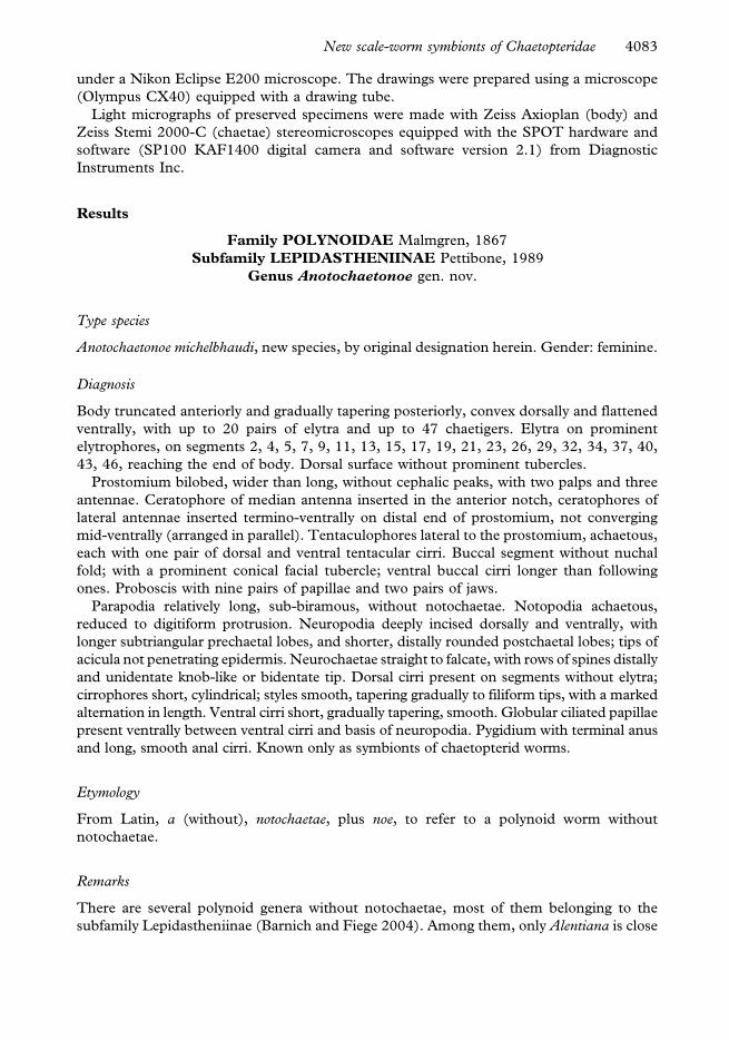
Results
Family POLYNOIDAE Malmgren, 1867
Subfamily LEPIDASTHENIINAE Pettibone, 1989
Genus Anotochaetonoe gen. nov.
Type species
Anotochaetonoe michelbhaudi, new species, by original designation herein. Gender: feminine.
Diagnosis
Body truncated anteriorly and gradually tapering posteriorly, convex dorsally and flattened
ventrally, with up to 20 pairs of elytra and up to 47 chaetigers. Elytra on prominent
elytrophores, on segments 2, 4, 5, 7, 9, 11, 13, 15, 17, 19, 21, 23, 26, 29, 32, 34, 37, 40,
43, 46, reaching the end of body. Dorsal surface without prominent tubercles.
Prostomium bilobed, wider than long, without cephalic peaks, with two palps and three
antennae. Ceratophore of median antenna inserted in the anterior notch, ceratophores of
lateral antennae inserted termino-ventrally on distal end of prostomium, not converging
mid-ventrally (arranged in parallel). Tentaculophores lateral to the prostomium, achaetous,
each with one pair of dorsal and ventral tentacular cirri. Buccal segment without nuchal
fold; with a prominent conical facial tubercle; ventral buccal cirri longer than following
ones. Proboscis with nine pairs of papillae and two pairs of jaws.
Parapodia relatively long, sub-biramous, without notochaetae. Notopodia achaetous,
reduced to digitiform protrusion. Neuropodia deeply incised dorsally and ventrally, with
longer subtriangular prechaetal lobes, and shorter, distally rounded postchaetal lobes; tips of
acicula not penetrating epidermis. Neurochaetae straight to falcate, with rows of spines distally
and unidentate knob-like or bidentate tip. Dorsal cirri present on segments without elytra;
cirrophores short, cylindrical; styles smooth, tapering gradually to filiform tips, with a marked
alternation in length. Ventral cirri short, gradually tapering, smooth. Globular ciliated papillae
present ventrally between ventral cirri and basis of neuropodia. Pygidium with terminal anus
and long, smooth anal cirri. Known only as symbionts of chaetopterid worms.
Etymology
From Latin, a (without), notochaetae, plus noe, to refer to a polynoid worm without
notochaetae.
Remarks
There are several polynoid genera without notochaetae, most of them belonging to the
subfamily Lepidastheniinae (Barnich and Fiege 2004). Among them, only Alentiana is close
New scale-worm symbionts of Chaetopteridae 4083

to Anotochaetonoe in having fewer than 50 segments. However, they differ in: (1) the
arrangement of elytra in posterior part of the body: on chaetigers 23, 26, 29, 32, 34, 37…
(Anotochaetonoe) or 23, 26, 29, 32, 34, 36, 37… (Alentiana); (2) in the shape of neuropodia:
long, with longer subtriangular prechaetal lobes and shorter, distally rounded postchaetal
lobes (Anotochaetonoe) or relatively short with both lobes rounded (Alentiana).
Anotochaetonoe resembles the long-bodied genus Telolepidasthenia in the arrangement of
elytra on segments 23, 26, 29, 32, 34, 37; in the presence of globular ciliated papillae on the
lower margin of neuropodia; and in their life habits as symbionts with chaetopterid worms.
However in the new genus, the number of chaetigers is fewer than 50, neuropodia have
longer, subtriangular prechaetal lobes and shorter, distally rounded postchaetal lobes, and
neurochaetae have unidentate and bidentate tips, while Telolepidasthenia has more than 60
chaetigers, neuropodia with both lobes rounded and neurochaetae with entire tips.
Anotochaetonoe michelbhaudi sp. nov.
(Figures 1–5)
Type locality
N’Kossa petrol field (5u16.529S, 11u33.879E) coast of the Republic of Congo.
Material examined
Holotype: inside the tube of Phyllochaetopterus sp.: Atlantic Ocean, coast of the Republic of
Congo, N’Kossa petrol field, st. 13(3), 5u16.529S, 11u33.879E, sandy silt, 180 m depth, 11
April 2003, one specimen (MNCN 16.01/10388). Paratype: inside the tube of
Spiochaetopterus sp.: Atlantic Ocean, coast of the Republic of Congo, Emeraude petrol field,
st. 1, 4u59.419S, 11u45.019E, 70 m depth, silt, 15 November 1999 (MNCN 16.01/10389).
Etymology
The species name michelbhaudi refers to the polychaete taxonomist, biologist, and ecologist
Dr Michel Bhaud from the Observatoire Oceanologique of Banyuls (CNRS, France) to
acknowledge his insightful contributions to the general knowledge of polychaetes,
especially chaetopterids, and to the present study in particular.
Description
Body truncate anteriorly, gradually tapering posteriorly, convex dorsally and flattened
ventrally, holotype with 20 pairs of elytra and 47 segments; paratype with 18 pairs of elytra
and 41 segments; holotype 14 mm long and 1.0 mm wide without parapodia, paratype
10.5 mm long and 0.3 mm wide without parapodia, both specimens 2 mm wide with
parapodia (Figure 1A).
Prostomium hexagonal, bilobed, wider than long, without cephalic peaks, median notch
a V-shaped gap between anterior ends of superior prostomial lobes, filled by median
ceratophore (Figures 1B, C, 2A). Two pairs of large dark brown eyes, ovate or round in
shape, the anterior pair larger than the posterior one; anterior eyes located immediately in
front of widest part of prostomium, positioned laterally and orientated antero-laterally;
4084 T. A. Britayev & D. Martin
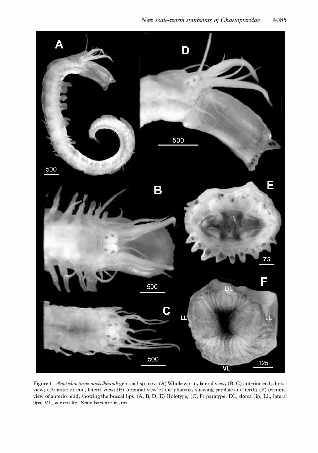
Figure 1. Anotochaetonoe michelbhaudi gen. and sp. nov. (A) Whole worm, lateral view; (B, C) anterior end, dorsal
view; (D) anterior end, lateral view; (E) terminal view of the pharynx, showing papillae and teeth; (F) terminal
view of anterior end, showing the buccal lips. (A, B, D, E) Holotype; (C, F) paratype. DL, dorsal lip; LL, lateral
lips; VL, ventral lip. Scale bars are in mm.
New scale-worm symbionts of Chaetopteridae 4085
posterior eyes smaller, at posterior edge of prostomium in its widest part, orientated dor-
sally; eyes of a pair (i.e. anterior or posterior) separated by one eye-diameter or less. Median
antenna as long as lateral ones (holotype) or 1.5 times longer (paratype), 2.5 (holotype) or
4.5 times longer than prostomium (paratype), inserted anteriorly in median notch, with
relatively long conical ceratophores. Lateral antennae inserted termino-ventrally on distal
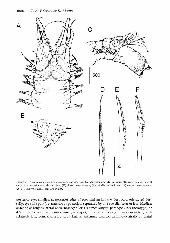
Figure 2. Anotochaetonoe michelbhaudi gen. and sp. nov. (A) Anterior end, dorsal view; (B) anterior end, lateral
view; (C) posterior end, dorsal view; (D) dorsal neurochaeta; (E) middle neurochaeta; (F) ventral neurochaeta.
(A–F) Holotype. Scale bars are in mm.
4086 T. A. Britayev & D. Martin

end of prostomium, slightly ventrally to median antenna, with relatively long cylindrical
ceratophores not converging mid-ventrally (i.e. parallel). All antennae gradually tapering,
smooth. Palps stout, conical, gradually tapering, 1.5 times longer than antennae and 4.5
times longer than prostomium in holotype, missing in paratype, smooth.
First segment not visible dorsally; tentaculophores long, achaetous, but with aciculae;
two pairs of tentacular cirri, very long, slightly exceeding in length the median antenna
(paratype) or 1.3 times longer (holotype); dorsal and ventral tentacular cirri similar in
length or dorsal ones slightly longer than ventral ones, smooth (Figures 1D, 2C). Buccal
Figure 3. Anotochaetonoe michelbhaudi gen. and sp. nov., elytron of paratype from unknown segment. (A) Entire
view; (B) detail of the structure of the dorsal surface showing the polygonal units of pigmentation; (C) detail of the
anterior edge. Scale bars are in mm.
New scale-worm symbionts of Chaetopteridae 4087
segment without nuchal fold; with prominent conical facial tubercle; mouth surrounded by
two lateral, cushion-shaped lips and lobular ventral and dorsal lips consisting of nine or ten
lobes, more prominent in median parts of the lips (Figure 1F); with subbiramous parapodia
and one pair of ventral cirri, shorter than ventral tentacular cirri and substantially longer
than ventral cirri of subsequent segments, gradually tapering, smooth; pharynx with nine
dorsal and nine ventral large terminal papillae and two pairs of brown jaws (Figure 1E).
Elytra up to 20 pairs on chaetigers 2, 4, 5, 7, alternating on segments to 23, 26, 29, 32,
34, 37, 40, 43, 46; present to posterior end. Most elytra still in place in both specimens,
relatively small, slightly overlapping antero-posteriorly, covering dorsum in anterior fourth
(Figure 2A) but with mid-dorsal area uncovered on most of the body. All elytra oval,
slightly folded along the external margin, delicate, smooth except for some scarce,
irregularly distributed micropapillae, transparent, with light brown pigmentation arranged
in polygonal units in anterior part of elytron (Figure 3A–C); intensity of pigmentation
decreasing along the body from anterior to posterior end.
Body without prominent dorsal tubercles. Dorsum of body segments without distinct
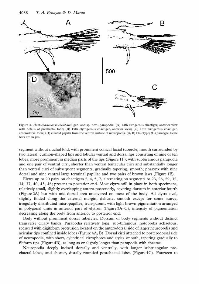
transverse ciliary bands. Parapodia relatively long, sub-biramous; notopodia achaetous,
reduced with digitiform protrusion located on the anterodorsal side of larger neuropodia and
acicular tips confined inside lobes (Figure 4A, B). Dorsal cirri attached to posterodorsal side
of neuropodia, with short, cylindrical cirrophores and styles smooth, tapering gradually to
filiform tips (Figure 4B), as long as or slightly longer than parapodia with chaetae.
Neuropodia deeply incised dorsally and ventrally, with longer subtriangular pre-
chaetal lobes, and shorter, distally rounded postchaetal lobes (Figure 4C). Fourteen to
Figure 4. Anotochaetonoe michelbhaudi gen. and sp. nov., parapodia. (A) 14th cirrigerous chaetiger, anterior view
with details of prechaetal lobe; (B) 15th elytrigerous chaetiger, anterior view; (C) 13th cirrigerous chaetiger,
anterodorsal view; (D) ciliated papilla from the ventral surface of neuropodia. (A, B) Holotype; (C) paratype. Scale
bars are in mm.
4088 T. A. Britayev & D. Martin
28 neurochaetae per bundle, increasing in number from posterior to anterior chaetigers;
dorsal-most neurochaetae long, straight, slender, with a long serrated section (16–22 rows
of spines), gradually tapering to nearly filiform entire, knob-like tip with a minute distal
spherical swelling; rows of spines largest, most conspicuous at the basis of the serrated
section, smaller and closer together distally (Figure 2D); middle neurochaetae with
relatively shorter serrated section, bidentate, with small secondary tooth and slightly curved
tips (Figure 2E); lower neurochaetae stouter, shorter, with a few rows of spines (10–12) and
slightly curved, clearly bidentate tips and secondary tooth slender and orientated in parallel
to the axis of the chaeta (Figure 2F).
Ventral cirri from the third chaetiger to the end of the body short, gradually taper-
ing, smooth (Figure 4A–C). A single row of two to four globular ciliated papillae
(Figure 4D) extends from the ventral cirri to the ventral basis of parapodia (Figure 4A,
B). Pygidium with dorso-terminal anus and two pygidial cirri, located below anus, rela-
tively long, comparable in length with dorsal cirri in the last segments, smooth
(Figure 2B).
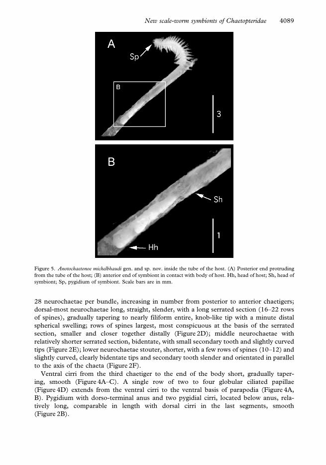
Figure 5. Anotochaetonoe michelbhaudi gen. and sp. nov. inside the tube of the host. (A) Posterior end protruding
from the tube of the host; (B) anterior end of symbiont in contact with body of host. Hh, head of host; Sh, head of
symbiont; Sp, pygidium of symbiont. Scale bars are in mm.
New scale-worm symbionts of Chaetopteridae 4089
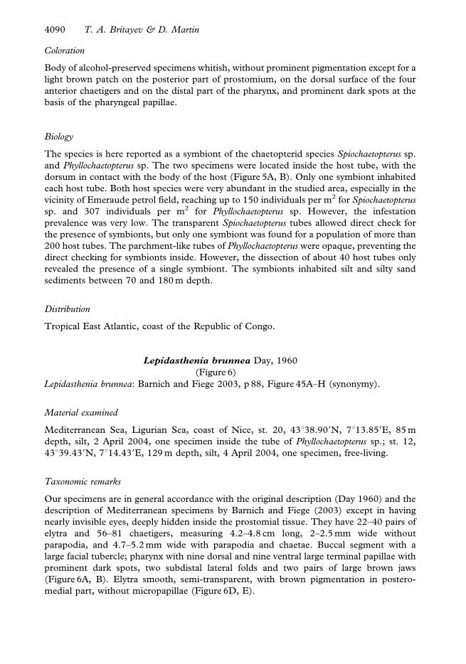
Coloration
Body of alcohol-preserved specimens whitish, without prominent pigmentation except for a
light brown patch on the posterior part of prostomium, on the dorsal surface of the four
anterior chaetigers and on the distal part of the pharynx, and prominent dark spots at the
basis of the pharyngeal papillae.
Biology
The species is here reported as a symbiont of the chaetopterid species Spiochaetopterus sp.
and Phyllochaetopterus sp. The two specimens were located inside the host tube, with the
dorsum in contact with the body of the host (Figure 5A, B). Only one symbiont inhabited
each host tube. Both host species were very abundant in the studied area, especially in the
vicinity of Emeraude petrol field, reaching up to 150 individuals per m2 for Spiochaetopterus
sp. and 307 individuals per m2 for Phyllochaetopterus sp. However, the infestation
prevalence was very low. The transparent Spiochaetopterus tubes allowed direct check for
the presence of symbionts, but only one symbiont was found for a population of more than
200 host tubes. The parchment-like tubes of Phyllochaetopterus were opaque, preventing the
direct checking for symbionts inside. However, the dissection of about 40 host tubes only
revealed the presence of a single symbiont. The symbionts inhabited silt and silty sand
sediments between 70 and 180 m depth.
Distribution
Tropical East Atlantic, coast of the Republic of Congo.
Lepidasthenia brunnea Day, 1960
(Figure 6)
Lepidasthenia brunnea: Barnich and Fiege 2003, p 88, Figure 45A–H (synonymy).
Material examined
Mediterranean Sea, Ligurian Sea, coast of Nice, st. 20, 43u38.909N, 7u13.859E, 85 m
depth, silt, 2 April 2004, one specimen inside the tube of Phyllochaetopterus sp.; st. 12,
43u39.439N, 7u14.439E, 129 m depth, silt, 4 April 2004, one specimen, free-living.
Taxonomic remarks
Our specimens are in general accordance with the original description (Day 1960) and the
description of Mediterranean specimens by Barnich and Fiege (2003) except in having
nearly invisible eyes, deeply hidden inside the prostomial tissue. They have 22–40 pairs of
elytra and 56–81 chaetigers, measuring 4.2–4.8 cm long, 2–2.5 mm wide without
parapodia, and 4.7–5.2 mm wide with parapodia and chaetae. Buccal segment with a
large facial tubercle; pharynx with nine dorsal and nine ventral large terminal papillae with
prominent dark spots, two subdistal lateral folds and two pairs of large brown jaws
(Figure 6A, B). Elytra smooth, semi-transparent, with brown pigmentation in postero-
medial part, without micropapillae (Figure 6D, E).
4090 T. A. Britayev & D. Martin
Biology
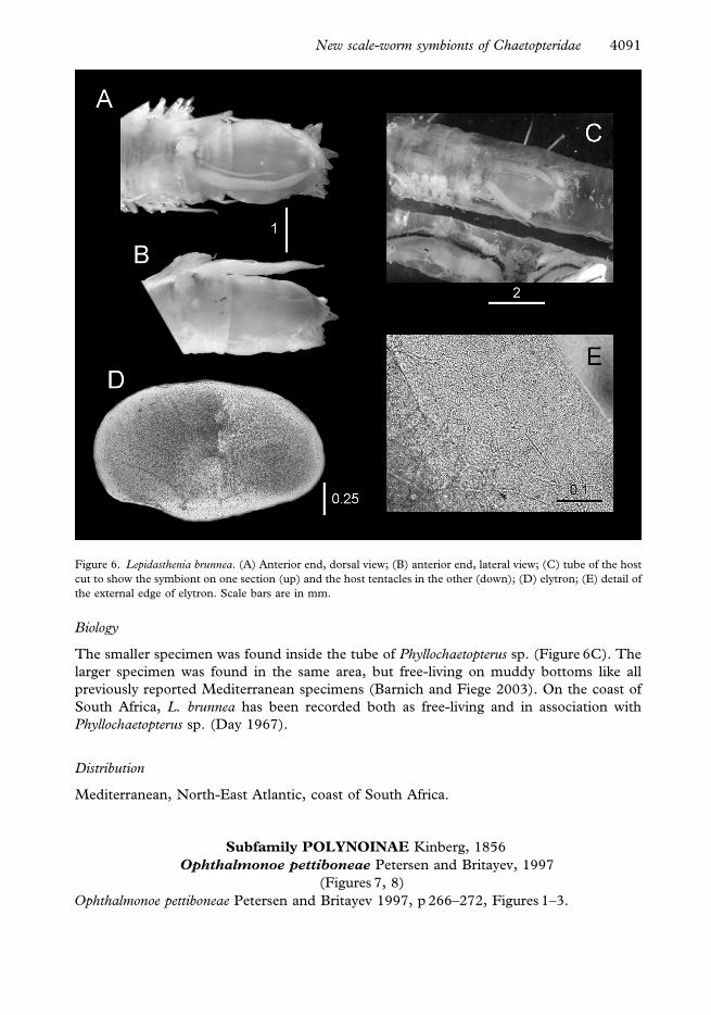
The smaller specimen was found inside the tube of Phyllochaetopterus sp. (Figure 6C). The
larger specimen was found in the same area, but free-living on muddy bottoms like all
previously reported Mediterranean specimens (Barnich and Fiege 2003). On the coast of
South Africa, L. brunnea has been recorded both as free-living and in association with
Phyllochaetopterus sp. (Day 1967).
Distribution
Mediterranean, North-East Atlantic, coast of South Africa.
Subfamily POLYNOINAE Kinberg, 1856
Ophthalmonoe pettiboneae Petersen and Britayev, 1997
(Figures 7, 8)
Ophthalmonoe pettiboneae Petersen and Britayev 1997, p 266–272, Figures 1–3.
Figure 6. Lepidasthenia brunnea. (A) Anterior end, dorsal view; (B) anterior end, lateral view; (C) tube of the host
cut to show the symbiont on one section (up) and the host tentacles in the other (down); (D) elytron; (E) detail of
the external edge of elytron. Scale bars are in mm.
New scale-worm symbionts of Chaetopteridae 4091
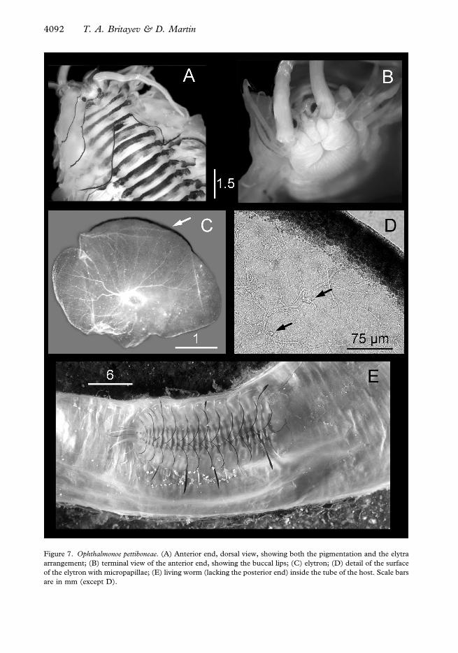
Figure 7. Ophthalmonoe pettiboneae. (A) Anterior end, dorsal view, showing both the pigmentation and the elytra
arrangement; (B) terminal view of the anterior end, showing the buccal lips; (C) elytron; (D) detail of the surface
of the elytron with micropapillae; (E) living worm (lacking the posterior end) inside the tube of the host. Scale bars
are in mm (except D).
4092 T. A. Britayev & D. Martin
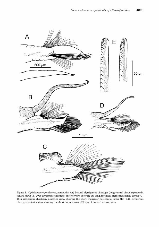
Figure 8. Ophthalmonoe pettiboneae, parapodia. (A) Second elytrigerous chaetiger (long ventral cirrus separated),
ventral view; (B) 20th cirrigerous chaetiger, anterior view showing the long, intensely pigmented dorsal cirrus; (C)
16th cirrigerous chaetiger, posterior view, showing the short triangular postchaetal lobe; (D) 40th cirrigerous
chaetiger, anterior view showing the short dorsal cirrus; (E) tips of hooded neurochaeta.
New scale-worm symbionts of Chaetopteridae 4093
Type locality
Banda Sea, Indonesia.
Material examined
South China Sea, Bay of Nhatrang, western point of Tre Island, 8–12 m, sandy silt, 29 June
2004, two specimens in the tubes of Chaetopterus sp., collected by I. Marin.
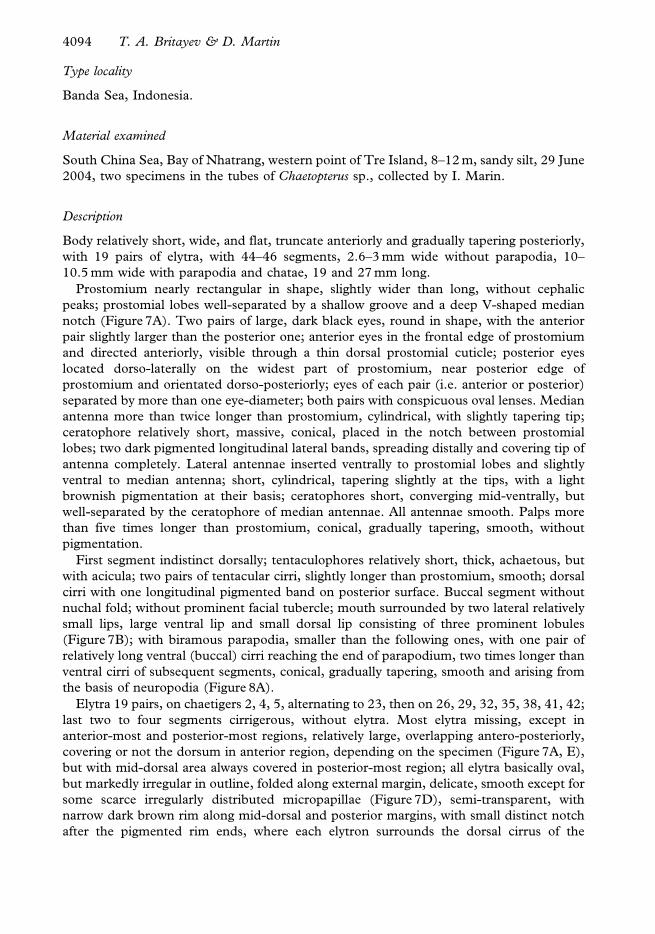
Description
Body relatively short, wide, and flat, truncate anteriorly and gradually tapering posteriorly,
with 19 pairs of elytra, with 44–46 segments, 2.6–3 mm wide without parapodia, 10–
10.5 mm wide with parapodia and chatae, 19 and 27 mm long.
Prostomium nearly rectangular in shape, slightly wider than long, without cephalic
peaks; prostomial lobes well-separated by a shallow groove and a deep V-shaped median
notch (Figure 7A). Two pairs of large, dark black eyes, round in shape, with the anterior
pair slightly larger than the posterior one; anterior eyes in the frontal edge of prostomium
and directed anteriorly, visible through a thin dorsal prostomial cuticle; posterior eyes
located dorso-laterally on the widest part of prostomium, near posterior edge of
prostomium and orientated dorso-posteriorly; eyes of each pair (i.e. anterior or posterior)
separated by more than one eye-diameter; both pairs with conspicuous oval lenses. Median
antenna more than twice longer than prostomium, cylindrical, with slightly tapering tip;
ceratophore relatively short, massive, conical, placed in the notch between prostomial
lobes; two dark pigmented longitudinal lateral bands, spreading distally and covering tip of
antenna completely. Lateral antennae inserted ventrally to prostomial lobes and slightly
ventral to median antenna; short, cylindrical, tapering slightly at the tips, with a light
brownish pigmentation at their basis; ceratophores short, converging mid-ventrally, but
well-separated by the ceratophore of median antennae. All antennae smooth. Palps more
than five times longer than prostomium, conical, gradually tapering, smooth, without
pigmentation.
First segment indistinct dorsally; tentaculophores relatively short, thick, achaetous, but
with acicula; two pairs of tentacular cirri, slightly longer than prostomium, smooth; dorsal
cirri with one longitudinal pigmented band on posterior surface. Buccal segment without
nuchal fold; without prominent facial tubercle; mouth surrounded by two lateral relatively
small lips, large ventral lip and small dorsal lip consisting of three prominent lobules
(Figure 7B); with biramous parapodia, smaller than the following ones, with one pair of
relatively long ventral (buccal) cirri reaching the end of parapodium, two times longer than
ventral cirri of subsequent segments, conical, gradually tapering, smooth and arising from
the basis of neuropodia (Figure 8A).
Elytra 19 pairs, on chaetigers 2, 4, 5, alternating to 23, then on 26, 29, 32, 35, 38, 41, 42;
last two to four segments cirrigerous, without elytra. Most elytra missing, except in
anterior-most and posterior-most regions, relatively large, overlapping antero-posteriorly,
covering or not the dorsum in anterior region, depending on the specimen (Figure 7A, E),
but with mid-dorsal area always covered in posterior-most region; all elytra basically oval,
but markedly irregular in outline, folded along external margin, delicate, smooth except for
some scarce irregularly distributed micropapillae (Figure 7D), semi-transparent, with
narrow dark brown rim along mid-dorsal and posterior margins, with small distinct notch
after the pigmented rim ends, where each elytron surrounds the dorsal cirrus of the
4094 T. A. Britayev & D. Martin
following segment (Figure 7C). Pigmentation intensity similar in all elytra along the body
(Figure 7E).
Body without prominent dorsal tubercles. Dorsal surface with one conspicuous
transverse dark-brown band across the middle of each segment, slightly wider mid-dorsally
in anterior segments (Figure 7A). Pigmented band on cirrigerous segments reaching basis
of cirrophores and continuing on posterior side of cirrophores and dorsal cirri (Figure 8B,
D). Dark band on elytragerous segments always in the middle, reaching top of
elytrophores. Segment 5 with oval, mid-dorsal black spot posterior to transverse band
(Figure 7A). Body non-pigmented ventrally. Transverse ciliary bands absent (at least
indistinct under stereomicroscope).
Parapodia relatively long, biramous; notopodia, small, conical, located at the antero-
dorsal side of neuropodia; dorsal cirri attached to postero-dorsal side of parapodia, with
relatively massive, cylindrical, short cirrophores (Figure 8B–D). Styles smooth, cylindrical,
tapering gradually to filiform tips (Figure 8B, D), alternating in length from similar (or
slightly shorter than) to longer than parapodia with chaetae. Dorsal cirri on cirrigerous
segments 3 and 6 short, on segment 8 long, on 10 short, then alternating long and short up
to the end of the body. Long cirri with dark longitudinal pigmented bands, covering
completely the cirri on their terminal fourth section; short cirri with less intense pigmented
bands, not reaching their tips.
Neuropodia longer and wider than notopodia, with row of cilia along ventral side.
Prechaetal neuropodial lobe elongated, nearly digitiform, substantially longer than the
triangular postchaetal lobe (Figure 8B, D). Distal region of postchaetal lobes with diffuse
brown pigmentation, slightly more intense dorsally.
Notochaetae arranged in a fan-shaped bundle orientated nearly horizontally, virtually all
of them on the same level, numerous, increasing in number from 70 (chaetiger 2) to more
than 150 (chaetiger 20), more slender than neurochaetae, fine, long, capillaries, smooth (at
least serration indistinct under 10006 with immersion oil).
Neurochaetae numerous, increasing in number from more than 80 (chaetiger 2) to more
than 240 (chaetiger 20), stout, nearly cylindrical, some chaetae slightly expanded in distal
hooded part; tips hooded, blunt and nearly rounded, with internal part slightly curved and
unidentate; with rows of narrow petaloid spines, smaller and closer together distally;
number of rows increasing from ventral-most (14–26) to dorsal-most notochaetae (up to
87) (Figure 8E).
Ventral cirri from the second chaetiger to the end of the body; relatively long at the
second chaetiger (Figure 8A); on subsequent segments, located on the first third of the
neuropodia, at the same level as notopodia, being twice or more shorter than prechaetal
lobes, conical, gradually tapering, smooth (Figure 8B–D); ventral cirri without coloration.
Anus dorso-terminal, two pygidial cirri, located below anus, slightly longer than last pair
of dorsal cirri.
Coloration
The worms preserved in alcohol have distinctive pigmentation on the dorsal surface. The
body is basically light brownish and has a conspicuous transverse dark brown or black band
crossing each segment. On segment 5, there is an additional characteristic oval, mid-dorsal,
black spot posterior to the transverse band (Figure 7A). The longest dorsal cirri have more
intense dark pigmentation than the smaller ones. The ventral body surface is always non-
pigmented. In living worms, the coloration is brighter and a transverse white band in front
New scale-worm symbionts of Chaetopteridae 4095
of the dorsal segmental dark brown band becomes visible in some chaetigers. A similar
white pigmentation is also conspicuous on dorsal cirri, antennae, and external margins of
elytra (Figure 7E).
Taxonomic remarks
The discovery of the Vietnamese specimens confirmed the number of elytra and seg-
ments reported for the holotype and contributed to the description of the species with
some important characteristics. In particular, the structure of the buccal lips, which was not
originally described since the pharynx of the holotype was fully everted, and the alter-
nation in length of the dorsal cirri. These features are useful additions to the specific
diagnosis. The new specimens agree in general with the holotype description, but it is
necessary to mention several minute differences. The prostomium of the holotype is
nearly hexagonal, instead of rectangular, as in the Vietnamese specimens. However,
the pharynx of the holotype is fully extended so this may have changed the shape of
the prostomium. According to Petersen and Britayev (1997) ‘‘neurochaetae … becom-
ing slightly broader at point where hood begins; … internal part of chaetae unidentate’’.
In the Vietnamese specimens, the distal part of most neurochaetae is cylindrical, with-
out any extension, and the internal part of the chaetal tips is unidentate and slightly
curved. Moreover, the rows of spines in notochaetae were indistinct even under a
magnification of 10006 (with immersion oil), as well as the transverse ciliary bands
described in the holotype (under binocular microscope). However, we suggest that these
slight differences probably reflect an intra-specific variation in the species morphology and/
or the quality of the optics employed. The pigmentation pattern of Vietnamese specimens
also agrees with that of the holotype and seems to be a remarkable species-specific
characteristic of O. pettiboneae, confirming the differences in pigmentation between O.
pettiboneae and O. tonkinika (Uschacov, 1992) as suggested by Petersen and Britayev
(1997).
Biology
The species lives symbiotically inside the tubes of a chaetopterid polychaete belonging to
the genus Chaetopterus (Figure 7E). Each studied chaetopterid tube contained one
symbiont. The two Vietnamese specimens are females and contained oocytes of 95–
130 mm (mean value 116 mm, n513), with large (about 55 mm in diameter) spherical
nucleus and small (about 11 mm in diameter) nucleolus. The spermatozoa of O. pettiboneae
belong to a primitive type, with rounded head of about 1.3 mm in diameter (Petersen and
Britayev 1997). Both features are usually linked to a broadcast reproductive strategy
(Thorson 1950; Rouse 1999).
The Vietnamese specimens of O. pettiboneae shared the host tube with two other
symbiotic species, an unidentified carapid fish and a porcellanid crab belonging to the
genus Pachycheles.
Distribution
Coasts of Ambon Island (Indonesia, Banda Sea) and Tre Island (Bay of Nhatrang, South
coast of Vietnam, South China Sea). Known only as symbiont of Chaetopterus sp., collected
from 8–12 m depth in silty sand sediments.
4096 T. A. Britayev & D. Martin
Discussion
A total of 19 species of polychaetes, including Anotochaetonoe chaetopterana, are currently
known in association with chaetopterids (see Martin and Britayev 1998 for a full list).
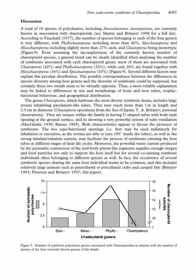
According to Fauchald (1977), the number of species belonging to each of the four genera
is very different, with Phyllochaetopterus including more than 40%, Spiochaetopterus and
Mesochaetopterus including slightly more than 27% each, and Chaetopterus being monotypic
(Figure 9). Even assuming the incompleteness of the currently known number of
chaetopterid species, a general trend can be clearly identified when analysing the number
of symbionts associated with each chaetopterid genus: most of them are associated with
Chaetopterus (42%) and Phyllochaetopterus (32%), while only 26% are found together with
Mesochaetopterus (16%) and Spiochaetopterus (10%) (Figure 9). Several different factors may
explain this peculiar distribution. The possible correspondence between the differences in
species diversity among host genera and the diversity of symbionts could be supposed, but
certainly these two trends seem to be virtually opposite. Thus, a more reliable explanation
may be linked to differences in size and morphology of hosts and host tubes, trophic-
functional behaviour, and geographical distribution.
The genus Chaetopterus, which harbours the most diverse symbiotic fauna, includes large
worms inhabiting parchment-like tubes. They may reach more than 1 m in length and
2.5 cm in diameter (Chaetopterus specimens from the Sea of Japan; T. A. Britayev, personal
observations). They are unique within the family in having U-shaped tubes with both ends
opening at the ground surface, and in showing a very powerful system of tube ventilation
(MacGinitie 1939; Barnes 1965). Both characteristics appear to favour the presence of
symbionts. The two equi-functional openings (i.e. they may be used indistinctly for
inhalation or excretion, as the worms are able to turn 180u inside the tubes), as well as the
strong inhalant/exhalant current, may facilitate the process of symbionts entering the host
tubes at different stages of their life cycles. Moreover, the powerful water current produced
by the peristaltic contraction of the mid-body piston-like segments supplies enough oxygen
and food particles not only to support the host itself but for several co-existing symbiont
individuals often belonging to different species as well. In fact, the occurrence of several
symbiotic species sharing the same host individual seems to be common, and this includes
relatively large animals such as pinnotherid or porcellanid crabs and carapid fish (Britayev
1993; Petersen and Britayev 1997; this paper).
Figure 9. Number of symbiotic polychaete species associated with Chaetopteridae in relation with the number of
species of the four currently known genera of the family.
New scale-worm symbionts of Chaetopteridae 4097
The other three genera of Chaetopteridae have trophic-functional behaviour similar to
Chaetopterus, although some of them may filtrate directly from the water column with the
help of tentacles instead of using mucus bags (e.g. Mesochaetopterus sp., D. Martin, personal
observations). Conversely, all of them are similar in having relatively uniform tube-building
structures, distinct from that of Chaetopterus (Barnes 1965; Sendall et al. 1995). In spite of
some differences in tube building material, all three genera have one tube end opening at
the ground level and the other more or less deep within the sediment, probably reflecting
different tube ventilation or pellet disposal procedures (Barnes 1965). Moreover, the tubes
tend to be relatively narrow, even for the largest known Mesochaetopterus species. For
instance, M. taylori measures more than 2 m long and less than 1 cm wide (MacGinitie and
MacGinitie 1968; Sendall et al. 1995) and Mesochaetopterus sp. may reach more than 2.5 m
long and about 0.7 cm wide (D. Martin, unpublished results). A normal width–length ratio
in Spiochaetopterus solotarius is 0.6:100 mm (Bhaud et al. 1994), while the width–length
ratio of Phyllochaetopterus sp. may range from 0.2:20 cm (Republic of Congo) to
0.6:.30 cm (France) as found by D. Martin.
The species of both Spiochaetopterus and Mesochaetopterus may be considered as
cosmopolitan, while Phyllochaetopterus tends to be widespread in subtropical and tropical
waters (M. Bhaud, personal communication). This distinction does not agree with the fact
that the latter also differs from the former two in harbouring more symbiotic species. On
the contrary, excluding Chaetopterus, there seems to be a clear relationship between intra-
generic host species diversity and symbiont diversity, both being higher in Phyllochaetopterus
and uniformly low for the other two genera (Figure 9).
According to our preliminary data, the diversity of symbiotic polychaete species
associated with each chaetopterid genus is related to: (1) the trophic-functional behaviour
and tube structure, Chaetopterus being distinct from the other three genera; and (2) the
intra-generic species diversity. However, explanation of the characteristics of the
Chaetopteridae as hosts of symbiotic polychaete is still not clear enough and thus, this
seems to be an interesting subject for further studies. Special attention should be addressed
to the increasing evidence that most chaetopterid species formerly reported as
cosmopolitan are, in fact, complexes of several species, each of them with restricted
biogeographical distributions. Studies of chaetopterid behaviour will also contribute but,
certainly, the most relevant approach would be to analyse the host–symbiont relationships
in vivo.
Acknowledgements
This study was supported by a mobility grant of the Spanish Ministry of Education and
Science (ref. no. SAB2003-0268), by the Federal Program ‘‘World Ocean. The studies of
World Ocean nature, the dynamic of ecosystems’’ of the Russian Ministry of Sciences and
Technologies and by the Russian Foundation for Basic Research (grant 05-04-48350). The
study has also been partly financed by a research contract between the CEAB (CSIC) and
CREOCEAN (France). We wish to thank I. N. Marin (A. N. Severtzov Institute of Ecology
and Evolution, Russia) who collected the specimens of O. pettiboneae and Dr E. Dutrieux
(CREOCEAN, France) who collected one of the specimens of the new genus (from
Emeraude, Republic of Congo) and the material from Nice. O. V. Savinkin is the author of
the pictures of the living O. pettiboneae inside the host tube and collected additional material
on Vietnamese chaetopterids. Drs R. Barnich and D. Fiege (Forschungsinstitut
Senckenberg, Germany) read a preliminary version of the manuscript and we highly
4098 T. A. Britayev & D. Martin
appreciate their thoughtful comments on scale-worm taxonomy and fruitful discussion of
the diagnosis of the new genus. We also wish to thank Dr M. Bhaud (Observatoire
Oceanologique de Banyuls, France), who helped us with the identification of the
Mediterranean and Atlantic chaetopterid hosts and whose vast knowledge on this family
allowed us to get a clear picture of their taxonomy, biology, and ecology.
References
Barnes RD. 1965. Tube-building and feeding in chaetopterid polychaetes. Biological Bulletin, Marine Biological
Laboratory, Woods Hole 129:217–233.
Barnich R, Fiege D. 2003. The Aproditoidea (Annelida: Polychaeta) of the Mediterranean Sea. Abhandlungen der
Senckenbergischen Naturforschenden Gesellschaft 559:1–167.
Barnich R, Fiege D. 2004. Revision of the genus Lepidastheniella Monro, 1924 (Polychaeta: Polynoidae:
Lepidastheniinae) with notes on the subfamily Lepidastheniinae and the description of a new species. Journal
of Natural History 38:863–876.
Bhaud M, Lastra MC, Petersen ME. 1994. Redescription of Spiochaetopterus solitarius (Rioja, 1917), with notes on
tube structure and comments on the generic status (Polychaeta: Chaetopteridae). Ophelia 40:115–133.
Britayev TA. 1993. Pilargis berkeleyae (Polychaeta, Pilargidae) as a commensal of a sedentary polychaete
Chaetopterus cautus (Chaetopteridae). Zoologicheskii Zhurnal 72:147–151. (Rus).
Day JH. 1960. The polychaete fauna of South Africa. Part 5. Errant species dredged off Cape coasts. Annals of the
South African Museum 45:261–373.
Day JH. 1967. A monograph on the polychaetes of Southern Africa. Part 1, Errantia. London: Trustees of the
British Museum (Natural History). 656 p.
Fauchald K. 1977. Polychaete worms, definitions and keys to the orders, families and genera. Natural History
Museum of Los Angeles County Science Service 28:1–190.
MacGinitie GE. 1939. The method of feeding of Chaetopterus. Biological Bulletin, Marine Biological Laboratory,
Woods Hole 77:115–118.
MacGinitie GE, MacGinitie N. 1968. Natural history of marine animals. New York: McGraw-Hill Book Co.
Martin D, Britayev TA. 1998. Symbiotic polychaetes: review of known species. Oceanography and Marine
Biology: An Annual Review 36:217–340.
Petersen ME, Britayev TA. 1997. A new genus and species of polynoid scaleworm commensal with Chaetopterus
appendiculatus Grube from the Banda Sea (Annelida: Polychaeta), with a review of commensals of
Chaetopteridae. Bulletin of Marine Science 60:261–276.
Rouse GW. 1999. Polychaete sperm: phylogenetic and functional considerations. Hydrobiologia 402:215–224.
Sendall KA, Fontaine AR, O’Foighil D. 1995. Tube morphology and activity patterns related to feeding and tube-
building in the polychaete Mesochaetopterus taylori Potts. Canadian Journal of Zoology 73:509–517.
Thorson G. 1950. Reproductive and larval ecology of marine bottom invertebrates. Biological Reviews 25:1–45.
New scale-worm symbionts of Chaetopteridae 4099
Copyright © 2022 FDOKUMEN