Peripheral nerve-derived HIV1 is predominantly CCR5-dependent and causes neuronal degeneration and...
16
Peripheral nerve-derived HIV-1 is predominantly CCR5-dependent and causes neuronal degeneration and neuroinflammation Gareth Jones a , Yu Zhu a , Claudia Silva a , Shigeki Tsutsui a , Carlos A. Pardo b , Oliver T. Keppler c , Justin C. McArthur b , Christopher Power a, T a University of Calgary, Calgary, Canada b Johns Hopkins University, School of Medicine, Baltimore, MD 21205-2196, USA c Department of Virology, University of Heidelberg, Heidelberg, Germany Received 20 September 2004; returned to author for revision 29 October 2004; accepted 17 January 2005 Abstract HIV-related peripheral neuropathy is a major neurological complication of HIV infection, although little is known about its pathogenesis. We amplified HIV-1 C2V3 envelope sequences from peroneal nerves obtained from HIV/AIDS patients. Sequence analysis and infectious recombinant viruses containing peripheral nerve-derived C2V3 sequences indicated a predominance of CCR5-dependent and macrophage- tropic HIV-1, although dual tropic viruses using both CCR5 and CXCR4 were identified. The neuropathogenic effects of recombinant HIV-1 clones were investigated using a novel dorsal root ganglion culture system that was comprised of sensory neurons, macrophages and Schwann cells from transgenic rats expressing human CD4 and CCR5 on monocytoid cells. Despite restricted viral replication, HIV-1 infection caused a reduction in the percentage of neurons with neuritic processes together with significant neurite retraction, which was accompanied by induction of IL-1h and TNF-a expression, depending on the individual virus. Our results suggest that HIV-1 infection of the peripheral nervous system causes axonal degeneration, possibly through the induction of pro-inflammatory cytokines. D 2005 Elsevier Inc. All rights reserved. Keywords: HIV-1; Co-receptor; Tropism; Peripheral nerve; Neuropathy; Dorsal root ganglion; Neuroinflammation Introduction Peripheral neuropathy has become the chief neurological syndrome observed among persons infected with human immunodeficiency virus type-1 (HIV-1) in the developed world (Brinley et al., 2001). Of these peripheral neuro- pathies, HIV-associated sensory neuropathies (HIV-SN) are the most common forms recognized, affecting approxi- mately 30% of both adults and children with acquired immunodeficiency syndrome (AIDS) (Araujo et al., 2000; Keswani et al., 2002; Tagliati et al., 1999). Furthermore, Morgello et al. (2004) have recently demonstrated HIV-SN prevalence rates of up to 52%, including patients who receive highly active antiretroviral therapy (HAART). HIV- SN includes both ddistal sensory polyneuropathyT (DSP) and dantiretroviral toxic neuropathyT (ATN), conditions which frequently overlap (Keswani et al., 2002). HIV-SN is predominately defined by disabling pain with parathesiae, gait instability and autonomic dysfunction (reviewed in Pardo et al., 2001). In addition, sub-clinical nerve damage may also occur in HIV-1-infected patients in the absence of symptoms of HIV-SN (Gastaut et al., 1989; Gulevich et al., 1992; Tagliati et al., 1999). The principal pathological feature of HIV-SN is defined by distal degeneration of long axons in a ddying backT pattern (Pardo et al., 2001). Prominent loss of the small, unmyelinated nociceptive sensory neurons occurs although reduced density of small and large myelinated axonal fibers has also been observed (Araujo et al., 2000; Griffin and 0042-6822/$ - see front matter D 2005 Elsevier Inc. All rights reserved. doi:10.1016/j.virol.2005.01.027 T Corresponding author. Department of Clinical Neurosciences, Neuro- science Research Group, HMRB 150, 3330 Hospital Drive NW, Calgary AB, Canada T2N 4N1. Fax: +1 403 283 8731. E-mail address: [email protected] (C. Power). Virology 334 (2005) 178 – 193 www.elsevier.com/locate/yviro
-
Upload
independent -
Category
Documents
-
view
0 -
download
0
Transcript of Peripheral nerve-derived HIV1 is predominantly CCR5-dependent and causes neuronal degeneration and...

www.elsevier.com/locate/yviro
Virology 334 (20
Peripheral nerve-derived HIV-1 is predominantly CCR5-dependent and
causes neuronal degeneration and neuroinflammation
Gareth Jonesa, Yu Zhua, Claudia Silvaa, Shigeki Tsutsuia, Carlos A. Pardob, Oliver T. Kepplerc,
Justin C. McArthurb, Christopher Powera,TaUniversity of Calgary, Calgary, Canada
bJohns Hopkins University, School of Medicine, Baltimore, MD 21205-2196, USAcDepartment of Virology, University of Heidelberg, Heidelberg, Germany
Received 20 September 2004; returned to author for revision 29 October 2004; accepted 17 January 2005
Abstract
HIV-related peripheral neuropathy is a major neurological complication of HIV infection, although little is known about its pathogenesis.
We amplified HIV-1 C2V3 envelope sequences from peroneal nerves obtained from HIV/AIDS patients. Sequence analysis and infectious
recombinant viruses containing peripheral nerve-derived C2V3 sequences indicated a predominance of CCR5-dependent and macrophage-
tropic HIV-1, although dual tropic viruses using both CCR5 and CXCR4 were identified. The neuropathogenic effects of recombinant HIV-1
clones were investigated using a novel dorsal root ganglion culture system that was comprised of sensory neurons, macrophages and
Schwann cells from transgenic rats expressing human CD4 and CCR5 on monocytoid cells. Despite restricted viral replication, HIV-1
infection caused a reduction in the percentage of neurons with neuritic processes together with significant neurite retraction, which was
accompanied by induction of IL-1h and TNF-a expression, depending on the individual virus. Our results suggest that HIV-1 infection of the
peripheral nervous system causes axonal degeneration, possibly through the induction of pro-inflammatory cytokines.
D 2005 Elsevier Inc. All rights reserved.
Keywords: HIV-1; Co-receptor; Tropism; Peripheral nerve; Neuropathy; Dorsal root ganglion; Neuroinflammation
Introduction
Peripheral neuropathy has become the chief neurological
syndrome observed among persons infected with human
immunodeficiency virus type-1 (HIV-1) in the developed
world (Brinley et al., 2001). Of these peripheral neuro-
pathies, HIV-associated sensory neuropathies (HIV-SN) are
the most common forms recognized, affecting approxi-
mately 30% of both adults and children with acquired
immunodeficiency syndrome (AIDS) (Araujo et al., 2000;
Keswani et al., 2002; Tagliati et al., 1999). Furthermore,
Morgello et al. (2004) have recently demonstrated HIV-SN
0042-6822/$ - see front matter D 2005 Elsevier Inc. All rights reserved.
doi:10.1016/j.virol.2005.01.027
T Corresponding author. Department of Clinical Neurosciences, Neuro-
science Research Group, HMRB 150, 3330 Hospital Drive NW, Calgary
AB, Canada T2N 4N1. Fax: +1 403 283 8731.
E-mail address: [email protected] (C. Power).
prevalence rates of up to 52%, including patients who
receive highly active antiretroviral therapy (HAART). HIV-
SN includes both ddistal sensory polyneuropathyT (DSP) anddantiretroviral toxic neuropathyT (ATN), conditions which
frequently overlap (Keswani et al., 2002). HIV-SN is
predominately defined by disabling pain with parathesiae,
gait instability and autonomic dysfunction (reviewed in
Pardo et al., 2001). In addition, sub-clinical nerve damage
may also occur in HIV-1-infected patients in the absence of
symptoms of HIV-SN (Gastaut et al., 1989; Gulevich et al.,
1992; Tagliati et al., 1999).
The principal pathological feature of HIV-SN is defined
by distal degeneration of long axons in a ddying backTpattern (Pardo et al., 2001). Prominent loss of the small,
unmyelinated nociceptive sensory neurons occurs although
reduced density of small and large myelinated axonal fibers
has also been observed (Araujo et al., 2000; Griffin and
05) 178–193

G. Jones et al. / Virology 334 (2005) 178–193 179
McArthur, 1998). Demyelination, if present, is not segmen-
tal and may be the result of axonal degeneration. The precise
mechanism(s) underlying the neuropathogenesis of HIV-SN
remains unclear. Both the dindirectT neurotoxicity of
products secreted within the nerve by activated macro-
phages and the ddirectT effect of viral proteins on dorsal root
ganglion (DRG) neurons have been suggested. In support of
the former, prominent infiltration of peripheral nerve by
both inflammatory macrophages and T-cells has been
demonstrated in HIV/AIDS patients compared to uninfected
controls (de la Monte et al., 1988; Pardo et al., 2001;
Rizzuto et al., 1995). Furthermore, in one study, the severity
of peripheral neuropathy was correlated with the degree of
inflammatory cell infiltration (Griffin and McArthur, 1998).
Lymphocyte and macrophage infiltrates are also seen within
the DRG of AIDS patients, with concomitant presence of
pro-inflammatory cytokines including tumor necrosis factor-
a (TNF-a), interferon-g (IFN-g), interleukin-1 (IL-1) and
interleukin-6 (IL-6) (Nagano et al., 1996; Rizzuto et al.,
1995; Shapshak et al., 1995; Yoshioka et al., 1994).
Several groups have demonstrated the presence of HIV-1
transcripts and proteins in peripheral nerves and DRG
(Brannagan et al., 1997; Cornford et al., 1992; Gherardi et
al., 1989; Grafe and Wiley, 1989; Nagano et al., 1996;
Rizzuto et al., 1995; Vital et al., 1992; Yoshioka et al.,
1994). However, it is generally held that HIV-1 replication
in peripheral nerves is sparse, and is restricted to the
macrophage cell population. While successful isolation of
whole virus from ex vivo peripheral nerve tissue of HIV/
AIDS patients has been reported (Chaunu et al., 1989;
Cornblath et al., 1987; de la Monte et al., 1988; Ho et al.,
1985), little is known about the nature of the virus isolated
from peripheral nerves and whether it differs between HIV/
AIDS patients with or without HIV-SN. In this study, we
have characterized the co-receptor usage and cell tropism of
HIV-1 derived from peroneal nerve tissue samples from
HIV/AIDS patients both with and without HIV-SN.
Recombinant HIV-1 expressing envelope sequences from
peripheral nerve-derived virus were constructed, and their
potential neurotoxic effects were investigated in vitro using
a novel DRG mixed cell culture system from rats transgenic
for human CD4 and human CCR5 (Keppler et al., 2002).
Lastly, the induction of host immune responses following
exposure to nerve-derived HIV-1 in both the in vitro cultures
and in the peroneal nerve tissue samples was investigated by
real time RT-PCR analysis.
Results
Detection of HIV-1 genomes in peripheral nerve
Although HIV-1 antigens have been detected in peripheral
nerve previously (Rizzuto et al., 1995), we investigated the
presence of HIV-1 provirus in peripheral nerve tissue from
twelve HIV/AIDS patients. Successful extraction of genomic
DNA from all 12 peroneal nerve samples was demonstrated
by PCR amplification of the GAPDH gene (data not shown).
HIV-1-specific amplicons corresponding to a 445-base pair
fragment of the proviral C2V3 region of the env gene were
detected in seven (HIV-SN, n = 4; HIV-NSN, n = 3) of 12
patient samples (58%), using established protocols (Chesebro
et al., 1992; Power et al., 1994; Van Marle et al., 2002).
Having demonstrated the presence of HIV-1 provirus in
peripheral nerves, we next investigated whether the nerve
was a site of viral transcription. Total RNA was extracted
from 10 out of the 12 nerve samples, and was used as a
template for cDNA synthesis. The generation of cDNA was
confirmed by PCR amplification of the GAPDH gene (data
not shown). In contrast to the HIV-1 provirus analysis, HIV-1
C2V3 sequences derived from RNA were detected only in
one of ten nerve samples (patient 417, with HIV-SN). The
detection of HIV-1 provirus in over half of the samples
studied demonstrates the presence of HIV-1-infected cells
within the peripheral nerve of HIV/AIDS patients both with
and without sensory neuropathy. However, the low level
detection of HIV-1 RNA suggested that there was limited
viral replication at this site.
Analysis of HIV-1 env sequences
Studies of HIV-1 infection of the central nervous system
have demonstrated that polymorphisms within the HIV-1
env gene distinguish patients with and without HIV-1
associated dementia (Kuiken et al., 1995; Power et al.,
1994). Since HIV-1 provirus was detected in the peripheral
nerve of patients both with and without HIV-SN, we
sequenced the amplified C2V3 region of the HIV-1 env
gene and investigated the phylogenetic relationships of virus
sequences from patients with and without sensory neuro-
pathy (Fig. 1A). As the nerve-derived C2V3 sequences were
obtained from clade B infected HIV/AIDS patients, we
chose to root the neighbor-joining tree against the distantly
related HIV-1 clade D strain NDK, permitting comparisons
to be made between the virus sequences obtained from
patients with and without sensory neuropathy. Although
these sequences were obtained from a single clone per
patient, they were identical to that obtained by direct
sequencing of the C2V3 PCR amplicon. To assess the
molecular diversity within the nerve from individual
patients, we sequenced a minimum of three extra clones
per patient in individual PCR reactions. In three individuals,
the sequence of the env amplicon in 2 out of 3 clones was
identical to that obtained by direct sequencing, while
the third clone differed by only one amino acid (99%
homology). For the remaining individuals, all clones
demonstrated identical sequences to that obtained by direct
sequencing (data not shown), suggesting limited molecular
diversity within the C2V3 env region from the nerve of
individual patients. The macrophage-tropic and CCR5-
dependent brain-derived JRFL and SF162 strains were
included in the analysis as control viruses isolated from

Fig. 1. Analysis of HIV-1 envelope sequences of peripheral nerve-C2V3 recombinant HIV-1 clones. (A) Phylogenetic neighbor-joining tree (using the Kimura
2-parameter model) based the C2V3 sequences obtained from peripheral nerve and plasma of HIV/AIDS patients. The tree is rooted against the C2V3 sequence
of the HIV-1 clade D strain NDK, and bootstrap values greater than 70 are indicated (bootstrap analysis of 1000 replicates). Bold type indicates patients with
HIV-SN. (B) V3 loop amino acid sequences of HIV-1 derived from the nerve tissue or plasma of HIV/AIDS patients aligned relative to the HIV-1 NL4-3 strain.
Predicted co-receptor usage was analyzed as described in Materials and methods.
G. Jones et al. / Virology 334 (2005) 178–193180
the nervous system, while NL4-3 was included as a
prototypic CXCR4-dependent isolate. The C2V3 nucleotide
sequences from the three patients without sensory neuro-
pathy (495NSN-nerve, 402NSN-nerve and 403NSN-nerve)
were more closely related to sequences from patients with
neuropathy than they were to each other, indicating that there
was no clustering of the C2V3 region on the basis of clinical
diagnosis of HIV-SN. Similar results were obtained when the
inferred amino acid sequences were analyzed (data not
shown). HIV-1 proviral (417SN-nerve) and viral (417cSN-
nerve) C2V3 sequences were similar for patient 417, with
the only difference being a four amino acid deletion in the
C2 region for 417SN-nerve (data not shown).
For patients 491 and 416, viral RNA was also extracted
from plasma samples obtained 3 and 8 months prior to the
autopsy. Sequence analysis revealed a 99% homology
between 491SN-nerve and 491SN-plasma samples (sequen-
ces differed by 2 out of 141 amino acids) and a 95%
sequence homology between 416SN-nerve and 416SN-
plasma (sequences differed by 7 out of 144 amino acids).
Having shown the presence of HIV-1 provirus in the
peripheral nerve of HIV/AIDS patients, we then examined

G. Jones et al. / Virology 334 (2005) 178–193 181
the potential co-receptor usage of these viruses. HIV-1 co-
receptor usage was initially analyzed by evaluating the
charge and amino acid composition of the V3 loop from the
amplified C2V3 region (Fig. 1B), using methods previously
described (Briggs et al., 2000). The V3 loop sequences of
two known macrophage-tropic and CCR5-dependent iso-
lates (JRFL and SF162) were included. Using this approach,
the predicted co-receptor usage for these isolates matched
their known phenotypes. Of the seven peripheral-nerve
derived HIV-1 sequences, five (365SN, 416SN, 402NSN,
403NSN and 495NSN) were predicted to be CCR5-depend-
ent, while two (417SN and 491SN) were predicted to use
both CCR5 and CXCR4 as a co-receptor. Where matched
plasma samples were available, similar virus phenotypes
were predicted. In addition, the V3 loop sequences of nerve-
derived HIV-1 provirus and HIV-1 virus for patient 417
Fig. 2. Both CCR5-dependent and CXCR4-dependent HIV-1 are present in the p
HeLa-CD4/CXCR4/CCR5 or (B) GHOST/CXCR4 and GHOST/CCR5 cells were
firefly luciferase gene. As controls, target cells were also infected with supernatan
pNL-Luc-E�R� alone (pLu). Results are presented as the mean relative light uni
(417SN and 417cSN, respectively) were identical and
predicted dual tropism for CCR5 and CXCR4.
Investigation of cell tropism and co-receptor usage
To investigate HIV-1 co-receptor usage, pseudotyped
viruses that contained the firefly luciferase gene and
expressed the nerve-derived C2V3 region were constructed
from three predicted CCR5-using strains (NL-365, NL-402
and NL-416) and one predicted dual tropic strain (NL-
417c). NL4-3 and NL-J1 were included as controls for HIV-
1 isolates that utilize either CXCR4 or CCR5, respectively.
NL-J1 is a recombinant strain that expresses the envelope
region of the HIV-1 JRFL strain in a NL4-3 backbone
(Zhang et al., 2003). All three predicted CCR5-using viruses
infected HeLa-CD4 cells expressing CCR5 (Fig. 2A, open
eripheral nerve tissue of HIV/AIDS patients. (A) HeLa-CD4/CXCR4 and
infected with pseudotyped HIV-1 expressing the nerve-C2V3 region and the
ts from non-transfected 293-T cells (mock) and 293-T cells transfected with
ts (RLU) and standard deviation of triplicate samples.

G. Jones et al. / Virology 334 (2005) 178–193182
bars), however, no infection was observed in HeLa-CD4
cells that expressed only CXCR4 (closed bars), clearly
demonstrating their dependence on CCR5 for infection. In
contrast, NL-417c-infected HeLa-CD4 cells expressing
CXCR4 alone or expressing both CXCR4 and CCR5.
These results confirmed that the biological phenotype for
co-receptor usage of peripheral nerve-derived HIV-1
matched that predicted by sequence analysis of the V3 loop
(Fig. 1B). However, as the two HeLa-CD4 cell lines both
constitutively expressed CXCR4, it was not possible to
determine whether NL-417c used only CXCR4 or in fact
was dual tropic (for both CXCR4 and CCR5). To address
this question, we tested the ability of these pseudotyped
viruses to infect the GHOST cell lines expressing either
CXCR4 or CCR5 alone (Fig. 2B). As expected, NL4-3 and
NL-J1 were shown to infect GHOST cells expressing either
CXCR4 or CCR5 respectively. Again, NL-365 and NL-416
only infected cells that expressed CCR5. In contrast, NL-
417c infected both GHOST/CXCR4 and GHOST/CCR5
cells, suggesting that as predicted by sequence analysis this
virus was dual tropic for either CXCR4 or CCR5.
Although CXCR4 and CCR5 are the major co-receptors
for HIV-1, other chemokine receptors have also been
demonstrated to mediate HIV-1 infection (Berger et al.,
1999). Therefore, we extended the co-receptor usage study
to include a cell line expressing CCR3. In addition to
expressing distinct chemokine receptors, the GHOST cell
lines are also stably transfected with a construct expressing
the humanized green fluorescent protein (hGFP) under the
control of the HIV-2 LTR. Therefore, HIV-1 infection of
these cells can be detected via induction of the hGFP gene.
Recombinant HIV-1 strains expressing the nerve-derived
C2V3 region in the NL4-3 backbone (herein to be known as
nerve-C2V3 recombinant HIV-1) were generated from four
HIV/AIDS patients (#365, #402, #416, #417c) and these
recombinant HIV-1 clones were shown to be replication-
competent in activated human PBMC (Fig. 3A). Mock
infection of the GHOST cell lines did not result in hGFP
expression, while infection with NL-J1 induced hGFP
expression in the GHOST/CCR5 cell line only (Fig. 3B).
Similarly, infection with the NL-365, NL-402 and NL-416
strains resulted in hGFP expression in only the GHOST/
CCR5 cells (data not shown). Infection of GHOST/CXCR4
cells, and to a lesser extent GHOST/CCR2b, was seen with
both NL4-3 and NL-417c strains, whereas the latter also
induced hGFP expression in GHOST/CCR5 cells. In
summary, all four recombinant HIV-1 strains expressing
the C2V3 region derived from peripheral nerve utilized
CCR5 for infection. For three of these viruses (NL-365, NL-
402 and NL-416), CCR5 appeared to be the primary co-
receptor used. However, NL-417c was also able to utilize
CXCR4 and, to a lesser extent, CCR2b (Fig. 3C).
To investigate the cell tropism of nerve-derived HIV-1,
human MDM cultures were exposed to recombinant
infectious HIV-1 clones expressing the nerve-derived
C2V3. NL-365, NL-402 and NL-416 demonstrated sus-
tained viral replication in human MDM (Fig. 3A). In
contrast, NL-417c failed to replicate in human MDM, with
culture supernatant HIV-1 p24 values similar to that of the
background (mock treated controls). Surprisingly, NL4-3
also replicated in MDM cultures at early time points,
although by day 12 post-infection HIV-1 p24 values in
culture supernatant were also approaching background
levels. Furthermore, NL4-3 failed to replicate in MDM
from two other individuals (data not shown), suggesting that
the replication observed in Fig. 3A was donor-specific.
Nonetheless, NL4-3 is generally considered to be a CXCR4-
dependent T-tropic isolate, although its replication in
macrophages has also been previously documented (Valen-
tin et al., 2000).
Infection of DRG cultures from human CD4 and CCR5
transgenic rats
The in vitro DRG cultures consisted of a mixed
population of cells containing neurons (MAP-2+, Fig.
4A), macrophages (ED-1+, Fig. 4D) and Schwann cells
(GFAP+, Fig. 4G). MAP-2 staining of neurons clearly
labeled not only the cell body, but also the neuritic
process (Fig. 4A, arrowed). Human CCR5 expression in
the DRG cultures was restricted to macrophages, as
evidenced by co-localization of CCR5 with Iba-1-express-
ing cells (Figs. 4E and F, arrowed cells and inset).
Similarly, human CD4 expression in the DRG cultures
was also restricted to macrophages, confirmed by co-
localization of CD4 with Iba-1-expressing cells (Figs. 4H
and I, arrowed cells). DRG cultures were infected with
the nerve-C2V3 recombinant HIV-1 clones (NL-365, NL-
402, NL-416 and NL-417c) or the brain-derived SF162
isolate. NL-J1 and NL4-3 were also included as controls
for CCR5-using and CXCR4-using viruses respectively.
Viral nucleocapsid protein (p24) expression could be
detected in macrophages (Figs. 4B and C, arrowed cells
and inset (representative image from SF162-infected
cultures)), but p24 was not detected in the supernatants
from infected cultures (data not shown). The presence of
HIV-1 RNA and provirus at day 1 and day 4 post-
infection was assessed using the nested PCR approach
described earlier. Viral RNA corresponding to the C2V3
region of the env gene was detected in cultures 1-day
post-infection, irrespective of whether the virus used
CCR5 or CXCR4 as a co-receptor (Fig. 4J). For all
clones used, HIV-1 provirus was detected at day 4 post-
infection, suggesting the persistence of virus-infected cells
in the DRG cultures. However, at this time point, viral
RNA was undetectable. The inability to detect viral RNA
at day 4 post-infection was likely not due to inadequate
RNA extraction and cDNA preparation, as the house
keeping gene h-actin could be amplified from the same
samples. In addition, viral RNA specific for the HIV-1
gene nef was also undetectable at day 4 post-infection
(data not shown). Taken together, these results demon-

Fig. 3. Cell tropism and co-receptor usage of nerve-C2V3 recombinant HIV-1. (A) Supernatants from 293-T cells transfected with different HIV-1 molecular
clones expressing nerve-C2V3 HIV-1 were used to infect activated human PBMC. NL4-3 and NL-J1 (a recombinant strain that expresses the envelope region
of the JRFL strain in a NL4-3 backbone) were included as controls for CXCR4-dependent or CCR5-dependent strains, respectively. Human MDM were
infected with PBMC-expanded recombinant HIV-1 (virus input titre of 5 ng p24 protein per culture). As a control, MDM were infected with culture
supernatants from activated PBMC that had not been exposed to HIV-1 (mock). Virus protein levels in culture supernatants were quantified using the HIV-1
p24 Antigen Capture Assay Kit at the indicated time points. Values are the mean of duplicate wells. (B) GHOST cell lines were infected with the recombinant
HIV-1 strains as described in Materials and methods. Infection of the different cell lines was evaluated by the induction of green fluorescent protein expression
3 days post-infection. (C) Summary of the co-receptor usage of the different nerve-C2V3 recombinant HIV-1 as indicated by infection of the GHOST cell lines.
(Original magnification �200).
G. Jones et al. / Virology 334 (2005) 178–193 183
strate that both CXCR4-using and CCR5-using HIV-1 are
capable of infecting the mixed cell DRG cultures.
However, the detection of viral RNA at day 1 post-
infection but not at day 4 suggests that there is limited
viral replication within these cultures.
Morphological effects of HIV-1 infection on DRG neurons
To assess the DRG culture system as a potential model
for evaluating neuropathogenic effects of HIV-1 infection in
the peripheral nervous system, a series of morphological

Fig. 4. DRG cultures from rats transgenic for human CD4 and CCR5 harbor HIV-1 provirus after infection with different HIV-1 strains. DRG cultures consist
of (A) neurons (MAP-2+) with neurite processes (arrowed), (D) macrophages (ED-1+) and (G) Schwann cells (GFAP+). (B and C) Following HIV-1 infection,
HIV-1 protein was detected in ED-1-expressing macrophages (representative images from SF162-infected cultures). Inset represents merged HIV-1 p24 and
ED-1 images. (E) Human CCR5 and (H) human CD4 expression co-localized with Iba-1-expressing macrophages (F and I). Inset represents merged CCR5 and
Iba-1 images. (J) The presence of HIV-1 viral RNA and proviral DNA was investigated using nested PCR to amplify the C2V3 region of the envelope gene
(detailed in methods section). Envelope specific amplicons (455 base pairs) were detected in cDNA samples at day 1 post-infection (but not at day 4) and in
DNA samples at day 4 post-infection. Mock represents DRG cultures infected with supernatants from activated PBMC that had not been exposed to HIV-1.
(Original magnification �400).
G. Jones et al. / Virology 334 (2005) 178–193184
examinations were performed on DRG cultures infected
with the nerve-C2V3 HIV-1 recombinant clones. These
studies comprised evaluation of neuronal soma numbers,
quantifying the percentage of neurons with neurite out-
growths, measuring neurite length and calculating the area
of the neuron soma (Fig. 5). To standardize the results
between experiments, neuronal soma numbers were
expressed as a percentage of the mock treated control for
that experiment. Total neuronal soma numbers were not
different in DRG cultures infected with the nerve-C2V3
recombinant HIV-1 compared to mock-infected controls
(Fig. 5A). In contrast, a reduction in the average percentage
of neurons with neuritic processes was observed in cultures
infected with the nerve-C2V3 recombinant HIV-1 compared
to the mock-infected controls (Fig. 5B). This reduction
reached statistical significance for two of the recombinant
strains, NL-402 and NL-417c, and was borderline signifi-
cant for NL-365 (P = 0.06). Neurite lengths in DRG
cultures infected with the different nerve-C2V3 recombinant
HIV-1 clones were significantly less than the mean value of
the mock-infected controls (Fig. 5C). Mean neurite lengths
were 410, 311, 312, 343 and 339 Am for mock, NL-365,

Fig. 5. DRG cultures infected with nerve-C2V3 recombinant HIV-1 clones exhibit neurite retraction and fewer processes. (A) Neuronal loss was not observed
in HIV-1-infected cultures compared to uninfected controls. (B) Neurons with neurite processes were reduced in HIV-1-infected cultures in a viral strain-
dependent manner compared to uninfected controls. (C) Neurite length was diminished in HIV-1-infected cultures compared to uninfected controls. (D)
Neuronal soma area was reduced in HIV-1-infected cultures depending on the viral strain. Circles represent (A) average of duplicate wells from three
independent experiments, or (B, C and D) individual wells from three independent experiments, with horizontal bars representing the mean value from three
experiments. (E) Supernatants from human CD4 and CCR5 transgenic rat cultured macrophages infected with nerve-C2V3 recombinant HIV-1-induced cell
death in LAN-2 cells. Data represent mean values and standard errors of the mean. TP b 0.05, TTP b 0.005, Student’s t test (versus mock); TTTP b 0.001
Mann–Whitney U test (versus mock).
G. Jones et al. / Virology 334 (2005) 178–193 185
NL-402, NL-416 and NL-417c infected cultures, respec-
tively, demonstrating a reduction in neurite length, ranging
from 16% to 24% of the mock-infected control, depending
on the virus strain. The majority of viruses containing the
nerve-C2V3 fragment had minimal effects on neuronal
soma size (Fig. 5D), with the exception of NL-365, which
induced a significant reduction in the average neuronal
soma size to 79% of the mock control (853 and 1083 Am2
for NL-365 and mock treated DRG cultures, respectively).
Thus, infection of DRG cultures with nerve-C2V3 recombi-
nant HIV-1 clones resulted in several morphological
changes in neurons, including a reduction in the percentage
of neurons with neurite outgrowth (Fig. 5B), neurite
retraction (Fig. 5C) and, to a lesser extent, a reduction in
neuronal soma area (Fig. 5D).
Host response in DRG cultures following HIV-1 infection
To investigate possible mechanisms underlying the
morphological changes described above, real time RT-

G. Jones et al. / Virology 334 (2005) 178–193186
PCR analysis of pro-inflammatory gene expression was
performed on the DRG cultures. Compared to mock-treated
controls, exposure to three out of the four different nerve-
C2V3 recombinant HIV-1 clones enhanced IL-1h expres-
sion in the DRG cultures at day 1 post-infection (Fig. 6A).
NL-365 and NL-416 induced approximately a two-fold
increase in expression while NL-417c induced a three-fold
Fig. 6. Expression of mRNA for (A) IL-1h, (B) TNF-a and (C) GFAP in
DRG cultures 24 h post-infection with different nerve-C2V3 recombinant
HIV-1 strains. Mock represents DRG cultures infected with supernatants
from activated PBMC that had not been exposed to HIV-1. Values were
normalized against the GAPDH mRNA level and are given as the fold
increase in expression relative to mock-infected cultures.
increase in expression. Exposure of the DRG cultures to the
different infectious HIV-1 clones had less effect on TNF-a
expression, with only two of the four viruses inducing
increased expression relative to the mock control (an
approximate two-fold increase for both NL-365 and NL-
402 at day 1 post-infection; Fig. 6B). The effect of HIV-1
exposure on Schwann cells of the DRG cultures was
investigated by analyzing GFAP expression. Compared to
mock-treated controls, three out of the four different HIV-1
strains enhanced GFAP expression in the DRG cultures at
day 1 post-infection (Fig. 6C). The ability of these viruses to
induce GFAP expression varied: NL-417c induced approx-
imately a three-fold increase in expression; NL-402 induced
a four-fold increase in expression while NL-365 induced
over a forty-fold increase in expression relative to the mock
control. Taken together, these results demonstrate that HIV-
1 infection of DRG cultures caused host immune activation
depending on the individual viral clone.
Given that induction of pro-inflammatory cytokines was
observed in DRG cultures infected with nerve-C2V3
recombinant HIV-1, we investigated whether soluble factors
released by HIV-1-infected macrophages might mediate
neuronal toxicity. Macrophages from human CD4 and
CCR5 transgenic rats were infected with different nerve-
C2V3 recombinant HIV-1 clones, and cell culture super-
natants were harvested 4 days post-infection. Application of
the supernatant to human LAN-2 cholinergic neuronal cells
resulted in significant cell death that was dependent on the
individual virus (Fig. 5E). HIV-1 proviral DNA was
detected in the macrophages, but HIV-1 p24 protein was
not detected in the supernatants (data not shown). These
latter findings imply that release of host factors from
macrophages following HIV-1 infection with nerve-C2V3
recombinant HIV-1 clones may mediate neuronal toxicity,
rather than the virus itself.
Host response in peroneal nerve tissue from HIV-SN and
HIV-NSN patients
Since nerve-C2V3 recombinant HIV-1 induced expres-
sion of inflammatory cytokines in the in vitro DRG culture
model, we next examined whether these cytokines were
expressed in the peroneal nerve tissue of HIV/AIDS
patients. IL-1h and IL-6 were detected in nerve tissue from
both groups (Figs. 7A and B), although TNF-a mRNA was
not detected in nerve tissue. The frequency of IL-1hdetection was slightly greater in HIV-SN compared to
HIV-NSN patients, with IL-1h transcripts detected in four
out of five (80%) HIV-SN samples compared to four out of
seven (57%) HIV-NSN samples (Fig. 7A). In contrast, the
frequency of IL-6 expression appeared to be similar, with
IL-6 transcripts found in three out of four (75%) HIV-SN
samples compared to six out of seven (86%) HIV-NSN
samples. Thus, induction of the cytokines IL-1h and IL-6
was evident in peripheral nerves of patients with or without
HIV-SN (Fig. 7C).

Fig. 7. Expression of mRNA for (A) IL-1h and (B) IL-6 in peripheral nerve
tissue from HIV/AIDS patients with and without peripheral neuropathy
(HIV-SN and HIV-NSN, respectively). N.D. denotes specific transcript was
not detected. (C) Pooled data from HIV-SN or HIV-NSN patients. Data
represent mean values and standard errors of the mean. In samples where no
specific transcripts were detected, the fold increase was assigned zero. (A,
B and C) Data were normalized against the GAPDH mRNA level and fold
increase in mRNA expression levels is given for each patient relative to the
average value across all patients.
G. Jones et al. / Virology 334 (2005) 178–193 187
Discussion
While HIV-SN affects nearly a third of all HIV/AIDS
patients, little is known about the extent or type of virus
infecting the peripheral nervous system. The results
presented herein demonstrate HIV-1 infection of peripheral
nerves occurs in HIV/AIDS patients both with and without
clinically evident neuropathy. Although HIV-1 genome was
present in over half of the peripheral nerve samples
analyzed, active viral replication was limited. All but two
of the patients studied had prior exposure to some type of
antiretroviral therapy and thus, it is likely that the restricted
viral replication may have been due to virological suppres-
sion. Plasma HIV-1 RNA levels were not available for these
patients because all of the samples were collected from
autopsies performed in the pre-HAART era. Despite limited
viral replication, nerve-C2V3 recombinant HIV-1 clones
induced neurite degeneration, and to a lesser extent, a
reduction in neuronal soma size in a DRG culture system. In
addition, HIV-1 infection of DRG cultures induced expres-
sion of host pro-inflammatory cytokines, which were also
detected in the peripheral nerve tissue from several HIV/
AIDS patients. Thus, the present studies indicate that viral
infection of peripheral nerve (despite low levels of
replication) caused neuropathogenic effects in a viral
sequence-dependent manner.
The present results also demonstrate that the majority of
viruses detected in peripheral nerve were CCR5-dependent
and exhibited macrophage tropism. These results reflect
HIV-1 infection of the central nervous system, where the
majority of viruses found in the brains of AIDS patients are
CCR5-dependent and macrophage-tropic (Albright et al.,
1999; Gorry et al., 2001; Ohagen et al., 2003; Power et al.,
1998). Although our results in general support the concept
that HIV-1 infection in the peripheral nerve is restricted to
the resident and/or infiltrating macrophage population, we
also demonstrated the presence of viruses that engaged both
CXCR4 and CCR5. Recombinant HIV-1 expressing the
C2V3 region from one of these patients (#417) replicated in
PBMC but not in MDM, suggesting that in some individuals
HIV-1 infection of peripheral nerve may also include
infiltrating T-cells as we did not see any evidence of
productive infection of Schwann cells.
We have developed a mixed cell in vitro culture system
to investigate the neuropathogenic effects of HIV-1 infection
on the peripheral nervous system. These cultures consisted
of the cell types found in the DRG of HIV-1-infected
patients, namely neurons, Schwann cells and macrophages.
In contrast to other in vitro models that studied the effects of
recombinant HIV-1 proteins on rat sensory neurons (Kes-
wani et al., 2003), we believe that the transgenic rat model
described herein allows us to investigate potential neuro-
toxic factors released from HIV-1-infected macrophages,
which are present in the peripheral nerve of HIV-1-infected
individuals, thereby recapitulating in vivo HIV-1 infection
of the peripheral nervous system. Of interest, detection of
G. Jones et al. / Virology 334 (2005) 178–193188
HIV-1 provirus in DRG cultures infected with the NL4-3
strain was unexpected, given that primary macrophages
from these human CD4 and CCR5 transgenic rats had
previously been shown to be refractory to entry with this
isolate using a luciferase reporter assay (Keppler et al.,
2002). Although not expressing human CXCR4, it is
possible that the expression of rat CXCR4 in conjunction
with human CD4 might allow for entry of CXCR4-
dependent HIV-1 into rat macrophages. Indeed, it has
previously been shown that transfection of rat CXCR4 into
the human CD4+ glioma cell lines U373MG and U87MG
rendered them permissive to infection with the HIV-1 NL4-
3 isolate (Pleskoff et al., 1997). Flow cytometric analysis
revealed a two-fold increase in the expression of both
human CD4 and human CCR5 on monocytes from the
transgenic rats described herein compared to human
monocytes (data not shown). Thus, this higher level of
CD4 expression may help to explain why the NL4-3 strain
could unexpectedly infect macrophages derived from these
transgenic animals. In addition, by using a sensitive nested
PCR approach, we may have detected NL4-3 provirus in the
macrophages of the DRG cultures that may have otherwise
been missed using a luciferase reporter assay. While viral
antigen (p24) was observed in both DRG macrophages and
bone marrow-derived macrophages by immunofluores-
cence, secreted p24 was not detected in supernatants from
the same cultures. This dichotomy may reflect a lack of
sensitivity in ELISA used herein or alternatively a failure to
release viral antigens from these cells. Indeed, it has been
reported that HIV-1 infection of human fetal DRG cultures
resulted in the expression of HIV-1 proviral DNA and
structural proteins in the absence of assembly and release of
mature virus (Kunsch and Wigdahl, 1991). Nonetheless, the
limited viral production resembles our findings in autopsied
nerves in HIV/AIDS patients, and moreover did not
preclude damage to DRG neurons and their processes
following infection. Thus, highly productive HIV-1 infec-
tion in the peripheral nerve may not be essential for the
development of nerve disease. Indeed, we have recently
demonstrated that neuronal cell death induced by super-
natants from HIV-1-infected macrophages was independent
of the replicative ability of the virus (Zhang et al., 2003).
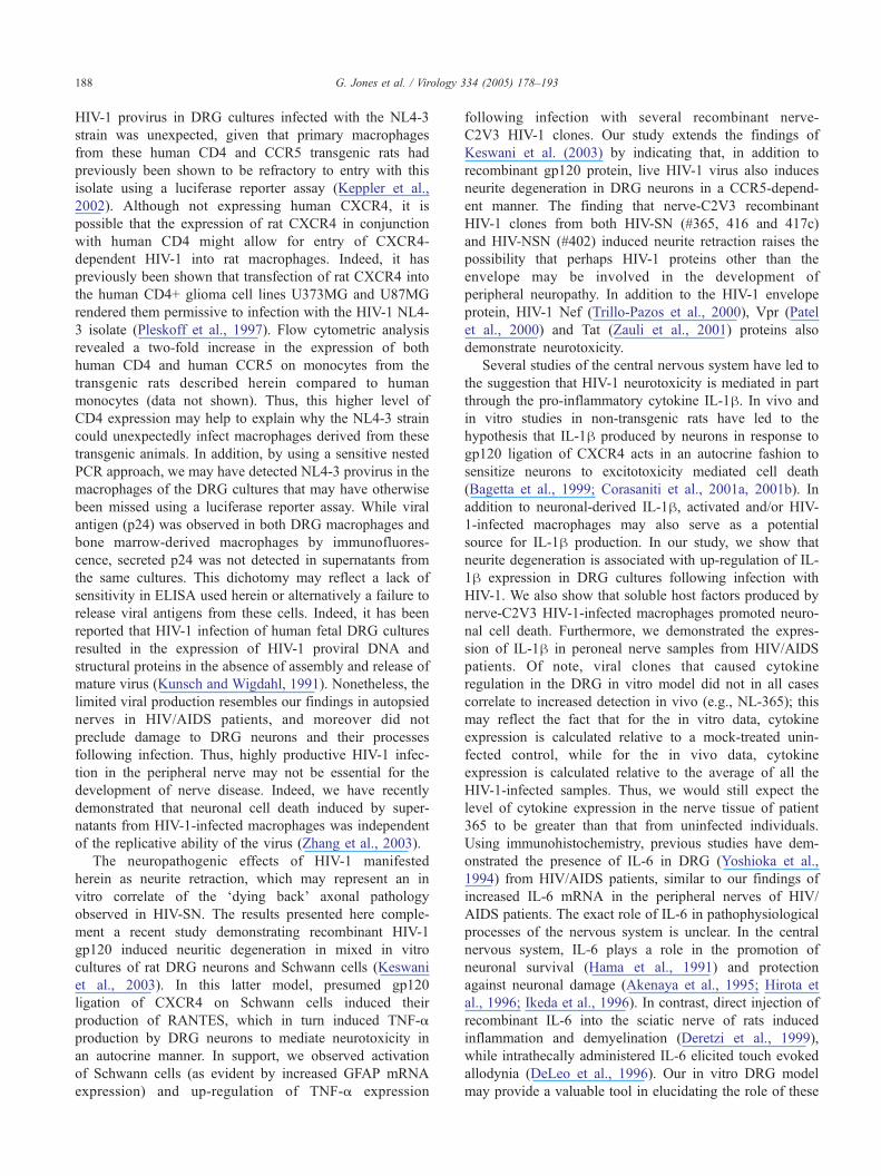
The neuropathogenic effects of HIV-1 manifested
herein as neurite retraction, which may represent an in
vitro correlate of the ddying backT axonal pathology
observed in HIV-SN. The results presented here comple-
ment a recent study demonstrating recombinant HIV-1
gp120 induced neuritic degeneration in mixed in vitro
cultures of rat DRG neurons and Schwann cells (Keswani
et al., 2003). In this latter model, presumed gp120
ligation of CXCR4 on Schwann cells induced their
production of RANTES, which in turn induced TNF-a
production by DRG neurons to mediate neurotoxicity in
an autocrine manner. In support, we observed activation
of Schwann cells (as evident by increased GFAP mRNA
expression) and up-regulation of TNF-a expression
following infection with several recombinant nerve-
C2V3 HIV-1 clones. Our study extends the findings of
Keswani et al. (2003) by indicating that, in addition to
recombinant gp120 protein, live HIV-1 virus also induces
neurite degeneration in DRG neurons in a CCR5-depend-
ent manner. The finding that nerve-C2V3 recombinant
HIV-1 clones from both HIV-SN (#365, 416 and 417c)
and HIV-NSN (#402) induced neurite retraction raises the
possibility that perhaps HIV-1 proteins other than the
envelope may be involved in the development of
peripheral neuropathy. In addition to the HIV-1 envelope
protein, HIV-1 Nef (Trillo-Pazos et al., 2000), Vpr (Patel
et al., 2000) and Tat (Zauli et al., 2001) proteins also
demonstrate neurotoxicity.
Several studies of the central nervous system have led to
the suggestion that HIV-1 neurotoxicity is mediated in part
through the pro-inflammatory cytokine IL-1h. In vivo and
in vitro studies in non-transgenic rats have led to the
hypothesis that IL-1h produced by neurons in response to
gp120 ligation of CXCR4 acts in an autocrine fashion to
sensitize neurons to excitotoxicity mediated cell death
(Bagetta et al., 1999; Corasaniti et al., 2001a, 2001b). In
addition to neuronal-derived IL-1h, activated and/or HIV-
1-infected macrophages may also serve as a potential
source for IL-1h production. In our study, we show that
neurite degeneration is associated with up-regulation of IL-
1h expression in DRG cultures following infection with
HIV-1. We also show that soluble host factors produced by
nerve-C2V3 HIV-1-infected macrophages promoted neuro-
nal cell death. Furthermore, we demonstrated the expres-
sion of IL-1h in peroneal nerve samples from HIV/AIDS
patients. Of note, viral clones that caused cytokine
regulation in the DRG in vitro model did not in all cases
correlate to increased detection in vivo (e.g., NL-365); this
may reflect the fact that for the in vitro data, cytokine
expression is calculated relative to a mock-treated unin-
fected control, while for the in vivo data, cytokine
expression is calculated relative to the average of all the
HIV-1-infected samples. Thus, we would still expect the
level of cytokine expression in the nerve tissue of patient
365 to be greater than that from uninfected individuals.
Using immunohistochemistry, previous studies have dem-
onstrated the presence of IL-6 in DRG (Yoshioka et al.,
1994) from HIV/AIDS patients, similar to our findings of
increased IL-6 mRNA in the peripheral nerves of HIV/
AIDS patients. The exact role of IL-6 in pathophysiological
processes of the nervous system is unclear. In the central
nervous system, IL-6 plays a role in the promotion of
neuronal survival (Hama et al., 1991) and protection
against neuronal damage (Akenaya et al., 1995; Hirota et
al., 1996; Ikeda et al., 1996). In contrast, direct injection of
recombinant IL-6 into the sciatic nerve of rats induced
inflammation and demyelination (Deretzi et al., 1999),
while intrathecally administered IL-6 elicited touch evoked
allodynia (DeLeo et al., 1996). Our in vitro DRG model
may provide a valuable tool in elucidating the role of these

G. Jones et al. / Virology 334 (2005) 178–193 189
different cytokines in peripheral nerve degeneration, as
there are few models of HIV-SN. Very recently, our group
has developed an in vivo model of lentivirus infection in
which feline immunodeficiency virus (FIV) was shown to
cause sensory neuropathy (Kennedy et al., 2004). Of
interest to the present study, peripheral nerve axonal injury
and inflammation occurs in the FIV model, but similar to
observations in autopsied human samples, there is not a
significant loss in DRG neurons (Power and Zochodne,
unpublished data). Thus, studies of HIV-SN can be
extended in several infection models to investigate the
neurotoxic effects of select anti-retroviral therapies and to
test the efficacy of potential neuroprotective agents.
Materials and methods
Patient samples
Peroneal nerve tissue was obtained from twelve male
HIV/AIDS patients at autopsy, five with HIV-SN (neuro-
pathic signs of reduced ankle reflexes and distal sensory
loss) and seven with no evidence of neuropathic signs or
symptoms (HIV-NSN). Both age and the last CD4 count
prior to autopsy were similar in the two groups, with a
median age of 39 and 48.5 years and a median CD4 T-cell
count of 24 and 27 cells/Al for the HIV-SN and HIV-NSN,
respectively. All HIV-SN patients had exposure to antire-
troviral therapy: three patients received AZT and ddI while
two patients received AZT and ddC. Five of the seven HIV-
NSN patients had exposure to antiretroviral therapy: four
patients received AZT alone, while one patient received
AZT, ddI, ddC and d4T. Unfortunately, plasma HIV-1 RNA
levels were not available.
Cell cultures
293T cells (American Type Culture Collection), HeLa-
CD4/CXCR4 (clone 1022) cells (Chesebro et al., 1991),
HeLa-CD4/CXCR4/CCR5 cells (Platt et al., 1998), the
human osteosarcoma cell lines GHOST CCR2b, GHOST
Hi-CCR5 and GHOST CXCR4 and human cholinergic
neuronal (LAN-2) cells were cultured as previously
described (Zhang et al., 2003). The GHOST cell lines
(catalogue numbers 3681, 3944 and 3685 respectively) were
obtained from the AIDS Research and Reference Reagent
Program, Division of AIDS, NIAD, NIH. Human peripheral
blood mononuclear cells (PBMC) were isolated from whole
blood of healthy donors using Histopaque (Sigma-Aldrich,
Oakville, Canada). Human monocyte-derived macrophages
(MDM) were generated as follows: human monocytes were
isolated from PBMC by adherence on poly-orthinate coated
tissue culture plates, after which non-adherent cells were
removed and the adherent cell population cultured in RPMI
1640 (Sigma-Aldrich) medium containing 10% FBS
(Sigma-Aldrich), 10% L929 cell-conditioned medium, as a
source of macrophage colony-stimulating factor-1 (Tsutsui
et al., 2004), and 1% penicillin-streptomycin (Sigma-
Aldrich). Cultures were washed every 2–3 days until the
cells exhibited macrophage morphology (Power et al.,
1998), demonstrating greater than 98% purity for macro-
phages as assessed by CD68 immunoreactivity (unpublished
observations). Rat bone marrow-derived macrophages were
isolated from the pelvis and femurs of adult Sprague–
Dawley rats transgenic for human CD4 and CCR5 (Keppler
et al., 2002) using culture methods previously described
(Tsutsui et al., 2004).
DNA/RNA isolation, PCR and sequencing
Genomic DNAwas isolated from peroneal nerve biopsies
via a phenol/chloroform extraction and ethanol precipitation
procedure. Genomic RNA was isolated from nerve tissue
using TRIzol (Life Technologies, Gaithersburg, MD)
according to the manufacturer’s protocol. Total RNA was
isolated, dissolved in diethylpyrocarbonate (DEPC)-treated
water and used for the synthesis of cDNA. The HIV-1 C2V3
envelope region was amplified from genomic DNA or
cDNA using a nested PCR protocol as previously described
(Power et al., 1994). PCR fragments corresponding to the
amplified C2V3 region (445 base pairs) were isolated from
agarose gel and cloned into the pSL1180 vector (Amersham
Biosciences Inc, Baie d’Urfe, Canada) using the StuI and
NheI sites. All reagents were obtained from New England
BioLabs Ltd. (Mississauga, Canada) and used following the
manufacturer’s specifications. Sequencing reactions and
DNA phylogenetic analysis was performed as previously
reported (Van Marle et al., 2002). HIV-1 co-receptor usage
was investigated by analyzing the V3 loop charge and
amino acid residues as previously described (Briggs et al.,
2000). Using this procedure, viral isolates can be predicted
to be (i) CCR5-dependent; (ii) CCR5/CXCR4 dual tropic; or
(iii) CXCR4-dependent.
Construction of recombinant infectious HIV-1 clones
Recombinant HIV-1 clones containing the C2V3 region
of nerve-derived envelope sequences (herein to be known as
nerve-C2V3 recombinant HIV-1) were constructed in the
genomic backbone of the molecular clone pNL4-3 as
previously described (Zhang et al., 2001). To generate
infectious virus, 293-T cells were transfected with 2 Ag of
plasmid DNA of the recombinant HIV-1 clones using
FuGENE6 (Roche Diagnostics Corporation, Indianapolis,
IN) according to the manufacturer’s protocol. Culture
supernatants were harvested 3 days post-transfection and
cleared of cellular debris by low-speed centrifugation. Virus
stocks were expanded by co-culture of the 293-T cell
transfection supernatants with PBMC, stimulated 3 days
previously with 5 Ag/ml of concanavalin A, and maintained
in RPMI 1640 medium with 10% FBS and 100 IU/ml
interleukin-2 (IL-2). Culture supernatants were removed at

G. Jones et al. / Virology 334 (2005) 178–193190
days 3, 7 and 10 post-infection, cleared of cellular debris by
low-speed centrifugation and viral protein levels were
quantified using the HIV-1 p24 Antigen Capture Assay
Kit (AIDS Vaccine Program, National Cancer Institute,
Frederick, MD.). Viral stocks were stored at �80 8C until
required.
Chemokine co-receptor utilization
The chemokine co-receptor utilization of the recombi-
nant HIV-1 strains was investigated using the previously
described luciferase reporter virus infection assay (Jian and
Zhao, 2003). Using FuGENE6 reagent, the molecular clones
of the recombinant HIV-1 strains were co-transfected into
293-T cells along with a plasmid expressing the firefly
luciferase gene within an env-inactivated HIV-1 clone
(pNL-Luc-E�R�; obtained through the AIDS Research
and Reference Reagent Program, catalogue number 3418).
Culture supernatants were collected 3 days post-trans-
fection, cleared of cell debris by low-speed centrifugation
and used to infect cell lines expressing different chemokine
receptors. Infection of the target cells by the pseudotyped
virus led to luciferase expression, which was quantified in
cell lysates 2 days post-infection using the Luciferase Assay
Kit (BD Biosciences, Mississauga, Canada) following the
manufacturer’s instructions. In addition to expressing
distinct chemokine receptors, the GHOST cell lines are also
stably transfected with a construct expressing humanized
green fluorescent protein (hGFP) under the control of the
HIV-2 LTR (HIV-1 infection of these cells can be detected
through induction of the hGFP gene). The GHOST cell lines
were seeded overnight in 16-well chamber slides (Nalgene
Nunc International, Naperville, IL), infected with the
recombinant HIV-1 strains and cultured for 3 days,
following which cells were fixed with 4% formalin and
washed with PBS. Slides were mounted and induction of
hGFP expression analyzed via fluorescent microscopy using
a Zeiss Axioskop 2 upright microscope (Oberkochen,
Germany).
Dorsal root ganglia (DRG) cultures
DRGs were harvested from previously described adult
Sprague–Dawley rats transgenic for human CD4 and CCR5
(Keppler et al., 2002). DRG were cleared of connective
tissue, dissected into small pieces and digested for 90 min at
37 8C in DMEM (Sigma-Aldrich) containing 1 mg/ml
collagenase (Sigma-Aldrich), 0.5% trypsin (Gibco BRL,
Burlington, Canada) and 0.1 mg/ml DnaseI (Roche Diag-
nostics Corporation). Cells were harvested via centrifuga-
tion, re-suspended in DRG medium (DMEM containing
10% FBS, 5% horse serum (Gibco BRL), 5% L929 cell-
conditioned medium, 1% penicillin-streptomycin, 2 mM l-
glutamine and 1% N-2 supplement (Gibco BRL)), and
plated in either 8-well chamber slides or 24-well tissue
culture plates (Nalgene Nunc International), pre-coated with
a 1:2 dilution of Matrigel (BD Biosciences) in DMEM.
Culture medium was replaced after 24 h, and then every 2–3
days for the next week. DRG cultures were infected with
standardized viral inputs (HIV-1 p24 5 ng per culture) for 6
h, following which the cultures were washed to remove
input virus and cultured in DRG medium for a further 4
days, with medium replenished at day 2. As the recombinant
HIV-1 clones were grown in activated PBMC, DRG cultures
were treated with supernatant from activated but uninfected
PBMC (mock) as controls.
Immunocytochemistry
At day 4 post-infection, DRG cultures were washed with
PBS, fixed with 95% ethanol and blocked overnight at 4 8Cwith PBS containing 50% normal goat serum (NGS). After
removal of the blocking reagent the cells were incubated
overnight at 48C with either mouse anti-MAP-2 (clone HM-
2, 1:1000 dilution, Sigma), mouse anti-ED-1 (1:200
dilution, Chemicon International), rabbit anti-Iba-1 (1:500
dilution, Wako Chemicals), rabbit anti-GFAP (1:400 dilu-
tion, Dako), mouse anti-human CD4 (1:100 dilution, Dako),
mouse anti-human CCR5 (clone 2D7, 1:100 dilution, BD
Biosciences) or HIV-1SF2 p24 rabbit antiserum (1:1000
dilution, obtained through the AIDS Research and Refer-
ence Reagent Program, catalogue number 4250). Following
primary antibody staining, the cells were washed in PBS
and incubated with either Cy3-conjugated goat anti-mouse
(1:2000 dilution, Jackson ImmunoResearch Laboratories,
West Grove, PA) or Alex Fluor-488-conjugated goat anti-
rabbit (1:2000 dilution, Molecular Probes) secondary anti-
bodies. In addition, cultures labeled with rabbit anti-Iba-1
were incubated with a horseradish peroxidase-conjugated
secondary antibody, and the cells were developed with 3,3V-diaminobenzidine as the substrate. All antibody dilutions
were made with PBS containing 10% NGS and 0.1% Triton
X-100 (Sigma). Slides were mounted with Gelvatol and
viewed using a Zeiss Axioskop 2 upright fluorescent
microscope. For the DRG morphological studies, digital
images of MAP-2+ neurons from 12 fields of view per well
were captured using the Advanced Spot system (Diagnostic
Instruments, Sterling Heights, MI) and quantitative analysis
of neuron cell soma size and neurite lengths were performed
using the Scion Image program (Scion, Frederick, MD).
Real time RT-PCR
24 h post-infection, DRG cultures were washed with
PBS, lysed in TRIzol and genomic DNA and RNA isolated
in accordance with the manufacturer’s guidelines. Total
RNA was isolated, dissolved in DEPC-treated water and
used for the synthesis of cDNA. The primers used in the real
time PCR were as follows: glyceraldehyde-3-phosphate
dehydrogenase (GAPDH) (Silva et al., 2003); GFAP (Latour
et al., 2003); IL-1h, forward primer 5V-CCA AAG AAG
AAG ATG GAA AAG CG-3V and reverse primer 5V-GGT

G. Jones et al. / Virology 334 (2005) 178–193 191
GCT GAT GTA CCA GTT GGG-3V (Tm 58 8C); TNF-a,forward primer 5V-CTA TCT GGG AGG GGT CTT CC-3Vand reverse primer 5V-GGT TGA GGG TGT CTG AAG
GA-3V(Tm 58 8C); IL-6, forward primer 5V-ACC CCT GAC
CCA ACC ACA AAT-3Vand reverse primer 5V-AGC TGC
GCA GAATGA GAT GAG-3V(Tm 55 8C); h-actin forward
primer 5V-GGA TGC AGA AGG AGA TCA CTG-3V andreverse primer 5V-CGA TCC ACA CGG AGT ACT TG-3V(Tm 56 8C). Semiquantitative analysis was performed by
monitoring in real time the increase in the fluorescence of
the SYBR-green dye (Molecular Probes) on a Bio-Rad i-
Cycler (Bio-Rad Laboratories, Hercules, CA). Proper
amplification was confirmed by performing melting-curve
analysis. Real-time fluorescence measurements were per-
formed and a threshold cycle value for each gene of interest
was determined, as previously reported (Power et al., 2003).
All data were normalized against the GAPDH or h-actinmRNA level and expressed as fold increases relative to the
mock control.
In vitro indirect neurotoxicity assay
Bone marrow-derived macrophages from rats transgenic
for human CD4 and CCR5 were infected with nerve-C2V3
recombinant viruses (HIV-1 p24 5 ng per culture). Day four
cell culture supernatants were cleared of cellular debris by
low speed centrifugation and applied to differentiated
human cholinergic neuronal (LAN-2) cells, as previously
described (Zhang et al., 2003). After 24 h of exposure,
LAN-2 cell death was quantified via trypan blue exclusion.
Statistical analysis
Statistical analysis was performed using GraphPad InStat
version 3.01 software. The effect of HIV-1 infection on
neurite length and neuronal soma size was examined using
z score analysis. z scores were calculated for each experi-
ment relative to the mock control group. In this way, the z
score denotes how far and in which way the neurite lengths
and neuronal soma size for the virus treated cultures deviate
from the mean values of the mock controls, and is
expressed in units of the standard deviation for the mock
control values. Statistical analysis was performed on the
pooled z scores from three independent experiments.
Acknowledgments
The authors thank Ahmet Hoke, Doug Zochodne and
Mark Goldsmith for helpful discussions and Peter Hauer for
technical assistance. These studies were supported by the
Canadian Institutes of Health Research (CIHR), the Cana-
dian Foundation for AIDS Research (CANFAR), the
Deutsche Forschunggemeinschaft (DFG, KE/742) (OTK),
and the NIH grants NS44807 (JCM), NS049465 (JCM),
R01-NS46262-01 (CP) and subcontract R0051-B (OTK)
from NIH grant R01-MH64396. ST is a Multiple Sclerosis
Society of Canada (MSSC) Fellow.
References
Akenaya, Y., Takahashi, M., Hatanaka, H., 1995. Interleukin-1 beta
enhances survival and interleukin-6 protects against MPP+ neuro-
toxicity in cultures of fetal rat dopaminergic neurons. Exp. Neurol. 136
(1), 44–52.
Albright, A.V., Shieh, J.T., Itoh, T., Lee, B., Pleasure, D., O’Connor, M.J.,
Doms, R.W., Gonzalez-Scarano, F., 1999. Microglia express CCR5,
CXCR4, and CCR3, but of these, CCR5 is the principal coreceptor for
human immunodeficiency virus type 1 dementia isolates. J. Virol. 73
(1), 205–213.
Araujo, A.P., Nascimento, O.J., Garcia, O.S., 2000. Distal sensory
polyneuropathy in a cohort of HIV-infected children over five years
of age. Pediatrics 106 (3), E35.
Bagetta, G., Corasaniti, M.T., Berliocchi, L., Nistico, R., Giammarioli,
A.M., Malorni, W., Aloe, L., Finazzi-Agro, A., 1999. Involvement of
interleukin-1beta in the mechanism of human immunodeficiency virus
type 1 (HIV-1) recombinant protein gp120-induced apoptosis in the
neocortex of rat. Neuroscience 89 (4), 1051–1066.
Berger, E.A., Murphy, P.M., Farber, J.M., 1999. Chemokine receptors as
HIV-1 coreceptors: roles in viral entry, tropism, and disease. Annu. Rev.
Immunol. 17, 657–700.
Brannagan III, T.H., Nuovo, G.J., Hays, A.P., Latov, N., 1997. Human
immunodeficiency virus infection of dorsal root ganglion neurons
detected by polymerase chain reaction in situ hybridization. Ann.
Neurol. 42 (3), 368–372.
Briggs, D.R., Tuttle, D.L., Sleasman, J.W., Goodenow, M.M., 2000.
Envelope V3 amino acid sequence predicts HIV-1 phenotype
(co-receptor usage and tropism for macrophages). AIDS 14 (18),
2937–2939.
Brinley Jr., F.J., Pardo, C.A., Verma, A., 2001. Human immunodeficiency
virus and the peripheral nervous system workshop. Arch. Neurol. 58
(10), 1561–1566.
Chaunu, M.P., Ratinahirana, H., Raphael, M., Henin, D., Leport, C., Brun-
Vezinet, F., Leger, J.M., Brunet, P., Hauw, J.J., 1989. The spectrum of
changes on 20 nerve biopsies in patients with HIV infection. Muscle
Nerve 12 (6), 452–459.
Chesebro, B., Wehrly, K., Metcalf, J., Griffin, D.E., 1991. Use of a new
CD4-positive HeLa cell clone for direct quantitation of infectious
human immunodeficiency virus from blood cells of AIDS patients.
J. Infect. Dis. 163 (1), 64–70.
Chesebro, B., Wehrly, K., Nishio, J., Perryman, S., 1992. Macrophage-
tropic human immunodeficiency virus isolates from different patients
exhibit unusual V3 envelope sequence homogeneity in comparison with
T-cell-tropic isolates: definition of critical amino acids involved in cell
tropism. J. Virol. 66 (11), 6547–6554.
Corasaniti, M.T., Bilotta, A., Strongoli, M.C., Navarra, M., Bagetta, G.,
Di Renzo, G., 2001. HIV-1 coat protein gp120 stimulates inter-
leukin-1beta secretion from human neuroblastoma cells: evidence for
a role in the mechanism of cell death. Br. J. Pharmacol. 134 (6),
1344–1350.
Corasaniti, M.T., Piccirilli, S., Paoletti, A., Nistico, R., Stringaro, A.,
Malorni, W., Finazzi-Agro, A., Bagetta, G., 2001. Evidence that the
HIV-1 coat protein gp120 causes neuronal apoptosis in the neocortex of
rat via a mechanism involving CXCR4 chemokine receptor. Neurosci.
Lett. 312 (2), 67–70.
Cornblath, D.R., McArthur, J.C., Kennedy, P.G., Witte, A.S., Griffin, J.W.,
1987. Inflammatory demyelinating peripheral neuropathies associated
with human T-cell lymphotropic virus type III infection. Ann. Neurol.
21 (1), 32–40.
Cornford, M.E., Ho, H.W., Vinters, H.V., 1992. Correlation of neuro-
muscular pathology in acquired immune deficiency syndrome patients

G. Jones et al. / Virology 334 (2005) 178–193192
with cytomegalovirus infection and zidovudine treatment. Acta Neuro-
pathol. (Berl.) 84 (5), 516–529.
de la Monte, S.M., Gabuzda, D.H., Ho, D.D., Brown Jr., R.H., Hedley-
Whyte, E.T., Schooley, R.T., Hirsch, M.S., Bhan, A.K., 1988.
Peripheral neuropathy in the acquired immunodeficiency syndrome.
Ann. Neurol. 23 (5), 485–492.
DeLeo, J.A., Colburn, R.W., Nichols, M., Malhotra, A., 1996. Interleukin-
6-mediated hyperalgesia/allodynia and increased spinal IL-6 expression
in a rat mononeuropathy model. J. Interferon Cytokine Res. 16 (9),
695–700.
Deretzi, G., Pelidou, S.H., Zou, L.P., Quiding, C., Zhu, J., 1999. Local
effects of recombinant rat interleukin-6 on the peripheral nervous
system. Immunology 97 (4), 582–587.
Gastaut, J.L., Gastaut, J.A., Pellissier, J.F., Tapko, J.B., Weill, O., 1989.
Peripheral neuropathies in human immunodeficiency virus infection.
A prospective study of 56 patients. Rev. Neurol. (Paris) 145 (6–7),
451–459.
Gherardi, R., Lebargy, F., Gaulard, P., Mhiri, C., Bernaudin, J.F., Gray, F.,
1989. Necrotizing vasculitis and HIV replication in peripheral nerves.
N. Engl. J. Med. 321 (10), 685–686.
Gorry, P.R., Bristol, G., Zack, J.A., Ritola, K., Swanstrom, R., Birch, C.J.,
Bell, J.E., Bannert, N., Crawford, K., Wang, H., Schols, D., De Clercq,
E., Kunstman, K., Wolinsky, S.M., Gabuzda, D., 2001. Macrophage
tropism of human immunodeficiency virus type 1 isolates from brain
and lymphoid tissues predicts neurotropism independent of coreceptor
specificity. J. Virol. 75 (21), 10073–10089.
Grafe, M.R., Wiley, C.A., 1989. Spinal cord and peripheral nerve pathology
in AIDS: the roles of cytomegalovirus and human immunodeficiency
virus. Ann. Neurol. 25 (6), 561–566.
Griffin, J.W., McArthur, J.C., 1998. Peripheral neuropathies associated with
HIV infection. In: Gendelman, H.E., Epstein, L., Swindells, S. (Eds.),
The neurology of AIDS. Chapman and Hall, New York, pp. 275–291.
Gulevich, S.J., Kalmijn, J.A., Thal, L.J., Iragui-Madoz, V., McCutchan,
J.A., Kennedy, C., Grant, I., 1992. Sensory testing in human
immunodeficiency virus type 1-infected men. HIV neurobehavioral
research center group. Arch. Neurol. 49 (12), 1281–1284.
Hama, T., Kushima, Y., Miyamoto, M., Kubota, M., Takei, N., Hatanaka,
H., 1991. Interleukin-6 improves the survival of mesencephalic
catecholaminergic and septal cholinergic neurons from postnatal, two-
week-old rats in cultures. Neuroscience 40 (2), 445–452.
Hirota, H., Kiyama, H., Kishimoto, T., Taga, T., 1996. Accelerated nerve
regeneration in mice by upregulated expression of interleukin (IL) 6 and
IL-6 receptor after trauma. J. Exp. Med. 183 (6), 2627–2634.
Ho, D.D., Rota, T.R., Schooley, R.T., Kaplan, J.C., Allan, J.D., Groopman,
J.E., Resnick, L., Felsenstein, D., Andrews, C.A., Hirsch, M.S., 1985.
Isolation of HTLV-III from cerebrospinal fluid and neural tissues of
patients with neurologic syndromes related to the acquired immunode-
ficiency syndrome. N. Engl. J. Med. 313 (24), 1493–1497.
Ikeda, K., Iwasaki, Y., Shiojima, T., Kinoshita, M., 1996. Neuroprotective
effect of various cytokines on developing spinal motoneurons following
axotomy. J. Neurol. Sci. 135 (2), 109–113.
Jian, H., Zhao, L.J., 2003. Pro-apoptotic activity of HIV-1 auxiliary
regulatory protein Vpr is subtype-dependent and potently enhanced by
nonconservative changes of the leucine residue at position 64. J. Biol.
Chem. 278 (45), 44326–44330.
Kennedy, J.M., Hoke, A., Zhu, Y., Johnston, J.B., van Marle, G., Silva, C.,
Zochodne, D.W., Power, C., 2004. Peripheral neuropathy in lentivirus
infection: evidence of inflammation and axonal injury. AIDS 18 (9),
1241–1250.
Keppler, O.T., Welte, F.J., Ngo, T.A., Chin, P.S., Patton, K.S., Tsou, C.L.,
Abbey, N.W., Sharkey, M.E., Grant, R.M., You, Y., Scarborough, J.D.,
Ellmeier, W., Littman, D.R., Stevenson, M., Charo, I.F., Herndier, B.G.,
Speck, R.F., Goldsmith, M.A., 2002. Progress toward a human CD4/
CCR5 transgenic rat model for de novo infection by human
immunodeficiency virus type 1. J. Exp. Med. 195 (6), 719–736.
Keswani, S.C., Pardo, C.A., Cherry, C.L., Hoke, A., McArthur, J.C., 2002.
HIV-associated sensory neuropathies. AIDS 16 (16), 2105–2117.
Keswani, S.C., Polley, M., Pardo, C.A., Griffin, J.W., McArthur, J.C.,
Hoke, A., 2003. Schwann cell chemokine receptors mediate HIV-1
gp120 toxicity to sensory neurons. Ann. Neurol. 54 (3), 287–296.
Kuiken, C.L., Goudsmit, J., Weiller, G.F., Armstrong, J.S., Hartman, S.,
Portegies, P., Dekker, J., Cornelissen, M., 1995. Differences in human
immunodeficiency virus type 1 V3 sequences from patients with and
without AIDS dementia complex. J. Gen. Virol. 76, 175–180.
Kunsch, C., Wigdahl, B., 1991. Maintenance of human immunodeficiency
virus type-1 proviral DNA in human fetal dorsal root ganglia neural
cells following a nonproductive infection. J. Leukocyte Biol. 49 (5),
505–510.
Latour, I., Hamid, J., Beedle, A.M., Zamponi, G.W., Macvicar, B.A., 2003.
Expression of voltage-gated Ca2+ channel subtypes in cultured
astrocytes. Glia 41 (4), 347–353.
Morgello, S., Estanislao, L., Simpson, D., Geraci, A., DiRocco, A., Gerits,
P., Ryan, E., Yakoushina, T., Khan, S., Mahboob, R., Naseer, M.,
Dorfman, D., Sharp, V., 2004. HIV-associated distal sensory polyneuro-
pathy in the era of highly active antiretroviral therapy: the Manhattan
HIV Brain Bank. Arch. Neurol. 61 (4), 546–551.
Nagano, I., Shapshak, P., Yoshioka, M., Xin, K., Nakamura, S., Bradley,
W.G., 1996. Increased NADPH-diaphorase reactivity and cytokine
expression in dorsal root ganglia in acquired immunodeficiency
syndrome. J. Neurol. Sci. 136 (1–2), 117–128.
Ohagen, A., Devitt, A., Kunstman, K.J., Gorry, P.R., Rose, P.P., Korber, B.,
Taylor, J., Levy, R., Murphy, R.L., Wolinsky, S.M., Gabuzda, D., 2003.
Genetic and functional analysis of full-length human immunodeficiency
virus type 1 env genes derived from brain and blood of patients with
AIDS. J. Virol. 77 (22), 12336–12345.
Pardo, C.A., McArthur, J.C., Griffin, J.W., 2001. HIV neuropathy: insights
in the pathology of HIV peripheral nerve disease. J. Peripher. Nerv.
Syst. 6 (1), 21–27.
Patel, C.A., Mukhtar, M., Pomerantz, R.J., 2000. Human immunodefi-
ciency virus type 1 Vpr induces apoptosis in human neuronal cells.
J. Virol. 74 (20), 9717–9726.
Platt, E.J.,Wehrly, K., Kuhmann, S.E., Chesebro, B., Kabat, D., 1998. Effects
of CCR5 and CD4 cell surface concentrations on infections by macro-
phagetropic isolates of human immunodeficiency virus type 1. J. Virol.
72 (4), 2855–2864.
Pleskoff, O., Sol, N., Labrosse, B., Alizon, M., 1997. Human
immunodeficiency virus strains differ in their ability to infect
CD4+ cells expressing the rat homolog of CXCR-4 (fusin). J. Virol.
71 (4), 3259–3262.
Power, C., McArthur, J.C., Johnson, R.T., Griffin, D.E., Glass, J.D.,
Perryman, S., Chesebro, B., 1994. Demented and nondemented patients
with AIDS differ in brain-derived human immunodeficiency virus type
1 envelope sequences. J. Virol. 68 (7), 4643–4649.
Power, C., McArthur, J.C., Nath, A., Wehrly, K., Mayne, M., Nishio, J.,
Langelier, T., Johnson, R.T., Chesebro, B., 1998. Neuronal death
induced by brain-derived human immunodeficiency virus type 1
envelope genes differs between demented and nondemented AIDS
patients. J. Virol. 72 (11), 9045–9053.
Power, C., Henry, S., Del Bigio, M.R., Larsen, P.H., Corbett, D., Imai, Y.,
Yong, V.W., Peeling, J., 2003. Intracerebral hemorrhage induces
macrophage activation and matrix metalloproteinases. Ann. Neurol.
53 (6), 731–742.
Rizzuto, N., Cavallaro, T., Monaco, S., Morbin, M., Bonetti, B., Ferrari, S.,
Galiazzo-Rizzuto, S., Zanette, G., Bertolasi, L., 1995. Role of HIV in
the pathogenesis of distal symmetrical peripheral neuropathy. Acta
Neuropathol. (Berl.) 90 (3), 244–250.
Shapshak, P., Nagano, I., Xin, K., Bradley, W., McCoy, C.B., Sun, N.C.,
Stewart, R.V., Yoshioka, M., Petito, C., Goodkin, K., et al., 1995. HIV-1
heterogeneity and cytokines, neuropathogenesis. Adv. Exp. Med. Biol.
373, 225–238.
Silva, C., Zhang, K., Tsutsui, S., Holden, J.K., Gill, M.J., Power, C., 2003.
Growth hormone prevents human immunodeficiency virus-induced
neuronal p53 expression. Ann. Neurol. 54 (5), 605–614.
Tagliati, M., Grinnell, J., Godbold, J., Simpson, D.M., 1999. Peripheral

G. Jones et al. / Virology 334 (2005) 178–193 193
nerve function in HIV infection: clinical, electrophysiologic, and
laboratory findings. Arch. Neurol. 56 (1), 84–89.
Trillo-Pazos, G., McFarlane-Abdulla, E., Campbell, I.C., Pilkington, G.J.,
Everall, I.P., 2000. Recombinant nef HIV-IIIB protein is toxic to human
neurons in culture. Brain Res. 864 (2), 315–326.
Tsutsui, S., Schnermann, J., Noorbakhsh, F., Henry, S., Yong, V.W.,
Winston, B.W., Warren, K., Power, C., 2004. A1 adenosine receptor
upregulation and activation attenuates neuroinflammation and demyeli-
nation in a model of multiple sclerosis. J. Neurosci. 24 (6), 1521–1529.
Valentin, A., Trivedi, H., Lu, W., Kostrikis, L.G., Pavlakis, G.N., 2000.
CXCR4 mediates entry and productive infection of syncytia-
inducing (X4) HIV-1 strains in primary macrophages. Virology 269
(2), 294–304.
Van Marle, G., Rourke, S.B., Zhang, K., Silva, C., Ethier, J., Gill, M.J.,
Power, C., 2002. HIV dementia patients exhibit reduced viral
neutralization and increased envelope sequence diversity in blood and
brain. AIDS 16 (14), 1905–1914.
Vital, A., Beylot, M., Vital, C., Delors, B., Bloch, B., Julien, J., 1992.
Morphological findings on peripheral nerve biopsies in 15 patients with
human immunodeficiency virus infection. Acta Neuropathol. (Berl.) 83
(6), 618–623.
Yoshioka, M., Shapshak, P., Srivastava, A.K., Stewart, R.V., Nelson, S.J.,
Bradley, W.G., Berger, J.R., Rhodes, R.H., Sun, N.C., Nakamura, S.,
1994. Expression of HIV-1 and interleukin-6 in lumbosacral dorsal root
ganglia of patients with AIDS. Neurology 44 (6), 1120–1130.
Zauli, G., Milani, D., Mirandola, P., Mazzoni, M., Secchiero, P., Miscia, S.,
Capitani, S., 2001. HIV-1 Tat protein down-regulates CREB tran-
scription factor expression in PC12 neuronal cells through a phos-
phatidylinositol 3-kinase/AKT/cyclic nucleoside phosphodiesterase
pathway. FASEB J. 15 (2), 483–491.
Zhang, K., Hawken, M., Rana, F., Welte, F.J., Gartner, S., Goldsmith, M.A.,
Power, C., 2001. Human immunodeficiency virus type 1 clade A and D
neurotropism: molecular evolution, recombination, and coreceptor use.
Virology 283 (1), 19–30.
Zhang, K., Rana, F., Silva, C., Ethier, J., Wehrly, K., Chesebro, B.,
Power, C., 2003. Human immunodeficiency virus type 1 envelope-
mediated neuronal death: uncoupling of viral replication and neuro-
toxicity. J. Virol. 77 (12), 6899–6912.