Competition between Folding, Native-State Dimerisation and Amyloid Aggregation in β-Lactoglobulin
13
Competition between Folding, Native-State Dimerisation and Amyloid Aggregation in β-Lactoglobulin Daizo Hamada 1,2,3 ⁎, Toshiki Tanaka 4 , Gian Gaetano Tartaglia 5 , Amol Pawar 5 , Michele Vendruscolo 5 , Mei Kawamura 6 , Atsuo Tamura 6 , Naoki Tanaka 7 and Christopher M. Dobson 5 ⁎ We show that a series of peptides corresponding to individual β-strands in native β-lactoglobulin readily form amyloid aggregates and that such aggregates are capable of seeding fibril formation by a full-length form of β- lactoglobulin in which the disulfide bonds are reduced. By contrast, pre- formed fibrils corresponding to only one of the β-strands that we con- sidered, βA, were found to promote fibril formation by a full-length form of β-lactoglobulin in which the disulfide bonds are intact. These results indicate that regions of high intrinsic aggregation propensity do not give rise to aggregation unless at least partial unfolding takes place. Further- more, we found that the high aggregation propensity of one of the edge strands, βI, promotes dimerisation of the native structure rather than misfolding and aggregation since the structure of βI is stabilised by the presence of a disulfide bond. These findings demonstrate that the inter- actions that promote folding and native-state oligomerisation can also result in high intrinsic amyloidogenicity. However, we show that the presence of the remainder of the sequence dramatically reduces the net overall aggregation propensity by negative design principles that we suggest are very common in biological systems as a result of evolutionary processes. © 2008 Elsevier Ltd. All rights reserved. 1 Department of Developmental Infectious Diseases, Osaka Medical Center for Maternal and Child Health, 840 Murodo, Izumi, Osaka 594-1101, Japan 2 Institute for Protein Research, Osaka University, 3-2 Yamadaoka, Suita, Osaka 565-0871, Japan 3 Division of Structural Biology (G-COE), Department of Biochemistry and Molecular Biology, Graduate School of Medicine, Kobe University, 7-5-1 Kusunoki-cho, Chuo-ku, Kobe 650-0017, Japan 4 Graduate School of Material Science, Nagoya Institute of Technology, Gokiso-cho, Nagoya 466-8555, Japan 5 Department of Chemistry, University of Cambridge, Lensfield Road, Cambridge CB2 1EW, UK 6 Department of Chemistry, Graduate School of Science, Kobe University, 1-1 Rokkodai, Nada-ku, Kobe 657-8501, Japan 7 Department of Polymer Science and Engineering, Kyoto Institute of Technology, Matsugasaki, Sakyo, Kyoto 606-8585, Japan *Corresponding authors. D. Hamada is to be contacted at Division of Structural Biology (G-COE), Department of Biochemistry and Molecular Biology, Graduate School of Medicine, Kobe University, 7-5-1 Kusunoki-cho, Chuo-ku, Kobe 650-0017, Japan; C. M. Dobson, Department of Chemistry, University of Cambridge, Lensfield Road, Cambridge CB2 1EW, UK. E-mail addresses: [email protected]; [email protected]. Abbreviations used: RCM-β-lactoglobulin, reduced and carboxymethylated β-lactoglobulin; TEM, transmission electron microscopy. doi:10.1016/j.jmb.2008.12.038 J. Mol. Biol. (2009) 386, 878–890 Available online at www.sciencedirect.com 0022-2836/$ - see front matter © 2008 Elsevier Ltd. All rights reserved.
-
Upload
independent -
Category
Documents
-
view
2 -
download
0
Transcript of Competition between Folding, Native-State Dimerisation and Amyloid Aggregation in β-Lactoglobulin
doi:10.1016/j.jmb.2008.12.038 J. Mol. Biol. (2009) 386, 878–890
Available online at www.sciencedirect.com
Competition between Folding, Native-State Dimerisationand Amyloid Aggregation in β-Lactoglobulin
Daizo Hamada1,2,3⁎, Toshiki Tanaka4, Gian Gaetano Tartaglia5,Amol Pawar5, Michele Vendruscolo5, Mei Kawamura6, Atsuo Tamura6,Naoki Tanaka7 and Christopher M. Dobson5⁎
*Corresponding authors. D. HamadBiochemistry and Molecular BiologKobe 650-0017, Japan; C. M. DobsoCB2 1EW, UK. E-mail addresses: dAbbreviations used: RCM-β-lacto
electron microscopy.
0022-2836/$ - see front matter © 2008 E
We show that a series of peptides corresponding to individual β-strands innative β-lactoglobulin readily form amyloid aggregates and that suchaggregates are capable of seeding fibril formation by a full-length form of β-lactoglobulin in which the disulfide bonds are reduced. By contrast, pre-formed fibrils corresponding to only one of the β-strands that we con-sidered, βA, were found to promote fibril formation by a full-length form ofβ-lactoglobulin in which the disulfide bonds are intact. These resultsindicate that regions of high intrinsic aggregation propensity do not giverise to aggregation unless at least partial unfolding takes place. Further-more, we found that the high aggregation propensity of one of the edgestrands, βI, promotes dimerisation of the native structure rather thanmisfolding and aggregation since the structure of βI is stabilised by thepresence of a disulfide bond. These findings demonstrate that the inter-actions that promote folding and native-state oligomerisation can also resultin high intrinsic amyloidogenicity. However, we show that the presence ofthe remainder of the sequence dramatically reduces the net overallaggregation propensity by negative design principles that we suggest arevery common in biological systems as a result of evolutionary processes.
© 2008 Elsevier Ltd. All rights reserved.
1Department of DevelopmentalInfectious Diseases, OsakaMedical Center for Maternaland Child Health, 840 Murodo,Izumi, Osaka 594-1101, Japan2Institute for Protein Research,OsakaUniversity, 3-2Yamadaoka,Suita, Osaka 565-0871, Japan3Division of Structural Biology(G-COE), Department ofBiochemistry and MolecularBiology, Graduate School ofMedicine, Kobe University,7-5-1 Kusunoki-cho, Chuo-ku,Kobe 650-0017, Japan4Graduate School of MaterialScience, Nagoya Institute ofTechnology, Gokiso-cho, Nagoya466-8555, Japan5Department of Chemistry,University of Cambridge,Lensfield Road, CambridgeCB2 1EW, UK6Department of Chemistry,Graduate School of Science,Kobe University, 1-1 Rokkodai,Nada-ku, Kobe 657-8501, Japan7Department of Polymer Scienceand Engineering, Kyoto Instituteof Technology, Matsugasaki,Sakyo, Kyoto 606-8585, Japan
a is to be contacted at Division of Structural Biology (G-COE), Department ofy, Graduate School of Medicine, Kobe University, 7-5-1 Kusunoki-cho, Chuo-ku,n, Department of Chemistry, University of Cambridge, Lensfield Road, [email protected]; [email protected], reduced and carboxymethylated β-lactoglobulin; TEM, transmission
lsevier Ltd. All rights reserved.

879Fibril Formation versus Folding in β-Lactoglobulin
Received 31 July 2008;received in revised form10 December 2008;accepted 15 December 2008Available online24 December 2008
Keywords: fragment seeding; transmission electron microscopy; thioflavinT; fluorescence; negative design principle
Edited by K. KuwajimaFig. 1. Structure of bovine β-lactoglobulin. (a) Dimericnative-state structure from X-ray crystallography (ProteinData Bank ID 1BEB). The regions corresponding to thepeptides studied in this work are represented by differentcolors (βA is shown in red; βF, magenta; βG, cyan; βH,green; βI, yellow; and α1, blue). (b) Amino acid sequencesof the peptide fragments studied in this work. Thearomatic residues shown in lower case were added tothe original sequence in order to allow determination ofthe peptide concentration by means of UVabsorption. Theadditional aromatic residues should not significantlyinfluence the aggregation propensity of these peptidesaccording to the Zagg score from Zyggregator (Table 1).Residue numbers are relative to the sequence of the intactprotein.
Introduction
Despite the natural tendency of proteins to foldinto native structures unique to their amino acidsequences,1,2 they are capable of aggregating both invitro and in vivo into large assemblies with a varietyof morphological features.3–6 Of such aggregates,amyloid fibrils represent particularly well-orderedstructures that can be micrometers in length whilejust a few nanometers in diameter and containcross β-strands aligned perpendicularly to the fibrilaxis.7–11 Such fibrils are the subject of intense interestat the present time since their deposition in tissue isassociated with a range of increasingly commonhuman diseases, including both systemic and organ-specific conditions, such as Alzheimer's disease andtype II diabetes.4,12,13 Remarkably, many peptidesand proteins, and indeed homopolymers, such aspolyalanine and polythreonine, without any knowninvolvement in amyloid disease can also formamyloid fibrils in vitro,14–21 suggesting that theability to form amyloid fibrils is an intrinsic propertyof polypeptide chains.22 Furthermore, such struc-tures have the potential to generate new forms ofnanoscale biomaterials based on the amyloid fibril-lar architecture since they are characterised by well-ordered and stable structures that can be formed byself-association in aqueous solution.23–26 Thus,elucidation of the mechanism by which polypeptidechains convert into amyloid fibrils is an importantgoal in medical, biological and material sciences.Formation of amyloid fibrils appears to occur in a
nucleation-dependent manner that is followed by arapid extension process that gives rise to fibrillararchitectures.27 It has been observed by analysis ofnative-state structures that the existence of exposedβ-strands at the edge ofβ-sheet structures tends to beavoided since they help in the formation of β-aggre-gates, such as amyloid fibrils.28 Extensive surveys ofstructures in the Protein Data Bank suggest that avariety of mechanisms prevent the exposure ofhydrophobic surfaces in such edge strands (e.g., byformation of intermolecular β-sheets or by placinginward-pointing charged side chains).28
One of the proteins exemplifying the formersituation is bovine β-lactoglobulin, which has beenshown to be capable of forming amyloid fibrils byprolonged incubation at pH 7.0 in the presence of5.0 M urea.18 However, the reaction is relativelyslow and can take several weeks to reach comple-tion. β-Lactoglobulin forms a homodimeric native
state at neutral pH in the absence of denaturant,although it dissociates into monomeric but stillnatively folded species at pH values below 3.0.29 Inthe native state, the protein has a predominantly β-sheet structure, consisting of a β-barrel of eightantiparallel β-strands (A–H) and an additional edgeβ-strand (I) that is part of the dimer interface locatedat the edge of the β-sheet of each individualmolecule, although there is one major α-helix atthe C-terminus (Fig. 1). The β-barrel structure hasthe shape of a flattened cone resembling a calyxinvolving the N- and C-terminal β-sheets; the N-terminal β-sheet consists of the βB, βC and βDstrands, while the C-terminal one involves the βA,βE, βF, βG, βH and βI strands. The protein alsocontains two disulfide bonds (C66–C160 and C106–C119, linking strands βD–βI and βG–βH, respec-
880 Fibril Formation versus Folding in β-Lactoglobulin
tively) and one free cysteine residue (C121). Despitethe fact that the native structure of β-lactoglobulincontains a predominantly β-sheet structure, the se-quence of β-lactoglobulin encodes a relatively highα-helical propensity; indeed, the major folding inter-mediate observed to be populated during the fold-ing of the protein contains non-native α-helicalmotifs30–37 located mainly in the vicinity of theβA-strand region in the native structure.35
In this work, we describe studies on the relativeabilities of peptide fragments corresponding to theamino acid sequences of the β-strands and the α-helical regions of the hydrophobic core of native β-lactoglobulin to form amyloid fibrils and to seed theaggregation of full-length β-lactoglobulin. Theseresults reveal new insights into the general princi-ples underlying the competition between folding,native-state oligomerisation and multimolecularaggregation and provides specific informationabout the way these factors influence the behaviourof β-lactoglobulin itself.
Results and Discussion
Amyloid formation by peptide fragments ofβ-lactoglobulin
The peptide fragments that we have analysed herecorrespond to the βA, βF, βG, βH and βI strandsand the C-terminal α-helical region (α1) of native β-lactoglobulin (Fig. 1); the amino acid sequences are
shown in Fig. 1b. Except for βA, the peptides are alllocated at the C-terminal β-sheet or at the dimerinterface of the native protein. These fragments werechosen because the corresponding regions of thesequence form the hydrophobic core of the nativestructure of the full-length protein.36 Moreover, inthe process of folding from the denatured state, theβF, βG and βH strands have been found to assume anative-like structure rapidly after the initiation of thereaction.36 α1 (described as fragment 3 in Ref. 31 andas fragment 4 in Ref. 33) has previously been shownto possess a high intrinsic helical propensity. In thiscontext, it is particularly interesting that, in thenative state, βI is located at the edge of a C-terminalβ-sheet that is also involved in the interface of thenative homodimer of β-lactoglobulin.The kinetics of fibril formation by the peptide
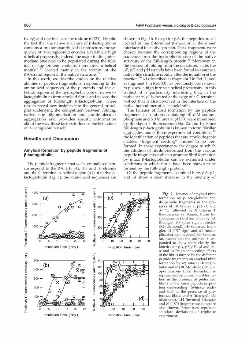
fragments in solutions containing 10 mM sodiumphosphate and 5.0 M urea at pH 7.0 were monitoredby thioflavin T fluorescence (Fig. 2a and b). Sincefull-length β-lactoglobulin is known to form fibrillaraggregates under these experimental conditions,18
the identification of peptides that are amyloidogenicenables “fragment seeding” studies to be per-formed. In these experiments, the degree to whichthe addition of fibrils preformed from the variouspeptide fragments is able to promote fibril formationby intact β-lactoglobulin can be examined underconditions in which fibrils have been shown to beformed by the full-length protein.Of the peptide fragments examined here, βA, βG
and βI show a clear increase in the intensity of
Fig. 2. Kinetics of amyloid fibrilformation by β-lactoglobulin andits peptide fragments in the pre-sence of 5.0 M urea at pH 7.0 and37 °C followed by thioflavin Tfluorescence. (a) Kinetic traces forspontaneous fibril formation by βA(triangle), βF (plus sign in circle),βG (diamond), βH (inverted trian-gle), βI (“X” sign) and α1 (multi-plication sign in circle). (b) Same as(a), except that the ordinate is ex-panded to show more clearly thekinetics for βA, βF, βH, βI and α1.(c and d) Fragment seeding effectsof the fibrils formed by the differentpeptide fragments on amyloid fibrilformation by (c) intact β-lactoglo-bulin and (d) RCM-β-lactoglobulin.Spontaneous fibril formation isrepresented by circles. Fibril forma-tion in the presence of preformedfibrils of the same peptide or pro-tein (self-seeding) (whisker ends)and that in the presence of pre-formed fibrils of βA (triangle), βG(diamond), βH (inverted triangle)and βI (“X”) (fragment seeding) arealso shown. Error bars representstandard deviations of triplicateexperiments.
881Fibril Formation versus Folding in β-Lactoglobulin
thioflavin T fluorescence after prolonged incubationat pH 7.0 in the presence of 5.0 M urea (Fig. 2a andb). The kinetic traces of the increase in fluorescencein solutions containing βA and βI are sigmoidalcurves, consistent with the idea that the formation ofamyloid fibrils occurs in a nucleation-dependentmanner. The kinetic profile for the solution contain-ing βG, on the other hand, does not show the pre-sence of a well-defined lag phase; the fluorescenceintensity of thioflavin T was found to increaseimmediately after the preparation of samples. Itshould be noted, however, that βG is highly inso-luble in water but soluble in the presence of dena-turants. The lack of an observable lag phase istherefore likely to reflect the fact that the solutionscontain preformed aggregates of βG prior toincubation and that the growth of fibrils does notrequire further nucleation events. The βGpeptide, inaddition, has a single free cysteine residue in thecentral region of its sequence that may permitintermolecular disulfide bonds to form under theconditions employed here, hence providing anadditional mechanism for nucleation. The long-term incubation in solution containing urea mayinduce carbamylation, thus potentially affecting the
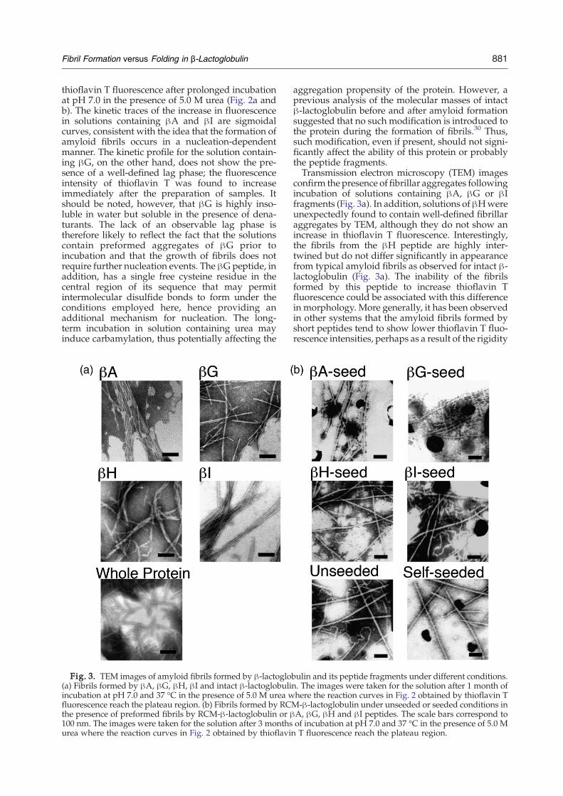
Fig. 3. TEM images of amyloid fibrils formed by β-lactoglo(a) Fibrils formed by βA, βG, βH, βI and intact β-lactoglobuliincubation at pH 7.0 and 37 °C in the presence of 5.0 M urea wfluorescence reach the plateau region. (b) Fibrils formed by RCthe presence of preformed fibrils by RCM-β-lactoglobulin or β100 nm. The images were taken for the solution after 3 monthsurea where the reaction curves in Fig. 2 obtained by thioflavi
aggregation propensity of the protein. However, aprevious analysis of the molecular masses of intactβ-lactoglobulin before and after amyloid formationsuggested that no such modification is introduced tothe protein during the formation of fibrils.30 Thus,such modification, even if present, should not signi-ficantly affect the ability of this protein or probablythe peptide fragments.Transmission electron microscopy (TEM) images
confirm the presence of fibrillar aggregates followingincubation of solutions containing βA, βG or βIfragments (Fig. 3a). In addition, solutions ofβHwereunexpectedly found to contain well-defined fibrillaraggregates by TEM, although they do not show anincrease in thioflavin T fluorescence. Interestingly,the fibrils from the βH peptide are highly inter-twined but do not differ significantly in appearancefrom typical amyloid fibrils as observed for intact β-lactoglobulin (Fig. 3a). The inability of the fibrilsformed by this peptide to increase thioflavin Tfluorescence could be associated with this differencein morphology. More generally, it has been observedin other systems that the amyloid fibrils formed byshort peptides tend to show lower thioflavin T fluo-rescence intensities, perhaps as a result of the rigidity
bulin and its peptide fragments under different conditions.n. The images were taken for the solution after 1 month ofhere the reaction curves in Fig. 2 obtained by thioflavin TM-β-lactoglobulin under unseeded or seeded conditions inA, βG, βH and βI peptides. The scale bars correspond toof incubation at pH 7.0 and 37 °C in the presence of 5.0 Mn T fluorescence reach the plateau region.

882 Fibril Formation versus Folding in β-Lactoglobulin
of the cross-β strands in the fibril structures (YujiGoto, personal communication).
Fragment seeding effects
Amyloid formation by large polypeptide chainsappears to be promoted by specific regions oftheir amino acid sequences, sometimes known as“aggregation-prone” regions, which, in addition, arelikely to be incorporated into the core of the fibrils.37
A major aim of the present study was to understandthe role played by such regions in the process offibril formation by β-lactoglobulin. As part of thisobjective, we adopted the strategy of adding fibrilsformed by the peptide fragments to solutions of thefull-length protein to explore their efficacy in pro-moting the formation of fibrils by the latter. Suchfragment seeding behaviour has been observed instudies on peptides and proteins, including insu-lin, hen lysozyme, islet amyloid polypeptide andβ2-microglobulin.38–43Figure 2c indicates the degree to which the fibrillar
aggregates formed by βA, βG, βH and βI fragmentsare capable of seeding the aggregation of the intactprotein in 5.0 M urea at pH 7.0. Of the fibrils formedby the four peptides, only those formed by the βAfragment were observed to detectably accelerate thefibril formation by full-length β-lactoglobulin; thekinetics of fibril formation are essentially unchangedby the presence of preformed fibrils of the otherfragments. This analysis suggests that the βA-strandregion, but not the regions corresponding to the βG,βH and βI strands, is likely to be directly involvedin promoting the formation of fibrils by full-lengthβ-lactoglobulin.However, these results do not rule out the
possibility that other regions of the sequence (e.g.,the βB, βC, βD and βE strands or loop regions) alsocontribute to the core region of the fibrillar structure.Indeed, the fact that several of the peptides studiedhere readily form amyloid fibrils supports the ideathat the ability to form fibrillar architectures is ageneric property of polypeptide chains.13 For largepolypeptide chains, in particular, different regions ofthe molecules may well assemble in a range of dis-tinct ways to generate cross-β core regions and giverise to the well-established structural polymorphismor heterogeneity of these fibrils.44–48 This observa-tion is consistent with the fact that very differentmorphologies of fibrillar structures are observed forfull-length β-lactoglobulin under different condi-tions.18,49 It is also possible that the amyloid core isformed by various segments apart from one another.In this sense, further analysis of fragment seedingusing several peptides at the same time to makeseeds may clarify the regions required for stabilisa-tion of the amyloid core by β-lactoglobulin.
Implications for amyloid formation byβ-lactoglobulin
We used our previously published method50 tocalculate the intrinsic aggregation propensity of β-
lactoglobulin as a function of its sequence (seeMaterials and Methods). The results show that thepropensities of the regions corresponding to the βA,βG, βH and βI strands to aggregate are substantiallyhigher than those of the other regions of the sequence(Fig. 4; Table 1). Consistent with these predictions,amyloid fibrils are formed very rapidly by the βA,βG, βH and βI peptides in solution, as shown in Fig.2a and b. Addition of preformed fibrils of the βG, βHand βI peptides however does not detectablypromote fibril formation by full-length β-lactoglo-bulin under the conditions used here, suggestingthat the intrinsic propensity for aggregation ofsegments of a protein sequence is not the only factorthat determines the aggregation process of the intactmolecule. Indeed, only preformed fibrils of βA,which in fact has a slightly lower intrinsic propensityfor aggregation than do βG, βH and βI (Fig. 4; Table1), were found to accelerate the formation of fibrilsby full-length β-lactoglobulin. It is well establishedthat, for intact proteins, only part of the polypeptidechain needs to be involved in the formation of thecross-β core of amyloid fibrils.51–54 The present re-sults imply that the region corresponding to the βAstrand could be specifically involved in stabilisingthe cross-β structure of full-length β-lactoglobulinamyloid fibrils.In order to understand the differences between the
fragment seeding effects of fibrils corresponding tothe βA, βG, βH and βI peptides, despite the fact thatthey all have high intrinsic propensities for aggrega-tion, we calculated two additional aggregation pro-pensity profiles by taking into account the protec-tion of the aggregation-prone regions that will beprovided by the formation of the native structure ofthe protein. In this approach, the local stability of aregion in a folded protein is predicted from knowl-edge of its structure.55 When the stability of native-like structures in a localised region is very high, theeffective propensity for aggregation around suchregion is strongly reduced as the region itself willtend to remain in a native-like conformation evenunder at least moderately destabilising conditions orin the presence of local conformational fluctuations.The presence of such localised native-like structurewill reduce the accessibility for aggregation of theprotein through the association of this region. Wetherefore calculated the aggregation profile takinginto account the structure present in the monomericform of β-lactoglobulin (Fig. 4). In this new profile,except for the terminal regions of the sequence, theonly region with an aggregation propensity sig-nificantly above the average corresponds to βI inthe native fold. The fact that dimerisation occursthrough this β-strand shows the value of the methodfor predicting regions likely to form intermolecularβ-sheets in functional and pathogenic assemblies.This idea is supported by the fact that equine β-lactoglobulin, for which the propensity to aggregatein the βI strand is much lower (Fig. 4b), forms amonomeric native state. A comparison of the aminoacid sequences of bovine and equine β-lactoglobulinreveals a clear difference in the sequence in the

Fig. 4. Structural and physicochemical properties of β-lactoglobulin predicted from the amino acid sequence. (a)Predicted aggregation propensity profiles of bovine β-lactoglobulin at pH 7.0. The intrinsic aggregation profile (black) iscompared with the profiles obtained by incorporating conformational factors in the monomeric (red) and dimeric (violet)forms. Gray shaded areas indicate the locations of the peptides analysed in this work. (b) Predicted intrinsic aggregationprofile of equine β-lactoglobulin (black) at pH 7.0 in comparison with that of bovine β-lactoglobulin (red). (c) Sequencealignment of bovine and equine β-lactoglobulin showing a less conserved βI region.
883Fibril Formation versus Folding in β-Lactoglobulin
region of the βI strand in contrast to the highlyconserved sequences in the other regions (Fig. 4c).We calculated the second profile by considering
the structure of the dimeric native state of β-lactoglobulin (Fig. 4a). This profile is essentiallyidentical with the previous one, except in the regionof βI, where the aggregation propensity is muchreduced as a result of the burial of this strand in thedimer interface. In this profile, there is no region ofsignificant aggregation propensity, in agreementwith the observation that the native dimer of β-
Table 1. Aggregation propensities of peptides derivedfrom β-lactoglobulin
Peptide fragmenta Zaggb Amyloidogenicityc
Full-length β-lactoglobulin 0.87βA 1.36 HighβF 0.64 LowβG 1.48 HighβH 1.79 (1.73)d HighβI 2.28 (1.66) Highα1 0.00 (0.41) Low
a Sequence position of fragments in intact β-lactoglobulin(Fig. 1).
b Intrinsic aggregation propensity corresponding to the aminoacid sequence of the peptide at pH 7.0 calculated using theZyggregator method (see Materials and Methods for details).
c Qualitative experimental rates of growth of fibrils at pH 7.0 inthe presence of 5.0 M urea (Fig. 2a).
d Values calculated without considering the effect of artificiallyadded aromatic residues (Fig. 1).
lactoglobulin is highly soluble and must be desta-bilised in order to form amyloid aggregates effi-ciently and indicating that the native fold of theprotein has a strongly protective function againstaggregation. These results indicate that fibrilsformed by the βA, βG, βH and βI strands are likelyto be able to promote amyloid formation by full-length β-lactoglobulin if the structure of the latterbecomes substantially unfolded.
Role of the disulfide bonds in reducingamyloidogenicity
Further insight into the different roles played bythe βA, βG, βH and βI strands in the process offormation of amyloid fibrils by β-lactoglobulin isprovided by studies on the folding mechanism ofthis protein using amide hydrogen exchange andnuclear magnetic resonance spectroscopy.35,36,56,57These studies indicate that the βG and βH strandsstrongly favour the formation of a native-like β-sheet structure, both in intermediates formed earlyin the folding process35,36 and in those formed incold-denatured states.56 This finding can be ratio-nalised by noting the presence of a disulfide bondbetween the βG and βH strands (C106–C119) thatrestricts conformational freedom by preferentiallystabilising native-like structures around the βG andβH strands. We have previously shown that theprotein populates significantly not only the mono-meric and the dimeric native states but also apartially unfolded state at pH 7.0 and 37 °C in the

Fig. 5. Conformational properties of partially foldedintermediate state of β-lactoglobulin accumulated atpH 7.0 and 37 °C in the presence of urea. (a) CD spectraof the native (continuous line), intermediate (circles) andfully unfolded (dotted line) states. Error bars are alsoshown for the spectrum of the intermediate state. (b)Sequence position of fragments resistant against protei-nase K cleavage at pH 7.0 and 37 °C in the presence of5.0 M urea.
Table 2. Secondary structure contents of variousconformational states of β-lactoglobulin accumulated at37 °C and pH 7.0
State α-Helix (%) β-Sheet (%) Others (%)
Native 13±5 (6.8) 37±2 (51.2) 50±3 (42.0)Intermediate 11±4 31±6 58±3Intermediate at 4 °Ca 28±10 23±14 49±4Fully unfolded 9±7 32±2 59±6
The reported data are the average of the values estimated from theSELCON3, CONTIN and CDSSTR software in CDpro package.Values in parentheses were calculated from the crystal structure ofβ-lactoglobulin (Protein Data Bank ID 1BEB).
a Spectral data were taken from Ref. 30.
884 Fibril Formation versus Folding in β-Lactoglobulin
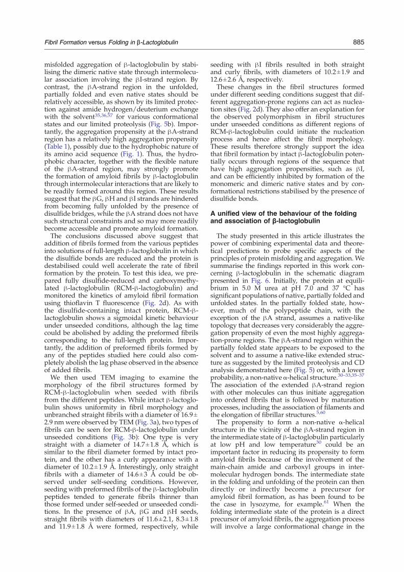
presence of 5.0 M urea.18 A similar result wasrecently obtained using an analysis of luminescencelifetime distributions58 that showed a significantpopulation (∼100%) of β-lactoglobulin in thepartially folded states in the presence of 5.0 M ureaat pH 7.0 and 37 °C. Therefore, under suchconditions, the partially folded conformation thatis most readily accessible to the protein is expectedto have stable native-like regions of β-sheet struc-ture in the vicinity of the βG and βH strands. Toinvestigate these possibilities, we characterised thestructural properties of an intermediate state accu-mulated at pH 7.0 and 37 °C during an equilibriumunfolding induced by urea using circular dichroism(CD) according to our previously established me-thod and limited proteolysis by proteinase K, a non-specific protease.We previously demonstrated that a partially
folded intermediate state with non-native α-helicalstructures accumulated during the guanidiniumhydrochloride unfolding at pH 2.0 and 4 °C byglobal-fitting analysis of far-UV CD spectra.30 Asimilar strategy has been applied here for ureaunfolding of β-lactoglobulin at pH 7.0 and 37°C(Fig. 5a; see also Supplementary Data). Unlike thespectrum obtained at low pH and low tempera-
ture,30 the CD spectrum of the intermediate state atpH 7.0 and 37 °C shown in Fig. 5a does not show asignificant increase of absolute ellipticity at 222 nm,which is diagnostic of the accumulation of α-helicalstructure. The spectrum was further analysed byCDpro,59 a software package that includes threeindependent programs (SELCON3, CDSSTR andCONTIN) to calculate the secondary structure con-tent. The results are summarised in Table 2. Accord-ing to this analysis, the secondary structure contentof the intermediate state is highly consistent withthat of the native state.Limited proteolysis of β-lactoglobulin by protei-
nase K was performed at pH 7.0 and 37 °C in thepresence of 5.0 M urea, and the resulting proteolysis-resistant fragments were examined by matrix-assisted laser desorption/ionisation time-of-flightmass spectrometry (Fig. 5b). The observed masses ofthese fragments were compared with the calculatedmasses of peptide fragments of β-lactoglobulin, thusenabling the identification of the regions of frag-ments resistant against proteinase K cleavage. Thisanalysis indicated that most fragments identifiedhere are located downstream of the βA-strandregion. According to Giurleo et al.58 almost 100%of β-lactoglobulin molecules assume a partiallyfolded structure at pH 7.0 and 37 °C in the presenceof 5.0 M urea.These results therefore suggest that the conforma-
tional state accumulated at pH 7.0 and 37 °C in thepresence of 5.0 M urea is a partially folded inter-mediate in which the N-terminal βA-strand regionin the native state is relatively flexible and accessibleto the proteolytic cleavage by proteinase K, whilethe downstream of the protein assumes relativelyrigid native-like structures resistant against protei-nase K cleavage.The native-like secondary structures in the par-
tially folded state of β-lactoglobulin identified hereshould act to inhibit at least to some extent theformation of aggregates, despite the fact that iso-lated peptides corresponding to these regions arereadily able to form amyloid fibrils. In addition, theC-terminal region close to the βI strand, which isinvolved in the dimer interface, contains anotherdisulfide bond between residue C66 and residueC160. This bond is likely to generate a conforma-tional restraint that will also inhibit the formation of
885Fibril Formation versus Folding in β-Lactoglobulin
misfolded aggregation of β-lactoglobulin by stabi-lising the dimeric native state through intermolecu-lar association involving the βI-strand region. Bycontrast, the βA-strand region in the unfolded,partially folded and even native states should berelatively accessible, as shown by its limited protec-tion against amide hydrogen/deuterium exchangewith the solvent35,36,57 for various conformationalstates and our limited proteolysis (Fig. 5b). Impor-tantly, the aggregation propensity at the βA-strandregion has a relatively high aggregation propensity(Table 1), possibly due to the hydrophobic nature ofits amino acid sequence (Fig. 1). Thus, the hydro-phobic character, together with the flexible natureof the βA-strand region, may strongly promotethe formation of amyloid fibrils by β-lactoglobulinthrough intermolecular interactions that are likely tobe readily formed around this region. These resultssuggest that the βG, βH and βI strands are hinderedfrom becoming fully unfolded by the presence ofdisulfide bridges, while the βA strand does not havesuch structural constraints and so may more readilybecome accessible and promote amyloid formation.The conclusions discussed above suggest that
addition of fibrils formed from the various peptidesinto solutions of full-length β-lactoglobulin in whichthe disulfide bonds are reduced and the protein isdestabilised could well accelerate the rate of fibrilformation by the protein. To test this idea, we pre-pared fully disulfide-reduced and carboxymethy-lated β-lactoglobulin (RCM-β-lactoglobulin) andmonitored the kinetics of amyloid fibril formationusing thioflavin T fluorescence (Fig. 2d). As withthe disulfide-containing intact protein, RCM-β-lactoglobulin shows a sigmoidal kinetic behaviourunder unseeded conditions, although the lag timecould be abolished by adding the preformed fibrilscorresponding to the full-length protein. Impor-tantly, the addition of preformed fibrils formed byany of the peptides studied here could also com-pletely abolish the lag phase observed in the absenceof added fibrils.We then used TEM imaging to examine the
morphology of the fibril structures formed byRCM-β-lactoglobulin when seeded with fibrilsfrom the different peptides. While intact β-lactoglo-bulin shows uniformity in fibril morphology andunbranched straight fibrils with a diameter of 16.9±2.9 nmwere observed by TEM (Fig. 3a), two types offibrils can be seen for RCM-β-lactoglobulin underunseeded conditions (Fig. 3b): One type is verystraight with a diameter of 14.7±1.8 Å, which issimilar to the fibril diameter formed by intact pro-tein, and the other has a curly appearance with adiameter of 10.2±1.9 Å. Interestingly, only straightfibrils with a diameter of 14.6±3 Å could be ob-served under self-seeding conditions. However,seeding with preformed fibrils of the β-lactoglobulinpeptides tended to generate fibrils thinner thanthose formed under self-seeded or unseeded condi-tions. In the presence of βA, βG and βH seeds,straight fibrils with diameters of 11.6±2.1, 8.3±1.8and 11.9±1.8 Å were formed, respectively, while
seeding with βI fibrils resulted in both straightand curly fibrils, with diameters of 10.2±1.9 and12.6±2.6 Å, respectively.These changes in the fibril structures formed
under different seeding conditions suggest that dif-ferent aggregation-prone regions can act as nuclea-tion sites (Fig. 2d). They also offer an explanation forthe observed polymorphism in fibril structuresunder unseeded conditions as different regions ofRCM-β-lactoglobulin could initiate the nucleationprocess and hence affect the fibril morphology.These results therefore strongly support the ideathat fibril formation by intact β-lactoglobulin poten-tially occurs through regions of the sequence thathave high aggregation propensities, such as βI,and can be efficiently inhibited by formation of themonomeric and dimeric native states and by con-formational restrictions stabilised by the presence ofdisulfide bonds.
A unified view of the behaviour of the foldingand association of β-lactoglobulin
The study presented in this article illustrates thepower of combining experimental data and theore-tical predictions to probe specific aspects of theprinciples of protein misfolding and aggregation. Wesummarise the findings reported in this work con-cerning β-lactoglobulin in the schematic diagrampresented in Fig. 6. Initially, the protein at equili-brium in 5.0 M urea at pH 7.0 and 37 °C hassignificant populations of native, partially folded andunfolded states. In the partially folded state, how-ever, much of the polypeptide chain, with theexception of the βA strand, assumes a native-liketopology that decreases very considerably the aggre-gation propensity of even the most highly aggrega-tion-prone regions. The βA-strand region within thepartially folded state appears to be exposed to thesolvent and to assume a native-like extended struc-ture as suggested by the limited proteolysis and CDanalysis demonstrated here (Fig. 5) or, with a lowerprobability, a non-nativeα-helical structure.30–33,35–37
The association of the extended βA-strand regionwith other molecules can thus initiate aggregationinto ordered fibrils that is followed by maturationprocesses, including the association of filaments andthe elongation of fibrillar structures.5,60
The propensity to form a non-native α-helicalstructure in the vicinity of the βA-strand region inthe intermediate state of β-lactoglobulin particularlyat low pH and low temperature30 could be animportant factor in reducing its propensity to formamyloid fibrils because of the involvement of themain-chain amide and carboxyl groups in inter-molecular hydrogen bonds. The intermediate statein the folding and unfolding of the protein can thendirectly or indirectly become a precursor foramyloid fibril formation, as has been found to bethe case in lysozyme, for example.61 When thefolding intermediate state of the protein is a directprecursor of amyloid fibrils, the aggregation processwill involve a large conformational change in the
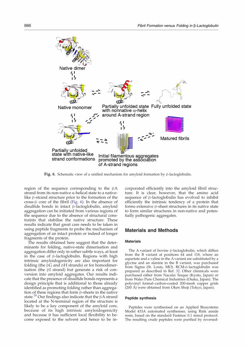
Fig. 6. Schematic view of a unified mechanism for amyloid formation by β-lactoglobulin.
886 Fibril Formation versus Folding in β-Lactoglobulin
region of the sequence corresponding to the βAstrand from its non-native α-helical state to a native-like β-strand structure prior to the formation of thecross-β core of the fibril (Fig. 6). In the absence ofdisulfide bonds in intact β-lactoglobulin, amyloidaggregation can be initiated from various regions ofthe sequence due to the absence of structural cons-traints that stabilise the native structure. Theseresults indicate that great care needs to be taken inusing peptide fragments to probe the mechanism ofaggregation of an intact protein or indeed of longerfragments of the protein.The results obtained here suggest that the deter-
minants for folding, native-state dimerisation andaggregation differ only in rather subtle ways, at leastin the case of β-lactoglobulin. Regions with highintrinsic amyloidogenicity are also important forfolding (the βG and βH strands) or for homodimer-isation (the βI strand) but generate a risk of con-version into amyloid aggregates. Our results indi-cate that the presence of disulfide bonds represents adesign principle that is additional to those alreadyidentified as promoting folding rather than aggrega-tion of these regions that form β-sheets in the nativestate.28 Our findings also indicate that the βA strandlocated at the N-terminal region of the structure islikely to be a key component of the amyloid core,because of its high intrinsic amyloidogenicityand because it has sufficient local flexibility to be-come exposed to the solvent and hence to be in-
corporated efficiently into the amyloid fibril struc-ture. It is clear, however, that the amino acidsequence of β-lactoglobulin has evolved to inhibitefficiently the intrinsic tendency of a protein thatforms extensive β-sheet structures in its native stateto form similar structures in non-native and poten-tially pathogenic aggregates.
Materials and Methods
Materials
The A variant of bovine β-lactoglobulin, which differsfrom the B variant at positions 64 and 118, where anaspartate and a valine in the Avariant are substituted by aglycine and an alanine in the B variant, was purchasedfrom Sigma (St. Louis, MO). RCM-β-lactoglobulin wasprepared as described in Ref. 32. Other chemicals werepurchased either from Nacalai Tesque (Kyoto, Japan) orfrom Wako Pure Chemical Industries (Osaka, Japan). Thepolyvinyl formal–carbon-coated 200-mesh copper grids(200 Å) were obtained from Oken Shoji (Tokyo, Japan).
Peptide synthesis
Peptides were synthesised on an Applied BiosystemsModel 433A automated synthesiser, using Rink amideresin, based on the standard Fastmoc 0.1 mmol protocol.The resulting crude peptides were purified by reversed-

887Fibril Formation versus Folding in β-Lactoglobulin
phase HPLC, and the degree of purity was checked byanalytical reversed-phase HPLC and mass spectrometry.Since all the peptides contain tryptophan or tyrosineresidues, the peptide concentrations were determined byUV absorption spectroscopy using the extinction coeffi-cients calculated according to Gill and von Hippel.62
Kinetics of fibril formation by peptides usingthioflavin T fluorescence
Thioflavin T fluorescence spectra were obtained using aJASCO fluorimeter (Model FP-6500). Peptide solutions(1 mg mL−1) in 10 mM sodium phosphate, pH 7.0, and5.0 M urea were incubated at 37 °C. After given times ofincubation, 5-μL aliquots of the peptide solutions weremixed with 400 μL of 5 μM thioflavin T dissolved in10 mM sodium phosphate at pH 7.0, and the fluorescenceat 460–660 nm was measured with an excitation wave-length of 450 nm. For seeding experiments using frag-ments, preformed fibrils of the various peptides (initialconcentration of 1 mg mL−1) were broken into smallersegments by sonication, and 10 μL of the resulting solu-tions containing fragmented fibrils was added to solutionsof β-lactoglobulin at a concentration of 1 mg mL−1 anddissolved in 10 mM sodium phosphate at pH 7.0 and5.0 M urea. The samples were incubated at 37 °C, and thepresence of fibrils was monitored by thioflavin T fluo-rescence as described above.
Fibril imaging by TEM
TEM images of fibrils were acquired with a JEM-1200EXII transmission electron microscope (JEOL, Tokyo, Japan)with an acceleration voltage of 85 keV. The sampleswere negatively stained with 1.5%–2.0% phosphotung-state adjusted at pH 7.5 by adding sodium hydroxide.The reported diameters of fibrils are the average ofmore than 100 randomly selected sections of differentfibrils.
Predictions of amyloid aggregation propensities
Predictions were made by using the Zyggregator†algorithm,50,55 which we previously introduced to predictthe intrinsic aggregation propensities of amino acidsequences by considering the physicochemical propertiesof amino acids. In these predictions, a region of a poly-peptide sequence should meet two conditions in order topromote aggregation: (1) it should have a high intrinsicaggregation propensity ZP and (2) it should be sufficientlyunstable to have a significant propensity to form inter-molecular interactions. In order to describe the latter, weused the CamP method for the prediction of structuralflexibility (lnP) from the amino acid sequence.63 For thosevalues that have ZPN0, we modified the aggregationpropensity profile ZP by modulating it with lnP,
ZPSi =ZP
i 1� lnPi
15
� �;
thus resulting in the aggregation propensity profile ZPS
with structural corrections.
†http://www-vendruscolo.ch.cam.ac.uk/zyggregator.php
Limited proteolysis by proteinase K
β-Lactoglobulin (1 mg mL−1) was dissolved in 1 mL of10 mM sodium phosphate, pH 7.0, and 5.0 M urea. Onemicroliter of 10 ng mL−1 of proteinase K dissolved indistilled water was added into the solution containingβ-lactoglobulin, and the solution was incubated for 1 minat 37 °C. The cleavage reaction was quenched by addi-tion of phenylmethylsulfonyl fluoride at room tempera-ture. Disulfide bonds were then reduced by addition ofdithiothreitol (final concentration of 10 mM) for 1 h atroom temperature. The solution was treated by ZipTipwith 0.6 μL of C4 resin (Millipore, Billerica, MA). Massesof the peptides in the solution after ZipTip treatmentwere analysed by matrix-assisted laser desorption/ioni-sation time-of-flight mass spectrometry using AXIMA-CFR (SHIMADZU, Kyoto, Japan). α-Cyano-4-hydroxy-cinnamic acid was used as matrix. The fragments wereidentified by using FindPept tool in ExPaSy‡ server.Further details are provided as supplementary data.
CD spectra
Far-UV CD spectra were monitored at 37 °C using aJ-720 spectropolarimeter from JASCO (Tokyo, Japan).Protein samples were prepared in 10 mM sodiumphosphate and various concentrations of urea at pH 7.0.A protein concentration of 1 mg mL−1 and the cell of 0.1-mm path length were used. It was essential to use the cellwith such short path length and relatively high concentra-tion of protein in order to efficiently eliminate the highabsorption of urea, particularly at shorter wavelength,below ∼220 nm. Data were expressed as molar residueellipticity [θ].The global-fitting analysis was performed as described
in Ref. 30. In order to obtain the CD spectrum of anintermediate state and various thermodynamic para-meters to explain the urea-unfolding reactions, weintroduced the following assumptions:
(1) Three states, including native dimer (N), inter-mediate (I) and fully unfolded (U), were respon-sible for the urea-unfolding transition. Thus, theobserved ellipticity at wavelength x, [θ](x), in thepresence of a given concentration of urea is ex-pressed by a linear combination of the contributionfrom the three states:
u½ � xð Þ = fN u½ �N xð Þ + fI u½ �I xð Þ + fU u½ �U xð Þ
where fi and [θ]i(x) are the fraction and ellipticityat wavelength x of the species i (U, N or I),respectively.
(2) The free-energy difference between states i and j(ΔGij) is a linear function of urea concentration,ΔGij =ΔGij(H2O)−mij [urea], where ΔGij(H2O)and mij are the free-energy difference in theabsence of urea and a measure of the coopera-tivity for urea-unfolding transition, respectively.In the case of urea unfolding of β-lactoglobulinat pH 7.0, “N” corresponds to the dimeric nativestate; therefore, ΔGNI is also a function of totalconcentration of protein in solution and ureaconcentration.
‡kr.expasy.org/tools/findpept.html
888 Fibril Formation versus Folding in β-Lactoglobulin
For the global-fitting analysis, the CD spectra of β-lactoglobulin in the absence and presence of 7.0 M ureawere used as [θ]N(x) and [θ]U(x), respectively. [θ]I(x) of theintermediate state and other thermodynamic parameterswere included as fitting parameters. The data wereanalysed by IgorPro version 6.04 (Wavemetrics, OR).We also analysed the data according to a two-state
model including native and fully unfolded states. How-ever, this approach could not provide reasonable conver-gence of the fitting result; we thus adopted the three-stateassumption made above. Details of the results of global-fitting analysis are available as supplementary data.
Acknowledgements
This work was supported in part by Grants-in-Aidfor Scientific Research on Priority Areas (no.15076201) and the Global COE Program A08 fromthe Ministry of Education, Culture, Sports, Scienceand Technology of Japan (to D.H.) and throughgrants from the Leverhulme and Wellcome Trusts(to M.V and C. M. D.). D.H. acknowledges Dr.Takashi Fukui for assistance in CDpro analysis. A.P.acknowledges with gratitude the receipt of a Cam-bridge Gates Scholarship; M.V., that of a RoyalSociety University Research Fellowship and anEMBO Young Investigator award.
Supplementary Data
Supplementary data associated with this articlecan be found, in the online version, at doi:10.1016/j.jmb.2008.12.038
References
1. Dobson, C. M., Šali, A. & Karplus, M. (1998). Proteinfolding: a perspective from theory and experiment.Angew. Chem. 110, 908–935.
2. Anfinsen, C. B. (1973). Principles that govern thefolding of protein chains. Science, 181, 223–230.
3. Dobson, C. M. (2003). Protein folding and misfolding.Nature, 426, 884–890.
4. Stefani, M. (2004). Protein misfolding and aggrega-tion: new examples in medicine and biology of thedark side of the protein world. Biochim. Biophys. Acta,1739, 5–25.
5. Stefani, M. & Dobson, C. M. (2003). Protein aggrega-tion and aggregate toxicity: new insights into proteinfolding, misfolding diseases and biological evolution.J. Mol. Med. 81, 678–699.
6. Vendruscolo, M. & Dobson, C. M. (2005). Towardscomplete descriptions of the free-energy landscapesof proteins. Philos. Trans. R. Soc. London, Ser. A, 363,433–450.
7. Jaroniec, C. P., MacPhee, C. E., Bajaj, V. S., McMahon,M. T., Dobson, C. M. & Griffin, R. G. (2004). High-resolution molecular structure of a peptide in anamyloid fibril determined by magic angle spinningNMR spectroscopy. Proc. Natl Acad. Sci. USA, 101,711–716.
8. Khurana, R., Souillac, P. O., Coats, A. C., Ionescu-Zanetti, C., Solomon, A. & Fink, A. L. (2003). Amodel for amyloid fibril formation in immunoglo-bulin light chains based on comparison of amyloi-dogenic and benign proteins and specific antibodybinding. Amyloid, 10, 97–109.
9. Makin, O. S. & Serpell, L. C. (2002). Examining thestructure of the mature amyloid fibril. Biochem. Soc.Trans. 30, 521–525.
10. Nelson, R., Sawaya, M. R., Balbirnie, M., Madsen,A.Ø., Riekel, C., Grothe, R. & Eisenberg, D. (2005).Structure of the cross-β spine of amyloid-like fibrils.Nature, 435, 773–778.
11. Sawaya, M. R., Sambashivan, S., Nelson, R., Ivanova,M. I., Sievers, S. A., Apostol, M. I. et al. (2007). Atomicstructures of amyloid cross-β spines reveal variedsteric zippers. Nature, 447, 453–457.
12. Pepys, M. B. (2001). Pathogenesis, diagnosis andtreatment of systemic amyloidosis. Philos. Trans. R.Soc. London, Ser. B, 356, 203–210.
13. Dobson, C. M. (2001). The structural basis of proteinfolding and its links with human disease. Philos. Trans.R. Soc. London, Ser. B, 356, 133–145.
14. Chiti, F., Webster, P., Taddei, N., Clark, A., Stefani, M.,Ramponi, G. & Dobson, C. M. (1999). Designingconditions for in vitro formation of amyloid protofila-ments and fibrils. Proc. Natl Acad. Sci. USA, 96,3590–3594.
15. Chiti, F., Bucciantini, M., Capanni, C., Taddei, N.,Dobson, C. M. & Stefani, M. (2001). Solution condi-tions can promote formation of either amyloidprotofilaments or mature fibrils from the HypFN-terminal domain. Protein Sci. 10, 2541–2547.
16. Fändrich, M. & Dobson, C. M. (2002). The behaviourof polyamino acids reveals an inverse side chaineffect in amyloid structure formation. EMBO J. 21,5682–5690.
17. Fändrich, M., Fletcher, M. A. & Dobson, C. M. (2001).Amyloid fibrils from muscle myoglobin. Nature, 410,165–166.
18. Hamada, D. & Dobson, C. M. (2002). A kinetic studyof β-lactoglobulin amyloid fibril formation promotedby urea. Protein Sci. 11, 2411–2426.
19. Guijarro, J. I., Sunde, M., Jones, J. A., Campbell, I. D. &Dobson, C. M. (1998). Amyloid fibril formation by anSH3 domain. Proc. Natl Acad. Sci. USA, 95, 4224–4228.
20. López de la Paz, M., Goldie, K., Zurdo, J., Lacroix, E.,Dobson, C. M., Hoenger, A. & Serrano, L. (2002). Denovo designed peptide-based amyloid fibrils. Proc.Natl Acad. Sci. USA, 99, 16052–16057.
21. Wilkins, D. K., Dobson, C. M. & Gross, M. (2000).Biophysical studies of the development of amyloidfibrils from a peptide fragment of cold shock proteinB. Eur. J. Biochem. 267, 2609–2616.
22. Dobson, C. M. (1999). Protein misfolding, evolutionand disease. Trends Biochem. Sci. 24, 329–332.
23. Hamada, D., Yanagihara, I. & Tsumoto, K. (2004).Engineering amyloidogenicity towards the develop-ment of nanofibrillar materials. Trends Biotechnol. 22,93–97.
24. Yeates, T. O. & Padilla, J. E. (2002). Designingsupramolecular protein assemblies. Curr. Opin. Struct.Biol. 12, 464–470.
25. Hamada, D., Tsumoto, K., Sawara, M., Tanaka, N.,Nakahira, K., Shiraki, K. & Yanagihara, I. (2008). Effectof an amyloidogenic sequence attached to yellowfluorescent protein. Proteins, 72, 811–821.
26. Knowles, T. P., Fitzpatrick, A. W., Meehan, S., Mott,H. R., Vendruscolo, M., Dobson, C. M. & Welland,
889Fibril Formation versus Folding in β-Lactoglobulin
M. E. (2007). Role of intermolecular forces in definingmaterial properties of protein nanofibrils. Science, 318,1900–1903.
27. Chiti, F. & Dobson, C. M. (2006). Protein misfolding,functional amyloid, and human disease. Annu. Rev.Biochem. 75, 333–366.
28. Richardson, J. S. & Richardson, D. C. (2002). Naturalβ-sheet proteins use negative design to avoid edge-to-edge aggregation. Proc. Natl Acad. Sci. USA, 99,2754–2759.
29. Hambling, S., MacAlpine, A. S. & Sawyer, L. (1992).β-Lactoglobulin. In Advanced Dairy Chemistry (Fox,P. F., ed), pp. 141–190, Elsevier, Amsterdam, TheNetherlands.
30. Hamada, D. & Goto, Y. (1997). The equilibrium inter-mediate of β-lactoglobulin with non-native α-helicalstructure. J. Mol. Biol. 269, 479–487.
31. Hamada, D., Kuroda, Y., Tanaka, T. & Goto, Y.(1995). High helical propensity of the peptide frag-ments derived from β-lactoglobulin, a predominantlyβ-sheet protein. J. Mol. Biol. 254, 737–740.
32. Hamada, D., Segawa, S. & Goto, Y. (1996). Non-native α-helical intermediate in the refolding of β-lactoglobulin, a predominantly β-sheet protein. Nat.Struct. Biol. 2, 868–874.
33. Kuroda, Y., Hamada, D., Tanaka, T. & Goto, Y. (1996).High helicity of peptide fragments corresponding toβ-strand regions of β-lactoglobulin observed by 2D-NMR spectroscopy. Fold Des. 1, 243–251.
34. Kuwajima, K., Yamaya, H. & Sugai, S. (1996). Theburst-phase intermediate in the refolding of β-lactoglobulin studied by stopped-flow circular dichro-ism and absorption spectroscopy. J. Mol. Biol. 264,806–822.
35. Kuwata, K., Shastry, R., Cheng, H., Hoshino, M., Batt,C. A., Goto, Y. & Roder, H. (2001). Structural andkinetic characterization of early folding events inβ-lactoglobulin. Nat. Struct. Biol. 8, 151–155.
36. Forge, V., Hoshino, M., Kuwata, K., Arai, M.,Kuwajima, K., Batt, C. A. & Goto, Y. (2000). Is foldingof β-lactoglobulin non-hierarchic? Intermediate withnative-like β-sheet and non-native α-helix. J. Mol. Biol.296, 1039–1051.
37. Shiraki, K., Nishikawa, K. & Goto, Y. (1995). Trifluoro-ethanol-induced stabilization of the α-helical structureof β-lactoglobulin: implication for non-hierarchicalprotein folding. J. Mol. Biol. 245, 180–194.
38. Devlin, G. L., Knowles, T. P., Squires, A., McCammon,M. G., Gras, S. L., Nilsson, M. R. et al. (2006). Thecomponent polypeptide chains of bovine insulinnucleate or inhibit aggregation of the parent proteinin a conformation-dependent manner. J. Mol. Biol. 360,497–509.
39. Hong, D. P. & Fink, A. L. (2005). Independent hetero-logous fibrillation of insulin and its B-chain peptide.Biochemistry, 44, 16701–16709.
40. Krebs, M. R., Wilkins, D. K., Chung, E. W., Pitkeathly,M. C., Chamberiain, A. K., Zurdo, J. et al. (2000).Formation and seeding of amyloid fibrils from wild-type hen lysozyme and a peptide fragment from theβ-domain. J. Mol. Biol. 300, 541–549.
41. Scrocchi, L. A., Ha, K., Chen, Y., Wu, L., Wang, F. &Fraser, P. E. (2003). Identification of minimal peptidesequences in the (8–20) domain of human islet amy-loid polypeptide involved in fibrillogenesis. J. Struct.Biol. 141, 218–227.
42. Kozhukh, G. V., Hagihara, Y., Kawakami, T., Hasegawa,K., Naiki, H. & Goto, Y. (2002). Investigation of apeptide responsible for amyloid fibril formation of
β2-microglobulin by Achromobacter protease I. J. Biol.Chem. 277, 1310–1315.
43. Jones, S., Manning, J., Kad, N. M. & Radford, S. E.(2003). Amyloid-forming peptides from β2-microglo-bulin—insights into the mechanism of fibril formationin vivo. J. Mol. Biol. 325, 249–257.
44. Ionescu-Zanetti, C., Khurana, R., Gillespie, J. R.,Petrick, J. S., Trabachino, L. C., Minert, L. et al.(1999). Monitoring the assembly of Ig light-chainamyloid fibrils by atomic force microscopy. Proc. NatlAcad. Sci. USA, 96, 13175–13179.
45. Chamberlain, A. K., MacPhee, C. E., Zurdo, J.,Morozova-Roche, L. A., Hill, H. A., Dobson, C. M. &Davis, J. J. (2000). Ultrastructural organization ofamyloid fibrils by atomic force microscopy. Biophys. J.79, 3282–3293.
46. Zurdo, J., Guijarro, J. I., Jimenez, J. L., Saibil, H. R. &Dobson, C. M. (2001). Dependence on solution con-ditions of aggregation and amyloid formation by SH3domain. J. Mol. Biol. 311, 325–340.
47. Petkova, A. T., Leapman, R. D., Guo, Z., Yau, W. M.,Mattson, M. P. & Tycko, R. (2005). Self-propagating,molecular-level polymorphism in Alzheimer's β-amyloid fibrils. Science, 307, 262–265.
48. Krishnan, R. & Lindquist, S. L. (2005). Structuralinsights into a yeast prion illuminate nucleation andstrain diversity. Nature, 435, 763–772.
49. Gosal, W. S., Clark, A. H. & Ross-Murphy, S. B. (2004).Fibrillar β-lactoglobulin gels: Part 1. Fibril formationand structure. Biomacromolecules, 5, 2408–2419.
50. Pawar, A. P., Dubay, K. F., Zurdo, J., Chiti, F.,Vendruscolo, M. & Dobson, C. M. (2005). Predictionof “aggregation-prone” and “aggregation-susceptible”regions in proteins associated with neurodegenerativediseases. J. Mol. Biol. 350, 379–392.
51. Glover, J. R., Kowal, A. S., Schirmer, E. C., Patino,M. M., Liu, J. J. & Lindquist, S. (1997). Self-seededfibers formed by Sup35, the protein determinant of[PSI+], a heritable prion-like factor of S. cerevisiae. Cell,89, 811–819.
52. Esteras-Chopo, A., Serrano, L. & Lopez de la Paz, M.(2005). The amyloid stretch hypothesis: recruitingproteins toward the dark side. Proc. Natl Acad. Sci.USA, 102, 16672–16677.
53. Ventura, S., Zurdo, J., Narayanan, S., Parreño, M.,Mangues, R., Reif, B. et al. (2004). Short amino acidstretches can mediate amyloid formation in globularproteins: the Src homology 3 (SH3) case. Proc. NatlAcad. Sci. USA, 101, 7258–7263.
54. Frare, E., Mossuto, M. F., Polverino de Laureto, P.,Dumoulin, M., Dobson, C. M. & Fonrana, A. (2006).Identification of the core structure of lysozyme amy-loid fibrils by proteolysis. J. Mol. Biol. 361, 551–561.
55. Tartaglia, G. G., Pawar, A. P., Campioni, S., Dobson,C. M., Chiti, F. & Vendruscolo, M. (2008). Predictionof aggregation-prone regions in structured proteins.J. Mol. Biol. 380, 425–436.
56. Katou, H., Hoshino, M., Kamikubo, H., Batt, C. A. &Goto, Y. (2001). Native-like β-hairpin retained in thecold-denatured state of bovine β-lactoglobulin. J. Mol.Biol. 310, 471–484.
57. Sakurai, K. & Goto, Y. (2007). Principal component ana-lysis of the pH-dependent conformational transitionsof bovine β-lactoglobulin monitored by heteronuclearNMR. Proc. Natl Acad. Sci. USA, 104, 15346–15351.
58. Giurleo, J. T., He, X. & Talaga, D. S. (2008). β-Lacto-globulin assembles into amyloid through sequentialaggregated intermediates. J. Mol. Biol. 381, 1332–1348.
59. Sreerama, N. & Woody, R. W. (2000). Estimation of

890 Fibril Formation versus Folding in β-Lactoglobulin
protein secondary structure from circular dichroismspectra: comparison of CONTIN, SELCON, andCDSSTR methods with an expanded reference set.Anal. Biochem. 287, 252–260.
60. Dumoulin, M. & Dobson, C. M. (2004). Probing theorigins, diagnosis and treatment of amyloid diseasesusing antibodies. Biochimie, 86, 589–600.
61. Canet, D., Last, A. M., Tito, P., Sunde, M., Spencer, A.,Archer, D. B. et al. (2002). Local cooperativity in the
unfolding of an amyloidogenic variant of humanlysozyme. Nat. Struct. Biol. 9, 308–315.
62. Gill, S. C. & von Hippel, P. H. (1989). Calculation ofprotein extinction coefficients from amino acidsequence data. Anal. Biochem. 189, 319–326.
63. Tartaglia, G. G., Cavalli, A. & Vendruscolo, M. (2007).Prediction of local structural stabilities of proteinsfrom their amino acid sequences. Structure, 15,139–143.