Molecular chaperones in protein folding and proteostasis
9
1 Department of Cellular Biochemistry, Max Planck Institute of Biochemistry, Am Klopferspitz 18, 82152 Martinsried, Germany. P roteins are the most versatile and structurally complex biological macromolecules. They are involved in almost every biological process. Mammalian cells typically express in excess of 10,000 different protein species, which are synthesized on ribosomes as linear chains of up to several thousand amino acids. To function, these chains must generally fold into their ‘native state’, an ensemble of a few closely related three-dimensional structures 1,2 . How this is accomplished and how cells ensure the conformational integrity of their proteome in the face of acute and chronic challenges constitute one of the most fundamental and medically relevant problems in biology. Central to this problem is that proteins must retain conformational flexibility to function, and thus are only marginally thermodynamically stable in their physiological environment. A substantial fraction of all proteins in eukaryotic cells (20–30% of the total in mammalian cells) even seem to be inherently devoid of any ordered three-dimensional structure and adopt folded conformations only after interaction with binding partners 3 . Aberrant behaviour of some of these metastable proteins, such as tau and α-synuclein, can give rise to the formation of fibrillar aggregates that are associated with dementia and Parkinson’s disease. Thus, protein quality control and the maintenance of proteome homeostasis (known as proteostasis) are crucial for cellular and organismal health. Proteostasis is achieved by an integrated network of several hundred proteins 4 , including, most prominently, molecular chaperones and their regulators, which assist in de novo folding or refolding, and the ubiquitin−proteasome system (UPS) and autophagy system, which mediate the timely removal of irreversibly misfolded and aggregated proteins. Deficiencies in proteostasis have been shown to facilitate the manifestation or progression of numerous diseases, such as neurodegeneration and dementia, type 2 diabetes, peripheral amyloidosis, lysosomal storage disease, cystic fibrosis, cancer and cardiovascular disease. A major risk factor for many of these ailments is advanced age. Indeed, studies in model organisms indicate that ageing is linked to a gradual decline in cellular proteostasis capacity 5,6 . Here we discuss recent insights into the mechanisms of chaperone- assisted protein folding and proteome maintenance. We focus on how proteins use the chaperone machinery to navigate successfully the complex folding-energy landscape in the crowded cellular environment. Understanding these reactions will guide future efforts to define the proteostasis network as a target for pharmacological intervention in diseases of aberrant protein folding. Fundamental role of molecular chaperones Many small proteins refold after their removal from denaturant in vitro, in the absence of other components or an energy source. This signifies that the amino-acid sequence, encoded in the DNA, contains all of the necessary information to specify the three-dimensional structure of a protein 1 . However, research over the past couple of decades has firmly established that in the cellular environment, many proteins require molecular chaperones to fold efficiently and on a biologically relevant timescale 7 . Why is this extra layer of complexity necessary? Although small proteins may fold at very fast speeds 8 (within microseconds), in dilute buffer solutions, larger, multidomain proteins may take minutes to hours to fold 9 , and often even fail to reach their native states in vitro. The folding of such proteins becomes considerably more challenging in vivo, because the cellular environment is highly crowded, with total cytosolic protein reaching concentrations of 300−400 g l −1 . The resultant excluded volume effects, although enhancing the functional interactions between macromolecules, also strongly increase the tendency of non-native and structurally flexible proteins to aggregate 10 . It seems likely, therefore, that the fundamental requirement for molecular chaperones arose very early during the evolution of densely crowded cells, owing to the need to minimize protein aggregation during folding and maintain proteins in soluble, yet conformationally dynamic states. Moreover, as mutations often disrupt the ability of a protein to adopt a stable fold 11 , it follows that the chaperone system provides a crucial buffer, allowing the evolution of new protein functions and phenotypic traits 11,12 . Some basics on protein folding and how it can go awry Because the number of possible conformations a protein chain can adopt is very large, folding reactions are highly complex and heterogeneous, relying on the cooperation of many weak, non-covalent interactions. In the case of soluble proteins, hydrophobic forces are particularly important in driving chain collapse and the burial of non- polar amino-acid residues within the interior of the protein (see ref. 13 for a discussion of membrane protein folding). Considerable progress has been made in recent years in understanding these reactions through biophysical experiments and theoretical analyses 1,2 . In the current model, polypeptide chains are thought to explore funnel- shaped potential energy surfaces as they progress, along several Most proteins must fold into defined three-dimensional structures to gain functional activity. But in the cellular environ- ment, newly synthesized proteins are at great risk of aberrant folding and aggregation, potentially forming toxic species. To avoid these dangers, cells invest in a complex network of molecular chaperones, which use ingenious mechanisms to prevent aggregation and promote efficient folding. Because protein molecules are highly dynamic, constant chaperone surveillance is required to ensure protein homeostasis (proteostasis). Recent advances suggest that an age-related decline in proteostasis capacity allows the manifestation of various protein-aggregation diseases, including Alzheimer’s disease and Parkinson’s disease. Interventions in these and numerous other pathological states may spring from a detailed under- standing of the pathways underlying proteome maintenance. Molecular chaperones in protein folding and proteostasis F. Ulrich Hartl 1 , Andreas Bracher 1 & Manajit Hayer-Hartl 1 324 | NATURE | VOL 475 | 21 JULY 2011 REVIEW doi:10.1038/nature10317 © 2011 Macmillan Publishers Limited. All rights reserved
-
Upload
biochem-mpg -
Category
Documents
-
view
0 -
download
0
Transcript of Molecular chaperones in protein folding and proteostasis
1Department of Cellular Biochemistry, Max Planck Institute of Biochemistry, Am Klopferspitz 18, 82152 Martinsried, Germany.
Proteins are the most versatile and structurally complex biological macromolecules. They are involved in almost every biological process. Mammalian cells typically express in excess of 10,000
different protein species, which are synthesized on ribosomes as linear chains of up to several thousand amino acids. To function, these chains must generally fold into their ‘native state’, an ensemble of a few closely related three-dimensional structures1,2. How this is accomplished and how cells ensure the conformational integrity of their proteome in the face of acute and chronic challenges constitute one of the most fundamental and medically relevant problems in biology.
Central to this problem is that proteins must retain conformational flexibility to function, and thus are only marginally thermodynamically stable in their physiological environment. A substantial fraction of all proteins in eukaryotic cells (20–30% of the total in mammalian cells) even seem to be inherently devoid of any ordered three-dimensional structure and adopt folded conformations only after interaction with binding partners3. Aberrant behaviour of some of these metastable proteins, such as tau and α-synuclein, can give rise to the formation of fibrillar aggregates that are associated with dementia and Parkinson’s disease. Thus, protein quality control and the maintenance of proteome homeostasis (known as proteostasis) are crucial for cellular and organismal health. Proteostasis is achieved by an integrated network of several hundred proteins4, including, most prominently, molecular chaperones and their regulators, which assist in de novo folding or refolding, and the ubiquitin−proteasome system (UPS) and autophagy system, which mediate the timely removal of irreversibly misfolded and aggregated proteins. Deficiencies in proteostasis have been shown to facilitate the manifestation or progression of numerous diseases, such as neurodegeneration and dementia, type 2 diabetes, peripheral amyloidosis, lysosomal storage disease, cystic fibrosis, cancer and cardiovascular disease. A major risk factor for many of these ailments is advanced age. Indeed, studies in model organisms indicate that ageing is linked to a gradual decline in cellular proteostasis capacity5,6.
Here we discuss recent insights into the mechanisms of chaperone-assisted protein folding and proteome maintenance. We focus on how proteins use the chaperone machinery to navigate successfully the complex folding-energy landscape in the crowded cellular environment. Understanding these reactions will guide future efforts to define the proteostasis network as a target for pharmacological intervention in diseases of aberrant protein folding.
Fundamental role of molecular chaperonesMany small proteins refold after their removal from denaturant in vitro, in the absence of other components or an energy source. This signifies that the amino-acid sequence, encoded in the DNA, contains all of the necessary information to specify the three-dimensional structure of a protein1. However, research over the past couple of decades has firmly established that in the cellular environment, many proteins require molecular chaperones to fold efficiently and on a biologically relevant timescale7. Why is this extra layer of complexity necessary?
Although small proteins may fold at very fast speeds8 (within microseconds), in dilute buffer solutions, larger, multidomain proteins may take minutes to hours to fold9, and often even fail to reach their native states in vitro. The folding of such proteins becomes considerably more challenging in vivo, because the cellular environment is highly crowded, with total cytosolic protein reaching concentrations of 300−400 g l−1. The resultant excluded volume effects, although enhancing the functional interactions between macromolecules, also strongly increase the tendency of non-native and structurally flexible proteins to aggregate10. It seems likely, therefore, that the fundamental requirement for molecular chaperones arose very early during the evolution of densely crowded cells, owing to the need to minimize protein aggregation during folding and maintain proteins in soluble, yet conformationally dynamic states. Moreover, as mutations often disrupt the ability of a protein to adopt a stable fold11, it follows that the chaperone system provides a crucial buffer, allowing the evolution of new protein functions and phenotypic traits11,12.
Some basics on protein folding and how it can go awryBecause the number of possible conformations a protein chain can adopt is very large, folding reactions are highly complex and heterogeneous, relying on the cooperation of many weak, non-covalent interactions. In the case of soluble proteins, hydrophobic forces are particularly important in driving chain collapse and the burial of non-polar amino-acid residues within the interior of the protein (see ref. 13 for a discussion of membrane protein folding). Considerable progress has been made in recent years in understanding these reactions through biophysical experiments and theoretical analyses1,2. In the current model, polypeptide chains are thought to explore funnel-shaped potential energy surfaces as they progress, along several
Most proteins must fold into defined three-dimensional structures to gain functional activity. But in the cellular environ-ment, newly synthesized proteins are at great risk of aberrant folding and aggregation, potentially forming toxic species. To avoid these dangers, cells invest in a complex network of molecular chaperones, which use ingenious mechanisms to prevent aggregation and promote efficient folding. Because protein molecules are highly dynamic, constant chaperone surveillance is required to ensure protein homeostasis (proteostasis). Recent advances suggest that an age-related decline in proteostasis capacity allows the manifestation of various protein-aggregation diseases, including Alzheimer’s disease and Parkinson’s disease. Interventions in these and numerous other pathological states may spring from a detailed under-standing of the pathways underlying proteome maintenance.
Molecular chaperones in protein folding and proteostasisF. Ulrich Hartl1, Andreas Bracher1 & Manajit Hayer-Hartl1
3 2 4 | N A T U R E | V O L 4 7 5 | 2 1 J U L Y 2 0 1 1
REVIEWdoi:10.1038/nature10317
© 2011 Macmillan Publishers Limited. All rights reserved
wtren
高亮
wtren
高亮
wtren
高亮
wtren
高亮
wtren
高亮
wtren
高亮
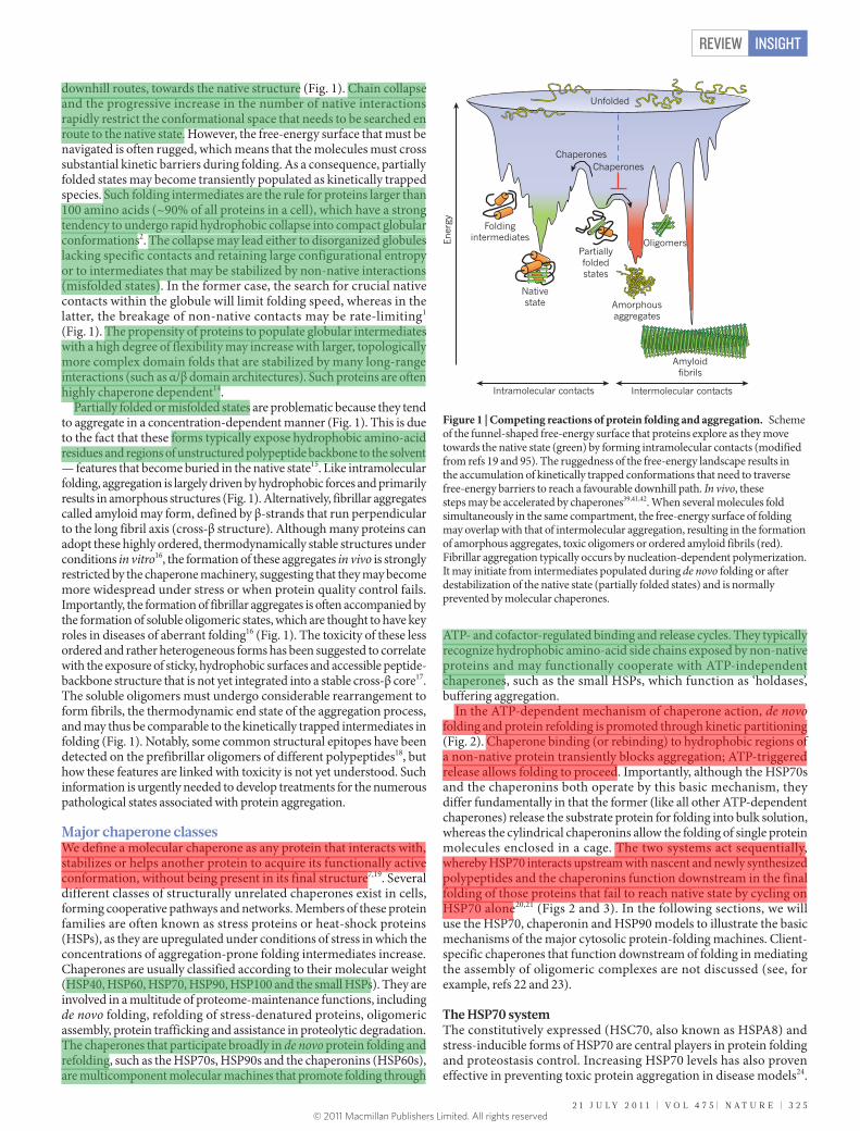
downhill routes, towards the native structure (Fig. 1). Chain collapse and the progressive increase in the number of native interactions rapidly restrict the conformational space that needs to be searched en route to the native state. However, the free-energy surface that must be navigated is often rugged, which means that the molecules must cross substantial kinetic barriers during folding. As a consequence, partially folded states may become transiently populated as kinetically trapped species. Such folding intermediates are the rule for proteins larger than 100 amino acids (~90% of all proteins in a cell), which have a strong tendency to undergo rapid hydrophobic collapse into compact globular conformations2. The collapse may lead either to disorganized globules lacking specific contacts and retaining large configurational entropy or to intermediates that may be stabilized by non-native interactions (misfolded states). In the former case, the search for crucial native contacts within the globule will limit folding speed, whereas in the latter, the breakage of non-native contacts may be rate-limiting1 (Fig. 1). The propensity of proteins to populate globular intermediates with a high degree of flexibility may increase with larger, topologically more complex domain folds that are stabilized by many long-range interactions (such as α/β domain architectures). Such proteins are often highly chaperone dependent14.
Partially folded or misfolded states are problematic because they tend to aggregate in a concentration-dependent manner (Fig. 1). This is due to the fact that these forms typically expose hydrophobic amino-acid residues and regions of unstructured polypeptide backbone to the solvent — features that become buried in the native state15. Like intramolecular folding, aggregation is largely driven by hydrophobic forces and primarily results in amorphous structures (Fig. 1). Alternatively, fibrillar aggregates called amyloid may form, defined by β-strands that run perpendicular to the long fibril axis (cross-β structure). Although many proteins can adopt these highly ordered, thermodynamically stable structures under conditions in vitro16, the formation of these aggregates in vivo is strongly restricted by the chaperone machinery, suggesting that they may become more widespread under stress or when protein quality control fails. Importantly, the formation of fibrillar aggregates is often accompanied by the formation of soluble oligomeric states, which are thought to have key roles in diseases of aberrant folding16 (Fig. 1). The toxicity of these less ordered and rather heterogeneous forms has been suggested to correlate with the exposure of sticky, hydrophobic surfaces and accessible peptide-backbone structure that is not yet integrated into a stable cross-β core17. The soluble oligomers must undergo considerable rearrangement to form fibrils, the thermodynamic end state of the aggregation process, and may thus be comparable to the kinetically trapped intermediates in folding (Fig. 1). Notably, some common structural epitopes have been detected on the prefibrillar oligomers of different polypeptides18, but how these features are linked with toxicity is not yet understood. Such information is urgently needed to develop treatments for the numerous pathological states associated with protein aggregation.
Major chaperone classes We define a molecular chaperone as any protein that interacts with, stabilizes or helps another protein to acquire its functionally active conformation, without being present in its final structure7,19. Several different classes of structurally unrelated chaperones exist in cells, forming cooperative pathways and networks. Members of these protein families are often known as stress proteins or heat-shock proteins (HSPs), as they are upregulated under conditions of stress in which the concentrations of aggregation-prone folding intermediates increase. Chaperones are usually classified according to their molecular weight (HSP40, HSP60, HSP70, HSP90, HSP100 and the small HSPs). They are involved in a multitude of proteome-maintenance functions, including de novo folding, refolding of stress-denatured proteins, oligomeric assembly, protein trafficking and assistance in proteolytic degradation. The chaperones that participate broadly in de novo protein folding and refolding, such as the HSP70s, HSP90s and the chaperonins (HSP60s), are multicomponent molecular machines that promote folding through
ATP- and cofactor-regulated binding and release cycles. They typically recognize hydrophobic amino-acid side chains exposed by non-native proteins and may functionally cooperate with ATP-independent chaperones, such as the small HSPs, which function as ‘holdases’, buffering aggregation.
In the ATP-dependent mechanism of chaperone action, de novo folding and protein refolding is promoted through kinetic partitioning (Fig. 2). Chaperone binding (or rebinding) to hydrophobic regions of a non-native protein transiently blocks aggregation; ATP-triggered release allows folding to proceed. Importantly, although the HSP70s and the chaperonins both operate by this basic mechanism, they differ fundamentally in that the former (like all other ATP-dependent chaperones) release the substrate protein for folding into bulk solution, whereas the cylindrical chaperonins allow the folding of single protein molecules enclosed in a cage. The two systems act sequentially, whereby HSP70 interacts upstream with nascent and newly synthesized polypeptides and the chaperonins function downstream in the final folding of those proteins that fail to reach native state by cycling on HSP70 alone20,21 (Figs 2 and 3). In the following sections, we will use the HSP70, chaperonin and HSP90 models to illustrate the basic mechanisms of the major cytosolic protein-folding machines. Client-specific chaperones that function downstream of folding in mediating the assembly of oligomeric complexes are not discussed (see, for example, refs 22 and 23).
The HSP70 systemThe constitutively expressed (HSC70, also known as HSPA8) and stress-inducible forms of HSP70 are central players in protein folding and proteostasis control. Increasing HSP70 levels has also proven effective in preventing toxic protein aggregation in disease models24.
Unfolded
Ener
gy
Foldingintermediates
Nativestate
Partiallyfoldedstates
Amorphousaggregates
Amyloid�brils
Oligomers
Intramolecular contacts Intermolecular contacts
ChaperonesChaperones
Figure 1 | Competing reactions of protein folding and aggregation. Scheme of the funnel-shaped free-energy surface that proteins explore as they move towards the native state (green) by forming intramolecular contacts (modified from refs 19 and 95). The ruggedness of the free-energy landscape results in the accumulation of kinetically trapped conformations that need to traverse free-energy barriers to reach a favourable downhill path. In vivo, these steps may be accelerated by chaperones39,41,42. When several molecules fold simultaneously in the same compartment, the free-energy surface of folding may overlap with that of intermolecular aggregation, resulting in the formation of amorphous aggregates, toxic oligomers or ordered amyloid fibrils (red). Fibrillar aggregation typically occurs by nucleation-dependent polymerization. It may initiate from intermediates populated during de novo folding or after destabilization of the native state (partially folded states) and is normally prevented by molecular chaperones.
2 1 J U L Y 2 0 1 1 | V O L 4 7 5 | N A T U R E | 3 2 5
REVIEW INSIGHT
© 2011 Macmillan Publishers Limited. All rights reserved
wtren
高亮
wtren
高亮
wtren
高亮
wtren
高亮
wtren
高亮
wtren
高亮
wtren
高亮
wtren
高亮
wtren
高亮
wtren
高亮
wtren
高亮
wtren
高亮
wtren
高亮
wtren
高亮
wtren
高亮
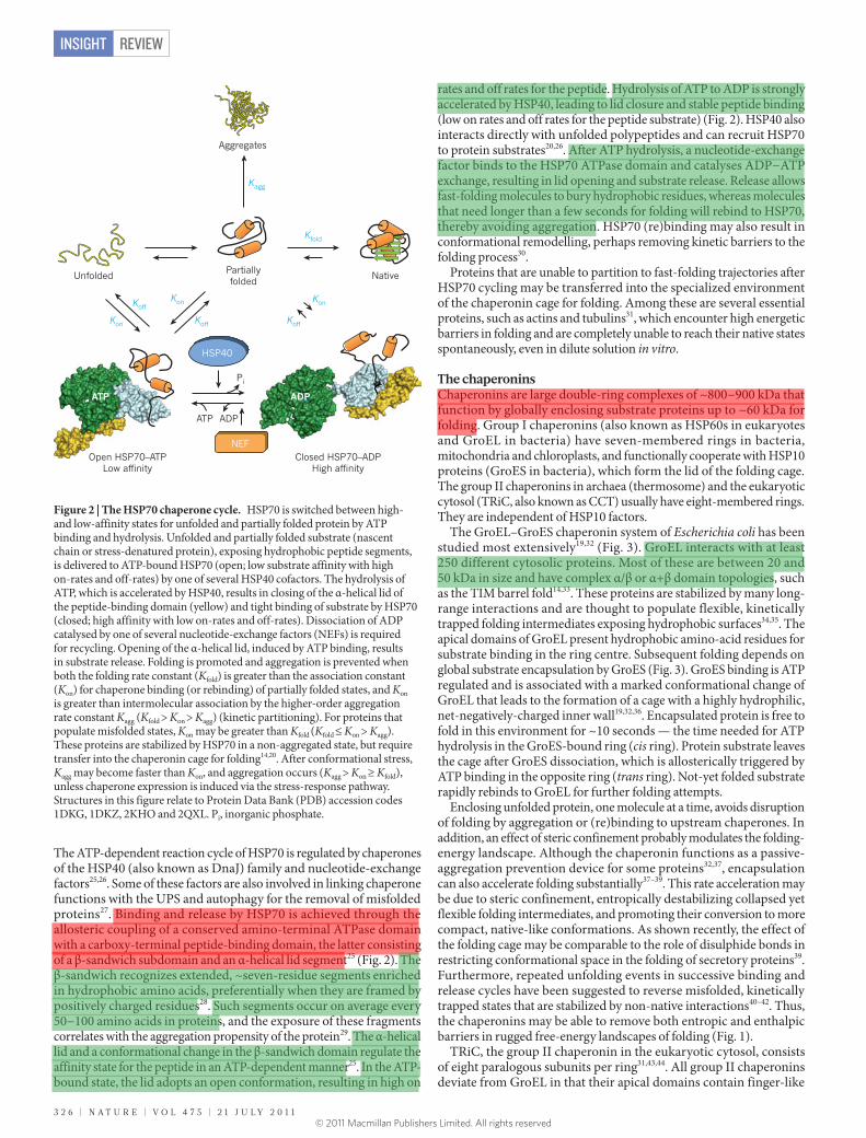
The ATP-dependent reaction cycle of HSP70 is regulated by chaperones of the HSP40 (also known as DnaJ) family and nucleotide-exchange factors25,26. Some of these factors are also involved in linking chaperone functions with the UPS and autophagy for the removal of misfolded proteins27. Binding and release by HSP70 is achieved through the allosteric coupling of a conserved amino-terminal ATPase domain with a carboxy-terminal peptide-binding domain, the latter consisting of a β-sandwich subdomain and an α-helical lid segment25 (Fig. 2). The β-sandwich recognizes extended, ~seven-residue segments enriched in hydrophobic amino acids, preferentially when they are framed by positively charged residues28. Such segments occur on average every 50−100 amino acids in proteins, and the exposure of these fragments correlates with the aggregation propensity of the protein29. The α-helical lid and a conformational change in the β-sandwich domain regulate the affinity state for the peptide in an ATP-dependent manner25. In the ATP-bound state, the lid adopts an open conformation, resulting in high on
rates and off rates for the peptide. Hydrolysis of ATP to ADP is strongly accelerated by HSP40, leading to lid closure and stable peptide binding (low on rates and off rates for the peptide substrate) (Fig. 2). HSP40 also interacts directly with unfolded polypeptides and can recruit HSP70 to protein substrates20,26. After ATP hydrolysis, a nucleotide-exchange factor binds to the HSP70 ATPase domain and catalyses ADP−ATP exchange, resulting in lid opening and substrate release. Release allows fast-folding molecules to bury hydrophobic residues, whereas molecules that need longer than a few seconds for folding will rebind to HSP70, thereby avoiding aggregation. HSP70 (re)binding may also result in conformational remodelling, perhaps removing kinetic barriers to the folding process30.
Proteins that are unable to partition to fast-folding trajectories after HSP70 cycling may be transferred into the specialized environment of the chaperonin cage for folding. Among these are several essential proteins, such as actins and tubulins31, which encounter high energetic barriers in folding and are completely unable to reach their native states spontaneously, even in dilute solution in vitro.
The chaperoninsChaperonins are large double-ring complexes of ~800−900 kDa that function by globally enclosing substrate proteins up to ~60 kDa for folding. Group I chaperonins (also known as HSP60s in eukaryotes and GroEL in bacteria) have seven-membered rings in bacteria, mitochondria and chloroplasts, and functionally cooperate with HSP10 proteins (GroES in bacteria), which form the lid of the folding cage. The group II chaperonins in archaea (thermosome) and the eukaryotic cytosol (TRiC, also known as CCT) usually have eight-membered rings. They are independent of HSP10 factors.
The GroEL–GroES chaperonin system of Escherichia coli has been studied most extensively19,32 (Fig. 3). GroEL interacts with at least 250 different cytosolic proteins. Most of these are between 20 and 50 kDa in size and have complex α/β or α+β domain topologies, such as the TIM barrel fold14,33. These proteins are stabilized by many long-range interactions and are thought to populate flexible, kinetically trapped folding intermediates exposing hydrophobic surfaces34,35. The apical domains of GroEL present hydrophobic amino-acid residues for substrate binding in the ring centre. Subsequent folding depends on global substrate encapsulation by GroES (Fig. 3). GroES binding is ATP regulated and is associated with a marked conformational change of GroEL that leads to the formation of a cage with a highly hydrophilic, net-negatively-charged inner wall19,32,36. Encapsulated protein is free to fold in this environment for ~10 seconds — the time needed for ATP hydrolysis in the GroES-bound ring (cis ring). Protein substrate leaves the cage after GroES dissociation, which is allosterically triggered by ATP binding in the opposite ring (trans ring). Not-yet folded substrate rapidly rebinds to GroEL for further folding attempts.
Enclosing unfolded protein, one molecule at a time, avoids disruption of folding by aggregation or (re)binding to upstream chaperones. In addition, an effect of steric confinement probably modulates the folding-energy landscape. Although the chaperonin functions as a passive-aggregation prevention device for some proteins32,37, encapsulation can also accelerate folding substantially37–39. This rate acceleration may be due to steric confinement, entropically destabilizing collapsed yet flexible folding intermediates, and promoting their conversion to more compact, native-like conformations. As shown recently, the effect of the folding cage may be comparable to the role of disulphide bonds in restricting conformational space in the folding of secretory proteins39. Furthermore, repeated unfolding events in successive binding and release cycles have been suggested to reverse misfolded, kinetically trapped states that are stabilized by non-native interactions40–42. Thus, the chaperonins may be able to remove both entropic and enthalpic barriers in rugged free-energy landscapes of folding (Fig. 1).
TRiC, the group II chaperonin in the eukaryotic cytosol, consists of eight paralogous subunits per ring31,43,44. All group II chaperonins deviate from GroEL in that their apical domains contain finger-like
Figure 2 | The HSP70 chaperone cycle. HSP70 is switched between high- and low-affinity states for unfolded and partially folded protein by ATP binding and hydrolysis. Unfolded and partially folded substrate (nascent chain or stress-denatured protein), exposing hydrophobic peptide segments, is delivered to ATP-bound HSP70 (open; low substrate affinity with high on-rates and off-rates) by one of several HSP40 cofactors. The hydrolysis of ATP, which is accelerated by HSP40, results in closing of the α-helical lid of the peptide-binding domain (yellow) and tight binding of substrate by HSP70 (closed; high affinity with low on-rates and off-rates). Dissociation of ADP catalysed by one of several nucleotide-exchange factors (NEFs) is required for recycling. Opening of the α-helical lid, induced by ATP binding, results in substrate release. Folding is promoted and aggregation is prevented when both the folding rate constant (Kfold) is greater than the association constant (Kon) for chaperone binding (or rebinding) of partially folded states, and Kon is greater than intermolecular association by the higher-order aggregation rate constant Kagg (Kfold > Kon > Kagg) (kinetic partitioning). For proteins that populate misfolded states, Kon may be greater than Kfold (Kfold ≤ Kon > Kagg). These proteins are stabilized by HSP70 in a non-aggregated state, but require transfer into the chaperonin cage for folding14,20. After conformational stress, Kagg may become faster than Kon, and aggregation occurs (Kagg > Kon ≥ Kfold), unless chaperone expression is induced via the stress-response pathway. Structures in this figure relate to Protein Data Bank (PDB) accession codes 1DKG, 1DKZ, 2KHO and 2QXL. Pi, inorganic phosphate.
NativePartiallyfolded
Aggregates
Open HSP70–ATPLow a�nity
Closed HSP70–ADPHigh a�nity
Pi
ADP ATP
Unfolded
Kon
Kon
Ko�
Kagg
Kfold
ADP ATP
NEF
HSP40
Ko�
Ko�
Kon
3 2 6 | N A T U R E | V O L 4 7 5 | 2 1 J U L Y 2 0 1 1
REVIEWINSIGHT
© 2011 Macmillan Publishers Limited. All rights reserved
wtren
高亮
wtren
高亮
wtren
高亮
wtren
高亮
wtren
高亮
wtren
高亮
wtren
高亮
wtren
高亮
wtren
高亮
wtren
高亮
protrusions, which act as an iris-like, built-in lid and replace the function of GroES. These segments open and close in an ATP-dependent protein-encapsulation cycle, similar in principle to that of GroEL–GroES44. However, the TRiC reaction cycle is much slower than that of GroEL, probably providing a substantially longer period of protein encapsulation and folding in the cage45. TRiC interacts with approximately 10% of newly synthesized cytosolic proteins, including actin and tubulins31,43. Interestingly, TRiC also functions in preventing the accumulation of toxic aggregates by the Huntington’s disease protein46–48.
The HSP90 systemHSP90 forms a proteostasis hub that controls numerous important signalling pathways in eukaryotic cells49. These pleiotropic functions include, among others, cell-cycle progression, telomere maintenance, apoptosis, mitotic signal transduction, vesicle-mediated transport, innate immunity and targeted protein degradation. Indeed, the evolution and maintenance of these functional networks is thought to depend on the ability of HSP90 to buffer the effects of structurally destabilizing mutations in the underlying protein complexes, thereby allowing the acquisition of new traits12.
HSP90 functions downstream of HSP70 in the structural maturation and conformational regulation of numerous signal-transduction molecules, such as kinases and steroid receptors49,50. It cooperates in this process with several regulators and co-chaperones, many of which use tetratricopeptide repeat (TPR) domains to dock onto HSP90. For example, the TPR protein HOP provides a direct link between HSP70 and HSP90, allowing substrate transfer51. Although the mechanism by which HSP90 and its cofactors mediate conformational changes
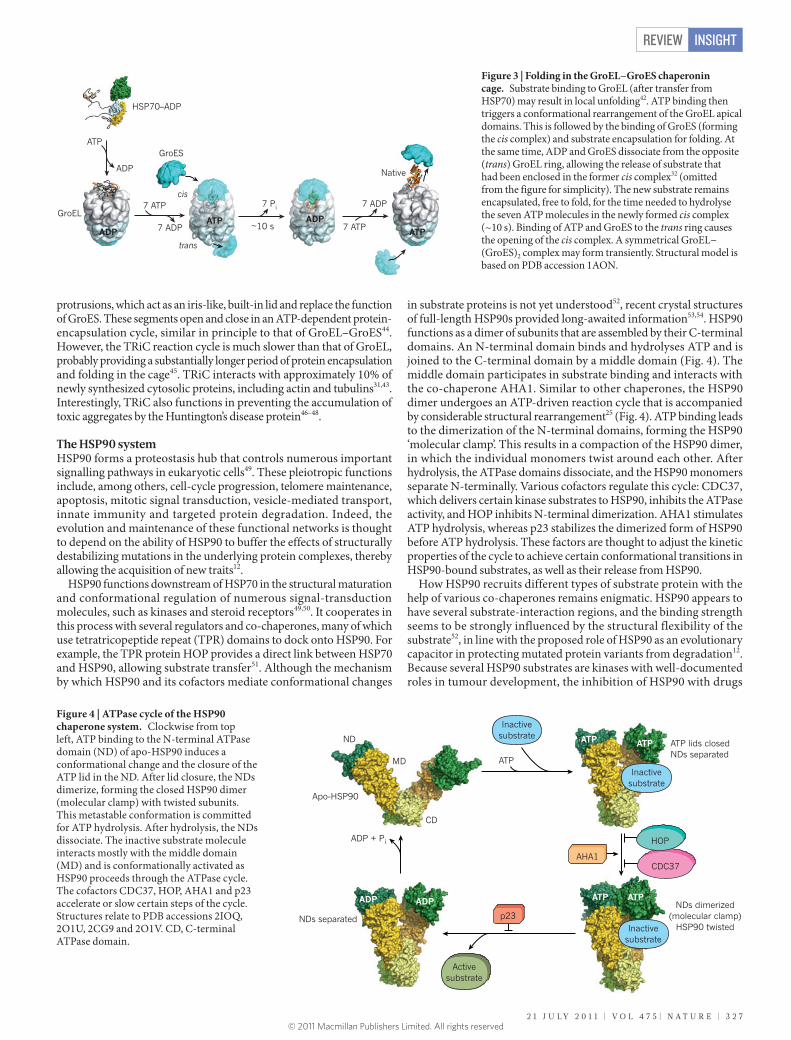
in substrate proteins is not yet understood52, recent crystal structures of full-length HSP90s provided long-awaited information53,54. HSP90 functions as a dimer of subunits that are assembled by their C-terminal domains. An N-terminal domain binds and hydrolyses ATP and is joined to the C-terminal domain by a middle domain (Fig. 4). The middle domain participates in substrate binding and interacts with the co-chaperone AHA1. Similar to other chaperones, the HSP90 dimer undergoes an ATP-driven reaction cycle that is accompanied by considerable structural rearrangement25 (Fig. 4). ATP binding leads to the dimerization of the N-terminal domains, forming the HSP90 ‘molecular clamp’. This results in a compaction of the HSP90 dimer, in which the individual monomers twist around each other. After hydrolysis, the ATPase domains dissociate, and the HSP90 monomers separate N-terminally. Various cofactors regulate this cycle: CDC37, which delivers certain kinase substrates to HSP90, inhibits the ATPase activity, and HOP inhibits N-terminal dimerization. AHA1 stimulates ATP hydrolysis, whereas p23 stabilizes the dimerized form of HSP90 before ATP hydrolysis. These factors are thought to adjust the kinetic properties of the cycle to achieve certain conformational transitions in HSP90-bound substrates, as well as their release from HSP90.
How HSP90 recruits different types of substrate protein with the help of various co-chaperones remains enigmatic. HSP90 appears to have several substrate-interaction regions, and the binding strength seems to be strongly influenced by the structural flexibility of the substrate52, in line with the proposed role of HSP90 as an evolutionary capacitor in protecting mutated protein variants from degradation12. Because several HSP90 substrates are kinases with well-documented roles in tumour development, the inhibition of HSP90 with drugs
p23
ATP
ADP + Pi
ATP ATP
ATP ATP ADP ADP
ATP lids closedNDs separated
NDs separated
NDs dimerized(molecular clamp)
HSP90 twisted
Apo-HSP90
AHA1CDC37
HOP
ND
MD
CD
Inactivesubstrate
Inactivesubstrate
Inactivesubstrate
Activesubstrate
Figure 4 | ATPase cycle of the HSP90 chaperone system. Clockwise from top left, ATP binding to the N-terminal ATPase domain (ND) of apo-HSP90 induces a conformational change and the closure of the ATP lid in the ND. After lid closure, the NDs dimerize, forming the closed HSP90 dimer (molecular clamp) with twisted subunits. This metastable conformation is committed for ATP hydrolysis. After hydrolysis, the NDs dissociate. The inactive substrate molecule interacts mostly with the middle domain (MD) and is conformationally activated as HSP90 proceeds through the ATPase cycle. The cofactors CDC37, HOP, AHA1 and p23 accelerate or slow certain steps of the cycle. Structures relate to PDB accessions 2IOQ, 2O1U, 2CG9 and 2O1V. CD, C-terminal ATPase domain.
~10 s
HSP70–ADP
7 Pi7 ATP
7 ADPATP
ADPADP
7 ATP
7 ADP
ATP
Native
ATP
ADP
cis
trans
GroES
GroEL
Figure 3 | Folding in the GroEL−GroES chaperonin cage. Substrate binding to GroEL (after transfer from HSP70) may result in local unfolding42. ATP binding then triggers a conformational rearrangement of the GroEL apical domains. This is followed by the binding of GroES (forming the cis complex) and substrate encapsulation for folding. At the same time, ADP and GroES dissociate from the opposite (trans) GroEL ring, allowing the release of substrate that had been enclosed in the former cis complex32 (omitted from the figure for simplicity). The new substrate remains encapsulated, free to fold, for the time needed to hydrolyse the seven ATP molecules in the newly formed cis complex (~10 s). Binding of ATP and GroES to the trans ring causes the opening of the cis complex. A symmetrical GroEL−(GroES)2 complex may form transiently. Structural model is based on PDB accession 1AON.
2 1 J U L Y 2 0 1 1 | V O L 4 7 5 | N A T U R E | 3 2 7
REVIEW INSIGHT
© 2011 Macmillan Publishers Limited. All rights reserved
such as geldanamycin has emerged as a promising strategy for the treatment of certain cancers55. These drugs specifically inhibit the ATPase function of HSP90. They will probably prove useful not only in cancer therapy but also in the treatment of viral diseases, owing to the fact that various pathogenic viruses hijack the HSP90 system and use it for capsid assembly56. However, the global inhibition of HSP90 is likely to result in a marked derangement of cellular circuitry, and it would be desirable to find ways to inhibit only specific aspects of HSP90 function.
From ribosome to folded protein The vectorial synthesis of polypeptides on the ribosome has important implications in the folding process that are only partly understood. Key questions concern the stage at which the nascent chain begins to fold and the extent to which the translation process modulates the free-energy landscape of folding. In addressing these issues, it is useful to first consider small, single-domain proteins, which tend to fold spontaneously in vitro. The translation process for such proteins seems to increase the risk of misfolding and aggregation considerably, because an incomplete nascent polypeptide is unable to fold into a stable native conformation57,58 and the local concentration of nascent chains in the context of polyribosomes is very high. Furthermore, the exit channel of the large ribosomal subunit, which is ~100 Å long but at most 20 Å wide, is unfavourable to folding beyond α-helices and small tertiary elements that may begin to form near the tunnel exit59–61; it
thus prevents the C-terminal 30−40 amino-acid residues of the chain from participating in long-range interactions that are necessary for cooperative domain folding. As a consequence, productive folding may occur only after the complete protein has emerged from the ribosome57,62. Because translation is relatively slow (~4−20 amino acids s−1), nascent chains are exposed in partially folded, aggregation-sensitive states for considerable periods of time. Moreover, non-native intrachain contacts formed during translation or interactions with the highly charged ribosomal surface could delay folding after completion of synthesis. For these reasons, nascent chains are thought to interact co-translationally with ribosome-bound chaperones, which inhibit their premature (mis)folding and maintain the nascent chain in a non-aggregated, folding-competent state (Fig. 5). For example, the bacterial trigger factor63 binds to the small titin I27 chain (~120 amino acids) throughout translation64, presumably delaying chain collapse until the complete β-sandwich domain has emerged from the ribosome and is available for folding. Moreover, the aggregation of nascent chains is disfavoured by the densely packed, pseudohelical arrangement of ribosomes in polyribosome complexes — an organization that maximizes the distance between nascent-chain exit sites on adjacent ribosomes65.
Although single-domain proteins will reach their native state post-translationally, multidomain proteins may undergo domain-wise co-translational folding, as independently folding structural units (~50–300 amino acids in length) emerge sequentially from the
~70%
Bacteria
N
HSP90system
Eukaryotes
mRNARibosome
TF
DnaKDnaJ
GroEL–GroES TRiC
~20%
N
~10%
N
+ GrpE, ATP(NEF)
Otherchaperones
+ ATP
NAC,Rac
HSP40HSP70
+ NEFs,+ ATP
HOP
~20%
N
N
+ ATP
Signalling
PFD
N
~10%
N
+ ATP
+ NEFs, ATP
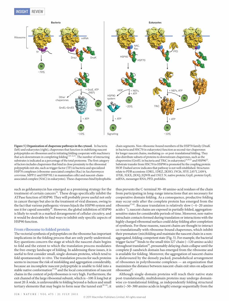
Figure 5 | Organization of chaperone pathways in the cytosol. In bacteria (left) and eukaryotes (right), chaperones that function in stabilizing nascent polypeptides on ribosomes and in initiating folding cooperate with machinery that acts downstream in completing folding7,19–21,31. The number of interacting substrates is indicated as a percentage of the total proteome. The first category of factors includes chaperones that bind in close proximity to the ribosomal polypeptide exit site, such as trigger factor (TF) in bacteria and specialized HSP70 complexes (ribosome-associated complex (Rac) in Saccharomyces cerevisiae, MPP11 and HSP70L1 in mammalian cells) and nascent-chain-associated complex (NAC) in eukaryotes). These chaperones bind hydrophobic
chain segments. Non-ribosome-bound members of the HSP70 family (DnaK in bacteria and HSC70 in eukaryotes) function as second-tier chaperones for longer nascent chains, mediating co- or post-translational folding. They also distribute subsets of proteins to downstream chaperones, such as the chaperonins (GroEL in bacteria and TRiC in eukaryotes)14,20,21 and HSP9052. Substrate transfer from HSC70 to HSP90 is promoted by the coupling protein HOP. Dashed arrow indicates that pathway is not well established. Structures relate to PDB accessions 1DKG, 1DKZ, 2KHO, 1W26, 3IYF, 2AVY, 2AW4, 1FXK, 3LKX, 2IOQ, 2QWR and 1NLT. N, native protein; GrpE, protein GrpE; mRNA, messenger RNA; PFD, prefoldin.
3 2 8 | N A T U R E | V O L 4 7 5 | 2 1 J U L Y 2 0 1 1
REVIEWINSIGHT
© 2011 Macmillan Publishers Limited. All rights reserved
ribosome66,67. This process avoids non-native interdomain contacts, thus smoothing the folding-energy landscape for large proteins66,68. Sequential domain folding during translation, which is highly efficient on eukaryotic ribosomes, probably promoted the explosive evolution of complex multidomain proteins in eukaryotes66,68. Co-translational folding is thought to be aided by the slower elongation speed of eukaryotic ribosomes (~4 amino acids s−1 in eukaryotes versus ~20 amino acids s−1 in bacteria) and as a result of various adaptations of the folding machinery. For example, eukaryotic ribosomes bind specialized HSP70 chaperone complexes (Fig. 5) and the binding and release of the canonical HSC70 from nascent chains may be coordinated with translation speed so as to support domain-wise folding. The eukaryotic chaperonin TRiC is recruited to nascent chains by HSC70 (ref. 69) and other upstream factors, such as prefoldin31, allowing co-translational folding. Moreover, fine-tuning of co-translational folding may be achieved by translational pausing at rare codons70. Overall, the eukaryotic translation and chaperone machinery has been highly optimized through evolution, ensuring efficient folding for the bulk of newly synthesized proteins71.
The chaperone pathways operating in the endoplasmic reticulum (ER) follow analogous organizational principles, but specialized machinery is used in disulphide-bond formation and the glycosylation of many secretory proteins72.
Proteome maintenance and the proteostasis networkAlthough it is generally accepted that the chaperone machinery is required for initial protein folding, we are only beginning to appreciate the extent to which many proteins depend on macromolecular assistance throughout their cellular lifetime to maintain or regain their functionally active conformations. Compared with prokaryotes, the proteomes of eukaryotic cells are highly complex, comprising a much greater number and diversity of multidomain proteins. In the dynamic cellular environment, these proteins constantly face numerous challenges to their folded states; these result from post-translational modifications (phosphorylation and acetylation), changes in cell physiology and alterations in the composition and concentration of small-molecule ligands that may influence protein stability4. Moreover, 20−30% of all proteins in mammalian cells are intrinsically unstructured3; that is, they may adopt defined three-dimensional conformations only after binding to other macromolecules or membrane surfaces. Such proteins probably require assistance to avoid aberrant interactions and aggregation,
particularly when their concentration is increased and they are not in complexes with partner molecules73.
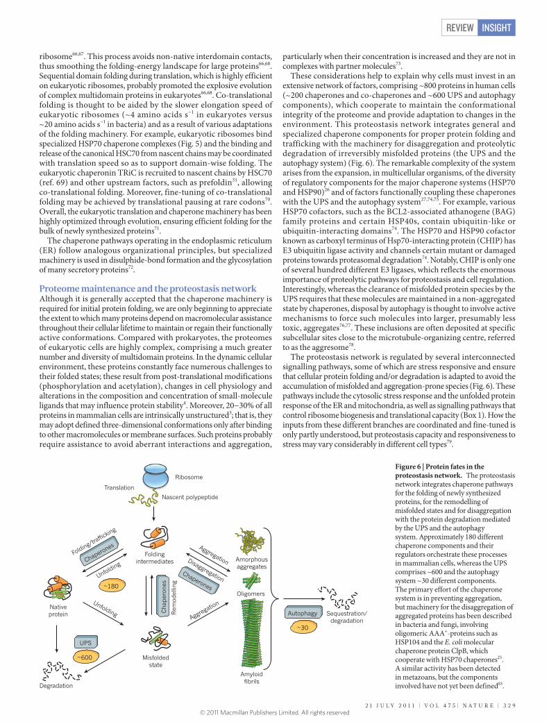
These considerations help to explain why cells must invest in an extensive network of factors, comprising ~800 proteins in human cells (~200 chaperones and co-chaperones and ~600 UPS and autophagy components), which cooperate to maintain the conformational integrity of the proteome and provide adaptation to changes in the environment. This proteostasis network integrates general and specialized chaperone components for proper protein folding and trafficking with the machinery for disaggregation and proteolytic degradation of irreversibly misfolded proteins (the UPS and the autophagy system) (Fig. 6). The remarkable complexity of the system arises from the expansion, in multicellular organisms, of the diversity of regulatory components for the major chaperone systems (HSP70 and HSP90)26 and of factors functionally coupling these chaperones with the UPS and the autophagy system27,74,75. For example, various HSP70 cofactors, such as the BCL2-associated athanogene (BAG) family proteins and certain HSP40s, contain ubiquitin-like or ubiquitin-interacting domains74. The HSP70 and HSP90 cofactor known as carboxyl terminus of Hsp70-interacting protein (CHIP) has E3 ubiquitin ligase activity and channels certain mutant or damaged proteins towards proteasomal degradation74. Notably, CHIP is only one of several hundred different E3 ligases, which reflects the enormous importance of proteolytic pathways for proteostasis and cell regulation. Interestingly, whereas the clearance of misfolded protein species by the UPS requires that these molecules are maintained in a non-aggregated state by chaperones, disposal by autophagy is thought to involve active mechanisms to force such molecules into larger, presumably less toxic, aggregates76,77. These inclusions are often deposited at specific subcellular sites close to the microtubule-organizing centre, referred to as the aggresome78.
The proteostasis network is regulated by several interconnected signalling pathways, some of which are stress responsive and ensure that cellular protein folding and/or degradation is adapted to avoid the accumulation of misfolded and aggregation-prone species (Fig. 6). These pathways include the cytosolic stress response and the unfolded protein response of the ER and mitochondria, as well as signalling pathways that control ribosome biogenesis and translational capacity (Box 1). How the inputs from these different branches are coordinated and fine-tuned is only partly understood, but proteostasis capacity and responsiveness to stress may vary considerably in different cell types79.
Foldingintermediates
Nativeprotein
Misfoldedstate
Amorphousaggregates
Amyloid�brils
Translation
Rem
odel
ling
Degradation
Unfolding
Unfolding
Disaggregation
Aggregation
Aggregation
Folding/tra�cki
ng
Sequestration/degradation
Oligomers
UPS
~600
Autophagy Cha
per
ones
Chaperones
Chaperones
~30
~180
Ribosome
Nascent polypeptide
Figure 6 | Protein fates in the proteostasis network. The proteostasis network integrates chaperone pathways for the folding of newly synthesized proteins, for the remodelling of misfolded states and for disaggregation with the protein degradation mediated by the UPS and the autophagy system. Approximately 180 different chaperone components and their regulators orchestrate these processes in mammalian cells, whereas the UPS comprises ~600 and the autophagy system ~30 different components. The primary effort of the chaperone system is in preventing aggregation, but machinery for the disaggregation of aggregated proteins has been described in bacteria and fungi, involving oligomeric AAA+-proteins such as HSP104 and the E. coli molecular chaperone protein ClpB, which cooperate with HSP70 chaperones25. A similar activity has been detected in metazoans, but the components involved have not yet been defined83.
2 1 J U L Y 2 0 1 1 | V O L 4 7 5 | N A T U R E | 3 2 9
REVIEW INSIGHT
© 2011 Macmillan Publishers Limited. All rights reserved
Proteostasis collapse in ageing and diseaseThe accumulation of misfolded and/or oxidized proteins in cells during ageing is a challenge to the proteostasis system and eventually results in the deposition of aggregates, as shown in model organisms such as Caenorhabditis elegans and Drosophila80,81. The inability of cells to restore normal proteostasis may result in disease, and even in cell death. Indeed, numerous diseases are now recognized to be associated with aberrant protein folding and are usually categorized as loss-of-function or toxic gain-of-function diseases, although specific pathological states often show elements of both groups. The former are generally caused by inherited mutations and include numerous disorders such as cystic fibrosis, lysosomal storage diseases and α1-antitrypsin deficiency. The latter, gain-of-function disorders, include type 2 diabetes and the major neurodegenerative conditions (Parkinson’s disease, Huntington’s disease, amyotrophic lateral sclerosis and Alzheimer’s disease) and are either sporadic or caused by mutations that render specific proteins more aggregation prone. These gain-of-function diseases are typically age related and are caused by the accumulation of amyloid or amyloid-like aggregates of the disease protein. A plausible explanation for the late onset of these diseases is provided by recent evidence from model organisms that the signalling pathways that regulate proteostasis are integrated with the genetic and epigenetic pathways that control longevity82,83 (Box 1). Thus, the age-related decline in proteostasis and specifically in the inability to upregulate chaperones in response to conformational stresses would trigger disease manifestation and, in turn, accelerate proteostasis collapse81,84,85.
Although the toxic principle operating in these disorders is far from understood, a consensus is emerging that soluble oligomeric aggregates, which may be ‘on-pathway’ or ‘off-pathway’ towards fibril formation, are the primary cytotoxic species16,18 (Fig. 1). One prominent hypothesis suggests that these oligomers expose promiscuous hydrophobic surfaces that can mediate aberrant interactions with several other proteins or with cellular membranes16,17. In support of this proposal, a recent proteomics study in human cells showed that certain metastable proteins are targeted preferentially by such interactions, resulting in their co-aggregation with the amyloidogenic disease protein86. The co-aggregating proteins are generally large in size and are enriched in intrinsically unstructured regions, properties that are coupled with a high degree of functionality. Accordingly, they tend to occupy essential hub positions in cellular protein networks, including transcriptional regulation, translation and maintenance of cell architecture, suggesting that their sequestration by the amyloid aggregates results in multifactorial toxicity. An interesting manifestation of this toxicity mechanism is the recent demonstration that aggregating mutant p53 may exert dominant oncogenic potential by sequestering wild-type p53 into co-aggregates, resulting in a complete loss of p53 function87.
Aggregate toxicity may be exacerbated by the inability of affected cells to adequately respond to stress stimuli86. This is consistent with
recent evidence that aberrantly folded protein species may interfere with central proteostasis functions, including protein folding and clearance mechanisms88,89. Notably, the overexpression of members of the HSP70 system has been shown to inhibit the formation of toxic oligomers and to prevent the formation of amyloid aggregates for different disease proteins24,90,91. In the case of polyglutamine-repeat proteins, which cause Huntington’s disease and several related neurodegenerative disorders, HSP70 cooperates with the chaperonin TRiC to prevent the accumulation of potentially toxic oligomers47, which is reminiscent of the functional cooperation between these chaperone systems in de novo protein folding.
On the basis of these findings, the pharmacological upregulation of chaperone function promises to open up new strategies for the treatment of numerous pathological states associated with aberrant folding and aggregation. Proof-of-principle experiments using small-molecule compounds to increase chaperone synthesis and rebalance proteostasis (for example, by activating heat-shock transcription factor-1 (HSF-1)-regulated pathways) have already demonstrated efficacy in loss-of-function and toxic gain-of-function disease models5,6,92,93. Likewise, recently identified proteasome activators94 have the potential to accelerate the clearance of toxic protein species, particularly when applied in combination with chaperone upregulation. Unlike conventional drugs, such ‘proteostasis regulators’ would not be disease-specific or protein-specific, and thus may be applicable to a whole group of related diseases — a new concept in medical practice.
OutlookStudies over the past two decades have provided fascinating insight into the mechanics of chaperone-assisted protein folding, but there are still major gaps in our understanding of how the pathways of folding in the cell differ from those studied in the test tube. Progress is being held back by the problem that the sophisticated biophysical methods used to characterize folding intermediates in vitro are not easily transferable to the in vivo situation. Major innovation potential can thus be expected from the development of advanced imaging techniques, eventually allowing us to monitor conformational changes in a single polypeptide chain as it emerges from the ribosome, performs its biological function and is finally degraded in the living cell. Much research will also be stimulated by the emerging concept that molecular chaperones function as the central element of a much larger cellular network of proteostasis control, comprising, in addition, the protein biogenesis machinery as well as the UPS and the autophagy system. Unravelling the complex regulatory circuitry of this network and understanding why it loses its grip during ageing will pose a major challenge for years to come. Solving this problem will require a broad systems-biology approach relying on a combination of ribosome profiling, quantitative proteomics and computational modelling. How cells react to conformational stress or proteostasis deficiency at the proteome level is unclear. Key questions include determining how certain aberrantly folding proteins aggregate
The expression of stress-inducible chaperone proteins (such as HSP70, HSP40, HSP90 and small HSPs) in the cytosol is governed by the heat-shock response96. The genes encoding these proteins are transcriptionally regulated by the HSF-1 and FOXO (DAF-16 in C. elegans) transcription factors.
The unfolded protein response (UPR)72 of the ER adjusts the folding capacity of the secretory pathway by upregulating ER chaperones and/or attenuating protein synthesis by means of the transcription factors IRE1, PERK and ATF6.
The mitochondrial UPR97,98 is activated by conformational stress in mitochondria and increases resistance to oxidative damage.
Ageing and longevity pathways are coupled to the regulation of stress-protective pathways5,99. Specifically, the upregulation of stress-protection factors such as chaperones by HSF-1 and FOXO is required for the lifespan-extending effect of mutations in the insulin and insulin-like growth factor I (IGF-I) receptor pathway. Autophagy, a process required for the recycling of organelles and the removal of large protein aggregates, is also necessary for lifespan extension and youthfulness in C. elegans. Autophagy is downregulated by the mammalian target of rapamycin (TOR) kinase when nutrients are plentiful99 and is upregulated by FOXO81. Dietary restriction, which extends lifespan in model organisms, is also coupled with HSF-1 and FOXO activation81,100.
BOX 1
Signalling pathways in proteostasis and ageing
3 3 0 | N A T U R E | V O L 4 7 5 | 2 1 J U L Y 2 0 1 1
REVIEWINSIGHT
© 2011 Macmillan Publishers Limited. All rights reserved
into toxic species whereas others are degraded, how the composition of the proteosome changes during ageing, what the signature of a youthful proteome is, and how we can find ways to maintain it for longer as we age. Addressing these and related issues not only offers great opportunities for intervention with numerous, currently incurable diseases but will also eventually reveal the fundamentally important relationship between proteostasis and longevity. ■
1. Dobson, C. M., Sali, A. & Karplus, M. Protein folding — a perspective from theory and experiment. Angew. Chem. Int. Edn Engl. 37, 868−893 (1998).
2. Bartlett, A. I. & Radford, S. E. An expanding arsenal of experimental methods yields an explosion of insights into protein folding mechanisms. Nature Struct. Mol. Biol. 16, 582−588 (2009).
3. Dunker, A. K., Silman, I., Uversky, V. N. & Sussman, J. L. Function and structure of inherently disordered proteins. Curr. Opin. Struct. Biol. 18, 756−764 (2008).
4. Powers, E. T., Morimoto, R. I., Dillin, A., Kelly, J. W. & Balch, W. E. Biological and chemical approaches to diseases of proteostasis deficiency. Annu. Rev. Biochem. 78, 959−991 (2009).
5. Morimoto, R. I. Proteotoxic stress and inducible chaperone networks in neurodegenerative disease and aging. Genes Dev. 22, 1427−1438 (2008).
6. Balch, W. E., Morimoto, R. I., Dillin, A. & Kelly, J. W. Adapting proteostasis for disease intervention. Science 319, 916−919 (2008).
7. Hartl, F. U. Molecular chaperones in cellular protein folding. Nature 381, 571−580 (1996).
8. Kubelka, J., Hofrichter, J. & Eaton, W. A. The protein folding ‘speed limit’. Curr. Opin. Struct. Biol. 14, 76−88 (2004).
9. Herbst, R., Schafer, U. & Seckler, R. Equilibrium intermediates in the reversible unfolding of firefly (Photinus pyralis) luciferase. J. Biol. Chem. 272, 7099−7105 (1997).
10. Ellis, R. J. & Minton, A. P. Protein aggregation in crowded environments. Biol. Chem. 387, 485−497 (2006).
11. Tokuriki, N. & Tawfik, D. S. Chaperonin overexpression promotes genetic variation and enzyme evolution. Nature 459, 668−671 (2009).
12. Rutherford, S. L. & Lindquist, S. Hsp90 as a capacitor for morphological evolution. Nature 396, 336−342 (1998).
This seminal study puts forward the idea that chaperones function in buffering the otherwise deleterious consequences of mutations.
13. Skach, W. R. Cellular mechanisms of membrane protein folding. Nature Struct. Mol. Biol. 16, 606−612 (2009).
14. Kerner, M. J. et al. Proteome-wide analysis of chaperonin-dependent protein folding in Escherichia coli. Cell 122, 209–220 (2005).
15. Eichner, T., Kalverda, A. P., Thompson, G. S., Homans, S. W. & Radford, S. E. Conformational conversion during amyloid formation at atomic resolution. Mol. Cell 41, 161−172 (2011).
This exciting paper describes, at atomic resolution, the structural features of a non-native folding intermediate that are critical for amyloidogenic aggregation.
16. Chiti, F. & Dobson, C. M. Protein misfolding, functional amyloid, and human disease. Annu. Rev. Biochem. 75, 333−366 (2006).
17. Bolognesi, B. et al. ANS binding reveals common features of cytotoxic amyloid species. ACS Chem. Biol. 5, 735−740 (2010).
This paper provides evidence that the exposure of hydrophobic surfaces by oligomeric aggregation intermediates correlates with their toxicity.
18. Kayed, R. et al. Common structure of soluble amyloid oligomers implies common mechanism of pathogenesis. Science 300, 486−489 (2003).
19. Hartl, F. U. & Hayer-Hartl, M. Converging concepts of protein folding in vitro and in vivo. Nature Struct. Mol. Biol. 16, 574−581 (2009).
20. Langer, T. et al. Successive action of DnaK, DnaJ and GroEL along the pathway of chaperone-mediated protein folding. Nature 356, 683−689 (1992).
21. Frydman, J., Nimmesgern, E., Ohtsuka, K. & Hartl, F. U. Folding of nascent polypeptide chains in a high molecular mass assembly with molecular chaperones. Nature 370, 111−117 (1994).
22. Ellis, R. J. Molecular chaperones: assisting assembly in addition to folding. Trends Biochem. Sci. 31, 395−401 (2006).
23. Liu, C. et al. Coupled chaperone action in folding and assembly of hexadecameric Rubisco. Nature 463, 197−202 (2010).
24. Auluck, P. K., Chan, H. Y. E., Trojanowski, J. Q., Lee, V. M. Y. & Bonini, N. M. Chaperone suppression of α-synuclein toxicity in a Drosophila model for Parkinson’s disease. Science 295, 865−868 (2002).
25. Mayer, M. P. Gymnastics of molecular chaperones. Mol. Cell 39, 321−331 (2010).
26. Kampinga, H. H. & Craig, E. A. The HSP70 chaperone machinery: J proteins as drivers of functional specificity. Nature Rev. Mol. Cell Biol. 11, 579−592 (2010).
27. Arndt, V. et al. Chaperone-assisted selective autophagy is essential for muscle maintenance. Curr. Biol. 20, 143−148 (2010).
28. Rüdiger, S., Buchberger, A. & Bukau, B. Interaction of Hsp70 chaperones with substrates. Nature Struct. Biol. 4, 342−349 (1997).
A key paper in understanding how chaperones recognize their substrates.29. Rousseau, F., Serrano, L. & Schymkowitz, J. W. H. How evolutionary pressure
against protein aggregation shaped chaperone specificity. J. Mol. Biol. 355, 1037−1047 (2006).
30. Sharma, S. K., De los Rios, P., Christen, P., Lustig, A. & Goloubinoff, P. The kinetic parameters and energy cost of the Hsp70 chaperone as a polypeptide unfoldase. Nature Chem. Biol. 6, 914−920 (2010).
31. Frydman, J. Folding of newly translated proteins in vivo: the role of molecular chaperones. Annu. Rev. Biochem. 70, 603−647 (2001).
32. Horwich, A. L. & Fenton, W. A. Chaperonin-mediated protein folding: using a central cavity to kinetically assist polypeptide chain folding. Q. Rev. Biophys. 42, 83−116 (2009).
33. Fujiwara, K., Ishihama, Y., Nakahigashi, K., Soga, T. & Taguchi, H. A systematic survey of in vivo obligate chaperonin-dependent substrates. EMBO J. 29, 1552−1564 (2010).
34. Raineri, E., Ribeca, P., Serrano, L. & Maier, T. A more precise characterization of chaperonin substrates. Bioinformatics 26, 1685−1689 (2010).
35. Tartaglia, G. G., Dobson, C. M., Hartl, F. U. & Vendruscolo, M. Physicochemical determinants of chaperone requirements. J. Mol. Biol. 400, 579−588 (2010).
36. Xu, Z. H., Horwich, A. L. & Sigler, P. B. The crystal structure of the asymmetric GroEL−GroES−(ADP)7 chaperonin complex. Nature 388, 741−749 (1997).
37. Brinker, A. et al. Dual function of protein confinement in chaperonin-assisted protein folding. Cell 107, 223−233 (2001).
38. Tang, Y. C. et al. Structural features of the GroEL−GroES nano-cage required for rapid folding of encapsulated protein. Cell 125, 903−914 (2006).
39. Chakraborty, K. et al. Chaperonin-catalyzed rescue of kinetically trapped states in protein folding. Cell 142, 112−122 (2010).
40. Thirumalai, D. & Lorimer, G. H. Chaperonin-mediated protein folding. Annu. Rev. Biophys. Biomol. Struct. 30, 245−269 (2001).
41. Lin, Z., Madan, D. & Rye, H. S. GroEL stimulates protein folding through forced unfolding. Nature Struct. Mol. Biol. 15, 303−311 (2008).
42. Sharma, S. et al. Monitoring protein conformation along the pathway of chaperonin-assisted folding. Cell 133, 142−153 (2008).
43. Munoz, I. G. et al. Crystal structure of the open conformation of the mammalian chaperonin CCT in complex with tubulin. Nature Struct. Mol. Biol. 18, 14−19 (2011).
44. Douglas, N. R. et al. Dual action of ATP hydrolysis couples lid closure to substrate release into the Group II chaperonin chamber. Cell 144, 240−252 (2011).
45. Reissmann, S., Parnot, C., Booth, C. R., Chiu, W. & Frydman, J. Essential function of the built-in lid in the allosteric regulation of eukaryotic and archaeal chaperonins. Nature Struct. Mol. Biol. 14, 432−440 (2007).
46. Kitamura, A. et al. Cytosolic chaperonin prevents polyglutamine toxicity with altering the aggregation state. Nature Cell Biol. 8, 1163−1170 (2006).
47. Behrends, C. et al. Chaperonin TRiC promotes the assembly of polyQ expansion proteins into nontoxic oligomers. Mol. Cell 23, 887−897 (2006).
48. Tam, S., Geller, R., Spiess, C. & Frydman, J. The chaperonin TRiC controls polyglutamine aggregation and toxicity through subunit-specific interactions. Nature Cell Biol. 8, 1155–1162 (2006).
49. Taipale, M., Jarosz, D. F. & Lindquist, S. HSP90 at the hub of protein homeostasis: emerging mechanistic insights. Nature Rev. Mol. Cell Biol. 11, 515−528 (2010).
50. McClellan, A. J. et al. Diverse cellular functions of the Hsp90 molecular chaperone uncovered using systems approaches. Cell 131, 121−135 (2007).
51. Scheufler, C. et al. Structure of TPR domain–peptide complexes: critical elements in the assembly of the Hsp70–Hsp90 multichaperone machine. Cell 101, 199−210 (2000).
52. Wandinger, S. K., Richter, K. & Buchner, J. The Hsp90 chaperone machinery. J. Biol. Chem. 283, 18473−18477 (2008).
53. Ali, M. M. U. et al. Crystal structure of an Hsp90–nucleotide–p23/Sba1 closed chaperone complex. Nature 440, 1013−1017 (2006).
54. Shiau, A. K., Harris, S. F., Southworth, D. R. & Agard, D. A. Structural analysis of E. coli hsp90 reveals dramatic nucleotide-dependent conformational rearrangements. Cell 127, 329−340 (2006).
55. Neckers, L. Heat shock protein 90: the cancer chaperone. J. Biosci. 32, 517−530 (2007).
56. Geller, R., Vignuzzi, M., Andino, R. & Frydman, J. Evolutionary constraints on chaperone-mediated folding provide an antiviral approach refractory to development of drug resistance. Genes Dev. 21, 195−205 (2007).
This seminal study describes the requirement of HSP90 in viral assembly, outlining a strategy for antiviral treatment based on HSP90 inhibition.
57. Eichmann, C., Preissler, S., Riek, R. & Deuerling, E. Cotranslational structure acquisition of nascent polypeptides monitored by NMR spectroscopy. Proc. Natl Acad. Sci. USA 107, 9111−9116 (2010).
58. Cabrita, L. D., Hsu, S. T., Launay, H., Dobson, C. M. & Christodoulou, J. Probing ribosome-nascent chain complexes produced in vivo by NMR spectroscopy. Proc. Natl Acad. Sci. USA 106, 22239−22244 (2009).
59. Lu, J. L. & Deutsch, C. Folding zones inside the ribosomal exit tunnel. Nature Struct. Mol. Biol. 12, 1123−1129 (2005).
60. Woolhead, C. A., McCormick, P. J. & Johnson, A. E. Nascent membrane and secretory proteins differ in FRET-detected folding far inside the ribosome and in their exposure to ribosomal proteins. Cell 116, 725−736 (2004).
61. O’Brien, E. P., Hsu, S.-T. D., Christodoulou, J., Vendruscolo, M. & Dobson, C. M. Transient tertiary structure formation within the ribosome exit port. J. Am. Chem. Soc. 132, 16928−16937 (2010).
62. Elcock, A. H. Molecular simulations of cotranslational protein folding: fragment stabilities, folding cooperativity, and trapping in the ribosome. PLoS Comput Biol. 2, e98 (2006).
63. Ferbitz, L. et al. Trigger factor in complex with the ribosome forms a molecular cradle for nascent proteins. Nature 431, 590−596 (2004).
64. Kaiser, C. M. et al. Real-time observation of trigger factor function on translating ribosomes. Nature 444, 455−460 (2006).
65. Brandt, F. et al. The native 3D organization of bacterial polysomes. Cell 136, 261−271 (2009).
2 1 J U L Y 2 0 1 1 | V O L 4 7 5 | N A T U R E | 3 3 1
REVIEW INSIGHT
© 2011 Macmillan Publishers Limited. All rights reserved

66. Netzer, W. J. & Hartl, F. U. Recombination of protein domains facilitated by co-translational folding in eukaryotes. Nature 388, 343−349 (1997).
67. Frydman, J., Erdjument-Bromage, H., Tempst, P. & Hartl, F. U. Co-translational domain folding as the structural basis for the rapid de novo folding of firefly luciferase. Nature Struct. Biol. 6, 697−705 (1999).
68. Agashe, V. R. et al. Function of trigger factor and DnaK in multidomain protein folding: increase in yield at the expense of folding speed. Cell 117, 199−209 (2004).
69. Cuellar, J. et al. The structure of CCT–Hsc70NBD suggests a mechanism for Hsp70 delivery of substrates to the chaperonin. Nature Struct. Mol. Biol. 15, 858−864 (2008).
70. Zhang, G. & Ignatova, Z. Generic algorithm to predict the speed of translational elongation: implications for protein biogenesis. PLoS ONE 4, e5036 (2009).
71. Vabulas, R. M. & Hartl, F. U. Protein synthesis upon acute nutrient restriction relies on proteasome function. Science 310, 1960−1963 (2005).
72. Buchberger, A., Bukau, B. & Sommer, T. Protein quality control in the cytosol and the endoplasmic reticulum: brothers in arms. Mol. Cell 40, 238−252 (2010).
73. Vavouri, T., Semple, J. I., Garcia-Verdugo, R. & Lehner, B. Intrinsic protein disorder and interaction promiscuity are widely associated with dosage sensitivity. Cell 138, 198−208 (2009).
74. Arndt, V., Rogon, C. & Hohfeld, J. To be, or not to be — molecular chaperones in protein degradation. Cell. Mol. Life Sci. 64, 2525−2541 (2007).
75. Gamerdinger, M. et al. Protein quality control during aging involves recruitment of the macroautophagy pathway by BAG3. EMBO J. 28, 889−901 (2009).
76. Kaganovich, D., Kopito, R. & Frydman, J. Misfolded proteins partition between two distinct quality control compartments. Nature 454, 1088−1095 (2008).
77. Iwata, A., Riley, B. E., Johnston, J. A. & Kopito, R. R. HDAC6 and microtubules are required for autophagic degradation of aggregated huntingtin. J. Biol. Chem. 280, 40282−40292 (2005).
78. Kopito, R. R. Aggresomes, inclusion bodies and protein aggregation. Trends Cell Biol. 10, 524−530 (2000).
79. Kern, A., Ackermann, B., Clement, A. M., Duerk, H. & Behl, C. HSF1-controlled and age-associated chaperone capacity in neurons and muscle cells of C. elegans. PLoS ONE 5, e8568 (2010).
80. David, D. C. et al. Widespread protein aggregation as an inherent part of aging in C. elegans. PLoS Biol. 8, e1000450 (2010).
81. Demontis, F. & Perrimon, N. FOXO/4E-BP signaling in Drosophila muscles regulates organism-wide proteostasis during aging. Cell 143, 813−825 (2010).
82. Morley, J. F. & Morimoto, R. I. Regulation of longevity in Caenorhabditis elegans by heat shock factor and molecular chaperones. Mol. Biol. Cell 15, 657−664 (2004).
This pioneering study provides important insight into the relationship between molecular chaperone functions and longevity.
83. Cohen, E., Bieschke, J., Perciavalle, R. M., Kelly, J. W. & Dillin, A. Opposing activities protect against age-onset proteotoxicity. Science 313, 1604−1610 (2006).
An exciting study demonstrating that active disaggregation and the forced formation of large inclusions prevent the accumulation of toxic aggregate species in C. elegans.
84. Ben-Zvi, A., Miller, E. A. & Morimoto, R. I. Collapse of proteostasis represents an early molecular event in Caenorhabditis elegans aging. Proc. Natl Acad. Sci. USA 106, 14914−14919 (2009).
85. Cohen, E. et al. Reduced IGF-1 signaling delays age-associated proteotoxicity in mice. Cell 139, 1157−1169 (2009).
86. Olzscha, H. et al. Amyloid-like aggregates sequester numerous metastable proteins with essential cellular functions. Cell 144, 67−78 (2011).
This paper demonstrates the existence of a metastable sub-proteome that is at risk of co-aggregating with amyloid-forming disease proteins.
87. Xu, J. et al. Gain of function of mutant p53 by coaggregation with multiple tumor suppressors. Nature Chem. Biol. 7, 285−295 (2011).
This interesting study expands the range of diseases promoted by proteostasis deficiency to cancer.
88. Bence, N. F., Sampat, R. M. & Kopito, R. R. Impairment of the ubiquitin−proteasome system by protein aggregation. Science 292, 1552−1555 (2001).
This key paper demonstrates that protein aggregation can interfere with protein degradation.
89. Gidalevitz, T., Ben-Zvi, A., Ho, K. H., Brignull, H. R. & Morimoto, R. I. Progressive disruption of cellular protein folding in models of polyglutamine diseases. Science 311, 1471−1474 (2006).
90. Schaffar, G. et al. Cellular toxicity of polyglutamine expansion proteins: mechanism of transcription factor deactivation. Mol. Cell 15, 95−105 (2004).
91. Lotz, G. P. et al. Hsp70 and Hsp40 functionally interact with soluble mutant huntingtin oligomers in a classic ATP-dependent reaction cycle. J. Biol. Chem. 285, 38183−38193 (2010).
92. Sittler, A. et al. Geldanamycin activates a heat shock response and inhibits huntingtin aggregation in a cell culture model of Huntington’s disease. Hum. Mol. Genet. 10, 1307−1315 (2001).
93. Mu, T. W. et al. Chemical and biological approaches synergize to ameliorate protein-folding diseases. Cell 134, 769−781 (2008).
94. Lee, B.-H. et al. Enhancement of proteasome activity by a small-molecule inhibitor of USP14. Nature 467, 179–184 (2010).
This study describes the first drug-like molecule that can activate proteasome function, thus providing a means to enhance the clearance of aberrantly folded proteins.
95. Jahn, T. R. & Radford, S. E. The Yin and Yang of protein folding. FEBS J. 272, 5962−5970 (2005).
96. Vabulas, R. M., Raychaudhuri, S., Hayer-Hartl, M. & Hartl, F. U. Protein folding in the cytoplasm and the heat shock response. Cold Spring Harb. Perspect. Biol. 2, a004390 (2010).
97. Ryan, M. T. & Hoogenraad, N. J. Mitochondrial-nuclear communications. Annu. Rev. Biochem. 76, 701−722 (2007).
98. Haynes, C. M. & Ron, D. The mitochondrial UPR — protecting organelle protein homeostasis. J. Cell Sci. 123, 3849−3855 (2010).
99. Kenyon, C. J. The genetics of ageing. Nature 464, 504−512 (2010).100. Westerheide, S. D., Anckar, J., Stevens, S. M., Jr, Sistonen, L. & Morimoto, R. I.
Stress-inducible regulation of heat shock factor 1 by the deacetylase SIRT1. Science 323, 1063−1066 (2009).
Acknowledgements We thank W. Balch, A. Dillin, J. Kelly, R. Morimoto and P. Reinhart for discussions about proteostasis, and thank the members of our laboratory for comments on the manuscript. We apologize to all those whose important work could not be cited owing to space limitations.
Author Information Reprints and permissions information is available at www.nature.com/reprints. The authors declare competing financial interests: details accompany the full-text HTML version of the paper at www.nature.com/nature. Readers are welcome to comment on the online version of this article at www.nature.com/nature. Correspondence should be addressed to F.U.H. ([email protected]).
3 3 2 | N A T U R E | V O L 4 7 5 | 2 1 J U L Y 2 0 1 1
REVIEWINSIGHT
© 2011 Macmillan Publishers Limited. All rights reserved