Vascular smooth muscle differentiation of murine stroma
14
Experimental Hematology 27 (1999) 1782–1795 0301-472X/99 $–see front matter. Copyright © 1999 International Society for Experimental Hematology. Published by Elsevier Science Inc. PII S0301-472X(99)00122-8 Vascular smooth muscle differentiation of murine stroma: A sequential model Jean-Paul Remy-Martin a , Aliette Marandin a , Bruno Challier a , Gabrielle Bernard a , Marie Deschaseaux a , Patrick Herve a , Yanzhang Wei b , Takashi Tsuji c , Robert Auerbach d , James E. Dennis e , Kateri A. Moore f , Joel S. Greenberger g , and Pierre Charbord a a E.T.S. de Franche-Comté and Departement d’Information Medicale, Besancon, France; b Department of Microbiology and Molecular Medicine, Clemson, SC; c Pharmaceutical Frontier Research Laboratory, JT Inc., Yokohama, Japan; d Laboratory of Developmental Biology, University of Wisconsin, Madison, WI; e Skeletal Research Center, Case Western Reserve University, Cleveland, OH; f Department of Molecular Biology, Princeton University, Princeton, NJ; g Department of Radiation Oncology, University of Pittsburgh, Medical School, Pittsburgh, PA (Received 3 March 1999; revised 24 August 1999; accepted 26 August 1999) Previous studies by our group showed that stromal cells from human long-term marrow cultures were mesenchymal cells following a vascular smooth muscle pathway. The present study using 58 immortalized stromal lines from different he- matopoietic sites was conducted to verify whether this hypoth- esis also held true for murine stroma. Principal components analysis performed using cytoskeletal and extracellular matrix proteins allowed the segregation of five factors explaining more than 70% of the variance. Factor I, including osteopon- tin and vimentin, and factor II, laminins and fibronectins, were representative of the mesenchyme. The remaining three factors were representative of vascular smooth muscle: factor III, including a SM actin, SM a actinin, SM22 a , EDa 1 fi- bronectin, and thrombospondin-1; factor IV, metavinculin and h-caldesmon; and factor V, smooth muscle myosin SM1 and desmin. All lines expressed factors I and II; 53 lines ex- pressed factor III, 35 lines expressed factor IV; and 11 lines ex- pressed factor V. A second principal components analysis in- cluding membrane antigens indicated the cosegregration of vascular cell adhesion molecule-1 with osteopontin and that of Ly6A/E with vimentin, whereas CD34 and Thy-1 appeared to be independent factors. The heterogeneity of vascular smooth muscle markers expression suggests that harmonious mainte- nance of hematopoiesis depends on the cooperation between different stromal cell clones. © 1999 International Society for Experimental Hematology. Published by Elsevier Science Inc. Keywords: Cytoskeleton—Extracellular matrix—Membrane antigens—Marrow culture—Factor analysis Introduction Maintenance of hematopoiesis in long-term marrow cul- tures strictly depends on the generation of an adherent layer [1,2]. The most primitive hematopoietic progenitors and, in the mouse, cells with long-term competitive repopulating ability are recovered from this layer, where they are brought together with microenvironmental cells that are considered to reproduce the microenvironment as defined from in vivo studies. Studies by our group showed that microenviron- mental cells from human long-term cultures consist mainly of two cell types: macrophages, similar to resident tissue- macrophages; and vascular smooth muscle (SM)-like stro- mal cells [3,4]. One may observe in stromal cells a time- dependent induction of genes usually expressed by vascular SM cells, which allows characterization of stromal cells as marrow myofibroblasts. Studies on murine long-term mar- row cultures [5] also showed that macrophages and myofi- broblasts are two major cell components generated in the adherent layer; however, these studies also disclosed the ex- istence of a third cell component, that of endothelial-like cells, whose presence in human cultures is minor or even undetectable. The respective role of microenvironmental cell popula- tions in the maintenance of hematopoiesis remains some- what elusive, although progress has been derived from the study of purified populations [6–8]. The simplest way to ob- tain purified populations of microenvironmental cells is to generate clonal immortalized cell lines, which has proved to be relatively easy for murine cells, because, presently, more than 100 murine stromal lines have been described. Re- markably, these lines were generated not only from marrow long-term cultures, but also from cultures of fetal liver, spleen, aorta-gonad mesonephros region, and yolk sac, i.e., from all sites of hematopoiesis during development. Functional studies using murine stromal cell lines have shed light on the many facets of stromal control of hemato- Offprint requests to: Pierre Charbord, U506, Hopital Paul Brousse, 12 Av Paul-Vaillant 94807, Couturier, Villejuif, France; E-mail: charbord@ vjf.inserm.fr
-
Upload
independent -
Category
Documents
-
view
3 -
download
0
Transcript of Vascular smooth muscle differentiation of murine stroma

Experimental Hematology 27 (1999) 1782–1795
0301-472X/99 $–see front matter. Copyright © 1999 International Society for Experimental Hematology. Published by Elsevier Science Inc.
PII S0301-472X(99)00122-8
Vascular smooth muscle differentiation of murine stroma: A sequential model
Jean-Paul Remy-Martin
a
, Aliette Marandin
a
, Bruno Challier
a
, Gabrielle Bernard
a
, Marie Deschaseaux
a
, Patrick Herve
a
, Yanzhang Wei
b
, Takashi Tsuji
c
, Robert Auerbach
d
, James E. Dennis
e
, Kateri A. Moore
f
, Joel S. Greenberger
g
, and Pierre Charbord
a
a
E.T.S. de Franche-Comté and Departement d’Information Medicale, Besancon, France;
b
Department of Microbiology and Molecular Medicine, Clemson, SC;
c
Pharmaceutical Frontier Research Laboratory, JT Inc., Yokohama, Japan;
d
Laboratory of Developmental Biology, University of Wisconsin, Madison, WI;
e
Skeletal Research Center, Case Western Reserve University, Cleveland, OH;
f
Department of Molecular Biology, Princeton University, Princeton, NJ;
g
Department of Radiation Oncology, University of Pittsburgh, Medical School, Pittsburgh, PA
(Received 3 March 1999; revised 24 August 1999; accepted 26 August 1999)
Previous studies by our group showed that stromal cells fromhuman long-term marrow cultures were mesenchymal cellsfollowing a vascular smooth muscle pathway. The presentstudy using 58 immortalized stromal lines from different he-matopoietic sites was conducted to verify whether this hypoth-esis also held true for murine stroma. Principal componentsanalysis performed using cytoskeletal and extracellular matrixproteins allowed the segregation of five factors explainingmore than 70% of the variance. Factor I, including osteopon-tin and vimentin, and factor II, laminins and fibronectins,were representative of the mesenchyme. The remaining threefactors were representative of vascular smooth muscle: factor
III, including
a
SM actin, SM
a
actinin, SM22
a
, EDa
1
fi-bronectin, and thrombospondin-1; factor IV, metavinculinand h-caldesmon; and factor V, smooth muscle myosin SM1and desmin. All lines expressed factors I and II; 53 lines ex-pressed factor III, 35 lines expressed factor IV; and 11 lines ex-pressed factor V. A second principal components analysis in-cluding membrane antigens indicated the cosegregration ofvascular cell adhesion molecule-1 with osteopontin and that ofLy6A/E with vimentin, whereas CD34 and Thy-1 appeared tobe independent factors. The heterogeneity of vascular smoothmuscle markers expression suggests that harmonious mainte-nance of hematopoiesis depends on the cooperation betweendifferent stromal cell clones. © 1999 International Society forExperimental Hematology. Published by Elsevier Science Inc.
Keywords:
Cytoskeleton—Extracellular matrix—Membrane
antigens—Marrow culture—Factor analysis
Introduction
Maintenance of hematopoiesis in long-term marrow cul-tures strictly depends on the generation of an adherent layer[1,2]. The most primitive hematopoietic progenitors and, inthe mouse, cells with long-term competitive repopulatingability are recovered from this layer, where they are broughttogether with microenvironmental cells that are consideredto reproduce the microenvironment as defined from in vivostudies. Studies by our group showed that microenviron-mental cells from human long-term cultures consist mainlyof two cell types: macrophages, similar to resident tissue-macrophages; and vascular smooth muscle (SM)-like stro-mal cells [3,4]. One may observe in stromal cells a time-dependent induction of genes usually expressed by vascularSM cells, which allows characterization of stromal cells asmarrow myofibroblasts. Studies on murine long-term mar-row cultures [5] also showed that macrophages and myofi-broblasts are two major cell components generated in theadherent layer; however, these studies also disclosed the ex-istence of a third cell component, that of endothelial-likecells, whose presence in human cultures is minor or evenundetectable.
The respective role of microenvironmental cell popula-tions in the maintenance of hematopoiesis remains some-what elusive, although progress has been derived from thestudy of purified populations [6–8]. The simplest way to ob-tain purified populations of microenvironmental cells is togenerate clonal immortalized cell lines, which has proved tobe relatively easy for murine cells, because, presently, morethan 100 murine stromal lines have been described. Re-markably, these lines were generated not only from marrowlong-term cultures, but also from cultures of fetal liver,spleen, aorta-gonad mesonephros region, and yolk sac, i.e.,from all sites of hematopoiesis during development.
Functional studies using murine stromal cell lines haveshed light on the many facets of stromal control of hemato-
Offprint requests to: Pierre Charbord, U506, Hopital Paul Brousse,12 Av Paul-Vaillant 94807, Couturier, Villejuif, France; E-mail: [email protected]

J.-P. Remy-Martin et al./Experimental Hematology 27 (1999) 1782–1795
1783
poiesis. Some lines allow the maintenance of both B lym-phopoiesis and myelopoiesis, depending on culture condi-tions [9,10]. In contrast, other lines appear to supportexclusively B lymphopoiesis or myelopoiesis [11], whereasothers more specifically are promoting erythropoiesis in thepresence of erythropoietin [12]. Great variability betweenlines has been observed in terms of primitive stem cellmaintenance. A minority of lines of marrow or fetal liver or-igin appears to allow the survival or even the proliferationin vitro of stem cells with repopulating ability [13–15]. Forother lines, expansion of mature cells is observed at the ex-pense of stem cell maintenance, indicating that cells fromthese lines favor stem cell commitment [14]. Another set ofresults indicating the functional relevance of the lines hasbeen the isolation of novel mediators. Interleukin 6 [16] andstem cell factor [17,18] have been isolated from murinestromal cell lines, which were hematopoietically inactive,although they provided essential synergistic factors. Othernovel molecules have been isolated from murine lines, e.g.,adhesion molecules, such as CD44 [19], and mediators in-volved in the interaction between stroma and primitivecells, such as
dlk
related to the notch/delta/serrate family[20]. Other applications of stromal cell lines has been in thestudy of human hematopoiesis [21–25] and for transplanta-tion studies [26].
It appeared to us that few of the cell lines that have beenextremely useful for the study of human or murine hemato-poiesis and the unraveling of new mediators had been char-acterized in terms of cell lineage. Although some lines hadbeen described as endothelial, preadipocytic, or osteoblas-tic, the question as to whether or not these lines were madeof myofibroblasts has been addressed in very few studies[27,28]. This work addresses this question by studying anumber of markers on 58 murine stromal cell lines derivedfrom bone marrow, fetal liver, spleen, and yolk sac. Morespecifically, we tried to answer two questions, that of themesenchymal origin of the lines and that of a potential vas-cular SM differentiation that might be specific for hemato-poietic sites. The marker proteins we selected belonged tothe cytoskeleton (vimentin, desmin,
a
SM actin, SM
a
acti-nin, SM myosin heavy chains, SM22
a
, metavinculin, andheavy chain caldesmon [h-caldesmon]), to the extracellularmatrix (laminins, EDa
2
and EDa
1
fibronectins, osteopon-tin, thrombospondin-1), or were membrane antigens alreadydescribed as expressed by cells from some stromal lines(Ly6A/E, vascular cell adhesion molecule 1 [VCAM-1],CD34, Thy-1). From principal components analysis (PCA)of the data, a model of differentiation for the different stro-mal cell lines is proposed. A sequential model, comprisingfour steps, each characterized by the induction of a differentset of genes, would account for the transition from imma-ture mesenchymal cells to vascular SM-like stromal cells.This model makes possible the classification of the differentlines according to the step where they leave the pathway.This classification reveals the broad extent of stromal heter-
ogeneity, suggesting that, for harmonious maintenance ofhematopoiesis, there should be cooperation between differ-ent stromal clones.
Materials and methods
Cell lines
The different cell lines are listed in Table 1. For each line, the tis-sue of origin is given as bone marrow (Ma), fetal liver (FL), spleen(Sp), yolk sac (YS), leukemia (Le), or endothelioma (En) cells. Wecultured the lines using medium and conditions indicated by thedifferent investigators that provided the lines. All lines were stud-ied when stromal layers were fully confluent, i.e., at a time when itcould be reasonably assumed that the phenotype was stable (whichwas checked for four lines). All lines have been published, and ref-erences are given in Table 1. However, some lines have been usedso extensively that only a few articles about these lines are dis-cussed in this report.
Western blot studies
Western blot studies were performed as previously described [4].Fully confluent adherent layers were detached in phosphate-buf-fered saline with 10 mM of ethylenediaminotetraacetic acid (PBS-EDTA) and sonicated in the same buffer with protease inhibitorcocktail. The amount of proteins for each sample was measuredusing bicinchoninic acid protein assay reagent (BCA kit) obtainedfrom Pierce Chemical Co. (Rockford, IL). The samples weremixed with an equal volume of 2
3
concentrated sample buffer,consisting of 4% (w/v) sodium dodecyl sulfate (SDS), 125 mMTris-HCl pH 6.8, 20% (v/v) glycerol, 4% (v/v)
b
mercaptoethanol,0.05% (w/v) bromophenol blue, and heated at 100
8
C for 5 minutes.Proteins were separated in SDS-polyacrylamide gel (SDS-PAGE).The concentration of polyacrylamide was 10% for actins and vi-mentin, 7.5% for desmin and osteopontin, and 6% for caldesmons,vinculins, laminins, fibronectins, and thrombospondin, with a re-ticulation rate reduced to 1.33% in 6% gel slab for heavy chains ofmyosin. Twenty protein
m
g was loaded per lane. After SDS-PAGE, proteins were electrophoretically transferred onto nitrocel-lulose membrane. Membranes were incubated for 1 hour in 5% (w/v) nonfat dried milk in Tris buffer saline (TBS) with 0.1% Tween20 (TTBS), washed two times in TTBS, and incubated overnight at4
8
C with the appropriate dilution of primary antibody. After threewashes in TTBS, the membrane was incubated for 1 hour withhorseradish peroxidase-linked secondary antibody or biotinylatedsecondary antibody (Amersham, Little Chalfont, UK) and washedthree times in TTBS. Blots were developed 1) directly with 4-chloro-1-naphtol for proteins in high amounts (actins, vimentin, fibronectins,laminins); 2) after 30 minutes of incubation with streptavidin-horse-radish peroxidase for vinculins, desmin, and thrombospondin-1; or 3)using the chemiluminescence method (ECL kit, Amersham) forcaldesmons, myosin heavy chains, laminin
b
1, and osteopontin. Foreach protein, an internal reference control protein was used to mini-mize the variation of loading of sample or revelation of the blot.The control was
b
cytoplasmic actin, except for h-caldesmon, whereit was l-caldesmon; metavinculin, where it was vinculin; and SMmyosin heavy chains SM1 (204 kDa) and SM2 (200 kDa), where itwas nonmuscle myosin heavy chain (197 kDa). To quantify theband intensity, blots or films were scanned with an Ultroscan LaserDensitometer (Pharmacia-LKB, Uppsala, Sweden) and analyzed

1784
J.-P. Remy-Martin et al./Experimental Hematology 27 (1999) 1782–1795
with Gelscan XL software
(Pharmacia-LKB). Antibodies and con-ditions used for Western blot studies are summarized in Table 2.
Immunofluorescence studies
Immunofluorescence studies were performed as previously de-scribed [4]. Confluent adherent layers were treated with trypsin.Ten thousand cells were seeded per chamber of eight-chamberslides. Slides were examined a few days later (usually 3–7 days),when the layer had reached maximal confluency. We looked forthe presence of
a
SM actin
1
, vimentin
1
, and desmin
1
cells, andthat of laminin and fibronectin within the cells and/or extracellularmatrix. Antibodies used were the same as those described forWestern blot studies. For lines for which SM myosin heavy chainswere detected using Western blot studies, we also checked for thepresence of SM-specific myosin (SM myosin) positive cells usingthe Myos Aor polyclonal antibody described by Benzonana et al.[50]. In our experience, Western blot studies were negative using1E12 monoclonal antibody, which recognizes SM
a
actinin [51].In contrast, 1E12 labeled distinct microfilaments; therefore, wechecked for the presence of SM actinin
1
cells in all lines. For lami-nin and fibronectin, the evaluation was qualitative for the presenceor absence within the cytoplasm and/or the extracellular matrix.For
a
SM actin, vimentin, desmin, SM
a
actinin, and SM myosin,the evaluation was quantitative. We measured on 10 to 100 areasof 1 mm
2
the number of cells with actin, vimentin, desmin, and SMmyosin filaments, and we related this number to the total cellnumber.
Flow cytometric analysis
The percentage and mean fluorescence intensity of
a
SM actin
1
cells were evaluated by flow cytometric analysis on permeabilizedcells. Confluent adherent layers from flasks were treated withtrypsin. After washing with PBS, cells were fixed with 80% (v/v)ethanol in PBS at
2
20
8
C overnight. Cells then were permeabilizedfurther in PBS/Tween (0.25% w/v) for 15 minutes at 4
8
C. Treatedcells were incubated with anti-
a
SM actin monoclonal antibody(clone 1A4) or with an irrelevant isotype control (Dako, Glostrup,Danemark) for 30 minutes at 4
8
C. They were washed with PBSand incubated for 30 minutes at 4
8
C with fluorescein isothiocyan-ate-conjugated goat anti-mouse IgG (Sigma) diluted 1:100 in PBS,resuspended in PBS, and passaged through Becton Dickinson(Mountain View, CA) FACSort equipped with the Lysis II™ soft-ware program. Twenty thousand events for each sample were re-corded. For samples containing
a
SM actin
1
cells, the mean fluo-rescence intensity was always at least twice that obtained with theirrelevant control.
We also looked for the presence of membrane antigens alreadydescribed as expressed by cells from few stromal lines: Ly6A/E(Sca-1), CD34, VCAM-1 and Thy-1, and for the absence of mark-ers of hematopoietic cells (CD45) and of endothelial cells (CD31,MECA-32, and VE-Cadherin). Rat anti-mouse unconjugated mono-clonal antibodies (MoAbs) were purchased from Pharmingen(Becton Dickinson): clone E13-161.7 for Ly6A/E, clone Ram34 forCD34, clone 429 for VCAM-1, clone G7 for Thy-1, clone 30F-11 forCD45, clone MEC13.3 for CD31, and clone 11D4.1 for VE-Cadherin.All these MoAbs were IgG2a, except for anti-CD45 and anti-
Table 1.
Cell lines used in this study
Line Tissue Provider Reference
WEHI-3 Le ATCCbEND.3 En ETCC 29EOMA En R. Auerbach 30HESS-1 and -5/SSXL no. 2, 7 and 17 Ma/Sp T. Tsuji 31GB1/6, D2XRII, OP1 Ma J. Greenberger 26, 3230R Ma D. Rennick 16BMS-1 & -2 Ma P. Kinkade 19, 27, 33SS1, OP42 Sp — 27, 34FBMD-1 Ma R. Ploemacher 23GY30 Ma C. Chabannon 16, 35MS-1 and -5 Ma K. Itoh 22, 36M2-10B4 Ma C. Eaves 37S17 Ma K. Dorshkind 9, 13MBA-1, -2.1, -13, -15 and 14F1.1 Ma D. Zipori 38, 39, 77SyS-1 Ma B. Péault 21
1
/
1
-1.LDA11 Ma S. Boswell 17, 18SR4987 Ma ATCC 40TC-1 Ma P. Quesenberry 41BMC2, 3, 9, 10, 11 Ma J. Dennis 42TPO-KO1 and 7 Ma F. Wendling 43FLS-3 and -5/MSS-31 and -62 FL/Sp N. Yanai 122012, 2018, 2058, BFC012, AFT024 FL K. Moore 15, 20, 24, 25AFT011, CFC-008, -032, -034, -037, 2058 FL — 14Sl4, Sl4-h220 and -248 FL D. Williams 44YS4 YS Y. Wei 45C-165, -166 and -167 YS R. Auerbach 46YSM-1 and -5 YS M. Yoder 47
ATCC
5
American Tissue Culture Collection; en
5
endothelioma; ETCC
5
European Tissue Culture Collection; FL
5
fetal liver; Le
5
leukemia; Ma
5
marrow; Sp
5
spleen; YS
5
yolk sac.

J.-P. Remy-Martin et al./Experimental Hematology 27 (1999) 1782–1795
1785
Thy-1, which were IgG2b and IgG2c, respectively. We also usedisotype-matched irrelevant antibodies (Pharmingen). Cells treatedwith trypsin were washed in PBS. Unfixed cells were incubated for30 minutes at 4
8
C with the appropriate dilution of MoAbs, washedtwice in PBS, and incubated for 30 minutes at 4
8
C with fluoresceinisothiocyanate-conjugated goat anti-rat IgG (Pharmingen) diluted1:100 in PBS. Cells were washed twice and analyzed in the FAC-Sort as described earlier. For each line, analyses were performed atleast three times using confluent layers at different passages.
Reverse transcriptase polymerase chain reaction
In the absence of an anti-SM22
a
antibody reactive with murineSM22
a
, we checked for the presence of SM22
a
mRNA using re-verse transcriptase polymerase chain reaction (RT-PCR). Totalcellular RNA was extracted from cells lysed directly in the cultureflask (confluent layers) by adding 1.5 mL of Trizol reagent (LifeTechnologies, Cergy Pontoise, France). cDNA synthesis was per-formed as previously described [52]. SM22
a
enzymatic amplifica-tion of the specific cDNA sequences by PCR was performed usingthe following sense and antisense primers selected from the cDNAsequence [53]. Sequence (located in exon 3) for the 3
9
primer was:gCA AAT Tgg TgA ACA gCC Tg. Sequence (located in exon 5)for the 5
9
primer was: TgC TTC CCC TCC TgC AgT T. Sequence(located in exons 4 and 5) for the probe used for the Southern was:gCT CCT ggg CTT TCT TCA TAA ACC. PCR products wereelectrophoresed in a 2% agarose gel, transferred onto nylon mem-brane, and probed with a 3
9
end-labeling synthetic oligonucleotideselected from the cDNA sequence. As described previously [52],we always used an internal control to normalize values of PCRproducts to mRNA of a “housekeeping” gene, that of
b
cytoplas-mic actin. After revelation, we measured the relative band inten-sity using densitometry (Biorad Gel D 1000 System). (Ivry-Sur,Seine) Relative band intensity for a given cell line was expressedas the percentage of the value obtained for the SSXL no. 17 line.
Statistical analysis
PCA was performed to examine whether and how markers couldbe grouped into factors. Starting with a certain number of originalvariables (17 to 25 in this study), PCA builds a certain amount(less than, or equal to, the number of original variables) of uncor-related factors. Each of these factors, which are linear functions ofrelated variables, explains part of the measured variance. We re-tained factors with eigenvalues
$
1. This allowed us to explainmore than 70% of the total variance of the results. The BMDP(Berkeley, CA) statistical software package (version 7.0) was usedin this study.
Analysis of variance was used to evaluate whether the expres-sion of membrane antigens was significantly different betweenlines from the distinctive steps of the linear differentiation model.
Results
Negative controls do not expressmost of the markers used in this study
The hypothesis we wanted to verify was that of a vascularSM differentiation pathway followed by stromal cell linesgenerated from different hematopoietic tissues. Therefore,our first task was to choose appropriate negative controlsthat should not express most of the marker proteins we hadselected. Negative controls used for this study were the he-matopoietic leukemic cell line WEHI-3 comprising CD45
1
/CD34
1
cells and two endothelial cell lines, EOMA andbEND.3, derived from murine endothelioma and compris-ing CD45
2
/CD34
1
cells. WEHI-3 cells expressed only vi-mentin and osteopontin. For bEND.3 and EOMA, lamininswere expressed, including laminin
b
1 for EOMA. EOMAcells also expressed weakly EDa
2
fibronectins. Immunoflu-
Table 2.
Conditions used for Western blot studies
AntibodyReference
proteinAntigen Abbreviation MW (kDa) Clone Source
b
Actin
b
45 AC-15 Sigma —
a
SM Actin ASMA 45 1A4 Sigma
b
Actin Vimentin Vim 55 13-2 Sigma
b
Actin Desmin Des 55 DE-R-11 Dako
b
Actin Fibronectins FNp 230 Polyclonal Dako
b
Actin EDa
1
Fibronectin EDaFN 230 3E2 Sigma
b
Actin Laminins LMp 200 & 220 Polyclonal I Pasteur
b
Actin Laminin
b
1 LM
b
1 220 MAB (rat) Chemicon
b
Actin Thrombospondin-1 TSP 140 Polyclonal Ref. 48
b
Actin Osteopontin OSP 86 MPIIIB10 IHB
b
Actin l-Caldesmon l-Cald 75 C21 Sigma —h-Caldesmon h-Cald 137 C21 Sigma l CaldesmonNM Myosin NMM 197 5A6 Ref. 49 —SM1 Myosin SM1 204 h SMV Sigma NM Myosin SM2 Myosin 200 h SMV Sigma NM Myosin Vinculin V 130 11-5 Sigma —Metavinculin m 150 11-5 Sigma Vinculin
I Pasteur
5
Institut Pasteur, Lyon, France; Chemicon
5
Chemicon, Temecula, CA; IHB
5
Iowa Hybridoma Bank, IA.

1786
J.-P. Remy-Martin et al./Experimental Hematology 27 (1999) 1782–1795
orescence studies confirmed the presence of laminin
1
mate-rial in the cytoplasm of EOMA and bEND.3 cells and in theextracellular matrix of the EOMA cell layer. We alsochecked for the lack of any SM
a
actinin
1
cells and that ofmessenger RNA for SM22
a
in these lines.
Western blot studies reveal that most stromalcell lines are myofibroblastic, regardless of origin
A representative sample of results (26 of 58 lines) is shownin Table 3. All lines expressed vimentin, osteopontin, lami-nins (as recognized by the polyclonal antibody), and lami-nin
b
1. All lines, except for YS4, also expressed fibronec-tins (as recognized by the polyclonal antibody).
Most lines expressed EDa
1
fibronectin, thrombospon-din-1, and
a
SM actin. The other markers were less wellrepresented. Ten lines expressed h-caldesmon, 6 metavincu-lin, 5 SM1 myosin, and 3 desmin. These data are representa-tive of the whole population studied, as h-caldesmon,metavinculin, SM1 myosin, and desmin were expressed by50%, 22%, 19%, and 7% of the lines, respectively. Mostlines were characterized as myofibroblastic due to theirexpression of thrombospondin-1, EDa
1
fibronectin,
a
SMactin, h-caldesmon, metavinculin, SM1 myosin, anddesmin. The notable exceptions were MBA-2.1, 14 F1.1,
112
-1.LDA11, MSS-31, and YS4. Lines MBA-2.1,14F1.1, MSS-31, and YS4 have been described as endothe-lial-like [12,36,45], which would fit with our results.
112
1.LDA11 cells have been defined previously as com-prising putative adventitial reticular cells and macrophage-like cells [17]. We could confirm in this study the existenceof two cell types, a majority of small ovoid cells and a mi-nority of large polygonal cells. Studies using membrane an-tigens exclude the presence of macrophages. Accordinglywe would qualify 1121.LDA11 cells simply as mesen-chymal.
Studies using immunofluorescence in situ andRT-PCR confirm that most lines are myofibroblasticImmunofluorescence studies confirmed that all cells fromall lines were vimentin1. Laminin also was located in allcells from all lines within cytoplasmic structures not re-solved at the light microscopic level, although in a fewlines, cells with clearcut laminin1 cytoplasmic vesicleswere observed. In contrast, fibronectin was located in theextracellular matrix, forming a continuous network of fibersof different sizes. However, in a few lines, fibronectin alsowas detected within cells, in well-delineated cytoplasmicvesicles. Immunofluorescence studies indicated that the per-centage of cells with aSM actin microfilaments rangedfrom 0 to 100%. Two to three evaluations were made at dif-ferent time points when layers had reached maximal conflu-ency. Estimations for a given line were similar from onetime point to the other, although usually increasing (by atmost 15%) with the degree of confluency. The lowest val-
Table 3. Western blot results for 26 of the 58 stromal cell lines
Line Tiss Vim/b OSP/b LMp/b LMb1/b FNp/b EDaFN/b TSP/b ASMA/b h/l Cald m/V SM1/NMM Des/b
MBA-2.1 Ma 1.18 4.09 1.70 1.05 1.29 0 0 0 0 0 0 01/1-1.LDA 11 Ma 0.79 0.64 1.17 0.78 0.37 0 0.18 0 0 0 0 014F1.1 Ma 0.83 1.59 4.65 1.55 3.99 0.10 0 0.05 0 0 0 0GB1/6 Ma 0.92 1.28 2.47 0.99 4.80 0.81 0.44 0.64 0 0 0 0D2XR11 Ma 0.92 1.14 1.77 0.20 2.87 0.70 0.16 2.83 0 0 0 030R Ma 1.32 1.45 4.43 0.84 3.62 0.27 0.54 0.45 0 0 0 0M2-10B4 Ma 0.91 1.10 4.04 0.78 1.46 0.30 0.50 2.26 0 0 0 0MS-5 Ma 0.85 0.85 2.86 0.55 3.79 0.08 0.07 1.63 0 0 0 0S17 Ma 0.90 0.46 4.68 0.90 5.61 0.63 0.41 2.71 0 0 0 0BMC9 Ma 1.03 1.60 0.88 0.32 1.51 0.41 0.57 0.10 0 0 0 0TC-1 Ma 0.81 0.61 0.83 0.40 0.65 0.03 0.09 0.34 0.11 0 0 0TPO-KO1 Ma 1.00 1.18 2.70 0.67 0.94 0.56 0.40 1.16 0 0.011 0 0AFT024 FL 1.27 3.00 5.21 1.79 8.90 0.17 0.39 1.12 0 0 0 0CFC034 FL 0.90 3.14 3.51 0.81 10.33 0.72 0.24 1.02 0 0 0 0CFC032 FL 0.99 1.74 6.27 2.04 5.82 0.26 0.59 2.46 0.17 0 0 0FLS-3 FL 1.07 3.90 2.94 1.34 3.07 0.50 0.51 0.49 0.05 0.018 0 0CFC037 FL 0.92 2.01 5.69 1.84 4.57 0.10 0.23 1.07 0.06 0 0.18 02058 FL 0.74 2.00 10.77 2.50 4.12 0.14 0.89 0.17 0.38 0.005 0.47 0MSS-31 Sp 1.04 2.49 8.00 1.34 1.69 0 0 0 0 0 0 0MSS-62 Sp 0.94 1.69 3.50 1.23 1.90 0.51 0.73 1.69 0.44 0 0 0OP42 Sp 0.75 1.90 2.26 0.97 3.87 0.44 0.39 2.02 0.06 0 0 0SSXL no. 7 Sp 0.85 1.37 2.61 0.79 3.21 0.46 0.51 1.97 0 0.016 0 0SSXL no. 17 Sp 0.88 2.08 4.14 2.30 3.96 1.04 0.62 4.59 0.58 0.124 0.24 0.115YS4 YS 0.80 0.10 2.78 2.67 0 0 0 0 0 0 0 0YSM5 YS 0.76 0.58 1.39 0.76 0.47 0.18 0.15 1.75 0.04 0 0.14 0.025C166 YS 0.66 1.40 1.61 1.19 1.29 0.99 0.57 2.63 0.57 0.039 1.12 1.022

J.-P. Remy-Martin et al./Experimental Hematology 27 (1999) 1782–1795 1787
ues (0–4%) were obtained for 1121.LDA11, MBA-2.1,14 F1.1, and MSS-31, indicating that, using our method ofdetection, aSM actin was not detected on Western blotswhen less than 5% of the cells were positive. For lines withhigh percentages of aSM actin1 cells (70–100%), stress fi-bers were very thin and were arranged linearly and regularlywithin the cytoplasm of widespread cells (Figs. 1A–B). Incontrast, in lines with low to moderate percentages of aSMactin1 cells (40–70%), many positive cells were dendritic-like, extending thin cytoplasmic protrusions far away fromthe nucleus (Fig. 1E). In aSM actin1 cells from yolk saclines, aSM actin1 microfilaments comprised both stress fi-bers and a cortical network running underneath the cyto-plasmic membrane (Figs. 1C–D).
The percentage of aSM actin1 cells correlated strongly(r2 5 0.467, p , 0.0001) with the value obtained for theaSM actin band using Western blotting (Fig. 2). Lines witha high aSM actin band ($1 arbitrary unit) comprised atleast 60% actin1 cells, whereas lines with a low aSM actinband (0.05 , ASMA/b , 1) might comprise a high per-centage of cells with very thin, albeit still detectable, aSMactin1 microfilaments. Nine lines (FLS-3, FLS-5, GY30,30R, BMC2, 2BMC11, 2012, 2058, CFC008) steadily com-prised 5 to 40% aSM actin1 cells. This relatively low per-centage was due to the presence of fat-laden, aSM actin2
cells in lines 30R, BMC2, and 2BMC11. In GY30, 2012,2058, and CFC008, it might be related to a high growth rate,implying a high microfilament turnover rate. For FLS-3 and
Figure 1. Immunofluorescence studies. (A) OP-1 bone marrow cells labeled with anti-aSM actin monoclonal antibody 1A4 (3 400). In 23 (of 25) flattenedand regular cells, aSM actin1 fine and linear microfilaments form an array of stress fibers so dense as to obscure the cell nucleus. (B) BFC012 fetal liver cellslabeled with 1A4 (3 250). In 13 (of 14) flattened and regular cells, aSM actin1 fine and linear microfilaments are observed. The density of the stress fibers isless dense than that shown in A, in that the cell nucleus is clearly visible. (C) C166 yolk sac cells labeled with 1A4 (3 180). In all rounded cells, two arrays ofaSM actin1 microfilaments are observed: stress fibers ending perpendicular to the cell membrane and a cortical network running underneath the cytoplasmicmembrane. (D) Higher magnification of microphotograph C (3 640). Close-up view of an ovoid cell, seen in the lower left corner of C, stressing the dual net-work of aSM actin1 microfilaments. (E) AFT024 fetal liver cells labeled with 1A4 (3 400). In all dendritic-like cells with slender and gracile protrusions oftheir cytoplasm, a very fine network of barely detectable aSM actin1 microfilaments is observed, giving the cell a silky appearance. (F) OP42 splenic cellslabeled with 1E12, an anti-SM actinin monoclonal antibody (3 170). In all (from the area shown) flattened and regular cells, linear SM a actinin microfila-ments are observed, arranged in a dense network of stress fibers. (G) C166 yolk sac cells labeled with Myos Aor, a polyclonal antibody directed at smoothmuscle myosin (3 600). SM myosin is present in the stress fibers in all four cells from this area. (H) C166 yolk sac cells labeled with an anti-desmin mono-clonal antibody (3 400). In 9 (of 10) cells, desmin1 intermediate filaments are thicker and coarser than the microfilaments shown in the other microphoto-graphs; they are nonlinear and clearly delineate the nucleus.

1788 J.-P. Remy-Martin et al./Experimental Hematology 27 (1999) 1782–1795
FLS-5, low percentages of aSM actin1 cells might reflecttrue clonal heterogeneity, as will be discussed later.
The percentage of SM a actinin1 cells correlated withthe percentage of aSM actin1 cells (r2 5 0.137, p 5 0.004).However, in positive lines, the percentage of SM a actinin1
cells was low, which probably is due to the low affinity ofthe 1E12 antibody for murine SM a actinin.
Finally, we checked for the presence of SM22a1 cells,using RT-PCR. The amount of SM22a was correlated to thepercentage of aSM actin1 cells (r2 5 0.151, p 5 0.0025) orto the amount of aSM actin (r2 5 0.232, p 5 0.0001) as eval-uated by Western blotting.
Positive SM actinin1 cells clearly presented SM actinin1
microfilaments (Fig. 1F). Likewise, SM myosin was locatedin microfilaments (Fig. 1G), whereas desmin was found inintermediate filaments (Fig. 1H).
Flow cytometric studies revealwidespread expression of Ly6A/E and VCAM-1and more restricted expression of Thy-1 and CD34More than 10% of cells were Thy-11 in 14 lines (Fig. 3D),CD341 in 27 lines (Fig. 3C), VCAM-11 in 51 lines (Fig.3B), and Ly6A/E1 in 45 lines (Fig. 3A). The pattern of anti-body staining, which was similar for each of the four anti-bodies used, was unusual, because for a number of cell linesthe relative mean fluorescence intensity was relatively low(,20 for Ly6A/E and ,10 for VCAM-1, CD34, and Thy-1)until the frequency of positive cells was greater than 70%.This unusal staining pattern cannot be due to nonspecificstaining, as might be observed with mesenchymal cells withsignificant autofluorescence and nonspecific antibody ab-
sorbence, because clearcut negative results were obtainedusing isotype-matched irrelevant antibodies (Fig. 3G), anti-CD45 (Fig. 3F), anti-CD31 (Fig. 3E), anti–VE-Cadherin, andMECA32 (not shown). The heterogeneity of antigen expres-sion between cells of a given cell line should account for thepeculiar pattern of antibody staining. Moreover, the patternof antigen expression was characteristic of each line, be-cause distinct evaluations made at different time points gavesimilar results (not shown).
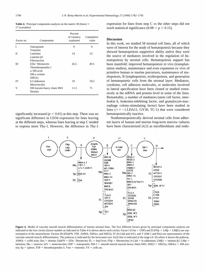
PCA suggests a sequential model of differentiationfrom mesenchymal cells to vascular SM-like cellsTwo analyses were performed sequentially. The first analy-sis was carried out on a matrix 58 (lines) 3 17 (variables),using variables representative of mesenchymal or vascularSM cells. This analysis indicated that five factors accountedfor more than 70% of the variance of the results (Table 4).Two factors were representative of the mesenchyme: factorI included osteopontin and vimentin, and factor II includedlaminins, laminin b1, and fibronectins. The remaining threefactors were representative of vascular SM cells. Factor IIIincluded aSM actin (evaluated by Western blotting, immu-nofluorescence and flow cytometry), EDa1 fibronectin,thrombospondin-1, SM a actinin, and SM22a. Factor IV in-cluded h-caldesmon and metavinculin. Factor V includedSM myosin heavy chain SM1 and desmin. From these data,we established a sequential five-step model of differentia-tion from mesenchymal cells to vascular SM cells (Fig. 4).This model makes it possible to classify the different linesaccording to the point where they leave the pathway. Ap-proximatively 10% of the stromal cell lines, three of mar-row origin (MBA-2.1, 14F1.1, and 1121.LDA11), onesplenic (MSS-31), and one from yolk sac (YS4), leave thepathway at step A, because they express only the mesenchy-mal markers (components from factors I and II). Thirty per-cent of the cell lines, 12 marrow lines, but also five fetalliver lines and one splenic, leave the pathway at step B, be-cause they express the mesenchymal markers (factors I andII) and components from factor III (the early vascular SMdifferentiation markers, EDa1 fibronectin, thrombospondin-1, aSM actin, and, for some lines, SM a actinin andSM22a). Forty percent of the lines, 15 marrow cell lines,but also five fetal liver lines and four splenic lines, leave thepathway at step C, because they express markers from fac-tors I, II, III, and IV (h-caldesmon and/or metavinculin).Twenty percent of the lines leave the pathway at step D, be-cause they express markers from factors I, II, III, IV, and V(SM myosin heavy chain SM1 6 desmin). At this stage,there were no longer bone marrow cell lines, but only fivelines from the fetal liver, one line from the spleen, and fivelines from the yolk sac. Representative profiles of proteinsdetected by Western blotting are shown in Figure 5.
The second analysis (Table 5) included the membraneantigens for which we did not have a differentiation cue.Therefore, it was performed on the matrix 58 (lines) 3 25
Figure 2. Regression analysis between the percentage of aSM actin1 cellsand the aSM actin band intensity. The aSM actin band intensity (related tothat of b cytoplasmic actin) in arbitrary units (a.u.) as given by Westernblot studies (see Methods) is compared with the percent of aSM actin1
cells as measured using immunofluorescence in situ. Although the regres-sion is simple and linear, the plot appears logarithmic due to the logarith-mic scale adopted for the y-axis.

J.-P. Remy-Martin et al./Experimental Hematology 27 (1999) 1782–1795 1789
(variables). Eight factors accounted for more than 70% ofthe variance. In addition to factors III, IV, and V representa-tive of vascular SM cells and to factor II representative ofthe mesenchyme, we found factor Ia comprising both vi-mentin and Ly6A/E and factor Ib comprising both os-teopontin and VCAM-1. The two remaining factors, which
did not correlate with any of the others, were Thy-1 andCD34 (factors VII and VIII, respectively).
The pattern of expression of the four membrane antigensaccording to the differentiation step is shown in Figure 6.There was a significant (0.01 , p , 0.001) decline ofLy6A/E for lines leaving at step D, whereas VCAM-1 was
Figure 3. Membrane antigen expression. (A–F) Percentage of positive cells is given on the x-axis, and the relative mean fluorescence intensity (rMFI) isgiven on the y-axis. Both values are corrected from that of the irrelevant isotype control (see Methods). The logarithmic scale on the y-axis allows better visu-alization of lines with low antigen density (3–10 times that of the isotype control). (A) Results for Ly6A/E. (B) Results for VCAM-1. (C) Results for CD34.(D) Results for Thy-1. (E) Results for CD31. Cells from all stromal lines were negative. In contrast, positive results were obtained for the control cell linesWEHI-3, EOMA, and bEND.3. (F) Results for CD45. Cells from all stromal lines were negative. Positive results were obtained only with the leukemia cellline WEHI-3. (G) Example of fluorescence histograms for Ly6A/E. Cells from FLS-5 do not express the antigen (histogram superimposed on that of the iso-type control). Approximately 40% of the cells from TC-1, 65% of the cells from OP-42, and 100% of the cells from GB1/6 are positive. Note that, for a givencell line, the larger the percentage of positive cells, the more intense the fluorescence intensity. This example of histograms illustrates the relationship shown in A.
1790 J.-P. Remy-Martin et al./Experimental Hematology 27 (1999) 1782–1795
significantly increased (p , 0.01) at this step. There was nosignificant difference in CD34 expression for lines leavingat the different steps, whereas lines leaving at step C tendedto express more Thy-1. However, the difference in Thy-1
expression for lines from step C vs the other steps did notreach statistical significance (0.08 , p , 0.15).
DiscussionIn this work, we studied 58 stromal cell lines, all of whichwere of interest for the study of hematopoiesis because theyshowed hematopoiesis supportive ability and/or they werethe source of mediators involved in the regulation of he-matopoiesis by stromal cells. Hematopoiesis support hasbeen manifold: improved hematopoiesis in vivo (transplan-tation studies), maintenance and even expansion ex vivo ofprimitive human or murine precursors, maintenance of my-elopoiesis, B lymphopoiesis, erythropoiesis, and generationof hematopoietic cells from the stromal layer. Mediators,cytokines, cell adhesion molecules, or molecules involvedin lateral specification have been cloned or studied exten-sively at the mRNA and protein level in some of the lines.Remarkably, a number of mediators (stem cell factor, inter-leukin 6, leukemia-inhibiting factor, and granulocyte-mac-rophage colony-stimulating factor) have been studied inlines (1121.LDA11, GY30, TC-1) that were consideredhematopoietically inactive.
Nonhematopoietically derived stromal cells from adher-ent layers of human and murine long-term marrow cultureshave been characterized [4,5] as myofibroblasts and endo-
Table 4. Principal components analysis on the matrix 58 (lines) 3 17 (variables)
Factor no. Components
Percent of varianceexplained
Cumulative value
I Osteopontin 9 9Vimentin
II Laminins 14 23Laminin b1Fibronectins
III EDa1 fibronectin 26.5 49.5Thrombospondin-1a SM actinSM a actininSM22a
IV h-Caldesmon 10 59.5Metavinculin
V SM myosin heavy chain SM1 11.5 71Desmin
Figure 4. Model of vascular smooth muscle differentiation of murine stromal lines. The five different factors given by principal components analysis areindicated in the five circles (factor number as indicated in Table 4 is shown above each circle). Factor I (Vim 1 OSP) and II (FNp 1 LMp 1 LMb1) are rep-resentative of the mesenchyme. Factors III (EDaFN, TSP, ASMA, SMAct, and SM22), IV (h-Cald and mV), and V (SM-1 and Des) are representative of thevascular smooth muscle differentiation. The pathway is indicated by the horizontal axis. Each line is indicated at the stage (A–D) where it leaves the pathway.ASMA 5 aSM actin; Des 5 desmin; EdaFN 5 EDa1 fibronectin; FL 5 fetal liver; FNp 5 fibronectins; h-Cald 5 h-caldesmon; LMb1 5 laminin b1; LMp 5laminins; Ma 5 marrow; mV 5 metavinculin; OSP 5 osteopontin; SM-1 5 smooth muscle myosin heavy chain SM1; SM22 5 SM22a; SMAct 5 SM acti-nin; Sp 5 spleen; TSP 5 thrombospondin-1; Vim 5 vimentin; YS 5 yolk sac.

J.-P. Remy-Martin et al./Experimental Hematology 27 (1999) 1782–1795 1791
thelial cells. However, although more than 100 murine stro-mal cell lines have been generated, few of them have beendefined in terms of cell lineage. The reasons for this partialcharacterization are multiple. First, stromal cell lines havebeen generated for more than 20 years. The oldest cell linesstudied in this work, D2XR11 [32], MBA-1, MBA-2.1,MBA-13, 14F1.1 [38], TC-1 [41], GY30 [35], and SyS-1from the AC series [54], were generated at a time when fewmarkers were available. Second, most investigators were,mainly and for justifiable reasons, preoccupied with the func-
tional role of the lines they generated. Third, investigatorsoften were more interested in the characterization of adipo-genic or osteoblastic lineages [33,39,42]. One of the reasonsfor carrying out the present work was the apparent gap ofknowledge between what was known about primary cul-tures and stromal cell lines.
Our approach was based on three major assumptions: 1)that the phenotype of individual stromal cell lines representdifferent maturational states of normal stromal differentia-tion, 2) that stromal cell differentiation is a hierarchical pro-cess, and 3) that multiple stromal lines from a variety of dif-ferent tissue sources are all part of the same differentiationpathway. Using cytoskeletal and extracellular matrixmarker proteins representative of mesenchymal or vascularSM cells, we found that 90% of the murine stromal linescould be characterized as myofibroblasts, i.e., fibroblastswith differentiation markers of SM cells [55]. PCA allowedreduction of 17 factors down to five, each being a linearcombination of related variables. We had no difficulty con-sidering that factor II “laminins 1 laminin b1 1 fibronec-tins” given by the analysis would be representative of mes-enchymal cells, according to what is known of theexpression during ontogeny of the molecules that this factorencompasses [56–58]. Factor I, given by the analysis, com-prised the variables vimentin and osteopontin. Vimentin is awell-recognized component of mesenchymal cell intermedi-ate filaments. Considering that osteopontin was first de-scribed as an abundant glycoprotein of bone cells, then wasfound as currently synthesized by SM cells and involved inrepair and tissue remodeling, and was observed as early dur-ing development as the gastrulation stage [59], its associa-tion with vimentin as a mesenchymal factor is not surpris-ing. The three remaining factors from the analysis were
Figure 5. Profiles for representative lines leaving at steps A, B, C, and D.The band intensity [in arbitrary units(a.u.)] for proteins studied by Westernblotting is given on the z-axis. The markers are given on the x-axis and thelines on the y-axis. Abbreviations as in Figure 4.
Table 5. Principal components analysis on the matrix 58 (lines) 3 25 (variables)
Factor no. Components
Percentof varianceexplained
Cumulativevalue
Ia VimentinLy6A/E 9 9
Ib Osteopontin 8 17VCAM-1
II Laminins 9.5 26.5Laminin b1Fibronectins
III EDa1 fibronectin 17.5 44Thrombospondin-1aSM actinSM a actininSM22a
IV h-Caldesmon 7 51Metavinculin
V SM myosin heavy chain SM1 8 59Desmin
VI Thy-1 9 68VII CD34 8 76
Figure 6. Pattern of expression of the membrane antigens according to thedifferentiation pathway depicted in Figure 4. The percentage of positivecells (mean 6 SEM) from lines leaving at steps A, B, C, and D is shownfor the four markers, Ly6A/E, VCAM-1, CD34, and Thy-1. Asterisksabove the bars indicate that the mean percentage for this step is signifi-cantly different (p . 0.01) from those for the other steps.

1792 J.-P. Remy-Martin et al./Experimental Hematology 27 (1999) 1782–1795
clearly indicative of vascular SM differentiation. Factor IIIassociated three vascular SM cytoskeletal components(aSM actin, SM a actinin, and SM22a) and two extracellu-lar matrix molecules (EDa1 fibronectin and thrombospon-din-1). The expression of EDa1 fibronectin, developmen-tally regulated in SM cells [60], underlines the similarity ofmarrow stroma with subendothelial SM cells, as alreadyemphasized for human stroma [4,61]. A similar conclusionmay be drawn for thrombospondin-1, which is known to besynthesized by vascular SM cells [62], upregulated in theearly phases of experimental arterial lesion [63], and in-volved in vascular SM proliferation [64] and migration [65].Factor IV associated two vascular SM-specific actin-bind-ing proteins generated by alternative splicing (metavinculinand h-caldesmon). Factor V associated desmin, the compo-nent of muscle cell intermediate filaments, and the SM myo-sin heavy chain SM1. The significance and regulation dur-ing development of vascular SM of these cytoskeletalproteins was reviewed by Gluckhova and Koteliansky [60]and by Owens [66].
Given that the expression of the five factors obtained byPCA was in decreasing order in the different cell lines (58expressed factors I and II, 53 expressed most of the compo-nents of factor III, 35 expressed factor IV, and 11 expressedfactor V) and with regard to the hypotheses mentioned ear-lier, we devised a sequential differentiation model compris-ing four steps (Fig. 4). This model enables classification ofthe lines according to the step where they exit the pathway.At step A, we found four stromal cell lines already known tobe endothelial-like and one line previously described as re-ticular adventitial and which we would qualify as mesen-chymal. All other lines left at step B, C, or D due to their ex-tent of vascular SM differentiation. We were surprised tofind five fetal liver, one spleen, and five (of six) yolk saccell lines at step D, which indicated advanced vascular SMdifferentiation. The presence of yolk sac cell lines at thisstage was all the more unexpected because the cells previ-ously were characterized as endothelial [46,47]. Our hy-pothesis to explain such data is that mesenchymal precur-sors at the origin of yolk sac lines are endowed with manypotentialities: the microenvironment in vivo would favor anendothelial differentiation, whereas the culture conditionswould induce vascular SM differentiation. It is striking tonote that our model of stromal cell differentiation fits withthe model of differentiation devised from the developmentalstudy of vascular SM cells [66], discriminating betweengenes expressed early during development, (aSM actin andSM a actinin), at mid-stage (SM22a, h-caldesmon, andmetavinculin), or during late development (SM myosinheavy chains and desmin).
Some membrane proteins used for characterization ofmurine hematopoietic progenitor cells and precursors(Ly6A/E, VCAM-1, CD34, and Thy-1) have been describedon a few stromal lines [27,46,67–72]. Having no differentia-tion cue for these markers, we speculated how their expres-
sion would fit with our differentiation model. We foundwidespread expression of Ly6A/E and VCAM-1, whereasthat of CD34 and Thy-1 was more restricted. Another PCAincluding these membrane antigens indicated that Ly6A/Eand VCAM-1 segregated with vimentin and osteopontin,respectively, which leads to consideration of these mem-brane antigens as specific for mesenchymal cells and,therefore, not restricted to hematopoietic or endothelialcells. Widespread expression of Ly6A/E on cells from thevasculature has been observed [73]. In contrast, CD34 andThy-1 were not correlated with each other or with any ofthe other factors. However, expression of Thy-1 was in-creased, although not significantly, in cells from lines fromstep C. Further studies using more lines should confirm themodulation of this factor with the vascular SM differentia-tion of murine stroma. CD34 expression was unchangedfollowing the different steps, indicating, in particular, thatCD34 is not a marker for more “primitive” murine stromalcells.
The heterogeneity of expression of the markers thatcould be studied on a cell-to-cell basis (aSM actin, SM aactinin, membrane antigens) was remarkable (Figs. 2 and3). For example, expression of aSM actin in cells from 30R,BMC2, and 2058 did not exceed 40%, although the lineswere studied several times. Even more stricking was thecontinuum of expression of Ly6A/E (Fig. 3A) with regardto both the percentage of positive cells and the density ofantigens per cell. The simple explanation for this heteroge-neity, that the lines were no longer clonal, having been de-rived during subcultures, would fit with the concept of ge-netic instability of cultured cells. However, this explanationwould hardly account for the restricted marker expressionby cells from lines generated more than 20 years ago (e.g.,TC-1 or S17) and would not fit with the stability of markerexpression found for a given cell line studied at differenttime points (including different passages). Heterogeneitywithin a stromal clone might be related 1) to its kinship withvascular SM cells known to be able to shift from a contrac-tile to a synthetic phenotype [66] or 2) to its derivation froma mesenchymal stem cell [74,75] able to differentiate fol-lowing distinct lineages. According to the model devised byGimble et al. [76], the progeny of stromal stem cells stillmight be able to modulate from one pathway to the other.This concept would fit with the fact that certain cell linescharacterized as myofibroblastic in this work, such as BMS-2 and MBA-15, or the lines provided by Dennis and Caplan,previously had been described as preadipocytic with dualdifferentiation toward adipocytes and osteoblasts dependingon culture conditions [33], as osteoblastic [39] or as bothadipocytic and osteogenic [42].
The purpose of the model we propose is to allow a classi-fication revealing the broad extent of stromal cell heteroge-neity with clusters of lines at four distinct stages. This heter-ogeneity suggests that, for harmonious maintenance ofhematopoiesis (maintenance of pluripotent hematopoietic

J.-P. Remy-Martin et al./Experimental Hematology 27 (1999) 1782–1795 1793
progenitors, commitment, and maturation toward distincthematopoietic lineages, balance between proliferation andapoptosis), there should be cooperation between differentstromal clones, which also has been suggested by otherstudies [11,14,15,77]. More specifically, it is of interest tonote that many lines (S17, M2-10B4, SyS-1, MS-5, AFT024,CFC034, 2012) clustered at step B are supportive for hema-topoietic pluripotent hematopoietic progenitors. Furtherstudies that refine the present work by including more dif-ferentiation markers and analysis of a set of mediators (cy-tokines, adhesion molecules) should clarify the stromal cellprofile required for the different hematopoiesis supportivefunctions.
AcknowledgmentsThe authors are indebted to Donna Rennick (Palo Alto, CA), PaulKinkade (Oklahoma City, OK), Rob Ploemacher (Rotterdam, TheNetherlands), Christian Chabannon (Marseille, France), KatsuhikoItoh (Hamburg, Germany), Connie Eaves (Vancouver, Canada),Kenneth Dorshkind (Riverside, CA), Dov Zipori (Rehovot, Israel),Bruno Peault (Nogent-sur-Marne, France), Scott Boswell (India-napolis, IN), Peter Quesenberry (Charlottesville, VA), FrançoiseWendling (Villejuif, France), Nobuaki Yanaï (Sendai, Japan),David Williams (Indianapolis, IN), and Marvin Yoder (Indianapo-lis, IN) for providing the lines. The authors are indebted to ChantalLegrand (Paris, France), Catherine Klotz (Gif-sur-Yvette, France),Guilio Gabbiani (Geneva, Switzeland), and Charles Little (Charles-ton, SC) for providing antibodies against thrombospondin-1, non-muscle myosin, smooth muscle myosin, and SM a actinin. The au-thors also are indebted to Victor E. Koteliansky (Biogen, Cambridge,MA) for reviewing the manuscript. This work was supported bygrants from INSERM (CRI no. 950401), from “Fonds d’Organisa-tion pour la Recherche en Transfusion Sanguine” and from “Asso-ciation pour la Recherche sur le Cancer” (grant no. 1613).
References1. Dexter TM, Allen TD, Lajtha LG (1976) Conditions controlling the
proliferation of haemopoietic stem cells in vitro. J Cell Physiol 91:3352. Gartner S, Kaplan HS (1980) Long-term culture of human bone mar-
row cells. Proc Natl Acad Sci U S A 77:47563. Charbord P, Tamayo E, Saeland S, Duvert V, Poulet J, Gown AM,
Hervé P (1991) Granulocyte-macrophage colony-stimulating factor(GM-CSF) in human long-term bone marrow cultures: endogeneousproduction in the adherent layer and effect of exogeneous GM-CSF ongranulomonopoiesis. Blood 78:1230
4. Galmiche MC, Koteliansky VE, Hervé P, Charbord P (1993) Stromalcells from human long-term marrow cultures are mesenchymal cellsthat differentiate following a vascular smooth muscle differentiationpathway. Blood 82:66
5. Penn PE, Jiang DZ, Fei RG, Sitnicka E, Wolf NS (1993) Dissectingthe hematopoietic microenvironment. IX. Further characterization ofmurine bone marrow stromal cells. Blood 81:1205
6. Moreau I, Duvert V, Caux C, Galmiche MC, Charbord P, BanchereauJ, Saeland S (1993) Myofibroblastic stromal cells isolated from humanbone marrow induce the proliferation of both early myeloid and Blymphoid cells. Blood 82:2396
7. Li J, Sensebé L, Hervé P, Charbord P (1997) Nontransformed colony-
derived stromal cell lines from normal human marrows. III. The main-tenance of hematopoiesis from CD341 cell populations. Exp Hematol25:582
8. Sitnicka E, Wang QR, Tsai S, Wolf NS (1995) Support versus inhibi-tion of hematopoiesis by two characterized stromal cell types. StemCells 13:655
9. Collins LS, Dorshkind K (1987) A stromal cell line from myeloidlong-term bone marrow cultures can support myelopoiesis and B lym-phopoiesis. J Immunol 138:1082
10. Hunt P, Robertson D, Weiss D, Rennick D, Lee F, Witte ON (1987) Asingle bone marrow-derived stromal cell type supports the in vitrogrowth of early lymphoid and myeloid cells. Cell 48:997
11. Friedrich C, Zausch E, Sugrue SP, Gutierrez-Ramos JC (1996) He-matopoietic supportive functions of mouse bone marrow and fetal livermicroenvironment: dissection of granulocyte, B-lymphocyte, and hema-topoietic progenitor support at the stroma cell clone level. Blood 87:4596
12. Ohneda O, Yanai M, Obinata M (1990) Microenvironment created bystromal cells is essential for a rapid expansion of erythroid cells inmouse fetal liver. Development 110:379
13. Wineman JP, Nishikawa SI, Muller-Sieburg CE (1993) Maintenanceof high levels of pluripotent hematopoietic stem cells in vitro: effect ofstromal cells and c-kit. Blood 81:365
14. Wineman JP, Moore KA, Lemischka IR, Muller-Sieburg CE (1996)Functional heterogeneity of the hematopoietic microenvironment: rarestromal elements maintain long-term repopulating stem cells. Blood87:4082
15. Moore KA, Ema H, Lemischka IR (1997) In vitro maintenance of highlypurified, transplantable hematopoietic stem cells. Blood 89:4337
16. Chiu CP, Moulds C, Coffman RL, Rennick D, Lee F (1988) Multiplebiological activities are expressed by a mouse interleukin 6 cDNAclone isolated from bone marrow stromal cells. Proc Natl Acad Sci U S A85:7099
17. Boswell HS, Mochizuki DY, Burgess GS, Gillis S, Walker EB, Ander-son D, Williams DE (1990) A novel mast cell growth factor (MCGF-3)produced by marrow-adherent cells that synergizes with interleukin 3and interleukin 4. Exp Hematol 18:794
18. Williams DE, Eisenman J, Baird A, Rauch C, Van Bess K, March CJ,Park LS, Martin U, Mochizucki DY, Boswell HS, Burgess GS, Cos-man D, Lyman SD (1990) Identification of a ligand for the c-kit proto-oncogene. Cell 63:167
19. Miyake K, Medina KL, Hayashi SI, Ono S, Hamaoka T, Kincade PW(1990) Monoclonal antibodies to Pgp-1/CD44 block lympho-hemopoiesis in long-term bone marrow cultures. J Exp Med 171:477
20. Moore KA, Pitowski B, Witte L, Hicklin D, Lemischka IR (1997) He-matopoietic activity of a stromal cell transmembrane protein contain-ing epidermal growth factor-like repeat motifs. Proc Natl Acad Sci U SA 94:4011
21. Baum CM, Weissman IL, Tsukamoto AS, Buckle AM, Peault B(1992) Isolation of a candidate human hematopoietic stem-cell popula-tion. Proc Natl Acad Sci U S A 89:2804
22. Isaad C, Croisille L, Katz A, Vainchenker W, Coulombel L (1993) Amurine stromal cell line allows the proliferation of very primitive hu-man CD3411/CD382 progenitor cells in long-term cultures andsemi-solid assays. Blood 81:2916
23. Breems DA, Blokland EAW, Neben S, Ploemacher RE (1994) Fre-quency analysis of human primitive haematopoietic stem cell subsetsusing a cobblestone area forming cell assay. Leukemia 8:1095
24. Thiemann FT, Moore KA, Smogorzewska EM, Lemischka IR, CrooksGM (1998) The murine stromal cell lines AFT024 acts specifically onhuman CD341CD382 progenitors to maintain primitive function andimmunophenotype in vitro. Exp Hematol 26:612
25. Miller JS, McCullar V, Punzel M, Lemischka IR, Moore KA (1999)Single adult human CD341/lin2/CD382 progenitors give rise to nat-ural killer cells, B-lineage cells, dendritic cells, and myeloid cells.Blood 93:96
26. Anklesaria P, Kase K, Glowacki J, Holland CA, Sakakeeny MA,

1794 J.-P. Remy-Martin et al./Experimental Hematology 27 (1999) 1782–1795
Wright JA, Fitzgerald TJ, Lee CY, Greenberger JS (1987) Engraftmentof a clonal bone marrow stromal cell line in vivo stimulates hemato-poietic recovery from total body irradiation. Proc Natl Acad Sci U S A84:7681
27. Pietrangeli CE, Hayashi SI, Kincade PW (1988) Stromal cell lineswhich support lymphocyte growth: characterization, sensitivity to radi-ation and responsiveness to growth factors. Eur J Immunol 18:863
28. Peled A, Zipori D, Abramsky O, Ovadia H, Shezen E (1991) Expres-sion of alpha-smooth muscle actin in murine bone marrow stromalcells. Blood 78:304
29. Montesano R, Pepper MS, Möehle-Steinlein U, Risau W, Wagner EF,Orci L (1990) Increased proteolytic activity is responsible for the aber-rant morphogenetic behavior of endothelial cells expressing the middleT oncogene. Cell 62:435
30. Obeso J, Weber J, Auerbach R (1990) A hemangioendothelioma-derived cell line: its use as a model for the study of endothelial cell bi-ology. Lab Invest 63:259
31. Tsuji T, Ogasawara H, Aoki Y, Tsurumak Y, Kodama H (1996) Char-acterization of murine stromal cell clones established from bone mar-row and spleen. Leukemia 10:803
32. Greenberger JS, Sakakeeny MA, Davis LM, Moloney WC, Reid D(1984) Biologic properties of factor-independent non adherent he-matopoietic and adherent preadipocyte cell lines derived from continu-ous bone marrow culture. Leuk Res 8:363
33. Dorheim MA, Sullivan M, Dandapani V, Wu X, Hudson J, SegariniPR, Rosen DM, Aulthouse AL, Gimble JM (1993) Osteoblastic geneexpression during adipogenesis in hematopoietic supporting murinebone marrow stromal cells. J Cell Physiol 154:317
34. Smithson G, Medina K, Ponting I, Kincade PW (1995) Estrogen sup-presses stromal cell-dependent lymphopoiesis in culture. J Immunol155:3409
35. Rennick D, Yang G, Gemmell L, Lee F (1987) Control of hemopoiesis bya bone marrow stromal cell clone: lipopolysaccharide and interleukin-1 inducible production of colony-stimulating factors. Blood 69:682
36. Itoh K, Tezuka H, Sakoda H, Konno M, Nagata K, Uchiyama T,Uchino H, Mori KJ (1989) Reproducible establishment of hemopoieticsupportive stromal cell lines from murine bone marrow. Exp Hematol17:145
37. Lemoine FM, Dedhar S, Lima GM, Eaves CJ (1990) Transformation-associated alterations in interactions between pre-B cells and fibronec-tin. Blood 76:2311
38. Zipori D, Toledo J, von der Mark K (1985) Phenotypic heterogeneityamong stromal cell lines from mouse bone marrow disclosed in theirextracellular matrix composition and interactions with normal and leu-kemic cells. Blood 66:447
39. Benayahu D, Fried A, Zipori D, Wientroub S (1991) Subpopulationsof marrow stromal cells share a variety of osteoblastic markers. CalcifTissue Int 49:202
40. Pessina A, Neri MG, Mineo E, Piccirillo M, Gribaldo L, Brambilla P,Zaleskis G, Ujhazy P (1997) Expression of B cell markers on SR-4987cells derived from murine bone marrow stroma. Exp Hematol 25:536
41. Song ZX, Shadduck RK, Innes DJ, Waheed A Jr, Quesenberry PJ(1985) Hematopoietic factor production by a cell line (TC-1) derivedfrom adherent murine marrow cells. Blood 66:273
42. Dennis JE, Caplan AI (1996) Analysis of the developmental potentialof conditionally immortal marrow-derived mesenchymal progenitorcells isolated from the H-2Kb-tsA58 transgenic mouse. Connect TissRes 35:93
43. De Sauvage F, Carver-Moore K, Luoh SM, Ryan A, Dowd M, EatenDL, Moore MW (1996) Physiological regulation of early and latestages of megakaryocytopoiesis by thrombopoietin. J Exp Med 183:651
44. Toksoz D, Zsebo KM, Smith KA, Hu S, Brankow D, Suggs SV, MartinFH, Williams DA (1992) Support of human hematopoiesis in long-termbone marrow cultures by murine stromal cells selectively expressingthe membrane-bound and secreted forms of the human homolog of thesteel gene product, stem cell factor. Proc Natl Acad Sci U S A 89:7350
45. Wei Y, Quertermous T, Wagner TE (1995) Directed endothelial differ-entiation of cultured embryonic yolk sac cells in vivo provides a novelcell-based system for gene therapy. Stem Cells 13:541
46. Wang SJ, Greer P, Auerbach R (1996) Isolation and propagation ofyolk-sac-derived endothelial cells from a hypervascular transgenicmouse expressing a gain of function FPS/FES proto-oncogene. InVitro Cell Dev Biol Animal 32:292
47. Yoder MC, Papaioannou VE, Breitfeld PP, Williams DA (1994) Mu-rine yolk sac endoderm- and mesoderm-derived cell lines support invitro growth and differentiation of hematopoietic cells. Blood 83:2436
48. Hourdillé P, Hasitz M, Belloc F, Nurden AT (1985) Immunocy-tochemical study of the binding of fibrinogen and thrombospondin toADP- and thrombin-stimulated human platelets. Blood 65:912
49. Cavaillé F, Fournier T, Dallot E, Dhellemes C, Ferré F (1995) Myosinheavy chain isoform expression in human myometrium: presence of anembyronic nonmuscle isoform in leiomyomas and in cultured cells.Cell Motil Cytoskel 30:183
50. Benzonana G, Skalli O, Gabbiani G (1988) Correlation between thedistribution of smooth muscle or non muscle myosins and a-smoothmuscle actin in normal and pathological soft tissues. Cell Motil Cy-toskeleton 11:260
51. Hungerford JE, Hoeffler JP, Bowers CW, Dahm LM, Falchetto R,Shabanowitz J, Hunt DF, Little CD (1997) Identification of a novelmarker for primordial smooth muscle and its differential expressionpattern in contractile vs noncontractile cells. J Cell Biol 137:925
52. Sensebé L, Deschaseaux M, Li J, Hervé P, Charbord P (1996) Thebroad spectrum of cytokine gene expression by myoid cells from thehuman marrow microenvironment. Stem Cells 15:133
53. Solway J, Seltzer J, Samaha FF, Kim S, Alger LE, Niu Q, MorriseyEE, Ip HS, Parmacek MS (1995) Structure and expression of a smoothmuscle cell-specific gene, SM22a. J Biol Chem 270:13460
54. Whitlock CA, Tidmarsh GF, Muller-Sieburg C, Weissman IL (1987)Bone marrow stromal cell lines with lymphopoietic activity expresshigh levels of a pre-B neoplasia-associated molecule. Cell 48:1009
55. Sappino AP, Schurch W, Gabbiani G (1990) Biology of disease. Dif-ferentiation repertoire of fibroblastic cells: expression of cytoskeletalproteins as marker of phenotypic modulations. Lab Invest 63:144
56. Cooper AR, MacQueen HA (1983) Subunits of laminin are differen-tially synthesized in mouse eggs and early embryos. Dev Biol 96:467
57. Thomas T, Dziadek M (1993) Genes coding for basement membraneglycoproteins laminin, nidogen, and collagen IV are differentially ex-pressed in the nervous system and by epithelial, endothelial, and mes-enchymal cells of the mouse embryo. Exp Cell Res 208:54
58. Hynes R (1989) Fibronectins. New York: Springer Verlag59. Schwartz SM (1997) Smooth muscle migration in atherosclerosis and
restenosis. J Clin Invest 99:281460. Gluckhova M, Koteliansky V (1995) Integrins, cytoskeletal and extra-
cellular matrix proteins in developing smooth muscle cells of humanaorta. In: SM Schwarz, RP Mecham (eds.) The vascular smooth mus-cle cell. San Diego: Academic Press, p. 37
61. Li J, Sensebé L, Hervé P, Charbord P (1995) Non-transformed colony-derived stromal cell lines from normal human marrows. II. Phenotypiccharacterization and differentiation pathway. Exp Hematol 23:133
62. Raugi GJ, Mumby SM, Abbott-Brown D, Bornstein P (1982) Thrombo-spondin: synthesis and secretion by cells in culture. J Cell Biol 95:351
63. Miano JM, Vlasic N, Tota RR, Stemerman MB (1993) Smooth musclecell immediate-early gene and growth factor activation follows vascu-lar injury. A putative in vivo mechanism for autocrine growth. Arterio-scler Thromb 13:211
64. Majack RA, Goodman LV, Dixit VM (1988) Cell surface thrombo-spondin is functionally essential for vascular smooth muscle cell pro-liferation. J Cell Biol 106:415
65. Yabkowitz R, Mansfield PJ, Ryan US, Suchard SF (1993) Thrombo-spondin mediates migration and potentiates platelet-derived growthfactor-dependent migration of calf pulmonary artery smooth musclecells. J Cell Physiol 157:24

J.-P. Remy-Martin et al./Experimental Hematology 27 (1999) 1782–1795 1795
66. Owens GK (1995) Regulation of differentiation of vascular smoothmuscle cells. Physiol Rev 75:487
67. Izon DJ, Oritani K, Hamel M, Revilla Calvo C, Boyd RL, KincadePW, Kruisbeek AD (1996) Identification and functional analysis ofLy-6A/E as a thymic and bone marrow stromal antigen. J Immunol156:2391
68. Satoh M, Mioh H, Shiotsu Y, Ogawa Y, Tamaoki T (1997) Mousebone marrow stromal cell line MC3T3-G2/PA6 with hematopoietic-supporting activity expresses high levels of stem cell antigen Sca-1.Exp Hematol 25:972
69. Krause DS, Ito T, Fackler MJ, Smith OM, Collector MI, Sharkis SJ,Stratford May W (1994) Characterization of murine CD34, a markerfor hematopoietic progenitor and stem cells. Blood 84:691
70. Lin G, Finger E, Gutierrez-Ramos JC (1995) Expression of CD34 inendothelial cells, hematopoietic progenitors and nervous cells in fetaland adult mouse tissues. Eur J Immunol 25:1508
71. Kinashi T, St. Pierre Y, Springer TA (1995) Expression of glycophos-phatidylinositol-anchored and -non-anchored isoforms of vascular cell
adhesion molecule 1 in murine stromal and endothelial cells. J LeukocBiol 37:168
72. Yanai T, Sekine C, Yagita H, Obinata M (1994) Roles for integrin verylate activation antigen-4 in stroma-dependent erythropoiesis. Blood10:2844
73. Van de Rijn M, Heimfeld S, Spangrude GJ, Weissman IL (1989)Mouse hematopoietic stem-cell antigen Sca-1 is a member of the Ly-6antigen family. Proc Natl Acad Sci U S A 86:4634
74. Maximow AA (1924) Relation of blood cells to connective tissues andendothelium. Physiol Rev 4:533
75. Caplan AI (1991) Mesenchymal stem cells. J Orthop Res 9:64176. Gimble JM, Robinson CE, Wu X, Kelly K (1996) The function of adi-
pocytes in the bone marrow stroma: an update. Bone 19:42177. Knospe WH, Husseini SG, Zipori D, Fried W (1993) Hematopoiesis
on cellulose ester membrane. XIII. A combination of cloned stromalcells is needed to establish a hematopoietic microenvironment support-ive of trilineal hematopoiesis. Exp Hematol 21:257