Biochemical characterization of nuclear receptors for vitamin D 3 and glucocorticoids in prostate...
7
Biochemical characterization of nuclear receptors for vitamin D 3 and glucocorticoids in prostate stroma cell microenvironment Alejandro A. Hidalgo a,b , Viviana P. Montecinos a , Roberto Paredes a , Alejandro S. Godoy a , Eileen M. McNerney a , Heribelt Tovar a , Diego Pantoja a , Candace Johnson b , Donald Trump c , Sergio A. Onate a,d,⇑ a Laboratory of Molecular Endocrinology, Department of Physiopathology, University of Concepcion, Concepcion, Chile b Department of Molecular Pharmacology and Therapeutics, NY, USA c Department of Medicine, Roswell Park Cancer Institute, Buffalo, NY, USA d Department of Urology, State University of New York at Buffalo, NY, USA article info Article history: Received 28 June 2011 Available online 5 July 2011 Keywords: Vitamin D receptor Glucocorticoids receptor Coactivators Corepressors Prostate cancer Cellular microenvironment Transcription regulation abstract The disruption of stromal cell signals in prostate tissue microenvironment influences the development of prostate cancer to androgen independence. 1a,25-Dihydroxyvitamin D 3 (1,25D 3 ) and glucocorticoids, either alone or in combination, have been investigated as alternatives for the treatment of advanced pros- tate cancers that fails androgen therapies. The effects of glucocorticoids are mediated by the intracellular glucocorticoid receptor (GR). Similarly, the effect of 1,25D 3 is mediated by the 1,25D 3 nuclear receptor (VDR). In this study, fibroblasts from benign- (BAS) and carcinoma-associated stroma (CAS) were isolated from human prostates to characterize VDR and GR function as transcription factors in prostate stroma. The VDR-mediated transcriptional activity assessed using the CYP24-luciferase reporter was limited to 3-fold induction by 1,25D 3 in 9 out of 13 CAS (70%), as compared to >10-fold induction in the BAS clinical sample pair. Expression of His-tagged VDR (Ad-his-VDR) failed to recover the low transcriptional activity of the luciferase reporter in 7 out of 9 CAS. Interestingly, expression of Ad-his-VDR successfully recovered receptor-mediated induction in 2 out of the 9 CAS analyzed, suggesting that changes in the receptor pro- tein itself was responsible for decreased response and resistance to 1,25D 3 action. Conversely, VDR-med- iated transcriptional activity was more efficient in 4 out of 13 CAS (30%), as compared to the BAS sample pair. Consistent with the reduced response to 1,25D 3 observed in CAS, chromatin immunoprecipitation (ChIP) assays indicated decreased recruitment of coactivators SRC-1/CBP, without major changes in the recruitment of VDR to the CYP24 promoter. In addition, we observed that GR-mediated transcriptional activity was also altered in CAS, as compared to BAS. Disruption of coactivators SRC-1/CBP recruitment may promote hormone resistance in CaP, and highlights the relevance of molecular diagnosis and drug design in tumor cell microenvironment. Ó 2011 Elsevier Inc. All rights reserved. 1. Introduction Growth factors produced in prostate stroma regulate prolifer- ation and differentiation of the glandular epithelia, in an andro- gen-dependent manner [1]. Changes in stromal-epithelial cell interactions during carcinogenesis generate a cellular microenvi- ronment that fosters local tumor growth, invasion and progres- sion to androgen independence [2]. Fibroblasts found in the stroma of invasive tumors are known as reactive stroma, and are involved in the paracrine activity of growth factors and proteases that alter cell growth and extracellular matrix to facil- itate invasion [2]. Additional steroid hormones, including 1a,25- dihydroxyvitamin D 3 (1,25D 3 ) and glucocorticoids, are important modulators of the stromal-epithelial cell signaling interactions in the prostate [3,4]. Prostate tumor cells, over time, acquire the ability to grow in the presence of blockade testicular and adrenal androgens. Clinical trials demonstrate that alternative treatment options, at this stage, are limited [3]. Calcitriol, the active form of vitamin D, either alone or in combination with glucocorticoids, has been investigated for the mitigation of prostate cancer progression that acquires the ability to grow, despite the androgen blockade. The 1,25D 3 poten- tial as an anticancer drug is mediated by the intracellular vitamin D receptor (VDR), and includes the induction of cell-cycle arrest, dif- ferentiation and apoptosis, as well as decreased invasiveness and 0006-291X/$ - see front matter Ó 2011 Elsevier Inc. All rights reserved. doi:10.1016/j.bbrc.2011.06.181 ⇑ Corresponding author at: Laboratory of Molecular Endocrinology, Department of Physiopathology, University of Concepcion, Concepcion, Chile. Fax: +56 41 220 6465. E-mail address: [email protected] (S.A. Onate). Biochemical and Biophysical Research Communications 412 (2011) 13–19 Contents lists available at ScienceDirect Biochemical and Biophysical Research Communications journal homepage: www.elsevier.com/locate/ybbrc
-
Upload
independent -
Category
Documents
-
view
1 -
download
0
Transcript of Biochemical characterization of nuclear receptors for vitamin D 3 and glucocorticoids in prostate...
Biochemical and Biophysical Research Communications 412 (2011) 13–19
Contents lists available at ScienceDirect
Biochemical and Biophysical Research Communications
journal homepage: www.elsevier .com/locate /ybbrc
Biochemical characterization of nuclear receptors for vitamin D3
and glucocorticoids in prostate stroma cell microenvironment
Alejandro A. Hidalgo a,b, Viviana P. Montecinos a, Roberto Paredes a, Alejandro S. Godoy a,Eileen M. McNerney a, Heribelt Tovar a, Diego Pantoja a, Candace Johnson b, Donald Trump c,Sergio A. Onate a,d,⇑a Laboratory of Molecular Endocrinology, Department of Physiopathology, University of Concepcion, Concepcion, Chileb Department of Molecular Pharmacology and Therapeutics, NY, USAc Department of Medicine, Roswell Park Cancer Institute, Buffalo, NY, USAd Department of Urology, State University of New York at Buffalo, NY, USA
a r t i c l e i n f o a b s t r a c t
Article history:Received 28 June 2011Available online 5 July 2011
Keywords:Vitamin D receptorGlucocorticoids receptorCoactivatorsCorepressorsProstate cancerCellular microenvironmentTranscription regulation
0006-291X/$ - see front matter � 2011 Elsevier Inc. Adoi:10.1016/j.bbrc.2011.06.181
⇑ Corresponding author at: Laboratory of Moleculaof Physiopathology, University of Concepcion, Concep6465.
E-mail address: [email protected] (S.A. Onate).
The disruption of stromal cell signals in prostate tissue microenvironment influences the development ofprostate cancer to androgen independence. 1a,25-Dihydroxyvitamin D3 (1,25D3) and glucocorticoids,either alone or in combination, have been investigated as alternatives for the treatment of advanced pros-tate cancers that fails androgen therapies. The effects of glucocorticoids are mediated by the intracellularglucocorticoid receptor (GR). Similarly, the effect of 1,25D3 is mediated by the 1,25D3 nuclear receptor(VDR). In this study, fibroblasts from benign- (BAS) and carcinoma-associated stroma (CAS) were isolatedfrom human prostates to characterize VDR and GR function as transcription factors in prostate stroma.The VDR-mediated transcriptional activity assessed using the CYP24-luciferase reporter was limited to3-fold induction by 1,25D3 in 9 out of 13 CAS (70%), as compared to >10-fold induction in the BAS clinicalsample pair. Expression of His-tagged VDR (Ad-his-VDR) failed to recover the low transcriptional activityof the luciferase reporter in 7 out of 9 CAS. Interestingly, expression of Ad-his-VDR successfully recoveredreceptor-mediated induction in 2 out of the 9 CAS analyzed, suggesting that changes in the receptor pro-tein itself was responsible for decreased response and resistance to 1,25D3 action. Conversely, VDR-med-iated transcriptional activity was more efficient in 4 out of 13 CAS (30%), as compared to the BAS samplepair. Consistent with the reduced response to 1,25D3 observed in CAS, chromatin immunoprecipitation(ChIP) assays indicated decreased recruitment of coactivators SRC-1/CBP, without major changes in therecruitment of VDR to the CYP24 promoter. In addition, we observed that GR-mediated transcriptionalactivity was also altered in CAS, as compared to BAS. Disruption of coactivators SRC-1/CBP recruitmentmay promote hormone resistance in CaP, and highlights the relevance of molecular diagnosis and drugdesign in tumor cell microenvironment.
� 2011 Elsevier Inc. All rights reserved.
1. Introduction
Growth factors produced in prostate stroma regulate prolifer-ation and differentiation of the glandular epithelia, in an andro-gen-dependent manner [1]. Changes in stromal-epithelial cellinteractions during carcinogenesis generate a cellular microenvi-ronment that fosters local tumor growth, invasion and progres-sion to androgen independence [2]. Fibroblasts found in thestroma of invasive tumors are known as reactive stroma, andare involved in the paracrine activity of growth factors and
ll rights reserved.
r Endocrinology, Departmentcion, Chile. Fax: +56 41 220
proteases that alter cell growth and extracellular matrix to facil-itate invasion [2]. Additional steroid hormones, including 1a,25-dihydroxyvitamin D3 (1,25D3) and glucocorticoids, are importantmodulators of the stromal-epithelial cell signaling interactionsin the prostate [3,4].
Prostate tumor cells, over time, acquire the ability to grow inthe presence of blockade testicular and adrenal androgens. Clinicaltrials demonstrate that alternative treatment options, at this stage,are limited [3]. Calcitriol, the active form of vitamin D, either aloneor in combination with glucocorticoids, has been investigated forthe mitigation of prostate cancer progression that acquires theability to grow, despite the androgen blockade. The 1,25D3 poten-tial as an anticancer drug is mediated by the intracellular vitamin Dreceptor (VDR), and includes the induction of cell-cycle arrest, dif-ferentiation and apoptosis, as well as decreased invasiveness and
14 A.A. Hidalgo et al. / Biochemical and Biophysical Research Communications 412 (2011) 13–19
angiogenesis in several tumor models [3,4]. High VDR expressionin prostate tumors is associated with reduced lethality in advancedprostate cancer [4]. However, potential benefits of high-dose treat-ment with 1,25D3 in patients with advanced prostate cancer werehampered by development of hypercalcemia resulting fromchanges in calcium and phosphate metabolism [3].
Glucocorticoids have been used in clinical trials to treat this in-duced hypercalcaemia, in order to decrease intestinal absorption ofcalcium, increase renal secretion, and decrease synthesis of adrenalandrogens in response to the negative feed-back mechanism on thepituitary–adrenal axis. The intracellular glucocorticoids receptor(GR) may mediate a direct effect on cellular signals that relatesto induction of programmed cell death and decreased expressionof cytokines and angiogenic factors, thus becoming relevant tothe potential treatment of advanced stages [3,4]. However, thedevelopment of glucocorticoids resistance in cancer cells also lim-its its therapeutic use.
Regulation of transcriptional programs by VDR and GR requiresreceptor interacting proteins, identified as corepressors and coacti-vators, to silence or increase the expression of hormonally regu-lated genes [5]. The first corepressors identified were theSilencing Mediator for Retinoid and Thyroid hormone (SMRT)and the Nuclear Receptor Corepressor (NCoR) [10]. The bindingof hormone induces the release of the corepressors and recruit-ment of coactivators to achieve full receptor transcriptional capac-ity [5]. Classic coactivators for nuclear receptor is the SteroidReceptor Coactivator-1 (SRC-1/p160) family of proteins [6], andthe general CBP/p300/pCAF [7]. The intrinsic histone acetyl-trans-ferase activity present in this coactivator complex enables chroma-tin remodeling for better access of the RNA pol II pre-initiationcomplex to activate transcription [5,8].
In this study, expression of GR and VDR was characterized inprostate cells isolated from BAS and CAS microenvironment.Short-term cultures expressed the stroma cell-specific markersVimentin, Desmin, and smooth muscle actin. Both cell types ex-pressed GR and VDR in the nuclear compartment and activatedthe expression of target genes in a ligand-specific manner. How-ever, the ability of GR and VDR to drive gene expression differedin CAS compared to BAS. Reduced VDR-mediated transcriptionalactivity in CAS was consistent with decreased recruitment ofSRC-1 and CBP coactivators to the VDR complex. Decreased SRC-1/CBP coactivators recruitment may result in hormone resistanceto glucocorticoids and 1,25D3 in stromal cell microenvironmentduring cancer progression. Characterization of coregulator profilein prostate stroma microenvironment may provide novel perspec-tives in the treatment of prostate cancer, as well as othermalignancies.
2. Materials and methods
2.1. Isolation of stromal cells from prostate
Prostate tissue was obtained from the Translational ResearchTissue Support Resource at Roswell Park Cancer Institute underInternal Review Board approved protocols. Prostatectomy speci-mens are procured in the pathology core, and classified as normal,hyperplastic, prostatic intraepithelial neoplasia (PIN) or cancer.The remaining pathology core tissue, otherwise discarded, wasuse in this study. Stromal derived cells were obtained from 14 pa-tients scheduled for radical prostatectomy. Prostate tissues fromthe malignant sample and benign counterpart were subjected tocollagenase type I digestion, and short-term prostatic primary cellcultures were maintained in 10% FBS containing media for 4–5 pas-sages, as described [9]. After passage 4 and 5, cultures were above95% enriched in fibroblast-like stromal cells. Primary cell culture at
these early passages can be routinely cryopreserved liquid N2, andrecovered with 95% viability [9]. For this study, BAS and CAS weregrown in parallel and used between passages 4 and 5.
2.2. Ligand-binding assays
BAS and CAS cells were pre-incubated overnight in RPMI-1640media containing 5% (v/v) charcoal-stripped FBS and used for bind-ing assays with radio-labeled [3H]-dexamethasone. To calculatethe dissociation constant (Kd) of GR for dexamethasone, cells wereincubated 4 h at 37 �C in increasing concentrations of [3H]-dexa-methasone (Perkin Elmer, Boston, MA) ranging from 0 to 10 nM.Cells were washed three times with ice-cold PBS, bound R1881was extracted in ethanol, and released label measured using scin-tillation spectrometry. The Kd for dexamethasone was determinedusing Scatchard analysis with Kd values representing the averageof three independent experiments, each performed in triplicate.To calculate the hormone binding activity for VDR in BAS andCAS, cells were pre-incubated overnight in RPMI-1640 media con-taining 5% (v/v) charcoal-stripped FBS and used for binding assayswith radio-labeled [3H]-1,25D3 at 1 mM in absence and 10-fold ex-cess of unlabeled ligand. Bound [3H]-1,25D3 was extracted in eth-anol and measured using scintillation spectrometry as above (LS6500 Multi-Purpose Scintillation Counter, Beckman Coulter, Fuller-ton, CA).
2.3. VDR and GR immunostaining
Stromal cells grown on glass slides chambers (Nalge Nunc,Naperville, IL) for 24 h were incubated in DMEM containing 10%dextran-charcoal treated FBS for hormone withdrawal prior totreatment with 50 nM of 1,25D3 or ethanol vehicle for 1 h at37 �C. For GR analysis, cells were treated with 5 nM Dexametha-sone, or as indicated in figure legends. Cells were fixed with 4%formaldehyde, and immunostained for VDR using mouse mono-clonal anti human VDR antibody (D6) (1/500 diluted; Santa CruzBiotechnology, Santa Cruz, CA), or normal rabbit IgG as a control(1/500 diluted, SC-2027; Santa Cruz Biotechnology, Santa Cruz,CA). Donkey anti-mouse Cy3 conjugated (1/1000 diluted; Chem-icon, Temecula, CA) was used as secondary antibody. Nuclei weredetected using DAPI staining [18]. Anti-human GR antibody wasused for GR immunostaining (H-300, Santa Cruz BiotechnologyINC, Santa Cruz, CA) or anti-GR (Affinitty Bioreagents Inc, Golden,Co).
2.4. Adenoviral Expression Vectors
Adenoviral expression vectors encoding the luciferase reporterunder the control of the CYP24 promoter and encoding for wildtype human VDR and GR, were produced in HEK-293QB1 cells byco-transfecting cells with the pBHGlox(delta)E1,3 Cre adenoviralgenomic DNA, and either pDC311-CYP24-luciferase reporter orthe His-tagged wild type VDR adenoviral expression shuttle vec-tors (Microbix, Ontario, Canada). The resulting adenoviral expres-sion vectors expressing CYP24-luciferase (Ad-CYP24-luc) and thehuman VDR (Ad-his-VDR) were isolated and titer by DNA content,as recommended by manufacturer protocols (Microbix, Ontario,Canada). Luciferase reporter assay was determined using 2 � 104
BAS or CAS cells in absence and presence of 1,25D3, from 10�12
to 10�6 M final concentrations [9].
2.5. Chromatin Immunoprecipitation (ChIP) assay
ChIP assay was used to determine VDR and coregulator recruit-ment of corepressor SMRT and coactivator SRC-1 to the CYP24promoter [9]. Stromal cells grown in 10% charcoal-striped FBS were
A.A. Hidalgo et al. / Biochemical and Biophysical Research Communications 412 (2011) 13–19 15
infected with Ad-CYP24-Luc reporter for 3 h at 37 �C in CO2 incuba-tor for 36 h and treated with hormone for 1 h at 37 �C. Cells werecross-linked with 1% formaldehyde for 10 min and soluble chroma-tin obtained using sonication. Fifty micrograms of protein extractwas used for immunoprecipitation with specific antibodies, asindicated in the Figure legends. Immunoisolated complexes wereincubated for 4 h at 65 �C to reverse cross-linking and immune iso-lated DNA fragments containing the VDR transcriptional com-plexes amplified using PCR with primers specific for the CYP24promoter primers (forward: cgttggaagcacacccggtg; reverse:ccaccccggagataaccccc). PCR product was fractionated onto PAGE5–20% gradient, and visualized using ethidium bromide staining.
3. Results
3.1. Characterization of primary cultures of human prostatic BAS andCAS cells
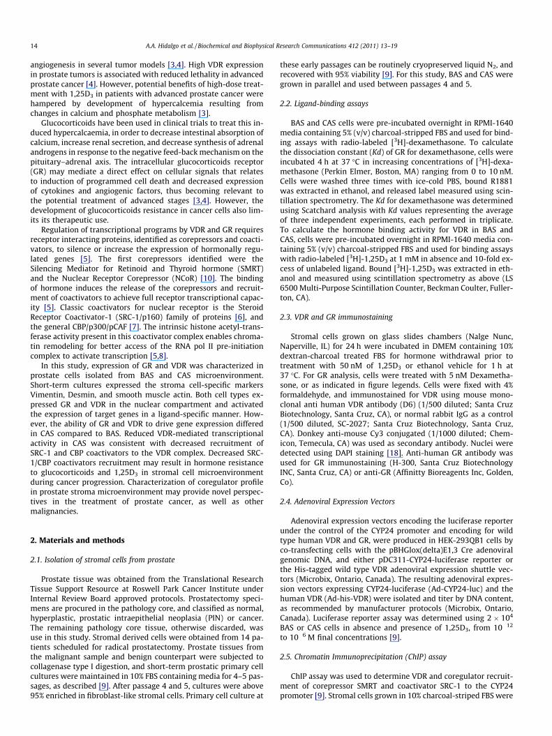
Prostate stromal cells in primary cell cultures were positive forvimentin and desmin, indicating the presence of fibroblast in theisolated stroma (Fig. 1A). Expression of smooth muscle actin(SMA) indicated the presence of myoblast in primary cell cultures(Fig. 1A). Other stromal cell markers previously reported in ourlaboratory include KGF, FGF2, ps20 and vimentin [9]. In addition,cell cultures were negative for epithelial cell markers (PSA andcytokeratin 18) after passage three. Thus, cell cultures were en-riched for fibroblasts and myoblast present in the cell populationof the prostate stroma.
3.2. Characterization of GR and VDR in BAS and CAS
Ligand dependent cytoplasmic-nuclear trafficking of GR andVDR was monitored in both BAS and CAS cell using immunofluo-rescence with receptor specific antibodies. In the absence of added
Fig. 1. Immunostaining analysis of stromal markers and nuclear receptors in BAS andanalyzed for expression of stromal-specific cell markers Vimentin, Desmin, and SmoothThe inset indicates nuclei staining with DAPI. (B). Immune fluorescence detection of nanalyzed in absence and presence of Dexamethasone (10 nM) and 1,25D3 (10 nM), as ipresence of their cognate hormone.
glucocorticoids, GR protein was localized in the cytoplasm in BASand CAS cells (Fig. 1B). The addition of glucocorticoids localizedGR to the nuclei in both cell types (Fig. 1B). In contrast, VDRexpression was nuclear, in the absence and presence of added1,25D3 ligand in BAS and CAS cells (Fig. 1B). The presence of GRand VDR in the nuclear compartment suggests functional receptor.However, specific transcription assays are required to demonstratethat GR and VDR are working as transcription factors in isolatedBAS and CAS cells.
3.3. Functional characterization of GR and VDR in BAS and CAS
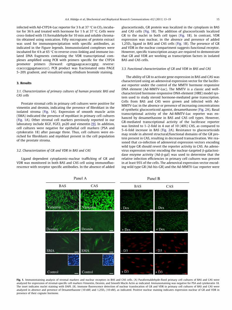
The ability of GR to activate gene expression in BAS and CAS wascharacterized using an adenoviral expression vector for the lucifer-ase reporter under the control of the MMTV hormone responsiveDNA element (Ad-MMTV-Luc). The MMTV is a classic and well-characterized hormone-responsive DNA-element (HRE) model sys-tem used to study steroid hormone-mediated gene transcription.Cells from BAS and CAS were grown and infected with Ad-MMTV-Luc in the absence or presence of increasing concentrationsof synthetic glucocorticoid agonist, dexamethasone (Fig. 2A). Basaltranscriptional activity of the Ad-MMTV-Luc reporter was en-hanced by dexamethasone in BAS and CAS cell types. However,GR-mediated transcriptional activity of the luciferase reporterwas limited to 1–2-fold in 4 out of 10 (40%) CAS, as compared to5–6-fold increase in BAS (Fig. 2A). Resistance to glucocorticoidsmay reside in altered structural/functional domains of the GR pro-tein present in CAS, resulting in decreased transactivation. We rea-soned that co-infection of adenoviral expression vectors encodingwild type GR should revert the reporter activity in CAS. An adeno-virus expression vector encoding the nuclear-targeted b-galactosi-dase enzyme activity (Ad-b-gal) was used to determine that therelative infection efficiencies in primary cell cultures was presentin at least 95% of the cells. The adenoviral expression vector encod-ing wild type GR (Ad-his-GR) and the Ad-MMTV-Luc reporter were
CAS cells. (A) Paraformaldehyde-fixed primary cell cultures of BAS and CAS wereMucle Actin as indicated. Immunostaining was negative for PSA and cytekeratin 18.uclear translocation of GR and VDR in primary cell cultures of BAS and CAS werendicated. Positive nuclear staining indicates expression nuclear of GR and VDR in
Fig. 2. GR-mediated transcriptional activity in BAS and CAS isolated from different prostates BAS and CAS cells isolated from prostate samples, representing two differentresponses to glucocorticoids, are indicated in panels (i) A–C and (ii) D–F. Isolated cells from each prostate sample were infected with Ad-MMTV-Luc alone (A and D), or incombination with Ad-his-GR (B and E). The MMTV-luciferase reporter activity driven by endogenous GR was determined in absence and presence of increasingconcentrations of Dexamethasone, as indicated (A and D). The MMTV-luciferase reporter activity driven by exogenous Ad-his-GR was determined in absence and presence ofincreasing concentrations of adenoviral particles (Ad-his-GR) in cells exposed to Dexamethasone at 10 nM (filled circles) or vehicle control (empty circles), as indicated (B andF). Relative [3H]-Dexamethasone-binding activity determined relative expression of endogenous and exogenous His-tagged GR in BAS and CAS (C and F).
16 A.A. Hidalgo et al. / Biochemical and Biophysical Research Communications 412 (2011) 13–19
use to co-infect BAS and CAS, in the absence or presence of dexa-methasone (Fig. 2B). The expression of Ad-his-GR resulted in a li-gand-dependent increased luciferase reporter in BAS and CAS, ascompared to endogenous GR. However, exogenous GR transactiva-tion of the reporter in CAS remained limited, as compared to BAS(Fig. 2B). Radioligand binding assays using [3H]-dexamethasonedemonstrated that the expression of GR was similar in BAS andCAS (Fig. 2C). Interestingly, GR-mediated transcriptional activityof the MMTV-luciferase reporter in CAS cells isolated from a differ-ent clinical samples, was more efficiently induced by dexametha-sone in a concentration-dependent manner, as compared to thereporter activity in BAS (Fig. 2D). Exogenous Ad-his-GR resultedin a ten-fold increase of luciferase reporter in both cell types, andGR maintained higher transcriptional capacity of the reporter inCAS as compared to BAS (Fig. 2E), despite similar levels of receptorexpression (Fig. 2F). This highlights the potential role of accessoryproteins that affect GR efficiency.
The VDR-mediated transcriptional activity in BAS and CAS wasdetermined using the adenoviral expression vector encoding theluciferase reporter under the control of the CYP24 promoter. Basaltranscriptional activity of the Ad-CYP24-Luc reporter was en-hanced by 1,25D3 in BAS and CAS. However, VDR-mediated tran-scriptional activity was limited to 1–2-fold in 8 out of 14 (57%)
CAS, as compared to 10–20-fold increase in BAS. Differences inVDR transactivation was observed at increasing concentrations of1,25D3, from 10�12 to 10�6 M (Fig. 3A). Co-infection of Ad-CYP24-Luciferase and an adenoviral expression vector encodingHis-tagged-VDR (Ad-his-VDR) resulted in increased reporter activ-ity in BAS and CAS (Fig. 3B). The exogenous Ad-his-VDR trans-acti-vation of the reporter remained limited in 5 out of 8 CAS, ascompared to BAS. Hormone binding assays using [3H]-1,25D3 andWestern immunoblotting demonstrated VDR expression in BASand CAS (Fig. 3C and D, respectively). Expression of Ad-his-VDRin 3 out of 8 CAS that exhibited limited VDR transactivaation com-pared to BAS (Fig. 3E), efficiently recovered reporter activity in a li-gand-dependent manner (Fig. 3F). Hormone binding and Westernimmunoblot demonstrate VDR expression in BAS and CAS(Fig. 3G and H). Therefore, resistance to 1,25D3 in these 3 clinicalsamples may reside in altered structural/functional domains ofVDR protein in CAS, as compared to BAS (Fig. 3E and F). In addition,VDR-mediated transcriptional activity induced by hormone wasobserved to be more efficient in 3 out of 14 CAS, as compared toBAS (Fig. 3I). Overexpression of the receptor increased VDR-medi-ated transactivation in CAS and BAS (Fig. 3J). However, differencesin the luciferase reporter activity, observed between CAS and BAS,continued after expression of exogenous VDR (Fig. 3J). Hormone-
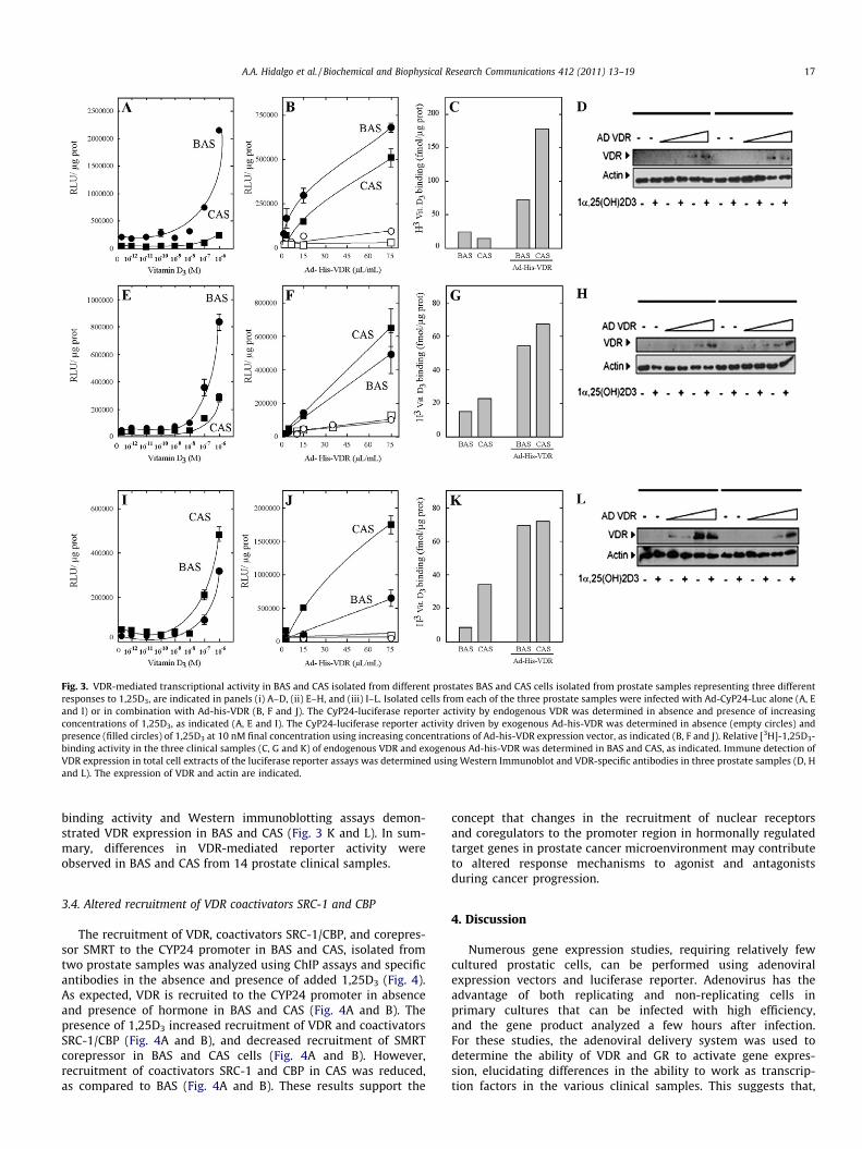
Fig. 3. VDR-mediated transcriptional activity in BAS and CAS isolated from different prostates BAS and CAS cells isolated from prostate samples representing three differentresponses to 1,25D3, are indicated in panels (i) A–D, (ii) E–H, and (iii) I–L. Isolated cells from each of the three prostate samples were infected with Ad-CyP24-Luc alone (A, Eand I) or in combination with Ad-his-VDR (B, F and J). The CyP24-luciferase reporter activity by endogenous VDR was determined in absence and presence of increasingconcentrations of 1,25D3, as indicated (A, E and I). The CyP24-luciferase reporter activity driven by exogenous Ad-his-VDR was determined in absence (empty circles) andpresence (filled circles) of 1,25D3 at 10 nM final concentration using increasing concentrations of Ad-his-VDR expression vector, as indicated (B, F and J). Relative [3H]-1,25D3-binding activity in the three clinical samples (C, G and K) of endogenous VDR and exogenous Ad-his-VDR was determined in BAS and CAS, as indicated. Immune detection ofVDR expression in total cell extracts of the luciferase reporter assays was determined using Western Immunoblot and VDR-specific antibodies in three prostate samples (D, Hand L). The expression of VDR and actin are indicated.
A.A. Hidalgo et al. / Biochemical and Biophysical Research Communications 412 (2011) 13–19 17
binding activity and Western immunoblotting assays demon-strated VDR expression in BAS and CAS (Fig. 3 K and L). In sum-mary, differences in VDR-mediated reporter activity wereobserved in BAS and CAS from 14 prostate clinical samples.
3.4. Altered recruitment of VDR coactivators SRC-1 and CBP
The recruitment of VDR, coactivators SRC-1/CBP, and corepres-sor SMRT to the CYP24 promoter in BAS and CAS, isolated fromtwo prostate samples was analyzed using ChIP assays and specificantibodies in the absence and presence of added 1,25D3 (Fig. 4).As expected, VDR is recruited to the CYP24 promoter in absenceand presence of hormone in BAS and CAS (Fig. 4A and B). Thepresence of 1,25D3 increased recruitment of VDR and coactivatorsSRC-1/CBP (Fig. 4A and B), and decreased recruitment of SMRTcorepressor in BAS and CAS cells (Fig. 4A and B). However,recruitment of coactivators SRC-1 and CBP in CAS was reduced,as compared to BAS (Fig. 4A and B). These results support the
concept that changes in the recruitment of nuclear receptorsand coregulators to the promoter region in hormonally regulatedtarget genes in prostate cancer microenvironment may contributeto altered response mechanisms to agonist and antagonistsduring cancer progression.
4. Discussion
Numerous gene expression studies, requiring relatively fewcultured prostatic cells, can be performed using adenoviralexpression vectors and luciferase reporter. Adenovirus has theadvantage of both replicating and non-replicating cells inprimary cultures that can be infected with high efficiency,and the gene product analyzed a few hours after infection.For these studies, the adenoviral delivery system was used todetermine the ability of VDR and GR to activate gene expres-sion, elucidating differences in the ability to work as transcrip-tion factors in the various clinical samples. This suggests that,
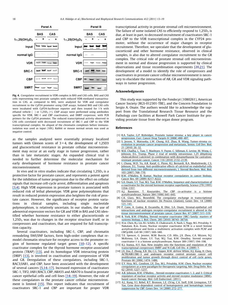
Fig. 4. Coregulator recruitment to VDR complex in BAS and CAS cells. BAS and CAScells representing two prostate samples with reduced VDR-mediated transactiva-tion in CAS, as compared to BAS, were analyzed for VDR and coregulatorrecruitment to the CyP24 promoter using ChIP assays. Isolated BAS and CAS cellswere incubated with CyP24-luciferase reporter and then treated for 1 h withethanol vehicle (�) or 1,25D3 (+). ChIP assays were performed using antibodiesspecific for VDR, SRC-1 and CBP coactivators, and SMRT corpressor, with PCRprimers for the CyP24 promoter. The reduced transcriptional activity observed inCAS cells correlated with decreased recruitment of SRC-1 and CBP to the VDRtranscriptional complex. An aliquot of the chromatin complexes before immuno-isolation was used as input (10%). Rabbit or mouse normal serum was used asnegative control.
18 A.A. Hidalgo et al. / Biochemical and Biophysical Research Communications 412 (2011) 13–19
as the samples analyzed were essentially primary localizedtumors with Gleeson scores of 3 + 4, the development of 1,25D3and glucocorticoid resistance in prostate cellular microenviron-ment may occur at an early stage in tumor progression, ratherthan in late metastatic stages. An expanded clinical trial isneeded to further determine the molecular mechanism forearly development of hormone resistance in prostate cancermicroenvironment.
In vivo and in vitro studies indicate that circulating 1,25D3 is aprotective factor for prostate cancer, and represents a potent agentfor the inhibition of tumor progression due to the effect on cellularsignals that increase cell-cycle arrest, differentiation and apoptosis[3,4]. High VDR expression in prostate tumors is associated withreduced risk of lethal phenotype. VDR gene polymorphisms thatresult in reduced protein expression also heighten the risk of pros-tate cancer. However, the significance of receptor protein varia-tions in clinical samples, including single nucleotidepolymorphism, is relatively uncertain. In our studies, the use ofadenoviral expression vectors for GR and VDR in BAS and CAS iden-tified whether hormone resistance to either glucocorticoids or1,25D3 was due to changes in the receptor structure itself, or incorepressors and coactivators, that are required for full transcrip-tion capacity.
Several coactivators, including SRC-1, CBP, and chromatinremodeling SWI/SNF factors, form high-order complexes that re-model chromatin structure for receptor access to the promoter re-gion of hormone regulated target genes [10–12]. A specificcoactivator complex for the thyroid hormone receptor-associatedprotein (TRAP) [13], and its homologue VDR interacting protein(DRIP) [13], is involved in coactivation and corepression of VDRand GR. Deregulation of these coregulators, including SRC-1,RAC3/IAB1, and CBP, have been associated with the developmentof several cancers [5,14–17]. Increased expression of coactivatorsSRC-1, TIF2, IAB1/RAC3, CBP, ARA55 and ARA70 is found in prostatecancer epithelial cells and cell lines [14–19]. However, the role ofthese coregulators in the prostate cancer cellular microenviron-ment is limited [15]. This report indicates that recruitment ofcoactivators SRC-1 and CBP are important for proper VDR
transcriptional activity in prostate stromal cell microenvironment.The failure of some isolated CAS to efficiently respond to 1,25D3 isdue, at least in part, to decreased recruitment of coactivators SRC-1and CBP to the VDR transcriptional complex in the CYP24 pro-moter, without the occurrence of major changes in receptorrecruitment. Therefore, we speculate that the development of glu-cocorticoid and other hormone resistance, observed in clinicalsamples, is also due to altered coregulator recruitment to the GRcomplex. The critical role of prostate stromal cell microenviron-ment in normal and disease progression is supported by clinicalobservations and tissue recombination experiments [20,21]. Thedevelopment of a model to identify the role of corepressors andcoactivators in prostate cancer cellular microenvironment is neces-sary to elucidate the interaction of AR, GR and VDR signaling path-ways in tumor progression.
Acknowledgments
This study was supported by the Fondecyt (1080261), AmericanCancer Society (RGS-012301-TBE), and the Concern Foundation toSergio A. Onate. The authors would like to acknowledge the sup-port from the Translational Research Tissue Resource and thePathology core facilities at Roswell Park Cancer Institute for pro-viding prostate tissue from the organ donor program.
References
[1] R.A. Taylor, G.P. Risbridger, Prostatic tumor stroma: a key player in cancerprogression, Curr. Cancer Drug Targets 8 (2008) 490–497.
[2] S. Josson, Y. Matsuoka, L.W. Chung, H.E. Zhau, R. Wang, Tumor-stroma co-evolution in prostate cancer progression and metastasis, Semin. Cell Dev. Biol.21 (2010) 26–32.
[3] M.K. Chadha, L. Tian, T. Mashtare, V. Payne, C. Silliman, E. Levine, M. Wong, C.Johnson, D.L. Trump, Phase 2 trial of weekly intravenous 1,25 dihydroxycholecalciferol (calcitriol) in combination with dexamethasone for castration-resistant prostate cancer, Cancer 116 (2010) 2132–2139.
[4] I. Chung, W.D. Yu, A.R. Karpf, G. Flynn, R.J. Bernardi, R.A. Modzelewski, C.S.Johnson, D.L. Trump, Anti-proliferative effects of calcitriol on endothelial cellsderived from two different microenvironments, J. Steroid Biochem. Mol. Biol.103 (2007) 768–770.
[5] B.W. O’Malley, R. Kumar, Nuclear receptor coregulators in cancer biology,Cancer Res. 69 (2009) 8217–8222.
[6] S.A. Onate, S.Y. Tsai, M.J. Tsai, B.W. O’Malley, Sequence and characterization ofa coactivator for the steroid hormone receptor superfamily, Science 270 (1995)1354–1357.
[7] A.J. Bannister, T. Kouzarides, The CBP co-activator is a histoneacetyltransferase, Nature 384 (1996) 641–643.
[8] C.K. Glass, M.G. Rosenfeld, The coregulator exchange in transcriptionalfunctions of nuclear receptors [In Process Citation], Genes Dev. 14 (2000)121–141.
[9] P. Cano, A. Godoy, R. Escamilla, R. Dhir, S.A. Onate, Stromal-epithelial cellinteractions and androgen receptor-coregulator recruitment is altered in thetissue microenvironment of prostate cancer, Cancer Res. 67 (2007) 511–519.
[10] B. York, B.W. O’Malley, Steroid receptor coactivator (SRC) family: masters ofsystems biology, J. Biol. Chem. 285 (2010) 38743–38750.
[11] H.w. Chen, R.j. Lin, R.l. Schiltz, D. Chakravarti, A. Nash, L. Nagy, M.l. Privalsky, Y.Nakatani, R.m. Evans, Nuclear receptor coactivator ACTR is a novel histoneacetyltransferase and forms a multimeric activation complex with PCAF andCBP/p300, Cell 90 (1997) 569–580.
[12] T.E. Spencer, G. Jenster, M.M. Burcin, C.D. Allis, J.X. Zhou, C.A. Mizzen, N.J.McKenna, S.A. Onate, S.Y. Tsai, M.J. Tsai, B.W. Omalley, Steroid receptorcoactivator-1 is a histone acetyltransferase, Nature 389 (1997) 194–198.
[13] K.J. Stanya, H.Y. Kao, New insights into the functions and regulation of thetranscriptional corepressors SMRT and N-CoR, Cell Div. 4 (2009) 7.
[14] J.X. Zou, Z. Zhong, X.B. Shi, C.G. Tepper, R.W. deVere White, H.J. Kung, H. Chen,ACTR/AIB1/SRC-3 and androgen receptor control prostate cancer cellproliferation and tumor growth through direct control of cell cycle genes,Prostate 66 (2006) 1474–1486.
[15] E.Y. Hsia, M.L. Goodson, J.X. Zou, M.L. Privalsky, H.W. Chen, Nuclear receptorcoregulators as a new paradigm for therapeutic targeting, Adv. Drug Deliv. Rev.62 (2010) 1227–1237.
[16] A.B. Johnson, B.W. O’Malley, , Steroid receptor coactivators 1, 2, and 3: Criticalregulators of nuclear receptor activity and steroid receptor modulator (SRM)-based cancer therapy, Mol. Cell Endocrinol. (2011).
[17] A.L. Kung, V.I. Rebel, R.T. Bronson, L.E. Ch’ng, C.A. Sieff, D.M. Livingston, T.P.Yao, Gene dose-dependent control of hematopoiesis and hematologic tumorsuppression by CBP, Genes Dev. 14 (2000) 272–277.

A.A. Hidalgo et al. / Biochemical and Biophysical Research Communications 412 (2011) 13–19 19
[18] B. Comuzzi, C. Nemes, S. Schmidt, Z. Jasarevic, M. Lodde, A. Pycha, G. Bartsch, F.Offner, Z. Culig, A. Hobisch, The androgen receptor co-activator CBP is up-regulated following androgen withdrawal and is highly expressed in advancedprostate cancer, J. Pathol. 204 (2004) 159–166.
[19] C. Nessler-Menardi, I. Jotova, Z. Culig, I.E. Eder, T. Putz, G. Bartsch, H. Klocker,Expression of androgen receptor coregulatory proteins in prostate cancer andstromal-cell culture models, Prostate 45 (2000) 124–131.
[20] IG. Schauer, DR. Rowley, The functional role of reactive stroma in benignprostatic hyperplasia, Differentiation (2011 June 9).
[21] A. Planche, M. Bacac, P. Provero, C. Fusco, M. Delorenzi, JC. Stehle, I.Stamenkovic, Identification of prognostic molecular features in thereactive stroma of human breast and prostate cancer, PLoS One 6(2011) e18640.